


Highlights
What are the main findings?
- Patients with moderate AD showed poorer visual acuity, a more remote near point of convergence, and poorer stereopsis; an exophoric alignment pattern was frequent in both groups.
- Patients with moderate AD showed lower clinical NSUCO scores for saccadic and smooth pursuit movements.
- Video-oculography was feasible only in a subgroup of patients with AD; without control recordings, these observations remain exploratory and descriptive.
What are the implications of the main findings?
- Routine visual and oculomotor assessment may help identify functional difficulties in patients with moderate AD, although these findings should not be interpreted as disease-specific diagnostic markers.
- The deterioration of the visual and oculomotor function in AD may contribute to the functional decline of this type of patients.
Abstract
Background/Objectives: Alzheimer’s disease (AD) is a progressive neurodegenerative disorder that, in addition to its characteristic cognitive decline, may be associated with impairments in visual and oculomotor function. The aim of this study was to characterize and compare a battery of visual, binocular, and oculomotor skills in patients with moderate AD and age-comparable older adults without a known diagnosis of cognitive impairment or neurodegenerative disease. Methods: A cross-sectional comparative observational study was conducted in 16 patients with AD and 20 age-comparable controls. Monocular and binocular visual acuity at distance and near, near point of convergence (NPC), stereopsis using the Randot® test, ocular alignment, fixation, and clinical oculomotor function using the NSUCO test were assessed. In a subgroup of patients with AD, an objective assessment was also attempted using GazeLab® video-oculography. Results: No between-group differences in age were observed (p = 0.757). The AD group showed worse binocular distance visual acuity (0.16 ± 0.13 vs. 0.01 ± 0.05 logMAR, p = 0.003), a trend to more remote NPC (16.50 ± 5.35 vs. 10.80 ± 4.56 cm, p = 0.466), poorer stereopsis (188.5 ± 64.14 vs. 80.00 ± 24.48 arcseconds, p = 0.004), and lower NSUCO scores for saccadic and pursuit movements (p < 0.001). Video-oculography was feasible in a subgroup of patients with AD and yielded exploratory, descriptive observations; equivalent recordings were not obtained in control participants. Conclusions: The patients with moderate AD showed poorer performance across several visual, binocular, and oculomotor measures. These findings support the relevance of incorporating visual assessment into the functional characterization of AD.
1. Introduction
Alzheimer’s disease (AD) is the most common cause of dementia and is characterized by a progressive neurodegenerative process affecting multiple brain networks. Although impairments in memory and other cognitive domains dominate its clinical presentation, the disease may also compromise different levels of the visual system. Available evidence describes abnormalities in both the afferent visual pathway and cortical and oculomotor networks, with potential effects on visual acuity, contrast sensitivity, color vision, visual field, motion perception, and visuomotor behavior [1,2,3,4,5].
Oculomotor abnormalities are of particular interest because they depend on frontal, parietal, subcortical, and cerebellar circuits that also contribute to attention, response inhibition, and other executive functions. Systematic reviews and instrumental studies have described increased latency, antisaccade errors, reduced accuracy, changes in gain, and abnormalities in fixation stability and visual exploration in mild cognitive impairment and AD [6,7,8,9,10,11,12]. However, the heterogeneity of tasks, devices, diagnostic definitions, and clinical stages still limits the translation of these parameters into biomarkers for routine use. The most recent systematic review of eye tracking in AD and related dementias specifically emphasizes the need for better-characterized cohorts and standardized protocols before individual diagnostic application [6].
Visual impairment is particularly prominent in posterior cortical atrophy, a neurodegenerative syndrome characterized by progressive visuoperceptual and visuospatial dysfunction with relative preservation of other cognitive functions early in the course of the disease. Although Alzheimer’s pathology is its most frequent underlying etiology, this phenotype must be distinguished from the typical amnestic presentation of AD [13,14]. Therefore, the presence of visual abnormalities in patients with AD does not, by itself, imply that these abnormalities are specific to the disease or that they necessarily precede its cognitive manifestations.
From an optometric and neuro-ophthalmological perspective, there is interest in identifying simple, reproducible, and well-tolerated tests that can characterize the functional impact of AD on vision and ocular motility. This approach is clinically relevant because unrecognized visual impairment may interfere with reading, mobility, spatial orientation, and performance on cognitive tasks with a visual component. Nevertheless, detecting functional impairment must be distinguished from validating a biomarker: the latter requires standardization, control of ocular confounding factors, external validation, and demonstration of adequate sensitivity and specificity [1,3,6].
The aim of the present study was to preliminarily characterize and compare visual acuity, binocular convergence, stereopsis, ocular alignment, fixation, and clinical oculomotor performance in patients with moderate AD and age-comparable older adults without a known diagnosis of cognitive impairment or neurodegenerative disease. The feasibility of incorporating objective video-oculography in a subgroup of patients was also explored, with the aim of complementing the clinical examination and defining a battery of measures suitable for evaluation in subsequent studies with larger samples.
2. Materials and Methods
2.1. Participants and Ethical Considerations
A cross-sectional comparative observational study was conducted. Recruitment and clinical examinations were carried out from July 2021 to May 2022. Sixteen patients (10 women and 6 men) with a previously established clinical diagnosis of mild or moderate Alzheimer’s disease and 20 age-comparable control participants (12 women and 8 men) without a known diagnosis of cognitive impairment, dementia, or another neurodegenerative disease were included. Patients with AD were assessed at the facilities of the Association of Relatives of People with Alzheimer’s Disease of Elche (AFAE Elche), whereas the control group was examined at Punto Óptico Elche. Disease severity had been classified by the clinical team at AFAE using the Global Deterioration Scale (GDS); all included patients were in GDS stages 4 or 5, corresponding to moderate dementia. Individual MMSE (Mini-Mental State Examination) or CDR (Clinical Dementia Ratio) scores were not available. Before the examination, the available clinical information for participants with AD was reviewed, including the degree of cognitive impairment and history of cataract surgery. All participants with AD were receiving pharmacological treatment for the disease, although the specific agents and doses had not been recorded systematically. Age, sex, habitual use of optical correction, and the presence of pseudophakia were recorded in both groups.
All participants received information about the nature and objectives of the study and provided informed consent before the examination; when appropriate, the information and consent process was conducted with their legal representative. The study was conducted in accordance with the principles of the Declaration of Helsinki and was reviewed and approved by the Ethics Committee of the University of Alicante.
2.2. Inclusion and Exclusion Criteria
Patients in the AD group were considered eligible if they had a documented clinical diagnosis of Alzheimer’s disease, had been classified as GDS stage 4 or 5 (moderate dementia), were older than 60 years, and had sufficient ability to understand simple instructions and cooperate with the basic visual tests. Participants were excluded if they had other known neurological or neurodegenerative disorders likely to interfere substantially with the examination, active or severe ocular disease preventing performance or interpretation of the tests, or insufficient ability to cooperate with the basic clinical protocol. An antecedent of stable cataract surgery was not an exclusion criterion and was specifically recorded as a clinical characteristic of the sample.
The control group consisted of age-comparable older adults with no known diagnosis of cognitive impairment, dementia, or another neurodegenerative disease and who were able to complete the same examination protocol. No formal cognitive screening was administered; eligibility was based on the available clinical history and the absence of a known diagnosis of cognitive impairment or neurodegenerative disease. Ocular and neurological exclusion criteria equivalent to those applied to the AD group were used. Corrected ametropia or stable cataract surgery was not a reason for exclusion; use of optical correction and pseudophakia were recorded for descriptive purposes and considered when interpreting the results.
2.3. Visual and Oculomotor Examination Protocol
The examination followed the same clinical protocol in both groups. Each test was performed using the participant’s habitual optical correction for distance or near, as appropriate for the testing distance. Participants underwent periodic ophthalmological follow-up outside the study; however, no study-specific refraction or comprehensive ocular health examination was performed, and it was therefore not possible to confirm that the habitual prescription was fully up to date. The two groups were examined at different clinical sites, and the examiners were not masked to group status. Instructions were given briefly and directly and were repeated when necessary to facilitate understanding, without altering the purpose or recording criterion of each test.
Visual acuity (VA) was measured using logMAR optotypes, both monocularly and binocularly and at distance and near. Results were expressed in logMAR units, with higher values representing poorer VA. The use of a logarithmic scale provides a regular progression between acuity levels and facilitates quantitative comparison between observations. The near point of convergence (NPC) was assessed using a fixation target that was moved slowly along the midline toward the bridge of the nose. Participants were instructed to maintain fixation on the target as it approached, and the convergence break point was recorded in centimeters, identified by the perception of diplopia or by an observable loss of binocular convergence. A higher value corresponded to a more remote NPC and therefore poorer near-convergence performance. Stereopsis was assessed at near using the Randot® test (Stereo Optical, Chicago, IL, USA), with habitual optical correction when required. Stereoacuity was expressed in arcseconds, with higher values indicating poorer stereoscopic discrimination. Randot® is a widely used clinical test for quantifying stereopsis using binocular disparity stimuli [15].
Ocular alignment was examined at distance and near using cover test, and binocular motor status was recorded as exophoria, esophoria, or orthophoria, with no numerical data associated due to the difficulty of performing a measurement with prism bar in AD patients. The analysis was intended to describe the distribution of the observed motor pattern in each group rather than to establish a diagnosis of binocular anomaly based on this variable.
Clinical oculomotor function was assessed using an adaptation of the NSUCO (Northeastern State University College of Optometry Oculomotor Test), a clinical procedure for evaluating pursuit and saccadic eye movements [16,17]. The test was performed binocularly using small colored fixation targets positioned in front of the participant. For pursuits, the target was moved along a circular trajectory in both directions; for saccadic movements, participants were asked to alternate fixation between two horizontally separated targets. According to the recording system used in this study, ability, accuracy, and associated postural control were scored for each modality, yielding a maximum aggregate score of 15 points; lower scores represented poorer clinical performance. The original pediatric normative criteria were not applied. Scores were interpreted through direct comparison with the control group. Because this adapted scoring approach has not been validated in older adults, the aggregate score was interpreted as a measure of overall clinical oculomotor performance rather than as a normative diagnosis or an isolated measure of saccadic accuracy. Fixation stability was recorded during the oculomotor examination as a dichotomous clinical variable (stable or unstable) according to the participant’s ability to maintain gaze on the target during the task. This measure was considered a clinical estimate of fixation ability rather than an instrumental quantification of gaze dispersion.
2.4. Video-Oculography Assessment
In addition to the clinical examination, objective eye-movement recording was attempted in a subgroup of participants with AD using the GazeLab® video-oculography system (BCN Innova, Barcelona, Spain). The purpose of this component was to explore the feasibility of instrumental recording in this population and to complement, rather than confirm, the clinical NSUCO assessment. The device uses infrared eye-tracking technology with binocular cameras and provides quantitative outputs related to saccadic and pursuit eye movements.
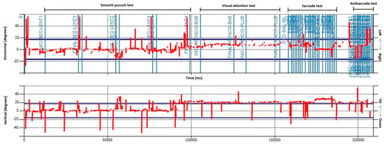
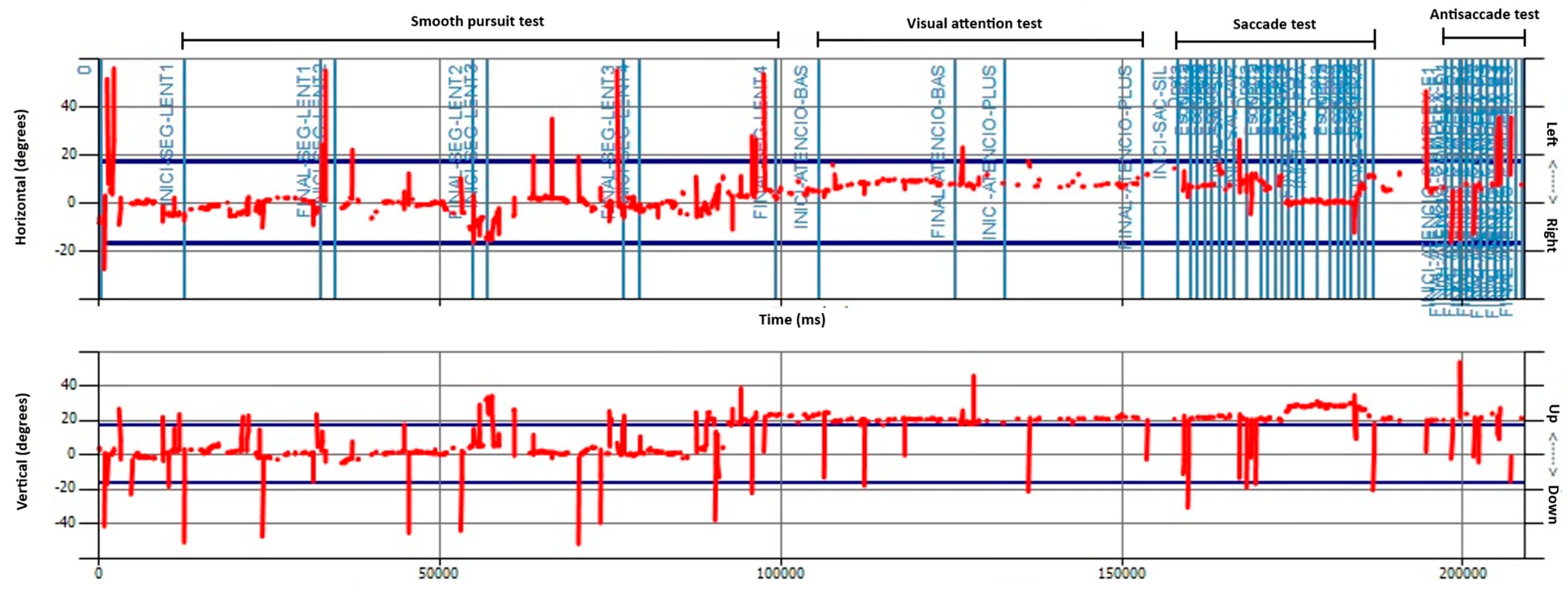
The GazeLab® system has demonstrated adequate reliability and validity for the objective measurement of ocular motility, showing agreement with standard clinical tests and other previously validated video-oculography systems [18]. For this exploratory component, a study-specific sequence of consecutive tests was administered (Figure 1). Equivalent recordings were not obtained in control participants, and no external normative thresholds were applied. Accordingly, the video-oculographic assessment was not intended to provide a between-group comparison or a diagnostic classification:
Figure 1.
Example of video-oculographic registry with the GazeLab system in one patient from the sample of subjects with Alzheimer’s disease.
- Smooth pursuit: Pursuit eye movements were assessed using a moving target following four different trajectories: an ellipse, a figure-eight (∞), a sinusoidal trajectory, and an atom-like trajectory. The number of saccadic movements per minute was recorded.
- Visual attention test: The number of distractions per minute was recorded under a basic attention paradigm, in which participants were instructed to maintain fixation on a static target.
- Saccadic eye movements: Saccades were assessed using letters (short targets), words (medium-length targets), and sentences (long targets). The number of eye movements performed per jump was recorded.
- Antisaccade task: Participants were instructed to suppress a reflexive saccade and generate a voluntary saccade in the opposite direction, while additional fixation targets appeared at different locations in the peripheral visual field. The percentage of correct responses was recorded.
Video-oculographic assessment could not be completed in all patients from the AD group owing to limitations associated with cognitive impairment, including difficulties understanding and following the test instructions, inability to maintain stable fixation, and fatigue during the assessment. These factors were considered relevant to the feasibility of the procedure in the study population and were considered in the interpretation of the findings.
2.5. Statistical Analysis
Statistical analysis was performed using SPSS v. 24.0 (IBM Corp., Armonk, NY, USA). Distribution of quantitative variables was examined using the Kolmogorov–Smirnov test. As most variables were not normally distributed, non-parametric procedures were used. Quantitative variables were described using mean and standard deviation, as well as median and range. The statistical significance of difference in such variables between the AD and control groups was assessed using the Mann–Whitney U test. Categorical variables were expressed as frequencies and percentages, and differences between groups were analyzed using the chi-square test. All tests were two-tailed and p < 0.05 was considered statistically significant. p-values were adjusted with the Benjamini–Hochberg procedure to control the risk of type I error due to the multiple comparisons done. Given the exploratory nature of the study and the number of variables analyzed, significance values were interpreted together with the magnitude and clinical consistency of the observed differences.
3. Results
3.1. Sample Characteristics and Overall Analysis
Thirty-six participants were analyzed: 16 with AD and 20 age-comparable controls without a known diagnosis of cognitive impairment or neurodegenerative disease. No statistically significant difference in age was observed between the groups (78.5 ± 9.4 vs. 79.6 ± 9.1 years; p = 0.757). Table 1 presents the complete descriptive data for the recorded variables and the significance values of differences between groups. The AD group used optical correction less frequently and had a lower proportion of pseudophakia than the control group (p = 0.014).
Table 1.
Sample characteristics and results of the comparative analysis between AD and control groups. Abbreviations: N, number of patients; SD, standard deviation; RE, right eye; LE, left eye; CDVA, corrected distance visual acuity; CNVA, corrected near visual acuity.
3.2. Visual Acuity
The AD group showed higher-distance logMAR values, indicating poorer VA, under both monocular and binocular viewing conditions (p ≤ 0.023; Table 1). In addition, the AD group exhibited greater variability in VA, as reflected by higher standard deviations. A similar pattern was observed for near visual acuity, with higher mean logMAR values in the AD group across all three measurement conditions (p ≤ 0.023), together with higher standard deviations, indicating greater interindividual variability in this group.
3.3. Ocular Alignment and Stereopsis
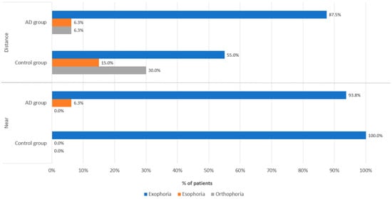
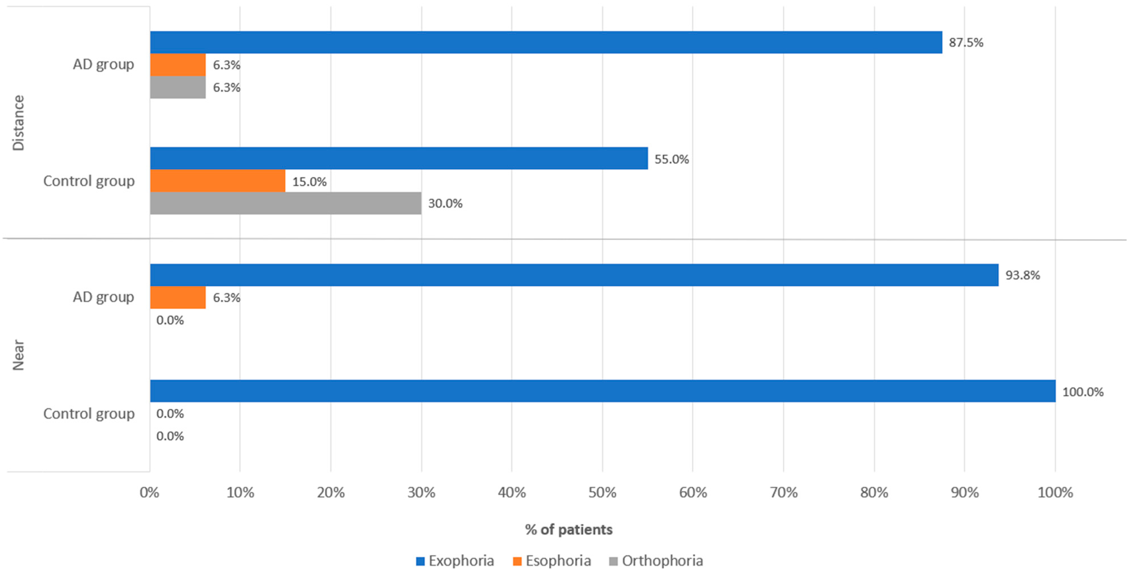
Figure 2 shows the distribution of the type of phoria measured in the patients evaluated. No tropia was found in any case. At a distance, 14 of the 16 participants with AD had exophoria (87.5%), whereas in the control group, 11 cases from the sample of 20 (55%) presented exophoria (p = 0.119). At near, exophoria was observed in 15 participants with AD (93.8%), whereas all 20 controls were classified as exophoric (100%) in the original record (p = 0.466). Near exophoria was recorded in both groups and was more frequent in controls, whereas most participants with AD also showed an exophoric alignment pattern at distance.
Figure 2.
Distribution of the type of phoria detected with cover test in AD and control groups.
The NPC break point tended to be more remote in the AD group, with a mean value of 16.50 ± 5.35 cm compared with 10.80 ± 4.56 cm in the control group, but differences did not reach statistical significance (p = 0.466). Stereopsis was also poorer in patients with AD: mean stereoacuity was 188.57 ± 64.14 arcseconds compared with 80.00 ± 24.48 arcseconds in controls (p = 0.004). Both findings indicate poorer binocular performance in the tasks assessed, although their interpretation should take the participants’ optical and ocular status into account. Stereopsis measurement was not possible in 9 subjects (56.3%) due to cognitive limitations.
3.4. Oculomotor Function and Fixation
NSUCO scores were significantly lower in the AD group both for smooth pursuits and saccadic movements (p < 0.001). Indeed, mean difference in NSUCO pursuit and saccade scores between control and AD groups were 11.62 and 7.80, respectively. Fixation stability was classified as stable in all cases in the control group, whereas only was considered stable in 5 patients (31.3%) in AD group (p < 0.001).
The GazeLab® examination was feasible in only a subgroup of 10 patients from the AD group. Table 2 summarizes the main outcomes obtained. A higher number of saccades per minute was observed during pursuit tasks involving more complex trajectories, such as sinusoidal and atom-like trajectories. Likewise, a greater number of eye movements per jump was detected during saccadic tasks involving longer targets (i.e., sentences). Only 5.67% of correct responses were detected during the antisaccadic task.
Table 2.
Results obtained in the video-oculographic evaluation with the GazeLab system in a subgroup of 10 AD patients. Abbreviations: SD, standard deviation.
4. Discussion
The main finding of this preliminary study is that patients with mild-to-moderate AD performed more poorly than controls across several components of visual, binocular, and oculomotor function. Differences involved VA, NPC, stereopsis, clinical saccade and pursuit scores, and fixation stability. Taken together, this pattern is consistent with contemporary reviews describing simultaneous impairment of afferent and efferent visual functions in AD [1,2,3,4,5,6,7,8,9]. However, the cross-sectional and exploratory nature of the study precludes determining whether the observed differences directly reflect the neurodegenerative process, concomitant ocular factors, or an interaction between the two.
Monocular and binocular VA was poorer in the AD group than in the control group. This finding is consistent with previous studies describing reduced VA and other visual functions in people with AD [1,2]. All visual tests were performed with the habitual distance or near correction appropriate for the task. Although participants underwent periodic ophthalmological follow-up, no study-specific refraction or comprehensive ocular health examination was performed. Consequently, the habitual correction cannot be considered confirmed best correction, and the influence of residual refractive error, cataract, or other ocular-media opacity cannot be excluded. VA also depends on retinal function, cortical processing, and the ability to respond to the task. The same testing procedure was applied in both groups; nevertheless, the substantial between-group differences in spectacle use and pseudophakia may have influenced the VA findings. Therefore, the poorer VA observed cannot be attributed exclusively to AD or to cortical dysfunction. In addition, studies linking visual abnormalities to patterns of cerebral neurodegeneration support the plausibility of a central component but do not eliminate the need for rigorous control of optical and ocular factors [19].
Stereopsis was markedly poorer in the AD group. Stereoscopic perception requires adequate binocular correspondence and the cortical integration of disparity signals across multiple visual areas [20]. Accordingly, reduced stereoacuity may reflect a combination of optical and binocular limitations, as well as reduced efficiency of visuoperceptual processing. In the present sample, however, the lack of comprehensive refractive and ocular characterization limits the interpretation of this finding. Thus, it should be regarded as a functional difference between groups rather than as isolated evidence of a cortical abnormality specifically associated with AD. Previous studies have shown that patients with AD and mild cognitive impairment retain the ability to process coarse binocular disparities, supporting their performance in everyday tasks such as reaching, grasping, driving, and navigation [21]. Conversely, other studies have reported that impaired depth perception in patients with AD may contribute to visuospatial deficits [22]. Discrepancies among studies may be attributable to differences in sample characteristics, including disease severity and the clinical stage of the patients included. In addition, stereopsis assessment may impose a substantial cognitive demand, potentially affecting test performance in patients with greater cognitive impairment. In the present study, stereopsis could not be assessed in 9 participants (56.3%) because of such limitations. Therefore, the possibility that cognitive impairment contributed to the observed group difference and to the high proportion of unmeasurable results should be considered when interpreting these findings.
The NPC was more remote in participants with AD. An exophoric alignment pattern was common in both groups at near and was also observed in most participants with AD at distance. Although exophoria is commonly observed at near, its predominance at distance in the AD group is less typical. This finding is presented as a descriptive observation, and its clinical relevance requires confirmation in larger samples. Convergence is a complex binocular response that integrates sensory, accommodative, and motor mechanisms and involves supranuclear and oculomotor circuits [23]. Our findings are consistent with those reported by Jiménez and coauthors, who observed altered vergence eye movements and pupil responses in patients with AD and mild cognitive impairment during an oddball task [24]. However, research on vergence abnormalities in AD remains limited, possibly in part because of the difficulties involved in performing these assessments in cognitively impaired patients. Further studies are therefore warranted using visual tests with lower cognitive demands that allow rapid and objective assessment of the vergence response. In addition, further research is needed to characterize the prevalence and clinical significance of binocular disorders in patients with AD. This is particularly relevant because other neurodegenerative diseases have also been associated with convergence insufficiency [25].
The largest descriptive differences were observed in clinical oculomotor performance. Patients with AD had lower NSUCO scores for both saccadic and smooth pursuit eye movements, consistent with previous studies documenting oculomotor abnormalities in AD using objective eye-tracking techniques [26,27,28]. It is important, however, to distinguish between these approaches. The NSUCO test provides an aggregate clinical assessment that combines ability, accuracy, and associated head or body movements. Consequently, its total score cannot be interpreted as an isolated measure of saccadic accuracy, nor does it directly quantify parameters such as latency, amplitude, gain, or velocity. Nevertheless, the reliability of this clinical assessment has been established [17], and NSUCO scores have also been shown to correlate with objective eye-tracking measures [29]. Therefore, our findings support previous evidence of altered saccadic and smooth pursuit performance in patients with AD. Video-oculography was feasible in only a subgroup of participants with AD and was not performed in the control group. Consequently, these recordings cannot provide a between-group comparison or establish diagnostic validity. The study-specific outputs should be regarded as preliminary observations of feasibility and task performance. Although the patterns observed during pursuit and saccadic tasks may be compatible with oculomotor difficulty, they may also reflect attentional load, comprehension, fatigue, and the degree of cognitive impairment. Controlled studies using standardized metrics are required before these observations can be interpreted clinically.
The GazeLab® technology also enabled the assessment of other visual functions, including visual attention and antisaccade performance. The presence of distractibility during the basic visual attention task was not unexpected, given that impairments in visual attention have been widely documented in patients with AD [30]. This finding is also consistent with the lower proportion of patients in the AD group who demonstrated stable fixation in the NSUCO evaluation. Attentional deterioration can occur during the early stages of AD and may contribute to functional decline [30]. In addition, a low proportion of correct responses was observed during the antisaccade task. An antisaccade is a voluntary eye movement directed away from, rather than toward, a newly presented visual stimulus and therefore requires the suppression of a reflexive saccadic response. Our findings are consistent with previous studies showing that patients with AD make more fixations toward distractors during visual attention tasks than age-matched neurologically healthy controls [31]. Similarly, the reduced performance observed in the antisaccade task is in agreement with previous reports of impaired inhibitory control of visually elicited saccades in AD [32]. Antisaccade performance has been proposed as a sensitive measure of executive and inhibitory control and may detect abnormalities even in patients with mild AD or at presumed prodromal stages of the disease [33]. Previous studies have also reported increased antisaccade error rates in patients with mild AD compared with age-matched controls, supporting the presence of impaired inhibitory control even in relatively early stages of the disease [33].
This study has several limitations that should be considered when interpreting the results. First, the sample size was small, and a convenience sampling approach was used, which limits the generalizability of the findings. The groups were recruited and examined at two different clinical sites, and the examiners were not masked to group status. This may have influenced subjective clinical outcomes. The control group did not undergo formal cognitive screening, and individual MMSE or CDR scores were unavailable for the AD group. Detailed medication, BMI, and comorbidity data were not collected systematically. All participants with AD were receiving pharmacological treatment, but the specific agents and doses were not recorded. Diabetes was not documented in the available clinical histories, whereas hypertension was common but was not systematically coded. These limitations restricted the clinical characterization of the groups and the assessment of potential confounding factors.
Although participants underwent periodic ophthalmological follow-up, the study did not include a study-specific refraction or a comprehensive ocular health examination. Habitual correction was used in both groups according to the testing distance, but residual refractive error, cataract, other ocular-media opacity, and the between-group differences in spectacle use and pseudophakia may have influenced VA, stereopsis, and NPC. In addition, several clinical tests contained a subjective component and depended on understanding the instructions, sustained attention, fatigue, and cooperation. Lower scores in the AD group may therefore reflect the combined influence of visual, oculomotor, and cognitive performance rather than an isolated visual or oculomotor deficit.
Second, the cross-sectional design precludes the establishment of temporal or causal relationships between visual abnormalities and cognitive decline. Nevertheless, the comparative design, involving participants with AD and age-comparable controls without a known diagnosis of cognitive impairment or neurodegenerative disease, allows the identification of patterns of between-group differences in selected clinical variables. The adapted NSUCO scoring approach has not been validated in older adults, and the original pediatric norms were not applied; the findings should therefore be interpreted as differences in overall clinical oculomotor performance. The dependence on participants’ understanding and cooperation also affected video-oculography, which could not be completed in the entire study sample. Although previous studies have investigated visual and oculomotor alterations in AD, a substantial proportion of the literature has focused on early-stage disease, including mild cognitive impairment, in which participants may be better able to perform cognitively demanding experimental tasks. Therefore, the present findings provide complementary information regarding the assessment of visual function in patients with more established cognitive impairment, while also highlighting the methodological challenges associated with testing this population. Furthermore, given the exploratory nature of the study and the large number of variables analyzed, statistical significance should be interpreted with caution. The video-oculographic component was restricted to a subgroup of participants with AD, without control recordings or external normative data, and used study-specific output metrics. These observations therefore represent feasibility data only and cannot support group-level or diagnostic inferences.
5. Conclusions
In this study, patients with moderate AD tend to perform more poorly than age-comparable controls in VA, stereopsis, fixation, and clinical measures of oculomotor function. Taken together, these preliminary findings describe clinically relevant group-level differences and support the potential value of incorporating visual function into the comprehensive assessment of these patients. The exophoric alignment pattern was present in both groups, and the lower NSUCO scores should be interpreted as reduced overall clinical oculomotor performance rather than as an isolated deficit in saccadic accuracy. Video-oculography may complement clinical oculomotor assessment in future research. In the present study, however, recordings were available only from a subgroup of participants with AD, without a control comparator or an external normative reference; these observations should therefore be regarded solely as preliminary feasibility data. More broadly, the observed clinical differences are not specific to AD and, given the current study design and sample size, should not be interpreted as independent diagnostic biomarkers. Confirmation of their clinical utility will require longitudinal studies with larger samples, rigorous control of refractive and ocular factors, and comprehensive cognitive and biological characterization of participants. Importantly, the present study included patients with moderate AD, a population in whom cognitive impairment may pose additional challenges for the performance of clinical and experimental visual assessments. Previous research has frequently included individuals with mild cognitive impairment or earlier stages of AD, although studies have also examined oculomotor alterations in patients with mild-to-moderate AD. Therefore, the present findings should be considered complementary to the existing literature and may contribute to a better understanding of the feasibility and clinical relevance of visual and oculomotor assessment in patients with more established cognitive impairment. In this context, combining easily administered optometric tests with objective measures of ocular motility represents a reasonable strategy for future research aimed at identifying complementary functional markers of disease-related changes.
Author Contributions
Conceptualization, G.A. and D.P.P.; methodology, G.A., A.M.M. and D.P.P.; software, G.A. and D.P.P.; validation, G.A., A.M.M. and D.P.P.; formal analysis, G.A. and D.P.P.; investigation, G.A., A.M.M. and D.P.P.; resources, G.A. and D.P.P.; data curation, G.A.; writing—original draft preparation, G.A.; writing—review and editing, G.A., A.M.M. and D.P.P.; visualization, D.P.P.; supervision, A.M.M. and D.P.P.; project administration, G.A. and D.P.P.; funding acquisition, D.P.P. All authors have read and agreed to the published version of the manuscript.
Funding
This research received no external funding.
Institutional Review Board Statement
The study was conducted in accordance with the Declaration of Helsinki and was approved by the Ethics Committee of the University of Alicante (protocol code UA-2019-03-25; approval date: 17 September 2019).
Informed Consent Statement
Informed consent was obtained from all subjects involved in the study.
Data Availability Statement
The data presented in this study are available on request from the corresponding author due to privacy.
Acknowledgments
We would like to thank the collaboration of the Association of Relatives of People with Alzheimer’s Disease of Elche (AFAE Elche) that offered its facilities for the clinical examinations and the access to AD patients. During the preparation of this manuscript, the authors used ChatGPT (OpenAI, GPT-5.6 Luna) solely for linguistic revision. The authors have reviewed and edited the output and take full responsibility for the content of this publication.
Conflicts of Interest
The authors declare no conflicts of interest.
References
- Alvite-Piñeiro, T.; López-López, M.; Regueiro, U.; Pías-Peleteiro, J.M.; Sobrino, T.; Lema, I. Visual function in alzheimer’s disease: Current understanding and potential mechanisms behind visual impairment. J. Clin. Med. 2025, 14, 5963. [Google Scholar] [CrossRef] [Scilit]
- Xu, Y.; Aung, H.L.; Hesam-Shariati, N.; Keay, L.; Sun, X.; Phu, J.; Honson, V.; Tully, P.J.; Booth, A.; Lewis, E.; et al. Contrast sensitivity, visual field, color vision, motion perception, and cognitive impairment: A systematic review. J. Am. Med. Dir. Assoc. 2024, 25, 105098. [Google Scholar] [CrossRef] [Scilit]
- Almario, G.; Piñero, D.P. Impact of Alzheimer’s disease in ocular motility and visual perception: A narrative review. Semin. Ophthalmol. 2022, 37, 436–446. [Google Scholar] [CrossRef] [Scilit]
- Scheltens, P.; De Strooper, B.; Kivipelto, M.; Holstege, H.; Chételat, G.; Teunissen, C.E.; Cummings, J.; van der Flier, W.M. Alzheimer’s disease. Lancet 2021, 397, 1577–1590. [Google Scholar]
- Salobrar-García, E.; de Hoz, R.; Ramírez, A.I.; López-Cuenca, I.; Rojas, P.; Vazirani, R.; Amarante, C.; Yubero, R.; Gil, P.; Pinazo-Durán, M.D.; et al. Changes in visual function and retinal structure in the progression of Alzheimer’s disease. PLoS ONE 2019, 14, e0220535. [Google Scholar] [CrossRef] [Scilit]
- Verón, G.L.; Juantorena, G.E.; Keller, G.; Crivelli, L.; Kamienkowski, J.E. Eye tracking as a diagnostic tool in Alzheimer’s disease, mild cognitive impairment, and related dementias: A systematic review. Alzheimer’s Dement. 2026, 18, e70238. [Google Scholar] [CrossRef] [Scilit]
- Sekar, A.; Panouillères, M.T.N.; Kaski, D. Detecting abnormal eye movements in patients with neurodegenerative diseases-current insights. Eye Brain 2024, 16, 3–16. [Google Scholar] [CrossRef] [Scilit]
- Eraslan Boz, H.; Koçoğlu, K.; Akkoyun, M.; Tüfekci, I.Y.; Ekin, M.; Özçelik, P.; Akdal, G. Uncorrected errors and correct saccades in the antisaccade task distinguish between early-stage Alzheimer’s disease dementia, amnestic mild cognitive impairment, and normal aging. Neuropsychol. Dev. Cogn. B Aging Neuropsychol. Cogn. 2024, 31, 457–478. [Google Scholar] [CrossRef] [Scilit]
- Opwonya, J.; Doan, D.N.T.; Kim, S.G.; Kim, J.I.; Ku, B.; Kim, S.; Park, S.; Kim, J.U. Saccadic eye movement in mild cognitive impairment and alzheimer’s disease: A systematic review and meta-analysis. Neuropsychol. Rev. 2022, 32, 193–227. [Google Scholar] [CrossRef] [Scilit]
- Hannonen, S.; Andberg, S.; Kärkkäinen, V.; Rusanen, M.; Lehtola, J.M.; Saari, T.; Korhonen, V.; Hokkanen, L.; Hallikainen, M.; Hänninen, T.; et al. Shortening of saccades as a possible easy-to-use biomarker to detect risk of Alzheimer’s disease. J. Alzheimers Dis. 2022, 88, 609–618. [Google Scholar] [CrossRef] [Scilit]
- Molitor, R.J.; Ko, P.C.; Ally, B.A. Eye movements in Alzheimer’s disease. J. Alzheimers Dis. 2015, 44, 1–12. [Google Scholar] [CrossRef] [Scilit]
- Shafiq-Antonacci, R.; Maruff, P.; Masters, C.; Currie, J. Spectrum of saccade system function in Alzheimer disease. Arch. Neurol. 2003, 60, 1272–1278. [Google Scholar] [CrossRef] [Scilit]
- Chapleau, M.; La Joie, R.; Yong, K.; Agosta, F.; Allen, I.E.; Apostolova, L.; Best, J.; Boon, B.D.C.; Crutch, S.; Filippi, M.; et al. Demographic, clinical, biomarker, and neuropathological correlates of posterior cortical atrophy: An international cohort study and individual participant data meta-analysis. Lancet Neurol. 2024, 23, 168–177. [Google Scholar] [CrossRef] [Scilit]
- Yong, K.X.X.; Graff-Radford, J.; Ahmed, S.; Chapleau, M.; Ossenkoppele, R.; Putcha, D.; Rabinovici, G.D.; Suarez-Gonzalez, A.; Schott, J.M.; Crutch, S.; et al. Diagnosis and management of posterior cortical atrophy. Curr. Treat. Options Neurol. 2023, 25, 23–43. [Google Scholar] [CrossRef] [Scilit]
- Fawcett, S.L.; Birch, E.E. Validity of the Titmus and Randot circles tasks in children with known binocular vision disorders. J. Am. Assoc. Pediatr. Ophthalmol. Strabismus 2003, 7, 333–338. [Google Scholar] [CrossRef] [Scilit]
- Bilbao, C.; Piñero, D.P. Distribution of visual and oculomotor alterations in a clinical population of children with and without neurodevelopmental disorders. Brain Sci. 2021, 11, 351. [Google Scholar] [CrossRef] [Scilit]
- Maples, W.C.; Atchley, J.; Ficklin, T. Northeastern State University College of Optometry’s Oculomotor Norms. J. Behav. Optom. 1992, 3, 143–150. [Google Scholar]
- Narváez Palazón, C.; Sánchez Ventosa, Á.; Nieves Moreno, M.; Redondo Ibáñez, A.; Gómez de Liaño Sánchez, R. Study of reliability and validity of VOG Perea® and GazeLab® and calculation of the variability of their measurements. Arch. Soc. Esp. Oftalmol. 2021, 96, 127–132. [Google Scholar] [CrossRef] [Scilit]
- Vidal, K.S.M.; Decleva, D.; Barboni, M.T.S.; Nagy, B.V.; Hidalgo de Menezes, P.A.; Aher, A.; Martins Coutinho, A.; Squarzoni, P.; de Paula Faria, D.; de Souza Duran, F.L.; et al. The association between acquired color deficiency and PET imaging of neurodegeneration in mild cognitive impairment and Alzheimer disease. Invest. Ophthalmol. Vis. Sci. 2022, 63, 20. [Google Scholar] [CrossRef] [Scilit]
- Parker, A.J. Binocular depth perception and the cerebral cortex. Nat. Rev. Neurosci. 2007, 8, 379–391. [Google Scholar] [CrossRef] [Scilit]
- Kim, N.G.; Lee, H.W. Stereoscopic depth perception and visuospatial dysfunction in Alzheimer’s disease. Healthcare 2021, 9, 157. [Google Scholar] [CrossRef] [Scilit]
- Mendez, M.F.; Cherrier, M.M.; Meadows, R.S. Depth perception in Alzheimer’s disease. Percept. Mot. Ski. 1996, 83, 987–995. [Google Scholar] [CrossRef] [Scilit]
- Gamlin, P.D. Neural mechanisms for the control of vergence eye movements. Ann. N. Y. Acad. Sci. 2002, 956, 264–272. [Google Scholar] [CrossRef] [Scilit]
- Jiménez, E.C.; Sierra-Marcos, A.; Romeo, A.; Hashemi, A.; Leonovych, O.; Bustos Valenzuela, P.; Solé Puig, M.; Supèr, H. Altered vergence eye movements and pupil response of patients with Alzheimer’s disease and mild cognitive impairment during an oddball task. J. Alzheimers Dis. 2021, 82, 421–433. [Google Scholar] [CrossRef] [Scilit]
- Herrero-Gracia, A.; Hernández-Andrés, R.; Luque, M.J.; Ciuffreda, K.J.; Díez-Ajenjo, M.A. Convergence insufficiency and Parkinson’s disease progression. Park. Relat. Disord. 2025, 133, 107341. [Google Scholar] [CrossRef] [Scilit]
- Koric, L.; Mourre, H.; Trompette, C.; Brillant, L.; Favre, M.; Trinquet, L.; Ceccaldi, M.; Felician, O. Enhancing atypical Alzheimer’s disease phenotyping through the oculomotor window in posterior cortical atrophy. J. Alzheimers Dis. 2026, 111, 609–623. [Google Scholar] [CrossRef] [Scilit]
- Yang, Q.; Wang, T.; Su, N.; Liu, Y.; Xiao, S.; Kapoula, Z. Long latency and high variability in accuracy-speed of prosaccades in Alzheimer’s disease at mild to moderate stage. Dement. Geriatr. Cogn. Disord. Extra 2011, 1, 318–329. [Google Scholar] [CrossRef] [Scilit]
- Garbutt, S.; Matlin, A.; Hellmuth, J.; Schenk, A.K.; Johnson, J.K.; Rosen, H.; Dean, D.; Kramer, J.; Neuhaus, J.; Miller, B.L.; et al. Oculomotor function in frontotemporal lobar degeneration, related disorders and Alzheimer’s disease. Brain 2008, 131, 1268–1281. [Google Scholar] [CrossRef] [Scilit]
- Bilbao, C.; Carrera, A.; Hernández-Andrés, R.; Piñero, D.P.; Remón Martín, L.; López-de-la-Fuente, M.J.; Casanovas-Marsal, J.O. Saccadic eye movements in childhood: A pilot study comparing clinical eye tracker software and the NSUCO. J. Optom. 2025, 18, 100565. [Google Scholar] [CrossRef] [Scilit]
- Rizzo, M.; Anderson, S.W.; Dawson, J.; Myers, R.; Ball, K. Visual attention impairments in Alzheimer’s disease. Neurology 2000, 54, 1954–1959. [Google Scholar] [CrossRef] [Scilit]
- Akkoyun, M.; Koçoğlu, K.; Eraslan Boz, H.; Tüfekci, I.Y.; Ekin, M.; Akdal, G. Visual search for real-world scenes in patients with Alzheimer’s disease and amnestic mild cognitive impairment. Brain Behav. 2024, 14, e3567. [Google Scholar] [CrossRef] [Scilit]
- Kahana Levy, N.; Lavidor, M.; Vakil, E. Prosaccade and antisaccade paradigms in persons with Alzheimer’s disease: A meta-analytic review. Neuropsychol. Rev. 2018, 28, 16–31. [Google Scholar] [CrossRef] [Scilit]
- Holden, J.G.; Cosnard, A.; Laurens, B.; Asselineau, J.; Biotti, D.; Cubizolle, S.; Dupouy, S.; Formaglio, M.; Koric, L.; Seassau, M.; et al. Prodromal Alzheimer’s disease demonstrates increased errors at a simple and automated anti-saccade task. J. Alzheimers Dis. 2018, 65, 1209–1223. [Google Scholar] [CrossRef] [Scilit]

Disclaimer/Publisher’s Note: The statements, opinions and data contained in all publications are solely those of the individual author(s) and contributor(s) and not of MDPI and/or the editor(s). MDPI and/or the editor(s) disclaim responsibility for any injury to people or property resulting from any ideas, methods, instructions or products referred to in the content. |
© 2026 by the authors. Licensee MDPI, Basel, Switzerland. This article is an open access article distributed under the terms and conditions of the Creative Commons Attribution (CC BY) license.