


Abstract
Frankliniella occidentalis, an important crop pest worldwide, is mainly controlled by chemical pesticides, triggering resistance in thrips populations. Among the biological control methods evaluated in recent years, entomopathogenic fungi (EPF) appear to be a promising solution. A self-inoculation method could be an alternative that would increase specificity and reduce application costs. This requires efficient compatible semiochemicals and available EPF. The present study therefore consists of a preliminary screening of these components and their compatibility. Fungal strains were isolated from thrips cadavers with fungal infection symptoms in thrips populations reared in insect rearing cages. Out of twenty-three isolated fungal strains, one strain (Bb1) of Akanthomyces lecanii showed some efficacy for F. occidentalis biocontrol. Other strains potentially pathogenic to humans were not considered. The pathogenicity of the Bb1 strain and nine other EPF strains from our collection was assessed using an exploratory method in which the fungal treatments were applied to F. occidentalis eggs laid on bean pods. The reduction in the number of surviving individuals in the emerging population was evaluated after 8 days. The Bb1 strain caused a statistically significant mortality of 75%. In addition to that, the ten EPF strains were exposed to seven semiochemicals at concentrations of 1% and 10%. All semiochemicals showed an inhibitory effect. Moreover, geraniol, nerol and linalool at 10% were specifically deleterious, with 41%, 71% and 82% fungal growth inhibition, respectively. However, the inhibitory effect of semiochemicals depends on the strains tested. Thus, the radial growth of strain Bb1 is particularly little affected by verbenone, neryl (S)-2-methylbutanoate, methyl isonicotinate, and p-anisaldehyde. This work yielded a fungal strain of interest and the selection of potentially compatible semiochemicals attractive to F. occidentalis.
1. Introduction
Thrips are insects constituting the order Thysanoptera. Most of the phytophagous species belong to the family Thripidae (more than 1400 species) [1,2]. The most damaging thrips species to crops are Frankliniella occidentalis Pergande, 1895 (Thysanoptera, Thripidae), also known as Western flower thrips (WFT), and Thrips tabaci Lindemann, 1888 (Thysanoptera, Thripidae), also known as the onion thrips [2,3].
The feeding behavior of these herbivorous insects, which involves piercing, crushing, and sucking, facilitates the transmission of viruses to plants, particularly tospoviruses (Orthotospovirus) [4,5]. Two species of viruses are of great concern due to the significant economic losses they cause to agriculture worldwide: tomato spotted wilt virus (TSWV) and impatiens necrotic spot virus (INSV) [6]. These insects, therefore, cause direct damage to plants through feeding and indirect damage through viral infections [4,5,6]. While chemical pesticides are the most widely used tool for controlling thrips [7,8,9,10], these pests, particularly Western flower thrips (WFT), can rapidly develop tolerance and/or resistance to plant protection products [11,12,13,14,15], thereby increasing the risk of widespread damage to field and greenhouse crops. For this reason, for the past 30 years, research has focused on biological alternatives within the framework of integrated pest management, using predatory or parasitoid macroorganisms, as well as microorganisms such as entomopathogenic fungi (EPF) and pheromones, either alone or in combination [9].
Semiochemicals associated with thrips have been the subject of extensive studies in the past 20 years [16,17,18]. Biopesticides can reduce the impact on non-target populations of arthropods, as well as the development of resistance, and are not harmful to mammals and humans. Among all organisms tested as biopesticides, entomopathogenic fungi (EPF) appear to be the most promising [16,17,18], as they have the advantage of reaching the soil-dwelling stage of the thrips. The application of EPF to WFT is, however, only available via flood spraying [16]. This means that a large quantity of inoculum must be applied to the fields, with a high failure rate, which significantly increases the cost of treatment. Furthermore, the application of fungal conidia to the above-ground parts of plants is subject to the deleterious effects of environmental factors such as UV radiation, temperature fluctuations and leaching, all well-known weaknesses of fungal biological control agents. Due to the low persistence of conidia in the environment, numerous repeated applications are therefore necessary, which increases the cost of treatment [16]. To overcome these drawbacks, drawing on similar work [18], an exploratory method was tested to assess the virulence of EPF on thrips, with the final objective of producing an autodissemination trap, combining pheromones or other semiochemicals and EPF, to attract a target insect by a semiochemical molecule into a trap containing spores of EPF, allowing its release to infect these congeners [18,19,20,21,22]. The application of conidia by an auto-inoculation trap would allow a more localized application compared to the flood spraying method and reduce the quantity of inoculum needed and the costs of treatment and its ecological impact [20,21]. To achieve this objective, known semiochemicals attractive to thrips [23,24,25] but not inhibiting EPF pathogenic to thrips must be identified.
The objectives of this preliminary work were to screen a collection of EPF for their virulence against F. occidentalis larvae and their compatibility with seven semiochemicals, including p-anisaldehyde, verbenone, linalool, geraniol, methyl-isonicotinate, nerol and the male aggregation pheromone neryl (S)-2-methylbutanoate synthesized for this work. To achieve these objectives, WFT populations were reared in insect rearing cages in climatic chambers. Fungi were isolated from thrips cadavers and genetically identified. A collection of ten EPF was exposed to seven semiochemicals at two concentrations. Thrips were exposed to the ten EPF strains. These efforts yielded a fungal strain pathogenic to WFT and weakly impacted by most semiochemicals, allowing for complementary study.
2. Materials and Methods
2.1. Fungal Isolates
Nine fungal isolates used during this study were selected from the entomopathogenic fungi collection of the Plants and Pathogens Laboratory of the Research Institute Land Nature Landscape [26] (Table 1). One additional new strain (Bb1) of Akanthomyces lecanii (Zimm.) Spatafora, Kepler & B. Shrestha (Hypocreales, Cordycipitaceae) has been isolated from thrips cadavers in the course of this study. The fungi were maintained throughout the experiments on PDA (Potato Dextrose Agar; Roth AG, Arlesheim, Switzerland) at room temperature in an incubator (Heraeus, B5060).
Table 1.
EPF strains used in this study (strains, species names and GenBank Accessions).
2.2. Bioprospecting of Phytophagous Thysanoptera and Identification
The F. occidentalis populations used to establish the rearing were obtained from bioprospection in greenhouses during this study. Unknown Thysanoptera samples were taken in the Geneva area in Switzerland between 15 February 2021 and 10 March 2021, from greenhouse crops of Chrysanthemum indicum L. (Asterales; Asteraceae), Cannabis sativa L. (Rosales; Cannabinaceae) and Zantedeschia aethiopica (L.) Spreng. (Arales; Araceae) showing symptoms of Thysanoptera damage.
The sampled plant leaves were placed in Microbox® plastic boxes (Sac O2, Deinze, Belgium) of 500 mL volume with a lid fitted with a 0.2 µm filter. The boxes containing the samples were immediately placed in pre-culture in a climatic chamber, DR-36VL (Percival, Perry, IA, USA), at 25 °C, with a relative humidity of 60–75% and a 16/8 (light/dark) photoperiod. Light was emitted by two neon lamps, DaylightStar T8 36W and GroLux T8 36W (Havells Sylvania Switzerland AG, Zürich, Switzerland), providing a photon flux density around 40 µmol m−2 s−1. The thrips were initially reared under these conditions on Chrysanthemum sp. leaves until all other metazoans had been completely eliminated. In a second step, following a well-established thrips rearing procedure [27], the three thrips populations were placed under the same climate conditions on Phaseolus vulgaris plants in an insect rearing cage, Bugdorm 4 Woven, with a nylon mesh of 160 µm (NHBS GmbH, Bonn, Germany). Thrips populations were identified morphologically up to the order level using a binocular magnifying glass, and genetically up to the species level. For this purpose, thirty individuals of each population were ground with a TissueLyser (QIAGEN, Venlo, The Netherlands) in 1 mL of DNA extraction buffer, adjusted with 25 µL/mL of proteinase K. DNA extraction was then carried out following a modified CTAB procedure [28], where beta-mercaptoethanol was replaced with 100 µL of sodium bisulfite.
The cytochrome oxidase subunit I (COI) taxonomic gene was then amplified in a Biometra thermocycler (Labgene Scientific SA, Châtel St-Denis, Switzerland) with the following degenerate primer pair: COI-CO2/COI-CO4 (Table 2) [29], using the amplification kit Bioline MyTaqTM (Labgene Scientific SA, Châtel-Saint-Denis, Switzerland). Amplification was carried out under the following conditions: an initial denaturation was performed at 95 °C for 3 min, followed by 37 cycles including denaturation at 95 °C for 20 s, hybridization at 52 °C for 20 s and elongation at 72 °C for 20 s each, terminated by an elongation phase at 72 °C for 20 min. PCR products were then purified with the kit Wizard® SV Gel and PCR Clean-Up System (Promega AG, Dübendorf, Switzerland) and sent for sequencing at Microsynth AG (Balgach, Switzerland). Sequences were then edited with Finch TV v.1.4 (https://finchtv.software.informer.com/1.4/, accessed on 15 April 2021) and compared to the NCBI GenBank sequence database using the NCBI Blast tool (https://blast.ncbi.nlm.nih.gov/Blast.cgi, accessed on 15 April 2021). Finally, the sequences were registered in the NCBI GenBank database (https://www.ncbi.nlm.nih.gov/genbank/, 10 May 2021).
Table 2.
List of primers used in the work.
2.3. Bioprospection of Fungal Isolates and Identification
The cadavers of thrips were collected using a fine brush (No. 10, Landi) from thrips colonies reared on French bean plants and previously identified as WFT. Corpses of thrips showing fungal sporulation were observed under a stereomicroscope for preliminary screening to exclude saprophytic fungi such as Penicillium. The remaining cadavers were individually placed on a Petri dish of PDA medium supplemented with 200 mg/L of chlortetracycline (Roth AG) and ampicillin (Roth AG) [31], then incubated in the dark for 7 days at 25 °C. This procedure was followed by regular subculturing until pure cultures were obtained on PDA. Subsequently, the pure cultures of Aspergillus flavus Link, 1809 (Eurotiales, Trichocomaceae) were identified morphologically based on fungal fruiting structures. Other pure cultures were grown on PDA and phenotypically characterized, including the colony margin, growth rate, and color of the surface and the reverse of the colony. Four distinct phenotypes were selected for DNA extraction. The strains were then grown in liquid culture in PDB (Potato Dextrose Broth; Roth AG) until enough mycelium was yielded, which was then ground under liquid nitrogen before DNA extraction using the same extraction procedure used for thrips. For genetic identification, the internal transcribed spacer (ITS) of the ribosomal DNA, a common taxonomic gene for fungi, was amplified with the ITS4/ITS5 primer pair [30] (Table 1). Amplification conditions were as follows: initial denaturation at 95 °C for 1 min, followed by 35 cycles of denaturation at 95 °C for 10 s, hybridization at 57 °C for 10 s and elongation at 72 °C for 10 s each, terminated by a final elongation phase at 72 °C for 10 min. PCR products were then purified with the kit Wizard® SV Gel and PCR Clean-Up System (Promega AG, Dübendorf, Switzerland) and sent for sequencing at Microsynth AG (Balgach, Switzerland). Sequences were then edited with Finch TV v.1.4 (https://finchtv.software.informer.com/1.4/) and compared to the NCBI GenBank sequence database with the NCBI Blast tool (https://blast.ncbi.nlm.nih.gov/Blast.cgi). Finally, the sequences were registered in the NCBI GenBank database (https://www.ncbi.nlm.nih.gov/genbank/).
2.4. In Vitro Application of Fungal Strains on the Developmental Stages of F. occidentalis
In order to address the recurring problem of thrips survival and the disappearance of the corpses with the IRAC method No. 10 and No. 14, an innovative methodology was developed to achieve the emergence of homogenate populations of larvae. Briefly, for each randomized experimental unit, an independent sample of twenty WFT females and two males was transferred into a 500 mL Microbox® (Sac O2) for reproducing and laying eggs for 24 h on a dwarf bean fruit from Morocco (Coop, Switzerland) under rearing conditions. To obtain a homogeneous relative humidity of 65% in the experimental boxes, the beans were standardized according to diameter, length, mass, variety, and production batch. The selected sex ratio was based on that observed in our rearing and was consistent with the ratio reported in the literature [32]. After this period, beans were cleaned of thrips and put in incubation under rearing conditions.
Each experimental unit consisted of three laying beans; three independent biological replicates were performed using randomly selected beans. For this purpose, females were collected from the same rearing unit for each replicate. In order to compare the quantity of individuals laid per experimental unit (bean), an ANOVA procedure, followed by Tukey’s multiple comparisons, was applied, using the Minitab statistical program (https://www.minitab.com/en-us/), to the total individuals, i.e., the number of individuals dead and alive after 8 days, until the appearance of the pupation stage. The normality and homogeneity of variance were verified before applying ANOVA.
In order to identify the candidates for further testing, we assessed the potential virulence of a fungal collection (Table 1) comprising three strains (1.1, 2.1, 11.4) of Beauveria bassiana and four strains (10.1, 32.1, 33.1, 34.2) of Metarhizium anisopliae, one strain (28.2) of Paecilomyces fumosoroseus Wize, 1904 (Hypocreales, Cordycipitacea), in a first test and Akanthomyces lecanii (Bb1) in a second test, on the larval stage of WFT. To obtain a juvenile thrips population, the same methodology as previously described was used. After inoculation of the eggs and for each fungal strain, the beans, previously cleaned of their thrips, were immersed for 30 s in 20 mL of a concentrated spore solution at 2 × 10⁶ CFU/mL in sterile water, supplemented with 10% Phosphate Buffer Saline (PBS) (Roth AG) and 0.03% Tween 20 (Roth AG). The concentration of the spore suspension, expressed as colony-forming units (CFU), was previously determined by plating the spore suspension on PDA medium in Petri dishes using a serial dilution method. After allowing them to dry for 24 h independently in sterile boxes, the beans were placed in new sterile 500 mL Microbox® (Sac O2) and put under rearing conditions. Moreover, the negative control consisted of beans not exposed to a fungal strain, but treated with a solution of 10% PBS and 0.03% Tween 20 in sterile demineralized water, which was prepared in parallel for both tests. A positive control, namely the commercial solution Naturalis-L® diluted to 10% (2.3 × 10⁶ CFU/mL), was run following the same protocol. The new homogeneous generation of healthy and feeding thrips was observable in treatments 3 to 4 days after oviposition. From then on, the count of the number of living and dead individuals was carried out daily in each experimental unit and terminated after 8 days, until the appearance of the pupation stage. Symptoms of fungal infection were observed on the retrieved corpses. However, due to the difficulty of handling and sterilizing them, no Koch postulates were performed. The experiment was conducted on experimental units composed of the population laid per bean. Each experimental unit was repeated three independent biological replicates using randomly selected beans, and the cumulative mortality rate was calculated with Abbott’s formula. Moreover, parametric ANOVA procedures, complemented by Dunnet’s multiple comparisons and Fisher’s pairwise comparison for the Bb1 test, were performed using the Minitab statistical program. The normality and homogeneity of variance were verified before applying ANOVA.
In addition, a third test was conducted to compare the commercial solution Naturalis-L® diluted to 1% with its strain ATCC74040, previously isolated in pure culture and concentrated to 2 × 10⁶ CFU/mL. The positive control for this test consisted of rapeseed oil diluted to 1%, to compare the impact of the oil-based formulation of Naturalis L® on the egg-laying population of F. occidentalis. A negative control, similar to that of the previous tests, was also included. The treatments and rearing conditions were carried out following a protocol identical to the previous test. The new generation of thrips was observable in the negative control 3 to 4 days after egg-laying. From this point onward, the number of live and dead individuals was counted daily in each experimental unit and stopped after 8 days, until the appearance of the pupation stage, as described above. Each experimental unit was composed of an egg-laying population per bean and was repeated in three independent biological replicates using randomly selected beans. The cumulative mortality rate was calculated with Abbott’s formula. Moreover, parametric ANOVAs, supplemented by Tukey multiple comparisons, were performed using the Minitab statistical software, as explained above. The normality and homogeneity of variance were verified before applying ANOVA.
2.5. Quick Identification of Inhibitory Action of Kairomones and Pheromones on Fungal Growth
The different attractive kairomones used here were provided by Sigma-Aldrich Chemie GmbH (Buchs, Switzerland). The aggregation pheromone neryl (S)-2-methylbutanoate was specifically synthesized for this work [33]. The inhibitory action of the potential attractive molecules used in this work was observed on the same fungal collection. A preliminary test was conducted to rapidly assess the inhibitory effect of these molecules; the radial growth of fungal isolates was observed following a published protocol [34]. For this purpose, a transplantation of fungal cultures less than 15 days old was performed using a 6 mm diameter cookie cutter [35] to a new Petri dish of PDA medium, fitted with a 25 mm × 34 mm sterile microscope glass slide harboring a 5 mm diameter blotting paper containing 20 µL of a solution of any of the tested semiochemicals [36]. Negative controls consisted of water and hexane alone. The test was then incubated in the dark at 25 °C for 9 days [34]. The semiochemicals neryl (S)-2-methylbutanoate (thrips pheromone), nerol, geraniol, linalool, methyl isonicotinate and verbenone, known to be attractive at concentrations of 1% and 10% [33], were diluted to 1% or 10% in hexane while p-anisaldehyde was diluted in sterile distilled water [36,37]. Fungal radial growth was measured on both axes every 48 h from day 3 to day 7 and compared to their negative control [34], i.e., each fungal isolate not exposed to the semiochemicals. The experiment was conducted in duplicate randomized experimental units consisting of a Petri dish like an independent sample. Statistical procedures, namely, a non-parametric Friedman test supplemented by post-hoc multiple comparisons test with Bonferroni adjustments, were performed using the statistical program Minitab. The radial growth inhibition rate was calculated with the following formula [38,39]:
(((diameter of control Thallus)-(diam. of tested Thallus))/(diam. of control Thallus))*100
3. Results
3.1. Bioprospecting and Identification of Phytophagous Thysanoptera
The three populations of Thysanoptera could be identified morphologically as belonging to the family Thripidae and genetically as belonging to the species Frankliniella occidentalis. In addition, DNA sequencing of the cytochrome oxidase subunit 1 (COX 1) gene showed that the thrips nuclei isolated from Zantedeschia sp. were affiliated with the cryptic subspecies G (GenBank Accession MZ519715), while those taken from Chrysanthemum sp. (GenBank Accession MZ519713) and Cannabis sp. (GenBank Accession MZ519714) have been associated with the cryptic subspecies L.
3.2. Bioprospecting and Identification of Fungal Strains
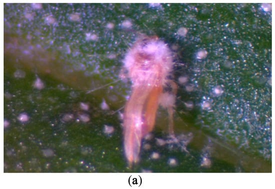
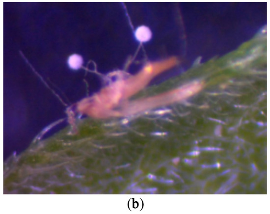
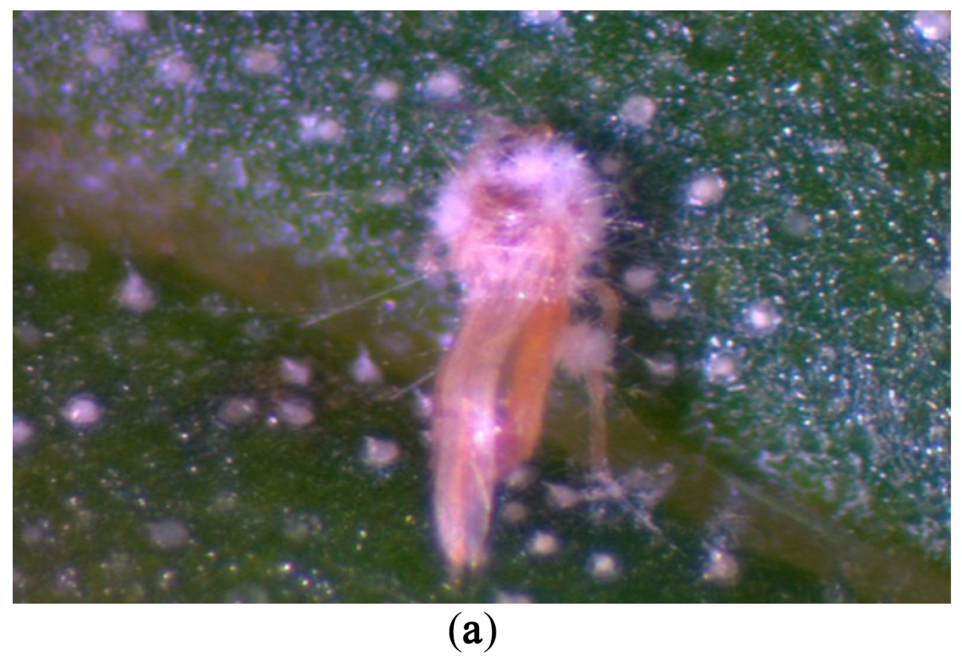
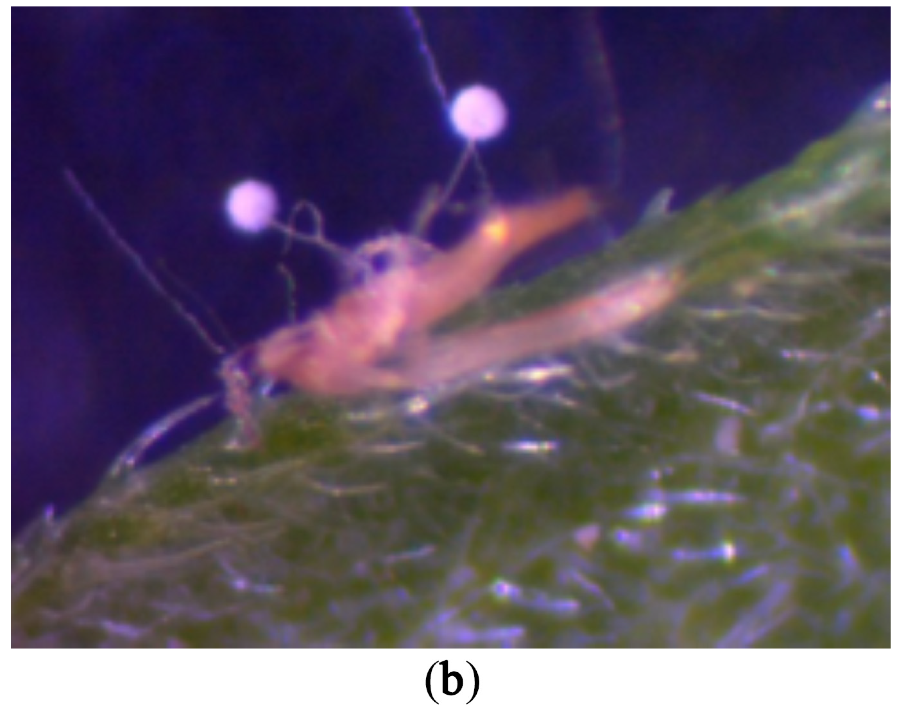
Of the twenty-one cadavers selected for an apparent fruiting phenotype (Figure 1), twenty-three pure strains representing four fungal species were isolated. All strains tested showed 100% query coverage and 100% sequence identity of the internal transcribed spacer (ITS) region of the ribosomal DNA with the corresponding reference sequences. The following GenBank accession numbers, ON103386, ON103385, ON103384, ON103383, and ON103382, are assigned to these strains: Akanthomyces lecanii isolate UASWS2695 BB1, Fusarium sp. isolate UASWS2694 THR25, Fusarium sp. isolate UASWS2693 THR18, Fusarium sp. isolate UASWS2692 THR12, and Fusarium oxysporum Snyder and Hansen, 1940 (Hypocreales, Nectriaceae) isolate UASWS2691 THR9, respectively.
Figure 1.
Phenotypic diversity of fungal fruiting bodies on F. occidentalis cadavers. (a) Typical fruiting body of Penicillium sp. (b) Typical fruiting body of Aspergillus sp.
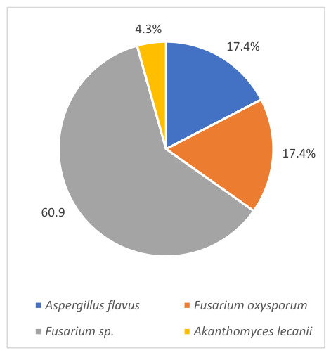
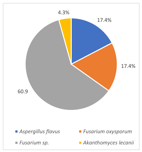
Indeed, the most represented genus was Fusarium, with 60.9% of Fusarium sp. and 17.4% of Fusarium oxysporum identified. Secondarily, Aspergillus flavus occurred with a prevalence of 17.4%, and finally, one strain of Akanthomyces lecanii was present at 4.3% (Figure 2).
Figure 2.
Percentage of fungal species occurrence from fructifications on thrips cadavers. Isolates were identified morphologically or genetically.
3.3. In Vitro Application of Fungal Strains on the Larval Stages of F. occidentalis
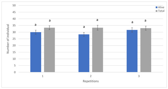
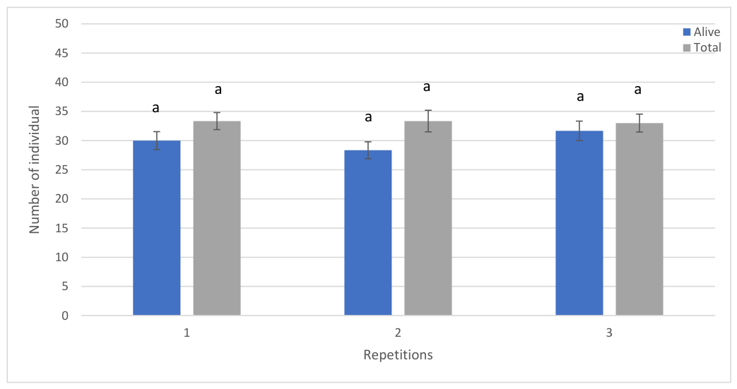
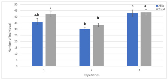
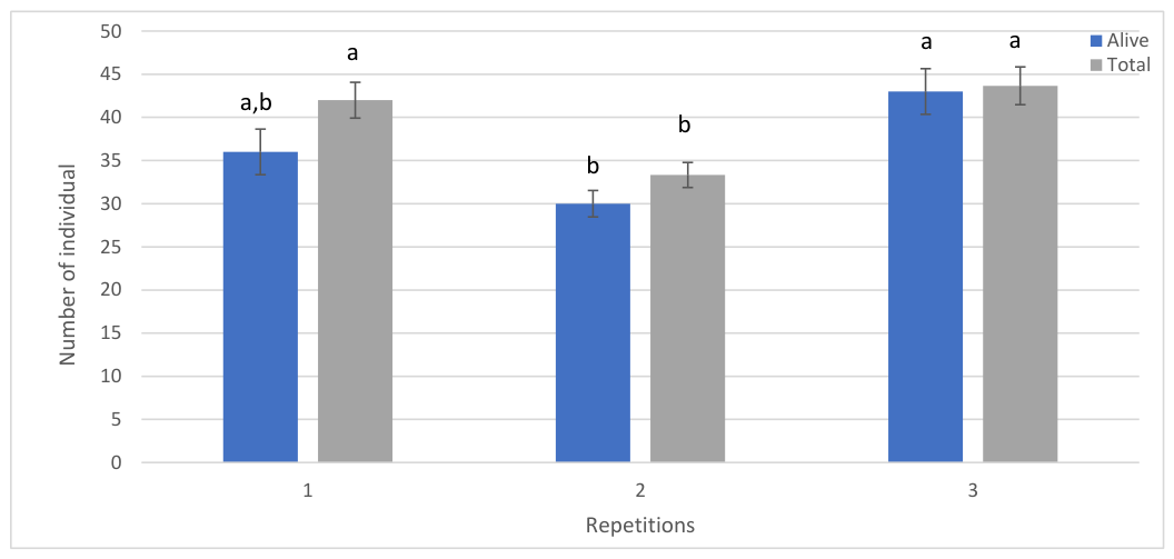
Oviposition of twenty WFT females over 24 h proved to be homogeneous. No statistically significant difference at the 5% threshold (p-value 0.986) was detected between the three averages representing nine replicates sampled on the same day and in the same insect rearing cage using ANOVA procedures (Figure 3).
Figure 3.
Average of dead and alive individuals identified 8 days after oviposition on beans by a population of twenty female Frankliniella occidentalis sampled simultaneously. Each repeated experimental unit (1, 2, 3) included 3 distinct batches. Means not sharing the same letters are significantly different in ANOVA procedures and Tukey’s multiple comparisons (p-values ≤ 5%), and error bars are represented by the standard error margin (SE).
On the other hand, significant differences were detected at the 5% threshold (p-values 0.024) between the egg-laying populations of WFT females originating from samples taken at different times (Figure 4).
Figure 4.
Average dead and alive individuals identified 8 days after oviposition on beans by a population of twenty Frankliniella occidentalis females sampled at different specific times. Each repeated experimental unit (1, 2, 3) comprised 3 distinct batches. Means not sharing the same letters are significantly different in ANOVA procedures and Tukey’s multiple comparisons (p-values ≤ 5%), and error bars are represented by the standard error margin (SE).
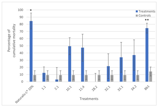
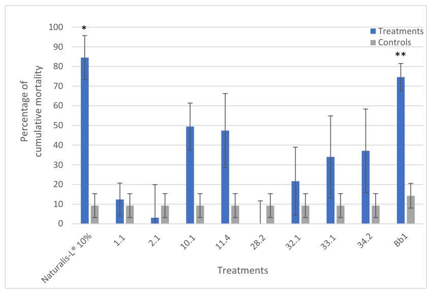
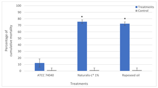
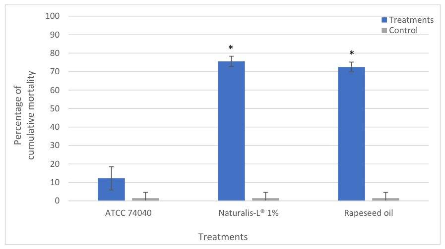
Strains applied to the larval stages of WFT showed different rates of population reduction. Indeed, the ANOVA procedures performed here demonstrated a statistical difference at the 5% and 10% thresholds. As a result, pairwise comparisons of Tukey, Dunnet and Fisher only detected significant differences between the A. lecanii Bb1 strain (p-values ≤ 0.003), the commercial product Naturalis-L® at 10% (p-value ≤ 0.075) and 1% (p-values ≤ 0.001), as well as rapeseed oil (p-values ≤ 0.001) with their negative controls. Due to the small number of repetitions and the formulation impact, the commercial product had a p-value greater than 5%; however, it showed a strong trend toward efficacy against the thrips population. These treatments demonstrated a cumulative mortality decrease of 74%, 84%, 75% and 73%, respectively (Figure 5 and Figure 6).
Figure 5.
Percentage of thrips cumulative mortality 8 days after exposure to fungal isolates and their negative controls for comparative purposes. The species corresponding to the strain numbers are given in Section 2.4. The positive control is the commercial product Naturalis-L®. Each treatment was repeated 3 times. Mortality percentages marked with an asterisk are significantly different from their controls in ANOVA and Dunnett’s multiple comparisons (* p-values ≤ 10%), or Fisher pairwise comparisons (** p-values ≤ 5%), and error bars represent the standard error margin (SE).
Figure 6.
Percentage of cumulative mortality of thrips 8 days after exposure to the commercial formulation Naturalis-L® and its pure strain Beauveria bassiana ATCC 74040 in comparison to the positive control, rapeseed oil, and the negative control. Each treatment was repeated 3 times. Mortality percentages marked with an asterisk are significantly different from their controls in ANOVA and Tukey multiple comparisons (* p-values ≤ 5%), and error bars represent the standard error margin (SE).
3.4. Fungal Growth-Inhibiting Actions of Kairomones and Pheromones
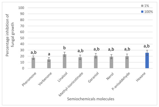
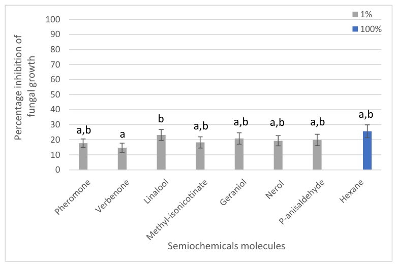
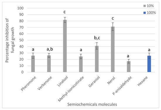
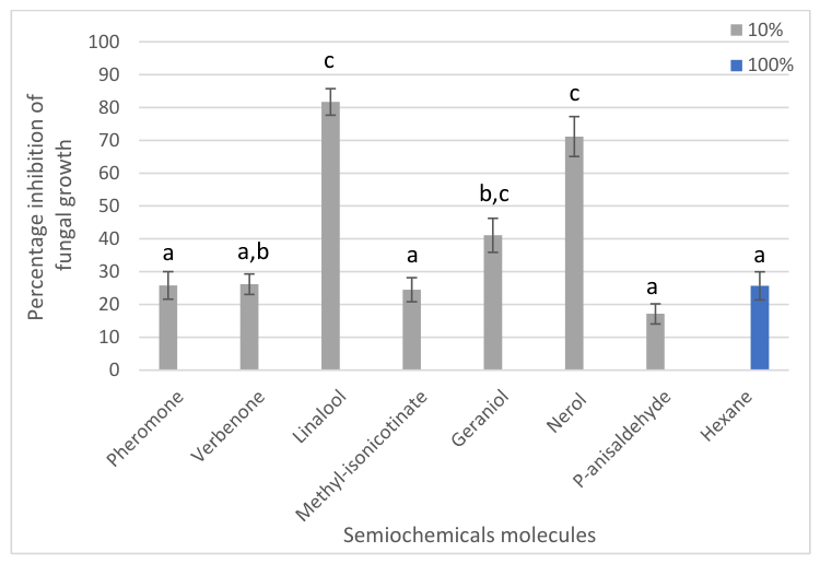
The attractive molecules tested here showed differences in the average percentage of growth inhibition for all strains tested. Indeed, linalool, nerol, and geraniol were found to be particularly harmful when applied at a 10% concentration, with 82%, 71%, and 41% average inhibition, respectively (Figure 7 and Figure 8).
Figure 7.
Average radial growth inhibition rates of all ten fungal isolate strains tested for each of the semiochemicals concentrated at 1%. Each treatment was repeated 2 times. Inhibition percentages not sharing the same letters are significantly different in the Friedman non-parametric test and post hoc multiple comparisons test with Bonferroni adjustments on ranks (p-values ≤ 5%), and error bars are represented by the standard error margin (SE).
Figure 8.
Average radial growth inhibition rate of all ten fungal isolate strains tested for each of the semiochemicals concentrated at 10%. Each treatment was repeated 2 times. Inhibition percentages not sharing the same letters are significantly different in the Friedman non-parametric test and post hoc multiple comparisons test with Bonferroni adjustments on ranks (p-values ≤ 5%), and error bars are represented by the standard error margin (SE).
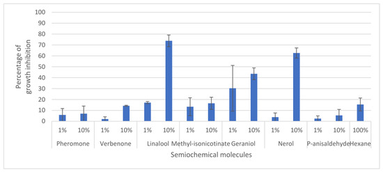
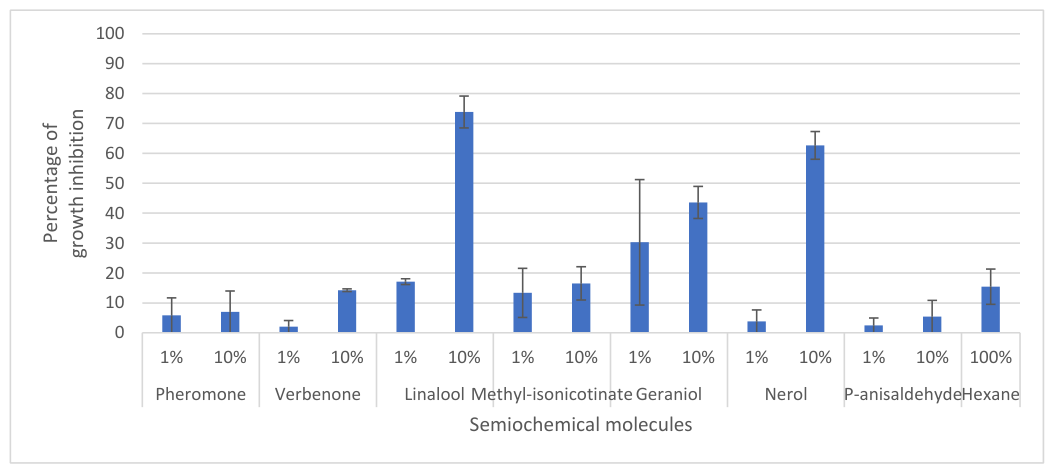
In addition, Friedman non-parametric tests showed a statistically significant difference in inhibition at the 1 ‰ threshold (p-value < 0.001) between treatments of the same concentrations. Thus, the multiple comparisons test with Bonferroni rank correction showed statistically significant inhibition at the 5% level (p < 0.05) for all molecules and concentrations applied compared to their control. Moreover, all molecules applied at 10% were also different from linalool, nerol, and geraniol at 10% concentrations. Finally, as shown in Figure 9, Akanthomyces lecanii strain Bb1 seems to be little affected by pheromone and p-anisaldehyde, as well as verbenone 1% and nerol 1%. All data on the effects of semiochemicals on the growth of the 9 other fungal strains are shown in Figures S1–S9.
Figure 9.
Percentage inhibition of radial growth of Akanthomyces lecanii strain Bb1 by semiochemical molecules as a function of their concentrations. Each treatment was repeated twice. Given the small number of repetitions, no statistical analysis was carried out. This graph is for descriptive purposes; only visual trends can be demonstrated. Error bars are represented by the standard error margin (SE).
4. Discussion
Three populations of Thysanoptera have been sampled from three different greenhouse crops and have been sustainably reared in in vitro conditions. They have been subsequently identified by amplification and sequencing of the taxonomic gene COI as belonging to the species Frankliniella occidentalis, using the pair of degenerate primers COI-CO2/COI-CO4, which amplify the COI taxonomic gene [40]. In addition, the population isolated from Z. aethiopica cultures differed by 3.15% from the other two isolates in the sequence of this mitochondrial gene, demonstrating the presence in Geneva of the two cryptic subspecies L and G of F. occidentalis [41].
Of the sixty-one thrips corpses sampled, twenty-one were selected based on the emerging fruiting body phenotype, i.e., 35% of the samples. Indeed, harvesting cadavers suggests an available time for growth on cadavers of saprophytic organisms. Moreover, only 4% of the fungal isolates identified in this study proved to be of interest. Indeed, a proportion of possibly entomopathogenic organisms could be used as phytosanitary products for reasons of risk to human or environmental health. In this context, 78% of the isolates were found to belong to the genus Fusarium, and 17% to the genus Aspergillus. These two genera are known to contain mycotoxin-producing strains with environmental and human pathogenic potential [42]. However, the strain of Akanthomomyces lecanii (Bb1) isolated during this work still represents a very good candidate [43]. Indeed, the Ve6 strain in the Mycotal® product from Koppert, formerly classified in the species Verticillium lecanii, is today reclassified as A. muscarium (syn: Lecanicillium muscarium) and is already approved for the biological control of F. occidentalis [44].
An innovative in vitro technique, adapted from the IRAC No. 14 [45], for applying fungal treatments to a sample of egg-laying WFT, made it possible to screen their entomopathogenic potential directly on homogeneous larval populations. This methodology has been successfully implemented to overcome the recurrent mortality observed in adult individuals of this same population that were kept alive in in vitro conditions with IRAC No. 14 [46]. EPF generally take more time to infect their host and reduce the population than chemical treatment [47]. So, to improve the longevity of targeted individuals under in vitro conditions, more robust methodologies to address this type of bias need to be developed. The methodology described here avoids the biases of mortality and disappearance of individuals in the test. In fact, this preliminary work has demonstrated that there is no variability in egg-laying and relative emerging population between individuals collected on the same day and from the same insect rearing cage. However, the opposite was observed when twenty WFT females from time-dispersed sampling laid eggs over 24 h. This suggests that it is essential to sample all laying WFT females on the same day and from the same insect rearing cage.
The Naturalis-L® commercial treatment diluted to 10% (2.3 × 10⁶ CFU/mL) caused the greatest incidence on the Thysanoptera population, with 85% cumulative mortality by the 7th day. However, given the small number of individuals emerging from the egg laying, we hypothesized that this could be correlated to the commercial formulation of this product, which appeared greasy and could therefore possibly cause suffocation to the eggs contained in the plant tissues [48]. Because we observed a low emergence of thrips when the Naturalis-L® formulation was applied in positive controls, the commercial B. bassiana strain ATCC 74040 was isolated from Naturalis-L® and applied without its commercial formulation at a concentration of 2.3 × 10⁶ CFU/mL. It only caused a 12% decrease in the emerging population. In addition, rapeseed oil and Naturalis-L®, both concentrated at 1%, showed a respective mortality of 73% and 75%, i.e., two similar mortalities at a threshold of 5%. Finally, only the fungal strain Bb1 caused a statistically significant cumulative mortality compared to its negative control, despite observable trends in virulence for M. anisopliae strains 10.1 and B. bassiana strain 11.4. Indeed, these last two strains did not statistically differ in terms of mortality compared to the 10% concentrated Naturalis-L®. In addition, lower mortalities were recorded in the immature stages compared to the adult stage of F. occidentalis [49]. Because of this, these last two strains cannot be ruled out of the process.
At the same time, tests using semiochemical compound attractants for WFT on fungal strains confirmed the fungistatic properties of all the compounds tested. Furthermore, geraniol, nerol, and linalool proved to be particularly harmful at a concentration of 10%. This confirms Mfuti’s observations [50] regarding the inhibitory effect of insect semiochemicals on fungal growth. However, four compounds, namely verbenone, p-anisaldehyde, methyl isonicotinate, and (S)-2-methylbutanoate of neryl, were identified as having a weakly inhibitory effect on the fungal growth of strains of A. lecanii (Bb1), M. anisopliae (10.1) and B. bassiana (11.4). The attractiveness of these four molecules to F. occidentalis was evaluated in a Y-shaped olfactometer, and the results have been published previously [33]. If none of the strains tested here showed tolerance towards all the tested molecules, certain strains such as B. bassiana strain (2.1) and M. anisopliae strains (32.1, 33.1, and 34.2) exhibited the greatest mean inhibitions.
Though these works point to the utility of these results for the objective of an autodissemination trap for F. occidentalis, one must also consider the complexity of multitrophic interactions between species and their contribution to ecological equilibrium when developing pest control, as stressed by Zhang et al. [51]. The fact that microbes could modify olfactory signals emitted by plants, which in turn would affect insect behavior, is an aspect to be explored in the next steps of the development of the autodissemination trap and in our further in vivo studies to be assessed for F. occidentalis in relation to a few major greenhouse crops.
5. Conclusions
This research led to the isolation and characterization of the virulence of the fungal strain Bb1 of Akanthomyces lecanii, which shows great potential for further development as a biopesticide against thrips species. Its compatibility with four semiochemical attractants of F. occidentalis is promising for the development of a self-inoculating trap against F. occidentalis. Further studies should, however, focus on evaluating the persistence of the germination capacity of strain Bb1 when exposed to semiochemicals. Furthermore, the LT50 and LC50 values still need to be estimated for strain Bb1.
Supplementary Materials
The following supporting information can be downloaded at https://www.mdpi.com/article/10.3390/microbiolres17090171/s1: Figure S1. Percentage inhibition of radial growths of Beauveria bassiana strain 1.1 by semiochemical molecules as a function of their concentrations; Figure S2. Percentage inhibition of radial growths of Beauveria bassiana strain 2.1 by semiochemical molecules as a function of their concentrations; Figure S3. Percentage inhibition of radial growths of Metarhizium anisopliae strain 10.1 by semiochemical molecules as a function of their concentrations; Figure S4. Percentage inhibition of radial growths of Beauveria bassiana strain 11.4 by semiochemical molecules as a function of their concentrations; Figure S5. Percentage inhibition of radial growths of strain 28.2 by semiochemical molecules as a function of their concentrations; Figure S6. Percentage inhibition of radial growths of Metarhizium anisopliae strain 32.1 by semiochemical molecules as a function of their concentrations; Figure S7. Percentage inhibition of radial growths of Metarhizium anisopliae strain 33.1 by semiochemical molecules as a function of their concentrations; Figure S8. Percentage inhibition of radial growths of Metarhizium anisopliae strain 34.2 by semiochemical molecules as a function of their concentrations; Figure S9. Percentage inhibition of radial growths of Beauveria bassiana strain ATCC 74040 by semiochemical molecules as a function of their concentrations.
Author Contributions
Conceptualization, C.D. and F.L.; methodology, C.D., Y.B., C.J.-F.C. and F.L.; validation, C.D. and F.L.; formal analysis, C.D.; investigation, C.D. and Y.B.; visualization, C.D.; resources, F.L.; data curation, C.D. and F.L.; writing—original draft preparation, C.D.; writing—review and editing, C.D. and F.L.; supervision, F.L. and C.J.-F.C.; project administration, F.L. and C.J.-F.C.; funding acquisition, F.L. and C.J.-F.C. All authors have read and agreed to the published version of the manuscript.
Funding
This research was funded through the P2-Interdisciplinary program of the HES-SO University of Applied Sciences and Arts, Western Switzerland, grant number 107077/IA-INTERDISC20.
Institutional Review Board Statement
Ethical review and approval were waived for this study because the experimental subjects were insects (Frankliniella occidentalis), which are invertebrates and are exempt from institutional animal care guidelines in Switzerland. Trials with invertebrates such as thrips do not require legal permits under the framework of the Federal Laws of Switzerland (The Animal Welfare Act (AniWA) and the Animal Welfare Ordinance (AniPO)).
Informed Consent Statement
Not applicable.
Data Availability Statement
The original contributions presented in this study are included in the article/Supplementary Material. Further inquiries can be directed to the corresponding author.
Acknowledgments
We are grateful to Bastien Cochard for his valuable and helpful advice.
Conflicts of Interest
The authors declare no conflicts of interest.
References
- Richard, O.W.; Davies, R.G. Thysanoptera (Physapoda: Thrips). Gen. Textb. Entomol. 1977, 1, 782–792. [Google Scholar] [CrossRef] [Scilit]
- Mound, L.A.; Teulon, D.A.J. Thysanoptera as Phytophagous Opportunists. In Thrips Biology and Management; Parker, B.L., Skinner, M., Lewis, T., Eds.; NATO ASI Series; Springer: Boston, MA, USA, 1995; Volume 276, pp. 3–19. [Google Scholar] [CrossRef] [Scilit]
- Lewis, T. Pest Thrips in Perspective. In Thrips as Crop Pests; Lewis, T., Ed.; CAB International: Wallingford, UK, 1997; pp. 1–13. [Google Scholar]
- Rotenberg, D.; Jacobson, A.L.; Schneweis, D.J.; Whitfield, A.E. Thrips Transmission of Tospoviruses. Curr. Opin. Virol. 2015, 15, 80–89. [Google Scholar] [CrossRef] [Scilit] [PubMed]
- He, Z.; Guo, J.F.; Reitz, S.R.; Lei, Z.R.; Wu, S.Y. A Global Invasion By The Thrip, Frankliniella occidentalis: Current Virus Vector Status and Its Management. Insect Sci. 2020, 27, 626–645. [Google Scholar] [CrossRef] [Scilit] [PubMed]
- Zhao, K.; Rosa, C. Thrips as the Transmission Bottleneck for Mixed Infection of Two Orthotospoviruses. Plants 2020, 9, 509. [Google Scholar] [CrossRef] [Scilit] [PubMed]
- Reitz, S.R.; Gao, Y.; Kirk, W.D.J.; Hoddle, M.S.; Leiss, K.A.; Funderburk, J.E. Invasion Biology, Ecology, and Management of Western Flower Thrips. Ann. Rev. Entomol. 2020, 65, 17–37. [Google Scholar] [CrossRef] [Scilit] [PubMed]
- Reitz, S.R.; Funderburk, J.E. Chap. 16. Management Strategies for Western Flower Thrips and the Role of Insecticides. In Insecticides—Pest Engineering; Perveen, F., Ed.; InTechOpen: London, UK, 2012; Volume 16, pp. 355–384. [Google Scholar] [CrossRef] [Scilit] [PubMed]
- Mouden, S.; Sarmiento, K.F.; Klinkhamer, P.G.; Leiss, K.A. Integrated Pest Management in Western Flower Thrips: Past, Present and Future. Pest Manag. Sci. 2017, 73, 813–822. [Google Scholar] [CrossRef] [Scilit] [PubMed]
- Herron, G.A.; Rophail, J.; Gullick, G.C. Laboratory-Based, Insecticide Efficacy Studies on Field-Collected Frankliniella occidentalis (Pergande) (Thysanoptera: Thripidae) and Implications for Its Management in Australia. Aust. J. Entomol. 1996, 35, 161–164. [Google Scholar] [CrossRef] [Scilit]
- Jensen, S.E. Insecticide Resistance in the Western Flower Thrips, Frankliniella occidentalis. Integr. Pest Manag. Rev. 2000, 5, 131–146. [Google Scholar] [CrossRef] [Scilit]
- Gao, Y.; Lei, Z.; Reitz, S.R. Western Flower Thrips Resistance to Insecticides: Detection, Mechanisms and Management Strategies. Pest Manag. Sci. 2012, 8, 1111–1121. [Google Scholar] [CrossRef] [Scilit] [PubMed]
- Jensen, S.E. Mechanisms Associated with Methiocarb Resistance in Frankliniella occidentalis (Thysanoptera: Thripidae). J. Econ. Entomol. 2000, 93, 464–471. [Google Scholar] [CrossRef] [Scilit]
- Herron, G.A.; James, T.M. Monitoring Insecticide Resistance in Australian Frankliniella occidentalis Pergande (Thysanoptera: Thripidae) Detects Fipronil and Spinosad Resistance. Aust. J. Entomol. 2005, 44, 299–303. [Google Scholar] [CrossRef] [Scilit]
- Cubillos-Salamanca, Y.P.; Rodríguez-Maciel, J.C.; Pineda-Guillermo, S.; Silva-Rojas, H.V.; Berzosa, J.; Tejeda-Reyes, M.A.; Rebollar-Alviter, Á. Identification of Thrips Species and Resistance of Frankliniella occidentalis (Thysanoptera: Thripidae) to Malathion, Spinosad, and Bifenthrin in Blackberry Crops. Fla. Entomol. 2019, 102, 738–746. [Google Scholar] [CrossRef] [Scilit]
- Koschier, E.H.; De Kogel, W.; Visser, H. Assessing the Attractiveness of Volatile Plant Compounds to Western Flower Thrips Frankliniella occidentalis. J. Chem. Ecol. 2000, 26, 2643–2655. [Google Scholar] [CrossRef] [Scilit]
- Kirk, W.D.J.; Jan de Kogel, W.; Koschier, E.H.; Teulon, D.A.J. Semiochemicals for Thrips and their use inPest Management. Ann. Rev. Entomol. 2021, 66, 101–119. [Google Scholar] [CrossRef] [Scilit] [PubMed]
- Díaz, M.A.; Osorio, C.; Coy-Barrera, E.; Rodríguez, D. Semiochemicals Associated with the Western Flower Thrips Attraction: A Systematic Literature Review and Meta-Analysis. Insects 2023, 14, 269. [Google Scholar] [CrossRef] [Scilit] [PubMed]
- Sharma, R.; Sharma, P. Fungal Entomopathogens: A Systematic Review. Egypt. J. Biol. Pest Control 2021, 31, 57. [Google Scholar] [CrossRef] [Scilit]
- Kivett, J.M.; Cloyd, R.A.; Bello, N.M. Evaluation of Entomopathogenic Fungi Against the Western Flower Thrips (Thysanoptera: Thripidae) Under Laboratory Conditions. J. Entomol. Sci. 2016, 51, 274–291. [Google Scholar] [CrossRef] [Scilit]
- Ludwig, S.W.; Oetting, R.D. Efficacy of Beauveria bassiana Plus Insect Attractants for Enhanced Control of Frankliniella occidentalis (Thysanoptera: Thripidae). Fla. Entomol. 2002, 85, 270–272. [Google Scholar] [CrossRef] [Scilit]
- Niassy, S.; Maniania, N.K.; Subramanian, S.; Gitonga, L.M.; Ekesi, S. Performance of a Semiochemical-Baited Autoinoculation Device Treated with Metarhizium anisopliae for Control of Frankliniella occidentalis on French Bean in Field Cages. Entomol. Exp. Appl. 2012, 142, 97–103. [Google Scholar] [CrossRef] [Scilit]
- Mfuti, D.K.; Subramanian, S.; Van Tol, R.W.; Wiegers, G.L.; De Kogel, W.J.; Niassy, S.; Du Plessis, H.; Ekesi, S.; Maniania, N.K. Spatial Separation of Semiochemical Lurem-TR and Entomopathogenic Fungi to Enhance Their Compatibility and Infectivity in an Autoinoculation System for Thrips Management. Pest Manag. Sci. 2015, 72, 131–139. [Google Scholar] [CrossRef] [Scilit] [PubMed]
- Mfuti, D.K.; Niassy, S.; Subramanian, S.; Du Plessis, H.; Ekesi, S.; Maniania, N.K. Lure and Infect Strategy for Application of Entomopathogenic Fungus for the Control of Bean Flower Thrips in Cowpea. Biol. Control 2017, 107, 70–76. [Google Scholar] [CrossRef] [Scilit]
- Mfuti, D.K.; Subramanian, S.; Niassy, S.; Salifu, D.; Du Plessis, H.; Ekesi, S.; Maniania, N.K. Screening for Attractants Compatible with Entomopathogenic Fungus Metarhizium anisopliae for Use in Thrips Management. Afr. J. Biotechnol. 2016, 15, 714–721. [Google Scholar] [CrossRef] [Scilit]
- Laurent, E.; Crovadore, J.; Cochard, B.; Pelleteret, P.; Chablais, R.; Bovigny, P.-Y.; Lefort, F. New Strains of Entomopathogenic Fungi Isolated from Organic Orchards in Switzerland. Commun. Agric. Appl. Biol. Sci. 2017, 82, 207–213. [Google Scholar]
- Steiner, M.Y.; Goodwin, S. Methods for Collecting and Rearing Thrips (Thysanoptera) and Their Natural Enemies. Aust. J. Entomol. 1998, 37, 101–106. [Google Scholar] [CrossRef] [Scilit]
- Lefort, F.; Douglas, G.C. An Efficient Micro-Method of DNA Isolation from Mature Leaves of Four Hardwood Tree Species Acer, Fraxinus, Prunus and Quercus. Ann. For. Sci. 1999, 56, 259–263. [Google Scholar] [CrossRef] [Scilit]
- Che, J.; Chen, H.-M.; Yang, J.-X.; Jin, J.-Q.; Jiang, K.E.; Yuan, Z.-Y.; Murphy, R.W.; Zhang, Y.-P. Universal COI Primers for DNA Barcoding Amphibians. Mol. Ecol. Res. 2012, 12, 247–258. [Google Scholar] [CrossRef] [Scilit] [PubMed]
- Sönmez, E.; Sevim, A.; Demirbağ, Z.; Demir, İ. Isolation, Characterization and Virulence of Entomopathogenic Fungi from Gryllotalpa gryllotalpa (Orthoptera: Gryllotalpidae). Appl. Entomol. Zool. 2016, 51, 213–223. [Google Scholar] [CrossRef] [Scilit]
- White, T.J.; Bruns, T.; Lee, S.; Taylor, J. Amplification and Direct Sequencing of Fungal Ribosomal RNA genes for Phylogenetics. PCR Protoc. A Guide Methods Appl. 1990, 38, 315–322. [Google Scholar] [CrossRef] [Scilit]
- DeGraaf, H.E.; Wood, G.M. An Improved Method For Rearing Western Flower Thrips Frankliniella occidentalis. Fla. Entomol. 2009, 92, 664–666. [Google Scholar] [CrossRef] [Scilit]
- Chappuis, C.J.F.; Cléroux, M.; Descombes, C.; Barth, Y.; Lefort, F. Attraction of Frankliniella occidentalis females Towards the Aggregation Pheromone Neryl (S)-2-Methylbutanoate and Kairomones in a Y-Olfactometer. Insects 2023, 14, 562. [Google Scholar] [CrossRef] [Scilit] [PubMed]
- Nana, P.; Maniania, N.K.; Maranga, R.O.; Boga, H.I.; Kutima, H.L.; Eloff, J.N. Compatibility Between Calpurnia aurea Leaf Extract, Attraction Aggregation, and Attachment Pheromone and Entomopathogenic Fungus Metarhizium anisopliae on Viability, Growth, and Virulence of the Pathogen. J. Pest Sci. 2012, 85, 109–115. [Google Scholar] [CrossRef] [Scilit]
- Matsuura, K.; Matsunaga, T. Antifungal Activity of a Termite Queen Pheromone Against Egg-Mimicking Termite Ball Fungi. Ecol. Res. 2015, 30, 93–100. [Google Scholar] [CrossRef] [Scilit]
- Mitaka, Y.; Mori, N.; Matsuura, K. Multi-Functional Roles of a Soldier-Specific Volatile as a Worker Arrestant, Primer Pheromone and an Antimicrobial Agent in a Termite. Proc. R. Soc. B Biol. Sci. 2017, 284, 20171134. [Google Scholar] [CrossRef] [Scilit] [PubMed]
- Cole, L.K.; Blum, M.S.; Roncadori, R.W. Antifungal Properties of the Insect Alarm Pheromones, Citral, 2-Heptanone, and 4-Methyl-3-Heptanone. Mycologia 1975, 67, 701–708. [Google Scholar] [CrossRef] [Scilit]
- Sirima, A.; Sereme, A.; Sereme, D.; Koïta, K.; Nana, T.A.; Sawadogo, M. Effets de Quatre Huiles Essentielles sur la Croissance Mycélienne Radiale d’un Isolat de Alternaria sp. au Burkina Faso. Int. J. Biol. Chem. Sci. 2020, 14, 762–771. [Google Scholar] [CrossRef] [Scilit]
- Koné, D.; Badou, O.J.; Bomisso, E.L.; Camara, B.; Ake, S. Activités In Vitro de Différents Fongicides sur la Croissance chez Mycosphaerella fijiensis var. difformis Stover et Dickson, Cladosporium musae Morelet et Deightoniella torulosa (Syd.) Ellis, Parasites Isolés de la Phyllosphère des Bananiers en Côte-d’Ivoire. Comptes Rendus—Biol. 2009, 332, 448–455. [Google Scholar] [CrossRef] [Scilit] [PubMed]
- Brunner, P.C.; Frey, J.E. Habitat-Specific Population Structure in Native Western Flower Thrips Frankliniella occidentalis (Insecta, Thysanoptera). J. Evol. Biol. 2010, 23, 797–804. [Google Scholar] [CrossRef] [Scilit] [PubMed]
- Mirnezhad, M.; Schidlo, N.; Klinkhamer, P.G.L.; Leiss, K.A. Variation in Genetics and Performance of Dutch Western Flower Thrips Populations. J. Econ. Entomol. 2012, 105, 1816–1824. [Google Scholar] [CrossRef] [Scilit] [PubMed]
- Meliani, H.; Makhloufi, A.; Cherif, A.; Mahjoubi, M.; Makhloufi, K. Biocontrol of toxinogenic Aspergillus flavus and Fusarium oxysporum f. sp. albedinis by two rare Saharan actinomycetes strains and LC-ESI/MS-MS profiling of their antimicrobial products. Saudi J. Biol. Sci. 2022, 29, 103288. [Google Scholar] [CrossRef] [Scilit] [PubMed]
- Gouli, V.V.; Gouli, S.Y.; Skinner, M.; Shternshis, M.V. Effect of the Entomopathogenic Fungi on Mortality and Injury Level of Western Flower Thrips, Frankliniella occidentalis. Arch. Phytopathol. Plant Prot. 2009, 42, 118–123. [Google Scholar] [CrossRef] [Scilit]
- Schreiter, G.; Butt, T.M.; Beckett, A.; Vestergaard, S.; Moritz, G. Invasion and Development of Verticillium lecanii in the Western Flower Thrips, Frankliniella occidentalis. Mycol. Res. 1994, 98, 1025–1034. [Google Scholar] [CrossRef] [Scilit]
- Insecticide Resistance Action Committee (IRAC). Susceptibility Test Methods Series, Method No: 014. Vol. 3. 1999. Available online: https://irac-online.org/methods/frankliniella-occidentalis-larvae/ (accessed on 15 February 2021).
- Insecticide Resistance Action Committee (IRAC). Susceptibility test methods Series Method No: 010. Vol. 3. 1999. Available online: https://irac-online.org/methods/frankliniella-occidentalis-adults/ (accessed on 15 February 2021).
- Pachamuthu, P.; Kamble, S.T. In Vivo Study on Combined Toxicity of Metarhizium anisopliae (Deuteromycotina: Hyphomycetes) Strain ESC-1 with Sublethal Doses of Chlopyrifos, Propetamphos and Cyfluthrin Against German Cockroach (Dictyoptera: Blattellidae). J. Econ. Entomol. 2000, 93, 60–70. [Google Scholar] [CrossRef] [Scilit] [PubMed]
- Martín-López, B.; Varela-Barrenechea, I.; Lores-Hermida, M. Eficacia de Aceites Vegetales, Minerales y de Pescado Frente a Frankliniella occidentalis (Pergande). Bol. Sanid. Veg. Plagas. 2004, 30, 177–183. [Google Scholar]
- Vestergaard, S.; Gillespie, A.T.; Butt, T.M.; Schreiter, G.; Eilenberg, J. Pathogenicity of the Hyphomycete Fungi Verticillium lecanii and Metarhizium anisopliae to the Western Flower Thrips, Frankliniella occidentalis. Biocontrol Sci. Technol. 1995, 5, 185–192. [Google Scholar] [CrossRef] [Scilit]
- Mfuti, D.K.; Tamiru, A.; Kirk, W.D.J.; Akinyemi, A.O.; Campbell, H.; O’brien, M.; Drijfhout, F.P.; Pope, T.W.; Niassy, S.; Subramanian, S. Exploiting Thrips Aggregation Pheromones to Develop a Lure-And-Kill Strategy for the Management of the Bean Flower Thrips. Agronomy 2021, 11, 1269. [Google Scholar] [CrossRef] [Scilit]
- Zhang, W.; Stelinski, L.L.; Mohamed, A.; Wang, G.; Tettamanti, G.; Chen, M.; Hong, M.; Daly, E.Z.; Bruin, J.; Renault, D.; et al. Unlocking Agro-Ecosystem Sustainability: Exploring the Bottom-Up Effects of Microbes, Plants, and Insect Herbivores. Integr. Zool. 2025, 20, 465–484. [Google Scholar] [CrossRef] [Scilit] [PubMed]

Disclaimer/Publisher’s Note: The statements, opinions and data contained in all publications are solely those of the individual author(s) and contributor(s) and not of MDPI and/or the editor(s). MDPI and/or the editor(s) disclaim responsibility for any injury to people or property resulting from any ideas, methods, instructions or products referred to in the content. |
© 2026 by the authors. Licensee MDPI, Basel, Switzerland. This article is an open access article distributed under the terms and conditions of the Creative Commons Attribution (CC BY) license.