


Abstract
Accurate analysis and diagnosis of parasitic infections traditionally require conventional laboratory microscopes, limiting accessibility in resource-limited and rural areas as these microscopes are expensive and not easily portable. Smartphone-attachable microscope devices offer an affordable, portable alternative. This study evaluated the accuracy and perceived microscope usability of three commercially available smartphone-attachable microscopes (uHandy, Foldscope, and Carson handy) compared to a conventional microscope using prepared parasitic ova and cyst slides. Evaluators captured images of prepared parasite slides using these devices and performed blinded identification. Agreement analysis of the species identification based on the images and assessment of the perceived usability features of each microscope were performed. Despite scoring lower than other microscopes for perceived usability, the Foldscope demonstrated the most accurate diagnosis consistently throughout the different parasites. The Carson handy received scores for magnification, lighting, and image quality that were comparable to a conventional microscope. There were no differences between the three smartphone-attachable microscopes in terms of slide compatibility, ease of image acquisition, stability, or portability. Improvements in imaging in the future along with access to standardized portable device could allow for quicker, more accurate diagnoses, and more tailored treatments leading to better outcomes for patients in neglected areas.
1. Introduction
Smartphones and their incorporated digital cameras have undergone great developments in recent years. Evolving to include multicamera systems, 3D acquisition, image stabilization, fast focusing and a high dynamic range, smartphone imaging technology continues to improve [1]. Smartphone-attachable devices are a growing trend in medicine for cost-effective, portable diagnostic workups.
Smartphone-attachable microscopy is an affordable and easy method for acquiring and sharing digital photomicrographs [2,3]. The field has moved from simple eyepiece adapters to modular bright-field, fluorescence, and lensless systems, but standardization and clinical validation are still lacking [4]. The adoption of smartphone use into everyday life has led to the rapid development of digital diagnostic tools [5,6]. Nevertheless, their use as point-of-care devices, i.e., their use to perform diagnostics at or near the site of patient care, is promising [7].
The incidence of parasitic infection is significantly higher in remote areas than in industrialized urban areas [8], due in part to a lack of modern sanitation and increased exposure to contaminated water [9]. In global medicine, the use of portable equipment is pivotal to diagnostics in the rural environment. Making clinical diagnoses in remote areas is often challenging due to resource limitations and a lack of laboratory support. Training community health workers on parasitic ova diagnosis using smartphone-attachable microscopy is feasible [10]. However, the accuracy of smartphone-attachable microscopes for such diagnostic applications has not yet been assessed.
With the recent rise in the use of smartphones as clinical tools, we aimed to determine the feasibility of identifying common parasites with a set of various smartphone-attachable microscopes. We evaluated these microscopes using slides containing intestinal parasites, rating smartphone image quality and perceived usability.
2. Material and Methods
2.1. Assessment of the Feasibility of Smartphone-Attachable Microscopes in the Identification of Parasitic Ova and Cysts
Commercially available prepared slides of 18 different parasitic ova and cysts (Supplementary Materials Table S1) were photographed with four different commercially available smartphone-attachable microscopes: Carson MicroFlip Pocket microscope (Carson Optical, Ronkonkoma, NY, USA) (called here Carson handy), uHandy Lite Mobile microscope (Aidmics Biotechnology Co., Taipei, Taiwan), and the Foldscope (Foldscope Instruments, Inc., Palo Alto, CA, USA) (Supplementary Materials Figure S1). In addition, images were also taken with a conventional upright light-transmitted microscope (Zeiss, Primostar, Dublin, CA, USA). Smartphone cameras used by the 5 evaluators included: iPhone 13, iPhone 14 Pro, iPhone 15, iPhone 16 Pro, and iPhone 17 Pro. The evaluators are medical students at different training years, so their knowledge of microbiology is not at the professional nor technician level.
At least two different slides per species were photographed independently by at least four of the authors, so true different fields of view per slide were available. Blinded photomicrographs were identified by three raters using a predetermined image guide composed of labeled parasite images taken with a conventional light-transmitted laboratory-grade microscope. Concordance and disagreement were statistically analyzed.
2.2. Microscope Accuracy Analysis
Identification accuracy was defined as the proportion of blinded photomicrographs correctly determined to represent the true parasite species. For each image, the ground-truth label (the known parasite species on the photographed slide) was compared against each rater’s identification.
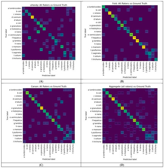
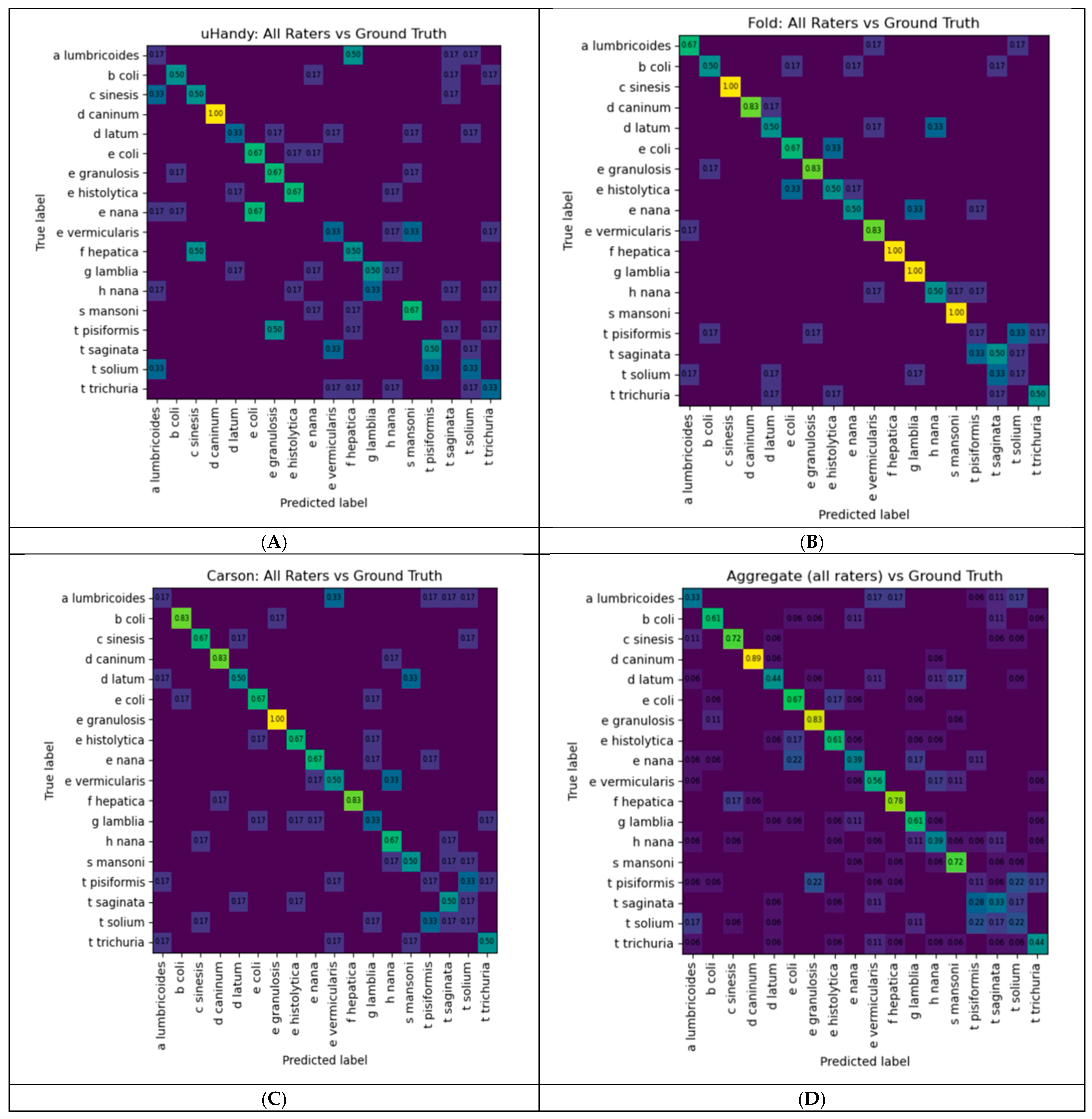
Each image was rated by two raters who did not take the image. Giving n = 18 images per pairwise comparison, the rater assignment followed a fixed rotation, and kappa was calculated within each device rather than pooled. Because each photomicrograph was identified by raters who had not photographed it, accuracy was computed per rater as the proportion of that rater’s identifications that were objectively correct, and then pooled across raters. Accuracy was stratified in two ways: by microscope, in which the dataset was subset to each device (Carson, uHandy, Foldscope) and accuracy was recomputed within each subset; by parasite species, in which the proportion of correct identifications was calculated for each organism. Per-species accuracy was computed for each microscope individually and for all raters collectively, and these values were visualized as heat-map tables (Figure 1). Aggregate agreement between raters’ identifications and ground truth was additionally summarized using normalized confusion matrices, in which rows corresponded to the actual organism and columns to rater identifications.
Figure 1.
Heat-map-style table showing levels of agreement of the raters’ diagnoses per parasitic species. Panels (A–C) depict microscopes from the brands uHandy, Foldscope, and Carson, respectively. Panel (D) depicts a heat-map-style table of the rater’s collective accuracy proportion per parasitic species.
All analyses were performed in Python (version [3.14]) using the pandas, NumPy, scikit-learn, and statsmodels libraries (Version is 3.14.0).
2.3. Perceived Microscope Usability
A rubric in a Likert scale ranging from “very poor” to “very good” evaluated the following criteria: magnification, lighting, acquired image quality, phone compatibility, slide compatibility, ease of image acquisition, stability, and portability. The rubric (Supplementary Materials Table S2) was completed by the five authors using the uHandy, Foldscope, the Carson handy, and a conventional transmitted light microscope (Supplementary Materials Table S2). Each evaluator captured images on all 5 microscopes. Evaluators’ ratings of each microscope were based on personal utilization and the images produced by the standardized slides of ova and cysts.
3. Results
3.1. Assessments of Smartphone-Attachable Microscopes in the Identification of Parasitic Ova and Cysts
We first assessed the percent accuracy of the diagnosis for each parasitic species based on the acquired digital images. The accuracy ranged from 0.407 to 0.657. Fleiss’ kappa coefficient across raters was 0.405. Pairwise Cohen’s kappa coefficient between the raters ranged from 0.255 to 0.49, which indicates that there was fair-to-moderate agreement between individuals that was beyond that expected by chance. The Fleiss’ kappa coefficient of 0.405 suggests that there was a fair-to-moderate agreement between all raters.
In addition, we also calculated inter-rater agreement examined by microscope. Because each sample photograph was rated by two raters who did not photograph it, agreement per microscope was calculated on a rotating two-rater design (n = 18 images per pairwise comparison). All-rater agreement was highest for the Foldscope (Fleiss’ κ = 0.469, 95% CI 0.32–0.60), followed by the Carson (κ = 0.372, 95% CI 0.22–0.50) and uHandy (κ = 0.370, 95% CI 0.22–0.50). Confidence intervals overlapped substantially across devices, so these differences should be interpreted as descriptive. The Foldscope showed the most consistent pairwise agreement (Cohen’s κ 0.29–0.59); the Carson’s pooled value was reduced by one discordant rater pair (κ = 0.06), which is likely a remnant of the n= 18 sample size and of having only two rater-pairs for any given agreement. This pattern was similar to that of identification accuracy, which was highest for the Foldscope and lowest for the uHandy.
3.2. Perceived Microscope Usability
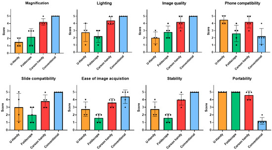
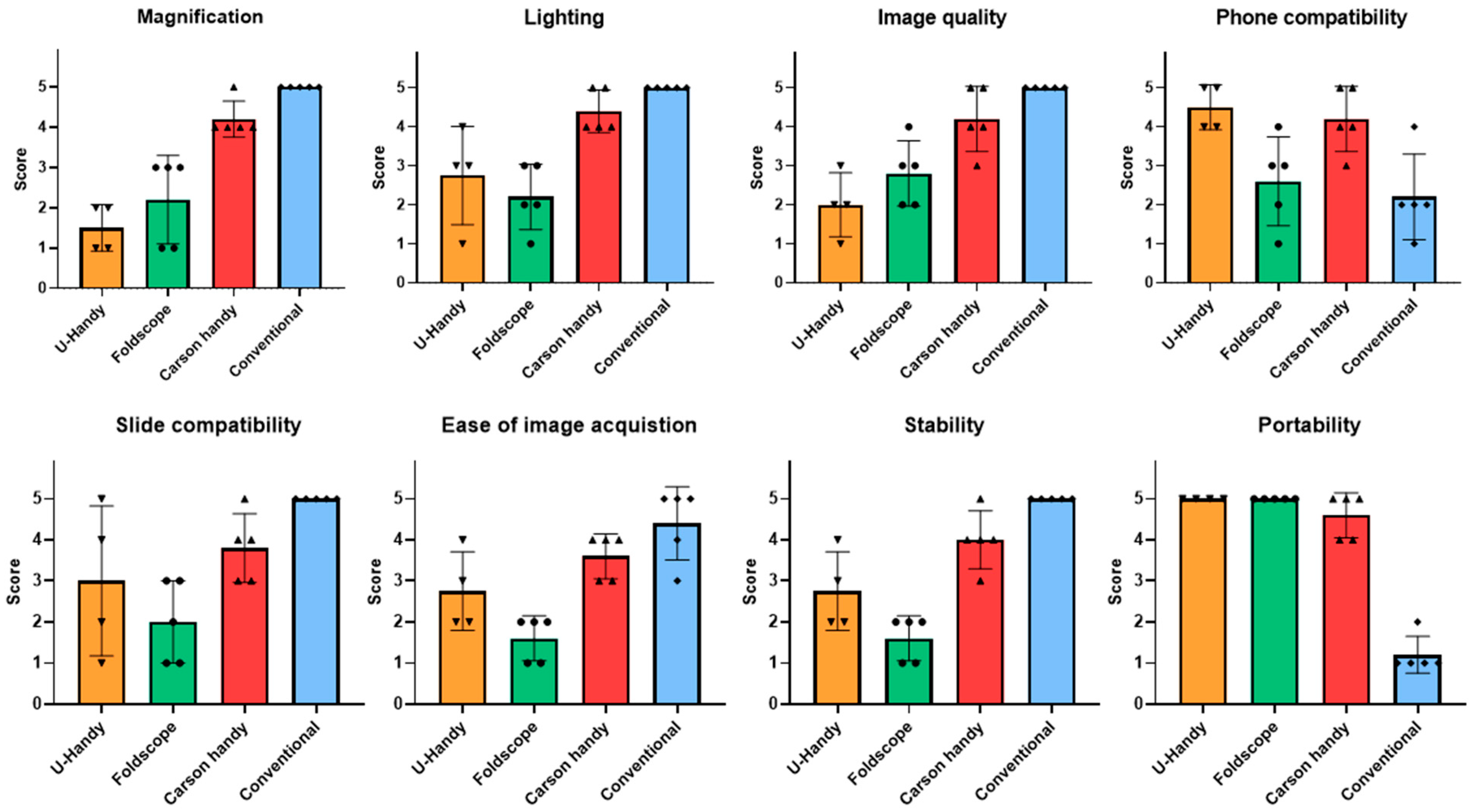
Usability was rated by five reviewers across eight dimensions on a five-point Likert scale (1 = very poor, 5 = very good) for three smartphone-attachable devices (uHandy, Foldscope, Carson handy) and a conventional lab-style microscope. The Carson handy received scores for magnification, lighting, and image quality that were comparable to a conventional microscope. The uHandy and the Carson handy scored highest in phone compatibility. There were no differences between the three smartphone-attachable microscopes in terms of slide compatibility, ease of image acquisition, stability, or portability (Figure 2). While the uHandy was rated poorest for magnification, and image quality, the Foldscope was rated poorest in slide compatibility, ease of image acquisition, and stability.
Figure 2.
Ratings for the 8 criteria per microscope (uHandy, Foldscope, Carson Handy, conventional) ranging from 1 (very poor) to 5 (very good). Scatter plot of individual values plus bar graph.
4. Discussion
Using an image guide, our data showed a fair-to-moderate inter-rater reliability of appropriately determining the parasitic species. The species Dipylidium caninum and Echinococcus granulosus had the highest identification accuracy across raters. This may be attributed to the hematoxylin and eosin stain used on these particular slides, making them more easily identifiable. The species within the Taenia genus were most often guessed incorrectly, likely due to their greater morphological similarity compared with the other species examined.
Surprisingly, accuracy analysis showed that the Foldscope demonstrated the highest overall inter-rater agreement between blinded participants in accurately identifying the parasitic ova and cysts. Notably, the raters’ guesses were least accurate when using the uHandy microscope. This was observed across usage of multiple iPhone models and smartphone-attachable microscopes. Issues such as the camera resolution have been studied elsewhere [11], demonstrating that as long as the phone has a greater than 5 MP camera, it is capable of single-cell imaging. Such resolution capability is independent of using an iPhone or an Android phone, but features such as automatic focus, exposure, and color gain standard on mobile phones can degrade image resolution and reduce accuracy of color capture if uncorrected [11].
Our study also aimed to determine which portable microscopes are more user-friendly. Although there were no differences between the three smartphone-attachable microscopes in terms of slide compatibility, ease of image acquisition, stability, or portability, the Carson handy received scores for magnification, lighting, and image quality that were comparable to a conventional microscope. This is promising, as an attachable smartphone device producing high-quality images can be considered more user-friendly compared to the more conventional microscopes. The Foldscope and uHandy scored lowest in magnification, lighting, image quality, and ease of image acquisition. These microscopes were, however, rated highly in compatibility and portability. Despite scoring lower than other microscopes for perceived usability, the Foldscope showed the greatest diagnosis agreement consistently throughout the different parasites.
Together, the user ratings demonstrate a trade-off between performance features and field practicality. Nevertheless, the Carson handy approached the conventional microscope on image-related dimensions while retaining portability, whereas the most portable devices (uHandy and Foldscope) were rated lowest on the dimensions relevant to sample identification. The perceived microscope usability ratings for the Carson handy were similar to a conventional microscope in many domains. Its ease of use and portability make it a feasible alternative for point-of-care identification and diagnosis of ova and parasites. These are inexpensive microscopes, especially compared to upright microscopes. Since this may be a limiting factor in resource-limited regions, their substantially less expensive cost to a conventional microscope may constitute a pivotal factor.
Since this study used prepared educational slides, our findings should not be extrapolated to a clinical microbiology setting, in which specimens vary in concentration, staining quality, overlapping organisms, or background debris. Most importantly, the accuracy of identification values found should not be interpreted as clinical diagnostic sensitivity or specificity.
Rural and resource-limited communities face unique challenges stemming from underfunding and lack of diagnostic equipment [12]. Lack of pathologists and essential pathology equipment such as compound light microscopes with high power magnification in rural areas leads to a huge gap in care for patients. This leads to barriers for the physicians in diagnosing and properly treating these rural and underserved populations. With the introduction of effective and inexpensive smartphone-attachable microscopes, providers would have the ability to effectively and efficiently diagnose and treat many blood- and stool-borne pathogens.
The challenges in providing high-quality, evidenced-based clinical care in resource-limited areas could be improved with the continued research, standardization, and adoption of these portable, inexpensive, smartphone-attachable devices. Attachable smartphone devices are a feasible option for rural communities with limited resources that require field testing and diagnosis [5]. Other pathogens, such as hematological parasites, and mycobacteria can also be visualized using smartphones attached to high-resolution and high-magnification microscopes [13,14].
Other promising portable smartphone diagnostic devices on the market include attachable smartphone devices that capture fluorescein-stained images [15]. Devices can also be used to quantify molecular assays, such as a lateral flow test and a CRISPR-Cas13a-based assay [14]. Besides hardware capability, a software interface should facilitate both data acquisition and analysis, allowing easy operation by the end user [16]. Smartphone app development would certainly aid in the identification, and even quantification, of the pathogens in the acquired images [17].
In the future, healthcare workers in resource-limited areas may be able to use inexpensive, portable smartphone-attachable microscopes as a point-of-care diagnostic tool for a wide range of pathologies. Such devices would allow for quicker, more accurate diagnoses, and more tailored treatments leading to better outcomes for patients in neglected areas.
Supplementary Materials
The following supporting information can be downloaded at https://www.mdpi.com/article/10.3390/ijmd1020006/s1, Figure S1: Four different commercially available smartphone-attachable microscopes; Table S1: Parasitic species used for identification; Table S2: User rating rubric.
Author Contributions
J.C.: Conceptualization, management, data curation, writing—original draft, manuscript editing. S.C. (Sarah Cameron): data acquisition, manuscript editing. S.C. (Skyler Colwell): data acquisition, data analysis, data curation. S.P.: data acquisition, data curation, writing—original draft. R.V.: data management, data acquisition, manuscript editing. All authors have read and agreed to the published version of the manuscript.
Funding
This research received no external funding.
Institutional Review Board Statement
Not applicable.
Data Availability Statement
Data available from authors upon reasonable request.
Conflicts of Interest
The authors declare no conflicts of interest.
References
- Blahnik, V.; Schindelbeck, O. Smartphone imaging technology and its applications. Adv. Opt. Technol. 2021, 10, 1450232. [Google Scholar] [CrossRef] [Scilit]
- Naqvi, A.; Manglik, N.; Dudrey, E.; Perry, C.; Mulla, Z.D.; Cervantes, J.L. Evaluating the performance of a low-cost mobile phone attachable microscope in cervical cytology. BMC Women’s Health 2020, 20, 60. [Google Scholar] [CrossRef] [Scilit] [PubMed]
- Roy, S.; Pantanowitz, L.; Amin, M.; Seethala, R.R.; Ishtiaque, A.; Yousem, S.A.; Parwani, A.V.; Cucoranu, I.; Hartman, D.J. Smartphone adapters for digital photomicrography. J. Pathol. Inform. 2014, 5, 24. [Google Scholar] [CrossRef] [Scilit] [PubMed]
- Sami, M.A.; Tayyab, M.; Parikh, P.; Govindaraju, H.; Hassan, U. A modular microscopic smartphone attachment for imaging and quantification of multiple fluorescent probes using machine learning. Analyst 2021, 146, 2531–2541. [Google Scholar] [CrossRef] [Scilit] [PubMed]
- Hernandez-Neuta, I.; Neumann, F.; Brightmeyer, J.; Ba Tis, T.; Madaboosi, N.; Wei, Q.; Ozcan, A.; Nilsson, M. Smartphone-based clinical diagnostics: Towards democratization of evidence-based health care. J. Intern. Med. 2019, 285, 19–39. [Google Scholar] [CrossRef] [Scilit] [PubMed]
- Raju, G.; Ranjan, A.; Banik, S.; Poddar, A.; Managuli, V.; Mazumder, N. A commentary on the development and use of smartphone imaging devices. Biophys. Rev. 2024, 16, 151–163. [Google Scholar] [CrossRef] [Scilit] [PubMed]
- Ong, D.S.Y.; Poljak, M. Smartphones as mobile microbiological laboratories. Clin. Microbiol. Infect. 2020, 26, 421–424. [Google Scholar] [CrossRef] [Scilit] [PubMed]
- Bryan, P.E.; Romero, M.; Sanchez, M.; Torres, G.; Gomez, W.; Restrepo, M.; Restrepo, A.; Mejia, R. Urban versus Rural Prevalence of Intestinal Parasites Using Multi-Parallel qPCR in Colombia. Am. J. Trop. Med. Hyg. 2020, 104, 907–909. [Google Scholar] [CrossRef] [Scilit] [PubMed]
- Ahmed, M. Intestinal Parasitic Infections in 2023. Gastroenterol. Res. 2023, 16, 127–140. [Google Scholar] [CrossRef] [Scilit] [PubMed]
- Ameen, E.; Zhu, S.N.; Guzman, C.M.; Taub, E.; Siles, C.; Sanchez, G.M.; Vilcarromero, S.; Ramal, C.; Tangoa, N.; Marcos, L.A. Feasibility of Training Community Health Workers to Use Smartphone-Attached Microscopy for Point-of-Care Visualization of Soil-Transmitted Helminths in the Peruvian Amazon. Am. J. Trop. Med. Hyg. 2023, 108, 1175–1182. [Google Scholar] [CrossRef] [Scilit] [PubMed]
- Skandarajah, A.; Reber, C.D.; Switz, N.A.; Fletcher, D.A. Quantitative imaging with a mobile phone microscope. PLoS ONE 2014, 9, e96906. [Google Scholar] [CrossRef] [Scilit] [PubMed]
- Anticona Huaynate, C.F.; Pajuelo Travezano, M.J.; Correa, M.; Malpartida, H.M.; Oberhelman, R.; Murphy, L.L.; Paz-Soldan, V.A. Diagnostics barriers and innovations in rural areas: Insights from junior medical doctors on the frontlines of rural care in Peru. BMC Health Serv. Res. 2015, 15, 454. [Google Scholar] [CrossRef] [Scilit] [PubMed]
- Breslauer, D.N.; Maamari, R.N.; Switz, N.A.; Lam, W.A.; Fletcher, D.A. Mobile phone based clinical microscopy for global health applications. PLoS ONE 2009, 4, e6320. [Google Scholar] [CrossRef] [Scilit] [PubMed]
- Diaz de Leon Derby, M.; Moussa, Z.L.; Ng, C.F.; Bhuiya, A.M.; Cabrera, J.P.; Banik, D.; Delahunt, C.B.; Keller, M.D.; Le Ny, A.M.; Garcia-Villena, J.; et al. NTDscope: A multi-contrast portable microscope for disease diagnosis. PLoS Glob. Public Health 2026, 6, e0005937. [Google Scholar] [CrossRef] [Scilit] [PubMed]
- Puthalath, A.S.; Gupta, N.; Samanta, R.; Singh, A.; Kumawat, D.; Mittal, S.K. Cobalt blue light unit filter—A smartphone attachment for blue light photography. Indian J. Ophthalmol. 2021, 69, 2841–2843. [Google Scholar] [CrossRef] [Scilit] [PubMed]
- Hunt, B.; Ruiz, A.; Pogue, B. Smartphone-based imaging systems for medical applications: A critical review. J. Biomed. Opt. 2021, 26, 040902. [Google Scholar] [CrossRef] [Scilit] [PubMed]
- D’Ambrosio, M.V.; Bakalar, M.; Bennuru, S.; Reber, C.; Skandarajah, A.; Nilsson, L.; Switz, N.; Kamgno, J.; Pion, S.; Boussinesq, M.; et al. Point-of-care quantification of blood-borne filarial parasites with a mobile phone microscope. Sci. Transl. Med. 2015, 7, 286re4. [Google Scholar] [CrossRef] [Scilit] [PubMed]
Disclaimer/Publisher’s Note: The statements, opinions and data contained in all publications are solely those of the individual author(s) and contributor(s) and not of MDPI and/or the editor(s). MDPI and/or the editor(s) disclaim responsibility for any injury to people or property resulting from any ideas, methods, instructions or products referred to in the content. |
© 2026 by the authors. Licensee MDPI, Basel, Switzerland. This article is an open access article distributed under the terms and conditions of the Creative Commons Attribution (CC BY) license.