


Abstract
Background: Terminal sterilization is a regulatory requirement for most medical devices. However, conventional sterilization methods can compromise temperature-sensitive and biologically reactive components. This study evaluates the sterilization efficacy and preliminary material compatibility of a novel room-temperature sterilization modality using gaseous nitric oxide (NO). Methods: NO was delivered using standardized Sterile Solution® (Sterile State Inc., Grand Rapids, MI, USA). To characterize microbial inactivation kinetics, the decimal reduction time (D-value) of commercial biological indicators (BIs), Bacillus atrophaeus Apex® Discs, was determined in accordance with ISO 11138. BIs were exposed to NO at ambient temperature (18.9–22.0 °C). D-values were calculated using both the survivor curve method (Holcomb–Spearman–Karber) and the fraction-negative method (Stumbo–Murphy–Cochran). Material compatibility was evaluated across four primary medical device and packaging plastics, including polyethylene (PE), poly(ethylene terephthalate) (PET), polypropylene (PP), and polyvinyl chloride (PVC)), representing 70% of the industry market share. After exposure to NO sterilization cycles, mechanical and chemical properties were characterized using tensile testing (ASTM D882-18) and Fourier-transform infrared (FTIR) spectroscopy relative to unexposed controls. Results: The calculated D-values ranged from 1.31 (78.7 min) to 1.34 h (80.4 min) across both analytical methods, corresponding to an estimated 6-Log reduction in Bacillus atrophaeus BIs in approximately 8 h. Meanwhile, plastics exposed to the NO sterilization cycle showed no significant changes in mechanical or chemical properties. Conclusions: These findings demonstrate that NO sterilization achieves semi-Log-linear inactivation of a widely accepted BI and represents a promising alternative to conventional gaseous sterilization methods such as ethylene oxide (EtO). By achieving a reliable sterility assurance level under mild ambient conditions without adversely affecting the physicochemical properties of commonly used plastic materials, NO sterilization may offer a practical solution for thermosensitive and delicate medical devices.
1. Introduction
The US Food and Drug Administration (FDA) enforces strict pre-market sterilization and sterile labelling mandates based on patient contact; specifically, all critical devices, such as surgical implants, catheters, and needles, which contact tissues or vascular systems must undergo validated terminal sterilization before commercialization. It is estimated that ten billion medical devices are sold sterile in the US annually, with roughly 50% relying on ethylene oxide (EtO) and over 40% on radiation to meet strict regulatory sterility assurance levels (SALs) [1]. Currently, the FDA and Environmental Protection Agency (EPA) are leading a coordinated multi-year effort to reduce the medical industry’s systemic reliance on EtO sterilization [2]. This regulatory shift is driven by the well-documented carcinogenic profile, acute toxicity, and significant physical hazards associated with EtO exposure [3]. The goals of this project are to identify alternative sterilization modalities, proving efficacy and feasibility that will lead to a significant industry-wide reduction in environmental EtO emissions.
Nitric oxide (NO) is a gaseous molecule naturally produced in humans by nitric oxide synthase (NOS). In macrophages, inducible nitric oxide synthase (iNOS) generates high levels of NO to neutralize pathogenic microorganisms [4]. Since NO is a highly reactive free radical, it exerts antimicrobial activity through multiple mechanisms, including DNA damage, S-nitrosylation of protein thiol groups, and lipid peroxidation, leading to irreversible damage to microbial genetic material, structural and functional proteins, and cell membrane systems [5]. Unlike conventional antibiotics, NO acts through multiple lethal targets, making it a promising candidate for a range of medical applications, particularly against multidrug-resistant (MDR) microorganisms [6,7,8]. To date, there is no FDA-cleared or commercially available medical device terminally sterilized using NO gas as the sterilant. The FDA maintains a precise and stringent definition of sterilization, requiring a 12-Log reduction in appropriate bioburdens, typically biological indicators (BIs) containing bacterial endospores, which are often considered the most resistant species. There is currently a lack of documentation regarding the lethal effects of NO on endospores, as well as a deficiency in material compatibility data. Therefore, this preliminary study aims to bridge these research gaps and provide the necessary information to facilitate further studies and application of NO in sterilizing medical devices.
We developed a sustained NO delivery unit where NO was used as a gas sterilant inside an airtight device that achieved over Log 6 reduction in BI spores including Bacillus atrophaeus (B. atrophaeus) at room temperature (RT; in this study defined as 18–23 °C) [9]. One reason EtO remains a leading sterilization method for medical devices is that it operates at relatively low temperatures (37–63 °C) and is widely considered to be compatible with most plastic materials [10,11]. Critical medical devices, such as flexible endoscopes, implantable pacemakers, advanced catheters, and drug-eluting stents, often contain highly temperature-sensitive materials like digital sensors, onboard batteries, plastics, and bioactive molecules. Thus, conventional autoclave or dry-heat sterilization becomes entirely incompatible, as the intense heat would severely shatter optics, destroy electronics, warp polymers, and denature active pharmaceutical ingredients. Moreover, many polymers are vulnerable to high-energy radiation, which can degrade polymer chains. As our NO sterilization can be performed at true RT, we sought to further investigate its sterilization efficacy kinetics and material compatibility.
Calculation of the D-value (decimal reduction time) is the most efficient way to establish a quantitative baseline for the efficacy of a sterilization process. The D-value is defined as the exposure time required to achieve a 90% reduction (or one Log10 reduction) in a specific microbial population. D-value is a fundamental mathematic metric for evaluating sterilization efficacy. It enables comparison of novel and established methods, quantifies microbial resistance, and helps optimize the conditions to achieve a target SAL. Moreover, D-value calculations can support an overkill approach, in which the exposure time needed for a 6-Log reduction is doubled to provide a theoretical 12-Log reduction in the target microorganism population to add margin for safety and meet FDA’s 12-Log reduction requirement for claiming medical device sterility. In this study, D-values were determined based on ISO 11138 [12], using both the survivor curve and fraction-negative methods. Then, material compatibility to the NO sterilization cycle was evaluated by exposing commonly used polymer-based medical device components, including polyethylene (PE), polyethylene terephthalate (PET), polypropylene (PP), and polyvinyl chloride (PVC), and assessing changes in mechanical and chemical properties based on ASTM D882-18 [13] and Fourier-transform infrared (FTIR) spectroscopy.
2. Materials and Methods
2.1. Reagents
Sterile Solution® 0.2 M from Sterile State Inc. (Grand Rapids, MI, USA) was used as the sustained NO source according to product instructions for use [9].
2.2. Biological Indicators
Commercially available Apex® Discs from Mesa Labs (Lakewood, CO, USA) were used for this study. These are commercial BIs for hydrogen peroxide (H2O2) sterilization validation, and each disc includes the organisms inoculated on the surface of a stainless-steel disc with a Tyvek envelope containing B. atrophaeus endospores (reorder number: GRS-090). For the survivor curve method, manufacturing LOTs are AG-29, AG-36, AG-37, and AG-38, with corresponding spore populations of 5.3 × 10⁶, 1.3 × 10⁶, 1.8 × 10⁶, and 1.5 × 10⁶, respectively, according to product sheets. For the fraction-negative method, LOT# is AG-41, with the 2.9 × 10⁶ spore population. All the BIs were stored according to the manufacturer’s instructions, which included storage in a 2–8 °C refrigerator and within the manufacturer’s zip-lock bag that included a desiccant pack until use.
2.3. Sterilization Conditions and Physical Parameter Monitoring
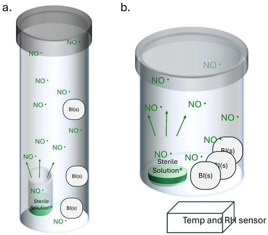
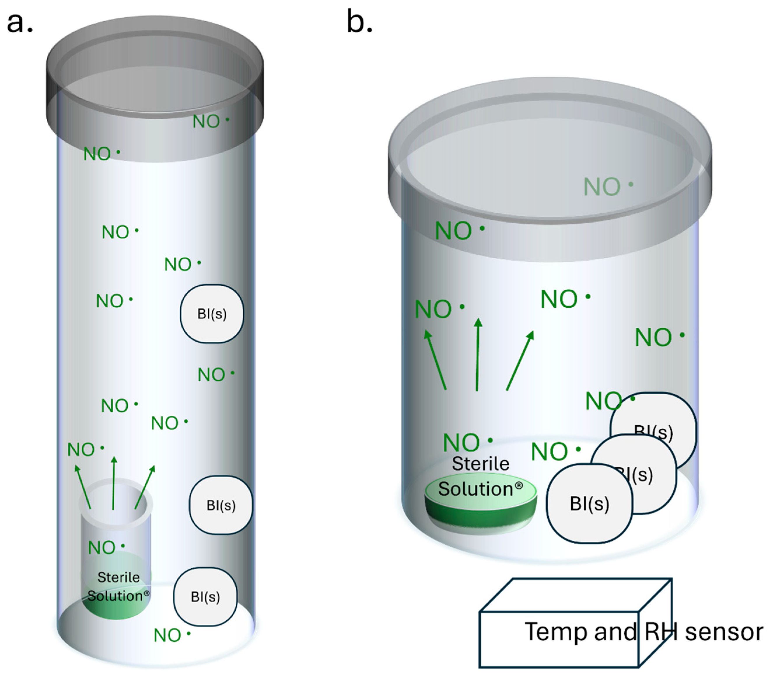
Gas sterilant NO was delivered by 0.2 M Sterile Solution® (Sterile State Inc. Grand Rapids, MI, USA). For the survivor curve method, 0.5 mL of freshly prepared Sterile Solution® was added into a scintillation vial (v = 5 mL, inner diameter of 1.5 cm). The vial was placed upright into a 50 mL conical tube, which was the sterilization vessel. BIs of the same LOT number were loaded into one sterilization vessel, with care taken to avoid stacking the BIs on top of each other. The sterilization vessel was then tightly capped and placed upright on the lab counter. Within each independent study, triplicate samples were processed at each defined exposure time point (Scheme 1a). For the fraction-negative method, 1.5 mL of freshly prepared Sterile Solution® was added into a 1-inch-diameter dish. The dish was then placed into a 4 oz container which functioned as the sterilization vessel. BIs were placed into the same container, adjacent to the container wall with the printed side facing inwards. Each container contained four BIs without stacking. The sterilization vessel was tightly capped and placed upright on the lab counter (Scheme 1b). The experiment was carried out at RT (18–23 °C) and the temperature was monitored and recorded by calibrated Govee sensors placed next to the sterilization vessels.
Scheme 1.
Experimental configurations using the Sterile State™ NO sterilization devices. NO gas was delivered via the 0.2 M Sterile Solution® reagent (Sterile State Inc.). The sterilization units were sealed to allow NO gas to reach a working concentration above 100 PPM (or 1 a.u.). To generate the Log reduction (survivor) curve data, an airtight 50 mL conical tube served as the sterilization chamber (a), whereas an airtight 4 oz container was used for the fraction-negative assays (b). For each test run, a sterilization load of up to three commercial BIs was placed inside device (a) and up to four commercial BIs were placed inside device (b). The sterilization cycles were carried out at RT for various durations for the designated tests. Experiment temperature was monitored and recorded by Govee sensors placed next to the sterilization vessels.
2.4. Survivor Curve Method Procedures
To characterize the inactivation kinetics, independent NO sterilization modules were configured for exposure intervals of 2, 4, 6, and 8 h. At least three BIs were used for each LOT at each time point. For t = 0, BIs were not placed in the sterilization vessel and were enumerated using a 1:10,000 dilution to determine the recoverable colony-forming units (CFUs). For samples harvested at t = 2, 4, 6, and 8 h, the timing of the sterilization was within ± 1 min of the intended timing, the NO sterilization units were opened at the end of each cycle, and the BIs were retrieved for serial dilutions and spread-plating. All the agar plates were cultured in the 30–35 °C moisturized bacterial incubator. If the agar plates were found to have no growth after overnight incubation, these plates were placed back into the incubator for an additional 2 days to account for potential delayed outgrowth from injured spores. When obviously visible colonies were seen, the recovered colony numbers were quantitated using manual counting methods as CFUs. Based on the recovered CFUs, the Log10 of surviving spores was plotted against NO exposure time. According to ISO11138, the D-value calculation is expressed as:
where N0 is the initial population and Nt represents the final population after exposure time t. To take the whole curve into consideration, the data was processed with linear regression and line fitting, and the D-value was obtained as the negative reciprocal of the slope of the line.
2.5. Fraction-Negative Method’s Procedures
BIs were exposed to NO RT sterilization for different durations, including 6, 8, 9, 10, 10.5 and 11 h. n = 20 samples were loaded for each time point for a total of 120 samples. At the end of the treatment duration, within ±1 min, the BI envelopes were collected, and the BI discs were placed on the Mueller–Hinton agar plates (Hardy Diagnostics, Santa Maria, CA, USA) for culturing. Plates were kept in a moisturized 35 °C incubator for a minimum of 3 days to examine whether there was growth of the BIs.
D-value calculation was based on ISO 11138-1 2017(E) [12], using the D.3.1 Holcomb–Spearman–Karber procedure (HSKP) as follows:
where UHSK is the mean time to sterility, and is calculated as follows:
N0 is the average viable count per indicator determined by the total viable count method. In our study, we used the manufacturer’s BI population for this LOT certified as 2.9 × 10⁶.
Ui is the ith treatment time;
k is the first exposure point with all BI units being negative.
To calculate Ui, we use the following equation:
where χi should be calculated according to:
and γi should be calculated according to:
ri represents the number of test samples showing no growth after NO sterilization at time ti; ni is the total number of test samples exposed to the NO sterilization at time ti.
The 95% confidence interval for D-value (p = 0.05), Dcalc, is expressed as:
where V is calculated through:
And a is calculated through:
2.6. Initial Material Compatibility Sample Preparation
Plastic film materials and real medical devices were exposed to one standard RT NO sterilization cycle, specifically, a 500 μm thick high-density polyethylene (PE) sheet (McMaster-Carr 8619K743, USA), 500 μm thick medical-grade polyester (PET) sheet (Goex MEDPET®90, Janesville, WI, USA), 500 μm thick medical-grade blended polypropylene (PP) sheet (Goex CONTOUR® 70, Janesville, WI, USA), 500 μm thick clear chemical-resistant polyvinyl chloride (PVC) film (McMaster-Carr 87875K17, Taiwan), PE and PET film-based packaging materials (<100 μm thick sheet, Amcor), PP sample vial (1.5 cm O.D, 5.6 cm tall, Uline ST-5ML), and PVC catheter tubing (5 mm O.D, 6 feet, Stellaris Elite®). Polymer samples (n = 5 for mechanical test and n = 3 for FTIR test) were exposed to NO sterilization cycles, while control samples were placed right next to the treatment groups without placing them into the NO sterilization unit. No pre-exposure was applied for NO sterilization cycles. Material characterizations were performed on unexposed control samples (0 h exposure). Right after the completion of the NO sterilization cycles, mechanical testing was completed within 8 h, while FTIR spectroscopy characterization was completed within 4 h post-exposure to minimize potential ambient ageing effects.
2.7. Tensile Testing
The test was conducted based on ASTM D882-18, Tensile Testing of Thin Plastic Film. Specimens were cut to dimensions of 200 × 20 mm plastic sheets (n = 5 for each type). The mechanical tester Mark 10 F1505 with IntelliMESUR (Version 2.3.0) software was used to perform the tensile test and analyze stress–strain curves. A grip separation rate of 500 mm/s was used for the PE, PP, and PVC films. A grip separation rate of 50 mm/s was used for the PET film. Stress–strain curves were reported and maximum stress, strain at rupture, and tensile modulus were calculated according to the stress–strain curves.
2.8. FTIR Characterization
Infrared (IR) absorbance was measured using a Thermo Fisher Nicolet iS50 Smart iTX-diamond (Thermo Fisher Scientfic, Waltham, MA, USA) instrument, and data was collected with OMNIC (Version 2.20) software. The measurement method was set to 32 scans and a resolution of 4. After background collection, test samples were secured on the attenuated total reflectance (ATR) pressure device and positioned on the crystal within the stainless-steel plate. Raw spectra were then collected and saved as SPA files, which were subsequently exported as CSV files for data processing.
2.9. Data and Statistical Analysis
Data was reported in the form of mean ± SD. A sample size ≥ 3 was used. Sample mean, standard deviation, D-value, liner regression, trendline, and p-value of t-test (unpaired two-tail at a significance level of 0.05) were all calculated using MS Excel.
3. Results
3.1. D-Value Calculation by Survivor Curve Method
After different NO exposure treatments, BIs were collected and recovered on the plates and quantified as CFU Count. The results are included in the table in Appendix A Table A1. Incorporating the dilution ratios that were used for each countable plate, the recoverable CFU numbers per BI were calculated as shown within Appendix A Table A2. Based on the recoverable CFU numbers per BI, the Log10 numbers were calculated. The results are listed in Table 1.
Table 1.
Log10 of surviving BI spore CFUs for each BI sample.
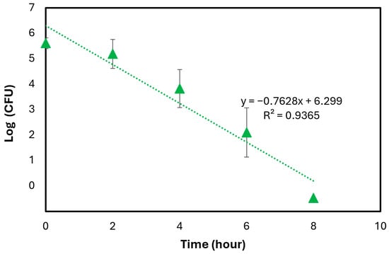
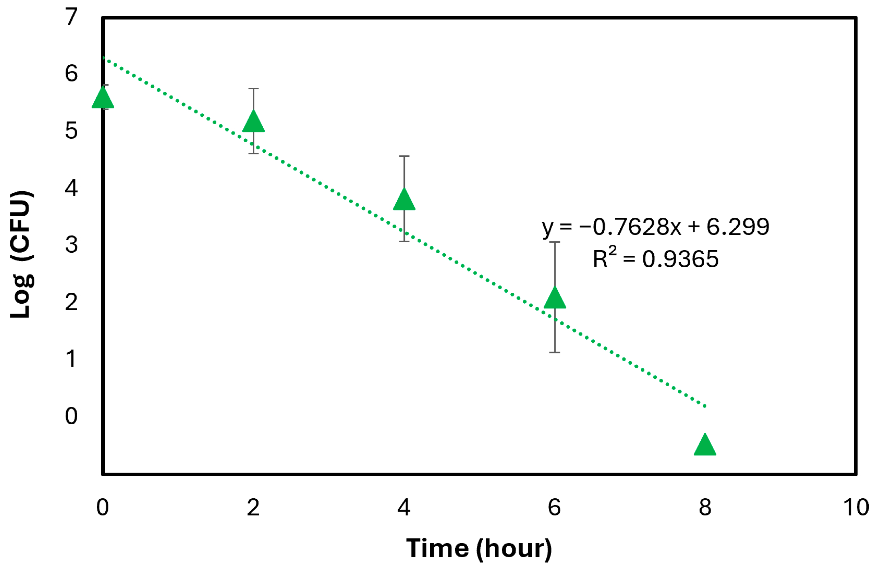
The D-value was determined by plotting the mean Log10 number of survivors (n = 21) against their respective exposure intervals, as shown in Figure 1. When no living bacteria are recovered, the Log10 of zero is −∞, which is not a usable value. For this reason, Log101 is used, which assumes that there is one colony remaining in these groups. This is an expected underestimate of the sterilization efficacy, resulting in a larger D-value, and will support the establishment of a higher SAL. Then, a linear regression analysis was performed on the resulting survivor curve, where the D-value was calculated as the negative reciprocal of the slope (D-value = −(1/−0.7628) = 1.31 h or 78.7 min).
Figure 1.
Survivor curve of Apex® Disc B. atrophaeus BIs established by using n = 21 samples.
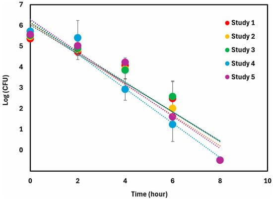
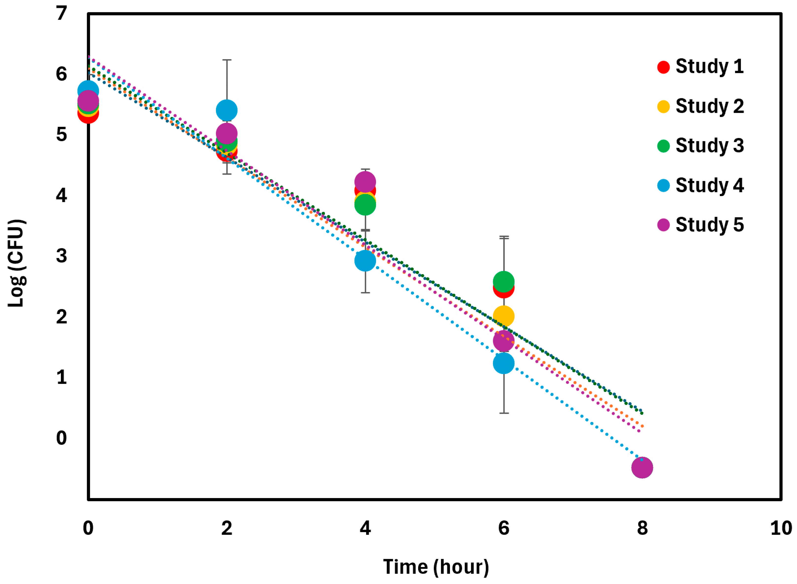
To understand the reliability and repeatability of the result, we break down the result by investigating the five independent studies. The individual plot of each study is presented in Figure 2.
Figure 2.
Summary of survivor curve of Apex® Disc B. atrophaeus BIs against different NO RT sterilization durations for all replicates.
The D-value based on each individual study was calculated using its corresponding curve slope (Table 2). The coefficient of determination, or R2 value, measures the proportion of variance. With a mean R2 value of 0.92 and S.D. < 0.03, this study quantitatively expresses the similarity of the results between different independent studies and when using different manufacturing lots of Apex® BI. The mean calculated D-value is 1.34 h (or 80.4 min). The standard deviation is 6.8% of the average.
Table 2.
D-value calculations from different independent studies.
D-values obtained across all experimental configurations were highly comparable and reproducible. The five independent survivor curves exhibited narrow variance, with a 19.5% maximum difference between extremes, which aligns with the <20% threshold considered consistent and acceptable by ISO 11138-1. A one-sample t-test revealed no statistically significant difference (p-value = 0.55) between the mean of the multi-curve approach and the single, high-sample-size (n = 21) protocol (Table 3). In addition, all individual curves exceeded the ISO 11138 requirement for semi-logarithmic linearity, with all R2 values above the threshold of 0.8. Overall, these results support the methodological robustness of our D-value calculation.
Table 3.
D-value calculations from integrated data.
3.2. Fraction-Negative Method’s BI Growth Results and D-Value Calculation
BIs exposed to different NO sterilization durations were cultured for bacterial growth, with the examination shown in Table 4.
Table 4.
BI growth result.
Calculation of D-value was performed according to the Holcomb–Spearman–Karber procedure (HSKP) as described in ISO 11138-1 2017(E), D.3.1 (see Methods section); the results are summarized in Table 5 and Table 6.
Table 5.
Sterilization results data summary.
Table 6.
Calculation of D-value and its 95% confidence interval according to ISO 11138-1 2017(E), D.3.1.
Based on the fraction-negative method, the D-value of Apex® B. atrophaeus for NO RT sterilization is determined to be 1.32 ± 0.05 h (79.3 ± 2.7 min), which is comparable to the calculated D-value according to the individual survivor curve method (p = 0.65) with only 1.34% difference.
3.3. Initial Material Compatibility: Mechanical Properties
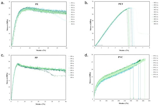
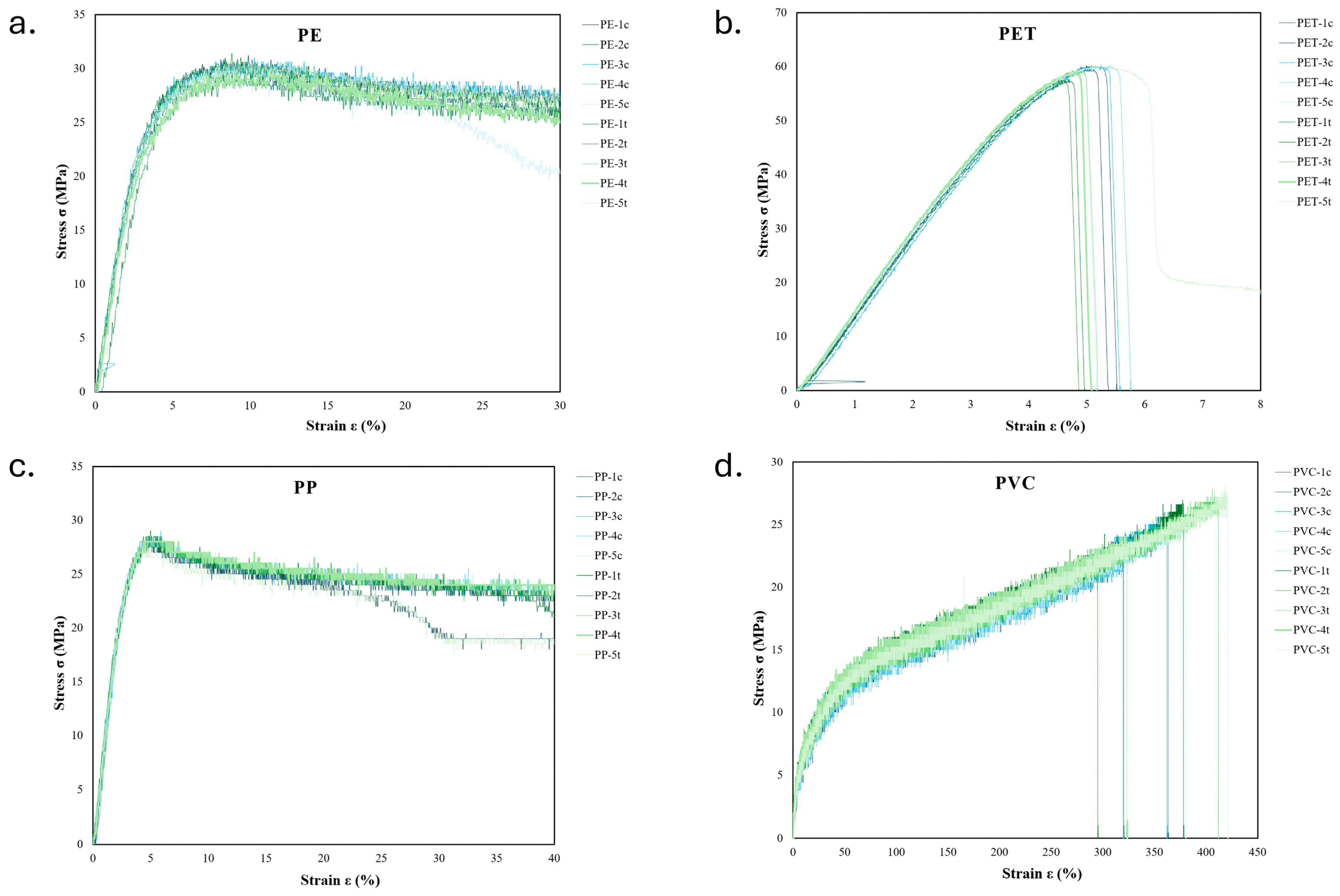
We selected four types of the most common plastic materials, PE, PET, PP, and PVC, and exposed them to NO sterilization cycles for an initial assessment of material response to NO gas sterilization. Figure 3 displays the stress–strain curves for all tested polymer samples. The high degree of overlap between the control (blue) and NO-treated (green) curves indicates that the sterilization cycles had a negligible impact on the baseline mechanical performance of these four materials. Comprehensive quantitative metrics and statistical analyses that can further support these observations are summarized in Table 7.
Figure 3.
Stress–strain curves of tested plastic samples with (treatment groups in green) and without (control groups in blue) NO sterilization cycles for (a) PE, (b) PET, (c) PP, and (d) PVC (n = 5 per experimental group; in the legend control replicates are denoted with “c” in the end, and treated replicates with “t”).
Table 7.
NO’s effect on mechanical properties of common plastic materials.
3.4. Initial Material Compatibility: Chemical Properties
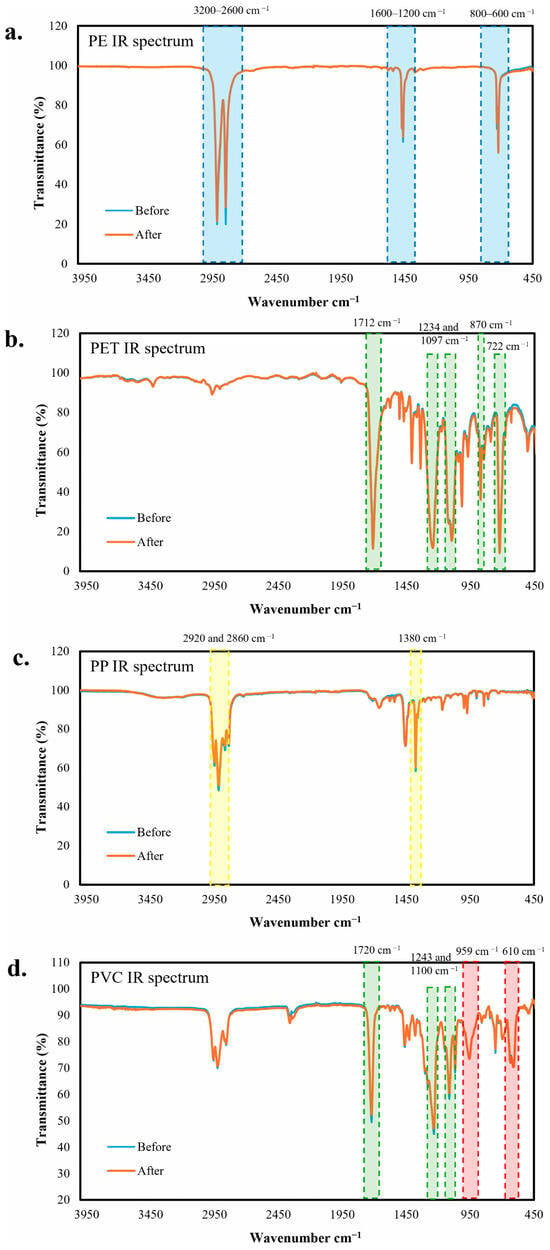
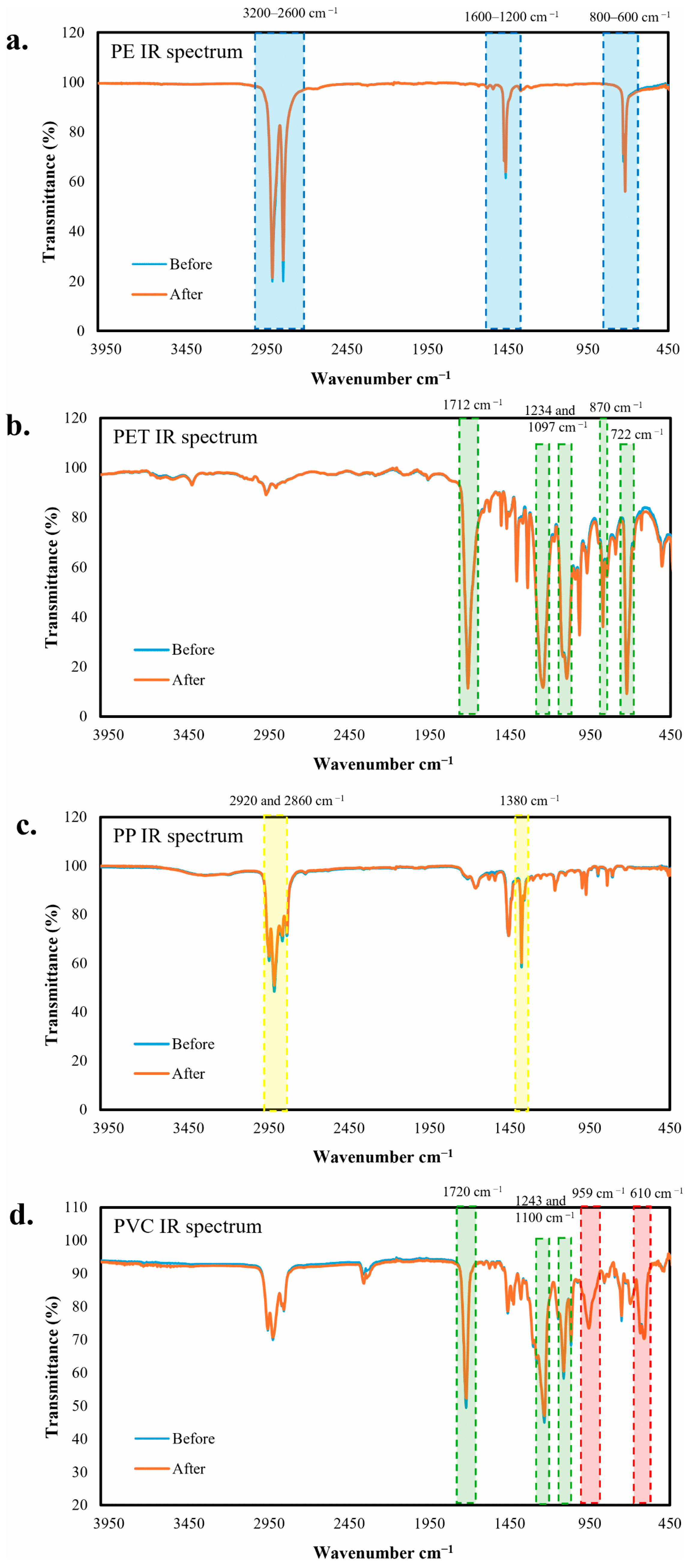
The FTIR spectra of the evaluated polymers confirmed their chemical structures and demonstrated material stability post-NO sterilization cycles. For PE, the spectrum displays a characteristic aliphatic C-H stretching band across 3200–2600 cm−1, C-H bending vibration within 1600–1200 cm−1, and the classic CH2 rock vibration between 800 and 600 cm−1 (Figure 4a, blue boxes). The PET structure is confirmed by prominent polyester signals, including the C=O ester carbonyl stretch at 1712 cm−1, the aromatic C-C-O stretch at 1243 cm−1, and the O-C-C stretch at 1097 cm−1. Additionally, the aromatic benzene ring is identified by distinct out-of-plane C-H bending peaks at approximately 870 and 722 cm−1 (Figure 4b, green boxes). PP exhibits a prominent methyl group (-CH3) bending vibration near 1380 cm−1 (Figure 4c, yellow boxes). Furthermore, due to the presence of both methyl and methylene groups, the C-H stretching region is more complex, resolving into distinct methylene (-CH2-) asymmetric and symmetric stretching vibrations at approximately 2920 and 2860 cm−1, respectively. In the PVC spectrum, prominent ester bands observed in the vicinities of 1720, 1243, and 1100 cm−1 (Figure 4d, green boxes) indicate the presence of a terephthalate ester plasticizer, consistent with the flexible feature of our PVC sample. The underlying PVC matrix is confirmed by the out-of-plane C-H bending at 959 cm−1 and the characteristic C-Cl stretch at 610 cm−1 (Figure 4d, red boxes). After NO sterilization, no peak shifts or new absorbance bands were observed in any of the polymer spectra, indicating that the current NO sterilization cycle does not induce significant chemical degradation or structural alterations in these four plastic materials.
Figure 4.
FTIR spectrometry characterization of different plastic materials with and without exposure to NO sterilization cycle treatment for (a) PE, (b) PET, (c) PP, and (d) PVC.
4. Discussion
The sterilization efficacy and material compatibility of terminal sterilization are critical to medical devices. This study focuses on the understanding of sterilization kinetics of the NO RT sterilization process using both the survivor curve and fraction-negative methods to determine the D-value of the B. atrophaeus Apex® BIs (Figure 1 and Figure 2) and examining the mechanical (Figure 3) and chemical property (Figure 4) alteration after NO sterilization cycles. The results demonstrated a high degree of correlation between the two D-value calculation methodologies, with calculated D-values ranging from 1.31 to 1.34 h (p = 0.55 and p = 0.65). Based on these kinetics, a Log 6 spore reduction can be achieved within 8 h of NO exposure at RT. Utilizing these D-values as the empirical foundation for an overkill sterilization cycle, a total exposure time of 16 h is expected to achieve the 12-Log reduction (or SAL of 10−6) required by the FDA, thereby ensuring a robust safety margin for medical device sterilization.
The ISO 11138-1 standard outlines and recommends both the survivor curve method (direct enumeration) and fraction-negative methods as standardized approaches for D-value calculation. To determine the D-value using the survivor curve method, ISO 11138 recommends using a sample size of n = 20 per exposure time point, or averaging the results of four independent survivor curves. In our work, this was achieved by conducting five independent studies with a total sample size of n = 21 (n = 9 for Study 1, and n = 3 for each of the remaining four studies). The resulting data demonstrated high consistency across all replicates. The survivor curve method quantifies surviving microorganisms via serial dilution and plate counting to plot the Log10 of survivors against exposure time. This semi-Log plot demonstrates the linear trend of microbial inactivation over time, while the negative reciprocal of the slope of the resulting Log reduction curve indicates the D-value. This linear regression-based approach is highly effective for characterizing the death kinetics and demonstrating whether microorganisms exhibit a linear Log reduction over time against a new sterilization method. The fraction-negative method is employed during the lethal boundary phase, where survivor concentrations are too low for direct enumeration. In this approach, rather than performing plate counts, a large cohort of BIs are subjected to sterilization exposure and subsequently monitored during incubation for a binary test, growth or no growth, after the sterilization cycle. D-values are then derived using probabilistic models such as the Stumbo–Murphy–Cochran or the HSKP methods. Following ISO 11138, our results show excellent consistency across these different methods. Note that industry standards such as ISO 11138 and ISO 17665 [14] conventionally dictate reporting D-values in minutes. In our case, doing so can imply an unwarranted level of precision, because the NO sterilization cycle at RT is significantly longer than traditional methods and our variance between replicate studies already exceeds one minute. To ensure scientific accuracy regarding experimental precision and to facilitate practical interpretation, our D-values are reported in our study in both minutes and hours.
While the D-value of gaseous sterilization is typically dependent on sterilant concentration, temperature and humidity, this study utilized a standardized 0.2 M Sterile Solution®, which maintained NO levels at 1–2 a.u. (the critical unit defined for sterilization applications) and an RH of 82–94% at ambient RT (monitored as 18.9 to 22.0 °C), allowing the D-value calculation to rely primarily on the quantification of surviving spores at discrete time intervals.
In the gas sterilization chamber, volume and geometric configuration (such as internal lumens or intricate interstitial spaces) significantly impact gas diffusion and overall sterilization efficacy. In traditional gas sterilization chambers where sterilant is forced into packaging from the outside of a carton/pallet and then must remove sterilant after the cycle completes, the volume and geometric configuration (such as internal lumens or intricate interstitial spaces) significantly impact gas diffusion and overall sterilization efficacy. These industrial systems use massive chambers often over 100 m3, where products are densely stacked on pallets. This arrangement results in long and difficult diffusion pathways that necessitate aggressive pressure and deep-vacuum cycling to force EtO penetration and removal. In addition, due to this large industrial load volume, sampling BIs at varying time intervals across multiple independent runs is highly costly. Our current design focuses on localized, single-unit packaging (volume < 1 L), fundamentally reducing the physical diffusion distances. NO possesses superior thermodynamic advantages, including lower molecular mass (30 g/mol vs. 44 g/mol for EtO) and an exceptionally high vapour pressure at ambient temperatures (boiling point of −152 °C vs. 10.4 °C for EtO). These properties render NO inherently more diffusible and reduce the risk of localized condensation gradients (the ideal gas laws evenly distribute gas in uniform concentrations). We have also conducted preliminary tests using BIs loaded within catheter lumens and inside various Tyvek® packaging geometries. Successful spore inactivation has been observed, although longer exposure times were required. Our current data successfully demonstrates the quantifiable sterilization efficacy of NO as a highly capable low-temperature sterilant by calculating the D-value of commercial BIs, establishing a baseline that warrants further comprehensive characterizations.
The selection of Apex® Discs, which utilize B. atrophaeus (Strain ATCC 9372) inoculated onto a 304 stainless-steel disc carrier, provided a rigorous worst-case challenge. The non-absorptive stainless-steel substrate is specifically engineered for a gaseous sterilant to prevent the interference often seen with porous materials such as cellulose. Furthermore, the medical-grade Tyvek® packaging allows gas permeability during the cycle while maintaining a sterile barrier for post-processing handling. While B. atrophaeus D-values in vaporized H2O2 are typically characterized in several minutes at temperatures between 24 and 45 °C [15,16], NO sterilization achieves lethality at significantly lower ambient temperatures. Although the NO procedure appears slower than H2O2 vapour, it operates at true ambient RT, pressure, and oxygen levels. However, the potential diffusion-limiting effects of the Tyvek® pouch on NO gas were not systemically evaluated in this study. It is very likely that the intrinsic lethality of NO is higher in the absence of barrier materials. In fact, although R2 values indicated great linearity of the Log reduction curves, a minor lag phase was observed at the onset of the sterilization cycle (from 0 to 2 h), characterized by a brief delay before the establishment of first-order inactivation kinetics. This shoulder effect is likely attributable to the diffusion-limiting properties of the packaging material. Consequently, the intrinsic D-value of the NO gas itself may be lower than the reported ones, further reinforcing the 16 h overkill cycle as a conservative and high-safety-margin parameter. Furthermore, a significant operational advantage of the NO sterilization method is the elimination of the extensive preconditioning and aeration phases required by the conventional EtO method. While EtO typically requires a preconditioning temperature and a humidity time of up to 24 h before sterilization and an 8–12 h outgassing phase to mitigate carcinogenic residuals, the low concentrations of NO used in this process allow for rapid pre- and post-cycle handling. Upon exposure to ambient air, residual NO and its oxidation products are rapidly oxidized and diluted in seconds [17,18]. The ultimate oxidation product is nitrate, which is completely safe for both humans and the environment, well aligned with FDA Innovation Challenge 2: reduce ethylene oxide emissions.
Currently, EtO is considered to be the most material-friendly sterilization method that is compatible with many temperature- and moisture-sensitive materials, especially polymers [10,11,19,20,21]. Some sources even describe EtO as universally compatible with plastics [22]. This statement seems controversial when looking at EtO’s mode of action [23], that is, EtO deactivates microorganisms by alkylation. It is well documented that the epoxide ring of EtO reacts with nucleophiles, undergoing ring-opening reactions for alkylation [24]. Nucleophiles such as hydroxyls, amines, halides, carboxylics, and thiols are very common in medical devices and biological and pharmaceutical molecules. Many descriptions of material compatibility with sterilization methods are vague. In this paper, we specifically selected four types of polymer materials that covered almost 70% of current medical device manufacturing on the market (PE, PET, PP, and PVC) [25]. PE represents the most prevalent polymeric biomaterial used in single-use medical device manufacturing and sterile barrier packaging systems. PET is widely deployed for thermoformed structural trays. PP serves as the dominant thermoplastic for recyclable medical components. PVC dominates medical catheter and intravenous tubing applications. We examined the material compatibility after NO sterilization cycles in terms of mechanical properties and chemical components. The tensile test indicates no statistical compromising of mechanical properties after NO sterilization is detected (Table 7), while FTIR supports the result by showing that no chemical structure alteration is detected in these four polymer materials (Figure 4). We understand our limitation is the thoroughness of the material compatibility test, as there are vast options of polymer choices for medical device applications. Even with the same polymer backbone, there are many additives and forms of polymers that could cause different conclusions; therefore, more tests should be carried out for a comprehensive understanding of NO sterilization material compatibility.
5. Conclusions
NO-based RT sterilization produces semi-Log-linear bacterial endospore inactivation while maintaining excellent compatibility with common medical device polymer materials. Specifically, a survivor curve study confirmed the Apex® B. atrophaeus BIs exhibited a linear Log reduction over time against NO RT sterilization, and the calculated D-value was 1.31–1.34 h (or 78.7–80.4 min) based on the integrated method and the average of five individual studies. The D-value based on the fraction-negative method is calculated as 1.32 ± 0.05 h (79.3 ± 2.7 min), which aligns with the results from the survivor curve method. To achieve a SAL of 10−6, a 16 h sterilization cycle at RT is recommended when using Apex® B. atrophaeus BIs. NO RT sterilization demonstrates high compatibility with commonly used plastics for medical device manufacturing, including PE, PET, PP, and PVC. In short, NO RT sterilization has enormous potential for medical device sterilization applications as an alternative method to EtO.
Author Contributions
Conceptualization: W.H. and M.C.F.; data curation: W.H. and E.S.; formal analysis: W.H. and E.S.; funding acquisition: M.C.F.; investigation: W.H. and E.S.; methodology: W.H. and M.C.F.; project administration: W.H. and M.C.F.; resources: M.C.F.; supervision: W.H. and M.C.F.; validation: W.H.; visualization: W.H.; writing—original draft preparation: W.H.; writing—review and editing: W.H., E.S., and M.C.F. All authors have read and agreed to the published version of the manuscript.
Funding
This study was funded and conducted by Sterile State Inc.
Data Availability Statement
The experimental data supporting the findings of this study is available from the corresponding author upon reasonable request. Specific details regarding the proprietary NO delivery product are omitted due to trade secret protections.
Conflicts of Interest
This study was funded and conducted by Sterile State Inc. The funder provided support in the form of salaries for authors, but did not have any role in the experiment design, data collection and analysis. The funder was involved in the decision to submit the work for publication. All authors declare that the research was conducted in the absence of any commercial influences that could be construed as a potential conflict of interest.
Abbreviations
The following abbreviations are used in this manuscript:
| ATR | Attenuated total reflectance |
| BI | Biological indicator |
| CFU | Colony-forming unit |
| EPA | Environmental Protection Agency |
| EtO | Ethylene oxide |
| FDA | Food and Drug Administration |
| FTIR | Fourier-transform infrared |
| H2O2 | Hydrogen peroxide |
| HSKP | Holcomb–Spearman–Karber procedure |
| iNOS | Inducible nitric oxide synthase |
| IR | Infrared |
| MDR | Multidrug-resistant |
| NO | Nitric oxide |
| PE | Polyethylene |
| PET | Polyethylene terephthalate |
| PP | Polypropylene |
| PVC | Polyvinyl chloride |
| RT | Room temperature |
| SAL | Sterility assurance level |
| S.D. | Standard deviation |
Appendix A
Table A1.
The recovered CFUs as colonies quantified on each plate.
Table A2.
Calculation of surviving BI CFUs according to the dilution ratios.
References
- FDA. Sterilization for Medical Devices; FDA: Silver Spring, MD, USA, 2021.
- EPA. Reducing Ethylene Oxide and Glutaraldehyde Use; EPA: Washington, DC, USA, 2002.
- EPA. National Emission Standards for Hazardous Air Pollutants: Ethylene Oxide Emissions Standards for Sterilization Facilities Residual Risk and Technology Review. Fed. Regist. 2024, 89, 24090–24203. [Google Scholar]
- Sweet, M.J.; Ramnath, D.; Singhal, A.; Kapetanovic, R. Inducible antibacterial responses in macrophages. Nat. Rev. Immunol. 2025, 25, 92–107. [Google Scholar] [PubMed]
- Fang, F.C. Perspectives series: Host/pathogen interactions. Mechanisms of nitric oxide-related antimicrobial activity. J. Clin. Investig. 1997, 99, 2818–2825. [Google Scholar] [CrossRef] [Scilit] [PubMed]
- Bu, F.; Kang, X.; Tang, D.; Liu, F.; Chen, L.; Zhang, P.; Feng, W.; Yu, Y.; Li, G.; Xiao, H. Enhancing near-infrared II photodynamic therapy with nitric oxide for eradicating multidrug-resistant biofilms in deep tissues. Bioact. Mater. 2024, 33, 341–354. [Google Scholar] [CrossRef] [Scilit] [PubMed]
- Rouillard, K.R.; Novak, O.P.; Pistiolis, A.M.; Yang, L.; Ahonen, M.J.; McDonald, R.A.; Schoenfisch, M.H. Exogenous nitric oxide improves antibiotic susceptibility in resistant bacteria. ACS Infect. Dis. 2020, 7, 23–33. [Google Scholar] [CrossRef] [Scilit] [PubMed]
- Schairer, D.O.; Chouake, J.S.; Nosanchuk, J.D.; Friedman, A.J. The potential of nitric oxide releasing therapies as antimicrobial agents. Virulence 2012, 3, 271–279. [Google Scholar] [CrossRef] [Scilit] [PubMed]
- He, W.; Osborne, J.; Frost, M.C. Using nitric oxide (NO)for gas sterilization at room temperature: A novel sterilization modality. PLoS ONE 2026. Manuscript submitted for publication. [Google Scholar] [CrossRef] [Scilit]
- Pereira, A.; Nakka, G.; Gupta, S. Beyond Ethylene Oxide (EtO): A comprehensive review of sustainable sterilization technologies for medical devices. Biomed. Ther. Lett. 2025, 12, 1161. [Google Scholar] [CrossRef] [Scilit]
- Mendes, G.C.; Brandao, T.R.; Silva, C.L. Ethylene oxide sterilization of medical devices: A review. Am. J. Infect. Control 2007, 35, 574–581. [Google Scholar] [CrossRef] [Scilit] [PubMed]
- ISO 11138-1; Sterilization of Health Care Products—Biological Indicators—Part 1: General Requirements. 3rd ed. International Organization for Standardization: Geneva, Switzerland, 2017.
- ASTM D882; Standard Test Method for Tensile Properties of Thin Plastic Sheeting. ASTM International: West Conshohocken, PA, USA, 2018.
- ISO 17665; Sterilization of Health Care Products—Moist Heat—Requirements for the Development, Validation and Routine Control of a Sterilization Process for Medical Devices. International Organization for Standardization: Geneva, Switzerland, 2024.
- McEvoy, B.; Maksimovic, A.; Rowan, N.J. Geobacillus stearothermophilus and Bacillus atrophaeus spores exhibit linear inactivation kinetic performance when treated with an industrial scale vaporized hydrogen peroxide (VH2O2) sterilization process. J. Appl. Microbiol. 2023, 134, lxac028. [Google Scholar] [PubMed]
- Pottage, T.; Macken, S.; Giri, K.; Walker, J.; Bennett, A. Low-temperature decontamination with hydrogen peroxide or chlorine dioxide for space applications. Appl. Environ. Microbiol. 2012, 78, 4169–4174. [Google Scholar] [CrossRef] [Scilit] [PubMed]
- Hakim, T.; Sugimori, K.; Camporesi, E.; Anderson, G. Half-life of nitric oxide in aqueous solutions with and without haemoglobin. Physiol. Meas. 1996, 17, 267–277. [Google Scholar] [CrossRef] [Scilit] [PubMed]
- Thomas, D.D.; Liu, X.; Kantrow, S.P.; Lancaster, J.R. The biological lifetime of nitric oxide: Implications for the perivascular dynamics of NO and O2. Proc. Natl. Acad. Sci. USA 2001, 98, 355–360. [Google Scholar] [CrossRef] [Scilit]
- Brouwer, H.D.; Maqsood, M. Comparison of the optical and mechanical properties of plastics before and after ethylene oxide sterilization. Polym. Med. 2025, 55, 79–87. [Google Scholar] [CrossRef] [Scilit] [PubMed]
- Massey, L.K. The Effect of Sterilization Methods on Plastics and Elastomers; William Andrew: New York, NY, USA, 2004. [Google Scholar]
- McEvoy, B.; Wiehle, S.B.; Gordon, K.; Kearns, G.; Laranjeira, P.; McLees, N. Advancing the sustainable use of ethylene oxide through process validation. Biomed. Instrum. Technol. 2021, 55, 35–44. [Google Scholar] [CrossRef] [Scilit] [PubMed]
- Rutala, W.A.; Weber, D.J. Sterilization of 20 billion medical devices by ethylene oxide (ETO): Consequences of ETO closures and alternative sterilization technologies/solutions. Am. J. Infect. Control 2023, 51, A82–A95. [Google Scholar] [CrossRef] [Scilit] [PubMed]
- Manson, M.M. Epoxides–is there a human health problem? Occup. Environ. Med. 1980, 37, 317–336. [Google Scholar] [CrossRef] [Scilit] [PubMed]
- Parker, R.-E.; Isaacs, N. Mechanisms of epoxide reactions. Chem. Rev. 1959, 59, 737–799. [Google Scholar] [CrossRef] [Scilit]
- Gill, Y.Q.; Khurshid, M.; Abid, U.; Ijaz, M.W. Review of hospital plastic waste management strategies for Pakistan. Environ. Sci. Pollut. Res. 2022, 29, 9408–9421. [Google Scholar] [CrossRef] [Scilit] [PubMed]

Disclaimer/Publisher’s Note: The statements, opinions and data contained in all publications are solely those of the individual author(s) and contributor(s) and not of MDPI and/or the editor(s). MDPI and/or the editor(s) disclaim responsibility for any injury to people or property resulting from any ideas, methods, instructions or products referred to in the content. |
© 2026 by the authors. Licensee MDPI, Basel, Switzerland. This article is an open access article distributed under the terms and conditions of the Creative Commons Attribution (CC BY) license.