


Abstract
To realize the high-value valorization of cold-pressed walnut meal, walnut oligopeptides were fabricated via synchronous dual-enzyme hydrolysis combined with activated carbon decolorization and membrane separation purification. We optimized the overall preparation process and systematically characterized the products, including their amino acid profile, in vitro antioxidant capacity, cytoprotective effects against H2O2-triggered oxidative injury in PC12 cells, and regulatory activity toward acetylcholinesterase (AChE). The optimal hydrolysis conditions were identified as pH 10.0, total enzyme dosage of 11,000 U/g, a trypsin-to-alkaline protease ratio of 2.1:1, solid–liquid ratio of 1:26, temperature of 51 °C and reaction duration of 4 h, which produced a hydrolysis degree of 33.02%. The optimized decolorization parameters were pH 5.2, activated carbon dosage of 2.3%, treatment at 58 °C for 43 min, with a peptide recovery rate reaching 83.35%. Cold-pressed walnut meal is rich in glutamic acid, arginine and aspartic acid, which lay the molecular foundation for the bioactive properties of the derived oligopeptides. In vitro tests demonstrated that the oligopeptides possessed strong scavenging ability against hydroxyl, DPPH and superoxide anion radicals (clearance rates of 90.18%, 81.72% and 85.80%, respectively), and maintained 73.21% of antioxidant activity after simulated gastrointestinal digestion. Moreover, walnut oligopeptides at 0.8 mg/mL showed no cytotoxicity and afforded a 79.88% protective effect against oxidative damage. The peptides significantly boosted SOD and GSH-Px activities, lowered MDA accumulation, and strongly suppressed AChE activity, performing better than donepezil hydrochloride. This efficient, eco-friendly technique achieves high-value utilization of walnut processing by-products. The obtained oligopeptides possess great potential as natural antioxidants and neuroprotective ingredients for functional food development.
1. Introduction
Abundant walnut meal is produced as a primary by-product during walnut oil extraction. At present, most walnut meal is merely used as animal feed or fertilizer, resulting in low resource utilization efficiency and substantial waste. Walnut meal contains approximately 40% protein with a well-balanced amino acid profile [1]. Notably, glutamic acid, arginine, and aspartic acid are the most abundant amino acids, which are closely associated with the memory-enhancing properties of walnut. Meanwhile, walnut protein provides all eight essential amino acids required by the human body [2], demonstrating high nutritional value and promising potential for developing high-quality protein products.
Bioactive peptides (BAPs) are functional oligomers formed by the covalent bonding of amino acids, generally with molecular weights below 6000 Da [3,4]. Among them, oligopeptides with molecular weights less than 1000 Da exhibit superior intestinal absorption efficiency [5] and possess diverse bioactivities, including antioxidant, hypolipidemic, and memory-improving effects [6]. Previous studies have confirmed that walnut-derived peptides can alleviate oxidative damage, facilitate neural development, and improve learning and memory in mice. Animal experiments have verified that walnut-derived peptides can alleviate neuronal oxidative damage and ameliorate learning and memory deficits. Furthermore, Min et al. (2024) [7] further demonstrated that walnut peptides are capable of crossing the blood–brain barrier, directly acting on hippocampal neurons and mitigating abnormal synchronous neuronal firing induced by β-amyloid protein (Aβ), which provides critical evidence for the direct neuromodulatory effects of walnut peptides in the brain.
Walnut peptides are typically produced from defatted walnut cake via targeted enzymatic hydrolysis. After oral administration, dietary proteins are digested into small peptides before absorption and utilization [8]. Such small peptides not only serve as nutritional substrates but also exert critical regulatory functions in physiological processes. Moreover, walnut meal is rich in protein, calcium, phosphorus, iron, and other trace elements, and exerts brain-tonifying, antioxidant, anti-inflammatory, and immune-enhancing effects [9]. In addition, walnut peptides can effectively address the poor solubility of walnut protein, thus showing great potential for the development of high-value functional foods. Given the insufficient comprehensive utilization of walnut meal, researchers have devoted efforts to preparing bioactive peptides via optimized processes with diverse biological functions.
Currently, walnut peptides are mainly prepared by chemical, enzymatic, and fermentation extraction methods [10]. Chemical hydrolysis using acids or alkalis often causes amino acid destruction, low yield, and unstable product quality, making it unsuitable for industrial production. In contrast, enzymatic hydrolysis features high specificity, mild reaction conditions, high efficiency, minimal nutrient loss, and environmental safety, and has become the dominant approach for producing walnut protein peptides. Enzymatic hydrolysis has become the dominant preparation method for walnut bioactive peptides owing to its mild reaction conditions and full retention of nutrients. Wang et al. (2024) [11] prepared bioactive peptides from walnut protein using alkaline protease, which provides in vivo experimental support for the dual-enzyme hydrolysis system and in vitro neuroprotective outcomes of the present study.
Cold pressing is widely applied in walnut oil manufacturing owing to its low processing temperature, which effectively preserves the natural structure and bioactivity of nutrients [12]. In this study, walnut oligopeptides were prepared from cold-pressed walnut meal using synchronous dual-enzyme hydrolysis to shorten reaction time and increase the degree of hydrolysis. The product was further purified by activated carbon decolorization and membrane separation, followed by spray drying. The amino acid composition, molecular weight distribution, purity, stability, and in vitro antioxidant activity of the oligopeptides were systematically characterized. Furthermore, the protective effects against oxidative damage and memory-enhancing potential were evaluated using a PC12 cell model. This study provides theoretical and technical support for the high-value utilization of walnut meal and the development of walnut oligopeptides as functional food ingredients.
Study Objectives: 1. To establish a synchronous dual-enzyme preparation technology for walnut oligopeptides. 2. To systematically evaluate the in vitro antioxidant activity and gastrointestinal digestion stability of walnut oligopeptides. 3. To explore the neuroprotective and memory-improving effects of walnut oligopeptides based on the PC12 cell model.
Highlights: 1. Synchronous dual-enzyme hydrolysis with trypsin and alkaline protease was applied to cold-pressed walnut meal for the first time. 2. A full green process combining decolorization and membrane separation purification was established to tackle the industrial bottleneck of low purity in walnut polypeptide preparation. 3. The PC12 neuronal cell model was further employed to verify that the obtained oligopeptides exhibit dual functions of anti-oxidative damage and acetylcholinesterase (AChE) inhibition for memory improvement.
2. Materials and Methods
2.1. Determination of Protein Content and Amino Acid Composition in Walnut Meal
- (1)
- Determination of Protein Content
Protein content in walnut meal was determined according to the Kjeldahl method specified in GB 5009.5-2016 [13].
① Sample digestion: Potassium sulfate, copper sulfate, and sulfuric acid were added to the sample, followed by heating in an electrothermal mantle until the solution turned clear blue-green. After cooling to room temperature, the digest was transferred into a 100 mL volumetric flask.
② Distillation: An aliquot of the digested sample was transferred into the reaction chamber, followed by the addition of sodium hydroxide solution. Distillation was performed with cooling water circulation until the pH of the distillate became neutral.
③ Titration: The distillate was titrated with 0.1 mol/L HCl solution, and the volume consumed was recorded for calculation.
- (2)
- Determination of Amino Acid Composition
Amino acid composition of walnut meal was analyzed using an amino acid analyzer [14]. Briefly, 3 g of walnut protein was mixed with 15 mL of 0.1 mol/mL hydrochloric acid solution, followed by ultrasonic extraction for 30 min. The mixture was centrifuged, and the supernatant was filtered through a 0.22 μm membrane filter prior to injection into the amino acid analyzer.
2.2. Morphological Structure Analysis of Walnut Protein
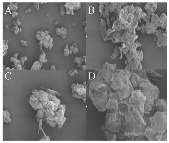
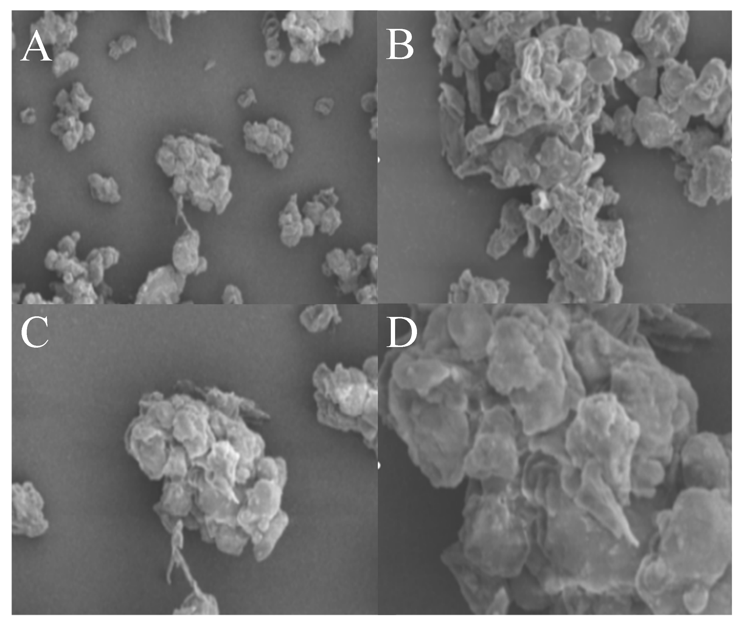
The morphological structure of cold-pressed walnut meal was observed using a scanning electron microscope (SEM) at magnifications ranging from 400× to 5000×.
2.3. Determination of Degree of Hydrolysis
The degree of hydrolysis (DH) was determined using the ninhydrin method [15].
- (1)
- Standard Curve Construction
A standard solution of glycine was prepared. Ninhydrin reagent was added, and the mixture was incubated in a boiling water bath. Subsequently, 40% ethanol solution was added, and the mixture was allowed to stand for 15 min. The absorbance was measured at 570 nm to establish the standard curve.
- (2)
- Determination of Free -NH2 Groups in Hydrolysate
The enzymatic hydrolysate was diluted appropriately, and the content of free -NH2 groups was determined following the procedure described above.
- (3)
- Calculation of Degree of Hydrolysis
F—conversion coefficient of walnut protein
N—protein content in the hydrolysate
walnut protein htot = 5.3
2.4. Determination of Peptide Content
Peptide content was determined using the Folin–phenol method [16]. Alkaline copper reagent was added, followed by the Folin–phenol reagent. The mixture was blended and allowed to stand, and the absorbance was measured at 500 nm to construct the standard curve.
The absorbance of the walnut oligopeptide solution was measured, and the peptide content was calculated accordingly.
2.5. Enzyme Screening
The effects of different proteases on the degree of hydrolysis (DH) of proteins in cold-pressed walnut meal were compared to select the optimal enzyme for preparing walnut oligopeptides via enzymatic hydrolysis.
2.6. Optimization of Enzymatic Hydrolysis Conditions for Walnut Oligopeptides
2.6.1. Single-Factor Experiments of Synchronous Dual-Enzyme Hydrolysis
- (1)
- Effects of total enzyme dosage on degree of hydrolysis
Under the initial pH of 9.0, trypsin and alkaline protease were added at dosages of 5000, 10,000, 15,000, 20,000 and 25,000 U per gram of walnut protein, respectively. The solid–liquid ratio was set at 1:20 (g/mL), the enzyme ratio of trypsin to alkaline protease was 2:1, the hydrolysis temperature was 50 °C, and the hydrolysis duration was 3 h. Subsequently, the degree of hydrolysis was determined.
- (2)
- Effects of enzyme ratio on degree of hydrolysis
The initial pH was fixed at 9.0, the total enzyme dosage was 10,000 U/g protein, and the solid–liquid ratio was 1:20 (g/mL). The ratios of trypsin to alkaline protease were set as 1:0, 1:1, 2:1, 3:1, 4:1 and 5:1, respectively. The hydrolysis was carried out at 50 °C for 3 h, and the corresponding degree of hydrolysis was measured.
- (3)
- Effects of hydrolysis pH on degree of hydrolysis
The initial pH values were adjusted to 7.0, 8.0, 9.0, 10.0 and 11.0 in sequence. The solid–liquid ratio was 1:20 (g/mL), total enzyme dosage was 10,000 U/g protein, enzyme ratio of trypsin to alkaline protease was 2:1, and the hydrolysis reaction was performed at 50 °C for 3 h. Finally, the degree of hydrolysis was detected.
- (4)
- Effects of solid–liquid ratio on degree of hydrolysis
The initial pH was set at 10.0, the total enzyme dosage was 10,000 U/g protein, and the trypsin-to-alkaline protease ratio was 2:1. The solid–liquid ratios were arranged as 1:10, 1:15, 1:20, 1:25, 1:30 and 1:35 (g/mL). The hydrolysis was conducted at 50 °C for 3 h, and the degree of hydrolysis was determined.
- (5)
- Effects of hydrolysis temperature on degree of hydrolysis
With the initial pH maintained at 10.0, the hydrolysis reactions were performed at 30, 40, 50, 60 and 70 °C for 3 h, respectively. Other fixed conditions were as follows: total enzyme dosage 10,000 U/g protein, enzyme ratio of trypsin to alkaline protease 2:1, solid–liquid ratio 1:25 (g/mL). The degree of hydrolysis was then measured.
- (6)
- Effects of hydrolysis time on degree of hydrolysis
Under the conditions of initial pH 10.0, total enzyme dosage 10,000 U/g protein, enzyme ratio 2:1 (trypsin/alkaline protease), solid–liquid ratio 1:25 (g/mL) and hydrolysis temperature 50 °C, the enzymatic hydrolysis was carried out for 1, 2, 3, 4 and 5 h, respectively, and the corresponding degree of hydrolysis was determined.
2.6.2. Plackett–Burman Design for Synchronous Dual-Enzyme Hydrolysis
Based on the above six single factors, a Plackett–Burman experimental design (Detailed experimental procedures are provided in the Supplementary Materials) was performed via Design-Expert 13.0 software. Four factors exerting the most significant effects on the response value (degree of hydrolysis) were screened out for subsequent response surface methodology experiments, as shown in Table 1.
Table 1.
Factor levels and codes of Plackett–Burman experimental design.
2.6.3. Response Surface Optimization of Synchronous Dual-Enzyme Hydrolysis
Four-factor and three-level experiments were designed using Design-Expert 13.0 software, as shown in Table 2.
Table 2.
Factors and levels of response surface experiments.
2.7. Separation and Purification of Walnut Oligopeptides
2.7.1. Single-Factor Experiments on Activated Carbon Decolorization
After adjusting the pH of the enzymatic hydrolysate, activated carbon powder was added for stirring and adsorption. The supernatant was collected by centrifugation to determine absorbance and calculate the decolorization rate. The peptide content before and after decolorization was measured by Folin–phenol method, and the peptide recovery rate was calculated.
2.7.2. Response Surface Optimization of Activated Carbon Decolorization
Four-factor three-level experiments were designed by Design-Expert 13.0 software, as shown in Table 3.
Table 3.
Factors and Levels of Response Surface Experiments.
2.7.3. Calculation of Decolorization Rate
2.7.4. Membrane Separation Technology
The decolorized solution was sequentially treated via microfiltration membrane (0.45 μm), ultrafiltration membrane (3 KDa) and nanofiltration to obtain walnut oligopeptide solution.
Calculation of peptide recovery rate:
2.8. Effects of Simulated Gastrointestinal Digestion on the Stability of Walnut Oligopeptides
2.8.1. Pepsin Digestion Assay [17]
The pH value of the walnut oligopeptide solution was adjusted to 2.0, and pepsin was subsequently added. The mixture was incubated at 37 °C for 2 h, and then subjected to enzyme inactivation at 90 °C for 15 min. After centrifugation, the collected supernatant was adjusted to pH 8.3, and its inhibitory effect on AChE activity was determined.
2.8.2. Trypsin Digestion Assay
The pH of the walnut oligopeptide solution was regulated to 7.5 prior to the addition of trypsin. The reaction system was incubated at 37 °C for 2 h, followed by heat inactivation at 90 °C for 15 min. The supernatant obtained by centrifugation was adjusted to pH 8.3 for the detection of its influence on AChE activity.
2.8.3. Sequential Digestion with Pepsin and Trypsin
The aforementioned experimental procedures were repeated to assess the effect on AChE activity.
where ΔA (ΔA = A1 − A2) denotes the absorbance reduction in the sample solution at 412 nm; ΔB (ΔB = B1 − B2) represents the absorbance reduction in the blank solution at 412 nm.
2.9. Study on Antioxidant Activity of Walnut Oligopeptides
2.9.1. Hydroxyl Radical Scavenging Effect of Walnut Oligopeptides
Vitamin C and walnut oligopeptide solutions with different concentrations were prepared. The experiment was performed following the kit instructions, and the hydroxyl radical scavenging rate was calculated.
2.9.2. DPPH Radical Scavenging Effect of Walnut Oligopeptides
Solutions of vitamin C and walnut oligopeptides at varied concentrations were prepared. The assay was conducted as instructed, and the DPPH radical scavenging rate was calculated.
2.9.3. Superoxide Anion Radical Scavenging Effect of Walnut Oligopeptides
Vitamin C and walnut oligopeptide solutions with different concentrations were prepared. The experiment was carried out according to the specification, and the superoxide anion radical scavenging rate was calculated.
2.10. Protective Effect Against Oxidative Damage and Memory-Improving Function of Walnut Oligopeptides
2.10.1. Culture of PC12 Cells
The original culture medium was discarded, and cells were rinsed with PBS. Cells were digested with 0.25% trypsin for 1–2 min. Once cells shrank into round shapes without detachment, complete medium was added to terminate digestion. After centrifugation, cells were seeded into new culture dishes.
2.10.2. Establishment of H2O2-Induced Oxidative Damage Model in PC12 Cells
PC12 cells were seeded into 96-well plates at a density of 1 × 10⁵ cells/mL with 100 μL per well, and incubated at 37 °C with 5% CO2 for 24 h. Blank group, positive control group and sample group were set up. The blank group was supplemented with 100 μL of complete medium. The experimental groups were treated with hydrogen peroxide at final concentrations of 0.1, 0.2, 0.3, 0.4 and 0.5 mmol/L, respectively. After 18 h of incubation under identical conditions, the medium was removed. Then 20 μL MTT solution was added and incubated for 4 h, followed by addition of 150 μL DMSO for oscillation dissolution. Absorbance was measured at 490 nm using a microplate reader. Cell viability was calculated to screen the optimal H2O2 concentration for model construction.
2.10.3. Effects of Walnut Oligopeptides at Different Concentrations on PC12 Cell Viability Detected by MTT Assay
MTT assay is widely applied to evaluate cell survival, proliferation and drug toxicity [18]. Reductase in viable cells reduces MTT into insoluble formazan deposits inside cells. The formazan crystals can be dissolved by DMSO, showing maximum absorbance at 490 nm. 100 μL complete medium was added to the blank group. Donepezil hydrochloride served as positive control. For sample groups, 100 μL complete medium containing walnut oligopeptides at concentrations of 0.4, 0.6, 0.8, 1.0, 1.2, 1.4 and 1.6 mg/mL was supplemented respectively. Each group had six replicates and underwent 24 h incubation. The medium was discarded afterwards, followed by addition of 20 μL MTT solution. After 4 h, 150 μL DMSO was added and shaken thoroughly for dissolution. Cell viability was calculated to select the appropriate peptide concentration for subsequent experiments.
2.10.4. Protective Effects of Different Concentrations of Walnut Oligopeptides on Oxidative Damage Model
100 μL complete medium was added to the blank group. Only hydrogen peroxide was administered to the model group. Donepezil hydrochloride was used as positive control. The sample groups were incubated with samples and hydrogen peroxide for 18 h, then the medium was removed. Twenty microliters of MTT solution was added, and 150 μL DMSO was supplemented 4 h later with shaking for dissolution. Cell viability was calculated according to Formula 5.6 to determine the optimal protective concentration of walnut oligopeptides. The activities of SOD, GSH-Px and MDA content in cells were detected using commercial kits.
2.10.5. Determination of Acetylcholinesterase Activity in PC12 Cells
Complete medium was added to the blank group, and only hydrogen peroxide was applied to the model group. Donepezil hydrochloride served as positive control. Cells in sample groups were treated with samples and hydrogen peroxide. After 18 h incubation, the medium was discarded. Cells were rinsed and lysed, and the supernatant was collected for subsequent measurement.
3. Results and Discussion
3.1. Determination of Protein Content and Amino Acid Composition
The protein content of cold-pressed walnut meal was 49.36 ± 0.23%. As shown in Table 4, walnut meal contains 16 kinds of amino acids. Glutamic acid (21.35%), arginine (18.83%) and aspartic acid (10.25%) are the predominant components. Glutamic acid can improve flavor and participate in brain energy metabolism [19]. Arginine can regulate body immunity, improve physiological functions and modulate neural activity [20]. The results indicate that cold-pressed walnut meal has favorable potential for food and health product development, serving as a high-quality by-product of plant protein resources.
Table 4.
Amino acid composition of cold-pressed walnut meal.
3.2. Study on Spatial Structure of Walnut Protein
The microstructure of walnut meal was observed by scanning electron microscope at magnifications ranging from 400 to 5000 times. The results are shown in Figure 1.
Figure 1.
SEM images of cold-pressed walnut meal. ((A) 400×, (B) 1000×, (C) 2000×, (D) 5000×).
It can be seen from Figure 1 that cold-pressed walnut meal presents a compact structure with irregular aggregated or massive particles lacking regular geometric shapes, which is a typical characteristic of natural plant protein–polysaccharide complexes. The particle surface is not completely smooth and shows slight folds and unevenness, inherent structural features of the raw material. Particle sizes vary from several to tens of micrometers, existing in dispersed or partially aggregated states.
3.3. Enzyme Screening
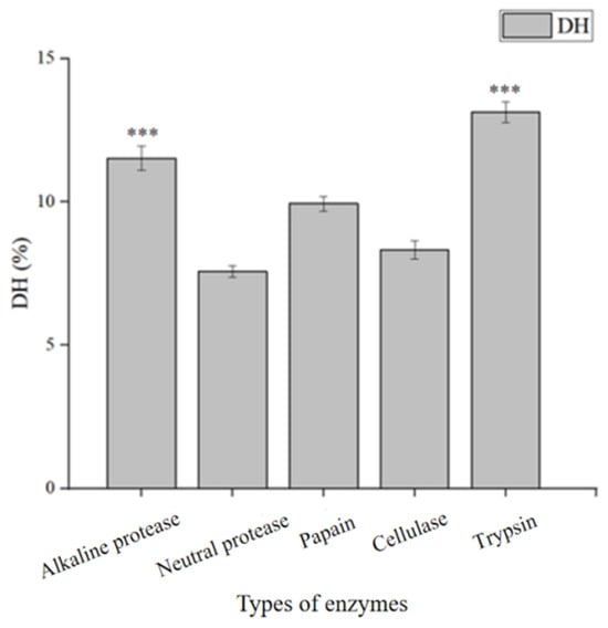
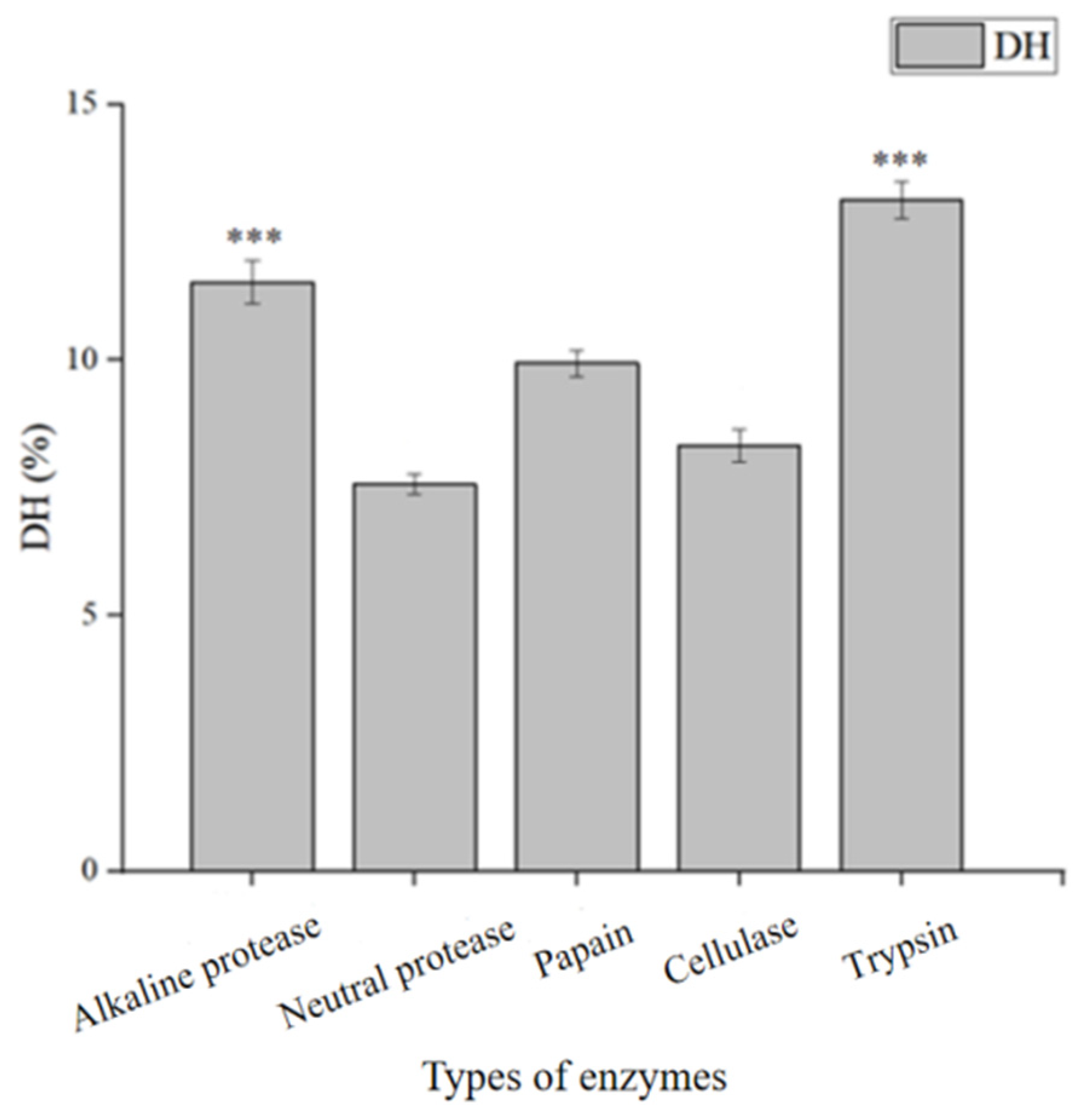
As shown in Figure 2, trypsin and alkaline protease exhibited extremely significant hydrolysis effects on walnut protein (p < 0.001), thus the two enzymes were selected for walnut oligopeptide preparation. Alkaline protease hydrolyzes hydrophobic amino acid residues, while trypsin acts on the carboxyl terminals of lysine and arginine.
Figure 2.
Effects of different proteases on hydrolysis degree. ***, (p < 0.001, extremely significant difference).
3.4. Optimization of Enzymatic Hydrolysis Conditions for Walnut Oligopeptides
3.4.1. Single-Factor Experimental Results of Simultaneous Double-Enzyme Hydrolysis
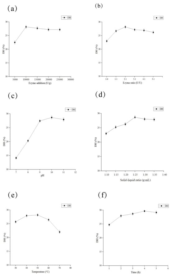
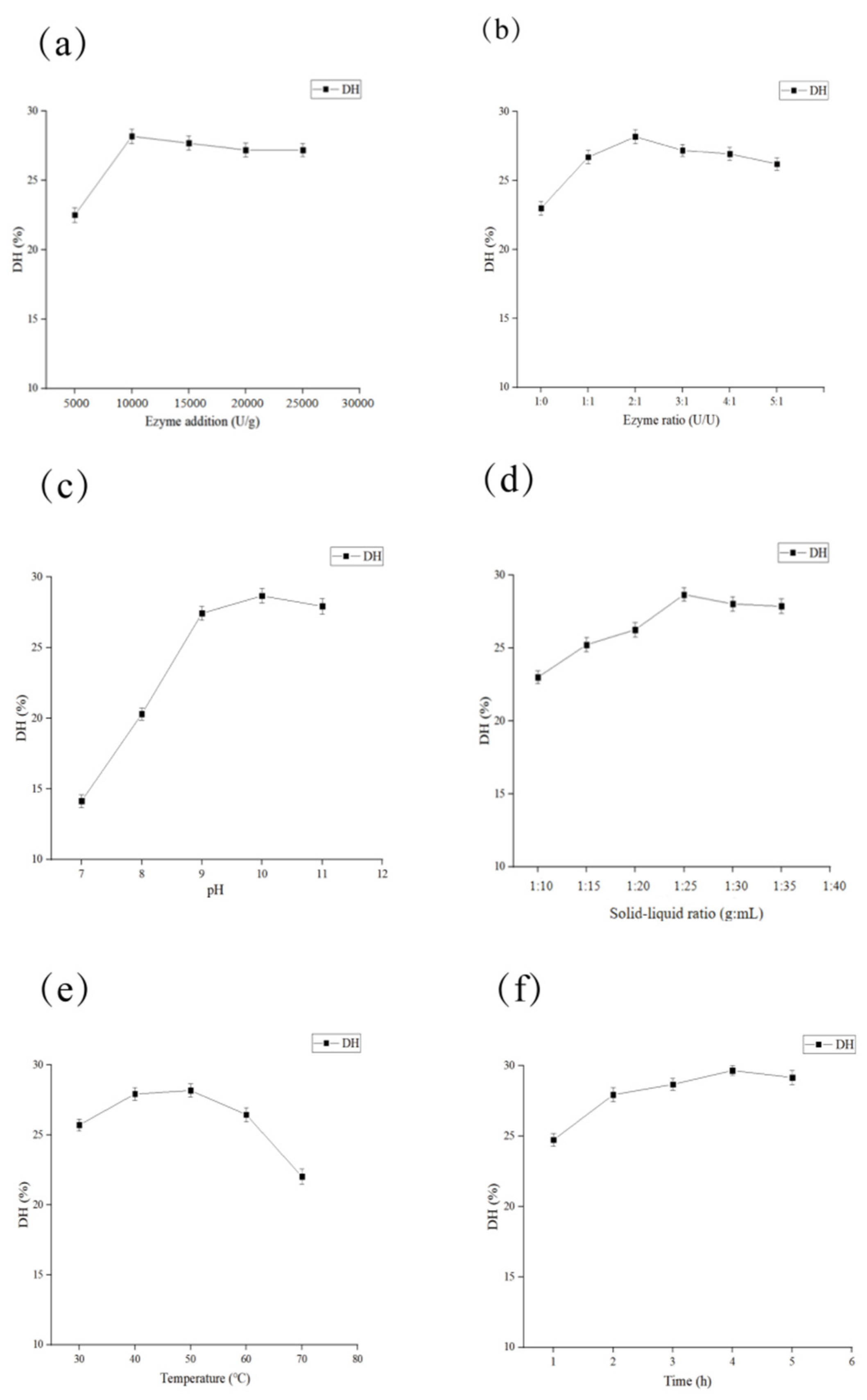
Walnut protein was hydrolyzed by trypsin and alkaline protease with different ratios, and the results are shown in Figure 3a. When the total enzyme dosage ranged from 5000 to 10,000 U/g(Pr), the hydrolysis degree rose with increased enzyme dosage. Excessively high dosage above 10,000 U/g(Pr) caused excessive protein hydrolysis into amino acids, reducing the hydrolysis degree. Thus, the optimal total enzyme dosage was determined as 10,000 U/g(Pr). As illustrated in Figure 3b, the hydrolysis degree increased first and then decreased with the changing enzyme ratio. The maximum hydrolysis degree was obtained at the trypsin to alkaline protease ratio of 2:1, which was selected as the optimal ratio.
Figure 3.
Single-factor results of simultaneous double-enzyme hydrolysis; (a) effect of total enzyme dosage on hydrolysis degree; (b) effect of compound enzyme ratio on hydrolysis degree; (c) effect of pH on hydrolysis degree; (d) effect of solid–liquid ratio on hydrolysis degree; (e) effect of hydrolysis temperature on hydrolysis degree; (f) effect of hydrolysis time on hydrolysis degree.
Strong alkaline conditions alter the spatial conformation of enzymes and reduce enzymatic activity. Figure 3c shows pH exerted a remarkable effect on hydrolysis degree. The highest hydrolysis degree occurred at pH 10.0, and declined when pH exceeded 10.0, so pH 10.0 was chosen as the optimal pH value. In Figure 3d, hydrolysis degree positively correlated with solid–liquid ratio and peaked at 1:25 (g/mL). Further increase in solid–liquid ratio brought no obvious improvement, hence 1:25 (g/mL) was confirmed as the optimal ratio.
Temperature affected hydrolysis degree in an upward-then-downward trend as shown in Figure 3e. The maximum hydrolysis degree was achieved at 50 °C, and high temperature impaired enzyme activity and lowered hydrolysis efficiency. Consequently, 50 °C was selected as the optimal hydrolysis temperature.
According to Figure 3f, hydrolysis degree increased along with reaction time and reached the maximum at 4 h. Therefore, 4 h was set as the optimal hydrolysis duration.
3.4.2. Plackett–Burman Design of Simultaneous Double-Enzyme Hydrolysis
Plackett–Burman experimental design was carried out using Design-Expert 13.0 software. The response values (hydrolysis degree) were determined under corresponding conditions, and the results are presented in Table S1 in Supplementary Materials.
Multivariate regression analysis was performed on the data, and the optimal equation with hydrolysis degree as the response value was obtained.
Y = 26.90 + 0.8175 A + 1.09 B + 0.1742 C − 0.9008 D + 1.28 E + 0.2042 F
Significance evaluation of each factor was obtained, as shown in Table S2 in the Supplementary Materials.
3.4.3. Response Surface Optimization of Simultaneous Double-Enzyme Hydrolysis
Experiments were conducted based on response surface design, as listed in Table S3 in the Supplementary Materials.
The results in Table S3 in the Supplementary Materials indicated that the experimental data fitted well with the regression model, which possessed practical significance. Based on F-values, the influencing order on walnut protein hydrolysis degree was E > B > D > A > F > C. The first four factors were selected for further response surface optimization and analysis.
- (1)
- Establishment and analysis of response model. Response surface data were analyzed via Design-Expert 13.0 software, as shown in Table S4 in the Supplementary Materials.
The response surface experimental data were fitted using Design-Expert 13.0 software, and the regression equation of hydrolysis degree related to total enzyme dosage (A), enzyme ratio (B), solid–liquid ratio (C) and hydrolysis temperature (D) was determined as follows:
DH(%) = 32.23 + 1.19 A + 0.1917 B + 0.2342 C + 0.2408 D + 0.0675 AB + 0.1675 AC − 0.28 AD +
0.29 BC + 0.1525 BD + 0.345 CD − 2.68 A2 − 1.63 B2 − 1.92 C2 − 2.88 D2
0.29 BC + 0.1525 BD + 0.345 CD − 2.68 A2 − 1.63 B2 − 1.92 C2 − 2.88 D2
As shown in Table S4 in the Supplementary Materials, the regression model was extremely significant (p < 0.0001), while the lack of fit was insignificant (p = 0.0830). The values of R2 = 0.9987 and adjusted R2 = 0.9974 indicated a favorable fitting performance of the model, which could reflect the variation in response values.
- (2)
- Analysis of interaction effects via response surface
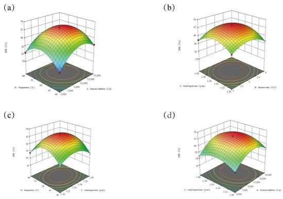
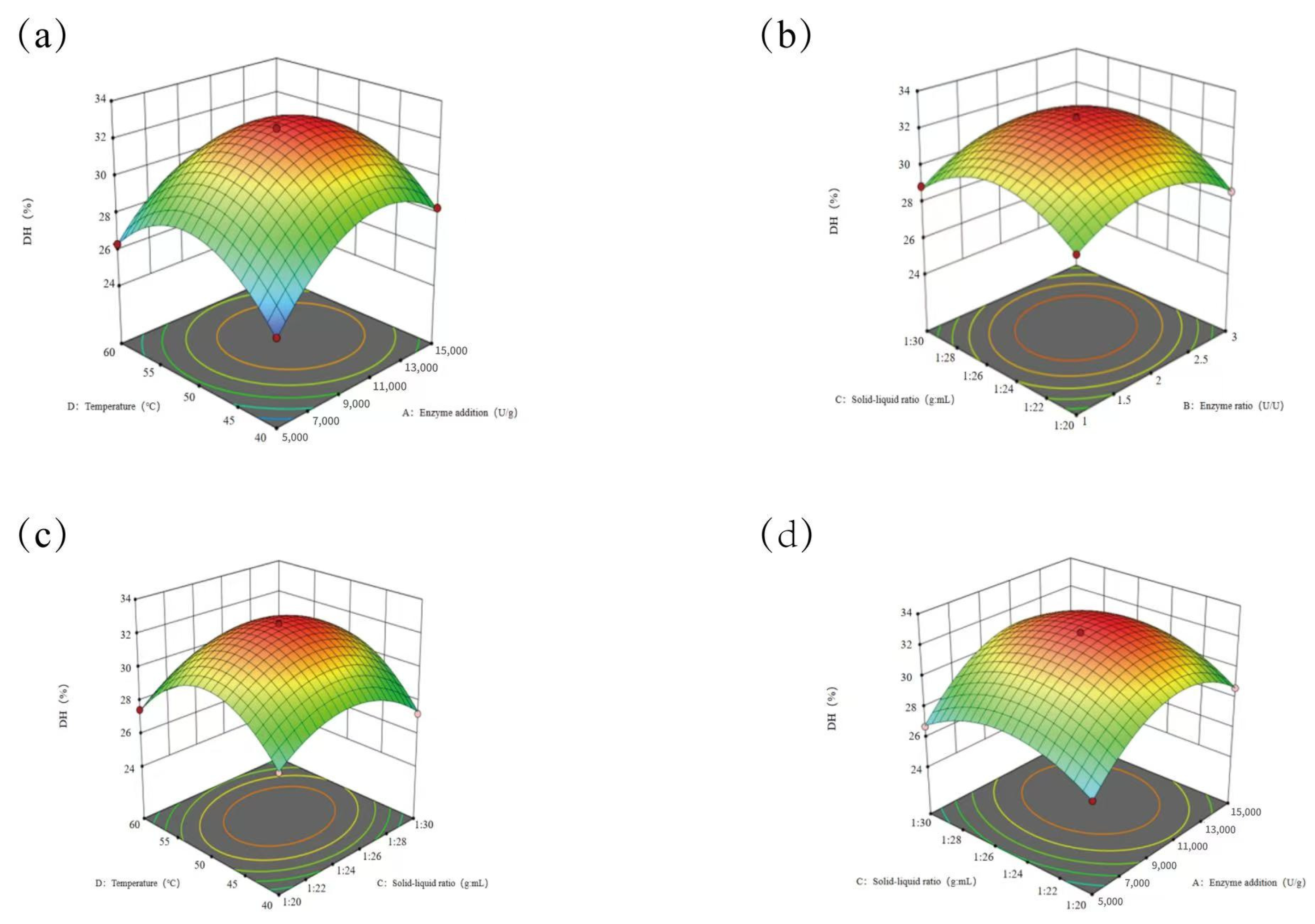
The interaction effects were analyzed, and the results are presented in Figure 4.
Figure 4.
Response surface interaction analysis. (a) Interactive effects of total enzyme dosage and hydrolysis temperature on hydrolysis degree; (b) interactive effects of solid–liquid ratio and enzyme ratio on hydrolysis degree; (c) interactive effects of solid–liquid ratio and hydrolysis temperature on hydrolysis degree; (d) interactive effects of solid–liquid ratio and total enzyme dosage on hydrolysis degree.
As shown in Figure 4, dense elliptical contour lines indicated strong interaction between factors. The influencing order on hydrolysis degree was total enzyme dosage > hydrolysis temperature > solid–liquid ratio > enzyme ratio.
- (3)
- Verification experiment
The optimal conditions for simultaneous double-enzyme hydrolysis were obtained via Box–Behnken design (Detailed experimental procedures are provided in the Supplementary Materials): pH 10.0, total enzyme dosage 11,000.09 U/g(Pr), trypsin-to-alkaline protease ratio 2.08:1, solid–liquid ratio 1:25.58 (g/mL), hydrolysis temperature 50.92 °C and hydrolysis time 4 h, with a predicted hydrolysis degree of 32.56%.
Verified experiments were conducted under adjusted practical conditions: pH 10.0, total enzyme dosage 11,000 U/g(Pr), enzyme ratio 2.1:1, solid–liquid ratio 1:26 (g/mL), hydrolysis temperature 51 °C and reaction duration 4 h. The actual hydrolysis degree reached 33.02%, consistent with the predicted value.
3.5. Isolation and Purification of Walnut Oligopeptides
Single-factor experiments on walnut oligopeptides isolation and purification are as follows:
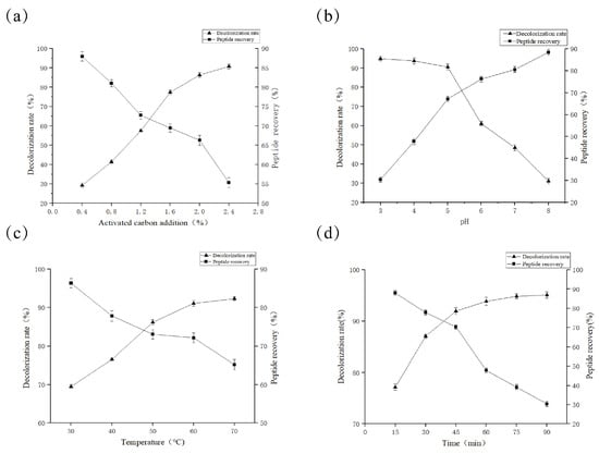
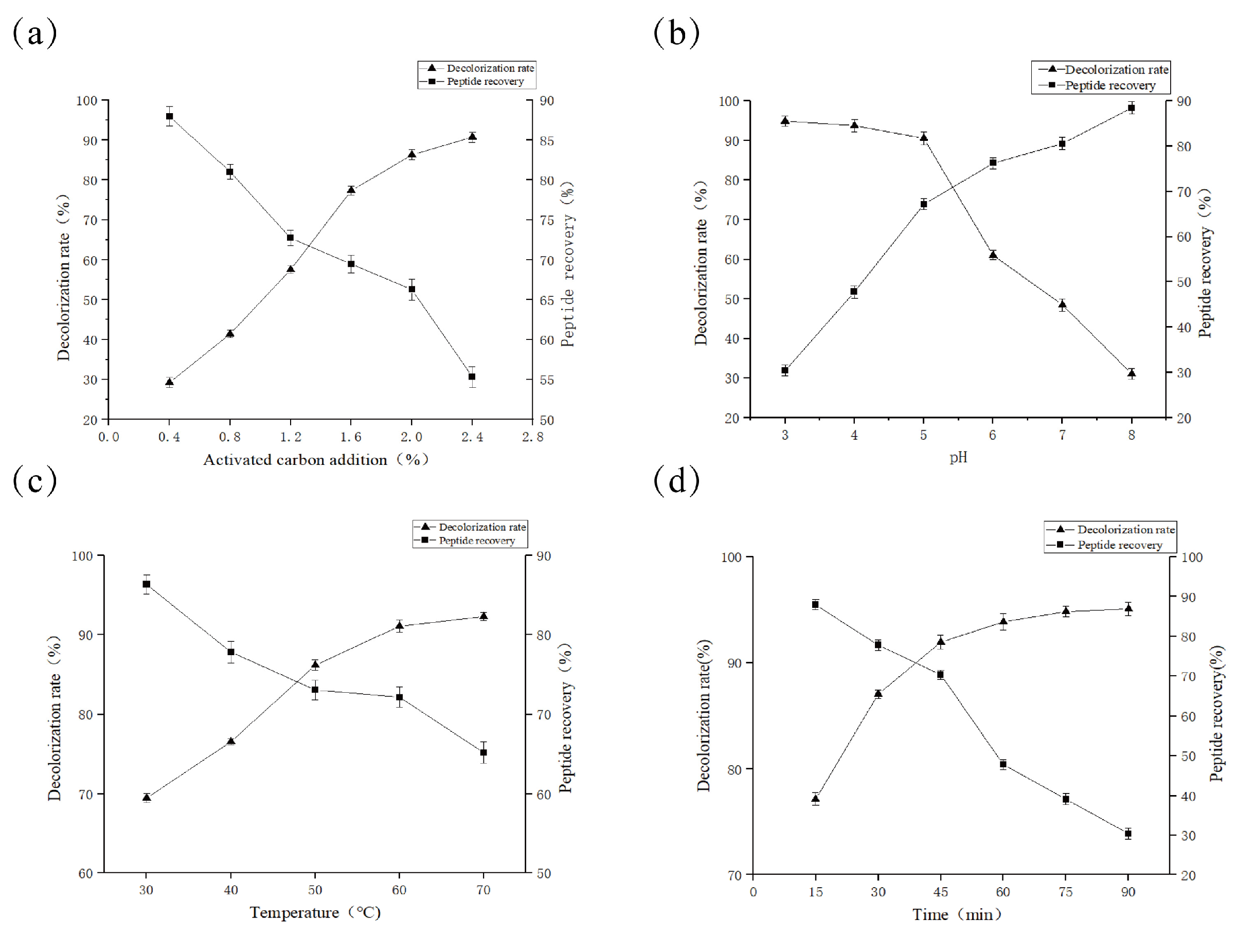
Activated carbon adsorbs macromolecular proteins and pigments in enzymatic hydrolysate, while causing loss of walnut oligopeptides simultaneously. As shown in Figure 5a, decolorization rate rises with increased carbon dosage, whereas peptide recovery declines sharply. The optimal dosage is determined to be 2.0% by comprehensive consideration.
Figure 5.
Effects of different parameters on decolorization rate and peptide recovery rate. (a) Activated carbon dosage; (b) pH value; (c) temperature; (d) treatment time.
Activated carbon presents better decolorization performance under acidic conditions. Figure 5b shows a high decolorization rate yet low peptide recovery when pH is below 5.0, and decolorization drops markedly above pH 5.0. The optimal pH is 5.0. In Figure 5c, temperature is positively correlated with decolorization rate and negatively correlated with peptide recovery. Peptide recovery decreases markedly above 60 °C; thus, 60 °C is selected as the optimal temperature. In Figure 5d, prolonged treatment improves decolorization but reduces peptide recovery, with a sharp decline after 45 min. The optimal treatment time is 45 min.
3.5.1. Response Surface Optimization
Experiments were carried out in accordance with response surface design, as shown in Table S5 in Supplementary Materials.
- (1)
- Establishment and analysis of response model. Response surface data were analyzed using Design-Expert 13.0 software, as shown in Table S6 in the Supplementary Materials.
The response surface experimental results were fitted using Design-Expert 13.0 software, and the regression equation of decolorization rate with activated carbon dosage (A), pH (B), temperature (C) and time (D) was determined as follows:
Decolorization rate(%) = 92.99 + 5.39 A − 4.58 B + 0.3275 C + 0.2267 D + 0.375 AB + 0.2125 AC − 0.045 AD
+ 0.1575 BC + 0.1225 BD − 0.2225 CD − 2.55 A − 2.59 B2 − 0.8243 C2 − 0.398 D
+ 0.1575 BC + 0.1225 BD − 0.2225 CD − 2.55 A − 2.59 B2 − 0.8243 C2 − 0.398 D
As shown in Table S6 in the Supplementary Materials, the regression model was extremely significant (p < 0.0001), and the lack of fit was insignificant (p = 0.0622). The R2 of 0.9992 and adjusted R2 of 0.9984 indicated a good fitting effect, which could reflect the variation in response values. According to F-values, the influencing order on decolorization rate was: activated carbon dosage > pH > temperature > time.
The experimental data were fitted via Design-Expert 13.0 software to establish the regression equation of peptide recovery rate against activated carbon dosage (A), pH (B), temperature (C) and time (D):
Peptide recovery rate(%) = 82.47 − 2.71 A + 4.69 B + 0.2283 C + 0.0633 D − 0.14 AB + 0.2325 A − 0.2450 AD
− 0.8725 BC − 0.56 BD − 0.575 CD − 2.16 A2 − 3.4 B2 − 1.74 C2 − 1.83 D2
− 0.8725 BC − 0.56 BD − 0.575 CD − 2.16 A2 − 3.4 B2 − 1.74 C2 − 1.83 D2
Table S7 in the Supplementary Materials shows that the regression model is extremely significant (p < 0.0001) and the lack of fit is insignificant (p = 0.0525). With R2 = 0.9941 and adjusted R2 = 0.9883, the model possesses favorable fitting performance and can reflect changes in response values. Based on F-values, the influence order on peptide recovery rate is: pH > activated carbon dosage > temperature > time.
3.5.2. Analysis of Response Surface Interaction
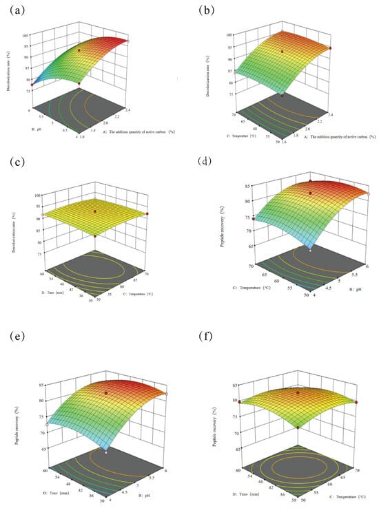
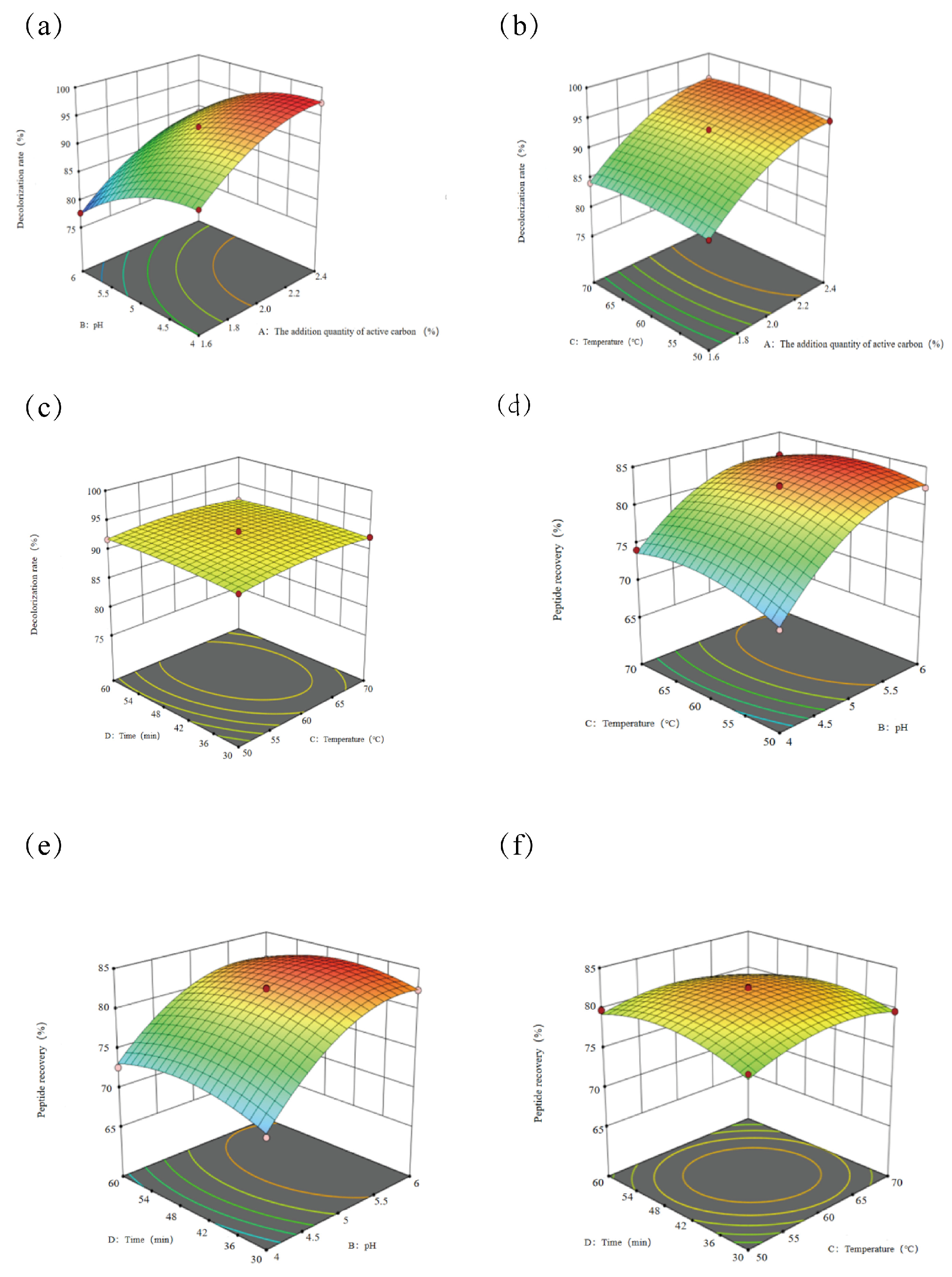
The interaction effects are analyzed and presented in Figure 6.
Figure 6.
Analysis of response surface interaction; (a) Interaction effect of activated carbon dosage and pH on decolorization rate; (b) Interaction effect of activated carbon dosage and temperature on decolorization rate; (c) Interaction effect of time and temperature on decolorization rate; (d) Interaction effect of pH and temperature on peptide recovery rate; (e) Interaction effect of time and pH on peptide recovery rate; (f) Interaction effect of temperature and time on peptide recovery rate.
As shown in Figure 6a, the dense semi-elliptical contour lines indicate a strong interaction, and pH exerts a weaker effect on decolorization rate than activated carbon dosage. In Figure 6b, dense semi-elliptical contours reveal notable interaction, with temperature having less influence than activated carbon dosage. Figure 6c shows sparse semicircular contours, suggesting weak interaction; temperature affects decolorization rate more obviously than time. Figure 6d presents dense semi-elliptical contours, reflecting strong interaction, and pH imposes greater impact on peptide recovery rate than temperature. Dense semi-elliptical contours in Figure 6e demonstrate strong interaction, and time has a weaker effect than pH. Sparse circular contours in Figure 6f indicate weak interaction, and time influences peptide recovery rate less than temperature.
- (2)
- Verification Experiment
Box–Behnken design determined the optimal decolorization conditions: pH 5.18, activated carbon dosage 2.33%, temperature 57.65 °C, duration 43.11 min. The predicted decolorization rate was 92.98% and the peptide recovery rate was 83.22%. Verification test under practical conditions of pH 5.2, activated carbon dosage 2.3%, temperature 58 °C and duration 43 min obtained a decolorization rate of 93.11% and a peptide recovery rate of 83.35%, consistent with predicted values.
3.6. Study on Antioxidant Activity of Walnut Oligopeptides
Pepsin and trypsin exist in the gastrointestinal tract, which readily degrade bioactive peptides and impair their physiological functions in vivo. Hence, simulating gastrointestinal digestion is of great significance [21].
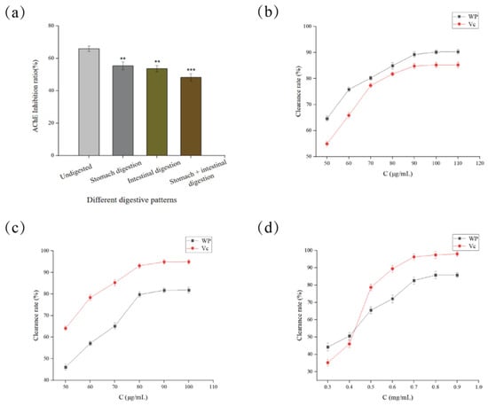
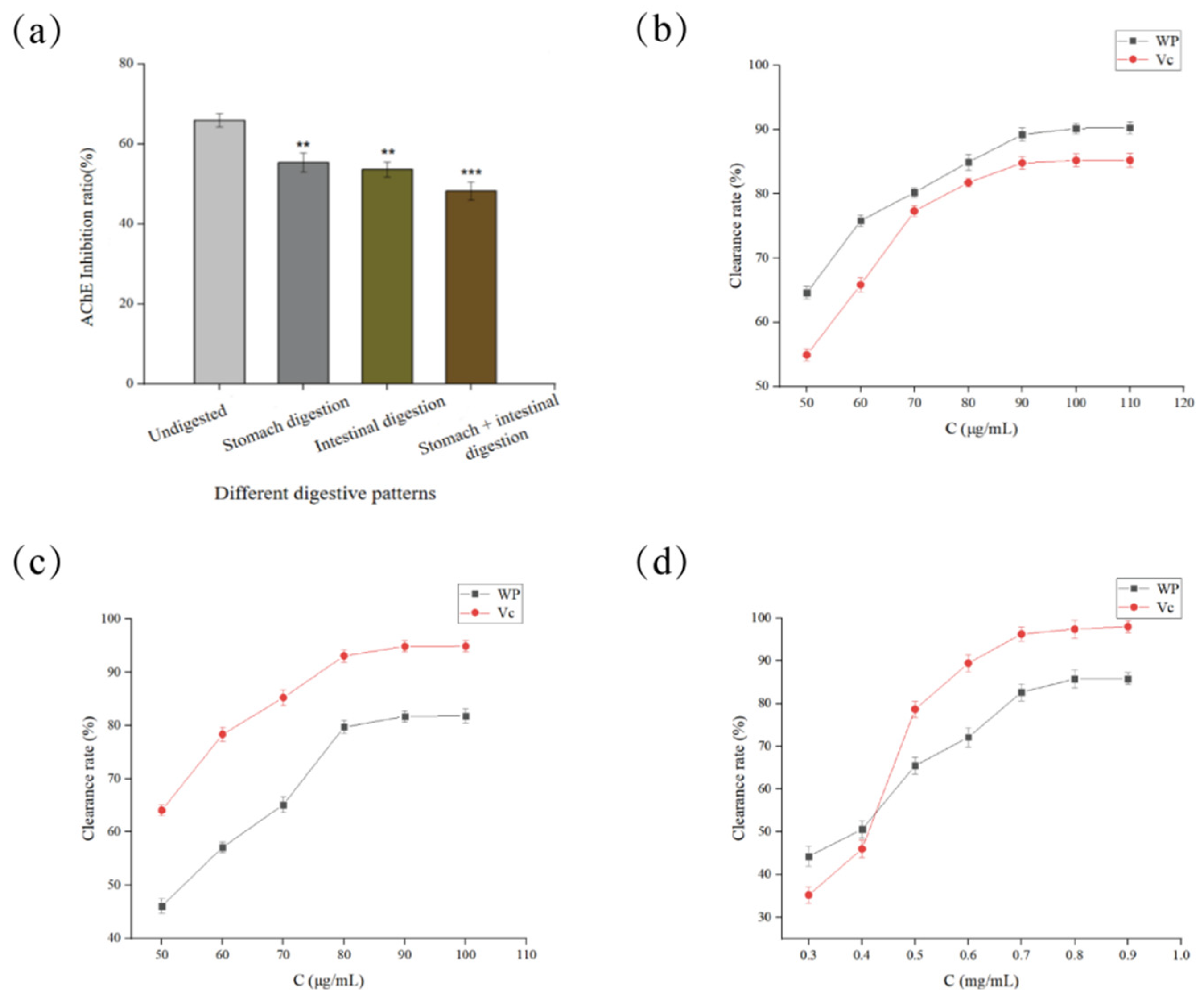
As shown in Figure 7a, the antioxidant activity of walnut oligopeptides decreased after gastrointestinal enzymatic digestion compared with the undigested group. The activity declined most significantly after treatment with pepsin combined with trypsin (p < 0.001), which was attributed to enzymatic hydrolysis that damaged active peptide structures. Nevertheless, the digested samples still retained favorable activity with a retention rate of 73.21%, indicating walnut oligopeptides possess potential to regulate AChE activity in vivo. The hydroxyl radical is the most reactive free radical, which rapidly reacts with various biomolecules and causes severe damage to organisms. Figure 7b reveals that walnut oligopeptides exhibited stronger hydroxyl radical scavenging capacity than vitamin C. The maximum scavenging rate reached 90.18% at 100 μg/mL, with an IC50 value of 38.50 μg/mL. DPPH is stable in organic solvents and requires light-proof storage. Its alcoholic solution appears purple with a maximum absorption peak at 517 nm [22]. Figure 7c shows DPPH scavenging activity positively correlated with sample concentration. Walnut oligopeptides showed weaker activity than vitamin C, with the peak scavenging rate of 81.72% at 90 μg/mL and an IC50 of 54.91 μg/mL. Superoxide anion can generate hydrogen peroxide and hydroxyl radical, triggering DNA and cell membrane damage. As presented in Figure 7d, walnut oligopeptides had lower superoxide anion scavenging ability than vitamin C. The optimal scavenging rate was 85.80% at 0.8 mg/mL, and the IC50 was 0.40 mg/mL.
Figure 7.
Study on antioxidant activity of walnut oligopeptides. (a) Effect of simulated gastrointestinal digestion system on the stability of walnut oligopeptides, (b) Scavenging Effect of Walnut Oligopeptides on Hydroxyl Radicals, (c) Scavenging Effect of Walnut Oligopeptides on DPPH Radicals, (d) Scavenging effect of walnut oligopeptides on superoxide anion radicals. ** (p < 0.01), highly significant difference; *** (p < 0.001), extremely significant difference.
3.7. Study on Oxidative Damage Protection and Memory Improvement Effects of Walnut Oligopeptides
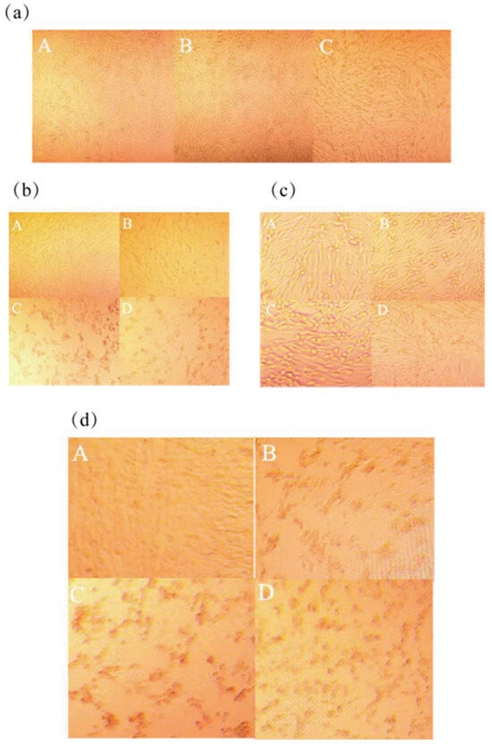
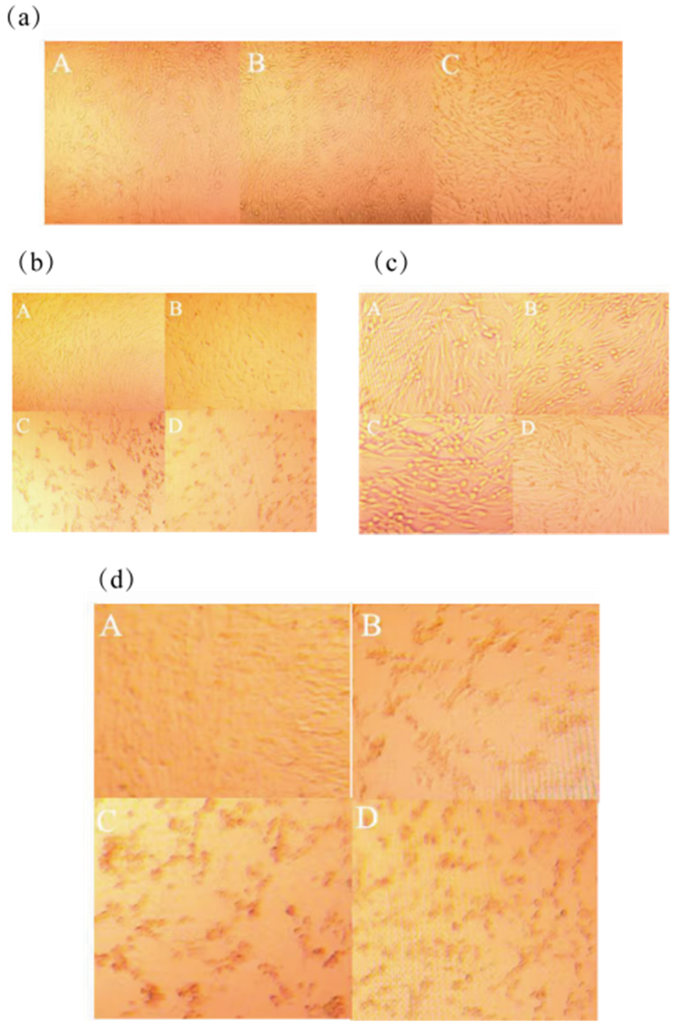
The morphology and growth status of PC12 cells were observed under an inverted phase contrast microscope (Figure 8a). PC12 cells in the blank group were sparse and polygonal in shape. After treatment with walnut oligopeptides, cells grew plump, interconnected and overlapped with each other. Hydrogen peroxide was used to induce PC12 cell damage. Cell viability was calculated to screen appropriate concentrations for establishing the oxidative damage model. MTT assay was adopted to evaluate cell viability, proliferation and drug cytotoxicity. Relevant results are shown in Figure 8b. Compared with the blank group, cell viability increased after walnut oligopeptide intervention, indicating no cytotoxicity or growth inhibition effect. Cell viability rose extremely significantly in the concentration range of 0.6–1.0 mg/mL (p < 0.001). The highest viability of 121.94 ± 0.39% was obtained at 0.8 mg/mL, which was markedly higher than the positive control donepezil hydrochloride group (p < 0.05). Thus, concentrations of 0.6, 0.8 and 1.0 mg/mL were selected for subsequent experiments. The protective effects of different concentrations of walnut oligopeptides on oxidatively damaged cells were detected by MTT assay (Figure 8d). Cell viability of the model group decreased extremely significantly versus the blank group (p < 0.001), confirming successful model establishment. Cell viability increased remarkably after walnut oligopeptide treatment compared with the model group (p < 0.001), verifying its protective effect on damaged PC12 cells. The optimal protective rate reached 79.88 ± 0.21% at 0.8 mg/mL, with a 25% viability elevation and 1.83% higher efficacy than donepezil hydrochloride. The in vitro cytoprotective results of the present study are consistent with the findings reported by Li et al. [23].
Figure 8.
Study on oxidative damage protection and memory improvement effects of walnut oligopeptides (a) Morphological observation of viable PC12 cells treated with walnut oligopeptides (10×; A. Blank group, B. Donepezil hydrochloride group, C. Walnut oligopeptide group) (b) Morphological observation of viable PC12 cells under different hydrogen peroxide concentrations (10×; A. Blank group, B. 100 μmol/L H2O2 group, C. 200 μmol/L H2O2 group, D. 300 μmol/L H2O2 group) (c) Morphological observation of viable PC12 cells treated with varying walnut oligopeptide concentrations (10×; A. Blank group, B. Donepezil hydrochloride group, C. 0.8 mg/mL walnut oligopeptide group, D. 0.6 mg/mL walnut oligopeptide group) (d) Morphological observation on protective effects of walnut oligopeptides against oxidative damage in PC12 cells (10×; A. Blank group, B. Model group, C. Donepezil hydrochloride group, D. 0.8 mg/mL walnut oligopeptide group).
3.8. Protective Effects of Different Concentrations of Walnut Oligopeptides on Oxidative Damage Model
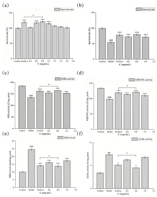
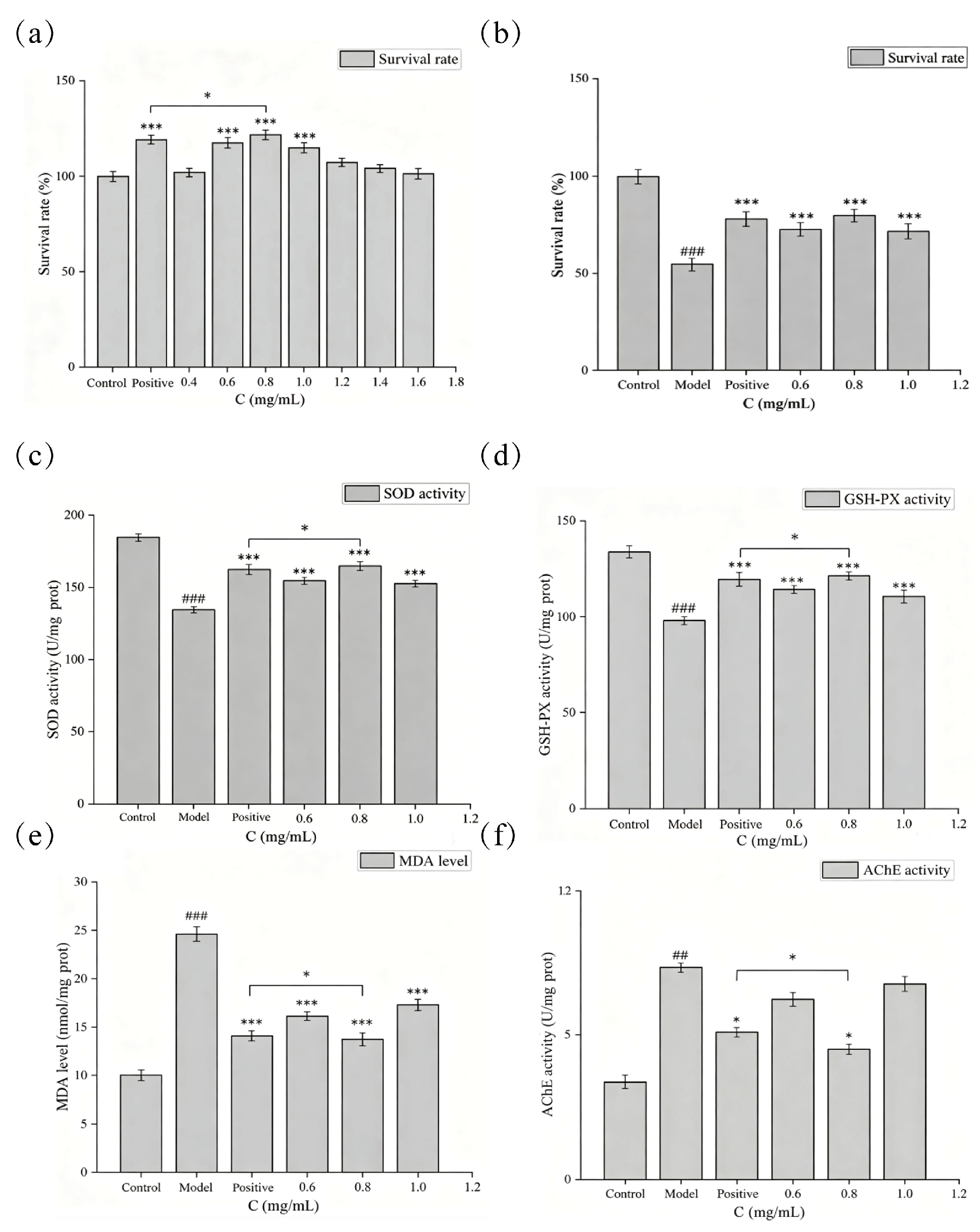
As shown in Figure 9a, cell viability increased after walnut oligopeptide treatment compared with the blank group, indicating no cytotoxicity or growth inhibition. Cell viability rose extremely significantly at 0.6–1.0 mg/mL (p < 0.001). The maximum viability of 121.94 ± 0.39% was achieved at 0.8 mg/mL, which was significantly higher than the donepezil hydrochloride positive control group (p < 0.05). Accordingly, concentrations of 0.6, 0.8 and 1.0 mg/mL were selected for subsequent tests.
Figure 9.
Protective effects of different concentrations of walnut oligopeptides on oxidative damage model; (a) Effects on PC12 cell viability; (b) Effects on viability of oxidatively damaged PC12 cells; (c) Effects on intracellular SOD activity; (d) Effects on intracellular GSH-PX activity; (e) Effects on intracellular MDA content; (f) Effects on intracellular AChE activity. * indicates (p < 0.05), *** indicates (p < 0.001); ## and ### represent (p < 0.01) and (p < 0.001), respectively.
In Figure 9b, cell viability of the model group dropped sharply relative to the blank group (p < 0.001), confirming successful model establishment. Cell viability increased remarkably after oligopeptide intervention (p < 0.001), demonstrating protective effects on oxidatively damaged PC12 cells. The 0.8 mg/mL group presented the optimal protective effect of 79.88 ± 0.21%, a 25% increase and 1.83% higher than the positive control. SOD is a key endogenous antioxidant enzyme that functions first in the reactive oxygen scavenging system. GSH-Px decomposes lipid peroxides with glutathione as a reductant and protects cell membranes and tissues from oxidative injury. Figure 9c,d revealed that SOD and GSH-Px activities in the model group decreased markedly (p < 0.001). Both enzyme activities were significantly elevated after walnut oligopeptide treatment (p < 0.001). The 0.8 mg/mL group obtained the highest SOD and GSH-Px activities of 164.94 ± 0.22 U/mg prot and 121.43 ± 0.56 U/mg prot, with increments of 30.15 U/mg prot and 23.22 U/mg prot, respectively, which were superior to the positive control (p < 0.05). Walnut oligopeptides could enhance antioxidant enzyme activities and alleviate oxidative damage. MDA content reflects cellular injury degree. Figure 9e showed remarkably elevated MDA levels in the model group (p < 0.001). MDA content declined significantly in all oligopeptide-treated groups (p < 0.001). The lowest MDA value of 13.70 ± 0.63 nmol/mg prot was observed at 0.8 mg/mL, obviously lower than the positive control (p < 0.05). The 0.8 mg/mL walnut oligopeptide exerted better protective efficacy against oxidative damage than donepezil hydrochloride.
Acetylcholinesterase (AChE) is a pivotal enzyme in nerve signal transmission. It catalyzes the hydrolysis of neurotransmitter acetylcholine into choline and acetic acid, thereby blocking neural signal transduction [24]. Acetylcholine content is closely associated with AChE activity. Hence, modulating AChE activity and elevating acetylcholine levels serve as vital strategies for ameliorating memory decline.
As illustrated in Figure 9f, AChE activity in the model group increased significantly compared with the blank group (p < 0.01), validating successful model construction. Oxidative stress triggers the upregulation of intracellular AChE activity. AChE activity declined significantly after walnut oligopeptide treatment, indicating its regulatory effect on this enzyme. The optimal effect was observed at 0.8 mg/mL with AChE activity of 4.51 ± 0.28 U/mg prot, which was distinctly lower than that of the donepezil hydrochloride group (p < 0.05). The results demonstrate that 0.8 mg/mL walnut oligopeptides can effectively inhibit AChE activity with superior efficacy to the positive control, elevate acetylcholine levels, and exert memory-improving capacity.
In addition to alleviating oxidative stress and regulating acetylcholinesterase activity, walnut peptides exert memory-improving effects by modulating the neural microenvironment. Xing et al. (2024) [25] isolated a heptapeptide TW-7 derived from walnuts, which can suppress excessive activation of microglia in the brain of mice with LPS-induced cognitive impairment, reduce aberrant microglial phagocytosis of synapses, and mitigate learning and memory decline caused by synaptic loss. These findings expand the potential therapeutic targets underlying the neuroprotective effects of walnut peptides.
4. Conclusions
In this study, cold-pressed walnut meal was used as raw material. Walnut oligopeptides with a high recovery rate were successfully prepared via simultaneous dual-enzyme hydrolysis combined with activated carbon decolorization and membrane separation purification, realizing high-value utilization of walnut processing by-products. The optimal enzymatic hydrolysis conditions were determined as pH 10.0, total enzyme dosage 11,000 U/g, trypsin-to-alkaline protease ratio 2.1:1, solid–liquid ratio 1:26, temperature 51 °C and hydrolysis duration 4 h, achieving a hydrolysis degree of 33.02%. The optimal decolorization parameters were pH 5.2, activated carbon dosage 2.3%, temperature 58 °C and reaction time 43 min. The final peptide recovery rate reached 83.35%. The obtained product was milky white with stable quality.
Amino acid analysis revealed that cold-pressed walnut meal was rich in glutamic acid, arginine and aspartic acid, laying a material foundation for the neuroprotective and nutritional functions of walnut oligopeptides. In vitro antioxidant assays showed that the oligopeptides possessed strong scavenging capacities against hydroxyl radical, DPPH radical and superoxide anion radical, with maximum scavenging rates of 90.18%, 81.72% and 85.80%, respectively. It still retained 73.21% of antioxidant activity after simulated gastrointestinal digestion, suggesting promising in vivo application prospects.
PC12 cell experiments verified that walnut oligopeptides exhibited no cytotoxicity and markedly promoted cell viability. At the concentration of 0.8 mg/mL, the protective rate against hydrogen peroxide-induced oxidative damage was 79.88%. It effectively increased intracellular SOD and GSH-Px activities, reduced MDA content and alleviated oxidative stress injury. Meanwhile, the oligopeptides significantly inhibited AChE activity with better efficacy than the positive control, donepezil hydrochloride, exerting neuroprotective and memory-improving effects by regulating the cholinergic system.
The walnut protein and walnut oligopeptides were only produced at laboratory bench scale in the present work, and no pilot or industrial scale-up experiments were carried out. Accordingly, relevant data regarding continuous production equipment compatibility and long-term economic cost evaluation for large-scale manufacturing are absent. In addition, the antioxidant and neuroprotective bioactivities of the obtained products were merely characterized via in vitro assays, without further validation through long-term toxicity assessments or dietary intervention animal models. Moreover, this study failed to explore the practical application performance of walnut protein and oligopeptides in typical food matrices, including beverages and bakery products.
This study clarified the preparation process and functional properties of walnut oligopeptides, providing a feasible strategy for resource utilization of walnut meal. Further research shall focus on separation and purification, structural identification and in vivo animal verification; explore its action mechanism in intervening neurodegenerative diseases, accelerate industrial application in brain-health food and dietary supplements, and broaden the application scope of plant-derived bioactive peptides in nutrition and healthcare fields. Subsequent research may adopt the research framework established by Li et al. (2025), [26] construct a D-galactose-induced aging mouse model, and explore the intrinsic mechanism by which walnut oligopeptides ameliorate age-related cognitive impairment from the perspective of the gut–brain axis. Combined with long-term in vivo toxicity tests, relevant data for product development can be supplemented and perfected.
Supplementary Materials
The following supporting information can be downloaded at: https://www.mdpi.com/article/10.3390/bioresourbioprod2030019/s1, Table S1: Plackett-Burman experimental design and response values; Table S2: Factor effect evaluation of Plackett-Burman experimental design; Table S3: Response surface experimental design and results; Table S4: Analysis of variance of regression model; Table S5: Response surface experimental design and results; Table S6: Analysis of variance of decolorization rate; Table S7: Analysis of variance of peptide recovery rate; Table S8: Abbreviations used in this paper.
Author Contributions
Methodology, C.L.; Validation, Y.L.; Formal analysis, F.W.; Investigation, N.S.; Resources, Conceptualization, Formal analysis, Y.W.; Data curation, C.Y.; Writing—original draft, X.Y.; Supervision, Conceptualization, Z.W.; Funding acquisition, C.Y. and Z.W. All authors have read and agreed to the published version of the manuscript.
Funding
This research was funded by the following projects: Heweixin Food Development Research Institute (grant number: W2025006).
Institutional Review Board Statement
Not applicable.
Informed Consent Statement
Not applicable.
Data Availability Statement
Data are not available for privacy reason.
Acknowledgments
We sincerely thank the School of Life Science and Technology, University of Jinan, for providing venue and facility support, and the Binzhou Weiqiao National Institute of Advanced Technology for technical support during the experiments.
Conflicts of Interest
Author Zhongzheng Wang was employed by the company Gansu Jiuyuan Agriculture and Forestry Technology Co., Ltd. The remaining authors declare that the research was conducted in the absence of any commercial or financial relationships that could be construed as a potential conflict of interest.
References
- Mao, X.; Hua, Y.; Chen, G. Amino acid composition, molecular weight distribution and gel electrophoresis of walnut proteins and protein fractionations. Int. J. Mol. Sci. 2014, 15, 2003–2014. [Google Scholar] [CrossRef] [Scilit] [PubMed]
- Sze-Tao, K.W.C.; Sathe, S.K. Walnuts: Proximate composition, protein solubility, protein amino acid composition and protein in vitro digestibility. J. Sci. Food Agric. 2000, 80, 1393–1401. [Google Scholar] [CrossRef] [Scilit]
- Korhonen, H.; Pihlanto, A. Food-derived bioactive peptides-opportunities for designing future foods. Curr. Pharm. Des. 2003, 9, 1297–1308. [Google Scholar] [CrossRef] [Scilit] [PubMed]
- Kitts, D.; Weiler, K. Bioactive proteins and peptides from food sources. Curr. Pharm. Des. 2003, 9, 1309–1323. [Google Scholar] [CrossRef] [Scilit] [PubMed]
- Shen, W.; Ma, S. Current knowledge of intestinal absorption of bioactive peptides. Food Funct. 2017, 8, 4306–4314. [Google Scholar] [CrossRef] [Scilit] [PubMed]
- Sowmya, K.; Bhat, M.I.; Bajaj, R.; Kapila, S.; Kapila, R. Antioxidative and anti-inflammatory potential with trans-epithelial transport of a buffalo casein-derived hexapeptide. Food Biosci. 2019, 28, 151–163. [Google Scholar] [CrossRef] [Scilit]
- Min, W.; Dang, Q.; Li, Z.; Zhao, X.; Shen, Y.; Fang, L.; Liu, C. Walnut-derived peptides cross the blood-brain barrier and ameliorate A(3-induced hypersynchronous neural network activity. Food Res. Int. 2024, 188, 115302. [Google Scholar] [CrossRef] [Scilit] [PubMed]
- Vinson, J.A.; Cai, Y. Especially walnuts, have both antioxidant quantity and efficacy and exhibit significant potential health benefits. Food Funct. 2012, 3, 134–140. [Google Scholar] [CrossRef] [Scilit] [PubMed]
- Ma, S.; Huang, D.; Zhai, M.; Yang, L.; Peng, S.; Chen, C.; Feng, X.; Weng, Q.; Zhang, B.; Xu, M. Isolation of a novel bio-peptide from walnut residual protein inducing apoptosis and autophagy on cancer cells. BMC Complement. Altern. Med. 2015, 15, 413. [Google Scholar] [CrossRef] [Scilit] [PubMed]
- Jahanbani, R.; Ghaffari, S.M.; Salami, M.; Vahdati, K.; Sepehri, H.; Sarvestani, N.N.; Sheibani, N.; Moosavi-Movahedi, A.A. Antioxidant and anticancer activities of walnut protein hydrolysates using different protease. Plant Foods Hum. Nutr. 2016, 71, 402–409. [Google Scholar] [CrossRef] [Scilit] [PubMed]
- Wei, W.; Wu, Q.; Wang, S.; Dong, C.; Shao, S.; Zhang, Z.; Zhang, X.; Zhang, X.; Kan, J.; Liu, F. Treatment with walnut peptide ameliorates memory impairment in zebrafish and rats: Promoting the expression of neurotrophic factors and suppressing oxidative stress. Food Funct. 2024, 15, 3762–3773. [Google Scholar] [CrossRef] [Scilit] [PubMed]
- Miao, W.; Li, Y.; Ma, S.; Jiang, J.; Liu, H.; Cai, X.; Qin, Z.; Wang, X. Effects of cold-pressing conditions on physicochemical and functional properties of cold-pressed tigernut oil and starch isolated from press-cake. Food Sci. 2022, 57, 662–675. [Google Scholar] [CrossRef] [Scilit]
- Sáez-Plaza, P.; Michałowski, T.; Navas, M.J.; Asuero, A.G.; Wybraniec, S. An Overview of the Kjeldahl Method of Nitrogen Determination. Part I. Early History, Chemistry of the Procedure, and Titrimetric Finish. Crit. Rev. Anal. Chem. 2013, 43, 178–223. [Google Scholar] [CrossRef] [Scilit]
- Lin, S.-D. Effect of Processing and Cooking on Physicochemical, Sensory, and Functional Properties of Food. Foods 2025, 14, 1598. [Google Scholar] [CrossRef] [Scilit] [PubMed]
- Sun, S.-W.; Lin, Y.-C.; Weng, Y.-M.; Chen, M.-J. Efficiency improvements on ninhydrin method for amino acid quantification. J. Food Compos. Anal. 2006, 19, 112–117. [Google Scholar] [CrossRef] [Scilit]
- Matsushita, S.; Iwami, N.; Nitta, Y. Colorimetric estimation of amino acids and peptides with the Folin phenol reagent. Anal. Biochem. 1966, 16, 365–371. [Google Scholar] [CrossRef] [Scilit] [PubMed]
- Brodkorb, A.; Egger, L.; Alminger, M.; Alvito, P.; Assunção, R.; Ballance, S.; Bohn, T.; Bourlieu-Lacanal, C.; Boutrou, R.; Carrière, F.; et al. INFOGEST static in vitro simulation of gastrointestinal food digestion. Nat. Protoc. 2019, 14, 991–1014. [Google Scholar] [CrossRef] [Scilit] [PubMed]
- Ghasemi, M.; Turnbull, T.; Sebastian, S.; Kempson, I. The MTT Assay: Utility, Limitations, Pitfalls, and Interpretation in Bulk and Single-Cell Analysis. Int. J. Mol. Sci. 2021, 12, 12827. [Google Scholar] [CrossRef] [Scilit] [PubMed]
- John, T.; Brosnan, M.E. Glutamate: A Truly Functional Amino Acid. Amino Acids 2013, 45, 413–418. [Google Scholar] [CrossRef] [Scilit] [PubMed]
- Pedrazini, M.C.; Martinez, E.F.; Santos, V.A.B.; Groppo, F.C. L-arginine: Its role in human physiology, in some diseases and mainly in viral multiplication as a narrative literature review. Int. J. Biol. Macromol. 2024, 10, 99. [Google Scholar] [CrossRef] [Scilit]
- Sheng, J.; Yang, X.; Chen, J.; Peng, T.; Yin, X.; Liu, W.; Liang, M.; Wan, J.; Yang, X. Antioxidative effects and mechanism study of bioactive peptides from defatted walnut (Juglans regia L.) meal hydrolysate. J. Agric. Food Chem. 2019, 67, 3305–3312. [Google Scholar] [CrossRef] [Scilit] [PubMed]
- Wu, H.; Chen, H.; Shiau, Y.S. Free amino acids and peptides asrelated to antioxidant properties inprotein hydrolysates of mackerel. Food Res. Int. 2003, 36, 949–957. [Google Scholar] [CrossRef] [Scilit]
- Feng, L.; Wu, Y.; Wang, J.; Han, Y.; Huang, J.; Xu, H. Neuroprotective Effects of a Novel Tetrapeptide SGGY from Walnut against H2O2-Stimulated Oxidative Stress in SH-SY5Y Cells: Possible Involved JNK, p38 and Nrf2 Signaling Pathways. Foods 2023, 12, 1490. [Google Scholar] [CrossRef] [Scilit] [PubMed]
- Wu, M.; Li, M.; Yuan, J.; Liang, S.; Chen, Z.; Ye, M.; Ryan, P.M.; Clark, C.; Tan, S.C.; Rahmani, J.; et al. Postmenopausal hormone therapy and alzheimer’s disease, dementia, and parkinson’s disease: A systematic review and time-response meta-analysis. Pharmacol. Res. 2020, 155, 104693. [Google Scholar] [CrossRef] [Scilit] [PubMed]
- Xing, Y.; Shi, H.; Gao, X.; Zhu, X.; Zhang, D.; Fang, L.; Wang, J.; Liu, C.; Wu, D.; Wang, X.; et al. Walnut-Derived Peptides Alleviate Learning and Memory Impairments in a Mice Model via Inhibition of Microglia Phagocytose Synapses. J. Agric. Food Chem. 2024, 72, 6478–6488. [Google Scholar] [CrossRef] [Scilit] [PubMed]
- Li, C.; Zheng, D.; Zhang, T.; Qian, S.; Liu, X.; Yang, Z.; Lim, K.-J.; Min, W.; Huang, J.; Yang, Y.; et al. Recombinant Walnut-Derived Peptide Ameliorates d-Galactose-Induced Cognitive Deficits. J. Agric. Food Chem. 2025, 73, 9712–9724. [Google Scholar] [CrossRef] [Scilit] [PubMed]

Disclaimer/Publisher’s Note: The statements, opinions and data contained in all publications are solely those of the individual author(s) and contributor(s) and not of MDPI and/or the editor(s). MDPI and/or the editor(s) disclaim responsibility for any injury to people or property resulting from any ideas, methods, instructions or products referred to in the content. |
© 2026 by the authors. Licensee MDPI, Basel, Switzerland. This article is an open access article distributed under the terms and conditions of the Creative Commons Attribution (CC BY) license.