


Abstract
Autism spectrum disorder (ASD) is characterized by persistent impairments in social communication together with restricted and repetitive patterns of behavior. Although ASD has traditionally been viewed primarily as a disorder of neural circuitry, increasing evidence indicates that interactions between the immune and nervous systems contribute substantially to brain development and cognitive function. This review develops a neuroimmune–cognitive–account of ASD by examining how immune signaling may influence neural organization and, in turn, cognitive function. Evidence from neuroimmunology, systems neuroscience, and experimental studies of neuromodulation is synthesized to examine relationships among immune signaling, neural network organization, and cognition. Disturbances in these processes have been associated with alterations in excitation–inhibition –balance and atypical large-scale connectivity, especially within networks supporting social cognition. We also examine the role of neuromodulatory systems, with particular emphasis on oxytocin and vasopressin, as intermediaries between biological regulation and cognitive processing. Experimental findings indicate that oxytocin can transiently modulate activity within social brain networks and increase the salience of socially relevant stimuli, although effects across clinical studies remain modest and inconsistent. The reviewed evidence suggests that disturbances in neuroimmune regulation may contribute to altered communication among distributed neural systems, providing one possible account of cognitive dysfunction in ASD. Social deficits, repetitive behaviors, and sensory differences reflect disturbances in the coordination of distributed neural systems rather than isolated impairments within single regions or pathways. This view has important implications for intervention, suggesting that approaches directed at individual molecular or neural targets alone are unlikely to produce broad or lasting effects. More effective strategies may require interventions that address interactions among immune function, neural dynamics, and cognitive processes. The resulting neuroimmune–cognitive account generates testable hypotheses concerning how immune processes may influence neural organization and cognition in ASD while providing a conceptual basis for future experimental and clinical research.
1. Introduction
Autism spectrum disorder (ASD) is a neurodevelopmental condition characterized by persistent impairments in social communication and interaction together with restricted and repetitive patterns of behavior. Although these features are defined behaviorally, they reflect underlying differences in the organization and function of cognitive and neural systems. Among these characteristics, social dysfunction is the most consistent and defining feature, emerging early in development and influencing subsequent cognitive, emotional, and behavioral trajectories (Frith & Frith, 2007; Müller et al., 2011).
Despite substantial advances in neuroscience, there is still no established pharmacological intervention that reliably addresses the core social deficits associated with ASD (Sikich et al., 2021; Preti et al., 2014). This limitation reflects a broader conceptual problem. Although numerous biological and cognitive mechanisms have been implicated in ASD, there remains no widely accepted framework explaining how immune signaling, neural development, large-scale network organization, and cognition are linked across levels of biological organization. Traditional models have emphasized abnormalities in neural circuitry, notably with disruptions in large-scale connectivity and atypical activation of brain regions supporting social cognition. Although these models have provided important insights, they do not fully account for the developmental origins, heterogeneity, and context-dependent features of cognitive impairment in ASD.
Increasing attention has turned toward neuroimmunology, the study of interactions between the nervous and immune systems as an important contributor to brain development and function. The nervous and immune systems form a highly interconnected and bidirectional network in which signaling molecules such as cytokines, chemokines, and growth factors may influence neural activity across the lifespan (Deverman & Patterson, 2009; Zipp et al., 2023). These interactions are not confined to pathological conditions but also subserve normal neurodevelopmental processes, including neurogenesis, synapse formation, and circuit refinement (L. Wang et al., 2025).
Immune-related processes are increasingly recognized as important regulators of neurodevelopmental trajectories. Cytokines regulate neuronal proliferation, differentiation, and survival as well as synaptic plasticity and connectivity (Deverman & Patterson, 2009; Linnerbauer et al., 2020; Monet & Quan, 2023). Microglia, the resident immune cells of the central nervous system, are critically involved in synaptic pruning, the developmental elimination of excess synaptic connections required for the establishment of efficient and specialized neural networks (Paolicelli et al., 2011). Disturbances in these processes may result in either excessive or insufficient pruning, both of which have been implicated in neurodevelopmental disorders, including ASD (Eltokhi, 2025). Growing evidence has prompted increasing interest in whether ASD may be understood not solely as a disorder of neural circuitry but also as one involving disturbances in neuroimmune–neural interactions. Immune signaling influences neural systems directly through modulation of synaptic transmission and plasticity and indirectly through alterations in large-scale network organization and excitation–inhibition balance (Imbriani et al., 2026).
Microglial and astrocytic dysfunction, for example, may alter the development of both excitatory and inhibitory synapses, contributing to network instability and altered information exchange across large-scale neural networks (Imbriani et al., 2026; Xiong et al., 2023). Such neurodevelopmental changes are singularly important during critical developmental periods, when neural systems are especially sensitive to biological and environmental influences.
These biological interactions become important when considering social cognition, the cognitive domain most consistently affected in ASD. Social cognition depends on coordinated activity across distributed neural networks, including the amygdala, medial prefrontal cortex, superior temporal sulcus, and cingulate cortex. These regions support functions such as emotion recognition, mental state attribution, and social attention. In ASD, these networks frequently show atypical activation and connectivity patterns, often interpreted as reflecting reduced interactions among distributed cortical networks. Neuroimmune mechanisms may contribute to these alterations through their effects on synaptic efficiency, neural responsiveness, and large-scale network organization.
In addition to influencing neural connectivity, neuroimmune processes interact with neuromodulatory systems supporting social behavior. Among the most extensively studied are the oxytocin (OT) and arginine vasopressin (AVP) systems. These neuropeptides support social bonding, attachment, and prosocial behavior across species (Carter, 2014). In humans, oxytocin has been associated with enhanced emotion recognition, eye gaze, and interpersonal trust (Meyer-Lindenberg et al., 2011).
A growing body of work has examined the role of oxytocin in ASD. Experimental studies suggest that oxytocin administration can improve performance on social cognition tasks and increase attention to socially relevant cues (Guastella et al., 2010). Functional neuroimaging studies further show that oxytocin modulates activity in brain regions associated with social processing, including the amygdala and mesocorticolimbic reward systems (Gordon et al., 2013). At the same time, meta-analytic findings indicate that the behavioral effects of oxytocin are generally modest and variable, with inconsistent effects on both social cognition and repetitive behaviors (Preti et al., 2014).
The heterogeneity of these outcomes highlights an important issue: interventions targeting a single neuromodulatory system within a complex biological network are unlikely to produce uniform effects across individuals. Genetic background, receptor expression, developmental timing, and environmental context all may influence interactions between neuroimmune and neuromodulatory systems. Variation in the oxytocin receptor gene (OXTR), for example, has been associated with differences in social behavior and neural structure, to a greater extent within the amygdala (Tost et al., 2010). These studies reinforce the view that ASD is a heterogeneous condition arising through interactions among multiple biological systems rather than from a single underlying deficit.
Many of these biological processes converge on mechanisms that are thought to influence functional communication across cortical systems. Social cognition depends on coordinated interactions among perception, attention, memory, and predictive processes. Disturbances affecting the neural systems supporting these functions may contribute to the characteristic difficulties in social understanding and interaction observed in ASD. Neuroimmune influences may participate in these disturbances through effects on synaptic connectivity, excitation–inhibition balance, and the salience of socially relevant stimuli.
The purpose of the present review is not simply to summarize evidence from neuroimmunology, systems neuroscience, and cognitive science but to integrate these studies into a multilevel conceptual account of cognitive dysfunction in ASD. Rather than treating immune, neural, and cognitive processes as separate domains, we consider how interactions among these systems may contribute to the development and expression of ASD. We argue that cognition should be understood as an emergent property of interacting neuroimmune–neural systems and examine how disturbances across these systems may contribute to the social and behavioral characteristics of ASD.
Specifically, we review evidence concerning (i) neuroimmune influences on brain development, (ii) large-scale brain connectivity, (iii) neuromodulatory systems mediate aspects of social cognition, and (iv) experimental findings relating oxytocin to neural and cognitive processing. Building on this evidence, we develop a neuroimmune–cognitive framework that generates testable hypotheses regarding how immune signaling may influence neural organization and, ultimately, cognitive function in ASD while remaining compatible with existing neuroscientific models of the disorder.
Because the objective of this article is conceptual integration rather than quantitative evidence synthesis, the review was conducted as a narrative review. The methods used to identify and select the literature are described in the following section.
2. Review Methodology
The present article was conducted as a narrative review designed to integrate evidence from neuroimmunology, systems neuroscience, developmental neuroscience, and cognitive science relevant to autism spectrum disorder (ASD). The objective was not to provide a quantitative synthesis of the literature but to examine whether findings across these complementary fields could be integrated into a coherent conceptual account linking neuroimmune processes with cognitive dysfunction in ASD.
The literature was identified through searches of PubMed, Web of Science, and Scopus, supplemented by manual searches of reference lists from relevant publications. Search terms included combinations of autism spectrum disorder, ASD, neuroimmunology, microglia, astrocytes, cytokines, synaptic pruning, brain connectivity, functional connectivity, social cognition, oxytocin, vasopressin, neurodevelopment, maternal immune activation, and cognitive neuroscience. The literature published through 2026 was considered.
Priority was given to peer-reviewed original experimental, clinical, and translational studies. Seminal publications were retained where they established foundational concepts or experimental findings, while more recent studies were preferentially included to reflect current advances in the field. Review articles and meta-analyses were cited selectively to provide conceptual context, summarize established bodies of evidence, identify broader trends in the literature, and support areas in which extensive empirical findings have accumulated.
Studies were selected on the basis of their relevance to the central objective of the review: understanding how interactions among immune signaling, neural development, and large-scale brain organization may contribute to cognitive dysfunction in ASD. Because the purpose of the article was conceptual integration rather than systematic evidence synthesis, formal assessment of study quality, risk of bias, or quantitative meta-analysis was not undertaken. Consequently, the review is subject to the limitations inherent to narrative reviews, including the possibility of selection bias and incomplete coverage of all relevant publications.
3. Neuroimmunology and Brain Development
Neurodevelopment is increasingly recognized as a process shaped not only by neuronal mechanisms but also by dynamic interactions between the nervous and immune systems. The central nervous system (CNS) develops within a complex biological environment in which immune signaling molecules, glial cells, and peripheral immune influences collectively regulate the formation, refinement, and stabilization of neural circuits. These processes are especially critical during early development, when neural systems exhibit heightened plasticity and sensitivity to environmental and endogenous signals.
A central component of neuroimmune influence on brain development is the role of cytokines and other immune signaling molecules. Cytokines, including interleukins, tumor necrosis factor (TNF), and interferons, are not only mediators of immune responses but also regulators of neural development and synaptic plasticity. They may influence neuronal proliferation, migration, and differentiation, as well as synaptic transmission and long-term potentiation (Matta et al., 2019; Deverman & Patterson, 2009; Ashwood et al., 2011). Cytokine signaling must be tightly regulated; both insufficient and excessive signaling can disrupt normal developmental trajectories. Chronic or dysregulated cytokine activity has been associated with alterations in neuronal function and cognitive processes, supporting a potential pathway through which immune imbalance may contribute to cognitive dysfunction (Matta et al., 2019; Ashwood et al., 2011).
Among the most critical cellular mediators of neuroimmune interactions are microglia, the resident immune cells of the CNS. Microglia play a central role in shaping neural circuits during development through processes such as synaptic pruning, the selective elimination of excess synaptic connections. This process is essential for the refinement of neural networks and the establishment of efficient information processing systems. Microglia actively survey the neural environment and remove weak or redundant synapses, thereby contributing to the optimization of network architecture (Luo & Wang, 2024; Mordelt & de Witte, 2023; Paolicelli et al., 2011; Schafer et al., 2012). Disruptions in microglial function have been implicated in neurodevelopmental disorders, including ASD. Experimental models demonstrate that alterations in microglial activation, neuron–microglia signaling, or phagocytic function can impair synaptic pruning and maturation, resulting in altered functional connectivity and autism-relevant social behavioral deficits (Zhan et al., 2014). For example, impaired microglial-mediated synaptic pruning has been associated with abnormal connectivity patterns and deficits in social behavior. These findings suggest that early disruptions in these processes can have lasting cognitive consequences (Xiong et al., 2023). Excessive pruning may likewise reduce synaptic density and disrupt neural transmission, underscoring the importance of maintaining a precise balance in synaptic remodeling processes (Wu et al., 2024).
Microglia also play a crucial role in regulating the balance between excitatory and inhibitory (E/I) synaptic activity. This balance is essential for stable and flexible neural processing and is widely regarded as a key factor in the pathophysiology of ASD. Alterations in microglial function can disrupt E/I balance by differentially affecting excitatory and inhibitory synapses, leading to network-level dysregulation and impaired information processing (Wake et al., 2019; Xiong et al., 2023). Such imbalances may contribute to both the hyper-reactivity to sensory stimuli and the reduced functional communication among distributed neural regions often observed in ASD.
In addition to microglia, astrocytes play a complementary role in neuroimmune regulation of brain development. Astrocytes contribute to maintaining homeostasis within the neural environment, including the regulation of neurotransmitter levels, ion balance, and metabolic support. They also contribute to synaptic formation and plasticity by releasing signaling molecules and modulating synaptic transmission. In ASD, astrocytic dysfunction has been associated with altered glutamate metabolism and disruptions in synaptic signaling, further contributing to network-level abnormalities (Xiong et al., 2023).
The interaction between microglia and astrocytes is important in the context of neuroinflammation. Activated glial cells release cytokines and chemokines that influence neuronal activity and synaptic function. While acute neuroinflammatory responses can be protective and support repair processes, chronic or dysregulated inflammation can disrupt neural function and contribute to cognitive impairment (Matta et al., 2019). In ASD, evidence of altered glial activation and cytokine profiles suggests that neuroinflammatory processes may play a role in shaping atypical neural development (Erbescu et al., 2022; Ashwood et al., 2011).
Another critical dimension of neuroimmune influence involves the interaction between the CNS and peripheral systems, especially through the gut–brain–immune axis. The gut microbiota can modulate immune signaling and may influence microglial function through circulating metabolites and immune mediators. Dysregulation of this system has been associated with altered microglial activation states and changes in synaptic pruning, potentially affecting neural circuits supporting behavior (Krishnapriya et al., 2025). This highlights the extent to which neurodevelopment is influenced by systemic biological processes that extend beyond the brain itself.
Developmental timing is a crucial factor in determining the impact of neuroimmune processes. During critical periods of development, neural circuits undergo rapid formation and refinement, making them vulnerable to perturbations. Maternal immune activation (MIA), for example, has been associated with an increased risk of ASD, with experimental and epidemiological evidence suggesting that immune challenges during pregnancy may alter fetal brain development and contribute to later behavioral and cognitive changes (Kim et al., 2024). These findings underscore the importance of temporal dynamics in understanding how neuroimmune processes developmental trajectories.
Neuroimmune processes may also influence large-scale brain organization through their effects on synaptic development, connectivity, and network dynamics. Disturbances in these processes have been associated with alterations in both structural and functional connectivity observed in ASD. Network-level changes may reduce communication among distributed neural systems that support higher-order cognitive functions, including social cognition, attention, and executive control (Zwierko et al., 2026).
Neuroimmune influences on brain development are not uniform but vary across individuals based on genetic, environmental, and developmental factors. For example, sex-dependent differences in microglial function have been observed in experimental models, suggesting that biological variability may contribute to differences in ASD prevalence and presentation (Wu et al., 2024). Similarly, genetic factors that may influence immune signaling pathways may interact with environmental exposures to shape individual developmental outcomes.
Collectively, this body of work provides a biological basis for considering how neuroimmune processes may influence development of neural systems relevant to cognition. Rather than acting as a secondary or peripheral influence, the immune system plays a central role in shaping the structural and functional architecture of the brain. Disruptions in these processes can lead to alterations in neural connectivity and network dynamics, which in turn affect the development of cognitive systems.
The body of evidence raises the possibility that neuroimmune processes may affect the development and function of cognitive systems through their effects on neural organization. Social cognition depends on coordinated activity across distributed neural networks that must develop with appropriate patterns of connectivity and responsiveness. Disturbances in neuroimmune signaling therefore represent one potential mechanism through which these networks may develop atypically, with consequences for adaptive behavior and social functioning. The following sections examine evidence linking neuroimmune regulation with large-scale brain organization, neuromodulatory systems, and cognitive processes implicated in ASD. The proposed relationships among immune signaling, cellular processes, and synaptic development are summarized schematically in Figure 1.
Figure 1.
Neuroimmune regulation of synaptic rules and emergent network topology. Neuroimmune processes, including cytokine signaling and glial activity, regulate the parameters that govern synaptic formation, elimination, and the excitation–inhibition balance. These processes define a synaptic rule set characterized by formation probability, pruning probability, and stability constraints. The balance of these parameters determines the distribution of connections within developing neural networks, giving rise to specific topological properties such as path length and clustering. Variations in these rules lead to differences in large-scale network organization, ranging from distributed configurations that support efficient communication to more segregated structures with reduced communication efficiency across the network. We emphasize that network architecture emerges from underlying synaptic dynamics shaped by neuroimmune regulation, rather than from independent alterations at the network level. (The figure shown is a theoretical illustration summarizing concepts discussed in the text and is not an empirical representation).
The evidence reviewed in this section indicates that neuroimmune processes contribute to neural development at molecular, cellular, and systems levels. The body of evidence provides a biological basis for examining how disturbances in immune signaling may alter the organization of neural systems supporting cognition. Subsequent sections consider evidence linking these developmental processes to large-scale brain organization, social cognition, and neuromodulatory mechanisms relevant to ASD.
4. Neuroimmune Influences on Cognitive Systems in Autism Spectrum Disorder
The neuroimmune mechanisms discussed in the preceding section are not confined to cellular or molecular processes. Their potential significance extends to large-scale neural systems that support cognition. In autism spectrum disorder (ASD), alterations in immune signaling, glial function, and synaptic development have been associated with alterations in the organization and dynamics of large-scale neural networks. Changes in large-scale network organization may influence cognitive processes contributing to social cognition, behavioral flexibility, and predictive processing.
A prominent feature of ASD is atypical brain connectivity. Structural and functional imaging studies have reported complex patterns of altered local and long-range connectivity, including reduced long-range connectivity and increased local connectivity in some samples, although findings vary with age, brain region, and methodology (Müller et al., 2011; Jiang et al., 2022). This pattern has generally been interpreted as reflecting alterations in the balance between distributed and localized processing, with possible consequences for cognition. Long-range connectivity is generally thought to support communication among distributed cortical systems, whereas local connectivity supports more specialized processing within local circuits. When this balance is disrupted, cognitive processing may become highly detailed while remaining poorly coordinated across systems.
Neuroimmune processes provide a biologically plausible mechanism underlying these connectivity patterns. Cytokine activity, microglial function, and glial–neuronal interactions have been shown to influence synaptic development and pruning in experimental systems, thereby influencing the organization of developing neural networks. Evidence suggests that neuroimmune activation is associated with altered functional connectivity in ASD, particularly within networks subserving social processing and self-referential cognition (Zwierko et al., 2026). Elevated levels of pro-inflammatory cytokines, such as TNF-α and IL-1β, have been linked to abnormal connectivity in the frontal-temporal and precuneus networks, regions mediating social cognition and perspective-taking (Edmonson et al., 2014).
4.1. Brain Connectivity and Cognitive Integration
Altered connectivity has consequential implications for higher-order cognition. Efficient cognitive processing depends on communication among distributed neural systems supporting attention, memory, executive control, and social behavior. Disruptions affecting these networks may reduce the efficiency with which information is exchanged across cortical systems, contributing to fragmented or context-insensitive cognition.
Large-scale brain networks such as the default mode network (DMN), salience network, and frontoparietal control network are relevant in this context. These networks support internally directed cognition, attentional switching, and goal-directed behavior, respectively. In ASD, abnormalities involving these networks have been reported consistently, including reduced connectivity within the DMN and altered interactions between networks (Buckner et al., 2008; Uddin et al., 2013; Müller et al., 2011; M. Wang et al., 2023). Such alterations may impair communication across cortical networks required for adaptive cognition.
Neuroimmune mechanisms may contribute to these network-level alterations through effects on both synaptic connectivity and neural excitability. Disruptions in excitation–inhibition (E/I) balance, associated with both synaptic protein dysfunction and immune-mediated modulation, can alter the stability and flexibility of neural networks (Rubenstein & Merzenich, 2003; Jiang et al., 2022). These changes may increase neural noise or reduce signal coherence, thereby decreasing the efficiency of information processing across networks. These observations suggest that alterations in network organization may contribute to cognitive dysfunction in ASD. Figure 2 summarizes how differences in network topology may influence communication among distributed neural systems.
Figure 2.
Network topology and communication across distributed neural systems. Illustration of how large-scale network organization constrains the efficiency of communication among distributed neural systems. Networks with lower path length and moderate clustering support efficient communication across distributed regions, enabling coordinated system-level dynamics. In contrast, increases in path length and clustering reduce the efficiency of information transfer, limiting integration across the network. The figure emphasizes that individual regions do not determine integration capacity but emerge from the global topology of connections. Variations in path length and clustering impose structural constraints on communication efficiency, providing a mechanistic basis for understanding differences in cognitive integration. (The figure is a theoretical illustration summarizing concepts discussed in the text and does not represent empirical data).
4.2. Social Brain Networks and Social Cognition
Social cognition is among the most extensively studied cognitive domains in ASD. It depends on a distributed network of brain regions commonly referred to as the “social brain,” including the medial prefrontal cortex, temporoparietal junction, superior temporal sulcus, amygdala, and cingulate cortex (Frith & Frith, 2007; Müller et al., 2011). These regions support functions such as emotion recognition, mentalizing, and attribution of intentions.
In ASD, these networks often exhibit atypical patterns of activation and connectivity. Functional neuroimaging studies have demonstrated reduced connectivity between key regions of the social brain as well as altered relationships between social and sensorimotor networks (M. Wang et al., 2023). The evidence further suggests that social dysfunction in ASD may arise not only from deficits within individual regions but also from impaired coordination across the network as a whole.
Neuroimmune processes may contribute to these alterations in several ways. Disruptions in synaptic pruning and plasticity can affect the structural organization of social brain circuits. Cytokine signaling and glial activation may alter neural responsiveness, influencing how these circuits process socially relevant information. In addition, immune-mediated changes in neuromodulatory systems such as oxytocin and serotonin may alter the salience of social stimuli, further affecting social cognition.
At the cognitive level, these alterations may manifest as reduced sensitivity to social cues, difficulty interpreting the mental states of others, and impaired use of emotional and contextual information. These characteristics are unlikely to arise solely from dysfunction within isolated brain regions and are more consistent with disturbances affecting communication among distributed neural systems.
4.3. Repetitive Behavior, Predictive Processing, and Cortico-Striatal System
In addition to social deficits, ASD is characterized by restricted and repetitive patterns of behavior. These behaviors have been associated with dysfunction in cortico-striatal circuits, notably those involving the dorsomedial striatum and its connections with frontal cortical regions. These circuits are central for action selection, habit formation, and behavioral flexibility.
These circuits are influenced by both neurotransmitter systems, including dopamine and serotonin, and neuroimmune processes. Alterations in serotonergic signaling, including activity involving the 5HT2A receptor, have been implicated in the regulation of repetitive behaviors (Alvarez et al., 2022). At the same time, immune-mediated modulation of synaptic function may affect plasticity within cortico-striatal circuits, contributing to reduced flexibility and increased behavioral rigidity.
Repetitive behaviors may also reflect alterations in predictive processing. Predictive processing theory proposes that the brain continuously generates predictions about incoming sensory information and updates them based on error signals. In ASD, disturbances in neural connectivity and E/I balance may impair the updating of predictions, leading to overly stable or rigid internal models (Pellicano & Burr, 2012). Behaviorally, this rigidity may appear as repetitive actions or restricted interests.
Neuroimmune processes may contribute to these alterations through effects on the neural mechanisms underlying prediction and error correction. Changes in synaptic plasticity and neurotransmitter function, for example, may influence how prediction errors are encoded and integrated. Under such conditions, neural systems may favor stability over flexibility, resulting in reduced adaptability in both cognition and behavior.
4.4. Toward a Neuroimmune–Cognitive Account of ASD
Evidence reviewed thus far supports the possibility that neuroimmune processes may influence cognition through effects on neural systems operating at multiple levels. Rather than producing isolated abnormalities, disturbances in immune signaling may affect network organization, neuromodulation, and communication among distributed neural systems. These mechanisms provide a biologically plausible account of several cognitive characteristics observed in ASD while also accommodating variability arising from differences in immune function, genetic background, and developmental timing (Zwierko et al., 2026).
In the following section, we examine the oxytocin and vasopressin systems, two critical neuromodulatory systems through which neuroimmune processes may influence cognition. These systems provide an important link between biological regulation and social cognitive function and may offer insight into both mechanisms and potential treatment approaches in ASD.
5. Oxytocin, Vasopressin, and Social Cognition
Neuropeptides play an important role in the regulation of social behavior across species by modulating neural circuits supporting social cognition, attachment, and affiliative behavior. Among these, oxytocin (OT) and arginine vasopressin (AVP) have received particular attention because of their established involvement in social bonding, pair formation, and social recognition in both animal models and humans. These systems provide one pathway linking biological regulation and higher-order cognitive processes and are therefore relevant to understanding mechanisms underlying social dysfunction in autism spectrum disorder (ASD).
Oxytocin is synthesized primarily within the paraventricular and supraoptic nuclei of the hypothalamus and is released both centrally and peripherally. Within the brain, oxytocin acts on a distributed network of regions contributing to social processing, including the amygdala, nucleus accumbens, medial prefrontal cortex, and superior temporal regions. Through its effects on these systems, oxytocin modulates attention to social stimuli, emotional salience, and reward processing (Meyer-Lindenberg et al., 2011; Aspé-Sánchez et al., 2016). Vasopressin, although structurally related, shows partially distinct receptor distributions and has been more strongly associated with social memory, aggression, and territorial behavior.
Available evidence suggests that oxytocin and vasopressin drive social information processing by modulating neural systems participating in evaluating socially relevant information rather than by directly generating behavior. These neuropeptides affect how social information is perceived, evaluated, and acted upon. Oxytocin, for example, has been reported to enhance attention to socially relevant cues such as eye gaze, facial expressions, and vocal affect, thereby facilitating the extraction of socially meaningful information (Sikich et al., 2021). The available evidence suggests that oxytocin may influence salience attribution by biasing cognitive systems toward socially relevant inputs. Mayer et al. (2022), however, did not observe comparable effects.
In ASD, several lines of evidence suggest dysregulation of the oxytocin system. Genetic studies have identified associations between polymorphisms in the oxytocin receptor gene (OXTR) and social behavior, including variation in empathy, social cognition, and amygdala structure and function (Tost et al., 2010; Aspé-Sánchez et al., 2016). Alterations in baseline oxytocin levels and receptor expression have also been reported, although findings remain inconsistent across studies (Lal et al., 2025; Zhao et al., 2022). These observations suggest that variability in oxytocin signaling may contribute to heterogeneity within ASD.
Early experimental studies suggested that intranasal oxytocin could improve several aspects of social cognition in individuals with ASD, including recognition of emotional states, attention to socially relevant cues, and performance on theory-of-mind tasks (Hollander et al., 2007; Guastella et al., 2010; Stavropoulos & Carver, 2013). These early reports generated considerable interest in oxytocin as a potential therapeutic target.
Subsequent randomized clinical trials have produced considerably more variable results. Although some studies reported modest improvements in selected social outcomes, others failed to demonstrate clinically meaningful benefits across primary outcome measures (Sikich et al., 2021; Mayer et al., 2022). Differences in participant characteristics, treatment duration, dosage, outcome measures, and baseline biological variability have all been proposed as contributing factors.
At the neural level, functional neuroimaging studies provide additional evidence that oxytocin modulates activity within social brain networks. Administration of oxytocin has been associated with enhanced activation in regions including the amygdala, superior temporal sulcus, and medial prefrontal cortex during processing of socially relevant stimuli (Gordon et al., 2013). The available evidence suggests that oxytocin can increase the responsiveness of neural circuits participating in social cognition and social information processing.
Oxytocin does not appear to enhance all aspects of cognition uniformly. Its effects are context-dependent and appear to selectively influence socially relevant processing. Oxytocin has been shown to increase differentiation between social and nonsocial stimuli by enhancing neural responses to socially meaningful inputs while attenuating responses to irrelevant stimuli (Gordon et al., 2013). These effects may reflect modulation of selective attention and relevance filtering, processes frequently discussed in relation to ASD. The available evidence suggests that oxytocin may influence the allocation of processing resources to socially relevant information rather than fundamentally altering the underlying cognitive architecture supporting social behavior. Systematic reviews and meta-analyses consistently conclude that although oxytocin can influence specific aspects of social cognition under experimental conditions, evidence for sustained clinical benefit remains limited (Ooi et al., 2017; Preti et al., 2014). More recent meta-analytic evidence has reported a small but statistically significant effect of oxytocin administration on social outcomes, although the estimated effect was reduced after adjustment for publication bias and varied according to factors including dosage; findings for routinized behaviors remained inconclusive (Audunsdottir et al., 2024). One factor that may contribute to this variability is the interaction between oxytocin signaling and broader neuroimmune processes. Immune activity can influence oxytocin receptor expression and sensitivity as well as the release and transport of oxytocin within the brain. Conversely, oxytocin itself has been reported to exert anti-inflammatory effects, suggesting a bidirectional relationship between these systems (Zhao et al., 2022). This interaction raises the possibility that the effectiveness of oxytocin-based interventions may depend partly on the neuroimmune state of the individual. Oxytocin may influence network dynamics rather than acting as a direct determinant of behavior. By influencing neural synchrony and connectivity within and between large-scale brain networks, oxytocin may influence patterns of communication among distributed neural systems. Computational and neuroimaging analyses suggest that oxytocin can modify functional integration and network organization within the default mode and frontoparietal networks, with possible implications for social-cognitive processing (Zheng et al., 2021). These neural effects may influence how individuals process and respond to social information. Increased attention to social cues, improved emotion recognition, and greater motivation for social interaction may arise through modulation of underlying neural systems. However, modulation of neural responsiveness alone does not appear sufficient to normalize social cognition in ASD, suggesting that changes in network activity may not necessarily translate into long-term reorganization of distributed neural systems.
Although vasopressin has been studied less extensively in ASD, it provides complementary evidence on neuropeptide regulation of social cognition. AVP has been associated with social memory and encoding of social information, and alterations in vasopressin signaling have been linked to variation in social behavior. Like oxytocin, vasopressin interacts with neural circuits subserving reward and social processing, suggesting that it may contribute to the regulation of social cognition at multiple levels.
Studies of oxytocin and vasopressin indicate that these neuropeptide systems contribute to social cognition in both typical development and ASD. At the same time, the variability observed across experimental and clinical studies suggests that their effects depend on broader characteristics of the neural and neuroimmune systems in which they operate.
Within the context of this review, oxytocin and vasopressin serve as examples of neuromodulatory systems through which immune signaling, neural network dynamics, and cognitive processing may interact. Their study illustrates both the potential and the limitations of targeting individual biological systems within a complex neurodevelopmental disorder.
6. Experimental Evidence: Oxytocin Modulation of Neural and Cognitive Processing in ASD
A growing body of experimental work has examined the effects of oxytocin administration on neural activity and cognitive function in individuals with autism spectrum disorder (ASD). These studies provide empirical evidence relevant to understanding how neuroimmune–neuromodulatory systems may influence social cognition. Functional neuroimaging paradigms, in particular, have been used to examine how oxytocin alters processing of socially relevant compared with nonsocial stimuli, providing insight into neural mechanisms through which this neuropeptide may influence social cognitive processing.
One commonly used experimental approach contrasts neural responses to social and nonsocial stimuli. In a representative paradigm, participants perform tasks requiring judgments about either social stimuli, such as identifying mental states from images of human eyes, or nonsocial stimuli, such as categorizing images of vehicles. This design allows researchers to isolate neural systems engaged in social cognitive processing and to examine the modulatory effects of oxytocin under controlled conditions.
Functional magnetic resonance imaging (fMRI) studies using these paradigms have consistently identified a network of brain regions showing increased activation in response to social stimuli relative to nonsocial stimuli. These regions include the amygdala, medial prefrontal cortex, superior temporal sulcus, inferior frontal gyrus, cingulate cortex, and precuneus (Frith & Frith, 2007; Gordon et al., 2013). Together, these regions comprise major components of the so-called “social brain,” supporting functions such as emotion recognition, mental state attribution, and social attention.
Administration of intranasal oxytocin has been reported to modulate activity within this network. Oxytocin increases neural responses to socially relevant stimuli while producing comparatively limited or sometimes suppressive effects on responses to nonsocial stimuli (Gordon et al., 2013). In the Eyes versus Vehicles paradigm, for example, oxytocin administration is associated with increased activation in social brain regions during processing of eye stimuli relative to vehicle stimuli when compared with placebo conditions.
Oxytocin administration has been associated with enhanced activation in the right amygdala, a region mediating emotional salience and social relevance. Increased activity has also been reported in bilateral superior frontal gyri, medial prefrontal cortices, superior temporal sulci, inferior frontal gyri, and middle cingulate cortices (Gordon et al., 2013). These findings suggest that oxytocin can alter activity within neural systems mediating interpretation of and response to social information.
The observed neural effects have also been associated with measurable cognitive and behavioral changes in some experimental studies. Reported effects include increased willingness to engage in social interaction, improved comprehension of affective speech, enhanced recognition of emotional expressions, and improved performance on tasks involving inference of others’ mental states following oxytocin administration (Guastella et al., 2010; Hollander et al., 2007). Some studies have also reported reductions in repetitive behaviors (Alvarez et al., 2022). However, these findings have not been replicated consistently across larger clinical trials, indicating that behavioral effects remain variable.
Another feature of these experimental studies is the combination of neural and peripheral measures of oxytocin activity. In some paradigms, salivary oxytocin levels are measured before and after administration, allowing examination of relationships between peripheral changes in oxytocin and neural responses. Increases in salivary oxytocin have been correlated with changes in brain activity, especially within social brain networks (Gordon et al., 2013). The observed relationships support a relationship between systemic neurochemical changes and localized neural processing.
Despite these observations, the effects of oxytocin have not been consistently demonstrated across studies. Some investigations report modest improvements, whereas others have failed to identify significant behavioral effects, especially in longer-term administration trials (Preti et al., 2014; Sikich et al., 2021). Although single-dose administration may produce measurable changes in neural activation and short-term behavior, these changes have not consistently translated into sustained improvements in social functioning. This pattern suggests that transient modulation of neural activity alone may be insufficient to produce durable changes in cognitive organization.
One possible interpretation is that oxytocin enhances salience and processing of social information without fundamentally altering the underlying network organization supporting cognition. Oxytocin may function as a gain modulator, increasing the responsiveness of social brain circuits to relevant stimuli without reorganizing the broader neural system. Under these conditions, improvements in social cognition may remain transient and context-dependent.
An important implication of this work is the importance of broader system interactions. The effects of oxytocin may depend on the underlying condition of neural and immune systems, including factors such as receptor density, synaptic organization, and inflammatory signaling. Variability in these factors across individuals may contribute to heterogeneity in treatment response.
The differential effects of oxytocin on social and nonsocial processing may also provide insight into the nature of cognitive dysfunction in ASD. Selective enhancement of responses to social stimuli suggests that the primary difficulty may not involve the capacity to process social information itself, but rather the prioritization of socially relevant information within broader cognitive systems. Oxytocin may transiently shift this balance by increasing the relative weighting of social inputs within the cognitive system. In this sense, oxytocin may modulate the weighting of specific connections within existing networks without necessarily altering the underlying connections themselves, as illustrated in Figure 3.
Figure 3.
Neuromodulation alters functional dynamics without changing network topology. Illustration of how neuromodulatory influences, such as oxytocin, alter the functional state of neural networks by changing the relative weighting of existing connections rather than their structural organization. Changes in excitatory and inhibitory balance modify the strength and salience of specific pathways, leading to shifts in network activity patterns. These alterations can produce states of increased or decreased activation (e.g., overexcitation or underexcitation), affecting information processing and responsiveness to stimuli. However, the underlying network topology remains unchanged, indicating that neuromodulation operates on connection dynamics rather than structural architecture. This distinction illustrates that functional changes in cognition can arise from modulation of network activity without requiring reorganization of the network itself. (The figure is a theoretical illustration summarizing concepts discussed in the text and does not represent empirical data).
Experimental findings indicate that oxytocin can influence neural activity and cognitive processes related to social behavior in ASD, notably under controlled experimental conditions. The available evidence also suggests that these effects primarily reflect modulation of existing neural systems rather than reorganization of underlying network architecture. The variability observed across experimental and clinical studies further indicates that oxytocin is only one component of a more complex set of interactions involving immune signaling, neural organization, and cognitive function. The following section examines how these findings relate to interactions among immune signaling, neural organization, and cognitive function in ASD.
7. Neuroimmune–Cognitive Integration in ASD
The preceding sections reviewed evidence from neuroimmunology, systems neuroscience, and experimental studies of neuromodulation. Although no single line of evidence provides a complete explanation for ASD, collectively these findings support a multilevel account in which immune signaling, neural development, large-scale network organization, and cognitive processing interact across development. The present section synthesizes these observations into a neuroimmune–cognitive account and identifies testable implications arising from these relationships.
Substantial experimental evidence indicates that immune processes play an active role in brain development. Cytokine signaling, microglial activity, and glial–neuronal interactions influence the formation and refinement of synaptic connections, thereby shaping the structural architecture of neural systems (Paolicelli et al., 2011; Matta et al., 2019). These processes do more than support neural development; they contribute to determining which connections are strengthened, weakened, or eliminated. Neuroimmune dynamics may influence the organization of neural networks underlying cognition.
Alterations in neuroimmune signaling may consequently produce measurable changes in neural system properties. Such changes include disturbances in synaptic pruning, alterations in excitation–inhibition balance, and changes in the strength and distribution of connectivity across brain regions (Rubenstein & Merzenich, 2003; Xiong et al., 2023). At the network level, these alterations may appear as reduced long-range connectivity and altered communication among large-scale brain networks, notably those involved in social cognition and executive control (Müller et al., 2011; M. Wang et al., 2023).
Network-level alterations have important implications for cognitive function. Cognitive systems depend on efficient exchange of information across distributed networks linking perception, attention, memory, and action. When communication across these networks is disrupted, cognitive processing may become fragmented and less context-sensitive. Such fragmentation is especially apparent in social cognition, where effective interaction depends on rapid coordination of multiple sources of information, including facial expression, vocal tone, and situational context (Frith & Frith, 2007).
Neuromodulatory systems such as oxytocin and vasopressin occupy an intermediary position within the broader neuroimmune–neural network. Oxytocin, for example, modulates the salience of social stimuli and increases the responsiveness of social brain circuits (Meyer-Lindenberg et al., 2011; Gordon et al., 2013). These effects, however, depend on the underlying condition of neural networks and immune signaling. In systems where network organization and communication are already compromised, neuromodulatory enhancement may produce only limited or transient improvements.
This may also help explain the variability observed in experimental and clinical studies of oxytocin in ASD. Although oxytocin can increase attention to social stimuli and enhance activation within relevant neural circuits, it does not appear to fundamentally reorganize the network architecture supporting cognition. Its effects tend to be modest, context-dependent, and often short-lived (Preti et al., 2014; Sikich et al., 2021). The observed heterogeneity may reflect the complexity of the system being targeted rather than the simple failure of the intervention itself.
The evidence reviewed above supports four closely related conclusions that form the conceptual foundation of the present neuroimmune–cognitive framework. First, neuroimmune signaling contributes to the development and refinement of synaptic organization. Second, these developmental processes impact the organization of large-scale neural networks. Third, the efficiency with which distributed neural systems communicate constrains higher-order cognitive functions, especially those supporting social cognition and behavioral flexibility. Finally, neuromodulatory systems such as oxytocin may influence the functional state of these networks without necessarily altering their underlying structural organization. These conclusions provide a testable biological account linking neuroimmune processes with network architecture and cognitive function in ASD.
On this basis, ASD may be viewed as involving disturbances in communication among neural systems arising, in part, from neuroimmune dysregulation. Social deficits, repetitive behaviors, and sensory differences need not be interpreted as independent abnormalities but may instead represent different expressions of altered organization across distributed neural systems. Repetitive behaviors, for example, may arise from reduced flexibility within cortico-striatal circuits, whereas social deficits may reflect impaired integration within social brain networks. Both may derive from disruptions affecting the same broader system.
Neuroimmune processes exert strong effects during critical developmental periods when neural circuits are being established and refined. Perturbations occurring during these periods may therefore have disproportionate and lasting effects on cognitive function (Kim et al., 2024). This temporal dimension may help explain why ASD manifests early in development and why earlier intervention is often associated with more favorable outcomes.
The proposed framework also provides a basis for understanding heterogeneity in ASD. Variation in immune function, genetic background, and environmental exposure may help explain heterogeneity in ASD. Differences in cytokine profiles, microglial activity, or neuromodulatory receptor expression, for example, may contribute to distinct developmental trajectories even among individuals sharing a common diagnosis. Such variability underscores the importance of more individualized approaches to both research and intervention.
The available evidence indicates that interventions directed at isolated biological targets may be insufficient when disturbances involve interacting neural and immune systems. Rather than focusing exclusively on a single neurotransmitter or brain region, future interventions may need to address interactions among immune, neural, and cognitive systems. Such approaches may combine pharmacological, behavioral, and cognitive interventions while also considering immune function and developmental timing. These proposed relationships among immune signaling, neural organization, network dynamics, and cognitive function are summarized in Figure 4.
Figure 4.
Multilevel relationships among neuroimmune signaling, neural organization, and cognitive function. Schematic representation of the hierarchical relationships linking neuroimmune processes, synaptic dynamics, network topology, and cognitive function. Neuroimmune signaling establishes synaptic rule sets that determine the formation and elimination of connections, giving rise to specific network topologies (Figure 1). These topological properties constrain the communication efficiency across distributed neural systems (Figure 2). Neuromodulatory influences, such as oxytocin, alter the functional weighting of existing connections, shifting network activity patterns without modifying underlying structural topology (Figure 3). These dynamic changes can transiently enhance or reduce integration capacity depending on the balance of excitatory and inhibitory influences. (The figure is a theoretical illustration summarizing concepts discussed in the text and does not represent empirical data).
The relationships outlined here may extend beyond ASD. If neuroimmune processes contribute to the development and organization of neural systems supporting cognition, similar mechanisms may also contribute to other neurodevelopmental and neuropsychiatric disorders characterized by altered network organization. This possibility remains to be tested. Examining interactions among immune signaling, neural dynamics, and cognitive function provides a means of linking biological mechanisms with higher-order cognition while generating testable hypotheses about how neuroimmune processes shape the emergence and organization of cognitive systems.
8. Clinical and Cognitive Implications
The neuroimmune–cognitive relationships discussed throughout this review have implications for both understanding and treatment of ASD. Conceptualizing ASD as involving dysregulation across interacting systems linking immune signaling, neural development, neural organization, and cognitive function challenges approaches that focus primarily on isolated symptoms or single biological targets. We instead emphasize the importance of interventions addressing coordination and interaction among multiple systems.
One implication concerns the limited efficacy of current pharmacological treatments. Despite extensive investigation of neuromodulatory systems such as oxytocin and serotonin, no pharmacological intervention has consistently demonstrated strong effects on core social deficits in ASD. As discussed previously, oxytocin administration can enhance aspects of social cognition and increase responsiveness within social brain circuits (Gordon et al., 2013; Guastella et al., 2010). These effects, however, are often modest, variable, and difficult to sustain over time (Preti et al., 2014; Sikich et al., 2021).
Pharmacological interventions generally target specific molecular pathways or receptor systems, whereas ASD involves alterations spanning multiple levels of biological and cognitive organization. Modulation of a single neuromodulatory system, such as the oxytocinergic system, may temporarily alter neural responsiveness without reorganizing distributed neural networks underlying cognitive function. These interventions target individual components of the system rather than the interactions that give rise to cognitive function.
This may also help explain variability in treatment response among individuals with ASD. Genetic background, immune function, developmental history, and system architecture may all influence responsiveness to intervention. Variability in oxytocin receptor expression or function, for example, may alter the effectiveness of oxytocin administration (Tost et al., 2010). Likewise, differences in baseline neuroimmune states, including inflammatory activity or microglial activation, may influence the responsiveness of neural systems to pharmacological modulation (Matta et al., 2019). These observations emphasize the importance of considering individual biological differences when evaluating treatment outcomes.
Developmental timing is another important consideration. Neuroimmune processes exert strong effects during critical developmental periods when neural circuits are actively forming and being refined. Interventions occurring during these periods may therefore have greater long-term effects than those introduced later in development. Early modulation of immune signaling or synaptic plasticity, for example, may influence developmental trajectories of network architecture and cognitive function (Kim et al., 2024). This highlights the importance of early detection and intervention in ASD and suggests that the developmental stage should be considered carefully when designing treatment strategies.
An additional implication is that intervention may benefit from strengthening communication among cognitive systems rather than targeting isolated functions. Many traditional interventions focus on specific domains such as language, social interaction, or behavioral control. Although these approaches may produce meaningful improvements, they may not fully address broader disturbances in coordination across cognitive systems. A more integrative approach would aim to strengthen interactions among perception, attention, memory, and action in ways that improve coherence and flexibility of cognitive processing.
This view is consistent with emerging multimodal approaches to intervention. Combining behavioral therapies with pharmacological treatments, for example, may produce complementary effects by influencing both neural responsiveness and cognitive learning processes. Similarly, interventions incorporating sensory, motor, and cognitive components may help strengthen communication across distributed neural systems and support more adaptive behavior. Such approaches are compatible with the view that cognition emerges through interaction among multiple systems rather than through isolated functional modules.
The neuroimmune–cognitive relationship also has implications for the interpretation of experimental findings. Improvements in neural activation or short-term behavioral performance following intervention should not necessarily be interpreted as evidence of lasting changes in cognitive organization. Studies of oxytocin, for example, suggest that acute changes in neural responsiveness do not consistently translate into sustained improvements in social functioning (Preti et al., 2014). This distinction underscores the importance of evaluating both immediate and longer-term intervention effects together with their influence on broader system organization.
The framework developed here also raises the possibility that interventions directed toward immune processes may influence cognitive outcomes. Modulation of inflammatory pathways or microglial activity could potentially affect synaptic plasticity, network organization, and ultimately cognitive function. Although such approaches remain largely experimental, they represent an important direction for future research given increasing evidence linking immune dysregulation to ASD (Erbescu et al., 2022).
Another implication concerns heterogeneity within ASD. The multilevel nature of the disorder suggests that individuals may show different patterns of dysfunction across immune, neural, and cognitive systems. A uniform treatment approach is therefore unlikely to be effective across all cases. More individualized or precision-based approaches may be needed that consider profiles of neuroimmune function, neural connectivity, and cognitive performance. Advances in neuroimaging, genetics, and biomarker identification may assist in the development of such approaches.
Traditional clinical endpoints frequently emphasize observable behavioral changes, such as reductions in repetitive behavior or improvements in social interaction. Although these measures remain important, they may not fully capture underlying changes in cognitive processes or system organization. Inclusion of measures related to neural connectivity, network dynamics, and communication among distributed cognitive systems may provide a more complete assessment of intervention effects.
The evidence reviewed here suggests that ASD is more appropriately understood in terms of interacting immune, neural, and cognitive systems than as a collection of independent deficits. This interpretation has implications for intervention because treatments directed at individual molecular targets or behavioral symptoms alone may be insufficient to address the broader biological and cognitive processes contributing to the disorder. Approaches that combine biological, behavioral, and cognitive interventions may therefore prove more effective than strategies directed toward a single level of organization.
9. Conclusions
ASD has traditionally been described in terms of behavioral symptoms or localized neural dysfunction. Although these approaches have provided important insights, they do not fully explain the complexity, variability, and developmental character of the condition. The evidence reviewed here is consistent with the view that ASD arises through interactions among immune signaling, neural system organization, and cognitive integration. The framework developed in this review links biological system dynamics with the emergence and organization of cognitive processes.
Neuroimmune processes play an important role in brain development. Cytokine signaling, microglial activity, and glial–neuronal–interactions contribute to the formation and refinement of neural circuits. Disturbances in these processes may alter synaptic organization, excitation–inhibition balance, and large-scale connectivity. These neural alterations can disrupt communication across distributed networks supporting cognition.
One consequence of such disruptions may be impaired communication among distributed cognitive systems. Higher-order cognitive functions, including social cognition, attention, and behavioral flexibility, depend on efficient exchange of information across multiple domains and scales. In ASD, this coordination may be compromised, contributing to fragmented, context-insensitive, or rigid patterns of cognitive processing. Social deficits, repetitive behaviors, and sensory differences may therefore reflect different manifestations of broader disturbances in communication among distributed neural systems.
The oxytocin and vasopressin systems illustrate how biological modulation may influence cognitive processes. These neuromodulatory systems affect salience and processing of social information and can transiently enhance responsiveness within social brain networks. At the same time, variability and limited durability of these effects suggest that modulation of individual components alone is unlikely to reorganize broader neural systems supporting cognition. Achieving more substantial and sustained improvements will likely require interventions capable of influencing the interacting processes underlying cognitive function.
Developmental timing and individual variability are also important considerations. Neuroimmune processes exert especially strong effects during critical developmental periods, suggesting that early intervention may be important. Variation in genetic, immune, and neural factors may contribute to diverse developmental trajectories, emphasizing the need for more individualized approaches to research and treatment.
These findings shift emphasis away from isolated mechanisms toward interactions among immune, neural, and cognitive systems. Rather than seeking singular causes or treatments, they support the development of approaches addressing coordination across these systems. Such approaches may involve combinations of pharmacological, behavioral, and environmental interventions together with identification of biomarkers reflecting broader system states.
The neuroimmune–cognitive account may also have implications extending beyond ASD. The relationships considered here may also inform investigation of other neurodevelopmental and neuropsychiatric conditions in which immune function, neural organization, and cognition interact. Examining these relationships may help clarify how biological processes influence higher-order cognition across a broader range of developmental conditions.
Future research will need to clarify the mechanisms linking neuroimmune processes with neural and cognitive dynamics while determining the developmental conditions under which these interactions exert their greatest influence. Longitudinal studies of developmental trajectories, multimodal approaches integrating neuroimaging and immune profiling, and computational models of system dynamics are likely to be valuable in advancing this area of research.
The evidence reviewed here supports viewing ASD as a condition involving dysregulation across interacting immune, neural, and cognitive systems rather than as a disorder arising from isolated abnormalities. Continued integration of discoveries from neuroimmunology, systems neuroscience, and cognitive science should further improve our understanding of the biological mechanisms underlying cognitive dysfunction while informing interventions that more closely reflect the multilevel nature of the disorder.
Author Contributions
S.G.: conceptualization, investigation, formal analysis, methodology, writing—original draft, and writing—review and editing. G.L.: conceptualization, project administration, visualization, supervision, methodology, writing—original draft, and writing—review and editing, R.M.: conceptualization, writing—review and editing, and writing—original draft and editing. All authors have read and agreed to the published version of the manuscript.
Funding
This research received no external funding.
Data Availability Statement
No new data were created or analyzed in this study. Data sharing is not applicable to this article.
Acknowledgments
Artificial intelligence-assisted tools were used during preparation of the manuscript for language refinement, structural editing, and generation of illustrative figures. All scientific content, interpretation, literature verification, figure supervision, and final editorial decisions were performed and reviewed by the authors.
Conflicts of Interest
The authors declare no conflicts of interest.
References
- Alvarez, B. D., Cavazos, C., Morales, C. A., Lopez, S. M., & Amodeo, D. A. (2022). Impact of specific serotonin receptor modulation on restricted repetitive behaviors. Frontiers in Behavioral Neuroscience, 16, 1078983. [Google Scholar] [CrossRef] [Scilit] [PubMed]
- Ashwood, P., Krakowiak, P., Hertz-Picciotto, I., Hansen, R., Pessah, I., & Van de Water, J. (2011). Elevated plasma cytokines in autism spectrum disorders provide evidence of immune dysfunction and are associated with impaired behavioral outcome. Brain, Behavior, and Immunity, 25(1), 40–45. [Google Scholar] [CrossRef] [Scilit] [PubMed]
- Aspé-Sánchez, M., Moreno, M., Rivera, M. I., Rossi, A., & Ewer, J. (2016). Oxytocin and vasopressin receptor gene polymorphisms: Role in social behavior and autism spectrum disorder. Frontiers in Neuroscience, 10, 510. [Google Scholar] [CrossRef] [Scilit] [PubMed]
- Audunsdottir, K., Sartorius, A. M., Kang, H., Glaser, B. D., Iversen, I. A., Bærland, T., Westlye, L. T., Andreassen, O. A., Quintana, D. S., & Barth, C. (2024). The effects of oxytocin administration on social and routinized behaviors in autism: A preregistered systematic review and meta-analysis. Psychoneuroendocrinology, 167, 107067. [Google Scholar] [CrossRef] [Scilit] [PubMed]
- Buckner, R. L., Andrews-Hanna, J. R., & Schacter, D. L. (2008). The brain’s default network: Anatomy, function, and relevance to disease. Annals of the New York Academy of Sciences, 1124(1), 1–38. [Google Scholar] [CrossRef] [Scilit] [PubMed]
- Carter, C. S. (2014). Oxytocin pathways and the evolution of human behavior. Annual Review of Psychology, 65, 17–39. [Google Scholar] [CrossRef] [Scilit] [PubMed]
- Deverman, B. E., & Patterson, P. H. (2009). Cytokines and CNS development. Neuron, 64(1), 61–78. [Google Scholar] [CrossRef] [Scilit] [PubMed]
- Edmonson, C., Ziats, M. N., & Rennert, O. M. (2014). Altered glial marker expression in autistic post-mortem prefrontal cortex and cerebellum. Molecular Autism, 5(1), 3. [Google Scholar] [CrossRef] [Scilit] [PubMed]
- Eltokhi, A. (2025). Overactivation of microglia as a shared pathophysiology in different neuropsychiatric disorders. In PsychoNeuroImmunology: Volume 2: Interdisciplinary approaches to diseases (pp. 263–285). Springer. [Google Scholar]
- Erbescu, A., Papuc, S. M., Budisteanu, M., Arghir, A., & Neagu, M. (2022). Re-emerging concepts of immune dysregulation in autism spectrum disorders. Frontiers in Psychiatry, 13, 1006612. [Google Scholar] [CrossRef] [Scilit] [PubMed]
- Frith, C. D., & Frith, U. (2007). Social cognition in humans. Current Biology: CB, 17(16), R724–R732. [Google Scholar] [CrossRef] [Scilit] [PubMed]
- Gordon, I., Vander Wyk, B. C., Bennett, R. H., Cordeaux, C., Lucas, M. V., Eilbott, J. A., Zagoory-Sharon, O., Leckman, J. F., Feldman, R., & Pelphrey, K. A. (2013). Oxytocin enhances brain function in children with autism. Proceedings of the National Academy of Sciences of the United States of America, 110(52), 20953–20958. [Google Scholar] [CrossRef] [Scilit] [PubMed]
- Guastella, A. J., Einfeld, S. L., Gray, K. M., Rinehart, N. J., Tonge, B. J., Lambert, T. J., & Hickie, I. B. (2010). Intranasal oxytocin improves emotion recognition for youth with autism spectrum disorders. Biological Psychiatry, 67(7), 692–694. [Google Scholar] [CrossRef] [Scilit] [PubMed]
- Hollander, E., Bartz, J., Chaplin, W., Phillips, A., Sumner, J., Soorya, L., Anagnostou, E., & Wasserman, S. (2007). Oxytocin increases retention of social cognition in autism. Biological Psychiatry, 61(4), 498–503. [Google Scholar] [CrossRef] [Scilit] [PubMed]
- Imbriani, P., D’Ambra, C., De Mori, R., Ionta, M., Renna, A., & Bonsi, P. (2026). Dysregulation of immune mediators and synaptic plasticity in central nervous system disorders. Cells, 15(2), 201. [Google Scholar] [CrossRef] [Scilit] [PubMed]
- Jiang, C.-C., Lin, L.-S., Long, S., Ke, X.-Y., Fukunaga, K., Lu, Y.-M., & Han, F. (2022). Signalling pathways in autism spectrum disorder: Mechanisms and therapeutic implications. Signal Transduction and Targeted Therapy, 7, 229. [Google Scholar] [CrossRef] [Scilit] [PubMed]
- Kim, E., Huh, J. R., & Choi, G. B. (2024). Prenatal and postnatal neuroimmune interactions in neurodevelopmental disorders. Nature Immunology, 25(4), 598–606. [Google Scholar] [CrossRef] [Scilit] [PubMed]
- Krishnapriya, R., Kishanchand, D. K., Haack, I., Hosseini, S., Deepti, A., Sudhadevi, T., Michaelsen-Preusse, K., Sivan, U., Korte, M., & Sasidharan, B. C. P. (2025). Gut dysbiosis leads to cognitive decline through CNTF-mediated activation of microglia in mice. BMC Biology, 23(1), 355. [Google Scholar] [CrossRef] [Scilit] [PubMed]
- Lal, N., Song, B., Zhang, C., Al-Shehari, W. A., Jabeen, S., Ahmed, N., Edrees, W. H., Gupta, R., Soomro, S., Cui, F., & Qasem, E. A. (2025). An overview of oxytocin integrative mechanisms in autism spectrum disorder. Discover Mental Health, 5(1), 185. [Google Scholar] [CrossRef] [Scilit] [PubMed]
- Linnerbauer, M., Wheeler, M. A., & Quintana, F. J. (2020). Astrocyte crosstalk in CNS inflammation. Neuron, 108(4), 608–622. [Google Scholar] [CrossRef] [Scilit] [PubMed]
- Luo, Y., & Wang, Z. (2024). The impact of microglia on neurodevelopment and brain function in autism. Biomedicines, 12(1), 210. [Google Scholar] [CrossRef] [Scilit] [PubMed]
- Matta, S. M., Hill-Yardin, E. L., & Crack, P. J. (2019). The influence of neuroinflammation in autism spectrum disorder. Brain, Behavior, and Immunity, 79, 75–90. [Google Scholar] [CrossRef] [Scilit] [PubMed]
- Mayer, A. V., Preckel, K., Ihle, K., Piecha, F. A., Junghanns, K., Reiche, S., Rademacher, L., Müller-Pinzler, L., Stolz, D. S., Kamp-Becker, I., Stroth, S., Roepke, S., Küpper, C., Engert, V., Singer, T., Kanske, P., Paulus, F. M., & Krach, S. (2022). Assessment of reward-related brain function after a single dose of oxytocin in autism: A randomized controlled trial. Biological Psychiatry Global Open Science, 2(2), 136–146. [Google Scholar] [CrossRef] [Scilit] [PubMed]
- Meyer-Lindenberg, A., Domes, G., Kirsch, P., & Heinrichs, M. (2011). Oxytocin and vasopressin in the human brain: Social neuropeptides for translational medicine. Nature Reviews Neuroscience, 12(9), 524–538. [Google Scholar] [CrossRef] [Scilit] [PubMed]
- Monet, M. C., & Quan, N. (2023). Complex neuroimmune involvement in neurodevelopment: A mini-review. Journal of Inflammation Research, 16, 2979–2991. [Google Scholar] [CrossRef] [Scilit] [PubMed]
- Mordelt, A., & de Witte, L. D. (2023). Microglia-mediated synaptic pruning as a key deficit in neurodevelopmental disorders: Hype or hope? Current Opinion in Neurobiology, 79, 102674. [Google Scholar] [CrossRef] [Scilit] [PubMed]
- Müller, R. A., Shih, P., Keehn, B., Deyoe, J. R., Leyden, K. M., & Shukla, D. K. (2011). Underconnected, but how? A survey of functional connectivity MRI studies in autism spectrum disorders. Cerebral Cortex, 21(10), 2233–2243. [Google Scholar] [CrossRef] [Scilit] [PubMed]
- Ooi, Y. P., Weng, S. J., Kossowsky, J., Gerger, H., & Sung, M. (2017). Oxytocin and autism spectrum disorders: A systematic review and meta-analysis of randomized controlled trials. Pharmacopsychiatry, 50(1), 5–13. [Google Scholar] [CrossRef] [Scilit] [PubMed]
- Paolicelli, R. C., Bolasco, G., Pagani, F., Maggi, L., Scianni, M., Panzanelli, P., Giustetto, M., Ferreira, T. A., Guiducci, E., Dumas, L., Ragozzino, D., & Gross, C. T. (2011). Synaptic pruning by microglia is necessary for normal brain development. Science, 333(6048), 1456–1458. [Google Scholar] [CrossRef] [Scilit] [PubMed]
- Pellicano, E., & Burr, D. (2012). When the world becomes ‘too real’: A Bayesian explanation of autistic perception. Trends in Cognitive Sciences, 16(10), 504–510. [Google Scholar] [CrossRef] [Scilit] [PubMed]
- Preti, A., Melis, M., Siddi, S., Vellante, M., Doneddu, G., & Fadda, R. (2014). Oxytocin and autism: A systematic review of randomized controlled trials. Journal of Child and Adolescent Psychopharmacology, 24(2), 54–68. [Google Scholar] [CrossRef] [Scilit] [PubMed]
- Rubenstein, J. L., & Merzenich, M. M. (2003). Model of autism: Increased ratio of excitation/inhibition in key neural systems. Genes, Brain, and Behavior, 2(5), 255–267. [Google Scholar] [CrossRef] [Scilit] [PubMed]
- Schafer, D. P., Lehrman, E. K., Kautzman, A. G., Koyama, R., Mardinly, A. R., Yamasaki, R., Ransohoff, R. M., Greenberg, M. E., Barres, B. A., & Stevens, B. (2012). Microglia sculpt postnatal neural circuits in an activity and complement-dependent manner. Neuron, 74(4), 691–705. [Google Scholar] [CrossRef] [Scilit] [PubMed]
- Sikich, L., Kolevzon, A., King, B. H., McDougle, C. J., Sanders, K. B., Kim, S. J., Spanos, M., Chandrasekhar, T., Trelles, M. D. P., Rockhill, C. M., Palumbo, M. L., Witters Cundiff, A., Montgomery, A., Siper, P., Minjarez, M., Nowinski, L. A., Marler, S., Shuffrey, L. C., Alderman, C., … Veenstra-VanderWeele, J. (2021). Intranasal oxytocin in children and adolescents with autism spectrum disorder. The New England Journal of Medicine, 385(16), 1462–1473. [Google Scholar] [CrossRef] [Scilit] [PubMed]
- Stavropoulos, K. K., & Carver, L. J. (2013). Research review: Social motivation and oxytocin in autism–Implications for joint attention development and intervention. Journal of Child Psychology and Psychiatry, and Allied Disciplines, 54(6), 603–618. [Google Scholar] [CrossRef] [Scilit] [PubMed]
- Tost, H., Kolachana, B., Hakimi, S., Lemaitre, H., Verchinski, B. A., Mattay, V. S., Weinberger, D. R., & Meyer-Lindenberg, A. (2010). A common allele in the oxytocin receptor gene (OXTR) impacts prosocial temperament and human hypothalamic-limbic structure and function. Proceedings of the National Academy of Sciences of the United States of America, 107(31), 13936–13941. [Google Scholar] [CrossRef] [Scilit] [PubMed]
- Uddin, L. Q., Supekar, K., & Menon, V. (2013). Reconceptualizing functional brain connectivity in autism from a developmental perspective. Frontiers in Human Neuroscience, 7, 458. [Google Scholar] [CrossRef] [Scilit] [PubMed]
- Wake, H., Horiuchi, H., Kato, D., Moorhouse, A. J., & Nabekura, J. (2019). Physiological implications of microglia–synapse interactions. In Microglia: Methods and protocols (pp. 69–80). Springer. [Google Scholar]
- Wang, L., Wang, C., Moriano, J. A., Chen, S., Zuo, G., Cebrián-Silla, A., Zhang, S., Mukhtar, T., Wang, S., Song, M., Gomes de Oliveira, L., Bi, Q., Augustin, J. J., Ge, X., Paredes, M. F., Huang, E. J., Alvarez-Buylla, A., Duan, X., Li, J., & Kriegstein, A. R. (2025). Molecular and cellular dynamics of the developing human neocortex. Nature, 647(8088), 169–178. [Google Scholar] [CrossRef] [Scilit] [PubMed]
- Wang, M., Xu, D., Zhang, L., & Jiang, H. (2023). Application of multimodal mri in the early diagnosis of autism spectrum disorders: A review. Diagnostics, 13(19), 3027. [Google Scholar] [CrossRef] [Scilit] [PubMed]
- Wu, J., Zhang, J., Chen, X., Wettschurack, K., Que, Z., Deming, B. A., Olivero-Acosta, M. I., Cui, N., Eaton, M., Zhao, Y., Li, S. M., Suzuki, M., Chen, I., Xiao, T., Halurkar, M. S., Mandal, P., Yuan, C., Xu, R., Koss, W. A., … Yang, Y. (2024). Microglial over-pruning of synapses during development in autism-associated SCN2A-deficient mice and human cerebral organoids. Molecular Psychiatry, 29(8), 2424–2437. [Google Scholar] [CrossRef] [Scilit] [PubMed]
- Xiong, Y., Chen, J., & Li, Y. (2023). Microglia and astrocytes underlie neuroinflammation and synaptic susceptibility in autism spectrum disorder. Frontiers in Neuroscience, 17, 1125428. [Google Scholar] [CrossRef] [Scilit] [PubMed]
- Zhan, Y., Paolicelli, R. C., Sforazzini, F., Weinhard, L., Bolasco, G., Pagani, F., Vyssotski, A. L., Bifone, A., Gozzi, A., Ragozzino, D., & Gross, C. T. (2014). Deficient neuron-microglia signaling results in impaired functional brain connectivity and social behavior. Nature Neuroscience, 17(3), 400–406. [Google Scholar] [CrossRef] [Scilit] [PubMed]
- Zhao, F., Zhang, H., Wang, P., Cui, W., Xu, K., Chen, D., Hu, M., Li, Z., Geng, X., & Wei, S. (2022). Oxytocin and serotonin in the modulation of neural function: Neurobiological underpinnings of autism-related behavior. Frontiers in Neuroscience, 16, 919890. [Google Scholar] [CrossRef] [Scilit] [PubMed]
- Zheng, S., Punia, D., Wu, H., & Liu, Q. (2021). Graph theoretic analysis reveals intranasal oxytocin-induced network changes over frontal regions. Neuroscience, 459, 153–165. [Google Scholar] [CrossRef] [Scilit] [PubMed]
- Zipp, F., Bittner, S., & Schafer, D. P. (2023). Cytokines as emerging regulators of central nervous system synapses. Immunity, 56(5), 914–925. [Google Scholar] [CrossRef] [Scilit] [PubMed]
- Zwierko, B., Jaroch, A., Bracha, M., Mruk, B., & Walecki, J. (2026). The relationship between functional brain connectivity and neuroinflammatory processes-new insights into the pathomechanisms of ASD. Frontiers in Neuroscience, 20, 1787670. [Google Scholar] [CrossRef] [Scilit] [PubMed]
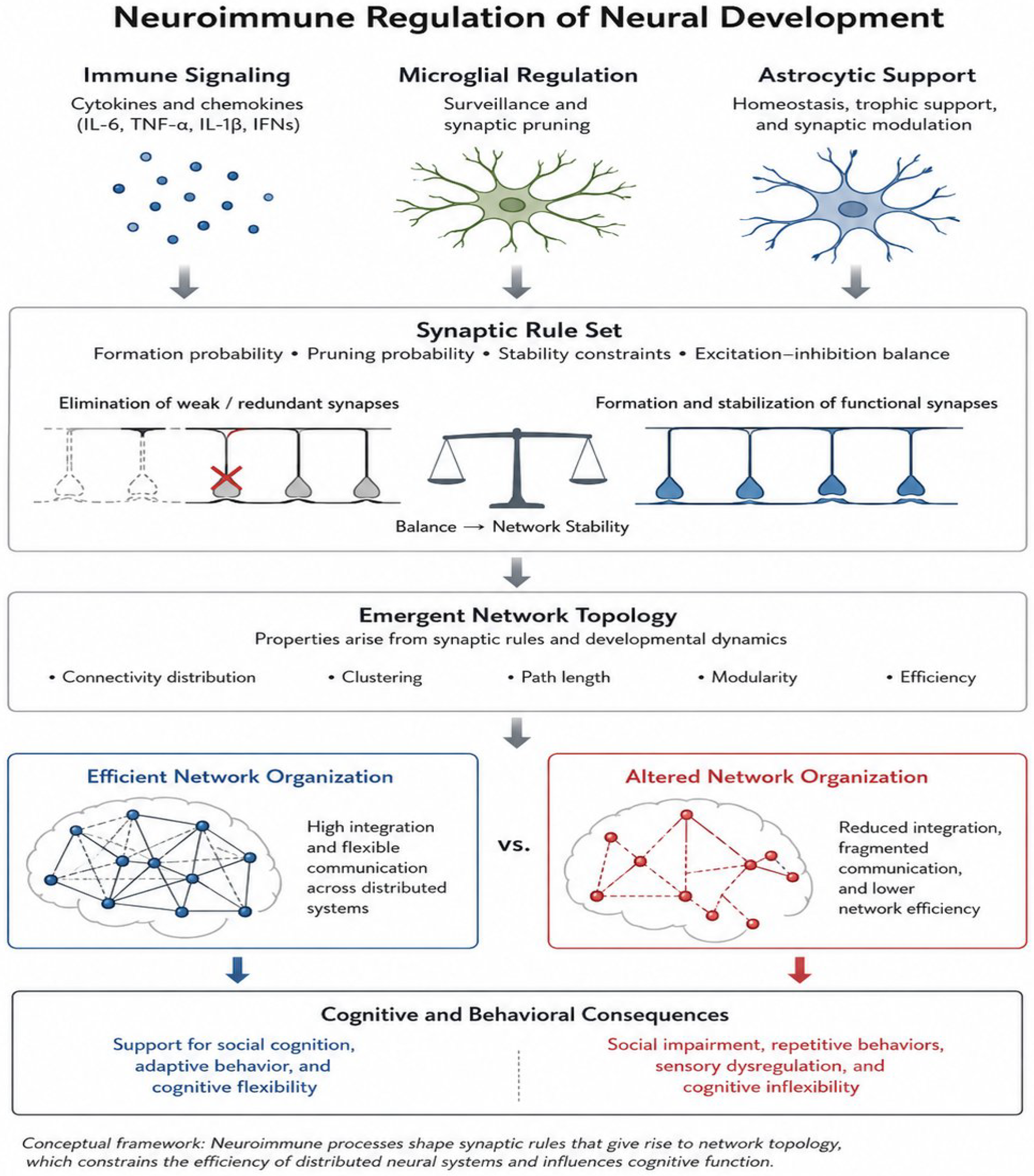
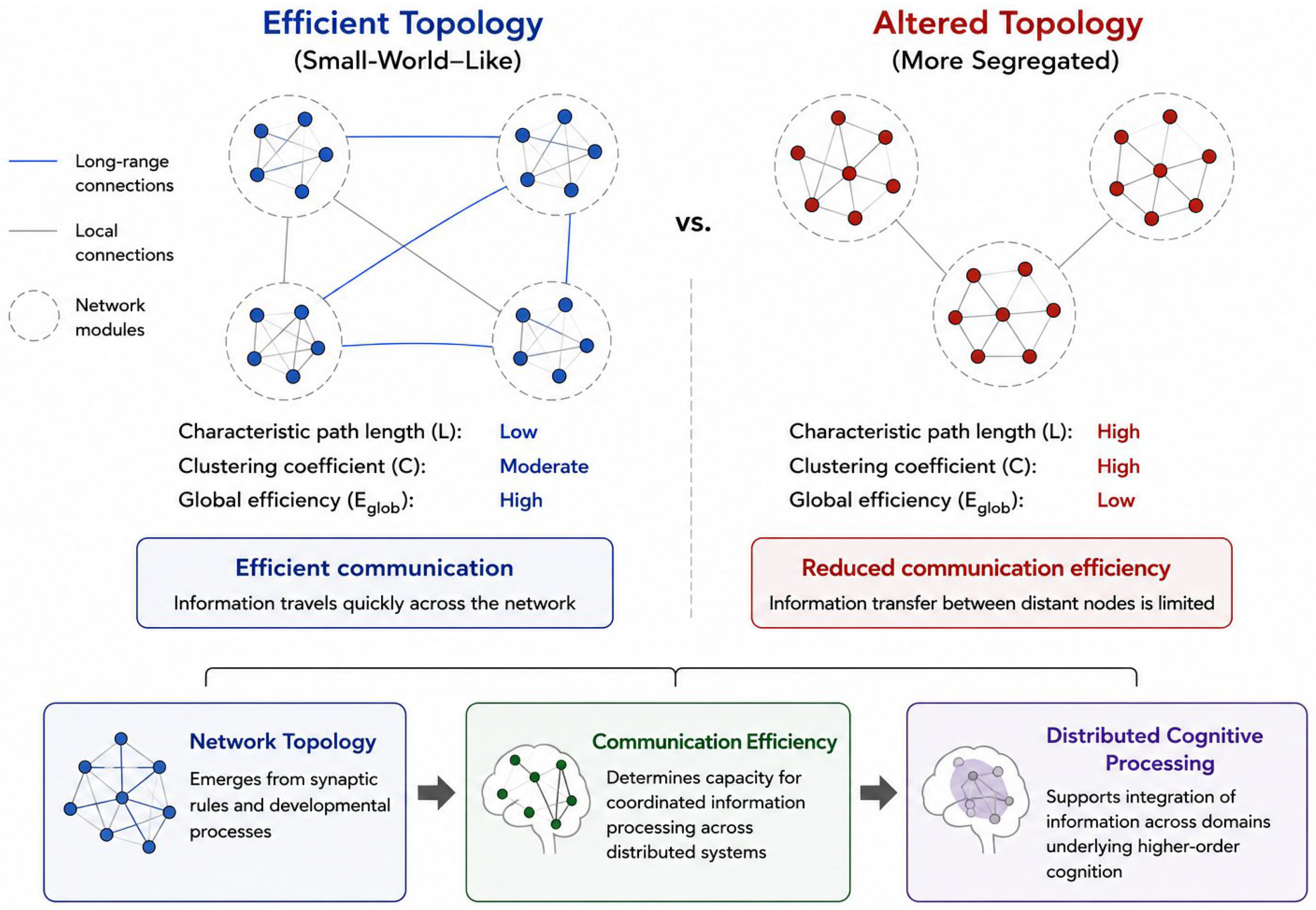
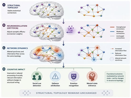
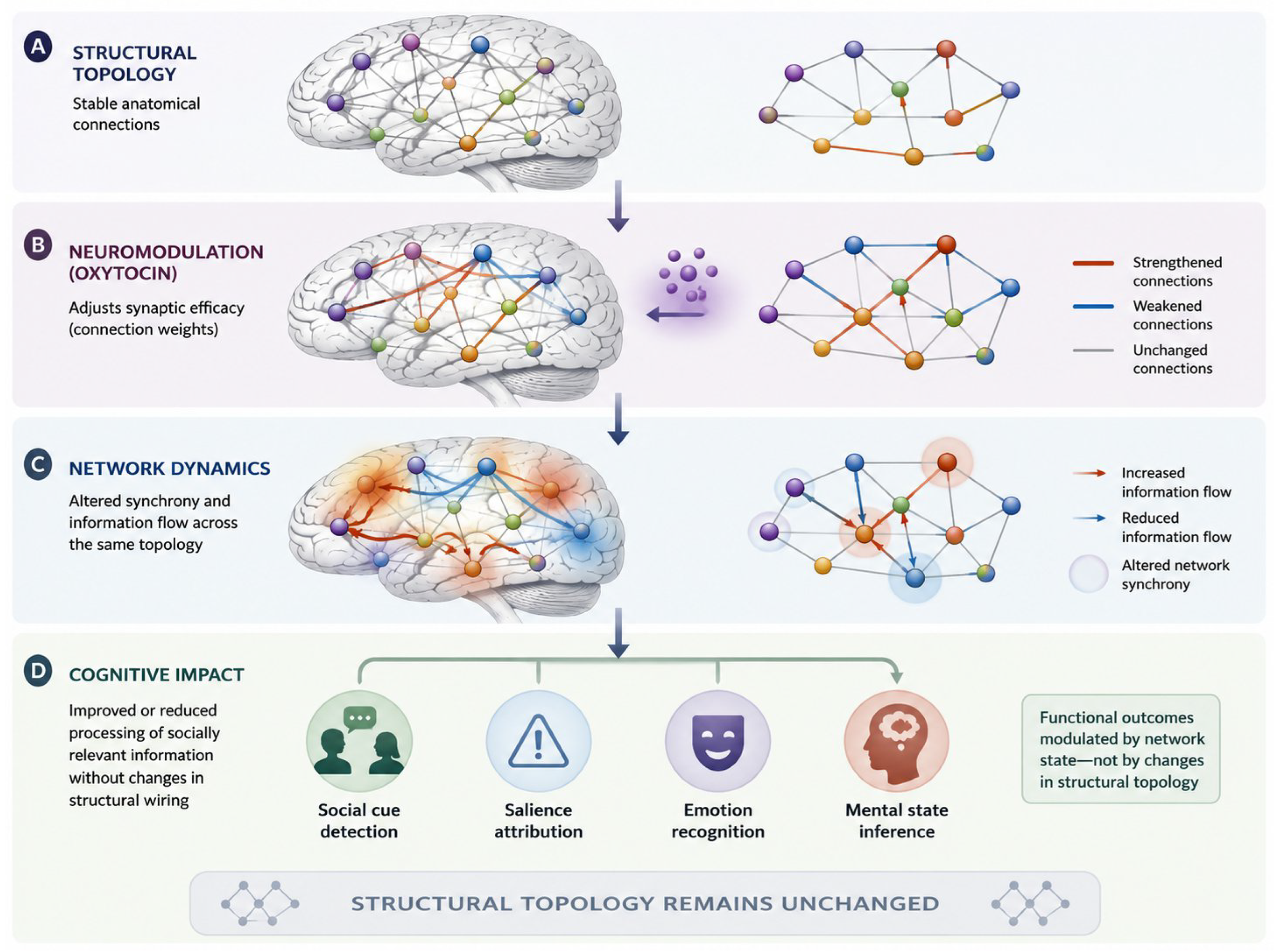
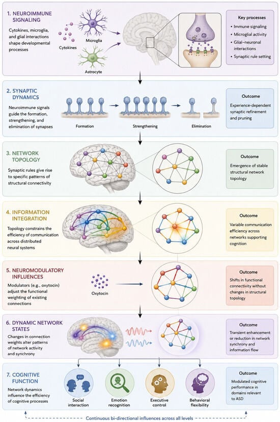
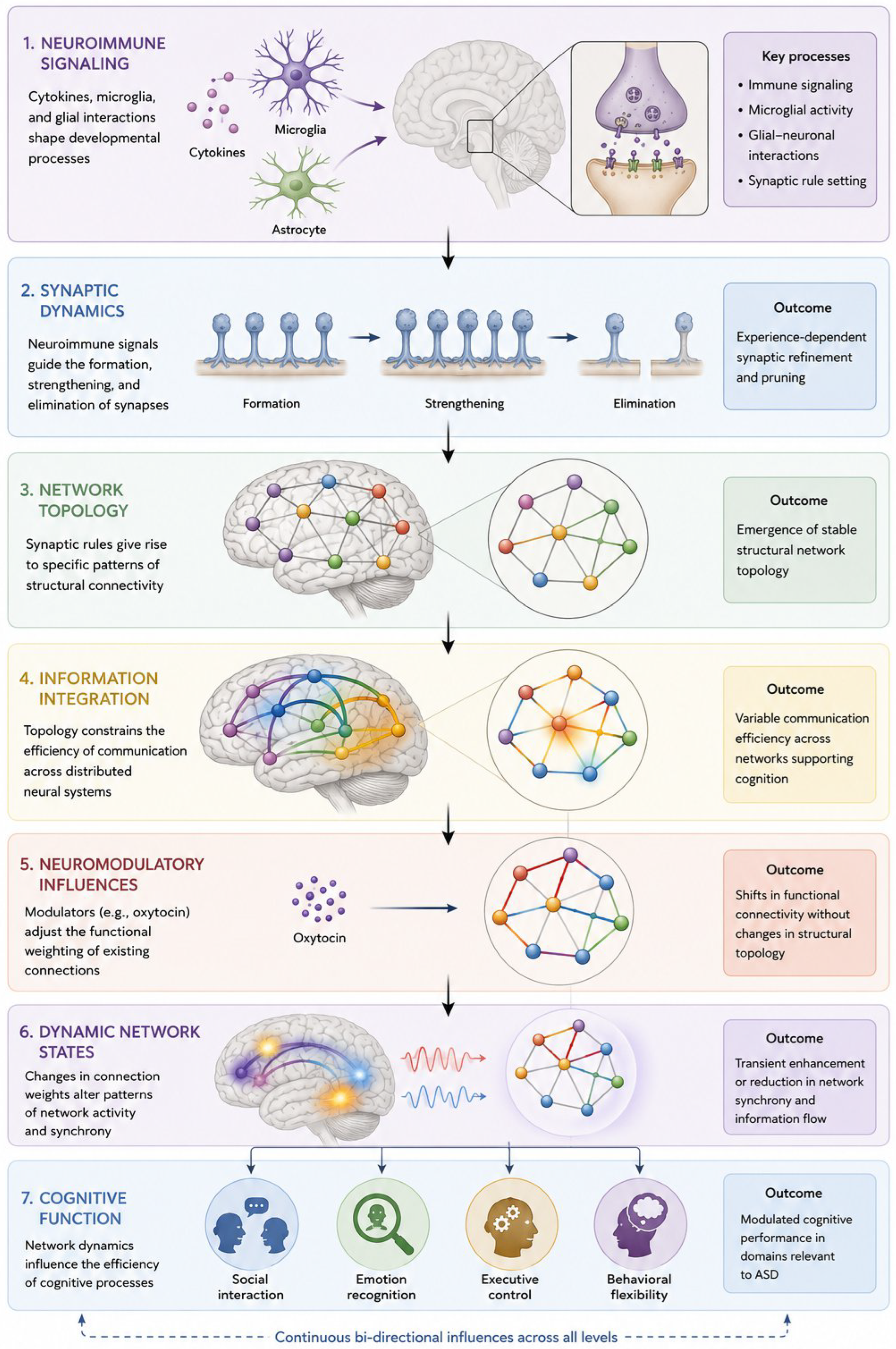
Disclaimer/Publisher’s Note: The statements, opinions and data contained in all publications are solely those of the individual author(s) and contributor(s) and not of MDPI and/or the editor(s). MDPI and/or the editor(s) disclaim responsibility for any injury to people or property resulting from any ideas, methods, instructions or products referred to in the content. |
© 2026 by the authors. Licensee MDPI, Basel, Switzerland. This article is an open access article distributed under the terms and conditions of the Creative Commons Attribution (CC BY) license.