


Abstract
Macroautophagy is a key adaptive process that enables tumour cells to withstand the cytotoxic stress induced by standard antitumour treatments, thereby contributing to therapeutic resistance. In this context, identifying regulators capable of modulating autophagy is crucial for restoring sensitivity to these treatments. The actin cytoskeleton has emerged as a regulatory axis of autophagy, governing both its dynamics and its functional control. Drugs that target actin, and in particular actin-binding proteins (ABPs), can modulate the autophagy triggered by antitumour treatments and thereby sensitise cancer cells. This review summarises recent advances in ABP-targeted strategies for modulating autophagy across different tumour contexts and examines their potential in combination therapies.
1. Introduction
Despite significant advances in oncology, chemotherapy resistance remains one of the most persistent obstacles in cancer treatment, contributing to therapeutic failure and poor patient survival. This resistance is multifactorial, arising from genetic, epigenetic and microenvironmental adaptations [1]. Among the compensatory survival mechanisms underlying this phenomenon, autophagy has been identified as a key mediator, enabling tumour cells to withstand the cytotoxic stress induced by antineoplastic treatments.
Autophagy appears to have a dual role in cancer that depends on tumour stage [2,3]. During the early stages of tumourigenesis, autophagy acts as a tumour-suppressive mechanism by removing damaged organelles, limiting oxidative stress, maintaining genomic stability and inducing apoptosis in abnormal cells, thereby preventing disease progression [4,5]. However, in advanced tumours, autophagy leads to the release of intracellular degradation products that serve as nutrients for adjacent malignant cells, which then rapidly divide and drive disease progression [6]. In addition, autophagy is activated in response to stimuli that affect the cellular microenvironment, such as starvation, hypoxia or oxidative stress. As a result, cells adapt to the new conditions [7]. Therefore, adaptive autophagy activation can allow subsets of tumour cells to survive chemotherapy or targeted therapies, acting as a reversible resistance mechanism [8,9]. This activation usually involves AMPK pathway activation, mTORC1 inhibition and stress-sensitive signalling cascades that enhance the autophagic machinery [6,10]. In this context, autophagy inhibition may sensitise tumours to therapy. However, in other contexts, autophagy blockade can exacerbate cellular dysfunction and trigger alternative forms of cell death, including non-apoptotic pathways [11]. Consequently, the decision to inhibit or exploit autophagy therapeutically depends on tumour stage, cellular context, the nature of the therapeutic stress and its interaction with other pathways such as apoptosis, metabolism and senescence.
In this complex landscape, the actin cytoskeleton has emerged as a key regulator of multiple stages of autophagy, offering a novel and potentially tumour-specific avenue to modulate this pathway therapeutically. This review highlights the role of actin-binding proteins (ABPs) in the autophagic process and explores how inhibitors targeting the actin–autophagy axis represent a compelling adjuvant strategy for the treatment of several tumour types.
2. Molecular Mechanisms and Pharmacological Modulation of Autophagy
2.1. Molecular Mechanisms of Autophagy
Autophagy is a complex, dynamic and conserved catabolic process that maintains cellular homeostasis and survival under stress conditions (starvation, hypoxia, oxidative stress and extracellular stimuli) by recycling macromolecules and organelles and removing damaged components [12,13,14,15]. Three main forms of autophagy have been described in mammalian cells—microautophagy, macroautophagy and chaperone-mediated autophagy—each involving distinct mechanisms for cargo delivery to the lysosome. The present review focuses on macroautophagy, hereafter referred to as autophagy, whose functional unit is the autophagosome, which sequesters cytoplasmic material and delivers it to lysosomes for degradation. Although the molecular machinery involved is extensive, autophagy can be conceptualised as a sequence of coordinated stages [8,16,17]. This process begins in a phosphatidylinositol 3-phosphate (PI3P)-rich structure associated with the endoplasmic reticulum known as the omegasome [18,19]. A double-membrane compartment called the phagophore then forms; as it expands, it curves to generate a spherical autophagosome that encloses the cargo. The autophagosome fuses its outer membrane with the lysosome, giving rise to the autolysosome, in which lysosomal enzymes degrade the cargo [20]. The resulting components are released into the cytoplasm, and the lysosomes are recycled to maintain cellular homeostasis [17,21,22].
Autophagy is closely linked to cellular signalling pathways, including the mechanistic target of rapamycin complex 1 (mTORC1), which negatively regulates the process [23,24]. Under normal conditions, mTORC1 is active and phosphorylates the Unc-51-like autophagy-activating kinase (ULK) complex, the primary autophagy-initiating factor, thereby suppressing its activity [25]. Under stress conditions, mTORC1 becomes inactive and dissociates from the ULK complex, which in turn phosphorylates and recruits class III phosphatidylinositol 3-kinase complex I (PI3KC3-C1)—comprising VPS34, ATG14 and Beclin-1, among other proteins—to the phagophore assembly site. The ULK complex phosphorylates Beclin-1, which interacts with VPS34 to promote PI3P synthesis [17,26,27,28,29,30]. This drives omegasome formation on the endoplasmic reticulum and PI3KC3-C1 recruitment [31], which then directs the omegasome towards the phagophore membrane via the effectors WIPI2 and DFCP1 [32]. Two ubiquitin-like conjugation systems coordinate phagophore expansion: the ATG12–ATG5–ATG16L1 system, recruited via the ATG16L1–WIPI2 interaction, dissociates upon autophagosome completion [33,34]; and the ATG8/LC3 system, whereby LC3-I conjugates with phosphatidylethanolamine to form LC3-II, which drives membrane extension [14,35]. Upon autophagosome completion, LC3-II is cleaved and degraded [36]. LC3-II is therefore widely used as a marker of autophagosome formation [14,36,37], though its accumulation does not necessarily reflect increased degradative activity; assessment therefore requires measurement of autophagic flux [38].
The autophagy receptor p62/SQSTM1 recognises and aggregates cargo, anchoring it to the phagophore through interaction with LC3 [39,40]. As LC3 interacts with p62 and autophagy inhibition elevates p62 levels, this protein is widely used as a marker of impaired or blocked autophagy [41,42].
Numerous cellular components are incorporated into the autophagosome membrane, delivered by ATG9-containing vesicles derived from cytoplasmic membranes [43,44]. After autophagosome formation, the vesicle translocates to the perinuclear region, where it fuses with the lysosome via SNARE proteins, RAB GTPases and ATG proteins to form the autolysosome [17,45,46,47]. LAMP1 and LAMP2 are lysosomal membrane proteins that maintain structural integrity and participate in autophagosome–lysosome fusion; they are routinely used as lysosomal markers [48,49,50]. The autophagic process concludes with cargo degradation, release of the resulting components into the cytoplasm and lysosomal recovery (Figure 1).
2.2. Pharmacological Modulation of Autophagy in Cancer
This mechanistic understanding of the autophagic pathway has enabled the identification of multiple pharmacological targets. Several compounds acting at distinct steps of the autophagic cascade have been evaluated in the oncological setting, either as standalone agents or in combination with standard chemotherapy.
To date, several compounds, mostly synthetic, are known to inhibit autophagy, including bafilomycin A1, 3-methyladenine (3-MA), chloroquine, hydroxychloroquine, spautin-1, Lys05 and PHY34. Whereas 3-MA and spautin-1 inhibit autophagy at early stages by blocking or degrading the PI3KC3 complex, the remaining compounds block late-stage autophagy by preventing autophagosome–lysosome fusion and/or lysosomal acidification [51,52,53,54]. Of these, only chloroquine and hydroxychloroquine are FDA-approved drugs currently evaluated in cancer clinical trials. Numerous studies have shown that autophagy-deficient cells are more sensitive to anticancer treatments [55,56]. Combining standard therapies with these autophagy inhibitors, therefore, represents a potential adjuvant strategy for cancer treatment [9,55,56].
Receptor tyrosine kinases (RTKs) regulate tumour cell growth and metastasis by activating signalling pathways such as PI3K/AKT/mTOR, which suppresses autophagy [57,58]. More than 40 RTK inhibitors have received FDA approval for oncological use, either as monotherapy or in combination with chemotherapy or immunotherapy [59,60]. However, their efficacy is limited by adverse effects and by resistance arising from RTK mutations or activation of compensatory signalling pathways, including autophagy [61,62].
Metformin, a widely used antidiabetic agent, has been shown to inhibit the proliferation of oral squamous cell carcinoma (OSCC) by promoting apoptosis and G1-phase cell-cycle arrest [63,64]. This drug can also induce protective autophagy in OSCC cells and, when combined with hydroxychloroquine, synergistically suppresses their growth in vivo without adverse effects [64]. These findings suggest that metformin may enhance the efficacy of antitumour treatments across several tumour types [64,65,66,67,68,69].
Microtubules are filaments composed of β-tubulin subunits that drive autophagosome movement towards lysosomes, participating in autophagosome–lysosome fusion and late autophagosome formation [70,71,72]. Vinca alkaloids and taxanes are plant-derived compounds with antitumour properties that regulate autophagy through the microtubule cytoskeleton [73,74]. Vinca alkaloids, including vinblastine and vincristine, bind microtubules and promote their depolymerisation [75]. Vincristine induces cytoprotective autophagy in neuroblastoma and oesophageal cancer cells, whereas vinblastine promotes accumulation of autophagic vacuoles and blocks autophagic flux in tumour cells by preventing autophagosome–lysosome fusion [73,76,77,78,79]. Paclitaxel, an antimicrotubule taxane from the Pacific yew, binds β-tubulin and stabilises non-functional microtubules, disrupting cell division and inducing tumour cell death [80,81]. Its effect on autophagy is cell-type dependent: in breast cancer cells, low concentrations inhibit autophagy via VPS34 inhibition [82]; in ovarian cancer cells, paclitaxel promotes cytoprotective autophagic flux [83,84]. Docetaxel induces non-protective autophagy in prostate cancer cells [73,85]. Azithromycin, a macrolide antibiotic with minimal toxicity, binds β-tubulin and inhibits autophagy in lung cancer cells by reducing lysosomal migration along microtubules, resulting in accumulation of enlarged autolysosomes containing undigested remnants [86].
The actin cytoskeleton is a dynamic network comprising different actin structures and actin-binding proteins (ABPs) that participates in key cellular functions such as intra- and intercellular movement and muscle contraction [87,88]. In addition, the actin cytoskeleton plays an important role in several stages of autophagy, including phagophore nucleation, membrane expansion, autophagosome trafficking and autophagosome–lysosome fusion [55,89,90,91,92]. Aguilera and colleagues observed that depolymerisation of actin filaments with latrunculin B and cytochalasin B inhibits autophagosome formation under starvation conditions, whereas stabilisation with jasplakinolide does not affect autophagy under these conditions [92]. Several studies have shown that the actin cytoskeleton is implicated in different cancer types, as its depolymerisation exerts an antitumour effect and many ABPs are dysregulated [93,94,95,96,97,98,99]. Compound 6K, the most effective of a series of 2-hydroxy-N-(arylalkyl)benzamide derivatives, disrupts the actin cytoskeleton in human melanoma cells, leading to LC3-II accumulation and blockade of autophagosome–lysosome fusion, and potentiates the antitumour activity of sorafenib [100]. Autophagy has also been implicated in tumour microenvironment mechanotransduction: greater extracellular matrix stiffness is associated with increased autophagy through the Rho/ROCK pathway in breast cancer cells [101].
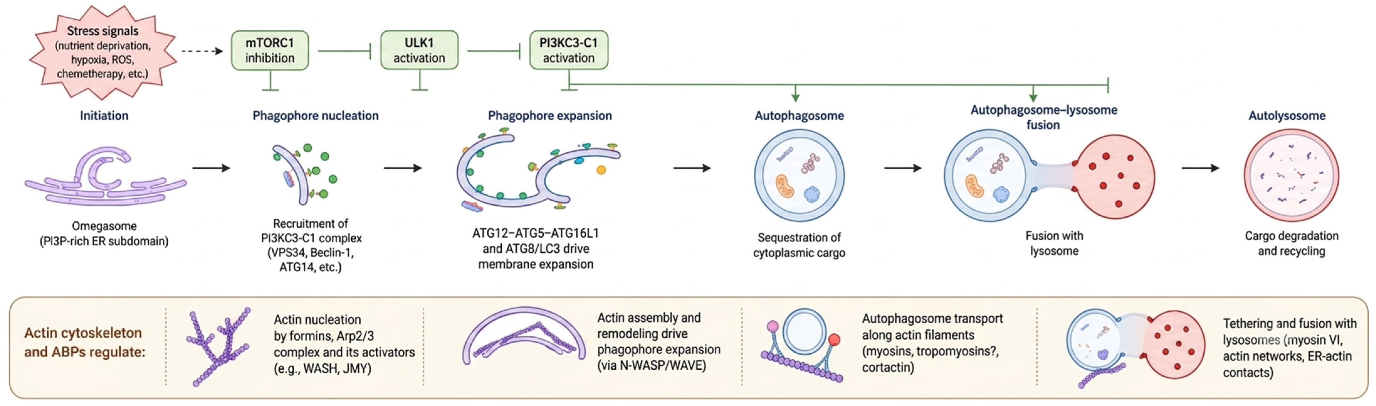
In summary, the autophagic process is a highly coordinated, multi-step pathway involving membrane remodelling, cargo sequestration and lysosomal degradation. Converging evidence highlights the actin cytoskeleton as a key regulator of several stages of this process, including phagophore nucleation, membrane expansion, vesicular trafficking and autophagosome–lysosome fusion (Figure 1).
Figure 1.
Overview of the autophagy process and points of regulation by the actin cytoskeleton. Autophagy is initiated by stress signals such as nutrient deprivation, hypoxia, reactive oxygen species (ROS) or chemotherapy, which inhibit mTORC1 and promote ULK1 activation. This leads to the recruitment of the PI3KC3-C1 complex to endoplasmic reticulum-associated omegasomes, triggering phagophore nucleation. Subsequent membrane expansion is driven by two ubiquitin-like conjugation systems (ATG12–ATG5–ATG16L1 and ATG8/LC3), resulting in autophagosome formation. The mature autophagosome then undergoes transport and fusion with lysosomes to form the autolysosome, where cargo degradation and recycling occur. The actin cytoskeleton and its associated actin-binding proteins (ABPs) regulate multiple stages of this process, including phagophore nucleation, membrane remodelling, vesicular trafficking and autophagosome–lysosome fusion.
3. Crosstalk Between Autophagy, Actin Cytoskeleton and Cancer
The actin cytoskeleton and its associated regulatory proteins modulate autophagy at multiple stages, from phagophore nucleation to autophagosome–lysosome fusion, thereby influencing tumour progression and the cellular response to antineoplastic treatments. The following subsections describe the key molecular players involved in this crosstalk and their pharmacological relevance.
3.1. Dynamics of Actin Polymerisation
Actin is a small protein (42 kDa) but highly abundant in the cell, accounting for 10% or more of total protein content. It is believed that the actin gene derives from the common ancestor that gave rise to the various forms of life on Earth and may even share its origin with the glycolytic enzyme hexokinase [102,103]. In humans, there are six actin-encoding genes expressed in different tissues [88,103,104]. Numerous mutations have been described in these genes, most of them nonsense and dominant, and consequently give rise to disease [88]. This demonstrates that actin plays an essential role in cellular function.
In eukaryotic cells, actin exists in a globular monomeric form (G-actin) and as polarised filaments (filamentous or F-actin) [87]. In its monomeric form, actin consists of four subdomains that fold around a central cleft where ATP binds [103]. In addition, a Mg2+ ion and various ABPs bind to this structure. However, G-actin does not hydrolyse ATP efficiently, so it tends to polymerise, forming polarised filaments composed of two helical strands of actin subunits wound into a right-handed helix [103,105]. These filaments have a barbed end (+) where ATP-bound G-actin monomers are incorporated and a pointed end (−) where the monomers are bound to ADP [87]. The barbed end is more dynamic because it elongates 10 times faster than the pointed end [106]. After monomer addition, ATP hydrolysis occurs, favouring polymerisation. Several studies suggest colocalisation between polymerised actin and key autophagy markers. ATG14 and Beclin-1 constitutively colocalise with F-actin [92]. However, ULK1, ATG5, ATG16 and LC3 colocalise with F-actin under starvation conditions [107]. This suggests a dynamic interaction between the actin cytoskeleton and the autophagic machinery depending on nutritional conditions.
Several inhibitors interfere with actin polymerisation, such as latrunculins, cytochalasins and jasplakinolide. Latrunculins A and B are macrolides derived from the marine sponge Latrunculia magnifica. Their structures contain a 2-thiazolidone ring, a 16-membered ring in latrunculin A and a 14-membered ring in latrunculin B, and although both bind G-actin monomers and inhibit polymerisation, latrunculin A is more effective than latrunculin B [108]. Cytochalasins are fungal metabolites that inhibit actin polymerisation by capping the barbed ends of filaments, but their mechanism of action is 10–100 times weaker than that of latrunculin A [109,110]. On the other hand, jasplakinolide, a cyclic peptide isolated from the marine sponge Jaspis johnstoni, stabilises actin filaments and at high doses or after long post-treatment times, this compound also promotes polymerisation [111,112]. As noted above, depolymerisation of actin filaments with latrunculin B and cytochalasin B has been observed to inhibit autophagosome formation under starvation conditions, whereas stabilisation with jasplakinolide does not affect autophagy under these conditions [92].
Profilin is a small protein (~13–14 kDa) that regulates actin polymerisation by binding ATP-bound monomeric actin and is involved in organ development, wound healing and immune functions [113,114,115]. Importantly, most cancers express lower amounts of profilin, and, therefore, focal adhesion is reduced and tumour malignancy is increased [98,116]. Overexpression of this protein in MDA-MB-231 triple-negative breast cancer cells has been shown to inhibit tumour initiation and growth [117]. Recently, Saurav and Kumar showed that trans retinoic acid (ATRA), the active form of vitamin A essential for neuronal and embryonic growth and development, induces profilin expression through transcriptional activation in MDA-MB-231 cells. As a result, AMPK is increased, mTOR is inhibited, ULK1 is upregulated and autophagy is induced but subsequently impaired at the degradation step, as evidenced by the accumulation of LC3-II, p62 and LAMP2, leading to cell apoptosis [118]. In addition, combining ATRA with other anticancer drugs, such as paclitaxel and vinblastine, enhances cell death and tumour regression [118,119]. However, Wang and colleagues found that profilin overexpression downregulates CD138, a membrane proteoglycan present in plasma cells and multiple myeloma cells, through autophagy in multiple myeloma cells. As a result, resistance to bortezomib, a proteasome inhibitor and the main treatment for this type of cancer, is induced [120,121,122].
3.2. Role of Actin Nucleating Factors
3.2.1. Arp2/3 Complex
It should be noted that de novo polymerisation of actin filaments is inefficient. Therefore, cells use other proteins that promote actin filament nucleation, such as the Arp2/3 complex, which promotes assembly of branched actin filaments. This complex is made up of seven polypeptides (Arp2, Arp3 and five additional subunits, ArpC1-5) stably bound together [123]. When the Arp2 and Arp3 subunits bind ATP, a conformational change occurs in the complex that brings them closer together. As a result, the complex binds to an existing actin filament through ArpC2 and ArpC4 subunits and initiates assembly of a new filament at a 70° angle [124,125,126,127,128,129]. One inhibitor of the Arp2/3 complex is the synthetic compound CK-666, which acts as a classic allosteric inhibitor by stabilising the inactive state of the complex [130]. It has been shown that, in some cancers, such as colorectal or breast cancer, the Arp2/3 complex is overexpressed, generally in advanced tumours with poor prognosis, and these tumour cells are sensitive to Arp2/3 inhibition [131,132,133]. Li and colleagues showed that the inhibitor CK-666 promotes autophagy and activation of the exosome pathway in human oesophageal cancer cells (KYSE-30) [134]. They also confirmed that high doses of this compound (100 μM) decrease the formation of tunnelling nanotubes (TNTs), actin-based membrane channels connecting two or more spatially separated cells and transporting vesicles, proteins and mitochondria [134,135,136]. These results, together with the demonstrated good clinical applicability and its synergy with other antitumour agents such as docetaxel, suggest that CK-666 could be used as an adjuvant therapy because it reduces transfer between cancer cells and normal cells, promotes autophagy and improves sensitivity to other drugs [134,137].
However, the Arp2/3 complex must be stimulated to increase its activity, and stimulation by actin nucleation-promoting factors (NPFs) is the best characterised mechanism [123,138]. In mammals, there are two types of NPFs, known as class I and class II. The class I NPF group is the largest. It includes N-WASP, the WASP family verprolin-homologous factors (WAVE), WASP- and WAVE-associated factors (WASH), WASP homologous proteins associated with actin, membranes and microtubules (WHAMM) and junction-mediating and regulatory proteins (JMY), which differ in their N-terminal sequence, which determines the mode of regulation [139].
The N-WASP group is the best characterised and comprises the WASP protein, which is expressed only in haematopoietic cells and whose mutation causes Wiskott-Aldrich syndrome, and N-WASP, which is expressed in most cell types and is autoinhibited by the intramolecular interaction between a GTPase-binding domain (GBD) located at the N-terminus and the acidic region of a WCA domain located at the C-terminus [140,141]. Binding of the GTPase Cdc42 and phosphatidylinositol (4,5)-bisphosphate (PIP2) activates the protein by relieving autoinhibition [123]. It has been shown that deletion of WASP reduces LC3-II expression and colocalisation of F-actin with both LC3 and p62 in cells infected with Escherichia coli. It has also been shown that deletion of WASP inhibits autophagosome formation. Therefore, WASP participates in autophagosome–lysosome fusion in an actin-dependent manner [17,142,143]. Biber and colleagues described that the small molecule compound SMC no. 13 is capable of specifically binding to Wasp, recruited and activated by Cdc42 at the plasma membrane, inducing its proteasomal degradation through ubiquitination in an actin-independent manner and inhibiting proliferation and migration in T-cell acute lymphoblastic leukaemia cells without affecting normal cells. In addition, administration of this compound reduced tumour volume and growth rate, increased the number of apoptotic cells and prolonged survival in mice without causing physical abnormalities or toxicity [144].
The WAVE group is composed of three isoforms (WAVE1-3), which lack an autoinhibitory mechanism and are activated by the GTPase Rac1, which cooperates with phosphatidylinositol (3,4,5)-trisphosphate (PIP3) and several protein kinases in this activation [145,146,147]. In autophagy, the WAVE group promotes autophagosome formation, since its silencing decreases both the LC3-II/LC3-I ratio and the number of autophagosomes [148].
WASH is a complex composed of five subunits (WASHC1-5) that is essential for nucleating F-actin from Arp2/3 in endosomes and lysosomes, and although its activation is carried out by Rho GTPases in Drosophila, this interaction is not sufficient to activate human WASH in vitro [149,150,151,152]. However, PIP3 and phosphatidylinositol 4-phosphate (PIP4) appear to be critical for recruitment and activation of WASH in endosomal microdomains [153,154]. In autophagy, WASH appears to have a dual role because, on the one hand, it is required for ATG9 trafficking and, on the other, it can negatively regulate the process through Beclin-1 and proteins of the PI3KC3-C1 complex [155,156,157].
JMY, initially discovered as a p53 regulatory cofactor, promotes actin polymerisation by binding to monomeric actin or through activation of the Arp2/3 complex [158,159,160]. Although the cytoskeletal functions of this protein vary depending on the cell line, it promotes neuronal dendritic branching and associates with actin in a Ca2+/calmodulin signalling-dependent manner [161,162]. Regarding its role in autophagy, a positive feedback loop has been described between this protein and LC3. JMY interacts with LC3 for autophagosome formation and, in turn, LC3 recruits JMY to the autophagosome membrane, promoting actin polymerisation through Arp2/3 [55,163,164].
The WHAMM subgroup lacks autoinhibition, and it is unclear whether it may be part of a complex [165]. It is localised to the cis-Golgi and to tubulovesicular membranes that move in an actin and microtubule-dependent manner, so it influences Golgi morphology and anterograde trafficking [123,165]. In autophagy, this subgroup participates in autophagosome formation since it colocalises with LC3 and p62 and in lysosomal recovery since it colocalises with and promotes autolysosome tubulation, although these functions are cell-type dependent [89,166]. In addition, WHAMM has been observed to negatively regulate autolysosome degradation under starvation conditions [166].
Cortactin is a class II NPF that cooperates with N-WASP to enhance activation of the Arp2/3 complex and stabilises branched actin. Its activity is regulated by post-translational modifications [167,168,169]. Thus, phosphorylation increases its activity, promoting autophagosome–lysosome fusion, whereas acetylation inhibits its activity and, therefore, autolysosome formation [170,171]. It has been shown that cortactin is a protein required for the stabilisation and maturation of invadopodia, membrane protrusions rich in actin filaments that are characteristic of tumour cells and that have the ability to degrade the underlying matrix, thereby favouring cancer cell invasion and metastasis [172]. Phosphorylation of this protein permits the assembly of the actin necessary for their formation [172]. Zhou and colleagues observed that in cancer cells, p62 may act as a negative regulator of invadopodium formation, since under starvation autophagy is able to induce activation of the ERK1/2 kinases via p62, thus promoting cortactin phosphorylation and invadopodium formation [172].
3.2.2. Formins
Formins are proteins that nucleate unbranched actin filaments and are characterised by having two formin homology domains, FH1 and FH2, in their structure. The FH2 domain binds to the barbed end of actin filaments, preventing other proteins from terminating elongation and competing with formin inhibitors, thereby determining filament length [173,174]. The FH1 domain is proline-rich and binds profilin, promoting filament elongation [175]. In mammals there are approximately 15 formins grouped into seven subtypes according to the FH2 domain [176]. The formin FHOD1, also known as Formin Homology 2 Domain Containing 1, is a protein belonging to the Diaphanous-related formin family (DRFs), characterised by the presence of a GTP-binding domain (GBD), FH1 and FH2 domains, a coiled-coil region and a Diaphanous-like autoinhibitory domain. Traditionally, FHOD1 has been associated with regulation of actin cytoskeleton dynamics, participating in processes such as cell migration, adhesion and contractility. However, recent reviews have shown that FHOD1 also plays a relevant role in regulating the tumour microenvironment, acting not only on tumour cells but also on different immune components, including T cells, B cells, cancer-associated fibroblasts (CAFs) and various cytokines [177]. In this context, FHOD1 appears to be closely related to the epithelial–mesenchymal transition (EMT), a biological process by which epithelial cells lose their polarity and intercellular adhesion to acquire mesenchymal properties associated with increased migratory, invasive and metastatic capacity. EMT is also an important mechanism of therapeutic resistance and immune evasion. During this process, multiple tumours increase expression of PD-L1, an immunoinhibitory protein that interacts with programmed death 1 PD-1 on T lymphocytes and blocks their cytotoxic activity. According to Yang and colleagues, FHOD1 favours PD-L1 overexpression during EMT, thereby contributing to reinforcing the immunosuppressive state of the tumour microenvironment. This phenomenon is particularly relevant in CAFs, where PD-L1 appears to be functionally related to autophagy mechanisms. In particular, it has been described that CAFs derived from the stromal cell-derived factor CXCL12 can promote immune evasion in bladder cancer by inhibiting p62-mediated autophagic degradation of PD-L1, thus favouring its stabilisation and accumulation. Consequently, PD-L1 remains active for longer, enhancing tumour immune evasion. In this way, this mechanism contributes to an immunosuppressive tumour microenvironment, promoting both tumour progression and therapeutic resistance [177].
On the other hand, among the Diaphanous-related formins, DIAPH1 stands out; it is a RhoA effector formin that participates in linear actin polymerisation and in the regulation of vesicular trafficking and signalling routes such as AKT and β-catenin. DIAPH1 has been implicated in modulation of PD-L1 expression and stability, thereby connecting cytoskeletal dynamics with tumour immunosuppression. In this context, lovastatin, a naturally occurring statin commonly used to reduce blood cholesterol and triglyceride levels, acts as an inhibitor of HMG-CoA reductase, reducing mevalonate synthesis and the availability of isoprenoid intermediates required for prenylation of small GTPases such as RhoA. This inhibition compromises DIAPH1 activity, decreasing formin activity and reducing PD-L1 expression, which limits tumour immune evasion. Moreover, in glioblastoma, lovastatin has been associated with the induction of autophagy initiation but with the alteration of final autophagic flux, suggesting that this drug can initiate the autophagic process without completing lysosomal degradation. Therefore, lovastatin positions itself as a modulator of the formin–autophagy axis via PD-L1 [177].
3.3. Actin-Binding Proteins (ABPs)
ABPs are a group of proteins that regulate the availability of actin monomers for polymerisation, elongation or filament severing [87,88,103]. Among these proteins, myosins, tropomyosins, capping protein, cofilin, α-actinin and filamin A deserve special mention, as they contribute to context-dependent regulation of autophagic flux and tumour progression, as described in the following subsections.
3.3.1. Myosins
Myosins are motor proteins that use the energy released from ATP hydrolysis to generate force and movement along actin filaments, thereby driving their sliding and generating tension [178,179,180]. In humans, about 40 myosin genes have been described, encoding more than 20 distinct myosins that are grouped into 13 classes denoted by Roman numerals [181]. All myosins have three structural domains called the motor, neck and tail. The motor domain, located at the N-terminus, contains actin-binding sites and an ATPase domain. In the central region is the neck domain, which is composed of 1–6 IQ motifs. This domain binds light chains (regulatory or essential) and regulates the motor function of the protein. Finally, the tail domain, located at the C-terminus, is involved in protein dimerisation and contains cargo-binding domains that interact with adaptor proteins or lipids, determining protein function [179,182]. Of all myosins, only myosin I, II and VI have so far been implicated in autophagy [91]. Myosins can also cooperate with other components of the actin cytoskeleton, promoting cancer progression by regulating migration, invasion and intracellular transport [180]. Myosin IC has been described as having a dual role in membrane formation and vesicle transport during autophagy through intracellular cholesterol metabolism and actin cytoskeleton remodelling during autophagosome–lysosome fusion [183,184]. Cepharanthine, a bisbenzylisoquinoline alkaloid, has anticancer effects by modulating autophagy [185,186]. Some studies show that this drug can induce autophagy and apoptosis in several cancers through mTOR signalling, whereas others have shown that it inhibits autophagy because of the blockade of autophagosome–lysosome fusion in lung tumour cells [186,187]. Zhang and colleagues showed that cepharanthine inhibits autophagosome–lysosome fusion by blocking Myo1C-dependent F-actin assembly on both autophagosomes and lysosomes in breast cancer cells, since in the presence of this compound coprecipitation and colocalisation of LC3 and LAMP1 with Myo1C and F-actin are reduced, as is the interaction between LC3 and LAMP1, preventing autolysosome formation [184]. In addition, other myosin I isoforms also play a role in autophagy. Thus, myosin IB favours increased intracellular calcium levels, affecting autophagosome–lysosome fusion and may be involved in angiogenesis in colon cancer cells [32,188]. Myosin ID may regulate p62 dynamics through the actin cytoskeleton in selective autophagy, and myosin IG may participate in vesicle transport without significantly affecting autophagic flux [189,190].
Myosin II binds to polymerised actin, forming a contractile actomyosin system that regulates ATG9A vesicle trafficking from the trans-Golgi to the phagophore, facilitating autophagosome formation [191]. Blebbistatin, a 1-phenyl-2-pyrrolidinone derivative, is a specific inhibitor of skeletal and non-muscle myosin II that binds to its motor domain, stabilising it in the ADP/Pi-bound transition state and preventing force generation [192,193]. Jia and colleagues report that treatment with blebbistatin can suppress cell blebbing, protrusions on the plasma membrane of tumour cells that alter organisation and cell movement dependent on non-muscle myosin II contractility, thereby promoting tumour invasion and metastasis [194,195,196]. In this way, tumour proliferation is reduced, although no clinical studies on its applicability are available to date [197]. Moreover, contact inhibition, a property of normal cells, is lost in tumour cells. As a result, cell proliferation becomes uncontrolled, and the capacity of the cells to invade tissues increases. The mechanism underlying contact inhibition has further been linked to the HIPPO pathway [195]. Pavel and colleagues have shown that the high cell density resulting from loss of contact inhibition activates the HIPPO pathway and alters the localisation of HIPPO-dependent transcription factors, which reduces expression of myosin II and other actin genes. As a result, stress fibres are lost, trafficking of key autophagy proteins is defective, and autophagosome biogenesis is reduced [198].
Myosin VI is involved in anchoring and tension generation in the final stages of fusion of secretory vesicles with the plasma membrane [199]. In addition, unlike the other myosins, myosin VI moves toward the negative end of actin filaments, allowing retrograde cargo trafficking that ensures spatiotemporal coordination of autophagic flux [180]. Through interaction with the autophagy protein Tom1, this myosin promotes autophagosome maturation and fusion with lysosomes [180,200,201]. The compound 2,4,6-triiodophenol, also known as Bobel-24 or AM-24, is a non-steroidal anti-inflammatory drug that binds to myosin VI and can reduce its motor activity and impair its function during the final stages of secretory pathways [202,203]. However, no results on its therapeutic potential have been reported to date [180,203].
3.3.2. Tropomyosins
Tropomyosins are dimeric proteins composed of two coiled helices that bind to actin filaments, reinforcing and stabilising them. In addition, they regulate the motor function of myosins and inhibit actin polymerisation and branching formed by the Arp2/3 complex [204,205]. In mammals, there are four tropomyosin genes encoding 40 isoforms, which are classified according to molecular weight [97]. In recent years, tropomyosin has been considered an essential target in cancer treatment, since the expression profile of its isoforms in tumour cells differs from that in normal cells. In tumour cells, high molecular weight isoforms (Tpm2.1, Tpm1.4, Tpm1.6 and Tpm1.7) are expressed at lower levels than low molecular weight isoforms (especially Tpm3.1 and Tpm4.2). Three anti-tropomyosin drugs (TR-100, ATM1001 and ATM3507) have now been described that are able to inhibit the function of the Tpm3.1 isoform [97,206]. Compound TR-100 binds to the C-terminus of Tpm3.1, destabilising the actin filaments containing this isoform without affecting actin-tropomyosin binding [207]. As a result, tumour cell growth is delayed, and tumour cell death occurs. The combination of TR-100 with anti-microtubule agents such as vincristine or paclitaxel leads to complete cytotoxicity in some cell lines [208]. ATM3507 is a modification of TR-100 that improves its interaction with the C-terminus of Tpm3.1 [208]. Although survival in murine models treated with ATM3507 was not greater than in those treated with TR-100, the ATM3507-vincristine combination showed similar efficacy to the TR-100-vincristine combination. Both combinations increase the accumulation of condensed chromatin and abnormally organised mitotic spindles, leading to cell-cycle arrest at the G2 phase [208].
3.3.3. Capping Protein
Capping protein, or CapZ, is a heterodimer formed by α and β subunits that negatively regulates actin polymerisation by blocking incorporation of monomers at the barbed end of filaments [209]. As a result, CapZ participates in autophagy and cancer [210,211]. In autophagy, PI3P in the omegasome binds CapZ and displaces it from actin filaments, allowing polymerisation [107]. In addition, CapZ also participates in the final stage of autophagy by negatively regulating p62 degradation [55]. In the cancer context, the β subunit of this protein is used as a marker for breast cancer classification and staging, since it is overexpressed in this tumour type [210]. Vacuolin-1 is a compound that affects endosome or lysosome fusion and blocks endosome maturation, compromising lysosome biogenesis and function [210,212]. In recent years, this compound has been shown to be a potent anti-metastatic agent that may be used in combination with antitumour treatments. CapZ is a target of vacuolin-1 in endosomal trafficking and migration, and the compound can suppress metastasis of cancer cells in the murine 4T1 breast cancer line [210].
3.3.4. Cofilin
Cofilin, also called actin-depolymerising factor (ADF), is a small protein with high affinity for ADP-actin subunits, and its main function is to sever actin filaments. In mammals there are three known cofilin isoforms: non-muscle cofilin 1, muscle cofilin 2 and ADF or destrin [213,214,215]. It is an ABP whose activity is negatively regulated by phosphorylation. When dephosphorylated, it can bind actin [215]. Numerous studies show that cofilin overexpression is a marker of tumour progression and malignancy regardless of tumour type [215,216]. Cofilin promotes tumour cell motility by regulating actin cytoskeleton reorganisation, lamellipodium formation, dissolution of cell–cell adhesion and the adhesion-dependent migration mechanism, thus participating in cancer metastasis [215]. Moreover, Wu and colleagues have shown that reduction in cofilin expression in vitro significantly decreases tumour cell growth because cells arrest in the G1 phase of the cell cycle, lamellipodium formation is interrupted and invasion and metastasis are reduced [216]. Some studies have suggested that activated cofilin induces mitochondria-mediated cytotoxic apoptosis in tumour cells. Once dephosphorylated, cofilin translocates to the outer mitochondrial membrane, favouring mitochondrial fission and promoting cytochrome c release. Consequently, cell apoptosis occurs, affecting tumour expansion [217]. Cucurbitacin E is a member of the tetracyclic triterpenoid family extracted from cucurbitaceous plants that can induce autophagy by negatively regulating mTORC1 activity [218]. Nakashima and colleagues described that this compound is able to inhibit cofilin phosphorylation and, therefore, increases the proportion of G-actin in human U937 leukaemia cells [219]. Therefore, although cucurbitacin E modulates both autophagy and the actin cytoskeleton, this compound would not, on balance, represent an optimal antitumour strategy. Although cucurbitacin E promotes autophagy through mTORC1 inhibition—a potentially beneficial effect in the context of chemosensitisation—it simultaneously inhibits cofilin phosphorylation, increasing the pool of dephosphorylated cofilin. Since dephosphorylated cofilin is clinically associated with a worse disease prognosis and greater tumour aggressiveness, its pharmacological accumulation would be expected to counteract the intended antitumour effect. In other words, the pro-autophagic benefit of cucurbitacin E may be offset by the pro-tumorigenic consequences of sustained cofilin dephosphorylation, making the net therapeutic outcome uncertain [220]. Future therapies aim to maintain this protein in a phosphorylated state.
3.3.5. α-Actinin
α-Actinin (ACTN) is an ABP of the spectrin superfamily that is essential for anchoring and organising actin filaments. In mammals there are four isoforms of this protein, and all are characterised by being antiparallel dimers that crosslink actin filaments at their N- and C-termini. ACTN1 and ACTN4 are non-muscle isoforms that participate in crosslinking actin in stress fibres and anchoring it to focal adhesions, while ACTN2 and ACTN3 are striated muscle-specific isoforms that crosslink actin filaments to stabilise the sarcomere [221,222]. It has been shown that the ACTN4 isoform is involved in cancer metastasis through its participation in cell motility [223]. In addition, this protein has been identified as a positive marker for platinum-based adjuvant therapy in some cancers, such as lung cancer, and its high expression is associated with poor tumour prognosis [222]. ACTN4 has a dynamic localisation between the nucleus and the cytoplasm. In breast cancer cells, cytoplasmic localisation is linked to an infiltrative phenotype, and nuclear localisation suggests that ACTN4 could act as a co-activator of nuclear receptors to promote cell proliferation. Some inhibitors of ACTN4 are known, such as ellagic acid, a natural polyphenol present in fruits and vegetables, which inhibits ACTN4 and, therefore, growth and metastasis of breast cancer [224]. Although a direct mechanistic link between α-actinin and autophagy has not yet been established in the literature, ACTN4 has a key role as an actin-organising protein in the cancer context.
3.3.6. Filamin A
Filamin A, also known as ABP-280 or Filamin-1, is a protein that crosslinks actin filaments and acts as an intracellular signalling scaffold by binding to diverse signalling components [225]. At its amino terminus it possesses an actin-binding domain, and it is in the cytoplasm, where it can promote tumour progression, and in the nucleus, where it is downregulated in some cancer types. Functionally, this protein participates in morphogenesis and cell migration, as well as in cell–cell contacts and adherens junctions during development of blood vessels, the heart and the brain. Wang and colleagues observed that Filamin A expression is decreased in bladder cancer cells and that its overexpression can promote autophagy by increasing LC3-II expression and decreasing p62 expression [226]. Moreover, in this cancer type, overexpression of this protein arrests cells in G0/G1 and increases apoptosis, suppressing tumour proliferation, migration and invasion. However, these findings contrast with those observed in breast cancer and melanoma cells, where Filamin A overexpression promotes tumour malignancy. Therefore, in the case of bladder cancer, promoting Filamin A overexpression would be a strategy to achieve cytoprotective autophagy via actin. The synthetic drug PTI-125, or Simufilam, has been used in clinical trials for Alzheimer’s disease because it is able to restore altered forms of Filamin A, inducing a slower rate of neurodegeneration, among other effects. However, in that context the drug did not pass phase III clinical trials despite being safe and well tolerated by patients.
3.4. Rho GTPases as Master Regulators: Their Role in Mediating Actin–Autophagy Crosstalk
Rho family GTPases regulate actin cytoskeleton organisation, among other functions [227,228]. They are a subfamily of Ras GTPases with low molecular weight that act as molecular switches by oscillating between an active state (GTP-bound) and an inactive state (GDP-bound), promoted respectively by guanine nucleotide exchange factors (GEFs) and GTPase-activating proteins (GAPs) [228,229,230]. In their active state, they can bind effector proteins and trigger a signalling response. Although this family comprises 22 members in humans, the most studied are Cdc42, RhoA and Rac1, which regulate cell polarity and motility through the actin cytoskeleton, membrane trafficking and cell adhesion [227,228,231,232,233]. Cdc42 binds to the NPFs Wasp and N-Wasp to stimulate the Arp2/3 complex and, through formins, induces the formation of thin protrusions of the plasma membrane composed of parallel actin bundles known as filopodia, which serve to sense the extracellular environment [139,232,234,235]. RhoA, together with the kinase ROCK and the formin mDia1, gives rise to stress fibres formed by contractile actin-myosin bundles that are essential for focal adhesion maturation and the generation of traction forces [236,237]. Recently, Huang and colleagues described a positive feedback loop between RhoA and Arp2/3-complex-branched actin [100]. In addition, RhoA participates in assembly and constriction of the contractile actomyosin ring (CAR) during cytokinesis [231,238,239]. RhoA binds to and activates the formin mDia1 to assemble CAR filaments and, through the serine/threonine kinases ROCK and Citron, which phosphorylate the myosin II light chain, activates myosin II, which binds to actin filaments and drives CAR constriction [240,241]. Rac1 activates Arp2/3 through several NPFs, such as WAVE, to nucleate branched actin filaments that form lamellipodia, plasma membrane protrusions that facilitate cell spreading and adhesion [232,242,243,244]. In addition, both Rac1 and RhoA promote PIP2 formation at the plasma membrane by associating with PIP2 kinase isoforms, thereby altering actin dynamics in these regions [245].
Rac1 and Cdc42 are overexpressed in some cancers such as rhabdomyosarcoma and breast cancer [99,246]. Cdc42 regulates cell-cycle progression, migration, invasion and the autophagic response to the tumour microenvironment [99,247]. Rac1 plays an important role in cell adhesion, migration, cell-cycle progression and transcriptional regulation [248]. The Rac1 inhibitor EHT1864 has shown promising results in overcoming resistance in oestrogen receptor-positive (ER+) breast cancer. This compound specifically blocks the interaction of Rac1 with GTP, so the protein remains in its inactive form, affecting ER stability and transcriptional activity [248,249]. Another Rac1 inhibitor, NSC23766, inhibits Rac1 binding and activation via GEFs without affecting the binding or activation of other GTPases; its use in rhabdomyosarcoma cells increases expression of the autophagy markers LC3 and Beclin-1. As a result, this inhibitor promotes autophagy. It has also been observed to promote apoptosis in rhabdomyosarcoma cells [99,250]. These same results were observed with the Cdc42 GTPase inhibitor ZCL278, and together they suggest that both drugs could be used to treat this type of cancer [99].
It has been observed that, under starvation conditions, inhibition of RhoA-C by Clostridium botulinum toxin C3 decreases LC3-II levels in HeLa human cervical cancer cells [92]. Therefore, RhoA positively regulates autophagy under starvation conditions. In addition, Rac1 has been shown to inhibit autophagy in a cell type-dependent manner under starvation conditions, whereas Cdc42 has no effect on autophagy [92,251].
In A549 lung cancer cells, RhoA, together with p62 and LC3-II, is overexpressed, thereby inhibiting autophagy [252]. Belaid and colleagues described that autophagy negatively regulates RhoA activation during cytokinesis. Once active, RhoA is ubiquitinated and directed by the autophagy receptor p62/SQSTM1 to autophagosomes for degradation. Therefore, the actin cytoskeleton is altered, and tumour progression is reduced [252,253]. In addition, this autophagy-mediated regulation of RhoA is specific, as it has not been observed for RhoA activators or other GTPases such as Rac [253]. For this reason, stimulation of autophagy in this context is considered a possible antitumour treatment.
Recently, it has been reported that CCG-1423, an aromatic compound capable of inhibiting RhoA-driven transcription, reduces cytokine release through inhibition of secretory autophagy in bone marrow myelofibrosis models [254,255]. Shang and colleagues identified the aromatic compound G04, or Rhosin, as able to inhibit RhoA activation and suppress breast cancer cell proliferation and invasion in a dose-dependent manner without affecting other GTPases. In addition, Rhosin induces apoptosis without affecting cell-cycle progression or showing cytotoxicity [256]. Both CCG-1423 and Rhosin are inhibitors that non-specifically affect the three RhoA isoforms (RhoA, RhoB and RhoC) [255,256]. The aromatic compound O1 decreases the amount of active RhoA, alters actin cytoskeleton organisation and cell morphology, and increases mitotic duration with defective spindles in HeLa cells. In addition, it cannot be ruled out that this inhibitor has other targets besides RhoA, such as the GTPase Cdc42 involved in kinetochore-microtubule attachment during mitosis [257,258].
The molecular components of the actin cytoskeleton, the stages of the autophagic process they regulate, and the therapeutic strategies that emerge from modulating this axis are visually integrated in Figure 2.
Figure 2.
Overview of the tripartite relationship between the actin cytoskeleton, autophagy, and chemotherapy resistance. The left panel depicts the key components of the actin cytoskeleton involved in autophagy regulation, including nucleation factors (formins and the Arp2/3 complex), actin-binding proteins (ABPs: myosins, tropomyosins, CapZ, cofilin, α-actinin and filamin A) and Rho family GTPases (RhoA, Rac1 and Cdc42). The central panel illustrates the six sequential stages of macroautophagy—from stress-induced initiation to cargo degradation and recycling—and identifies the specific stages at which actin cytoskeleton remodelling is required: NPFs and Rho GTPases drive phagophore nucleation; actin remodelling supports membrane expansion; myosins and actin networks coordinate vesicle transport; and ABPs facilitate autophagosome–lysosome fusion. The right panel presents the therapeutic implications: promoting cytoprotective autophagy for tumour suppression; inhibiting cytotoxic autophagy to sensitise cancer cells to chemotherapy; and combining actin–autophagy modulators with standard treatments to reduce chemoresistance. NPFs: nucleation-promoting factors; ABPs: actin-binding proteins.
3.5. p38 Kinase as a Stress-Responsive Regulator
p38 is a kinase that is activated in response to various stimuli and is involved in regulating migration, differentiation and cell apoptosis, among other processes [259,260,261]. In addition, this protein has been described as having a dual role in melanoma cells depending on mutation in the RAS genes since p38 inversely regulates the expression levels of factors predicting the likelihood of metastasis in this tumour type [80,262,263]. Banik and colleagues showed that low concentrations of anisomycin, a protein synthesis inhibitor, for a short period (30 min) activate p38 in highly aggressive NRAS-mutant melanomas [264,265,266]. Consequently, mTOR is transiently activated, which in turn causes autophagy defects by reducing LC3-I/LC3-II conversion and alters actin cytoskeleton dynamics [266]. In this way, they were able to reduce tumour invasion. Since p38 overexpression persistently increases mTOR phosphorylation, both anisomycin and other compounds could be considered for treatment of this type of tumour.
Given the complexity of autophagy regulation and its dual role in cancer, multiple pharmacological strategies have been developed to target different stages of the autophagic pathway. These compounds, together with their mechanisms of action and therapeutic implications, are summarised in Table 1.
Table 1.
Pharmacological modulators of autophagy in cancer: mechanisms of action and therapeutic implications.
4. In Vivo Assessment of Cytotoxicity
It is well known that manipulation of actin produces pleiotropic and cytotoxic effects because it does not distinguish between tumour cell actin and normal cell actin. Thus, compounds that affect polymerisation and/or stability of actin filaments have not passed preclinical testing [51]. Because many ABPs in cancer cells show an expression pattern different from that in normal cells, targeting them specifically could be an alternative. This review presents numerous compounds with promising results for overcoming resistance to antitumour therapies via autophagy in in vitro assays. Importantly, many of them have also shown good efficacy and low toxicity in vivo. The combination of all-trans retinoic acid and vinblastine, at doses lower than those used in breast cancer treatment, achieved a reduction in tumour size without toxic effects in mice bearing xenografts [116]. Several studies have suggested that CK-666 can be administered directly to small animals, and its combination with docetaxel is more effective than docetaxel treatment in tissue isolated from patients with urothelial carcinoma [131]. It has been verified that, in isolated peripheral blood cells, SMC no. 13 neither affects the immune system nor induces cytokine release. Moreover, in mice this compound induces neither weight loss nor toxic effects [141]. Oral and intraperitoneal administration of vacuolin-1 decreases the number and size of mammary tumours in transgenic MMTV-PyMT mice without affecting body weight or liver or kidney function [207]. Zhang and colleagues observed that, in mice treated with the Cdc42 inhibitor ZCL-278, hepatic metastasis is suppressed without altering weight or metabolic function. Wan and colleagues reported that patients with lung cancer show improved responses and prolonged survival and that administration of the compound in mice decreases tumour volume without altering body weight. Other compounds, such as blebbistatin and the Rac1 inhibitor, present certain drawbacks. The use of blebbistatin is limited by its low water solubility and its sensitivity to blue light, which causes phototoxicity and cytotoxicity. Derivatives of this compound, para-aminoblebbistatin and para-nitroblebbistatin, lack phototoxic and cytotoxic activity [269,270], so evaluating their effect both in vitro and in vivo in the cancer context is particularly interesting. Regarding the Rac1 inhibitor NSC23766, it should be mentioned that its selectivity has been questioned since it acts as an antagonist at muscarinic acetylcholine receptors and N-methyl-D-aspartate receptors in rodent neurones. Therefore, the use of other Rac1-specific drugs that reduce side effects and toxicity in the cancer and actin–autophagy axis context is necessary [271]. The remaining compounds mentioned in this review, such as cepharanthine, anisomycin or 6K, lack toxicity and adverse effect studies in the treated context.
5. Discussion
Given the dual role of autophagy in cancer and its tight regulation by the actin cytoskeleton, targeting the actin–autophagy axis has emerged as a promising therapeutic strategy. This review has focused on stress-induced autophagy, for example, in response to starvation, which is a non-selective process. However, a nutrient-independent “basal” or quality-control autophagy also exists that is responsible for the selective destruction of damaged proteins and organelles that may be harmful to the cell. Lee and Yao have described that the ubiquitin-binding deacetylase HDAC6 is not required for selective autophagy but stimulates autophagosome–lysosome fusion, being recruited to ubiquitinated autophagic substrates and promoting cortactin-dependent F-actin remodelling [171]. Moreover, selective autophagy can degrade signalling proteins such as the GTPase RhoA. In this context, autophagy promotes degradation of active, membrane-associated RhoA specifically because it is sequestered by p62 into autolysosomes. Therefore, this quality-control mechanism acts to degrade and maintain an appropriate level of active RhoA for correct completion of cytokinesis, thus constituting a tumour-suppressive mechanism [272]. Therefore, the actin cytoskeleton also participates in selective and non-selective autophagy, and vice versa.
Pharmacological modulation of actin dynamics or ABPs, either alone or in combination with autophagy inhibitors and standard chemotherapeutics, may enhance treatment efficacy and overcome drug resistance (Figure 3). According to the reviewed studies, the molecular cancer subtype most frequently associated with the actin–autophagy axis is the MDA-MB-231 triple-negative breast cancer cell line, followed by the HCT116 colon cancer and A549 lung cancer cell lines, both harbouring KRAS mutations. Also dependent on this axis are the SKOV3 ovarian cancer cell line carrying a p53 tumour suppressor gene mutation and the KYSE-30 oesophageal cancer cell line carrying a p16 gene mutation. In the reviewed studies, most ABPs—including the Arp2/3 complex, cortactin, CapZ, cofilin, α-actinin ACTN4 and the RhoA, Rac1 and Cdc42 GTPases—are overexpressed, whereas profilin expression is reduced. In the case of filamin A, its expression pattern is tumour-type dependent. Furthermore, the expression pattern of tropomyosin isoforms is altered in tumour cells, with the Tpm3.1 isoform being overexpressed. Taken together, these data suggest that ABPs could serve as biomarkers of tumour progression along the actin–autophagy axis. However, to date only cofilin has been described as a tumour-type-independent marker of tumour progression and malignancy through mitochondria-mediated apoptosis and α-actinin ACTN4 as a positive marker for platinum-based adjuvant therapy, but neither in relation to autophagy. It is important to acknowledge, however, that these candidate biomarkers are mostly derived from in vitro evidence based on established cancer cell lines. Consequently, any claim of precision oncology utility for these markers must be regarded as preliminary, and clinical validation in patient-derived samples and primary tumour cohorts remains an essential priority before translational application can be proposed.
Figure 3.
Therapeutic strategies targeting the actin–autophagy axis to overcome chemotherapy resistance. Pharmacological targeting of the actin cytoskeleton and its associated proteins represents a promising strategy to modulate autophagy in cancer. Compounds that interfere with actin dynamics or ABPs (e.g., Arp2/3 inhibitors, myosin inhibitors, or tropomyosin-targeting agents) can be combined with autophagy modulators such as chloroquine, hydroxychloroquine, metformin or trans retinoic acid (ATRA), as well as with standard chemotherapeutic agents. These combined approaches may either inhibit autophagic flux or induce dysfunctional autophagy, ultimately enhancing chemosensitivity, promoting apoptosis and reducing tumour invasion and metastasis. The therapeutic outcome is highly dependent on tumour type, genetic background and the specific stage of autophagy targeted.
The data presented in this review indicate that the actin cytoskeleton and some of its associated proteins constitute an important regulatory axis of autophagy and could be used as a possible target to combat tumour cell resistance to chemotherapeutic agents involved in autophagy. This idea is consistent with recent literature, which describes autophagy as a process with dual functions in cancer: it can act as a suppressive mechanism in early phases, preserving genomic stability and cellular homeostasis, but it can also promote tumour survival, phenotypic plasticity and therapeutic resistance once the tumour is established [272]. In this sense, autophagy should be considered not as an isolated pathway but as an adaptive response integrated with metabolism, membrane trafficking and stress signalling. In addition, actin not only participates in autophagosome biogenesis but also in membrane transport and fusion with lysosomes, and ABPs also appear to play a key role [17]. This explains why inhibition of actin or its regulators can produce seemingly opposite effects depending on cell type, the stress applied and the autophagic phase affected.
This complexity is precisely what makes the use of various therapeutic strategies so interesting. For example, blocking proteins such as Arp2/3, WASP, Rac1, Cdc42, RhoA or myosins can prevent autophagy progression or, in other contexts, push the cell towards dysfunctional autophagy that ultimately favours cell death. Considering the advances presented in this review, it is important to highlight a divergent expression pattern of ABPs in the tumour context. Whereas most of the ABPs analysed, such as the Arp2/3 complex, myosins and various Rho family GTPases, are frequently overexpressed in solid tumours, correlating with an invasive phenotype and greater resistance to chemotherapy, profilin is a critical exception. Several studies have shown that profilin is usually underexpressed in breast, pancreas and kidney tumours, where it may act as a tumour suppressor whose loss promotes cytoskeletal instability. This distinction determines the therapeutic strategy to follow: for overexpressed ABPs, the focus is on using inhibitors such as CK-666 or blebbistatin to block their tumour-promoting activity; by contrast, in the case of profilin, current research is directed towards compounds that can increase its affinity for actin and restore its original biological function, thereby seeking to reverse cellular malignancy. An interesting case to explore is tropomyosin, since although the effect of anti-tropomyosin drugs on tumour cells through autophagy has not yet been studied, it has been shown that these agents affect glucose metabolism after Tpm3.1 inhibition [97]. Studying whether these compounds activate autophagy and, if so, whether it is cytoprotective, is particularly interesting for proposing anti-tropomyosin drugs as a possible therapy for this disease.
The question is not only whether autophagy is activated or not but also where in the process interference occurs and how intense it is. Pimentel et al. emphasise that autophagy can be modulated at multiple stages but that this very biological plasticity also makes it difficult to translate preclinical findings into uniform clinical efficacy. Consequently, the proposal to exploit actin as a therapeutic target must be read with caution and focused on the specific tumour context, thereby enabling a precision oncology approach for the different cancer types. Actin participates in essential cellular processes, so indiscriminate inhibition could cause toxicity outside the tumour. Recent literature on autophagy in cancer and its microenvironment reinforces this idea: the value of autophagy lies not only in the degradation of cellular material but also in its role in tumour stem cells, antitumour immunity and adaptation to the cellular niche [272]. In this way, modulating the actin–autophagy network opens the door to the discovery of targeted therapies against specific tumours that are highly dependent on this circuit to survive stress, metastasise or maintain resistant populations.
It is particularly interesting that most of the actin or ABP inhibitors mentioned in this review contain benzyl- or phenyl-type aromatic rings. This presence is not accidental but rather reflects fundamental principles of medicinal chemistry. First, aromatic rings allow the drug to interact non-covalently with components of cancer cells (such as proteins or DNA) through π-stacking interactions, which help the drug bind firmly to its biological target. In addition, their hydrophobic nature facilitates passage through cell membranes and precise fitting into hydrophobic pockets in protein receptors, significantly improving binding affinity. From a structural point of view, these systems provide chemical stability and facilitate the synthetic modifications needed to enhance the antimetastatic effect by inhibiting tumour migration and invasion. Likewise, their flat and rigid shape is crucial for molecular recognition, allowing the drug to be recognised by specific proteins involved in the metastasis process [273]. However, this same aromaticity can penalise solubility, permeability or metabolic stability when too many planar rings accumulate. For this reason, researchers usually try to limit the number of such aromatic rings (generally to fewer than three in the molecule) to improve their developability and pharmacological properties. In current optimisation processes, heterocycles or saturated bioisosteres are often explored as phenyl substitutes to balance biological affinity with an optimal pharmacokinetic profile [273]. In short, the abundance of benzyl rings in these inhibitors probably reflects their pharmacological utility but also a historical inertia in precision medicinal chemistry that is now being offset by more three-dimensional and less lipophilic designs.
Finally, it is important to mention that several compounds originally described for non-oncological diseases have been identified as having an intrinsic ability to inhibit cytoskeletal dynamics or modulate autophagy, allowing their potential use in cancer treatment. Notable examples include metformin, a first-line antidiabetic that regulates mTOR signalling; azithromycin, a macrolide antibiotic that has proven to be an effective inhibitor of autophagic flux in tumour cells through its binding to the microtubule cytoskeleton; and all-trans retinoic acid, the active form of vitamin A, as an inducer of profilin expression for the treatment of certain cancers. The use of these drugs, with already known safety profiles, as demonstrated by their low toxicity in in vivo studies, opens new possibilities for combination therapies aimed at overcoming chemoresistance by acting on the actin–autophagy axis.
6. Conclusions and Future Perspectives
The most promising future perspectives involve defining functional biomarkers that allow patients to be stratified according to their dependence on autophagy and the actin cytoskeleton. It will not be enough to measure LC3-II or p62 in isolation; these markers will need to be combined with autophagic flux reporters, actin organisation, mTOR/AMPK signalling, lysosomal status and, where possible, features of the tumour microenvironment. Current literature already suggests that autophagy participates in resistance, tumour stem cell survival and interaction with antitumour immunity, so biomarkers will need to be equally multifactorial. To date, however, no ABP expression profile has been validated as a biomarker of cancer in the actin–autophagy axis. Another priority line will be the design of more selective inhibitors with a better pharmacological profile. Since aromatic rings continue to be frequent, optimisation should aim to reduce the number of rings so that efficacy is maintained without compromising solubility, metabolism and drug distribution. This approach is not only consistent with contemporary medicinal chemistry but can also expand the therapeutic window of compounds that modulate actin or its regulators [273].
On the other hand, validation in models closer to the clinic will be essential. The use of patient-derived organoids, 3D cultures, stromal and immune co-cultures, and in vivo pharmacodynamic studies will help determine when manipulation of actin and autophagy is truly beneficial. Clinical trials with hydroxychloroquine show that the strategy is feasible but also that its efficacy will depend on the specific combination, the biological context and patient selection. Overall, the most realistic horizon is not a single universal therapy, but precision medicine based on the autophagic and cytoskeletal vulnerability of each tumour.
In short, this review highlights that, although the role of various antitumour drugs in cell-cycle arrest and apoptosis induction has been extensively studied, there is still a significant gap in knowledge regarding their real impact on the autophagic machinery. The main conclusion of this work is that, although the cytotoxic efficacy of many chemotherapeutic agents is known, their role in modulating autophagy has not yet been fully elucidated, and, crucially, neither has whether this cellular response acts as a cytoprotective mechanism that favours chemoresistance or as a harmful process that leads to tumour death. On this basis, it is proposed that integrating more specific therapeutic strategies is key to clinical success. The use of drugs that act directly on the actin–autophagy axis, such as vinca alkaloids or compounds that specifically inhibit or stimulate the actin cytoskeleton, represents a promising avenue as adjuvant therapies. Combining these specific modulators with the antitumour drugs already described would not only enhance treatment efficacy but also tilt the balance of autophagy towards the definitive elimination of tumour cells, overcoming the barriers of metabolic adaptation and drug resistance.
Author Contributions
Conceptualization, M.R.-L. and F.P.-R.; investigation, M.R.-L., A.J.P.-D. and F.P.-R.; data curation, M.R.-L.; writing—original draft preparation, M.R.-L.; writing—review and editing, M.R.-L. and F.P.-R.; visualisation, M.R.-L.; supervision, F.P.-R. All authors have read and agreed to the published version of the manuscript.
Funding
This research received no external funding.
Data Availability Statement
This research did not create new data.
Acknowledgments
During the preparation of this manuscript, the authors used Canva AI 2.0 for assistance in drafting and structuring the manuscript figures. The authors have reviewed and edited the output and take full responsibility for the content of this publication.
Conflicts of Interest
The authors declare no conflicts of interest. The funders had no role in the design of the study; in the collection, analyses, or interpretation of data; in the writing of the manuscript; or in the decision to publish the results.
References
- Lei, H.-T.; Wang, J.-H.; Yang, H.-J.; Wu, H.-J.; Nian, F.-H.; Jin, F.-M.; Yang, J.; Tian, X.-M.; Wang, H.-D. LncRNA-mediated cell autophagy: An emerging field in bone destruction in rheumatoid arthritis. Biomed. Pharmacother. 2023, 168, 115716. [Google Scholar] [CrossRef] [Scilit] [PubMed]
- Singh, S.S.; Vats, S.; Chia, A.Y.-Q.; Tan, T.Z.; Deng, S.; Ong, M.S.; Arfuso, F.; Yap, C.T.; Goh, B.C.; Sethi, G.; et al. Dual role of autophagy in hallmarks of cancer. Oncogene 2018, 37, 1142–1158. [Google Scholar] [CrossRef] [Scilit] [PubMed]
- Yu, Y.S.; Kim, I.S.; Baek, S.H. Decoding the dual role of autophagy in cancer through transcriptional and epigenetic regulation. FEBS Lett. 2025, 599, 2237–2249. [Google Scholar] [CrossRef] [Scilit] [PubMed]
- Djavaheri-Mergny, M.; Giuriato, S.; Tschan, M.P.; Humbert, M. Therapeutic Modulation of Autophagy in Leukaemia and Lymphoma. Cells 2019, 8, 103. [Google Scholar] [CrossRef] [Scilit] [PubMed]
- Kimmelman, A.C. The dynamic nature of autophagy in cancer. Genes. Dev. 2011, 25, 1999–2010. [Google Scholar] [CrossRef] [Scilit] [PubMed]
- Galluzzi, L.; Pietrocola, F.; Bravo-San Pedro, J.M.; Amaravadi, R.K.; Baehrecke, E.H.; Cecconi, F.; Codogno, P.; Debnath, J.; Gewirtz, D.A.; Karantza, V.; et al. Autophagy in malignant transformation and cancer progression. EMBO J. 2015, 34, 856–880. [Google Scholar] [CrossRef] [Scilit] [PubMed]
- Kroemer, G.; Mariño, G.; Levine, B. Autophagy and the integrated stress response. Mol. Cell 2010, 40, 280–293. [Google Scholar] [CrossRef] [Scilit] [PubMed]
- Li, Y.-J.; Lei, Y.-H.; Yao, N.; Wang, C.-R.; Hu, N.; Ye, W.-C.; Zhang, D.-M.; Chen, Z.-S. Autophagy and multidrug resistance in cancer. Chin. J. Cancer 2017, 36, 52. [Google Scholar] [CrossRef] [Scilit] [PubMed]
- Mele, L.; del Vecchio, V.; Liccardo, D.; Prisco, C.; Schwerdtfeger, M.; Robinson, N.; Desiderio, V.; Tirino, V.; Papaccio, G.; La Noce, M. The role of autophagy in resistance to targeted therapies. Cancer Treat. Rev. 2020, 88, 102043. [Google Scholar] [CrossRef] [Scilit] [PubMed]
- Yang, Z.J.; Chee, C.E.; Huang, S.; Sinicrope, F.A. The role of autophagy in cancer: Therapeutic implications. Mol. Cancer Ther. 2011, 10, 1533–1541. [Google Scholar] [CrossRef] [Scilit] [PubMed]
- Levy, J.M.M.; Towers, C.G.; Thorburn, A. Targeting autophagy in cancer. Nat. Rev. Cancer 2017, 17, 528–542. [Google Scholar] [CrossRef] [Scilit] [PubMed]
- Dikic, I.; Elazar, Z. Mechanism and medical implications of mammalian autophagy. Nat. Rev. Mol. Cell Biol. 2018, 19, 349–364. [Google Scholar] [CrossRef] [Scilit] [PubMed]
- Rabinowitz, J.D.; White, E. Autophagy and metabolism. Science 2010, 330, 1344–1348. [Google Scholar] [CrossRef] [Scilit] [PubMed]
- Ichimura, Y.; Kirisako, T.; Takao, T.; Satomi, Y.; Shimonishi, Y.; Ishihara, N.; Mizushima, N.; Tanida, I.; Kominami, E.; Ohsumi, M.; et al. A ubiquitin-like system mediates protein lipidation. Nature 2000, 408, 488–492. [Google Scholar] [CrossRef] [Scilit] [PubMed]
- Mizushima, N. Autophagy: Process and function. Genes. Dev. 2007, 21, 2861–2873. [Google Scholar] [CrossRef] [Scilit] [PubMed]
- Chen, Y.; Yu, L. Recent progress in autophagic lysosome reformation. Traffic 2017, 18, 358–361. [Google Scholar] [CrossRef] [Scilit] [PubMed]
- Dong, Y.; Quan, C. NPFs-mediated actin cytoskeleton: A new viewpoint on autophagy regulation. Cell Commun. Signal 2024, 22, 111. [Google Scholar] [CrossRef] [Scilit] [PubMed]
- Hayashi-Nishino, M.; Fujita, N.; Noda, T.; Yamaguchi, A.; Yoshimori, T.; Yamamoto, A. A subdomain of the endoplasmic reticulum forms a cradle for autophagosome formation. Nat. Cell Biol. 2009, 11, 1433–1437. [Google Scholar] [CrossRef] [Scilit] [PubMed]
- Ylä-Anttila, P.; Vihinen, H.; Jokitalo, E.; Eskelinen, E.-L. 3D tomography reveals connections between the phagophore and endoplasmic reticulum. Autophagy 2009, 5, 1180–1185. [Google Scholar] [CrossRef] [Scilit] [PubMed]
- Parzych, K.R.; Klionsky, D.J. An overview of autophagy: Morphology, mechanism, and regulation. Antioxid. Redox Signal 2014, 20, 460–473. [Google Scholar] [CrossRef] [Scilit] [PubMed]
- Yorimitsu, T.; Klionsky, D.J. Autophagy: Molecular machinery for self-eating. Cell Death Differ. 2005, 12, 1542–1552. [Google Scholar] [CrossRef] [Scilit] [PubMed]
- Nanayakkara, R.; Gurung, R.; Rodgers, S.J.; Eramo, M.J.; Ramm, G.; Mitchell, C.A.; McGrath, M.J. Autophagic lysosome reformation in health and disease. Autophagy 2023, 19, 1378–1395. [Google Scholar] [CrossRef] [Scilit] [PubMed]
- Galluzzi, L.; Pietrocola, F.; Levine, B.; Kroemer, G. Metabolic control of autophagy. Cell 2014, 159, 1263–1276. [Google Scholar] [CrossRef] [Scilit] [PubMed]
- Kim, J.; Kundu, M.; Viollet, B.; Guan, K.-L. AMPK and mTOR regulate autophagy through direct phosphorylation of Ulk1. Nat. Cell Biol. 2011, 13, 132–141. [Google Scholar] [CrossRef] [Scilit] [PubMed]
- Xiang, H.; Zhang, J.; Lin, C.; Zhang, L.; Liu, B.; Ouyang, L. Targeting autophagy-related protein kinases for potential therapeutic purpose. Acta Pharm. Sin. B 2020, 10, 569–581. [Google Scholar] [CrossRef] [Scilit] [PubMed]
- Liu, Y.; Yang, Q.; Chen, S.; Li, Z.; Fu, L. Targeting VPS34 in autophagy: An update on pharmacological small-molecule compounds. Eur. J. Med. Chem. 2023, 256, 115467. [Google Scholar] [CrossRef] [Scilit] [PubMed]
- Axe, E.L.; Walker, S.A.; Manifava, M.; Chandra, P.; Roderick, H.L.; Habermann, A.; Griffiths, G.; Ktistakis, N.T. Autophagosome formation from membrane compartments enriched in phosphatidylinositol 3-phosphate and dynamically connected to the endoplasmic reticulum. J. Cell Biol. 2008, 182, 685–701. [Google Scholar] [CrossRef] [Scilit] [PubMed]
- Park, J.-M.; Seo, M.; Jung, C.H.; Grunwald, D.; Stone, M.; Otto, N.M.; Toso, E.; Ahn, Y.; Kyba, M.; Griffin, T.J.; et al. ULK1 phosphorylates Ser30 of BECN1 in association with ATG14 to stimulate autophagy induction. Autophagy 2018, 14, 584–597. [Google Scholar] [CrossRef] [Scilit] [PubMed]
- Russell, R.C.; Tian, Y.; Yuan, H.; Park, H.W.; Chang, Y.-Y.; Kim, J.; Kim, H.; Neufeld, T.P.; Dillin, A.; Guan, K.-L. ULK1 induces autophagy by phosphorylating Beclin-1 and activating VPS34 lipid kinase. Nat. Cell Biol. 2013, 15, 741–750. [Google Scholar] [CrossRef] [Scilit] [PubMed]
- Panwar, V.; Singh, A.; Bhatt, M.; Tonk, R.K.; Azizov, S.; Raza, A.S.; Sengupta, S.; Kumar, D.; Garg, M. Multifaceted role of mTOR (mammalian target of rapamycin) signaling pathway in human health and disease. Signal Transduct. Target. Ther. 2023, 8, 375. [Google Scholar] [CrossRef] [Scilit] [PubMed]
- Joshi, A.S.; Zhang, H.; Prinz, W.A. Organelle biogenesis in the endoplasmic reticulum. Nat. Cell Biol. 2017, 19, 876–882. [Google Scholar] [CrossRef] [Scilit] [PubMed]
- Yu, Y.; Ren, Y.; Li, Z.; Li, Y.; Li, Y.; Zhang, Y.; Gui, R.; Cui, Y.; Qian, L.; Xiong, Y. Myo1b Promotes Premature Endothelial Senescence and Dysfunction via Suppressing Autophagy: Implications for Vascular Aging. Oxid. Med. Cell Longev. 2023, 2023, 4654083. [Google Scholar] [CrossRef] [Scilit] [PubMed]
- Dooley, H.C.; Razi, M.; Polson, H.E.; Girardin, S.E.; Wilson, M.I.; Tooze, S.A. WIPI2 links LC3 conjugation with PI3P, autophagosome formation, and pathogen clearance by recruiting Atg12-5-16L1. Mol. Cell 2014, 55, 238–252. [Google Scholar] [CrossRef] [Scilit] [PubMed]
- Mizushima, N.; Kuma, A.; Kobayashi, Y.; Yamamoto, A.; Matsubae, M.; Takao, T.; Natsume, T.; Ohsumi, Y.; Yoshimori, T. Mouse Apg16L, a novel WD-repeat protein, targets to the autophagic isolation membrane with the Apg12-Apg5 conjugate. J. Cell Sci. 2003, 116, 1679–1688. [Google Scholar] [CrossRef] [Scilit] [PubMed]
- Fujita, N.; Itoh, T.; Omori, H.; Fukuda, M.; Noda, T.; Yoshimori, T. The Atg16L complex specifies the site of LC3 lipidation for membrane biogenesis in autophagy. Mol. Biol. Cell 2008, 19, 2092–2100. [Google Scholar] [CrossRef] [Scilit] [PubMed]
- Kabeya, Y.; Mizushima, N.; Ueno, T.; Yamamoto, A.; Kirisako, T.; Noda, T.; Kominami, E.; Ohsumi, Y.; Yoshimori, T. LC3, a mammalian homologue of yeast Apg8p, is localized in autophagosome membranes after processing. EMBO J. 2000, 19, 5720–5728. [Google Scholar] [CrossRef] [Scilit] [PubMed]
- Ichimura, Y.; Komatsu, M. Selective degradation of p62 by autophagy. Semin. Immunopathol. 2010, 32, 431–436. [Google Scholar] [CrossRef] [Scilit] [PubMed]
- Klionsky, D.J.; Abdel-Aziz, A.K.; Abdelfatah, S.; Abdellatif, M.; Abdoli, A.; Abel, S.; Abeliovich, H.; Abildgaard, M.H.; Abudu, Y.P.; Acevedo-Arozena, A.; et al. Guidelines for the use and interpretation of assays for monitoring autophagy (4th edition). Autophagy 2021, 17, 1–382. [Google Scholar] [CrossRef] [Scilit] [PubMed]
- Pankiv, S.; Clausen, T.H.; Lamark, T.; Brech, A.; Bruun, J.-A.; Outzen, H.; Øvervatn, A.; Bjørkøy, G.; Johansen, T. p62/SQSTM1 binds directly to Atg8/LC3 to facilitate degradation of ubiquitinated protein aggregates by autophagy. J. Biol. Chem. 2007, 282, 24131–24145. [Google Scholar] [CrossRef] [Scilit] [PubMed]
- Kim, Y.-J.; Lee, H.-H.; Jung, T.Y.; Jeong, Y.-H.; Lim, K.-H.; Han, J.M. The Roles of SQSTM1/p62 in Selective Autophagy and Oncogenic Signaling. Int. J. Mol. Sci. 2026, 27, 2342. [Google Scholar] [CrossRef] [Scilit] [PubMed]
- Pugsley, H.R. Assessing Autophagic Flux by Measuring LC3, p62, and LAMP1 Co-localization Using Multispectral Imaging Flow Cytometry. J. Vis. Exp. 2017, 125, 55637. [Google Scholar] [CrossRef] [Scilit] [PubMed]
- Bjørkøy, G.; Lamark, T.; Brech, A.; Outzen, H.; Perander, M.; Øvervatn, A.; Stenmark, H.; Johansen, T. p62/SQSTM1 forms protein aggregates degraded by autophagy and has a protective effect on huntingtin-induced cell death. J. Cell Biol. 2005, 171, 603–614. [Google Scholar] [CrossRef] [Scilit] [PubMed]
- Holzer, E.; Martens, S.; Tulli, S. The Role of ATG9 Vesicles in Autophagosome Biogenesis. J. Mol. Biol. 2024, 436, 168489. [Google Scholar] [CrossRef] [Scilit] [PubMed]
- Millard, E.; Tooze, S.A. ATG9 Not Just an Autophagy Related Protein. J. Mol. Biol. 2025, 437, 169288. [Google Scholar] [CrossRef] [Scilit] [PubMed]
- Itakura, E.; Kishi-Itakura, C.; Mizushima, N. The hairpin-type tail-anchored SNARE syntaxin 17 targets to autophagosomes for fusion with endosomes/lysosomes. Cell 2012, 151, 1256–1269. [Google Scholar] [CrossRef] [Scilit] [PubMed]
- Wang, Z.; Miao, G.; Xue, X.; Guo, X.; Yuan, C.; Wang, Z.; Zhang, G.; Chen, Y.; Feng, D.; Hu, J.; et al. The Vici Syndrome Protein EPG5 Is a Rab7 Effector that Determines the Fusion Specificity of Autophagosomes with Late Endosomes/Lysosomes. Mol. Cell 2016, 63, 781–795. [Google Scholar] [CrossRef] [Scilit] [PubMed]
- Diao, J.; Liu, R.; Rong, Y.; Zhao, M.; Zhang, J.; Lai, Y.; Zhou, Q.; Wilz, L.M.; Li, J.; Vivona, S.; et al. ATG14 promotes membrane tethering and fusion of autophagosomes to endolysosomes. Nature 2015, 520, 563–566. [Google Scholar] [CrossRef] [Scilit] [PubMed]
- Cheng, X.-T.; Xie, Y.-X.; Zhou, B.; Huang, N.; Farfel-Becker, T.; Sheng, Z.-H. Revisiting LAMP1 as a marker for degradative autophagy-lysosomal organelles in the nervous system. Autophagy 2018, 14, 1472–1474. [Google Scholar] [CrossRef] [Scilit] [PubMed]
- Ngabire, D.; Kim, G.-D. Autophagy and Inflammatory Response in the Tumor Microenvironment. Int. J. Mol. Sci. 2017, 18, 2016. [Google Scholar] [CrossRef] [Scilit] [PubMed]
- Xu, J.; Gu, J.; Pei, W.; Zhang, Y.; Wang, L.; Gao, J. The role of lysosomal membrane proteins in autophagy and related diseases. FEBS J. 2024, 291, 3762–3785. [Google Scholar] [CrossRef] [Scilit] [PubMed]
- Wu, Y.-T.; Tan, H.-L.; Shui, G.; Bauvy, C.; Huang, Q.; Wenk, M.R.; Ong, C.N.; Codogno, P.; Shen, H.-M. Dual role of 3-methyladenine in modulation of autophagy via different temporal patterns of inhibition on class I and III phosphoinositide 3-kinase. J. Biol. Chem. 2010, 285, 10850–10861. [Google Scholar] [CrossRef] [Scilit] [PubMed]
- Liu, J.; Xia, H.; Kim, M.; Xu, L.; Li, Y.; Zhang, L.; Cai, Y.; Norberg, H.V.; Zhang, T.; Furuya, T.; et al. Beclin1 controls the levels of p53 by regulating the deubiquitination activity of USP10 and USP13. Cell 2011, 147, 223–234. [Google Scholar] [CrossRef] [Scilit] [PubMed]
- McAfee, Q.; Zhang, Z.; Samanta, A.; Levi, S.M.; Ma, X.-H.; Piao, S.; Lynch, J.P.; Uehara, T.; Sepulveda, A.R.; Davis, L.E.; et al. Autophagy inhibitor Lys05 has single-agent antitumor activity and reproduces the phenotype of a genetic autophagy deficiency. Proc. Natl. Acad. Sci. USA 2012, 109, 8253–8258. [Google Scholar] [CrossRef] [Scilit] [PubMed]
- Salvi, A.; Young, A.N.; Huntsman, A.C.; Pergande, M.R.; Korkmaz, M.A.; Rathnayake, R.A.; Mize, B.K.; Kinghorn, A.D.; Zhang, X.; Ratia, K.; et al. PHY34 inhibits autophagy through V-ATPase V0A2 subunit inhibition and CAS/CSE1L nuclear cargo trafficking in high grade serous ovarian cancer. Cell Death Dis. 2022, 13, 45. [Google Scholar] [CrossRef] [Scilit] [PubMed]
- Izdebska, M.; Zielińska, W.; Hałas-Wiśniewska, M.; Grzanka, A. Involvement of Actin in Autophagy and Autophagy-Dependent Multidrug Resistance in Cancer. Cancers 2019, 11, 1209. [Google Scholar] [CrossRef] [Scilit] [PubMed]
- Fernández, Á.F.; Sebti, S.; Wei, Y.; Zou, Z.; Shi, M.; McMillan, K.L.; He, C.; Ting, T.; Liu, Y.; Chiang, W.-C.; et al. Disruption of the beclin 1-BCL2 autophagy regulatory complex promotes longevity in mice. Nature 2018, 558, 136–140. [Google Scholar] [CrossRef] [Scilit] [PubMed]
- Schlessinger, J. Cell signaling by receptor tyrosine kinases. Cell 2000, 103, 211–225. [Google Scholar] [CrossRef] [Scilit] [PubMed]
- Pottier, C.; Fresnais, M.; Gilon, M.; Jérusalem, G.; Longuespée, R.; Sounni, N.E. Tyrosine Kinase Inhibitors in Cancer: Breakthrough and Challenges of Targeted Therapy. Cancers 2020, 12, 731. [Google Scholar] [CrossRef] [Scilit] [PubMed]
- Roskoski, R. Properties of FDA-approved small molecule protein kinase inhibitors. Pharmacol. Res. 2019, 144, 19–50. [Google Scholar] [CrossRef] [Scilit] [PubMed]
- Planchard, D.; Popat, S.; Kerr, K.; Novello, S.; Smit, E.F.; Faivre-Finn, C.; Mok, T.S.; Reck, M.; E Van Schil, P.; Hellmann, M.D.; et al. Correction to: “Metastatic non-small cell lung cancer: ESMO Clinical Practice Guidelines for diagnosis, treatment and follow-up”. Ann. Oncol. 2019, 30, 863–870. [Google Scholar] [CrossRef] [Scilit] [PubMed]
- Sunder, S.S.; Sharma, U.C.; Pokharel, S. Adverse effects of tyrosine kinase inhibitors in cancer therapy: Pathophysiology, mechanisms and clinical management. Signal Transduct. Target. Ther. 2023, 8, 262. [Google Scholar] [CrossRef] [Scilit] [PubMed]
- Liang, F.; Ren, C.; Wang, J.; Wang, S.; Yang, L.; Han, X.; Chen, Y.; Tong, G.; Yang, G. The crosstalk between STAT3 and p53/RAS signaling controls cancer cell metastasis and cisplatin resistance via the Slug/MAPK/PI3K/AKT-mediated regulation of EMT and autophagy. Oncogenesis 2019, 8, 59. [Google Scholar] [CrossRef] [Scilit] [PubMed]
- Zhou, J.; Massey, S.; Story, D.; Li, L. Metformin: An Old Drug with New Applications. Int. J. Mol. Sci. 2018, 19, 2863. [Google Scholar] [CrossRef] [Scilit] [PubMed]
- Zhao, W.; Chen, C.; Zhou, J.; Chen, X.; Cai, K.; Shen, M.; Chen, X.; Jiang, L.; Wang, G. Inhibition of Autophagy Promotes the Anti-Tumor Effect of Metformin in Oral Squamous Cell Carcinoma. Cancers 2022, 14, 4185. [Google Scholar] [CrossRef] [Scilit] [PubMed]
- Lu, T.; Li, M.; Zhao, M.; Huang, Y.; Bi, G.; Liang, J.; Chen, Z.; Zheng, Y.; Xi, J.; Lin, Z.; et al. Metformin inhibits human non-small cell lung cancer by regulating AMPK-CEBPB-PDL1 signaling pathway. Cancer Immunol. Immunother. 2022, 71, 1733–1746. [Google Scholar] [CrossRef] [Scilit] [PubMed]
- Li, C.-Q.; Liu, Z.-Q.; Liu, S.-S.; Zhang, G.-T.; Jiang, L.; Chen, C.; Luo, D.-Q. Transcriptome Analysis of Liver Cancer Cell Huh-7 Treated With Metformin. Front. Pharmacol. 2022, 13, 822023. [Google Scholar] [CrossRef] [Scilit] [PubMed]
- Lord, S.R.; Cheng, W.-C.; Liu, D.; Gaude, E.; Haider, S.; Metcalf, T.; Patel, N.; Teoh, E.J.; Gleeson, F.; Bradley, K.; et al. Integrated Pharmacodynamic Analysis Identifies Two Metabolic Adaption Pathways to Metformin in Breast Cancer. Cell Metab. 2018, 28, 679–688.e4. [Google Scholar] [CrossRef] [Scilit] [PubMed]
- Moro, M.; Caiola, E.; Ganzinelli, M.; Zulato, E.; Rulli, E.; Marabese, M.; Centonze, G.; Busico, A.; Pastorino, U.; de Braud, F.G.; et al. Metformin Enhances Cisplatin-Induced Apoptosis and Prevents Resistance to Cisplatin in Co-mutated KRAS/LKB1 NSCLC. J. Thorac. Oncol. 2018, 13, 1692–1704. [Google Scholar] [CrossRef] [Scilit] [PubMed]
- Zamanian, M.Y.; Golmohammadi, M.; Yumashev, A.; Hjazi, A.; Toama, M.A.; AbdRabou, M.A.; Gehlot, A.; Alwaily, E.R.; Shirsalimi, N.; Yadav, P.K.; et al. Effects of metformin on cancers in experimental and clinical studies: Focusing on autophagy and AMPK/mTOR signaling pathways. Cell Biochem. Funct. 2024, 42, e4071. [Google Scholar] [CrossRef] [Scilit] [PubMed]
- Mackeh, R.; Perdiz, D.; Lorin, S.; Codogno, P.; Poüs, C. Autophagy and microtubules—New story, old players. J. Cell Sci. 2013, 126, 1071–1080. [Google Scholar] [CrossRef] [Scilit] [PubMed]
- Jahreiss, L.; Menzies, F.M.; Rubinsztein, D.C. The itinerary of autophagosomes: From peripheral formation to kiss-and-run fusion with lysosomes. Traffic 2008, 9, 574–587. [Google Scholar] [CrossRef] [Scilit] [PubMed]
- Kimura, S.; Noda, T.; Yoshimori, T. Dynein-dependent movement of autophagosomes mediates efficient encounters with lysosomes. Cell Struct. Funct. 2008, 33, 109–122. [Google Scholar] [CrossRef] [Scilit] [PubMed]
- Xu, J.; Elshazly, A.M.; Gewirtz, D.A. The Cytoprotective, Cytotoxic and Nonprotective Functional Forms of Autophagy Induced by Microtubule Poisons in Tumor Cells-Implications for Autophagy Modulation as a Therapeutic Strategy. Biomedicines 2022, 10, 1632. [Google Scholar] [CrossRef] [Scilit] [PubMed]
- Martelli, A.; Omrani, M.; Zarghooni, M.; Citi, V.; Brogi, S.; Calderone, V.; Sureda, A.; Lorzadeh, S.; Rosa, S.C.d.S.; Grabarek, B.O.; et al. New Visions on Natural Products and Cancer Therapy: Autophagy and Related Regulatory Pathways. Cancers 2022, 14, 5839. [Google Scholar] [CrossRef] [Scilit] [PubMed]
- Downing, K.H. Structural basis for the interaction of tubulin with proteins and drugs that affect microtubule dynamics. Annu. Rev. Cell Dev. Biol. 2000, 16, 89–111. [Google Scholar] [CrossRef] [Scilit] [PubMed]
- Belounis, A.; Nyalendo, C.; Le Gall, R.; Imbriglio, T.V.; Mahma, M.; Teira, P.; Beaunoyer, M.; Cournoyer, S.; Haddad, E.; Vassal, G.; et al. Autophagy is associated with chemoresistance in neuroblastoma. BMC Cancer 2016, 16, 891. [Google Scholar] [CrossRef] [Scilit] [PubMed]
- Shan, C.; Hui, W.; Li, H.; Wang, Z.; Guo, C.; Peng, R.; Gu, J.; Chen, Y.; Ouyang, Q. Discovery of Novel Autophagy Inhibitors and Their Sensitization Abilities for Vincristine-Resistant Esophageal Cancer Cell Line Eca109/VCR. ChemMedChem 2020, 15, 970–981. [Google Scholar] [CrossRef] [Scilit] [PubMed]
- Punnonen, E.-L.; Reunanen, H. Effects of vinblastine, leucine, and histidine, and 3-methyladenine on autophagy in Ehrlich ascites cells. Exp. Mol. Pathol. 1990, 52, 87–97. [Google Scholar] [CrossRef] [Scilit] [PubMed]
- Xie, R.; Nguyen, S.; McKeehan, W.L.; Liu, L. Acetylated microtubules are required for fusion of autophagosomes with lysosomes. BMC Cell Biol. 2010, 11, 89. [Google Scholar] [CrossRef] [Scilit] [PubMed]
- Schiff, P.B.; Fant, J.; Horwitz, S.B. Promotion of microtubule assembly in vitro by taxol. Nature 1979, 277, 665–667. [Google Scholar] [CrossRef] [Scilit] [PubMed]
- Horwitz, S.B. Mechanism of action of taxol. Trends Pharmacol. Sci. 1992, 13, 134–136. [Google Scholar] [CrossRef] [Scilit] [PubMed]
- Veldhoen, R.A.; Banman, S.L.; Hemmerling, D.R.; Odsen, R.; Simmen, T.; Simmonds, A.J.; Underhill, D.A.; Goping, I.S. The chemotherapeutic agent paclitaxel inhibits autophagy through two distinct mechanisms that regulate apoptosis. Oncogene 2013, 32, 736–746. [Google Scholar] [CrossRef] [Scilit] [PubMed]
- Zhang, S.-F.; Wang, X.-Y.; Fu, Z.-Q.; Peng, Q.-H.; Zhang, J.-Y.; Ye, F.; Fu, Y.-F.; Zhou, C.-Y.; Lu, W.-G.; Cheng, X.-D.; et al. TXNDC17 promotes paclitaxel resistance via inducing autophagy in ovarian cancer. Autophagy 2015, 11, 225–238. [Google Scholar] [CrossRef] [Scilit] [PubMed]
- Xi, G.; Hu, X.; Wu, B.; Jiang, H.; Young, C.Y.; Pang, Y.; Yuan, H. Autophagy inhibition promotes paclitaxel-induced apoptosis in cancer cells. Cancer Lett. 2011, 307, 141–148. [Google Scholar] [CrossRef] [Scilit] [PubMed]
- Cristofani, R.; Marelli, M.M.; Cicardi, M.E.; Fontana, F.; Marzagalli, M.; Limonta, P.; Poletti, A.; Moretti, R.M. Dual role of autophagy on docetaxel-sensitivity in prostate cancer cells. Cell Death Dis. 2018, 9, 889. [Google Scholar] [CrossRef] [Scilit] [PubMed]
- Takano, N.; Hiramoto, M.; Yamada, Y.; Kokuba, H.; Tokuhisa, M.; Hino, H.; Miyazawa, K. Azithromycin, a potent autophagy inhibitor for cancer therapy, perturbs cytoskeletal protein dynamics. Br. J. Cancer 2023, 128, 1838–1849. [Google Scholar] [CrossRef] [Scilit] [PubMed]
- Mishra, M.; Huang, J.; Balasubramanian, M.K. The yeast actin cytoskeleton. FEMS Microbiol. Rev. 2014, 38, 213–227. [Google Scholar] [CrossRef] [Scilit] [PubMed]
- Parker, F.; Baboolal, T.G.; Peckham, M. Actin Mutations and Their Role in Disease. Int. J. Mol. Sci. 2020, 21, 3371. [Google Scholar] [CrossRef] [Scilit] [PubMed]
- Kast, D.J.; Dominguez, R. WHAMM links actin assembly via the Arp2/3 complex to autophagy. Autophagy 2015, 11, 1702–1704. [Google Scholar] [CrossRef] [Scilit] [PubMed]
- Kast, D.J.; Dominguez, R. The Cytoskeleton-Autophagy Connection. Curr. Biol. 2017, 27, R318–R326. [Google Scholar] [CrossRef] [Scilit] [PubMed]
- Kruppa, A.J.; Kendrick-Jones, J.; Buss, F. Myosins, Actin and Autophagy. Traffic 2016, 17, 878–890. [Google Scholar] [CrossRef] [Scilit] [PubMed]
- Aguilera, M.O.; Berón, W.; Colombo, M.I. The actin cytoskeleton participates in the early events of autophagosome formation upon starvation induced autophagy. Autophagy 2012, 8, 1590–1603. [Google Scholar] [CrossRef] [Scilit] [PubMed]
- Doller, A.; Badawi, A.; Schmid, T.; Brauß, T.; Pleli, T.; zu Heringdorf, D.M.; Piiper, A.; Pfeilschifter, J.; Eberhardt, W. The cytoskeletal inhibitors latrunculin A and blebbistatin exert antitumorigenic properties in human hepatocellular carcinoma cells by interfering with intracellular HuR trafficking. Exp. Cell Res. 2015, 330, 66–80. [Google Scholar] [CrossRef] [Scilit] [PubMed]
- Konishi, H.; Kikuchi, S.; Ochiai, T.; Ikoma, H.; Kubota, T.; Ichikawa, D.; Fujiwara, H.; Okamoto, K.; Sakakura, C.; Sonoyama, T.; et al. Latrunculin a has a strong anticancer effect in a peritoneal dissemination model of human gastric cancer in mice. Anticancer. Res. 2009, 29, 2091–2097. [Google Scholar] [PubMed]
- El Sayed, K.A.; Khanfar, M.A.; Shallal, H.M.; Muralidharan, A.; Awate, B.; Youssef, D.T.A.; Liu, Y.; Zhou, Y.-D.; Nagle, D.G.; Shah, G. Latrunculin A and its C-17-O-carbamates inhibit prostate tumor cell invasion and HIF-1 activation in breast tumor cells. J. Nat. Prod. 2008, 71, 396–402. [Google Scholar] [CrossRef] [Scilit] [PubMed]
- Würtemberger, J.; Tchessalova, D.; Regina, C.; Bauer, C.; Schneider, M.; Wagers, A.J.; Hettmer, S. Growth inhibition associated with disruption of the actin cytoskeleton by Latrunculin A in rhabdomyosarcoma cells. PLoS ONE 2020, 15, e0238572. [Google Scholar] [CrossRef] [Scilit] [PubMed]
- Meng, Y.; Huang, K.; Shi, M.; Huo, Y.; Han, L.; Liu, B.; Li, Y. Research Advances in the Role of the Tropomyosin Family in Cancer. Int. J. Mol. Sci. 2023, 24, 13295. [Google Scholar] [CrossRef] [Scilit] [PubMed]
- Ding, Z.; Joy, M.; Bhargava, R.; Gunsaulus, M.; Lakshman, N.; Miron-Mendoza, M.; Petroll, M.; Condeelis, J.; Wells, A.; Roy, P. Profilin-1 downregulation has contrasting effects on early vs late steps of breast cancer metastasis. Oncogene 2014, 33, 2065–2074. [Google Scholar] [CrossRef] [Scilit] [PubMed]
- Li, C.; Li, Z.; Song, L.; Meng, L.; Xu, G.; Zhang, H.; Hu, J.; Li, F.; Liu, C. GEFT Inhibits Autophagy and Apoptosis in Rhabdomyosarcoma via Activation of the Rac1/Cdc42-mTOR Signaling Pathway. Front. Oncol. 2021, 11, 656608. [Google Scholar] [CrossRef] [Scilit] [PubMed]
- Huang, Y.; Yi, X.; Kang, C.; Wu, C. Arp2/3-Branched Actin Maintains an Active Pool of GTP-RhoA and Controls RhoA Abundance. Cells 2019, 8, 1264. [Google Scholar] [CrossRef] [Scilit] [PubMed]
- Li, Y.; Randriantsilefisoa, R.; Chen, J.; Cuellar-Camacho, J.L.; Liang, W.; Li, W. Matrix Stiffness Regulates Chemosensitivity, Stemness Characteristics, and Autophagy in Breast Cancer Cells. ACS Appl. Bio Mater. 2020, 3, 4474–4485. [Google Scholar] [CrossRef] [Scilit] [PubMed]
- Gunning, P.W.; Ghoshdastider, U.; Whitaker, S.; Popp, D.; Robinson, R.C. The evolution of compositionally and functionally distinct actin filaments. J. Cell Sci. 2015, 128, 2009–2019. [Google Scholar] [CrossRef] [Scilit] [PubMed]
- Pollard, T.D. Actin and Actin-Binding Proteins. Cold Spring Harb. Perspect. Biol. 2016, 8, a018226. [Google Scholar] [CrossRef] [Scilit] [PubMed]
- Charles-Orszag, A.; Petek-Seoane, N.A.; Mullins, R.D. Archaeal actins and the origin of a multi-functional cytoskeleton. J. Bacteriol. 2024, 206, e0034823. [Google Scholar] [CrossRef] [Scilit] [PubMed]
- Dominguez, R.; Holmes, K.C. Actin structure and function. Annu. Rev. Biophys. 2011, 40, 169–186. [Google Scholar] [CrossRef] [Scilit] [PubMed]
- Blanchoin, L.; Boujemaa-Paterski, R.; Sykes, C.; Plastino, J. Actin dynamics, architecture, and mechanics in cell motility. Physiol. Rev. 2014, 94, 235–263. [Google Scholar] [CrossRef] [Scilit] [PubMed]
- Mi, N.; Chen, Y.; Wang, S.; Chen, M.; Zhao, M.; Yang, G.; Ma, M.; Su, Q.; Luo, S.; Shi, J.; et al. CapZ regulates autophagosomal membrane shaping by promoting actin assembly inside the isolation membrane. Nat. Cell Biol. 2015, 17, 1112–1123. [Google Scholar] [CrossRef] [Scilit] [PubMed]
- Spector, I.; Shochet, N.R.; Blasberger, D.; Kashman, Y. Latrunculins--novel marine macrolides that disrupt microfilament organization and affect cell growth: I. Comparison with cytochalasin D. Cell Motil. Cytoskelet. 1989, 13, 127–144. [Google Scholar] [CrossRef] [Scilit] [PubMed]
- Cooper, J.A. Effects of cytochalasin and phalloidin on actin. J. Cell Biol. 1987, 105, 1473–1478. [Google Scholar] [CrossRef] [Scilit] [PubMed]
- Fujiwara, I.; Zweifel, M.E.; Courtemanche, N.; Pollard, T.D. Latrunculin A Accelerates Actin Filament Depolymerization in Addition to Sequestering Actin Monomers. Curr. Biol. 2018, 28, 3183–3192.e2. [Google Scholar] [CrossRef] [Scilit] [PubMed]
- Bubb, M.R.; Spector, I.; Beyer, B.B.; Fosen, K.M. Effects of jasplakinolide on the kinetics of actin polymerization: An explanation for certain in vivo observations. J. Biol. Chem. 2000, 275, 5163–5170. [Google Scholar] [CrossRef] [Scilit] [PubMed]
- Peng, G.E.; Wilson, S.R.; Weiner, O.D. A pharmacological cocktail for arresting actin dynamics in living cells. Mol. Biol. Cell 2011, 22, 3986–3994. [Google Scholar] [CrossRef] [Scilit] [PubMed]
- Ding, Y.; Xu, L.; Chen, S.; Jovanovic, B.D.; Helenowski, I.B.; Kelly, D.L.; Catalona, W.J.; Yang, X.J.; Pins, M.; Ananthanarayanan, V.; et al. Characterization of a method for profiling gene expression in cells recovered from intact human prostate tissue using RNA linear amplification. Prostate Cancer Prostatic Dis. 2006, 9, 379–391. [Google Scholar] [CrossRef] [Scilit] [PubMed]
- Ding, Z.; Gau, D.; Deasy, B.; Wells, A.; Roy, P. Both actin and polyproline interactions of profilin-1 are required for migration, invasion and capillary morphogenesis of vascular endothelial cells. Exp. Cell Res. 2009, 315, 2963–2973. [Google Scholar] [CrossRef] [Scilit] [PubMed]
- Pollard, T.D.; Borisy, G.G. Cellular motility driven by assembly and disassembly of actin filaments. Cell 2003, 112, 453–465. [Google Scholar] [CrossRef] [Scilit] [PubMed]
- Witke, W.; Sutherland, J.D.; Sharpe, A.; Arai, M.; Kwiatkowski, D.J. Profilin I is essential for cell survival and cell division in early mouse development. Proc. Natl. Acad. Sci. USA 2001, 98, 3832–3836. [Google Scholar] [CrossRef] [Scilit] [PubMed]
- Jiang, C.; Ding, Z.; Joy, M.; Chakraborty, S.; Kim, S.H.; Bottcher, R.; Condeelis, J.; Singh, S.; Roy, P. A balanced level of profilin-1 promotes stemness and tumor-initiating potential of breast cancer cells. Cell Cycle 2017, 16, 2366–2373. [Google Scholar] [CrossRef] [Scilit] [PubMed]
- Saurav, S.; Manna, S.K. Profilin upregulation induces autophagy through stabilization of AMP-activated protein kinase. FEBS Lett. 2022, 596, 1765–1777. [Google Scholar] [CrossRef] [Scilit] [PubMed]
- Saurav, S.; Manna, S.K. Increased expression of Profilin potentiates chemotherapeutic agent-mediated tumour regression. Br. J. Cancer 2022, 126, 1410–1420. [Google Scholar] [CrossRef] [Scilit] [PubMed]
- Wang, Y.; Dai, Z.; Xiao, S.; Zeng, F.; Lu, Y. Profilin 1 induces drug resistance by downregulating CD138 expression via autophagy in multiple myeloma. Discov. Oncol. 2025, 16, 284. [Google Scholar] [CrossRef] [Scilit] [PubMed]
- Kozalak, G.; Bütün, I.; Toyran, E.; Koşar, A. Review on Bortezomib Resistance in Multiple Myeloma and Potential Role of Emerging Technologies. Pharmaceuticals 2023, 16, 111. [Google Scholar] [CrossRef] [Scilit] [PubMed]
- Lu, Y.; Wang, Y.; Xu, H.; Shi, C.; Jin, F.; Li, W. Profilin 1 induces drug resistance through Beclin1 complex-mediated autophagy in multiple myeloma. Cancer Sci. 2018, 109, 2706–2716. [Google Scholar] [CrossRef] [Scilit] [PubMed]
- Campellone, K.G.; Welch, M.D. A nucleator arms race: Cellular control of actin assembly. Nat. Rev. Mol. Cell Biol. 2010, 11, 237–251. [Google Scholar] [CrossRef] [Scilit] [PubMed]
- Aguda, A.H.; Burtnick, L.D.; Robinson, R.C. The state of the filament. EMBO Rep. 2005, 6, 220–226. [Google Scholar] [CrossRef] [Scilit] [PubMed]
- Espinoza-Sanchez, S.; Metskas, L.A.; Chou, S.Z.; Rhoades, E.; Pollard, T.D. Conformational changes in Arp2/3 complex induced by ATP, WASp-VCA, and actin filaments. Proc. Natl. Acad. Sci. USA 2018, 115, E8642–E8651. [Google Scholar] [CrossRef] [Scilit] [PubMed]
- Goley, E.D.; Rodenbusch, S.E.; Martin, A.C.; Welch, M.D. Critical conformational changes in the Arp2/3 complex are induced by nucleotide and nucleation promoting factor. Mol. Cell 2004, 16, 269–279. [Google Scholar] [CrossRef] [Scilit] [PubMed]
- Goley, E.D.; Welch, M.D. The ARP2/3 complex: An actin nucleator comes of age. Nat. Rev. Mol. Cell Biol. 2006, 7, 713–726. [Google Scholar] [CrossRef] [Scilit] [PubMed]
- Robinson, R.C.; Turbedsky, K.; Kaiser, D.A.; Marchand, J.-B.; Higgs, H.N.; Choe, S.; Pollard, T.D. Crystal structure of Arp2/3 complex. Science 2001, 294, 1679–1684. [Google Scholar] [CrossRef] [Scilit] [PubMed]
- Zencheck, W.D.; Xiao, H.; Nolen, B.J.; Angeletti, R.H.; Pollard, T.D.; Almo, S.C. Nucleotide- and activator-dependent structural and dynamic changes of arp2/3 complex monitored by hydrogen/deuterium exchange and mass spectrometry. J. Mol. Biol. 2009, 390, 414–427. [Google Scholar] [CrossRef] [Scilit] [PubMed]
- Hetrick, B.; Han, M.S.; Helgeson, L.A.; Nolen, B.J. Small molecules CK-666 and CK-869 inhibit actin-related protein 2/3 complex by blocking an activating conformational change. Chem. Biol. 2013, 20, 701–712. [Google Scholar] [CrossRef] [Scilit] [PubMed]
- Molinie, N.; Gautreau, A. The Arp2/3 Regulatory System and Its Deregulation in Cancer. Physiol. Rev. 2018, 98, 215–238. [Google Scholar] [CrossRef] [Scilit] [PubMed]
- Molinie, N.; Rubtsova, S.N.; Fokin, A.; Visweshwaran, S.P.; Rocques, N.; Polesskaya, A.; Schnitzler, A.; Vacher, S.; Denisov, E.V.; Tashireva, L.A.; et al. Cortical branched actin determines cell cycle progression. Cell Res. 2019, 29, 432–445. [Google Scholar] [CrossRef] [Scilit] [PubMed]
- Liu, Z.; Yang, X.; Chen, C.; Liu, B.; Ren, B.; Wang, L.; Zhao, K.; Yu, S.; Ming, H. Expression of the Arp2/3 complex in human gliomas and its role in the migration and invasion of glioma cells. Oncol. Rep. 2013, 30, 2127–2136. [Google Scholar] [CrossRef] [Scilit] [PubMed]
- Li, L.; Cai, S.; Chen, J.; Yin, Z.; Liu, J.; Shi, S.; Wang, W. CK-666 exerts anticancer effects by regulating autophagy, tunneling nanotubes and extracellular vesicles formation. Biomed. Pharmacother. 2025, 183, 117825. [Google Scholar] [CrossRef] [Scilit] [PubMed]
- Turos-Korgul, L.; Kolba, M.D.; Chroscicki, P.; Zieminska, A.; Piwocka, K. Tunneling Nanotubes Facilitate Intercellular Protein Transfer and Cell Networks Function. Front. Cell Dev. Biol. 2022, 10, 915117. [Google Scholar] [CrossRef] [Scilit] [PubMed]
- Keller, K.E.; Bradley, J.M.; Sun, Y.Y.; Yang, Y.-F.; Acott, T.S. Tunneling Nanotubes are Novel Cellular Structures That Communicate Signals Between Trabecular Meshwork Cells. Investig. Ophthalmol. Vis. Sci. 2017, 58, 5298–5307. [Google Scholar] [CrossRef] [Scilit] [PubMed]
- Mergault, C.; Lisée, F.; Tiroille, V.; Magnien, M.; Parent, C.; Mabonda, W.L.; Sizaret, D.; Jaillet, M.; Crestani, B.; Marchand-Adam, S.; et al. Inhibition of the Arp2/3 complex represses human lung myofibroblast differentiation and attenuates bleomycin-induced pulmonary fibrosis. Br. J. Pharmacol. 2022, 179, 125–140. [Google Scholar] [CrossRef] [Scilit] [PubMed]
- LeClaire, L.L.; Baumgartner, M.; Iwasa, J.H.; Mullins, R.D.; Barber, D.L. Phosphorylation of the Arp2/3 complex is necessary to nucleate actin filaments. J. Cell Biol. 2008, 182, 647–654. [Google Scholar] [CrossRef] [Scilit] [PubMed]
- Stradal, T.E.; Scita, G. Protein complexes regulating Arp2/3-mediated actin assembly. Curr. Opin. Cell Biol. 2006, 18, 4–10. [Google Scholar] [CrossRef] [Scilit] [PubMed]
- Bosticardo, M.; Marangoni, F.; Aiuti, A.; Villa, A.; Roncarolo, M.G. Recent advances in understanding the pathophysiology of Wiskott-Aldrich syndrome. Blood 2009, 113, 6288–6295. [Google Scholar] [CrossRef] [Scilit] [PubMed]
- Snapper, S.B.; Takeshima, F.; Antón, I.; Liu, C.-H.; Thomas, S.M.; Nguyen, D.; Dudley, D.; Fraser, H.; Purich, D.; Lopez-Ilasaca, M.; et al. N-WASP deficiency reveals distinct pathways for cell surface projections and microbial actin-based motility. Nat. Cell Biol. 2001, 3, 897–904. [Google Scholar] [CrossRef] [Scilit] [PubMed]
- Lee, P.P.; Lobato-Márquez, D.; Pramanik, N.; Sirianni, A.; Daza-Cajigal, V.; Rivers, E.; Cavazza, A.; Bouma, G.; Moulding, D.; Hultenby, K.; et al. Wiskott-Aldrich syndrome protein regulates autophagy and inflammasome activity in innate immune cells. Nat. Commun. 2017, 8, 1576. [Google Scholar] [CrossRef] [Scilit] [PubMed]
- Rivers, E.; Rai, R.; Lötscher, J.; Hollinshead, M.; Markelj, G.; Thaventhiran, J.; Worth, A.; Cavazza, A.; Hess, C.; Bajaj-Elliott, M.; et al. Wiskott Aldrich syndrome protein regulates non-selective autophagy and mitochondrial homeostasis in human myeloid cells. Elife 2020, 9, e55547. [Google Scholar] [CrossRef] [Scilit] [PubMed]
- Biber, G.; Ben-Shmuel, A.; Noy, E.; Joseph, N.; Puthenveetil, A.; Reiss, N.; Levy, O.; Lazar, I.; Feiglin, A.; Ofran, Y.; et al. Targeting the actin nucleation promoting factor WASp provides a therapeutic approach for hematopoietic malignancies. Nat. Commun. 2021, 12, 5581. [Google Scholar] [CrossRef] [Scilit] [PubMed]
- Innocenti, M.; Zucconi, A.; Disanza, A.; Frittoli, E.; Areces, L.B.; Steffen, A.; Stradal, T.E.B.; Di Fiore, P.P.; Carlier, M.-F.; Scita, G. Abi1 is essential for the formation and activation of a WAVE2 signalling complex. Nat. Cell Biol. 2004, 6, 319–327. [Google Scholar] [CrossRef] [Scilit] [PubMed]
- Ismail, A.M.; Padrick, S.B.; Chen, B.; Umetani, J.; Rosen, M.K. The WAVE regulatory complex is inhibited. Nat. Struct. Mol. Biol. 2009, 16, 561–563. [Google Scholar] [CrossRef] [Scilit] [PubMed]
- Lebensohn, A.M.; Kirschner, M.W. Activation of the WAVE complex by coincident signals controls actin assembly. Mol. Cell 2009, 36, 512–524. [Google Scholar] [CrossRef] [Scilit] [PubMed]
- Zhang, Z.; Wu, B.; Chai, W.; Cao, L.; Wang, Y.; Yu, Y.; Yang, L. Knockdown of WAVE1 enhances apoptosis of leukemia cells by downregulating autophagy. Int. J. Oncol. 2016, 48, 2647–2656. [Google Scholar] [CrossRef] [Scilit] [PubMed]
- Liu, R.; Abreu-Blanco, M.T.; Barry, K.C.; Linardopoulou, E.V.; Osborn, G.E.; Parkhurst, S.M. Wash functions downstream of Rho and links linear and branched actin nucleation factors. Development 2009, 136, 2849–2860. [Google Scholar] [CrossRef] [Scilit] [PubMed]
- Jia, D.; Gomez, T.S.; Metlagel, Z.; Umetani, J.; Otwinowski, Z.; Rosen, M.K.; Billadeau, D.D. WASH and WAVE actin regulators of the Wiskott-Aldrich syndrome protein (WASP) family are controlled by analogous structurally related complexes. Proc. Natl. Acad. Sci. USA 2010, 107, 10442–10447. [Google Scholar] [CrossRef] [Scilit] [PubMed]
- Gomez, T.S.; Gorman, J.A.; de Narvajas, A.A.-M.; Koenig, A.O.; Billadeau, D.D. Trafficking defects in WASH-knockout fibroblasts originate from collapsed endosomal and lysosomal networks. Mol. Biol. Cell 2012, 23, 3215–3228. [Google Scholar] [CrossRef] [Scilit] [PubMed]
- Fokin, A.I.; Gautreau, A.M. Assembly and Activity of the WASH Molecular Machine: Distinctive Features at the Crossroads of the Actin and Microtubule Cytoskeletons. Front. Cell Dev. Biol. 2021, 9, 658865. [Google Scholar] [CrossRef] [Scilit] [PubMed]
- Dong, R.; Saheki, Y.; Swarup, S.; Lucast, L.; Harper, J.W.; De Camilli, P. Endosome-ER Contacts Control Actin Nucleation and Retromer Function through VAP-Dependent Regulation of PI4P. Cell 2016, 166, 408–423. [Google Scholar] [CrossRef] [Scilit] [PubMed]
- Singla, A.; Fedoseienko, A.; Giridharan, S.S.P.; Overlee, B.L.; Lopez, A.; Jia, D.; Song, J.; Huff-Hardy, K.; Weisman, L.; Burstein, E.; et al. Endosomal PI(3)P regulation by the COMMD/CCDC22/CCDC93 (CCC) complex controls membrane protein recycling. Nat. Commun. 2019, 10, 4271. [Google Scholar] [CrossRef] [Scilit] [PubMed]
- Zavodszky, E.; Seaman, M.N.; Moreau, K.; Jimenez-Sanchez, M.; Breusegem, S.Y.; Harbour, M.E.; Rubinsztein, D.C. Mutation in VPS35 associated with Parkinson’s disease impairs WASH complex association and inhibits autophagy. Nat. Commun. 2014, 5, 3828. [Google Scholar] [CrossRef] [Scilit] [PubMed]
- Xia, P.; Wang, S.; Du, Y.; Zhao, Z.; Shi, L.; Sun, L.; Huang, G.; Ye, B.; Li, C.; Dai, Z.; et al. WASH inhibits autophagy through suppression of Beclin 1 ubiquitination. EMBO J. 2013, 32, 2685–2696. [Google Scholar] [CrossRef] [Scilit] [PubMed]
- Xia, P.; Wang, S.; Huang, G.; Du, Y.; Zhu, P.; Li, M.; Fan, Z. RNF2 is recruited by WASH to ubiquitinate AMBRA1 leading to downregulation of autophagy. Cell Res. 2014, 24, 943–958. [Google Scholar] [CrossRef] [Scilit] [PubMed]
- Shikama, N.; Lee, C.-W.; France, S.; Delavaine, L.; Lyon, J.; Krstic-Demonacos, M.; La Thangue, N.B. A novel cofactor for p300 that regulates the p53 response. Mol. Cell 1999, 4, 365–376. [Google Scholar] [CrossRef] [Scilit] [PubMed]
- Zuchero, J.B.; Coutts, A.S.; Quinlan, M.E.; La Thangue, N.B.; Mullins, R.D. p53-cofactor JMY is a multifunctional actin nucleation factor. Nat. Cell Biol. 2009, 11, 451–459. [Google Scholar] [CrossRef] [Scilit] [PubMed]
- Coutts, A.S.; Weston, L.; La Thangue, N.B. A transcription co-factor integrates cell adhesion and motility with the p53 response. Proc. Natl. Acad. Sci. USA 2009, 106, 19872–19877. [Google Scholar] [CrossRef] [Scilit] [PubMed]
- Kramer, D.A.; Piper, H.K.; Chen, B. WASP family proteins: Molecular mechanisms and implications in human disease. Eur. J. Cell Biol. 2022, 101, 151244. [Google Scholar] [CrossRef] [Scilit] [PubMed]
- Kühne, M.; Zepernick, A.-L.; Qualmann, B.; Kessels, M.M.; Izadi-Seitz, M. JMY powers dendritogenesis and is regulated by CaM revealing a general, critical principle in neuromorphogenesis. Commun. Biol. 2025, 8, 784. [Google Scholar] [CrossRef] [Scilit] [PubMed]
- Rottner, K.; Hänisch, J.; Campellone, K.G. WASH, WHAMM and JMY: Regulation of Arp2/3 complex and beyond. Trends Cell Biol. 2010, 20, 650–661. [Google Scholar] [CrossRef] [Scilit] [PubMed]
- Demonacos, C.; Krstic-Demonacos, M.; La Thangue, N.B. A TPR motif cofactor contributes to p300 activity in the p53 response. Mol. Cell 2001, 8, 71–84. [Google Scholar] [CrossRef] [Scilit] [PubMed]
- Campellone, K.G.; Webb, N.J.; Znameroski, E.A.; Welch, M.D. WHAMM is an Arp2/3 complex activator that binds microtubules and functions in ER to Golgi transport. Cell 2008, 134, 148–161. [Google Scholar] [CrossRef] [Scilit] [PubMed]
- Dai, A.; Yu, L.; Wang, H.-W. WHAMM initiates autolysosome tubulation by promoting actin polymerization on autolysosomes. Nat. Commun. 2019, 10, 3699. [Google Scholar] [CrossRef] [Scilit] [PubMed]
- Ammer, A.G.; Weed, S.A. Cortactin branches out: Roles in regulating protrusive actin dynamics. Cell Motil. Cytoskelet. 2008, 65, 687–707. [Google Scholar] [CrossRef] [Scilit] [PubMed]
- Martinez-Quiles, N.; Ho, H.-Y.H.; Kirschner, M.W.; Ramesh, N.; Geha, R.S. Erk/Src phosphorylation of cortactin acts as a switch on-switch off mechanism that controls its ability to activate N-WASP. Mol. Cell Biol. 2004, 24, 5269–5280. [Google Scholar] [CrossRef] [Scilit] [PubMed]
- Tehrani, S.; Tomasevic, N.; Weed, S.; Sakowicz, R.; Cooper, J.A. Src phosphorylation of cortactin enhances actin assembly. Proc. Natl. Acad. Sci. USA 2007, 104, 11933–11938. [Google Scholar] [CrossRef] [Scilit] [PubMed]
- Hasegawa, J.; Iwamoto, R.; Otomo, T.; Nezu, A.; Hamasaki, M.; Yoshimori, T. Autophagosome-lysosome fusion in neurons requires INPP5E, a protein associated with Joubert syndrome. EMBO J. 2016, 35, 1853–1867. [Google Scholar] [CrossRef] [Scilit] [PubMed]
- Lee, J.; Koga, H.; Kawaguchi, Y.; Tang, W.; Wong, E.; Gao, Y.; Pandey, U.B.; Kaushik, S.; Tresse, E.; Lu, J.; et al. HDAC6 controls autophagosome maturation essential for ubiquitin-selective quality-control autophagy. EMBO J. 2010, 29, 969–980. [Google Scholar] [CrossRef] [Scilit] [PubMed]
- Zhou, Z.; Zhao, J.; Liu, Y.; Yan, X.; Sun, H.; Xia, M.; Su, J. Autophagy promotes invadopodia formation in human ovarian cancer cells via the p62-extracellular signal-regulated kinase 1/2 pathway. Exp. Ther. Med. 2021, 22, 952. [Google Scholar] [CrossRef] [Scilit] [PubMed]
- Chesarone, M.A.; Goode, B.L. Actin nucleation and elongation factors: Mechanisms and interplay. Curr. Opin. Cell Biol. 2009, 21, 28–37. [Google Scholar] [CrossRef] [Scilit] [PubMed]
- Harris, E.S.; Li, F.; Higgs, H.N. The mouse formin, FRLalpha, slows actin filament barbed end elongation, competes with capping protein, accelerates polymerization from monomers, and severs filaments. J. Biol. Chem. 2004, 279, 20076–20087. [Google Scholar] [CrossRef] [Scilit] [PubMed]
- Romero, S.; Le Clainche, C.; Didry, D.; Egile, C.; Pantaloni, D.; Carlier, M.-F. Formin is a processive motor that requires profilin to accelerate actin assembly and associated ATP hydrolysis. Cell 2004, 119, 419–429. [Google Scholar] [CrossRef] [Scilit] [PubMed]
- Higgs, H.N.; Peterson, K.J. Phylogenetic analysis of the formin homology 2 domain. Mol. Biol. Cell 2005, 16, 1–13. [Google Scholar] [CrossRef] [Scilit] [PubMed]
- Yang, Y.; Kang, Z.; Cai, J.; Jia, S.; Fan, S.; Zhu, H. Role of FHOD1 in tumor cells and tumor immune microenvironment. Front. Immunol. 2025, 16, 1514488. [Google Scholar] [CrossRef] [Scilit] [PubMed]
- Koncina, E.; Letellier, E. Myosins: Driving us towards novel targets and biomarkers in cancer. Int. Rev. Cell Mol. Biol. 2020, 356, 291–322. [Google Scholar] [CrossRef] [Scilit] [PubMed]
- Masters, T.A.; Kendrick-Jones, J.; Buss, F. Calcium gets myosin VI ready for work. Proc. Natl. Acad. Sci. USA 2016, 113, 2325–2327. [Google Scholar] [CrossRef] [Scilit] [PubMed]
- Xiong, J.; Wen, X.; Chen, M.; Liu, F.; Zhou, J. Autophagy-related myosins: Critical roles and potential therapeutic effects in malignancies. Biochim. Biophys. Acta Rev. Cancer 2025, 1880, 189375. [Google Scholar] [CrossRef] [Scilit] [PubMed]
- Trivedi, D.V.; Nag, S.; Spudich, A.; Ruppel, K.M.; Spudich, J.A. The Myosin Family of Mechanoenzymes: From Mechanisms to Therapeutic Approaches. Annu. Rev. Biochem. 2020, 89, 667–693. [Google Scholar] [CrossRef] [Scilit] [PubMed]
- Krendel, M.; Mooseker, M.S. Myosins: Tails (and heads) of functional diversity. Physiology 2005, 20, 239–251. [Google Scholar] [CrossRef] [Scilit] [PubMed]
- Brandstaetter, H.; Kishi-Itakura, C.; A Tumbarello, D.; Manstein, D.J.; Buss, F. Loss of functional MYO1C/myosin 1c, a motor protein involved in lipid raft trafficking, disrupts autophagosome-lysosome fusion. Autophagy 2014, 10, 2310–2323. [Google Scholar] [CrossRef] [Scilit] [PubMed]
- Zhang, Y.; Jiang, X.; Deng, Q.; Gao, Z.; Tang, X.; Fu, R.; Hu, J.; Li, Y.; Li, L.; Gao, N. Downregulation of MYO1C mediated by cepharanthine inhibits autophagosome-lysosome fusion through blockade of the F-actin network. J. Exp. Clin. Cancer Res. 2019, 38, 457. [Google Scholar] [CrossRef] [Scilit] [PubMed]
- Law, B.Y.K.; Chan, W.K.; Xu, S.W.; Wang, J.R.; Bai, L.P.; Liu, L.; Wong, V.K.W. Natural small-molecule enhancers of autophagy induce autophagic cell death in apoptosis-defective cells. Sci. Rep. 2014, 4, 5510. [Google Scholar] [CrossRef] [Scilit] [PubMed]
- Tang, Z.-H.; Cao, W.-X.; Guo, X.; Dai, X.-Y.; Lu, J.-H.; Chen, X.; Zhu, H.; Lu, J.-J. Identification of a novel autophagic inhibitor cepharanthine to enhance the anti-cancer property of dacomitinib in non-small cell lung cancer. Cancer Lett. 2018, 412, 1–9. [Google Scholar] [CrossRef] [Scilit] [PubMed]
- Gao, S.; Li, X.; Ding, X.; Qi, W.; Yang, Q. Cepharanthine Induces Autophagy, Apoptosis and Cell Cycle Arrest in Breast Cancer Cells. Cell Physiol. Biochem. 2017, 41, 1633–1648. [Google Scholar] [CrossRef] [Scilit] [PubMed]
- Chen, Y.-H.; Xu, N.-Z.; Hong, C.; Li, W.-Q.; Zhang, Y.-Q.; Yu, X.-Y.; Huang, Y.-L.; Zhou, J.-Y. Myo1b promotes tumor progression and angiogenesis by inhibiting autophagic degradation of HIF-1alpha in colorectal cancer. Cell Death Dis. 2022, 13, 939. [Google Scholar] [CrossRef] [Scilit] [PubMed]
- Feng, X.; Du, W.; Ding, M.; Zhao, W.; Xirefu, X.; Ma, M.; Zhuang, Y.; Fu, X.; Shen, J.; Zhang, J.; et al. Myosin 1D and the branched actin network control the condensation of p62 bodies. Cell Res. 2022, 32, 659–669. [Google Scholar] [CrossRef] [Scilit] [PubMed]
- Groth-Pedersen, L.; Aits, S.; Corcelle-Termeau, E.; Petersen, N.H.T.; Nylandsted, J.; Jäättelä, M. Identification of cytoskeleton-associated proteins essential for lysosomal stability and survival of human cancer cells. PLoS ONE 2012, 7, e45381. [Google Scholar] [CrossRef] [Scilit] [PubMed]
- Wang, G.; Wang, T.; Hu, Y.; Wang, J.; Wang, Y.; Zhang, Y.; Li, F.; Liu, W.; Sun, Y.; Yu, B.; et al. NMMHC IIA triggers neuronal autophagic cell death by promoting F-actin-dependent ATG9A trafficking in cerebral ischemia/reperfusion. Cell Death Dis. 2020, 11, 428. [Google Scholar] [CrossRef] [Scilit] [PubMed]
- Kovács, M.; Tóth, J.; Hetényi, C.; Málnási-Csizmadia, A.; Sellers, J.R. Mechanism of blebbistatin inhibition of myosin II. J. Biol. Chem. 2004, 279, 35557–35563. [Google Scholar] [CrossRef] [Scilit] [PubMed]
- Limouze, J.; Straight, A.F.; Mitchison, T.; Sellers, J.R. Specificity of blebbistatin, an inhibitor of myosin II. J. Muscle Res. Cell Motil. 2004, 25, 337–341. [Google Scholar] [CrossRef] [Scilit] [PubMed]
- Weems, A.D.; Welf, E.S.; Driscoll, M.K.; Zhou, F.Y.; Mazloom-Farsibaf, H.; Chang, B.-J.; Murali, V.S.; Gihana, G.M.; Weiss, B.G.; Chi, J.; et al. Blebs promote cell survival by assembling oncogenic signalling hubs. Nature 2023, 615, 517–525. [Google Scholar] [CrossRef] [Scilit] [PubMed]
- Jia, W.; Czabanka, M.; Broggini, T. Cell blebbing novel therapeutic possibilities to counter metastasis. Clin. Exp. Metastasis 2024, 41, 817–828. [Google Scholar] [CrossRef] [Scilit] [PubMed]
- Charras, G. A short history of blebbing. J. Microsc. 2008, 231, 466–478. [Google Scholar] [CrossRef] [Scilit] [PubMed]
- Straight, A.F.; Cheung, A.; Limouze, J.; Chen, I.; Westwood, N.J.; Sellers, J.R.; Mitchison, T.J. Dissecting temporal and spatial control of cytokinesis with a myosin II Inhibitor. Science 2003, 299, 1743–1747. [Google Scholar] [CrossRef] [Scilit] [PubMed]
- Pavel, M.; Renna, M.; Park, S.J.; Menzies, F.M.; Ricketts, T.; Füllgrabe, J.; Ashkenazi, A.; Frake, R.A.; Lombarte, A.C.; Bento, C.F.; et al. Contact inhibition controls cell survival and proliferation via YAP/TAZ-autophagy axis. Nat. Commun. 2018, 9, 2961. [Google Scholar] [CrossRef] [Scilit] [PubMed]
- Bond, L.M.; Peden, A.A.; Kendrick-Jones, J.; Sellers, J.R.; Buss, F. Myosin VI and its binding partner optineurin are involved in secretory vesicle fusion at the plasma membrane. Mol. Biol. Cell 2011, 22, 54–65. [Google Scholar] [CrossRef] [Scilit] [PubMed]
- Hu, S.; Guo, Y.; Wang, Y.; Li, Y.; Fu, T.; Zhou, Z.; Wang, Y.; Liu, J.; Pan, L. Structure of Myosin VI/Tom1 complex reveals a cargo recognition mode of Myosin VI for tethering. Nat. Commun. 2019, 10, 3459. [Google Scholar] [CrossRef] [Scilit] [PubMed]
- Tumbarello, D.A.; Waxse, B.J.; Arden, S.D.; Bright, N.A.; Kendrick-Jones, J.; Buss, F. Autophagy receptors link myosin VI to autophagosomes to mediate Tom1-dependent autophagosome maturation and fusion with the lysosome. Nat. Cell Biol. 2012, 14, 1024–1035. [Google Scholar] [CrossRef] [Scilit] [PubMed]
- Trocóniz, I.F.; Zsolt, I.; Garrido, M.J.; Valle, M.; Antonijoan, R.M.; Barbanoj, M.J. Dealing with time-dependent pharmacokinetics during the early clinical development of a new leukotriene B4 synthesis inhibitor. Pharm. Res. 2006, 23, 1533–1542. [Google Scholar] [CrossRef] [Scilit] [PubMed]
- Heissler, S.M.; Selvadurai, J.; Bond, L.M.; Fedorov, R.; Kendrick-Jones, J.; Buss, F.; Manstein, D.J. Kinetic properties and small-molecule inhibition of human myosin-6. FEBS Lett. 2012, 586, 3208–3214. [Google Scholar] [CrossRef] [Scilit] [PubMed]
- Hatano, T.; Lim, T.C.; Billault-Chaumartin, I.; Dhar, A.; Gu, Y.; Massam-Wu, T.; Scott, W.; Adishesha, S.; Chapa-Y-Lazo, B.; Springall, L.; et al. mNG-tagged fusion proteins and nanobodies to visualize tropomyosins in yeast and mammalian cells. J. Cell Sci. 2022, 135, jcs260288. [Google Scholar] [CrossRef] [Scilit] [PubMed]
- Hitchcock-DeGregori, S.E.; Song, Y.; Greenfield, N.J. Functions of tropomyosin’s periodic repeats. Biochemistry 2002, 41, 15036–15044. [Google Scholar] [CrossRef] [Scilit] [PubMed]
- Stehn, J.R.; Haass, N.K.; Bonello, T.; Desouza, M.; Kottyan, G.; Treutlein, H.; Zeng, J.; Nascimento, P.R.; Sequeira, V.B.; Butler, T.L.; et al. A novel class of anticancer compounds targets the actin cytoskeleton in tumor cells. Cancer Res. 2013, 73, 5169–5182. [Google Scholar] [CrossRef] [Scilit] [PubMed]
- Bonello, T.T.; Janco, M.; Hook, J.; Byun, A.; Appaduray, M.; Dedova, I.; Hitchcock-DeGregori, S.; Hardeman, E.C.; Stehn, J.R.; Böcking, T.; et al. A small molecule inhibitor of tropomyosin dissociates actin binding from tropomyosin-directed regulation of actin dynamics. Sci. Rep. 2016, 6, 19816. [Google Scholar] [CrossRef] [Scilit] [PubMed]
- Currier, M.A.; Stehn, J.R.; Swain, A.; Chen, D.; Hook, J.; Eiffe, E.; Heaton, A.; Brown, D.; Nartker, B.A.; Eaves, D.W.; et al. Identification of Cancer-Targeted Tropomyosin Inhibitors and Their Synergy with Microtubule Drugs. Mol. Cancer Ther. 2017, 16, 1555–1565. [Google Scholar] [CrossRef] [Scilit] [PubMed]
- Edwards, M.; Zwolak, A.; Schafer, D.A.; Sept, D.; Dominguez, R.; Cooper, J.A. Capping protein regulators fine-tune actin assembly dynamics. Nat. Rev. Mol. Cell Biol. 2014, 15, 677–689. [Google Scholar] [CrossRef] [Scilit] [PubMed]
- Ye, Z.; Wang, D.; Lu, Y.; He, Y.; Yu, J.; Wei, W.; Chen, C.; Wang, R.; Zhang, L.; Zhang, L.; et al. Vacuolin-1 inhibits endosomal trafficking and metastasis via CapZbeta. Oncogene 2021, 40, 1775–1791. [Google Scholar] [CrossRef] [Scilit] [PubMed]
- Zientara-Rytter, K.; Subramani, S. Role of actin in shaping autophagosomes. Autophagy 2016, 12, 2512–2515. [Google Scholar] [CrossRef] [Scilit] [PubMed]
- Lu, Y.; Dong, S.; Hao, B.; Li, C.; Zhu, K.; Guo, W.; Wang, Q.; Cheung, K.-H.; Wong, C.W.; Wu, W.-T.; et al. Vacuolin-1 potently and reversibly inhibits autophagosome-lysosome fusion by activating RAB5A. Autophagy 2014, 10, 1895–1905. [Google Scholar] [CrossRef] [Scilit] [PubMed]
- Poukkula, M.; Kremneva, E.; Serlachius, M.; Lappalainen, P. Actin-depolymerizing factor homology domain: A conserved fold performing diverse roles in cytoskeletal dynamics. Cytoskeleton 2011, 68, 471–490. [Google Scholar] [CrossRef] [Scilit] [PubMed]
- Ishikawa-Ankerhold, H.C.; Daszkiewicz, W.; Schleicher, M.; Müller-Taubenberger, A. Actin-Interacting Protein 1 Contributes to Intranuclear Rod Assembly in Dictyostelium discoideum. Sci. Rep. 2017, 7, 40310. [Google Scholar] [CrossRef] [Scilit] [PubMed]
- Xu, J.; Huang, Y.; Zhao, J.; Wu, L.; Qi, Q.; Liu, Y.; Li, G.; Li, J.; Liu, H.; Wu, H. Cofilin: A Promising Protein Implicated in Cancer Metastasis and Apoptosis. Front. Cell Dev. Biol. 2021, 9, 599065. [Google Scholar] [CrossRef] [Scilit] [PubMed]
- Wu, Q.; Jiang, Y.; Cui, S.; Wang, Y.; Wu, X. The role of cofilin-l in vulvar squamous cell carcinoma: A marker of carcinogenesis, progression and targeted therapy. Oncol. Rep. 2016, 35, 2743–2754. [Google Scholar] [CrossRef] [Scilit] [PubMed]
- Hu, J.; Zhang, H.; Li, J.; Jiang, X.; Zhang, Y.; Wu, Q.; Shen, L.; Shi, J.; Gao, N. ROCK1 activation-mediated mitochondrial translocation of Drp1 and cofilin are required for arnidiol-induced mitochondrial fission and apoptosis. J. Exp. Clin. Cancer Res. 2020, 39, 37. [Google Scholar] [CrossRef] [Scilit] [PubMed]
- Zha, Q.-B.; Zhang, X.-Y.; Lin, Q.-R.; Xu, L.-H.; Zhao, G.-X.; Pan, H.; Zhou, D.; Ouyang, D.-Y.; Liu, Z.-H.; He, X.-H. Cucurbitacin E Induces Autophagy via Downregulating mTORC1 Signaling and Upregulating AMPK Activity. PLoS ONE 2015, 10, e0124355. [Google Scholar] [CrossRef] [Scilit] [PubMed]
- Nakashima, S.; Matsuda, H.; Kurume, A.; Oda, Y.; Nakamura, S.; Yamashita, M.; Yoshikawa, M. Cucurbitacin E as a new inhibitor of cofilin phosphorylation in human leukemia U937 cells. Bioorg Med. Chem. Lett. 2010, 20, 2994–2997. [Google Scholar] [CrossRef] [Scilit] [PubMed]
- Maimaiti, Y.; Liu, Z.; Tan, J.; Abudureyimu, K.; Huang, B.; Liu, C.; Guo, Y.; Wang, C.; Nie, X.; Zhou, J.; et al. Dephosphorylated cofilin expression is associated with poor prognosis in cases of human breast cancer: A tissue microarray analysis. Onco Targets Ther. 2016, 9, 6461–6466. [Google Scholar] [CrossRef] [Scilit] [PubMed]
- Hsu, C.P.; Moghadaszadeh, B.; Hartwig, J.H.; Beggs, A.H. Sarcomeric and nonmuscle alpha-actinin isoforms exhibit differential dynamics at skeletal muscle Z-lines. Cytoskeleton 2018, 75, 213–228. [Google Scholar] [CrossRef] [Scilit] [PubMed]
- Noureddine, M.; Mikolajek, H.; Morgan, N.V.; Denning, C.; Loughna, S.; Gehmlich, K.; Mohammed, F. Structural and functional insights into alpha-actinin isoforms and their implications in cardiovascular diseases and beyond. J. Gen. Physiol. 2025, 157, e202413684. [Google Scholar] [CrossRef] [Scilit] [PubMed]
- Araki, N.; Hatae, T.; Yamada, T.; Hirohashi, S. Actinin-4 is preferentially involved in circular ruffling and macropinocytosis in mouse macrophages: Analysis by fluorescence ratio imaging. J. Cell Sci. 2000, 113, 3329–3340. [Google Scholar] [CrossRef] [Scilit] [PubMed]
- Wang, N.; Wang, Q.; Tang, H.; Zhang, F.; Zheng, Y.; Wang, S.; Zhang, J.; Wang, Z.; Xie, X. Direct inhibition of ACTN4 by ellagic acid limits breast cancer metastasis via regulation of β-catenin stabilization in cancer stem cells. J. Exp. Clin. Cancer Res. 2017, 36, 172. [Google Scholar] [CrossRef] [Scilit] [PubMed]
- Zhou, A.-X.; Hartwig, J.H.; Akyürek, L.M. Filamins in cell signaling, transcription and organ development. Trends Cell Biol. 2010, 20, 113–123. [Google Scholar] [CrossRef] [Scilit] [PubMed]
- Wang, Z.; Li, C.; Jiang, M.; Chen, J.; Yang, M.; Pu, J. Filamin A (FLNA) regulates autophagy of bladder carcinoma cell and affects its proliferation, invasion and metastasis. Int. Urol. Nephrol. 2018, 50, 263–273. [Google Scholar] [CrossRef] [Scilit] [PubMed]
- Grubisha, M.; DeGiosio, R.; Wills, Z.; Sweet, R. Trio and Kalirin as unique enactors of Rho/Rac spatiotemporal precision. Cell Signal 2022, 98, 110416. [Google Scholar] [CrossRef] [Scilit] [PubMed]
- Wennerberg, K.; Rossman, K.L.; Der, C.J. The Ras superfamily at a glance. J. Cell Sci. 2005, 118, 843–846. [Google Scholar] [CrossRef] [Scilit] [PubMed]
- Song, S.; Cong, W.; Zhou, S.; Shi, Y.; Dai, W.; Zhang, H.; Wang, X.; He, B.; Zhang, Q. Small GTPases: Structure, biological function and its interaction with nanoparticles. Asian J. Pharm. Sci. 2019, 14, 30–39. [Google Scholar] [CrossRef] [Scilit] [PubMed]
- Toma-Fukai, S.; Shimizu, T. Structural Insights into the Regulation Mechanism of Small GTPases by GEFs. Molecules 2019, 24, 3308. [Google Scholar] [CrossRef] [Scilit] [PubMed]
- Chircop, M. Rho GTPases as regulators of mitosis and cytokinesis in mammalian cells. Small GTPases 2014, 5, e29770. [Google Scholar] [CrossRef] [Scilit] [PubMed]
- Ridley, A.J. Rho GTPases and actin dynamics in membrane protrusions and vesicle trafficking. Trends Cell Biol. 2006, 16, 522–529. [Google Scholar] [CrossRef] [Scilit] [PubMed]
- Jaffe, A.B.; Hall, A. Rho GTPases: Biochemistry and biology. Annu. Rev. Cell Dev. Biol. 2005, 21, 247–269. [Google Scholar] [CrossRef] [Scilit] [PubMed]
- Kater, S. The sensory-motor role of growth cone filopodia. Curr. Opin. Neurobiol. 1995, 5, 68–74. [Google Scholar] [CrossRef] [Scilit] [PubMed]
- Soriano, O.; Alcón-Pérez, M.; Vicente-Manzanares, M.; Castellano, E. The Crossroads between RAS and RHO Signaling Pathways in Cellular Transformation, Motility and Contraction. Genes 2021, 12, 819. [Google Scholar] [CrossRef] [Scilit] [PubMed]
- Pelham, R.J.; Wang, Y.-L. High resolution detection of mechanical forces exerted by locomoting fibroblasts on the substrate. Mol. Biol. Cell 1999, 10, 935–945. [Google Scholar] [CrossRef] [Scilit] [PubMed]
- Tojkander, S.; Gateva, G.; Lappalainen, P. Actin stress fibers--assembly, dynamics and biological roles. J. Cell Sci. 2012, 125, 1855–1864. [Google Scholar] [CrossRef] [Scilit] [PubMed]
- Bement, W.M.; Benink, H.A.; von Dassow, G. A microtubule-dependent zone of active RhoA during cleavage plane specification. J. Cell Biol. 2005, 170, 91–101. [Google Scholar] [CrossRef] [Scilit] [PubMed]
- Jordan, S.N.; Canman, J.C. Rho GTPases in animal cell cytokinesis: An occupation by the one percent. Cytoskeleton 2012, 69, 919–930. [Google Scholar] [CrossRef] [Scilit] [PubMed]
- Otomo, T.; Otomo, C.; Tomchick, D.R.; Machius, M.; Rosen, M.K. Structural basis of Rho GTPase-mediated activation of the formin mDia1. Mol. Cell 2005, 18, 273–281. [Google Scholar] [CrossRef] [Scilit] [PubMed]
- Kosako, H.; Yoshida, T.; Matsumura, F.; Ishizaki, T.; Narumiya, S.; Inagaki, M. Rho-kinase/ROCK is involved in cytokinesis through the phosphorylation of myosin light chain and not ezrin/radixin/moesin proteins at the cleavage furrow. Oncogene 2000, 19, 6059–6064. [Google Scholar] [CrossRef] [Scilit] [PubMed]
- Amann, K.J.; Pollard, T.D. The Arp2/3 complex nucleates actin filament branches from the sides of pre-existing filaments. Nat. Cell Biol. 2001, 3, 306–310. [Google Scholar] [CrossRef] [Scilit] [PubMed]
- Helgeson, L.A.; Nolen, B.J. Mechanism of synergistic activation of Arp2/3 complex by cortactin and N-WASP. Elife 2013, 2, e00884. [Google Scholar] [CrossRef] [Scilit] [PubMed]
- Suetsugu, S.; Kurisu, S.; Oikawa, T.; Yamazaki, D.; Oda, A.; Takenawa, T. Optimization of WAVE2 complex-induced actin polymerization by membrane-bound IRSp53, PIP(3), and Rac. J. Cell Biol. 2006, 173, 571–585. [Google Scholar] [CrossRef] [Scilit] [PubMed]
- Oude Weernink, P.A.; Schmidt, M.; Jakobs, K.H. Regulation and cellular roles of phosphoinositide 5-kinases. Eur. J. Pharmacol. 2004, 500, 87–99. [Google Scholar] [CrossRef] [Scilit] [PubMed]
- Schnelzer, A.; Prechtel, D.; Knaus, U.; Dehne, K.; Gerhard, M.; Graeff, H.; Harbeck, N.; Schmitt, M.; Lengyel, E. Rac1 in human breast cancer: Overexpression, mutation analysis, and characterization of a new isoform, Rac1b. Oncogene 2000, 19, 3013–3020. [Google Scholar] [CrossRef] [Scilit] [PubMed]
- Till, A.; Saito, R.; Merkurjev, D.; Liu, J.-J.; Syed, G.H.; Kolnik, M.; Siddiqui, A.; Glas, M.; Scheffler, B.; Ideker, T.; et al. Evolutionary trends and functional anatomy of the human expanded autophagy network. Autophagy 2015, 11, 1652–1667. [Google Scholar] [CrossRef] [Scilit] [PubMed]
- Sun, J.; Gaidosh, G.; Xu, Y.; Mookhtiar, A.; Man, N.; Cingaram, P.R.; Blumenthal, E.; Shiekhattar, R.; Goka, E.T.; Nimer, S.D.; et al. RAC1 plays an essential role in estrogen receptor alpha function in breast cancer cells. Oncogene 2021, 40, 5950–5962. [Google Scholar] [CrossRef] [Scilit] [PubMed]
- Onesto, C.; Shutes, A.; Picard, V.; Schweighoffer, F.; Der, C.J. Characterization of EHT 1864, a novel small molecule inhibitor of Rac family small GTPases. Methods Enzym. 2008, 439, 111–129. [Google Scholar] [CrossRef] [Scilit] [PubMed]
- Levay, M.; Krobert, K.A.; Wittig, K.; Voigt, N.; Bermudez, M.; Wolber, G.; Dobrev, D.; Levy, F.O.; Wieland, T. NSC23766, a widely used inhibitor of Rac1 activation, additionally acts as a competitive antagonist at muscarinic acetylcholine receptors. J. Pharmacol. Exp. Ther. 2013, 347, 69–79. [Google Scholar] [CrossRef] [Scilit] [PubMed]
- Zhu, W.L.; Hossain, M.S.; Guo, D.Y.; Liu, S.; Tong, H.; Khakpoor, A.; Casey, P.J.; Wang, M. A role for Rac3 GTPase in the regulation of autophagy. J. Biol. Chem. 2011, 286, 35291–35298. [Google Scholar] [CrossRef] [Scilit] [PubMed]
- Belaid, A.; Cerezo, M.; Chargui, A.; Corcelle–Termeau, E.; Pedeutour, F.; Giuliano, S.; Ilie, M.; Rubera, I.; Tauc, M.; Barale, S.; et al. Autophagy plays a critical role in the degradation of active RHOA, the control of cell cytokinesis, and genomic stability. Cancer Res. 2013, 73, 4311–4322. [Google Scholar] [CrossRef] [Scilit] [PubMed]
- Belaid, A.; Ndiaye, P.D.; Cerezo, M.; Cailleteau, L.; Brest, P.; Klionsky, D.J.; Carle, G.F.; Hofman, P.; Mograbi, B. Autophagy and SQSTM1 on the RHOA(d) again: Emerging roles of autophagy in the degradation of signaling proteins. Autophagy 2014, 10, 201–208. [Google Scholar] [CrossRef] [Scilit] [PubMed]
- Becker, I.C.; Barrachina, M.N.; Lykins, J.; Camacho, V.; Stone, A.P.; Chua, B.A.; Signer, R.A.; Machlus, K.R.; Whiteheart, S.W.; Roweth, H.G.; et al. Inhibition of RhoA-mediated secretory autophagy in megakaryocytes mitigates myelofibrosis in mice. bioRxiv 2024. [Google Scholar] [CrossRef] [Scilit] [PubMed]
- Evelyn, C.R.; Wade, S.M.; Wang, Q.; Wu, M.; IñiGuez-Lluhí, J.A.; Merajver, S.D.; Neubig, R.R. CCG-1423: A small-molecule inhibitor of RhoA transcriptional signaling. Mol. Cancer Ther. 2007, 6, 2249–2260. [Google Scholar] [CrossRef] [Scilit] [PubMed]
- Shang, X.; Marchioni, F.; Evelyn, C.R.; Sipes, N.; Zhou, X.; Seibel, W.; Wortman, M.; Zheng, Y. Small-molecule inhibitors targeting G-protein-coupled Rho guanine nucleotide exchange factors. Proc. Natl. Acad. Sci. USA 2013, 110, 3155–3160. [Google Scholar] [CrossRef] [Scilit] [PubMed]
- Yasuda, S.; Oceguera-Yanez, F.; Kato, T.; Okamoto, M.; Yonemura, S.; Terada, Y.; Ishizaki, T.; Narumiya, S. Cdc42 and mDia3 regulate microtubule attachment to kinetochores. Nature 2004, 428, 767–771. [Google Scholar] [CrossRef] [Scilit] [PubMed]
- Morishita, J.; Nurse, P. Identification of a small RhoA GTPase inhibitor effective in fission yeast and human cells. Open Biol. 2023, 13, 220185. [Google Scholar] [CrossRef] [Scilit] [PubMed]
- Kyriakis, J.M.; Avruch, J. Mammalian MAPK signal transduction pathways activated by stress and inflammation: A 10-year update. Physiol. Rev. 2012, 92, 689–737. [Google Scholar] [CrossRef] [Scilit] [PubMed]
- Bonney, E.A. Mapping out p38MAPK. Am. J. Reprod. Immunol. 2017, 77, e12652. [Google Scholar] [CrossRef] [Scilit] [PubMed]
- Cuenda, A.; Sanz-Ezquerro, J.J. p38gamma and p38delta: From Spectators to Key Physiological Players. Trends Biochem. Sci. 2017, 42, 431–442. [Google Scholar] [CrossRef] [Scilit] [PubMed]
- Puujalka, E.; Heinz, M.; Hoesel, B.; Friedl, P.; Schweighofer, B.; Wenzina, J.; Pirker, C.; Schmid, J.A.; Loewe, R.; Wagner, E.F.; et al. Opposing Roles of JNK and p38 in Lymphangiogenesis in Melanoma. J. Investig. Dermatol. 2016, 136, 967–977. [Google Scholar] [CrossRef] [Scilit] [PubMed]
- Wenzina, J.; Holzner, S.; Puujalka, E.; Cheng, P.F.; Forsthuber, A.; Neumüller, K.; Schossleitner, K.; Lichtenberger, B.M.; Levesque, M.P.; Petzelbauer, P. Inhibition of p38/MK2 Signaling Prevents Vascular Invasion of Melanoma. J. Investig. Dermatol. 2020, 140, 878–890.e5. [Google Scholar] [CrossRef] [Scilit] [PubMed]
- Hacker, E.; Nagore, E.; Cerroni, L.; Woods, S.L.; Hayward, N.K.; Chapman, B.; Montgomery, G.W.; Soyer, H.P.; Whiteman, D.C. NRAS and BRAF mutations in cutaneous melanoma and the association with MC1R genotype: Findings from Spanish and Austrian populations. J. Investig. Dermatol. 2013, 133, 1027–1033. [Google Scholar] [CrossRef] [Scilit] [PubMed]
- Lee, J.-H.; Choi, J.-W.; Kim, Y.-S. Frequencies of BRAF and NRAS mutations are different in histological types and sites of origin of cutaneous melanoma: A meta-analysis. Br. J. Dermatol. 2011, 164, 776–784. [Google Scholar] [CrossRef] [Scilit] [PubMed]
- Banik, I.; Ghosh, A.; Beebe, E.; Burja, B.; Bertoncelj, M.F.; Dooley, C.M.; Markkanen, E.; Dummer, R.; Busch-Nentwich, E.M.; Levesque, M.P. P38 Mediates Tumor Suppression through Reduced Autophagy and Actin Cytoskeleton Changes in NRAS-Mutant Melanoma. Cancers 2023, 15, 877. [Google Scholar] [CrossRef] [Scilit] [PubMed]
- Wan, M.; Zhou, J.; Xue, N.; Mei, J.; Zhou, J.; Zong, X.; Ding, J.; Li, Q.; He, Z.; Zhu, Y. Lovastatin-mediated pharmacological inhibition of Formin protein DIAPH1 suppresses tumor immune escape and boosts immunotherapy response. Int. Immunopharmacol. 2025, 144, 113637. [Google Scholar] [CrossRef] [Scilit] [PubMed]
- Pimentel, L.S.; Bastos, L.M.; Goulart, L.R.; Ribeiro, L.N.d.M. Therapeutic Effects of Essential Oils and Their Bioactive Compounds on Prostate Cancer Treatment. Pharmaceutics 2024, 16, 583. [Google Scholar] [CrossRef] [Scilit] [PubMed]
- Várkuti, B.H.; Képiró, M.; Horváth, I.Á.; Végner, L.; Ráti, S.; Zsigmond, Á.; Hegyi, G.; Lenkei, Z.; Varga, M.; Malnasi-Csizmadia, A. A highly soluble, non-phototoxic, non-fluorescent blebbistatin derivative. Sci. Rep. 2016, 6, 26141. [Google Scholar] [CrossRef] [Scilit] [PubMed]
- Képiró, M.; Várkuti, B.H.; Végner, L.; Vörös, G.; Hegyi, G.; Varga, M.; Málnási-Csizmadia, A. para-Nitroblebbistatin, the non-cytotoxic and photostable myosin II inhibitor. Angew. Chem. Int. Ed. Engl. 2014, 53, 8211–8215. [Google Scholar] [CrossRef] [Scilit] [PubMed]
- Sauzeau, V.; Beignet, J.; Bailly, C. Rac1 as a Target to Treat Dysfunctions and Cancer of the Bladder. Biomedicines 2022, 10, 1357. [Google Scholar] [CrossRef] [Scilit] [PubMed]
- Fierti, A.O.; Rozsa, R.G.; Potu, A.; Tajammal, A.; Li, H. Autophagy-Actin Crosstalk: Implications for Cellular Homeostasis and Disease. Cells 2026, 15, 665. [Google Scholar] [CrossRef] [Scilit] [PubMed]
- Subbaiah, M.A.M.; Meanwell, N.A. Bioisosteres of the Phenyl Ring: Recent Strategic Applications in Lead Optimization and Drug Design. J. Med. Chem. 2021, 64, 14046–14128. [Google Scholar] [CrossRef] [Scilit] [PubMed]
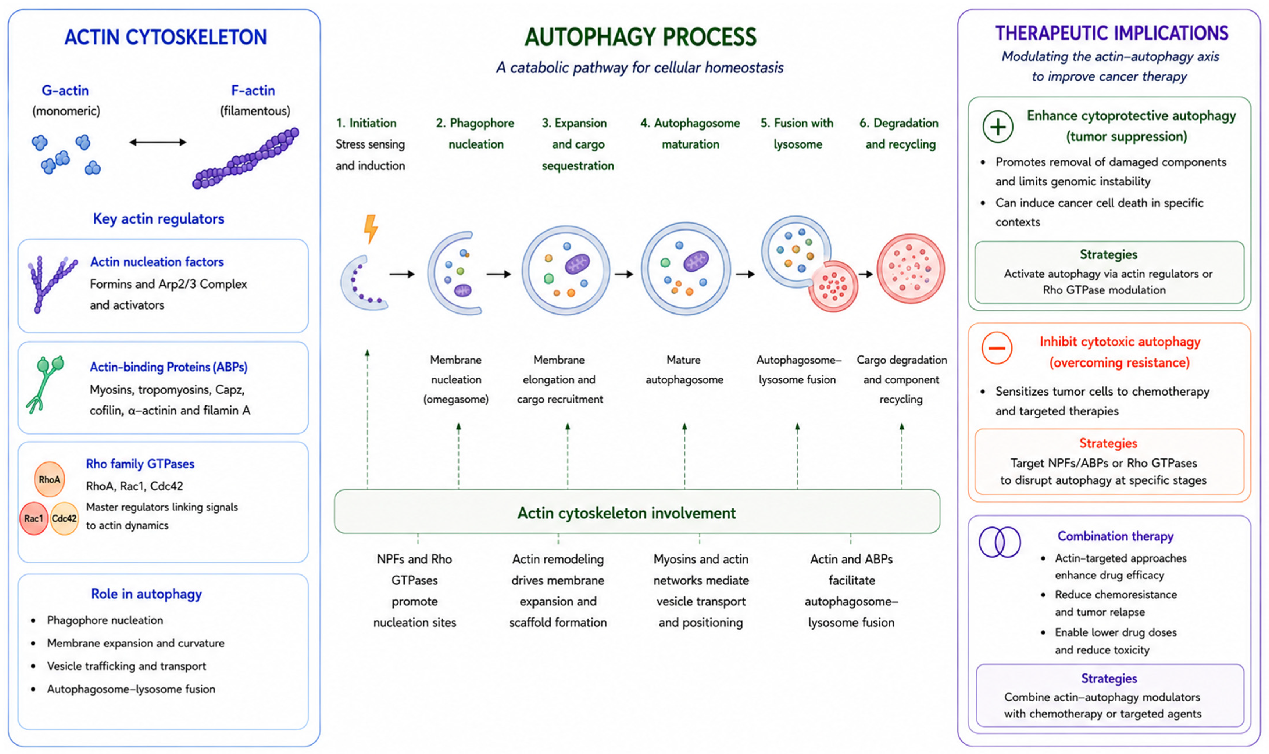
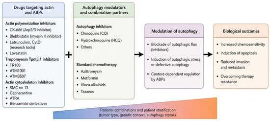
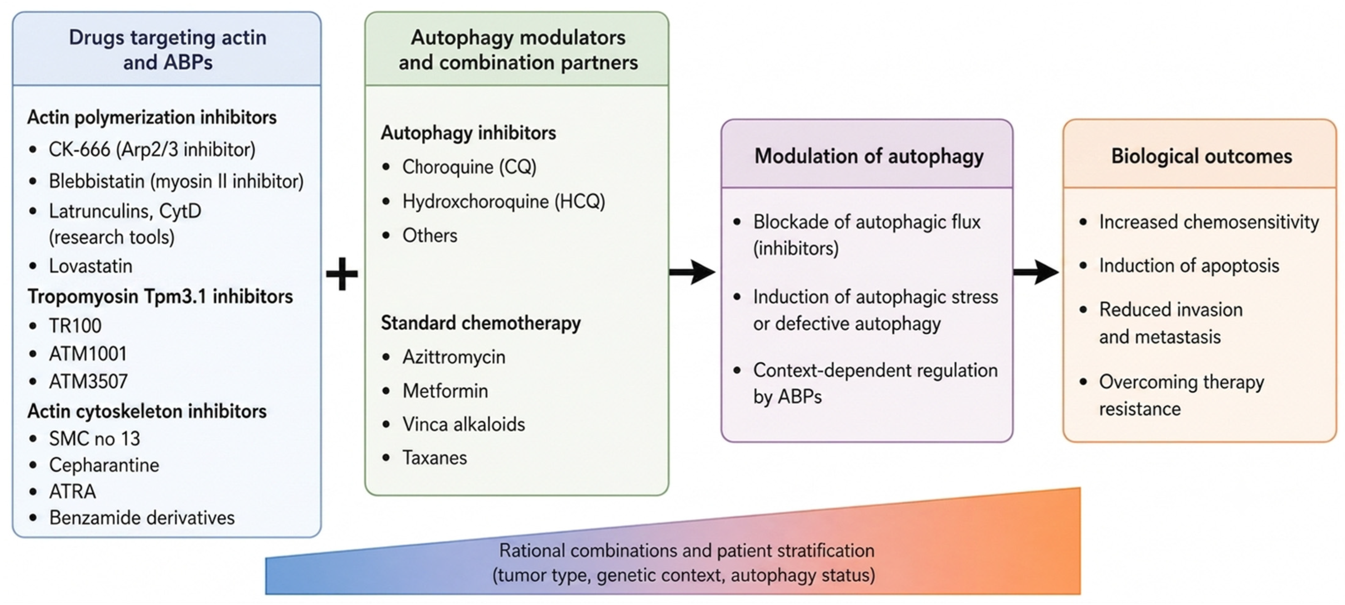

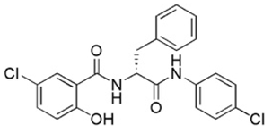
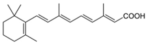
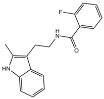
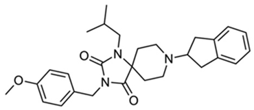
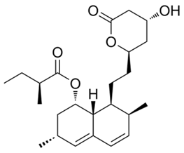
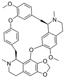
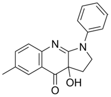
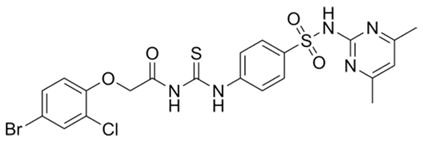
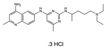
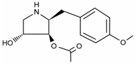
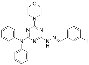
Disclaimer/Publisher’s Note: The statements, opinions and data contained in all publications are solely those of the individual author(s) and contributor(s) and not of MDPI and/or the editor(s). MDPI and/or the editor(s) disclaim responsibility for any injury to people or property resulting from any ideas, methods, instructions or products referred to in the content. |
© 2026 by the authors. Licensee MDPI, Basel, Switzerland. This article is an open access article distributed under the terms and conditions of the Creative Commons Attribution (CC BY) license.