


Abstract
Hemodialysis (HD) remains the most widely adopted renal replacement therapy for patients with end-stage kidney disease; however, its delivery entails a substantial environmental burden due to high water and energy consumption and extensive reliance on single-use synthetic polymeric membranes. As the global prevalence of chronic kidney disease increases, the ecological footprint of dialysis systems has become a critical challenge for sustainable healthcare. Conventional HD membranes, based on petroleum-derived polymers, provide controlled permeability but are inherently non-renewable, non-biodegradable, and susceptible to fouling and bio-incompatibility, underscoring the need for alternative, more sustainable materials. Chitosan has emerged as a promising biopolymer owing to its biodegradability, intrinsic antimicrobial activity, chemical versatility, and favorable hemocompatibility. This review presents a comprehensive analysis of chitosan-based hybrid membranes for HD, with emphasis on sustainability-driven material innovation. The structural chemistry and functional properties of chitosan are discussed in relation to molecular weight, degree of deacetylation, and supramolecular organization, followed by a comparative assessment of chitosan derived from crustacean, insect, fungal, and cephalopod sources. Attention is given to fungal chitosan as a naturally deacetylated, high-purity, and reproducible biomaterial aligned with circular bioeconomy principles. Eco-innovative extraction and purification strategies, including enzymatic and low-energy processes, are critically examined alongside membrane fabrication approaches such as polymer blending, electrospinning of hollow fibers, and functionalization strategies aimed at improving hemocompatibility, antimicrobial performance, and fouling resistance. Key challenges related to membrane reuse, scale-up, regulatory compliance, and clinical translation are also addressed. Overall, this review highlights fungal-derived chitosan as a sustainable platform for next-generation HD membranes.
1. Introduction
Hemodialysis (HD) remains the most widely adopted renal replacement therapy for patients with end-stage kidney disease, providing life-sustaining extracorporeal blood purification when renal function is irreversibly compromised. Advances in membrane engineering, fluid control, and clinical protocols have significantly improved patient survival and treatment efficiency in recent decades [1]. Nevertheless, the technological sophistication that underpins modern HD is accompanied by a substantial environmental burden. Each dialysis session requires large volumes of ultrapure water, considerable energy input, and extensive use of single-use consumables, including dialyzers, bloodlines, and ancillary plastics, most of which are derived from petroleum-based polymers [2,3]. As the global prevalence of chronic kidney disease (CKD) continues to rise, led by aging populations and increasing incidence of metabolic and cardiovascular disorders, the ecological footprint of HD systems has become a critical concern for sustainable healthcare [4]. In regions with high disease incidence, particularly across Europe and Mediterranean countries, the cumulative environmental impact of long-term dialysis therapy is especially pronounced. The combination of high patient numbers and resource-intensive treatment protocols amplifies pressures related to water consumption, carbon emissions, and medical waste generation [5]. Despite increasing awareness of these challenges, the material foundation of HD membranes has remained largely unchanged for decades. Commercial membranes are predominantly based on synthetic polymers such as polysulfone (PSu), polyethersulfone (PES), and polyacrylonitrile (PAN), selected for their mechanical robustness and reproducibility at industrial scale [3,6,7]. While these materials provide controlled permeability and structural stability, they are inherently non-renewable, non-biodegradable, and energy-intensive to manufacture. From a biological perspective, their surface chemistry often promotes protein fouling, platelet activation, and inflammatory responses, limiting long-term biocompatibility and functional longevity [7].
These limitations highlight the need for a paradigm shift in HD membrane design, moving beyond incremental optimization of existing synthetic materials toward alternative solutions that reconcile clinical performance with environmental responsibility. In this context, sustainability must be addressed holistically, encompassing renewable feedstocks, greener manufacturing processes, improved functional lifespan, and responsible end-of-life scenarios [3,8,9]. Biopolymers have emerged as promising candidates within this sustainability framework, offering natural abundance, structural versatility, and intrinsic biodegradability. Among them, chitosan has gained particular attention due to its favorable combination of physicochemical and biological properties [10]. Derived from the partial deacetylation of chitin, chitosan is a linear amino-polysaccharide characterized by excellent film-forming capacity, tunable molecular architecture, and high chemical reactivity [11]. Its polycationic nature enables electrostatic interactions with negatively charged biomolecules, conferring intrinsic antimicrobial activity and influencing protein adsorption behavior [12].
Beyond its inherent biological affinity, chitosan represents a versatile material platform due to the presence of reactive amino and hydroxyl groups that enable extensive chemical modification and functionalization [13,14]. These features allow for the incorporation of anticoagulant, antioxidant, and anti-inflammatory functionalities, enhancing hemocompatibility and therapeutic performance [15,16,17,18]. Furthermore, chitosan can be processed through established membrane fabrication techniques, including casting [19], phase inversion, and electrospinning, facilitating integration into existing industrial workflows [20,21]. The sustainability and performance of chitosan are strongly influenced by its biological origin and extraction pathway. While crustacean-derived chitosan has traditionally dominated commercial production, alternative sources such as insects, squid, and fungi are being increasingly explored [22]. In particular, fungal chitosan stands out as a naturally deacetylated biomaterial produced directly within the fungal cell wall, eliminating the need for harsh chemical treatments [23]. Controlled fermentation enables high purity, batch-to-batch reproducibility, and alignment with circular bioeconomy principles through the valorization of low-value biomass streams [24,25]. Recent advances in eco-innovative extraction and purification strategies further strengthen the sustainability profile of fungal chitosan [26,27]. Enzymatic treatments, green solvents, and low-energy physical processes reduce chemical input and preserve polymer integrity, enabling the production of medical-grade chitosan [27,28,29]. Against this background, this review provides a comprehensive and critical overview of chitosan-based hybrid membranes for HD, with particular emphasis on sustainability-driven material innovation. Structural chemistry, biological sources, eco-innovative extraction routes, membrane fabrication strategies, and functionalization approaches are discussed alongside challenges related to reuse, scale-up, regulatory compliance, and clinical translation. By integrating material science, biotechnology, and sustainability perspectives, this work aims to position chitosan, particularly fungal-derived chitosan, as a key enabler of next-generation, environmentally responsible HD technologies.
1.1. Hemodialysis and Environmental Burden
Hemodialysis (HD) remains the most widespread renal replacement therapy for patients with end-stage kidney disease, providing life-sustaining filtration when renal function is irreversibly impaired. However, the clinical and technical sophistication that enables this therapy also comes with a substantial environmental cost. Each dialysis session requires large volumes of ultrapure water, typically 120 to 180 liters per treatment, along with disposable dialyzers, blood lines, and ancillary plastics, most of which are derived from petroleum-based polymers such as PSU, PES, and PAN [30]. On a global scale, the estimated 3.8 million patients undergoing regular dialysis generate an enormous ecological footprint [4]. According to the most recent report from EDTA-ERTA (Figure 1), the incidence data and geographical distribution demonstrate the substantial burden of CKD across Europe [5].
Figure 1.
Incidence per million population by country/region by the beginning of 2023 [5].
Many countries exceed 150 new cases per million population, and several Mediterranean regions surpass 200–250 cases. Notably, Portugal shows one of the highest incidence rates (~205 cases per million population), placing the country among those with the greatest number of newly diagnosed patients each year [5]. This high disease burden means that a large and growing proportion of the population requires long-term renal replacement therapies, particularly HD [5]. Consequently, the environmental impact associated with dialysis procedures becomes increasingly relevant, especially regarding the generation and management of medical waste, including single-use consumables, plastics, and contaminated effluents. The convergence of a high number of CKD patients and the substantial waste output of dialysis units reinforces the urgency of developing more sustainable materials and waste-management strategies within nephrology care [31].
Beyond resource consumption and waste generation, the composition and life cycle of dialysis membranes also contribute to the environmental footprint of HD. The properties and limitations of currently available commercial membranes are discussed in detail in Section 4.1.
1.2. Sustainable Membranes
The concept of sustainability in HD goes beyond reducing plastic waste; it encompasses a holistic redesign of materials, processes, and life cycles. The next generation of dialysis membranes should not only ensure efficient toxin clearance and biocompatibility but also be manufactured from renewable feedstocks using green chemistry principles and designed for safe end-of-life degradation or recycling [30]. In this context, biopolymers have emerged as promising candidates due to their natural abundance, structural versatility, and intrinsic biodegradability [9]. Among natural polymers, chitosan stands out as a particularly attractive option. Derived from the partial deacetylation of chitin, chitosan is a linear amino-polysaccharide with excellent film-forming capacity, chemical reactivity, and biological affinity [10]. Its cationic nature under acidic conditions facilitates interaction with negatively charged biomolecules and confers inherent antimicrobial properties that are an advantage over synthetic polymers and potential membrane reuse [10,11,12,32]. Its structural and biological properties, source-dependent characteristics, processing strategies, and functionalization potential are discussed in the following sections.
2. Chitosan: Structure, Sources and Functional Properties
2.1. Structural Chemistry of Chitosan
Chitosan is a linear polysaccharide derived from the deacetylation of chitin, one of the most abundant biopolymers on Earth. Structurally, chitosan consists of β-(1→4)-linked D-glucosamine and N-acetyl-D-glucosamine units [10]. The degree of deacetylation (DD), which represents the proportion of glucosamine residues relative to N-acetylated units, plays a central role in determining its physicochemical and biological properties [33]. Typically, when the DD exceeds 50%, the polymer is considered chitosan rather than chitin [34]. The molecular weight (MW) of chitosan can vary substantially depending on the source material and processing conditions and influences solubility, viscosity, and functional activity [35]. An overview of the three types of chitosan, along with their MW ranges, DD and specific applications, is summarized in Table 1.
Table 1.
Classification of chitosan and its chemical properties and applications.
From a structural chemistry perspective, the primary amine groups located at the C-2 position of the glucosamine residues are the most distinctive feature of chitosan compared to other polysaccharides [41]. These protonatable amino groups confer a polycationic nature to chitosan in acidic media, allowing for electrostatic interactions with negatively charged molecules, including proteins, lipids, polyanions, and microbial cell membranes [42]. This reactivity is the foundation for its versatile functionality across biomedical, environmental, and industrial applications. Furthermore, chitosan exhibits significant hydrogen bonding capacity via its hydroxyl groups at the C-3 and C-6 positions, which influences crystallinity, film-forming ability, and intermolecular interactions [42,43]. The polymer typically exhibits semi-crystalline structures, with crystallinity governed by both the distribution of acetyl groups along the chain [44] and the degree of intra- and inter-chain hydrogen bonding. Solid-state nuclear magnetic resonance (NMR) and X-ray diffraction (XRD) studies have identified multiple polymorphic crystalline forms of chitosan (commonly referred to as chitosan I and II), reflecting variations in hydration and chain alignment. Such structural heterogeneity is not only of fundamental interest but also highly relevant for the tuning of functional materials [45,46]. Chitosan is also chemically versatile: the free amine and hydroxyl groups serve as reactive sites for chemical modifications, including acylation, quaternization, carboxylation, and grafting with functional moieties [41,47,48]. These derivatizations expand its solubility range, enhance stability, and confer targeted biological activities such as mucoadhesion, antimicrobial effects, or improved heparin binding. Importantly, this tunability makes chitosan a highly adaptable biomaterial platform capable of being engineered for applications as diverse as wound dressings, HD membranes, tissue engineering scaffolds, food packaging films, and controlled-release matrices [41,47,49,50]. The polycationic character and structural flexibility of chitosan also underpin its role as a chelating agent for divalent and trivalent metal ions. Binding occurs mainly through coordination with amine groups, although hydroxyl groups can also contribute [51,52,53]. This chelation capacity is relevant not only for wastewater treatment and heavy metal remediation but also for nutrient delivery in agriculture and modulation of trace elements in biological systems. Finally, chitosan’s supramolecular organization in aqueous environments deserves emphasis. In acidic solution, protonation of amino groups leads to an extended conformation due to repulsive electrostatic forces. As the pH approaches neutrality, deprotonation drives aggregation and precipitation. These transitions critically influence gel formation, film casting, and nanoparticle assembly. Consequently, an understanding of chitosan’s structural chemistry, at levels ranging from monomeric units to macromolecular assemblies, is fundamental to optimizing its processing and functional performance [10,54,55,56]. Another central feature of chitosan’s structural chemistry is its polydispersity. Unlike synthetic polymers with controlled chain lengths, naturally derived chitosan exhibits a broad distribution of MWs and DD [10,57,58]. This heterogeneity strongly affects reproducibility in both research and industrial contexts. To address this, controlled deacetylation strategies and enzymatic treatments are increasingly employed to generate well-defined chitosan oligomers with specific MWs and functionalities [59]. These oligomers often display distinct biological activities compared to the parent polymer, including immunomodulation, antioxidant potential, and prebiotic effects. Such advances in precision processing highlight the importance of structural characterization not merely as an analytical necessity, but as a foundation for tailoring performance [59,60,61]. Furthermore, the interplay between chitosan’s chemical structure and its physical behavior in aqueous systems has inspired new approaches in nanotechnology. Self-assembly into nanoparticles, nanofibers, or hydrogels occurs spontaneously under appropriate conditions, driven by a balance of electrostatic, hydrophobic, and hydrogen bonding interactions [62]. These nanoscale architectures preserve the fundamental chemistry of the parent polymer while imparting novel properties such as enhanced mucoadhesion, tunable drug release, and responsive swelling behavior. In this sense, the structural chemistry of chitosan does not remain confined to the level of repeating units or crystalline domains but extends into the supramolecular organization that defines its practical utility [63,64,65,66,67].
2.2. Comparison of Chitosan Sources
Although the basic repeating units of chitosan are structurally conserved, significant differences arise depending on its biological origin. Traditionally, crustacean shells (e.g., shrimp, crab, lobster) have been the dominant commercial source of chitin and chitosan, owing to the abundance of shellfish by-products from the seafood industry [68]. However, alternative sources, including insects [22], fungi [23], and cephalopods such as squid [69] are emerging as sustainable or functionally distinctive options [70] (Figure 2).
Figure 2.
Sources and chemical structures of chitin and chitosan and the conversion of chitin into chitosan through deacetylation. The red β symbols indicate the β-(1→4) glycosidic linkages between the monosaccharide units.
Crustacean chitosan is generally characterized by high crystallinity, reflecting the hierarchical organization of chitin in shell microfibrils embedded in a matrix of proteins and calcium carbonate [71]. Extraction typically requires strong deproteinization and decalcification steps using concentrated alkali and acid treatments [72]. Also, residual shellfish proteins can trigger allergic responses, limiting its use in biomedical and dietary formulations. In contrast, insect chitosan benefits from the growing edible insect industry, where exoskeletal residues can be valorized as a chitin-rich by-product [50]. Insects such as mealworms, crickets, and black soldier fly larvae contain significant amounts of chitin in their cuticle, typically with lower mineral content than crustacean shells [22]. However, insect-derived chitosan may also exhibit allergenic potential, particularly in individuals sensitive to insect proteins or chitin derivatives. Fungal chitosan, however, stands apart as the only naturally occurring form of chitosan, synthesized directly within the cell walls of certain fungi such as Aspergillus niger, Aspergillus oryzae and Rhizopus oryzae. This eliminates the need for post-deacetylation, simplifying processing and avoiding the use of harsh chemicals. Extraction typically involves mild alkaline treatments to remove proteins and glucans, resulting in high-purity material with low MW and a high DD, often exceeding 80%. These properties confer enhanced solubility in weakly acidic conditions and improved biocompatibility [73,74]. Controlled fermentation of fungal biomass also ensures reproducibility, independence from seasonal variation, and complete absence of allergenic components [75], making fungal chitosan highly suitable for medical and pharmaceutical use [23,74]. Also, squid-pen (β-chitin) represents a compelling biomass source for high-quality and non-allergenic chitosan. Schneider-Teixeira et al. (2025) [76] demonstrated that using High-Pressure Processing (HHP) as a pretreatment significantly improves deproteinization and extraction efficiency, leading to chitosan with fewer residual impurities and good structural attributes. In nanoparticle form, squid-pen chitosan displays exceptional antimicrobial performance: V. H. Nguyen (2021) [77] showed that by adjusting MW and DD, ultra-small chitosan nanoparticles (~30 nm) can be synthesized, and exhibit potent antibacterial activity against both Salmonella and Staphylococcus aureus at concentrations as low as 50 ppm. Moreover, under equivalent polymeric starting conditions, squid-pen-derived chitosan nanoparticles outperformed analogous particles from shrimp chitosan, likely owing to superior purity, favorable chain structure, and enhanced surface interactions with bacterial membranes [76,78]. From an ethical and sustainability perspective, non-crustacean sources— insect, fungal and squid—offer clear advantages. Fungal chitosan production can be scaled in bioreactors independently of agricultural or fishery cycles [23], insect farming relies on organic side-streams [50], and squid chitosan valorizes existing seafood by-products with minimal environmental cost [78]. Comparative analyses have also demonstrated that source-specific characteristics, such as acetylation pattern, crystallinity index, and polymer chain length, affect biological performance [51,52] (Table 2). This diversity indicates that chitosan should no longer be regarded as a single uniform biopolymer, but rather as a family of related biomaterials, each tailored by its biological origin to fulfil distinct functional, ecological, and industrial roles [26].
Table 2.
Comparative overview of chitosan from crustacean, insect, fungal and squid origins in terms of extraction, composition, and functional properties.
2.3. Antimicrobial, Hemocompatible and Biodegradable Properties
The functional properties of chitosan are central to its attractiveness as a biomaterial, with antimicrobial activity, hemocompatibility, and biodegradability being especially relevant for biomedical and environmental applications [26]. The antimicrobial activity of chitosan has been widely documented against bacteria, fungi, and even viruses [79]. Its mechanism of action is primarily attributed to electrostatic interactions between the positively charged amino groups of chitosan and the negatively charged microbial cell membranes [47]. This leads to increased membrane permeability, leakage of intracellular components, and eventual cell death. The efficacy depends strongly on MW, DD, solubility, and environmental conditions such as pH and ionic strength. Low MW chitosan tends to penetrate microbial membranes more easily, whereas high MW variants can form protective films that inhibit colonization. Additionally, chitosan can chelate essential trace metals, depriving microbes of nutrients. These multifaceted mechanisms render chitosan a powerful natural antimicrobial agent with reduced risk of resistance development compared to conventional antibiotics [80]. Similarly, in biomedical contexts, chitosan-based wound dressings have been shown to reduce biofilm formation, a property highly relevant in the management of chronic wounds. These broader ecological and physiological effects underscore the systemic impact of chitosan’s antimicrobial function, which transcends simple microbicidal activity [81]. Beyond its antimicrobial properties, chitosan can support blood contact applications when its surface chemistry is carefully controlled [13]. Nevertheless, its cationic amino groups may favor the adsorption of negatively charged plasma proteins and subsequent platelet adhesion and activation [16]. Although this hemostatic effect is advantageous in wound dressings, it is undesirable in HD membranes, where persistent platelet activation may promote thrombus formation and platelet consumption [82]. This risk can be mitigated by reducing the accessible positive surface charge and introducing hydrated, antithrombotic interfaces through heparin immobilization, blending with anionic or hydrophilic polymers, controlled crosslinking, or functionalization with nitric oxide donors. These strategies can limit nonspecific protein adsorption and platelet–surface interactions while preserving membrane permeability. Accordingly, platelet adhesion and activation, coagulation markers, and changes in platelet count should be evaluated under dynamic blood-flow conditions before clinical translation (Bowry et al., 2021) [83]. Surface modification strategies can further optimize hemocompatibility, for example, by blending chitosan with heparin to modulate anticoagulant properties or by introducing hydrophilic groups to minimize protein denaturation. In recent years, chitosan-based hemostatic dressings have gained regulatory approval and clinical adoption, highlighting its practical impact in trauma care and surgery [13,81,82].
Equally significant is chitosan’s biodegradability. In biological environments, chitosan is degraded by lysozyme and other glycosidases, primarily through hydrolysis of β-(1→4) linkages. The rate of biodegradation depends on DD, crystallinity, and MW: highly deacetylated and crystalline forms degrade more slowly, whereas amorphous and lower MW chitosan is rapidly metabolized [84,85]. The degradation products, mainly glucosamine and N-acetylglucosamine, are non-toxic, naturally occurring metabolites that can be incorporated into glycosaminoglycan and glycoprotein biosynthetic pathways. This biocompatible degradation profile is particularly valuable for medical applications, ensuring that implanted chitosan-based materials are gradually resorbed without eliciting harmful by-products [84]. The combination of antimicrobial activity, hemocompatibility, and biodegradability confers a unique competitive advantage to chitosan over many synthetic and natural biomaterials [13,85,86]. In wound healing, for example, chitosan simultaneously prevents infection, promotes hemostasis, and supports tissue regeneration [19,87]. In HD membranes, chitosan contributes to hemocompatibility while providing biodegradability that addresses end-of-life disposal concerns. Similarly, in drug delivery systems, chitosan’s enzymatic degradability ensures controlled release and safe clearance [85,88,89,90].
3. Eco-Innovative Extraction of Chitosan from Fungi
The production of chitosan from fungal sources has gained considerable attention as a sustainable alternative to crustacean-derived polymers. Unlike crustaceans, fungi naturally biosynthesize chitosan as part of their cell-wall structure, eliminating the need for aggressive chemical deacetylation. This biological advantage, coupled with the possibility of controlled fermentation, enables consistent and high-purity chitosan production while avoiding the seasonal, geographic, and allergenic constraints associated with marine by-products. Eco-innovative extraction from fungi aligns with circular bioeconomy principles, valorizing low-value biomass streams such as spent fermentation residues or fungal mycelium generated in the food, pharmaceutical, and biotechnology sectors [27,34]. However, the higher purity frequently attributed to fungal chitosan should not be considered universal, as it remains strongly dependent on the biological source and extraction conditions. In a direct comparative study, mushroom-derived chitosan contained 1.4% residual ash and 0.4% residual protein, compared with 2.1% ash and 0.4% protein for crab-derived chitosan and 0.8% ash and 0.3% protein for shrimp-derived chitosan (Periyannan et al., 2023) [91]. These findings indicate that fungal chitosan may present a lower mineral burden than some crustacean sources, while offering comparable residual protein levels. Fungal extraction methods are increasingly designed around “green chemistry” concepts, minimizing hazardous reagents, energy consumption, and waste generation. These processes rely on biological pretreatments, mild alkaline extraction, and enzymatic hydrolysis, often under optimized conditions that preserve the polymer’s functional integrity. In this context, the eco-innovative production of fungal chitosan is more than a process improvement: it represents an evolution toward sustainable biomaterial manufacturing suited for advanced biomedical and environmental technologies [28,29,92].
3.1. Fungal Strains for Chitosan Extraction: Selection Criteria
The choice of fungal strain is the cornerstone of any efficient chitosan production system. Ascomycota fungi, which include yeasts and filamentous fungi, have cell walls made of chitin and glucans, making them a potential source for producing chitosan. The structural composition of their cell walls, rich in glucosamine residues and low in interfering polysaccharides, facilitates extraction and purification. Strain selection also depends on physiological parameters such as growth rate, biomass yield, and tolerance to different substrates [93]. Fast-growing strains that can thrive on agro-industrial residues (e.g., molasses, wheat bran, or glycerol) are particularly desirable, as they enable cost-effective and scalable production within biorefinery models [29,74,94]. However, beyond the traditional zygomycetes, A. niger in particular has emerged as a strong candidate for eco-sustainable chitosan production. This species is classified as GRAS (Generally Recognized as Safe) by the U.S. FDA and has a long history of industrial use in the production of enzymes, organic acids, and fermented foods [95]. This safety status is crucial for the development of medical- or food-grade biomaterials. A. niger is especially valued for its robustness and adaptability; it can grow in a wide range of pH and nutrient conditions, efficiently converting low-cost agro-industrial residues into biomass. Several studies report that A. niger can accumulate up to 16–26% of its dry weight as chitin/chitosan-related polymers under optimized conditions, making it a practical and scalable source of fungal chitosan [25,93]. The cell walls of A. niger and A. oryzae contain higher proportions of β-glucans and galactomannans, which can complicate purification and reduce overall yield. Reported chitosan yields from Aspergillus biomass typically range between 5 and 12% after purification, depending on culture conditions and extraction methods [93]. These limitations can be mitigated through enzymatic cell-wall pretreatments, strain improvement, and process optimization. Emerging strategies, such as metabolic engineering to overexpress chitin deacetylase genes or redirect glucosamine biosynthetic pathways, are showing promise in increasing the chitosan fraction within Aspergillus cell walls. The production of chitosan by A. niger occurs naturally during the formation and maturation of its cell wall. The fungal wall contains a composite matrix of chitin, chitosan, β-glucans, and glycoproteins, whose proportions vary with growth phase and environmental conditions. During the vegetative mycelial stage, A. niger expresses chitin deacetylase enzymes, which partially convert chitin into chitosan. This biological deacetylation eliminates the need for aggressive chemical treatments commonly required for crustacean chitin, thus reducing energy consumption, reagent use, and effluent generation. The resulting chitosan displays a high DD (80–90%), moderate molecular weight, and low crystallinity, attributes that improve solubility in mildly acidic conditions and make it particularly suitable for film and membrane fabrication. From an environmental perspective, A. niger can be cultivated on a wide variety of agro-industrial residues, including molasses, corn steep liquor, fruit pulps, and glycerol, enabling its integration into circular biorefinery frameworks. These substrates not only reduce production costs but also valorize organic waste streams, reinforcing the ecological appeal of fungal chitosan. The fermentation process can be conducted under controlled aeration, pH, and temperature, ensuring reproducibility and consistency of polymer quality across batches. Furthermore, the mycelial biomass can be harvested and processed under mild alkaline extraction, yielding medical-grade chitosan with minimal purification steps and negligible residual proteins or minerals. The biological performance of A. niger-derived chitosan aligns well with the demands of biomedical applications. Its balanced molecular architecture confers strong film-forming ability, biocompatibility, and antimicrobial activity against both Gram-positive and Gram-negative bacteria [25,93]. In membrane systems, it enhances surface hydrophilicity and fouling resistance while maintaining structural integrity under physiological flow. Ultimately, A. niger exemplifies the intersection of biotechnology and sustainability in medical material development. Its established industrial use ensures scalability, its metabolic plasticity supports eco-efficient production, and its non-pathogenic nature guarantees biosafety. By harnessing A. niger as a renewable microbial factory for high-purity chitosan, it becomes possible to align the goals of environmental responsibility, clinical safety, and performance optimization [84,96]. In the broader context of sustainable nephrology, A. niger-derived chitosan thus represents not merely a substitute for synthetic polymers, but also a model for how biotechnological precision and ecological ethics can converge in the design of next-generation HD membranes [97,98].
3.2. Enzymatic and Green Extraction Techniques
Conventional chemical extraction of chitosan from fungal biomass has historically relied on aggressive reagents such as concentrated sodium hydroxide and hydrochloric acid for deproteinization and demineralization. While these methods can achieve reasonable yields, they are energy-intensive, generate large volumes of alkaline and acidic waste, and often lead to partial depolymerization or structural alteration of the polymer. The transition toward green and enzymatic extraction techniques therefore represents a crucial advancement, one that not only reduces environmental impact but also preserves the integrity and biocompatibility of fungal chitosan [99]. Enzymatic extraction builds upon the specificity of hydrolytic enzymes that selectively remove non-chitosan components from the fungal cell wall. Proteases (e.g., alcalase, papain) effectively hydrolyze residual proteins, while β-glucanases and cellulases target structural polysaccharides such as glucans and mannans. This controlled enzymatic hydrolysis gently exposes the chitosan framework without requiring strong alkaline treatment. In most protocols, a mild alkali wash (1–2 M NaOH at ≤60 °C) follows the enzymatic stage to solubilize residual lipids and pigments. Compared with traditional approaches, this combined enzymatic–mild-alkali method significantly improves polymer quality, minimizes MW degradation, and shortens purification time [79,99,100,101,102,103]. The optimization of enzymatic extraction parameters, enzyme concentration, temperature, pH, and incubation time is critical to balancing efficiency and cost. For instance, protease pretreatment at 45–55 °C followed by glucanase digestion at slightly acidic pH (5–6) often yields a clean cell-wall matrix with minimal polysaccharide contamination. Additionally, the reuse of immobilized enzymes has been successfully demonstrated, improving process economics and aligning with the principles of circular bio-manufacturing. The enzymatic hydrolysis stage can also be coupled with physical aids such as agitation, ultrasonication, or micro-filtration to enhance mass transfer and accelerate cell-wall disruption [79,104]. Beyond enzymatic hydrolysis, a variety of eco-innovative extraction technologies have been investigated to further reduce chemical input. Ultrasound-assisted extraction (UAE) employs acoustic cavitation to disrupt fungal cell walls, thereby increasing solvent penetration and diffusion of cell-wall polymers. When combined with dilute alkali, UAE can reduce extraction time from several hours to minutes while preserving MW [104,105]. Microwave-assisted extraction (MAE), on the other hand, provides rapid volumetric heating that softens the matrix and facilitates chitosan release at significantly lower chemical concentrations [106]. Both techniques are energy-efficient, scalable, and compatible with industrial continuous-flow systems. Emerging solvent-based alternatives, such as ionic liquids (ILs) and deep eutectic solvents (DESs), are also gaining traction for green extraction. These designer solvents can disrupt hydrogen bonding in the chitin–glucan network, allowing for selective dissolution of cell-wall components under mild conditions. Particularly, choline chloride-based DESs (combined with organic acids like lactic or citric acid) have demonstrated efficient extraction with minimal toxicity and complete solvent recyclability. Although these systems still require cost optimization and detailed biocompatibility assessment, they represent a good frontier in solvent-free biopolymer recovery [107]. A parallel development involves subcritical-water extraction (SWE), which exploits water at high temperature (120–180 °C) and pressure just below its critical point to achieve enhanced solvation power. SWE can solubilize proteins and glucans without strong chemicals, yielding chitosan with preserved DD and high purity. When powered by renewable energy, this approach offers one of the lowest environmental footprints among current extraction technologies [107,108]. Ultimately, enzymatic and green extraction technologies illustrate how biotechnology can reconcile efficiency and environmental stewardship. By substituting harsh reagents with biocatalysts, optimizing mild physical processes, and integrating energy-saving systems, the extraction of fungal chitosan becomes aligned with sustainable manufacturing paradigms. This shift not only supports regulatory trends favoring green chemistry but also enhances the polymer’s purity and biological performance, paving the way for its use in sensitive biomedical applications. Importantly, the extraction strategy influences not only the sustainability of the process but also the physicochemical characteristics of the resulting chitosan, which ultimately determines its suitability for HD membrane fabrication. Harsh alkaline treatments and prolonged extraction conditions may promote partial depolymerization, reducing molecular weight and viscosity, whereas milder enzymatic and green extraction routes generally better preserve polymer integrity (Zhang et al., 2026) [109]. Likewise, extraction conditions can affect the DD, a key parameter governing amino group availability, hydrophilicity, and chemical reactivity. Together, molecular weight, DD, viscosity, and polymer integrity strongly influence membrane-forming ability, mechanical strength, permeability, and the efficiency of subsequent surface functionalization (Zhang et al., 2026) [109]. Consequently, selecting an appropriate extraction strategy is essential not only for improving sustainability but also for obtaining chitosan with physicochemical properties tailored to high-performance HD membranes.
3.3. Purification and Characterization for Medical-Grade Chitosan
It is known that extraction conditions directly influence the physicochemical properties of chitosan and, to that end, comprehensive purification and characterization are essential to verify whether the extracted polymer possesses the quality required for biomedical membrane fabrication. The transition from crude fungal chitosan to medical-grade material demands stringent purification and exhaustive characterization. For biomedical applications such as HD membranes, tissue scaffolds, wound dressings, and controlled-release systems, purity and consistency are non-negotiable. Even trace contaminants, residual proteins, endotoxins, or heavy metals can provoke immune responses or alter biocompatibility. Therefore, the purification stage is designed to meet pharmaceutical Good Manufacturing Practice (GMP) standards, ensuring batch-to-batch reproducibility and compliance with international quality specifications. After extraction, the crude chitosan typically contains fragments of β-glucans, pigments, and other cellular residues. The first step of purification often involves dissolution–precipitation cycles: the polymer is solubilized in dilute acetic acid and re-precipitated by pH neutralization with sodium hydroxide. The purified polymer is then sterilized by gamma irradiation, ethylene-oxide exposure, or filtration, depending on the intended formulation [110]. Characterization follows purification to confirm chemical identity and functional suitability. Fourier-transform infrared spectroscopy (FTIR) and solid-state 13C NMR are the primary techniques for determining the DD and verifying the presence of characteristic amide (1650 cm−1) and amine (1590 cm−1) bands [111]. Gel-permeation chromatography (GPC) provides MW distribution and polydispersity indices, while thermogravimetric analysis (TGA) and differential scanning calorimetry (DSC) reveal thermal behavior relevant to processing stability [112]. Morphological evaluation via scanning electron microscopy (SEM) or atomic-force microscopy (AFM) helps correlate microstructure with film-forming and mechanical properties [113]. Biological testing completes the qualification process. Cytotoxicity assays (MTT, Live/Dead) assess cell viability in the presence of chitosan extracts, while hemolysis and coagulation tests determine blood compatibility. Advanced assays, such as complement activation, platelet adhesion, and protein adsorption, provide a comprehensive profile of hemocompatibility [114,115]. It should be noted that ISO 10993 (International Organization for Standardization, 2009) [116] and USP Class VI are not specific to chitosan but constitute general biocompatibility frameworks applicable to biomaterials and polymers intended for medical use. Materials designed for biomedical applications are typically evaluated according to these standards to ensure biological safety and, where relevant, hemocompatibility in blood-contacting devices. The specific set of tests required under ISO 10993 is not fixed and depends on the intended use, final form, duration of contact, and risk classification of the medical device. In practice, regulatory validation commonly focuses on core assessments such as cytotoxicity, irritation, and hemocompatibility, while more advanced evaluations—such as protein adsorption, complement activation, or platelet adhesion—may be informative from a research and performance perspective but are not always mandatory for regulatory approval [117,118,119].
4. Chitosan Hybrid Membranes
Building on the structural and functional properties described in Section 2, this section examines the incorporation of chitosan into hybrid membrane systems. Particular attention is given to polymer blending and electrospinning strategies designed to improve the mechanical stability, transport performance, and hemocompatibility required for HD applications.
4.1. Commercial HD Membranes: Properties and Limitations
At present, the development of chitosan-containing HD membranes remains predominantly at the laboratory and preclinical stages. Early approaches mainly explored chitosan as a membrane-forming material or surface coating, whereas more recent studies have incorporated chitosan into PES or PSU blends, immobilized chitosan nanoparticles within hollow-fiber membranes, or used chitosan as a functional platform for heparinization and other bioactive modifications [26,50,120]. The membranes that are currently in clinical use are mainly composed of synthetic polymers such as PSU, PES, PAN, cellulose acetate (CA), and poly(methyl methacrylate) (PMMA). These materials are selected for their mechanical stability, chemical resistance, and reproducibility at industrial scale. In addition, decades of clinical use have demonstrated their safety, reliability, and consistent dialysis performance, leading to widespread regulatory approval and well-established manufacturing processes. Although newer membrane materials are under development, few have yet demonstrated sufficient long-term clinical evidence, scalability, and cost-effectiveness to replace conventional synthetic membranes in routine HD practice [121]. Among them, PSU and PES dominate the market because they provide excellent hydraulic permeability, good thermal stability, and adequate protein adsorption control. Cellulose-based membranes, although partially replaced, are still employed for their hydrophilicity and low cost [122,123]. Despite their clinical success, synthetic membranes face several limitations in performance. Their hydrophobic nature often promotes protein fouling and platelet adhesion, leading to decreased efficiency during their use. Moreover, many synthetic membranes display limited selectivity for middle-MW toxins (e.g., β2-microglobulin, advanced glycation end products), which are crucial contributors to chronic inflammation and dialysis-related amyloidosis. Chemical modification, such as blending with hydrophilic polymers (e.g., PVP, PEG), can reduce fouling but does not fully restore hemocompatibility [123,124,125]. Another challenge relates to bio-inertness. While the lack of chemical reactivity is beneficial for stability, it also impedes beneficial biological interactions such as nitric oxide-mediated vasodilation or natural anticoagulation. Furthermore, most of these materials are non-biodegradable, creating waste-management issues and environmental impact after disposal. Current HD membranes thus represent a technological compromise, mechanically robust but biologically passive, that motivates research into hybrid materials integrating natural polymers like chitosan to achieve a more physiological interface between blood and membrane [126,127].
4.2. Chitosan Blends: Physicochemical and Biological Synergies
Blending chitosan with other polymers is an effective strategy to tailor its mechanical, chemical, and biological performance. As a single polymer, chitosan exhibits excellent biocompatibility, hemostatic and antimicrobial activity, but it also presents challenges such as brittleness when dry, limited mechanical strength, and solubility restricted to acidic conditions. Forming hybrid membranes allows these shortcomings to be compensated while preserving the beneficial biological features [120]. Physicochemical synergies arise when chitosan is blended with polymers such as poly(vinyl alcohol) (PVA), cellulose acetate (CA), polycaprolactone (PCL), or polyethylene oxide (PEO). The hydrogen bonding between hydroxyl and amino groups enhances interchain compatibility, improving film-forming ability and mechanical resilience [13,120]. Crosslinking agents like glutaraldehyde, genipin, or tripolyphosphate (TPP) can be introduced to regulate porosity and water stability, while also improving mechanical strength and structural integrity, yielding membranes with controllable swelling and diffusion characteristics. These modifications are particularly important in HD, where pore size distribution determines solute clearance efficiency [128,129,130,131]. In addition, the improved intermolecular interactions generated by hydrogen bonding contribute to the formation of smoother, more homogeneous and hydrophilic membrane surfaces. Increased hydrophilicity promotes the formation of a stable hydration layer that limits nonspecific protein adsorption, thereby reducing subsequent platelet adhesion and thrombus formation [132].
Biological synergies are equally significant. Beyond the physicochemical improvements provided by polymer blending, the intrinsic biological properties of chitosan further enhance membrane performance. The cationic amino groups of chitosan enable electrostatic interactions with plasma components and facilitate subsequent functionalization; however, excessive positive surface charge may also increase protein adsorption and platelet adhesion. Therefore, the hemocompatibility of chitosan blends depends on controlling surface charge and hydration through blending with anionic or hydrophilic polymers, crosslinking, or heparinization [133]. Moreover, when combined with anionic polymers such as alginate, hyaluronic acid, or heparin, polyelectrolyte complexes form spontaneously, imparting antithrombotic properties and enhancing bioactivity. For example, chitosan–heparin membranes can mimic endothelial surfaces by facilitating the controlled release of anticoagulants and reducing platelet aggregation [133,134]. Hybrid chitosan membranes also exhibit strong potential for drug- or nanoparticle-functionalized systems, where the polymer matrix serves as a reservoir for antioxidant, anticoagulant, and anti-inflammatory agents. This multifunctionality makes chitosan blends not only filters but also active bio-interfaces capable of modulating inflammatory and oxidative processes in dialysis patients. Recent studies have demonstrated that chitosan-based hybrid membranes can reach urea and creatinine permeability values comparable to those of commercial PES membranes while maintaining significantly lower protein fouling [135]. Their surface charge and hydrophilicity can be tuned by adjusting the blend ratio or crosslinking degree, enabling a delicate balance between permeability, selectivity, and hemocompatibility. From a materials perspective, blending fungal-derived chitosan offers an additional advantage: its higher purity and reproducibility ensure minimal batch variability, a key requirement for medical-grade products. Overall, chitosan blends constitute a versatile platform that unites mechanical stability with biological function, paving the way toward next-generation dialysis membranes that are both efficient and sustainable.
4.3. Electrospinning of Hollow Fibers: Parameters and Prospects
Electrospinning represents a powerful technique to produce nanoscale fibrous membranes with high surface area, interconnected porosity, and controllable morphology, features that are particularly attractive for HD applications. In this process, a polymer solution is subjected to a high-voltage electric field, ejecting a charged jet that solidifies into ultrafine fibers upon solvent evaporation [136]. For chitosan, successful electrospinning often requires blending with a carrier polymer such as PEO, PVA, or PCL to improve spinnability and prevent jet instability. The choice of solvent is also critical; mixtures of acetic acid or trifluoroacetic acid with water or ethanol are commonly used to ensure adequate polymer solvation and charge density [137]. Hollow fiber electrospinning extends this approach by employing coaxial spinnerets, where a core and shell solution are simultaneously extruded. This enables the fabrication of membranes with distinct inner and outer layers, an architecture that closely replicates commercial dialysis fibers. The inner lumen ensures blood flow, while the outer porous layer governs solute exchange and prevents clogging. Adjusting the flow ratio between core and shell, as well as the collector geometry, allows for precise control over wall thickness, pore size, and permeability [138].
The main stages involved in the electrospinning and assembly of hollow-fiber membranes for HD are schematically illustrated in Figure 3. Chitosan-containing hollow fibers provide unique advantages. Their inherent hydrophilicity enhances water flux and reduces fouling, while the amino functionalities can be exploited for further surface functionalization, for example, immobilization of heparin [139,140,141], nitric oxide donors [14]. or antioxidant nanoparticles [142,143]. These modifications can impart anticoagulant, anti-inflammatory, or antioxidative effects, transforming the membrane from a passive filter into an active therapeutic surface [144,145,146]. From a prospective viewpoint, electrospun chitosan hybrids could redefine the design of bioactive dialysis membranes. Advances in coaxial electrospinning, 3D fiber alignment, and post-treatments (plasma activation, crosslinking, or grafting) are expanding the scope of tunable fiber morphologies. Future research should focus on translating these laboratory-scale fibers into scalable manufacturing processes and validating their clinical performance in terms of toxin clearance, hemocompatibility, and long-term durability. Despite its considerable potential, the large-scale production of electrospun hollow fibers remains challenging due to relatively low production rates, the need for precise control of fiber morphology and reproducibility, and the complexity of integrating electrospinning into continuous manufacturing processes. In addition, ensuring batch-to-batch consistency, sterilization compatibility, and compliance with stringent regulatory requirements represents essential prerequisites for the clinical translation of electrospun HD membranes [147,148].
Figure 3.
Schematic representation of the electrospinning-based fabrication of hollow-fiber membranes for HD.
5. Functionalization Strategies
The functionalization of chitosan-based membranes plays a pivotal role in tailoring their physicochemical and biological performance for HD applications. By introducing specific functional groups, bioactive molecules, or nanostructured additives, the surface of the membrane can be modified to enhance antimicrobial activity, hemocompatibility, and selective transport properties [122,149,150].
Beyond membrane formulation, post-fabrication processing must be systematically optimized to support clinical translation. Neutralization, washing, crosslinking, and drying procedures should ensure the complete removal of residual solvents and crosslinking agents while preserving membrane porosity, mechanical stability, permeability, and surface bioactivity [8,98]. The selection of an appropriate sterilization method is equally critical, as steam, ethylene oxide, ionizing radiation, or low-temperature plasma treatments may alter chitosan molecular integrity, surface chemistry, swelling behavior, and the activity of immobilized bioactive compounds. Therefore, membrane performance and hemocompatibility should be reassessed after sterilization and storage. Furthermore, comparative life-cycle assessments should consider raw-material sourcing, reagent, water and energy consumption, membrane fabrication, clinical use, and end-of-life management. Such analyses are required to determine whether fungal chitosan-based membranes provide measurable environmental advantages over conventional synthetic HD membranes rather than assuming sustainability solely from their renewable origin [151].
Chitosan’s chemical versatility, owing to its reactive amino and hydroxyl groups, provides multiple routes for covalent grafting, ionic complexation, or physical adsorption of bioactive agents [48,152]. These modifications aim not only to prevent fouling and infection but also to mimic endothelial-like behavior, thereby reducing thrombogenicity and inflammatory responses. The following sections describe the main strategies employed for chitosan functionalization, focusing on antimicrobial reinforcement, heparin grafting, and other bioactive integrations that improve hemocompatibility and functionality [121,141,149,150,153,154,155]. As illustrated in Figure 4, the functionalization of chitosan-based HD membranes may improve their biological performance by conferring anticoagulant, vasodilatory, antimicrobial, antioxidant, and regenerative properties.
Figure 4.
Potential improvements conferred by the functionalization of chitosan-based HD membranes.
5.1. Antimicrobial Enhancement in Membranes
Chitosan inherently possesses antimicrobial activity, mainly due to its cationic nature. It is important to distinguish microbial biofilm formation from blood-side membrane fouling. In routine HD, biofilms develop primarily within the dialysate circuit and other water-contacting components, where prolonged exposure to aqueous conditions can support microbial attachment and growth. By contrast, the short single-use period of the hemodialyzer makes mature biofilm formation within the device less likely. The positively charged amino groups interact electrostatically with negatively charged microbial cell membranes, disrupting their integrity and leading to leakage of intracellular components. This intrinsic property already distinguishes chitosan from most synthetic polymers. However, when used in medical devices such as dialysis membranes, the antimicrobial effect can be further enhanced through surface functionalization with metallic nanoparticles, natural antimicrobial agents, or synergistic polymers [156]. Metallic nanoparticles, particularly silver (Ag), copper (Cu), and zinc oxide (ZnO), are among the most effective modifiers. When immobilized on the chitosan matrix, they generate reactive oxygen species (ROS) that induce oxidative stress in microbial cells, while the polymeric framework stabilizes the nanoparticles and limits their release rate. The result is a controlled antimicrobial action with minimal cytotoxicity. Silver–chitosan nanocomposites, for instance, have shown strong bactericidal activity against Staphylococcus aureus and Escherichia coli without compromising hemocompatibility [143,157,158]. Another eco-friendly approach involves the incorporation of plant-derived phenolic compounds, such as catechins, quercetin, or curcumin, into chitosan membranes. These polyphenols interact via hydrogen bonding or ionic interactions, imparting both antioxidant and antimicrobial properties. Their controlled release from the membrane can inhibit bacterial colonization and biofilm formation, which are persistent challenges in long-term dialysis [159,160]. In addition, enzymatic immobilization techniques can be employed to attach antimicrobial peptides (AMPs) or lysozyme to the chitosan surface. These biofunctional coatings mimic natural defense mechanisms, selectively targeting bacterial membranes without affecting mammalian cells. Such strategies, when combined with the inherent antimicrobial charge of chitosan, provide dual protection, chemical and biological, against nosocomial pathogens, significantly reducing infection risks during dialysis treatments [161,162,163].
5.2. Heparin-Grafting and Ionic Bonding Mechanisms
Heparinization represents one of the most effective functionalization strategies to improve the hemocompatibility of dialysis membranes [141]. Heparin, a highly sulfated polysaccharide, exhibits strong negative charge density, enabling it to inhibit thrombin and other clotting factors, thereby preventing blood coagulation upon contact with artificial surfaces. Chitosan’s polycationic structure provides a natural counterpart for ionic bonding with heparin, allowing for the formation of stable polyelectrolyte complexes or covalently grafted layers [15,140,164,165]. Two main approaches that are typically used are ionic complexation, in which the negatively charged sulfate groups of heparin interact electrostatically with the positively charged amino groups of chitosan. This simple and mild method preserves heparin’s biological activity and can be achieved by dipping the membrane in heparin solution under controlled pH and ionic strength. The resulting film confers anticoagulant properties and reduces platelet adhesion, while the dynamic equilibrium between bound and free heparin ensures a gradual, prolonged release [47,166,167]. Functional groups on heparin (–COOH or –OH) can react with the amino groups of chitosan through carbodiimide (EDC/NHS) coupling, glutaraldehyde crosslinking, or periodate oxidation followed by Schiff-base formation [168,169,170,171]. These reactions produce stable amide or imine bonds, ensuring long-term anticoagulant performance even under flow conditions. The surface potential and roughness of chitosan membranes can be tuned during this process to achieve optimal hemocompatibility [47,171]. Beyond traditional heparinization, hybrid systems have emerged combining chitosan–heparin complexes with nanoparticle carriers (e.g., Fe3O4 or SiO2) to achieve dual functions, anticoagulation and controlled delivery of anti-inflammatory or antioxidant molecules [142]. Such multifunctional surfaces imitate the endothelial glycocalyx, enabling the material to regulate both clotting and immune responses during dialysis. Importantly, these biointerfaces maintain the biodegradable and eco-friendly nature of chitosan, distinguishing them from synthetic coatings typically based on PEG or PVP [172].
5.3. Other Bioactive Functionalizations for Improved Hemocompatibility
Building on the intrinsic antimicrobial mechanisms described in Section 2.3, several functionalization strategies have been investigated to reinforce the antimicrobial performance of chitosan-based HD membranes. Beyond heparin grafting, several other functionalization strategies have been developed to enhance the bioactivity and hemocompatibility of chitosan membranes. Antioxidant molecules such as vitamin E, tannic acid, or curcumin have also been employed to mitigate oxidative stress induced during dialysis [173,174]. These molecules can be physically entrapped or covalently linked to chitosan, providing redox buffering capacity that protects circulating cells and proteins from damage. This antioxidant reinforcement is particularly relevant for patients undergoing repeated dialysis sessions, who often experience chronic systemic inflammation. Also, nanoparticle functionalization (e.g., with Fe3+, CeO2, or TiO2 nanoparticles) can impart additional catalytic or radical-scavenging properties, improving both hemocompatibility and membrane durability. When combined with chitosan’s own cationic framework, these nanoparticles establish hybrid systems capable of actively modulating biological responses rather than merely resisting them [64,175,176]. Therefore, functionalization transforms chitosan membranes from passive biopolymers into dynamic biointerfaces capable of interacting intelligently with the biological environment. Whether through antimicrobial modification, heparin complexation, or other bioactive integrations, these strategies represent essential steps toward developing advanced, sustainable, and clinically effective HD membranes.
6. Fouling Resistance and Membrane Reuse
One of the persistent challenges in HD is the gradual decline in membrane performance due to fouling, the accumulation of proteins, lipids, and microbial biofilms on the membrane surface. This phenomenon not only reduces permeability and selectivity but also increases the risk of infection and systemic inflammation. Addressing fouling through material innovation and functional surface design is therefore essential to improving the efficiency, safety, and sustainability of dialysis [122,177].
6.1. Biofilm Formation in HD
Biofilm formation on dialysis membranes arises from the initial adsorption of plasma proteins, which creates a conditioning layer that facilitates microbial attachment [39,178,179]. Once microorganisms adhere, they secrete extracellular polymeric substances (EPSs), forming a complex matrix that protects them from shear stress and disinfectants. Biofilms may develop both on the inner lumen of hollow fibers and in external flow circuits, especially during prolonged or repeated use [144]. Common pathogens include Staphylococcus aureus, Pseudomonas aeruginosa, and Candida albicans, whose persistence can lead to pyrogenic reactions and chronic inflammation in patients. In conventional synthetic membranes, hydrophobicity and surface roughness promote this colonization process. The EPS matrix strongly binds to hydrophobic regions, while limited surface charge inhibits repulsion of bacterial cells. Once established, biofilms are difficult to eradicate, even with sterilization or chemical cleaning. Hence, prevention through intrinsic material design, rather than post-formation treatment, is now considered the most effective strategy to combat biofilm-associated fouling in HD [178,180,181,182].
6.2. Role of Chitosan in Fouling Mitigation
Chitosan plays a dual role in mitigating fouling: it acts as both a passive barrier against adsorption and an active antimicrobial agent. Its hydrophilic and polycationic nature alters surface energy, reducing non-specific protein binding while repelling negatively charged microbial membranes [158]. When integrated into hybrid dialysis membranes, chitosan can delay the initial adhesion stage of fouling, maintaining higher flux stability during extended operation. Moreover, chitosan’s antimicrobial mechanism, based on electrostatic disruption of microbial membranes, prevents biofilm nucleation at an early stage [183,184,185]. The polymer can also be functionalized with nanoparticles or phenolic agents to enhance this effect, creating self-cleaning surfaces capable of oxidative degradation of organic residues. Studies have demonstrated that PES/chitosan or PVA/chitosan membranes exhibit significantly lower bovine serum albumin (BSA) adsorption and bacterial adhesion than unmodified controls, while maintaining comparable solute clearance rates [173,186,187]. At the physicochemical level, chitosan confers increased surface charge density and hydration-layer thickness, two factors known to improve antifouling behavior. Hydrated chitosan chains form a dynamic barrier that limits protein approach within the Debye length, while positive surface charge promotes electrostatic repulsion of most negatively charged biomolecules. This combination of steric and electrostatic effects constitutes a powerful mechanism for long-term fouling mitigation without the need for aggressive cleaning or regeneration [185,188].
6.3. Sterilization, Reuse Potential and Patient-Specific Applications
The sustainability and cost-effectiveness of dialysis systems depend heavily on the reusability of membranes. Conventional synthetic membranes are discarded after a single use due to concerns over fouling, structural fatigue, and contamination. Chitosan-based membranes, in contrast, can be designed for controlled sterilization and limited reuse, provided that their physicochemical integrity and biocompatibility are preserved [189,190]. Sterilization methods compatible with chitosan include gamma irradiation, ethylene oxide, and steam autoclaving under mild conditions. Among these, gamma irradiation is particularly effective, as it ensures microbial deactivation without major alteration of the polymer backbone when doses below 25 kGy are applied [191]. Ethylene oxide remains suitable for membranes containing sensitive additives, though careful aeration is required to eliminate residues. The intrinsic antimicrobial nature of chitosan also reduces the microbial load between sessions, potentially extending the safe reuse interval [192]. Looking forward, the concept of patient-specific dialysis membranes is gaining relevance. By tailoring the composition, pore size, and surface functionalization of chitosan hybrids to individual metabolic profiles or comorbidities, it becomes feasible to design membranes optimized for toxin removal, inflammatory control, or oxidative stress reduction [193,194].
7. Simulation and In Vitro Performance
The development of novel dialysis membranes requires a comprehensive understanding of mass transport, fluid dynamics, and biointerface behavior under realistic operational conditions. While in vitro testing remains essential for assessing biocompatibility and clearance performance, computational modelling offers a complementary and increasingly powerful tool for predicting and optimizing membrane performance before clinical implementation. Integrating both approaches, in silico and in vitro, can accelerate material screening, reduce experimental costs, and improve translational accuracy [17,195,196,197].
7.1. Dynamic Counter-Current Diffusion Models
Traditional HD systems operate under counter-current flow, where blood and dialysate move in opposite directions separated by a semi-permeable membrane. Mathematical diffusion models based on Fick’s law and mass balance equations are employed to simulate the clearance of solutes such as urea, creatinine, and β2-microglobulin. Incorporating time-dependent diffusion coefficients, boundary-layer effects, and transient fouling resistance enables accurate prediction of flux decline during repeated cycles [198,199]. Dynamic models can also account for non-idealities such as concentration polarization, membrane compaction, and the influence of molecular size and charge on transport rates. When applied to chitosan hybrid membranes, these simulations reveal how surface charge density and pore geometry affect solute selectivity. For instance, positively charged chitosan domains can enhance the diffusion of anionic toxins while limiting albumin loss, an effect that can be tuned via blending ratios or crosslinking degree [200,201].
7.2. Computational Fluid Dynamics in Membrane Testing
Computational fluid dynamics (CFD) has become a cornerstone for analyzing flow distribution, shear stress, and solute transport inside hollow-fiber modules. CFD simulations enable visualization of velocity profiles, pressure gradients, and dead zones that are difficult to measure experimentally. Such insights are crucial for preventing hemolysis and optimizing fiber-packing density. For chitosan-based systems, CFD studies can integrate reactive transport models that couple convective flow with surface reactions, such as adsorption of proteins or binding of charged toxins [175,202]. This allows for virtual testing of how membrane functionalization (e.g., heparin grafting or nanoparticle addition) influences hydrodynamics and biointeractions [139,203]. The flexibility of CFD also supports patient-specific design: by inputting individual blood viscosity or hematocrit parameters, it is possible to predict how a membrane will perform under different clinical scenarios before fabrication [204].
7.3. In Vitro vs. In Silico Performance Metrics
Bridging computational predictions with experimental validation is essential for translating chitosan-based membranes from research to clinical use. In vitro assays, such as diffusion cell tests, protein adsorption measurements, and whole-blood compatibility studies, provide empirical data on permeability, selectivity, and hemocompatibility [16,17,194]. These results can be used to calibrate in silico models, refining assumptions about membrane resistance, solute diffusivity, and reaction kinetics [203]. Conversely, simulation outputs guide experimental design by identifying optimal pore sizes, blend ratios, or operating pressures. Metrics such as mass transfer coefficient (K0A), solute clearance efficiency, and shear rate distribution can thus be compared across computational and experimental domains to assess model accuracy [203,204,205,206,207]. When in vitro and in silico data converge, confidence in the predicted clinical performance increases, facilitating regulatory acceptance and scale-up. The integration of simulation and experimental validation establishes a rational design framework for advanced membranes, one that connects material chemistry, process engineering, and biological response. This multidisciplinary synergy not only accelerates innovation but also supports the transition toward eco-sustainable dialysis technologies.
8. Industrial and Clinical Translation
A successful transition of chitosan hybrid membranes from laboratory-scale innovation to clinical-grade production requires an integrated framework encompassing material consistency, process scalability, regulatory alignment, and long-term sustainability. Each of these must be addressed to ensure that scientific innovation can evolve into a viable medical product capable of meeting industrial, environmental, and healthcare demands.
8.1. Scale-Up Considerations for Chitosan Hybrid Membranes
Scaling up the fabrication of chitosan hybrid membranes presents unique challenges, since bio-based polymers display greater variability than petrochemical counterparts [18,208,209,210]. At the processing level, laboratory methods such as phase inversion or electrospinning must be adapted to industrially viable continuous systems. Maintaining control over parameters such as solution viscosity, polymer concentration, flow rate, and drying temperature ensures stable pore morphology and surface chemistry [139]. Industrial electrospinning platforms, equipped with multi-needle or free-surface configurations, offer scalable routes for producing hollow fibers with reproducible diameters and high surface uniformity [186,187,203]. Inline analytical tools, such as FTIR or NIR spectroscopy, can monitor solvent content and functional group evolution in real time, guaranteeing batch homogeneity [146,211,212].
8.2. Integration in Circular Healthcare Models
The integration of chitosan-based membranes into circular healthcare systems represents a crucial step towards sustainable nephrology. Current dialysis technologies depend heavily on synthetic, non-biodegradable polymers, which generate significant environmental waste after a single use [26,213]. Chitosan membranes, by contrast, offer biodegradability, biocompatibility, and the possibility of controlled reuse without compromising patient safety. When produced from fungal sources, for example, these materials can be incorporated into biorefinery chains that valorize agro-industrial residues and food-processing by-products, creating a closed-loop production model aligned with green chemistry principles [94,214]. In a circular healthcare model, waste generated by one sector becomes a resource for another. Fungal chitosan derived from fermentation residues or lignocellulosic substrates exemplifies this philosophy, as it converts low-value biomass into a high-performance biomedical material. The potential for partial regeneration or sterilization between sessions could further reduce the ecological footprint of dialysis centers, provided that strict sterility and performance standards are maintained. Furthermore, the carbon-neutral production of chitosan, coupled with solvent recovery and water recirculation during membrane fabrication, aligns with European Union directives promoting sustainable and circular medical technologies [119]. In this sense, the transition to chitosan hybrid membranes transcends material innovation and embodies a systemic shift toward greener, more resilient healthcare ecosystems.
8.3. Long-Term Vision for Sustainable Nephrology
In the long-term vision of sustainable nephrology, chitosan-based membranes play a central role as the foundation of environmentally responsible and patient-centered dialysis systems. The ideal dialysis of the future will combine high clearance efficiency with minimal environmental impact, using biodegradable components produced from renewable feedstocks and fabricated through low-energy processes. Circular production cycles, where biopolymer residues are recovered, reprocessed, or safely biodegraded, will replace the linear “produce–use–discard” paradigm that dominates current dialysis practices [26,50,57,135,215,216,217]. Ultimately, chitosan’s intrinsic sustainability and biological compatibility position it as a key enabler of a new generation of dialysis technologies—ones that reconcile patient health, industrial scalability, and ecological responsibility within a single, coherent framework [216,218].
8.4. Economic and Regulatory Considerations
Economic feasibility and regulatory compliance are interdependent drivers of industrial translation. For chitosan hybrid membranes to reach the market, production costs must remain competitive with existing Psu membranes while providing measurable clinical and environmental advantages. The integration of chitosan production into existing fermentation or food-processing industries can significantly reduce raw-material costs and ensure year-round availability. Life-cycle assessments indicate that bio-based chitosan systems can achieve a lower environmental impact per dialysis session due to biodegradability and reduced waste management costs [74]. Regulatory approval, however, represents a more demanding frontier. As implantable or blood-contact materials, HD membranes are subject to strict medical-device frameworks such as ISO 10993 for biocompatibility, ISO 13485 (International Organization for Standardization, 2016) [218] for manufacturing quality, and MDR 2017/745 in the European Union. Consistent batch performance and the absence of cytotoxicity, hemolysis, and pyrogenic reactions are prerequisites for clinical validation [118,119,120]. Furthermore, for hybrid systems containing bioactive agents (such as heparin, nitric oxide donors, or nanoparticles), additional toxicological assessments and leaching studies are required. The transition from laboratory proof-of-concept to CE or FDA certification demands not only robust data but also long-term stability and reproducibility across production lots. The success of chitosan hybrid membranes will depend on the balance between economic scalability, regulatory transparency, and sustained demonstration of clinical benefits.
9. Challenges and Future Perspectives
While substantial progress has been achieved in developing chitosan hybrid membranes, several scientific, technological, and institutional challenges still limit their full clinical integration. These include the need for standardized material characterization, scalable fabrication protocols, and long-term clinical validation. At the same time, the rapid evolution of biomaterials science and digital manufacturing opens new directions for innovation, enabling chitosan membranes to progress from laboratory prototypes to intelligent, adaptive medical interfaces. Overcoming these barriers will define the trajectory of chitosan-based biomaterials in nephrology and broader biomedical applications. The next generation of chitosan-based membranes is expected to move beyond passive separation toward active, multifunctional systems. Emerging research explores the integration of sensing elements, stimuli-responsive polymers, and bioelectronic interfaces that allow for real-time monitoring of blood parameters or adaptive control of permeability [16,168]. Nanostructured chitosan composites incorporating conductive fillers, metal–organic frameworks, or enzyme-loaded nanoparticles could provide both selective adsorption and catalytic detoxification of uremic toxins. Additionally, 3D printing and advanced electrospinning techniques offer unprecedented control over pore architecture, fiber alignment, and hierarchical layering, enabling membranes with gradients of functionality tailored to specific physiological needs [136,146,183]. The convergence between chitosan chemistry and digital manufacturing also supports the rise of personalized dialysis. These technological trends indicate a paradigm shift: chitosan membranes will no longer serve merely as filters, but also as adaptive biointerfaces capable of responding dynamically to patient conditions and treatment goals [198,201].
10. Conclusions
This review highlights the urgent need to rethink HD membrane materials considering the growing environmental burden associated with conventional, petroleum-derived polymers. Chitosan-based hybrid membranes emerge as a compelling alternative, offering a unique combination of biodegradability, hemocompatibility, antimicrobial activity, and chemical versatility. Fungal-derived chitosan stands out as a sustainable and reproducible biomaterial, aligned with circular bioeconomy principles and well suited for medical-grade applications. Advances in eco-innovative extraction, purification, membrane fabrication, and functionalization strategies demonstrate that chitosan can be engineered into bioactive membranes capable of addressing both clinical performance requirements and sustainability goals. While challenges related to scale-up, regulatory validation, reuse, and clinical translation remain, the integration of material science, biotechnology, and green manufacturing positions chitosan, especially fungal chitosan, as a promising platform for next-generation, environmentally responsible HD technologies.
Author Contributions
M.M.: Writing—original draft, Formal analysis, Conceptualization. P.H.: Writing—review & editing, Visualization, Validation. S.B.-S.: Writing—review & editing, Visualization, Validation, Supervision, Investigation, Conceptualization. S.B.: Writing—review & editing, Visualization, Validation, Supervision, Investigation, Conceptualization. All authors have read and agreed to the published version of the manuscript.
Funding
This work was supported by National Funds from FCT—Fundação para a Ciência e a Tecnologia through projects UID/50016/2025 and LA/P/0076/2020 (https://doi.org/10.54499/LA/P/0076/2020). Maria Martingo acknowledges the support by FCT—Fundação para a Ciência e Tecnologia, for the doctoral grant with project reference 2024.03874.BDANA and DOI identifier https://doi.org/10.54499/2024.03874.BDANA and Sandra Borges was supported by national funds through FCT—Fundação para a Ciência e a Tecnologia, I.P., under contract ref. 2023.15056.TENURE.063.
Data Availability Statement
No new data were created or analyzed in this study. Data sharing is not applicable to this article.
Conflicts of Interest
Author Patrícia Henriques was employed by the company GOTECH Antimicrobial. The remaining authors declare that the research was conducted in the absence of any commercial or financial relationships that could be construed as a potential conflict of interest.
References
- Gagel, A.; Wiesen, G.; Stuard, S.; Canaud, B. Advances in Membrane, Dialyzer Design, and Related Monitoring Technologies for Hemodiafiltration: Translating Bench-Side Innovations to Bedside Applications. J. Clin. Med. 2026, 15, 1921. [Google Scholar] [CrossRef] [Scilit] [PubMed]
- Backston, K.J.; Thompson, C.J.; Raina, M.; Sathik, N.R.; Noh, L.S.; Lokkur, P.P.; Bansal, S.B. Green Dialysis Review: Reducing Water, Energy, and Waste. Hemodial. Int. 2025, 29, 463–471. [Google Scholar] [CrossRef] [Scilit] [PubMed]
- Sen Gupta, R.; Samantaray, P.K.; Bose, S. Going beyond Cellulose and Chitosan: Synthetic Biodegradable Membranes for Drinking Water, Wastewater, and Oil-Water Remediation. ACS Omega 2023, 8, 24695–24717. [Google Scholar] [CrossRef] [Scilit] [PubMed]
- Amane, M.; Dardona, Z.; Boussaa, S. Strategies for Reducing Water Consumption in Hemodialysis Therapy; Rapid Review. J. Fam. Med. Community Health 2023, 10, 11969. [Google Scholar]
- ERA Registry. ERA Registry Annual Report 2023; Amsterdam UMC, Location AMC, Department of Medical Informatics: Amsterdam, The Netherlands, 2025. [Google Scholar]
- Gul, A.; Hruza, J.; Yalcinkaya, F. Fouling and chemical cleaning of microfiltration membranes: A mini-review. Polymers 2021, 13, 846. [Google Scholar] [CrossRef] [Scilit] [PubMed]
- Iman, H.N.; Susilo, H.; Satriyatama, A.; Budi, I.D.M.; Kurnia, K.A.; Wenten, I.G.; Khoiruddin, K. Separation properties and fouling resistance of polyethersulfone membrane modified by fungal chitosan. BMC Chem. 2024, 18, 224. [Google Scholar] [CrossRef] [Scilit] [PubMed]
- Kumar Gupta, G.; De, S.; Franco, A.; Balu, A.M.; Luque, R. Sustainable Biomaterials: Current Trends, Challenges and Applications. Molecules 2015, 21, 48. [Google Scholar] [CrossRef] [Scilit] [PubMed]
- Salvay, A.G. Polysaccharide-Based Materials: Developments and Properties. Polymers 2025, 17, 3028. [Google Scholar] [CrossRef] [Scilit] [PubMed]
- Aranaz, I.; Alcántara, A.R.; Civera, M.C.; Arias, C.; Elorza, B.; Caballero, A.H.; Acosta, N. Chitosan: An overview of its properties and applications. Polymers 2021, 13, 3256. [Google Scholar] [CrossRef] [Scilit] [PubMed]
- Madera-Santana, T.J.; Herrera-Méndez, C.H.; Rodríguez-Núñez, J.R. An overview of the chemical modifications of chitosan and their advantages. Green Mater. 2018, 6, 131–142. [Google Scholar] [CrossRef] [Scilit]
- Signorini, L.; Marenzi, G.; Facente, A.; Marrelli, B.; Marano, R.M.; Valletta, A.; Pacifici, L.; Gasparro, R.; Sammartino, G.; Severino, M. Critical Overview on Pure Chitosan-based Scaffolds for Bone Tissue Engineering: Clinical insights in Dentistry. Int. J. Med. Sci. 2023, 20, 1527–1534. [Google Scholar] [CrossRef] [Scilit] [PubMed]
- Balan, V.; Verestiuc, L. Strategies to improve chitosan hemocompatibility: A review. Eur. Polym. J. 2014, 53, 171–188. [Google Scholar] [CrossRef] [Scilit]
- Lu, Y.; Slomberg, D.L.; Schoenfisch, M.H. Nitric oxide-releasing chitosan oligosaccharides as antibacterial agents. Biomaterials 2014, 35, 1716–1724. [Google Scholar] [CrossRef] [Scilit] [PubMed]
- Bračič, M.; Nagy, B.M.; Plohl, O.; Lackner, F.; Steindorfer, T.; Fischer, R.C.; Heinze, T.; Olschewski, A.; Kleinschek, K.S.; Nagaraj, C.; et al. Antithrombogenic polysaccharide coatings to improve hemocompatibility, protein-repellence, and endothelial cell response. IScience 2024, 27, 110692. [Google Scholar] [CrossRef] [Scilit] [PubMed]
- Rong, F.; Tang, Y.; Wang, T.; Feng, T.; Song, J.; Li, P.; Huang, W. Nitric Oxide-Releasing Polymeric Materials for Antimicrobial Applications: A Review. Antioxidants 2019, 8, 556. [Google Scholar] [CrossRef] [Scilit] [PubMed]
- Weber, M.; Steinle, H.; Golombek, S.; Hann, L.; Schlensak, C.; Wendel, H.P.; Avci-Adali, M. Blood-Contacting Biomaterials: In Vitro Evaluation of the Hemocompatibility. Front. Bioeng. Biotechnol. 2018, 6, 99. [Google Scholar] [CrossRef] [Scilit] [PubMed]
- Weber, N.; Wendel, H.P.; Ziemer, G. Hemocompatibility of heparin-coated surfaces and the role of selective plasma protein adsorption. Biomaterials 2002, 23, 429–439. [Google Scholar] [CrossRef] [Scilit] [PubMed]
- Dai, T.; Tanaka, M.; Huang, Y.Y.; Hamblin, M.R. Chitosan preparations for wounds and burns: Antimicrobial and wound-healing effects. Expert Rev. Anti-Infect. Ther. 2011, 9, 857–879. [Google Scholar] [CrossRef] [Scilit] [PubMed]
- Huang, C.; Thomas, N.L. Fabrication of porous fibers via electrospinning: Strategies and applications. Polym. Rev. 2020, 60, 595–647. [Google Scholar] [CrossRef] [Scilit]
- Huang, H.-F.; Peng, C.-F. Antibacterial and antifungal activity of alkylsulfonated chitosan. Biomark. Genom. Med. 2015, 7, 83–86. [Google Scholar] [CrossRef] [Scilit]
- Iber, B.T.; Kasan, N.A.; Torsabo, D.; Omuwa, J.W. A Review of Various Sources of Chitin and Chitosan in Nature. J. Renew. Mater. 2021, 10, 1097–1123. [Google Scholar] [CrossRef] [Scilit]
- Huq, T.; Khan, A.; Brown, D.; Dhayagude, N.; He, Z.; Ni, Y. Sources, production and commercial applications of fungal chitosan: A review. J. Bioresour. Bioprod. 2022, 7, 85–98. [Google Scholar] [CrossRef] [Scilit]
- Castro, R.; Guerrero-Legarreta, I.; Bórquez, R. Chitin extraction from Allopetrolisthes punctatus crab using lactic fermentation. Biotechnol. Rep. 2018, 20, e00287. [Google Scholar] [CrossRef] [Scilit] [PubMed]
- Marchezi, G.; Concolato, G.; Colla, L.; Piccin, J. Optimizing Submerged Fermentation Conditions of Aspergillus niger for Chitin Production. Appl. Biochem. Biotechnol. 2025, 197, 5790–5804. [Google Scholar] [CrossRef] [Scilit] [PubMed]
- Martingo, M.; Baptista-Silva, S.; Pintado, M.; Borges, S. A new vision upon hemodialysis: A shift from synthetic to sustainable chitosan membranes. Biomed. Eng. Adv. 2025, 9, 100175. [Google Scholar] [CrossRef] [Scilit]
- Sebastian, J.; Rouissi, T.; Brar, S.K. Chapter 14—Fungal chitosan: Prospects and challenges. In Handbook of Chitin and Chitosan; Gopi, S., Thomas, S., Pius, A., Eds.; Elsevier: Amsterdam, The Netherlands, 2020; pp. 419–452. [Google Scholar] [CrossRef] [Scilit]
- Den, W.; Sharma, V.K.; Lee, M.; Nadadur, G.; Varma, R.S. Lignocellulosic biomass transformations via greener oxidative pretreatment processes: Access to energy and value added chemicals. Front. Chem. 2018, 6, 141. [Google Scholar] [CrossRef] [Scilit] [PubMed]
- Veljković, V.B.; Đalović, I.G.; Siliveru, K.; Banković-Ilić, I.B.; Stamenković, O.S.; Mitrović, P.M.; Tasić, M.B.; Ciampitti, I.A.; Sikora, V.Š.; Prasad, P.V.V. Pretreatment Methods for Biofuel Production from Sorghum. In Sorghum in the 21st Century: Food—Fodder—Feed—Fuel for a Rapidly Changing World; Tonapi, V.A., Talwar, H.S., Are, A.K., Bhat, B.V., Reddy, C.R., Dalton, T.J., Eds.; Springer: Singapore, 2020; pp. 755–788. [Google Scholar] [CrossRef] [Scilit]
- Straits Research. Dialyzer Market Size & Outlook, 2025–2033; Straits Research: Pune, India, 2025. [Google Scholar]
- De Simone, E.; Morrone, M.C.; Tatarhan, E.I.; Pozzato, M.; Roccatello, D.; Fenoglio, R.; Sciascia, S. Green teams drive sustainability: A European Rare Kidney Disease Reference Network Survey on dialysis environmental practices. Clin. Kidney J. 2025, 18, sfaf276. [Google Scholar] [CrossRef] [Scilit] [PubMed]
- Gonciarz, W.; Balcerczak, E.; Brzeziński, M.; Jeleń, A.; Pietrzyk-Brzezińska, A.J.; Narayanan, V.H.B.; Chmiela, M. Chitosan-based formulations for therapeutic applications. A recent overview. J. Biomed. Sci. 2025, 32, 62. [Google Scholar] [CrossRef] [Scilit] [PubMed]
- Yadav, M.; Kaushik, B.; Rao, G.K.; Srivastava, C.M.; Vaya, D. Advances and challenges in the use of chitosan and its derivatives in biomedical fields: A review. Carbohydr. Polym. Technol. Appl. 2023, 5, 100323. [Google Scholar] [CrossRef] [Scilit]
- Kozma, M.; Acharya, B.; Bissessur, R. Chitin, Chitosan, and Nanochitin: Extraction, Synthesis, and Applications. Polymers 2022, 14, 3989. [Google Scholar] [CrossRef] [Scilit] [PubMed]
- Román-Doval, R.; Torres-Arellanes, S.P.; Tenorio-Barajas, A.Y.; Gómez-Sánchez, A.; Valencia-Lazcano, A.A. Chitosan: Properties and Its Application in Agriculture in Context of Molecular Weight. Polymers 2023, 15, 2867. [Google Scholar] [CrossRef] [Scilit] [PubMed]
- Zheng, B.; Wen, Z.S.; Huang, Y.J.; Xia, M.S.; Xiang, X.W.; Qu, Y.L. Molecular weight-dependent immunostimulative activity of low molecular weight chitosan via regulating NF-KB and AP-1 Signaling Pathways in RAW264.7 macrophages. Mar. Drugs 2016, 14, 169. [Google Scholar] [CrossRef] [Scilit] [PubMed]
- Zolfagharian, A.; Kaynak, A.; Noshadi, A.; Kouzani, A. System identification and robust tracking of a 3D printed soft actuator. Smart Mater. Struct. 2019, 28, 075025. [Google Scholar] [CrossRef] [Scilit]
- Cuong, H.N.; Minh, N.C.; Van Hoa, N.; Trung, T.S. Preparation and characterization of high purity β-chitin from squid pens (Loligo chenisis). Int. J. Biol. Macromol. 2016, 93, 442–447. [Google Scholar] [CrossRef] [Scilit] [PubMed]
- Garcia, L.G.S.; Guedes, G.M.d.M.; da Silva, M.L.Q.; Castelo-Branco, D.S.C.M.; Sidrim, J.J.C.; Cordeiro, R.d.A.; Rocha, M.F.G.; Vieira, R.S.; Brilhante, R.S.N. Effect of the molecular weight of chitosan on its antifungal activity against Candida spp. in planktonic cells and biofilm. Carbohydr. Polym. 2018, 195, 662–669. [Google Scholar] [CrossRef] [Scilit] [PubMed]
- Wantat, A.; Rojsitthisak, P.; Seraypheap, K. Inhibitory effects of high molecular weight chitosan coating on ‘Hom Thong’ banana fruit softening. Food Packag. Shelf Life 2021, 29, 100731. [Google Scholar] [CrossRef] [Scilit]
- Žigrayová, D.; Mikušová, V.; Mikuš, P. Advances in Chitosan Derivatives: Preparation, Properties and Applications in Pharmacy and Medicine. Gels 2024, 10, 701. [Google Scholar] [CrossRef] [Scilit] [PubMed]
- de Farias, B.S.; Sant’Anna Cadaval Junior, T.R.; de Almeida Pinto, L.A. Chitosan-functionalized nanofibers: A comprehensive review on challenges and prospects for food applications. Int. J. Biol. Macromol. 2019, 123, 210–220. [Google Scholar] [CrossRef] [Scilit] [PubMed]
- Lingait, D.; Rahagude, R.; Gaharwar, S.S.; Das, R.S.; Verma, M.G.; Srivastava, N.; Kumar, A.; Mandavgane, S. A review on versatile applications of biomaterial/polycationic chitosan: An insight into the structure-property relationship. Int. J. Biol. Macromol. 2024, 257, 128676. [Google Scholar] [CrossRef] [Scilit] [PubMed]
- Cui, J.; Yu, Z.; Lau, D. Effect of acetyl group on mechanical properties of chitin/chitosan nanocrystal: A molecular dynamics study. Int. J. Mol. Sci. 2016, 17, 61. [Google Scholar] [CrossRef] [Scilit] [PubMed]
- Chatterjee, S.; Adhya, M.; Guha, A.K.; Chatterjee, B.P. Chitosan from Mucor rouxii: Production and physico-chemical characterization. Process Biochem. 2005, 40, 395–400. [Google Scholar] [CrossRef] [Scilit]
- Nonappa; Lahtinen, M.; Ikonen, S.; Kolehmainen, E.; Kauppinen, R. Solid-State NMR, X-ray Diffraction, and Thermoanalytical Studies Towards the Identification, Isolation, and Structural Characterization of Polymorphs in Natural Bile Acids. Cryst. Growth Des. 2009, 9, 4710–4719. [Google Scholar] [CrossRef] [Scilit]
- Nicolle, L.; Journot, C.M.A.; Gerber-Lemaire, S. Chitosan functionalization: Covalent and non-covalent interactions and their characterization. Polymers 2021, 13, 4118. [Google Scholar] [CrossRef] [Scilit] [PubMed]
- Chen, Y.; Xiao, K.; Qu, X. Grafting Modification of Chitosan. In Biopolymer Grafting: Synthesis and Properties; Elsevier: Amsterdam, The Netherlands, 2018; pp. 295–364. [Google Scholar] [CrossRef] [Scilit]
- Lorkowska-Zawicka, B.; Kamiński, K.; Ciejka, J.; Szczubiałka, K.; Białas, M.; Okoń, K.; Adamek, D.; Nowakowska, M.; Jawień, J.; Olszanecki, R.; et al. Inactivation of heparin by cationically modified chitosan. Mar. Drugs 2014, 12, 3953–3969. [Google Scholar] [CrossRef] [Scilit] [PubMed]
- Martingo, M.; Baptista-Silva, S.; Mesquita, R.; Ferreira, J.P.; Borges, S.; Pintado, M. Exploring the potential of mealworm chitosan for hemodialysis applications. Sustain. Chem. Pharm. 2025, 45, 102013. [Google Scholar] [CrossRef] [Scilit]
- Cheung, R.C.F.; Ng, T.B.; Wong, J.H.; Chan, W.Y. Chitosan: An update on potential biomedical and pharmaceutical applications. Mar. Drugs 2015, 13, 5156–5186. [Google Scholar] [CrossRef] [Scilit] [PubMed]
- Torres, C.; Valerio, O.; Mendonça, R.T.; Pereira, M. Influence of chitosan protonation degree in nanofibrillated cellulose/chitosan composite films and their morphological, mechanical, and surface properties. Int. J. Biol. Macromol. 2024, 267, 131587. [Google Scholar] [CrossRef] [Scilit] [PubMed]
- Verma, C.; Quraishi, M.A. Chelation capability of chitosan and chitosan derivatives: Recent developments in sustainable corrosion inhibition and metal decontamination applications. Curr. Res. Green Sustain. Chem. 2021, 4, 100184. [Google Scholar] [CrossRef] [Scilit]
- Azueta-Aguayo, P.H.; Chuc-Gamboa, M.G.; Aguilar-Pérez, F.J.; Aguilar-Ayala, F.J.; Rodas-Junco, B.A.; Vargas-Coronado, R.F.; Cauich-Rodríguez, J.V. Effects of Neutralization on the Physicochemical, Mechanical, and Biological Properties of Ammonium-Hydroxide-Crosslinked Chitosan Scaffolds. Int. J. Mol. Sci. 2022, 23, 14822. [Google Scholar] [CrossRef] [Scilit] [PubMed]
- Gupta, S.; Vyas, S.P. Carbopol/chitosan based pH triggered in situ gelling system for ocular delivery of timolol maleate. Sci. Pharm. 2010, 78, 959–976. [Google Scholar] [CrossRef] [Scilit] [PubMed]
- Taokaew, S.; Kaewkong, W.; Kriangkrai, W. Recent Development of Functional Chitosan-Based Hydrogels for Pharmaceutical and Biomedical Applications. Gels 2023, 9, 277. [Google Scholar] [CrossRef] [Scilit] [PubMed]
- El-Araby, A.; Janati, W.; Ullah, R.; Ercisli, S.; Errachidi, F. Chitosan, chitosan derivatives, and chitosan-based nanocomposites: Eco-friendly materials for advanced applications (a review). Front. Chem. 2023, 11, 1327426. [Google Scholar] [CrossRef] [Scilit] [PubMed]
- Harugade, A.; Sherje, A.P.; Pethe, A. Chitosan: A review on properties, biological activities and recent progress in biomedical applications. React. Funct. Polym. 2023, 191, 105634. [Google Scholar] [CrossRef] [Scilit]
- Schmitz, C.; Auza, L.G.; Koberidze, D.; Rasche, S.; Fischer, R.; Bortesi, L. Conversion of chitin to defined chitosan oligomers: Current status and future prospects. Mar. Drugs 2019, 17, 452. [Google Scholar] [CrossRef] [Scilit] [PubMed]
- Chen, Y.J.; Sui, X.; Wang, Y.; Zhao, Z.H.; Han, T.H.; Liu, Y.J.; Zhang, J.N.; Zhou, P.; Yang, K.; Ye, Z.H. Preparation, structural characterization, biological activity, and nutritional applications of oligosaccharides. Food Chem. X 2024, 22, 101289. [Google Scholar] [CrossRef] [Scilit] [PubMed]
- Edo, G.I.; Mafe, A.N.; Razooqi, N.F.; Umelo, E.C.; Gaaz, T.S.; Isoje, E.F.; Igbuku, U.A.; Akpoghelie, P.O.; Opiti, R.A.; Essaghah, A.E.A.; et al. Advances in bio-polymer coatings for probiotic microencapsulation: Chitosan and beyond for enhanced stability and controlled release. Des. Monomers Polym. 2025, 28, 1–34. [Google Scholar] [CrossRef] [Scilit] [PubMed]
- Choi, J.; Hwang, D.S.; Lim, C.; Lee, D.W. Interaction mechanism between low molecular weight chitosan nanofilm and functionalized surfaces in aqueous solutions. Carbohydr. Polym. 2024, 324, 121504. [Google Scholar] [CrossRef] [Scilit] [PubMed]
- Herdiana, Y.; Febrina, E.; Nurhasanah, S.; Gozali, D.; Elamin, K.M.; Wathoni, N. Drug Loading in Chitosan-Based Nanoparticles. Pharmaceutics 2024, 16, 1043. [Google Scholar] [CrossRef] [Scilit] [PubMed]
- Ibrahim, M.A.; Alhalafi, M.H.; Emam, E.A.M.; Ibrahim, H.; Mosaad, R.M. A Review of Chitosan and Chitosan Nanofiber: Preparation, Characterization, and Its Potential Applications. Polymers 2023, 15, 2820. [Google Scholar] [CrossRef] [Scilit] [PubMed]
- Mohammed, M.A.; Syeda, J.T.M.; Wasan, K.M.; Wasan, E.K. An overview of chitosan nanoparticles and its application in non-parenteral drug delivery. Pharmaceutics 2017, 9, 53. [Google Scholar] [CrossRef] [Scilit] [PubMed]
- Pérez-Pacheco, Y.; Tylkowski, B.; García-Valls, R. Chitosan Micro/Nanocapsules in Action: Linking Design, Production, and Therapeutic Application. Molecules 2025, 30, 252. [Google Scholar] [CrossRef] [Scilit] [PubMed]
- Ways, T.M.M.; Lau, W.M.; Khutoryanskiy, V.V. Chitosan and its derivatives for application in mucoadhesive drug delivery systems. Polymers 2018, 10, 267. [Google Scholar] [CrossRef] [Scilit] [PubMed]
- Zhang, Z.; Ma, Z.; Song, L.; Farag, M.A. Maximizing crustaceans (shrimp, crab, and lobster) by-products value for optimum valorization practices: A comparative review of their active ingredients, extraction, bioprocesses and applications. J. Adv. Res. 2024, 57, 59–76. [Google Scholar] [CrossRef] [Scilit] [PubMed]
- Abdelmalek, B.E.; Sila, A.; Haddar, A.; Bougatef, A.; Ayadi, M.A. β-Chitin and chitosan from squid gladius: Biological activities of chitosan and its application as clarifying agent for apple juice. Int. J. Biol. Macromol. 2017, 104, 953–962. [Google Scholar] [CrossRef] [Scilit] [PubMed]
- Mersmann, L.; Souza, V.G.L.; Fernando, A.L. Green Processes for Chitin and Chitosan Production from Insects: Current State, Challenges, and Opportunities. Polymers 2025, 17, 1185. [Google Scholar] [CrossRef] [Scilit] [PubMed]
- Younes, I.; Rinaudo, M. Chitin and chitosan preparation from marine sources. Structure, properties and applications. Mar. Drugs 2015, 13, 1133–1174. [Google Scholar] [CrossRef] [Scilit] [PubMed]
- El-araby, A.; El Ghadraoui, L.; Errachidi, F. Physicochemical Properties and Functional Characteristics of Ecologically Extracted Shrimp Chitosans with Different Organic Acids during Demineralization Step. Molecules 2022, 27, 8285. [Google Scholar] [CrossRef] [Scilit] [PubMed]
- Alsohaimi, I.H. Comparative study of PS and PES and their sulfonated forms in antifouling behavior and rejection efficiency. J. King Saud. Univ. 2024, 36, 103576. [Google Scholar] [CrossRef] [Scilit]
- Crognale, S.; Russo, C.; Petruccioli, M.; D’annibale, A. Chitosan Production by Fungi: Current State of Knowledge, Future Opportunities and Constraints. Fermentation 2022, 8, 76. [Google Scholar] [CrossRef] [Scilit]
- Alimi, B.A.; Pathania, S.; Wilson, J.; Duffy, B.; Frias, J.M.C. Extraction, quantification, characterization, and application in food packaging of chitin and chitosan from mushrooms: A review. Int. J. Biol. Macromol. 2023, 237, 124195. [Google Scholar] [CrossRef] [Scilit] [PubMed]
- Schneider-Teixeira, A.; Míguez, N.; Plou, F.J.; Molina-García, A.D. An Efficient High-Pressure Solubilization Method for Chitosan Nanoparticle Generation. Food Bioprocess Technol. 2025, 18, 8071–8087. [Google Scholar] [CrossRef] [Scilit]
- Van Hoa, N.; Vuong, N.T.H.; Minh, N.C.; Cuong, H.N.; Trung, T.S. Squid pen chitosan nanoparticles: Small size and high antibacterial activity. Polym. Bull. 2021, 78, 7313–7324. [Google Scholar]
- Huang, Y.-L.; Tsai, Y.-H. Extraction of chitosan from squid pen waste by high hydrostatic pressure: Effects on physicochemical properties and antioxidant activities of chitosan. Int. J. Biol. Macromol. 2020, 160, 677–687. [Google Scholar] [CrossRef] [Scilit] [PubMed]
- Łubek-Nguyen, A.; Ziemichód, W.; Olech, M. Application of Enzyme-Assisted Extraction for the Recovery of Natural Bioactive Compounds for Nutraceutical and Pharmaceutical Applications. Appl. Sci. 2022, 12, 3232. [Google Scholar] [CrossRef] [Scilit]
- Yilmaz Atay, H. Antibacterial activity of chitosan-based systems. In Functional Chitosan: Drug Delivery and Biomedical Applications; Springer: Singapore, 2020; pp. 457–489. [Google Scholar] [CrossRef] [Scilit]
- Rajinikanth B, S.; Rajkumar, D.S.R.; K, K.; Vijayaragavan, V. Chitosan-Based Biomaterial in Wound Healing: A Review. Cureus 2024, 16, e55193. [Google Scholar] [CrossRef] [Scilit] [PubMed]
- Gheorghiță, D.; Moldovan, H.; Robu, A.; Bița, A.I.; Grosu, E.; Antoniac, A.; Corneschi, I.; Antoniac, I.; Bodog, A.D.; Băcilă, C.I. Chitosan-Based Biomaterials for Hemostatic Applications: A Review of Recent Advances. Int. J. Mol. Sci. 2023, 24, 10540. [Google Scholar] [CrossRef] [Scilit] [PubMed]
- Bowry, S.K.; Kircelli, F.; Himmele, R.; Nigwekar, S.U. Blood-incompatibility in haemodialysis: Alleviating inflammation and effects of coagulation. Clin. Kidney J. 2021, 14, i59–i71. [Google Scholar] [CrossRef] [Scilit] [PubMed]
- Atangana, E.; Ajiboye, T.O.; Mafolasire, A.A.; Ghosh, S.; Hakeem, B. Adsorption of Organic Pollutants from Wastewater Using Chitosan-Based Adsorbents. Polymers 2025, 17, 502. [Google Scholar] [CrossRef] [Scilit] [PubMed]
- Kean, T.; Thanou, M. Biodegradation, biodistribution and toxicity of chitosan. Adv. Drug Deliv. Rev. 2010, 62, 3–11. [Google Scholar] [CrossRef] [Scilit] [PubMed]
- Ke, C.L.; Deng, F.S.; Chuang, C.Y.; Lin, C.H. Antimicrobial actions and applications of Chitosan. Polymers 2021, 13, 904. [Google Scholar] [CrossRef] [Scilit] [PubMed]
- Mohandas, A.; Anisha, B.S.; Chennazhi, K.P.; Jayakumar, R. Chitosan–hyaluronic acid/VEGF loaded fibrin nanoparticles composite sponges for enhancing angiogenesis in wounds. Colloids Surf. B Biointerfaces 2015, 127, 105–113. [Google Scholar] [CrossRef] [Scilit] [PubMed]
- Bhattarai, N.; Gunn, J.; Zhang, M. Chitosan-based hydrogels for controlled, localized drug delivery. Adv. Drug Deliv. Rev. 2010, 62, 83–99. [Google Scholar] [CrossRef] [Scilit] [PubMed]
- da Silva, S.B.; Ferreira, D.; Pintado, M.; Sarmento, B. Chitosan-based nanoparticles for rosmarinic acid ocular delivery-In vitro tests. Int. J. Biol. Macromol. 2016, 84, 112–120. [Google Scholar] [CrossRef] [Scilit] [PubMed]
- Patel, M.P.; Patel, R.R.; Patel, J.K. Chitosan Mediated Targeted Drug Delivery System: A Review. J. Pharm. Pharm. Sci. 2010, 13, 536–557. Available online: www.cspsCanada.org (accessed on 12 January 2026). [CrossRef] [Scilit] [PubMed]
- Periyannan, K.; Selvaraj, H.; Subbu, B.; Pallikondaperumal, M.; Karuppiah, P.; Rajabathar, J.; Al-Lohedan, H.; Thangarasu, S. Green fabrication of chitosan from marine crustaceans and mushroom waste: Toward sustainable resource utilization. Green Process. Synth. 2023, 12, 20230093. [Google Scholar] [CrossRef] [Scilit]
- Olaniyan, O.F.; Ariwaodo, C.A.; Ibrahim, S.O.; Atolani, O.; Kambizi, L. Advances in green synthesis and application of nanoparticles from crop residues: A comprehensive review. Sci. Afr. 2025, 28, e02654. [Google Scholar] [CrossRef] [Scilit]
- Krake, S.; Conzelmann, C.; Heuer, S.; Dyballa, M.; Zibek, S.; Hahn, T. Production of chitosan from Aspergillus niger and quantitative evaluation of the process using adapted analytical tools. Biotechnol. Bioprocess Eng. 2024, 29, 942–954. [Google Scholar] [CrossRef] [Scilit]
- Berger, L.; Stamford, T.; Oliveira, K.; Pessoa, A.; Lima, M.; Pintado, M.; Câmara, M.; Franco, L.; Magnani, M.; Souza, E. Chitosan produced from Mucorales fungi using agroindustrial by-products and its efficacy to inhibit Colletotrichum species. Int. J. Biol. Macromol. 2017, 108, 635–641. [Google Scholar] [CrossRef] [Scilit] [PubMed]
- Blumenthal, C. Production of toxic metabolites in Aspergillus niger, Aspergillus oryzae, and Trichoderma reesei: Justification of mycotoxin testing in food grade enzyme preparations derived from the three fungi. Regul. Toxicol. Pharmacol. RTP 2004, 39, 214–228. [Google Scholar] [CrossRef] [Scilit] [PubMed]
- Bhatt, P.; Joshi, S.; Urper Bayram, G.M.; Khati, P.; Simsek, H. Developments and application of chitosan-based adsorbents for wastewater treatments. Environ. Res. 2023, 226, 115530. [Google Scholar] [CrossRef] [Scilit] [PubMed]
- Ahmad, A.; Tariq, S.; Zaman, J.U.; Martin Perales, A.I.; Mubashir, M.; Luque, R. Recent trends and challenges with the synthesis of membranes: Industrial opportunities towards environmental remediation. Chemosphere 2022, 306, 135634. [Google Scholar] [CrossRef] [Scilit] [PubMed]
- Boschetti-De-Fierro, A.; Voigt, M.; Storr, M.; Krause, B. MCO Membranes: Enhanced Selectivity in High-Flux Class. Sci. Rep. 2015, 5, 18448. [Google Scholar] [CrossRef] [Scilit] [PubMed]
- Pakizeh, M.; Moradi, A.; Ghassemi, T. Chemical Extraction and Modification of Chitin and Chitosan from Shrimp Shells. Eur. Polym. J. 2021, 159, 110709. [Google Scholar] [CrossRef] [Scilit]
- Adams, D. Fungal cell wall chitinases and glucanases. Microbiology 2004, 150, 2029–2035. [Google Scholar] [CrossRef] [Scilit] [PubMed]
- Ketemepi, H.K.; Awang; Bin, M.A.; Seelan, J.S.S.; Mohd Noor, N.Q.I. Extraction process and applications of mushroom-derived protein hydrolysate: A comprehensive review. Future Foods 2024, 9, 100359. [Google Scholar] [CrossRef] [Scilit]
- Paiva, P.; Teixeira, L.M.C.; Wei, R.; Liu, W.; Weber, G.; Morth, J.P.; Westh, P.; Petersen, A.R.; Johansen, M.B.; Sommerfeldt, A.; et al. Unveiling the enzymatic pathway of UMG-SP2 urethanase: Insights into polyurethane degradation at the atomic level. Chem. Sci. 2024, 16, 2437–2452. [Google Scholar] [CrossRef] [Scilit] [PubMed]
- Shin-Ya, Y.; Lee, M.-Y.; Hinode, H.; Kajiuchi, T. Effects of N-acetylation degree on N-acetylated chitosan hydrolysis with commercially available and modified pectinases. Biochem. Eng. J. 2001, 7, 85–88. [Google Scholar] [CrossRef] [Scilit] [PubMed]
- Sujinda, N.; Chaichana, N.; Saengsuwan, T.; Varith, J. Optimization of Enzymatic Extraction and Vacuum-Assisted Evaporation Parameters for Banana Syrup Production. Int. J. Food Sci. 2025, 2025, 8891536. [Google Scholar] [CrossRef] [Scilit] [PubMed]
- Thilakarathna, R.C.N.; Siow, L.F.; Tang, T.K.; Lee, Y.Y. A review on application of ultrasound and ultrasound assisted technology for seed oil extraction. J. Food Sci. Technol. 2023, 60, 1222–1236. [Google Scholar] [CrossRef] [Scilit] [PubMed]
- el Knidri, H.; Dahmani, J.; Addaou, A.; Laajeb, A.; Lahsini, A. Rapid and efficient extraction of chitin and chitosan for scale-up production: Effect of process parameters on deacetylation degree and molecular weight. Int. J. Biol. Macromol. 2019, 139, 1092–1102. [Google Scholar] [CrossRef] [Scilit] [PubMed]
- Thamer, I.; Mazurek-Budzyńska, M.; Kumaravel, V. Sustainable biopolymer design: Extraction of chitin and chitosan using natural deep eutectic solvents with improved antibacterial features. Mater. Des. 2025, 259, 114775. [Google Scholar] [CrossRef] [Scilit]
- Natolino, A.; Voce, S.; Alò, E.; Comuzzo, P. Exploring an eco-friendlier strategy for chitosan production and valuable compounds recovery from mushroom by-products with modified subcritical water. Innov. Food Sci. Emerg. Technol. 2025, 100, 103923. [Google Scholar] [CrossRef] [Scilit]
- Zhang, X.; Cheng, Y.; Jia, X.; Geng, D.; Bian, X.; Tang, N. Effects of Extraction Methods on Physicochemical and Structural Properties of Common Vetch Starch. Foods 2022, 11, 2920. [Google Scholar] [CrossRef] [Scilit] [PubMed]
- Hirayama, C.; Sakata, M. Chromatographic removal of endotoxin from protein solutions by polymer particles. J. Chromatogr. B Anal. Technol. Biomed. Life Sci. 2003, 781, 419–432. [Google Scholar] [CrossRef] [Scilit] [PubMed]
- He, Z.; Guo, M.; Fortier, C.; Cao, X.; Schmidt-Rohr, K. Fourier Transform Infrared and Solid State 13C Nuclear Magnetic Resonance Spectroscopic Characterization of Defatted Cottonseed Meal-Based Biochars. Mod. Appl. Sci. 2021, 15, 108. [Google Scholar] [CrossRef] [Scilit]
- Jędrzejczak, P.; Puszka, A.; Kubiak, A.; Podkościelna, B.; Klapiszewski, Ł. New lignin-based hybrid materials as functional additives for polymer biocomposites: From design to application. Int. J. Biol. Macromol. 2021, 190, 624–635. [Google Scholar] [CrossRef] [Scilit] [PubMed]
- Silva Bellucci, F.; Salmazo, L.O.; Budemberg, E.R.; Guerrero, A.R.; Aroca, R.F.; de Lima Nobre, M.A.; Job, A.E. Morphological characterization by SEM, TEM and AFM of nanoparticles and functional nanocomposites based on natural rubber filled with oxide nanopowders. Mater. Sci. Forum 2014, 798–799, 426–431. [Google Scholar] [CrossRef] [Scilit]
- Jakob, M.; Mahendran, A.R.; Gindl-Altmutter, W.; Bliem, P.; Konnerth, J.; Müller, U.; Veigel, S. The strength and stiffness of oriented wood and cellulose-fibre materials: A review. Prog. Mater. Sci. 2022, 125, 100916. [Google Scholar] [CrossRef] [Scilit]
- Pang, Y.; Qin, A.; Lin, X.; Yang, L.; Wang, Q.; Wang, Z.; Shan, Z.; Li, S.; Wang, J.; Fan, S.; et al. Biodegradable and biocompatible high elastic chitosan scaffold is cell-friendly both in vitro and in vivo. Oncotarget 2017, 8, 35583–35591. Available online: www.impactjournals.com/oncotarget/ (accessed on 30 December 2025). [CrossRef] [Scilit] [PubMed]
- ISO 10993-5:2009; Biological Evaluation of Medical Devices—Part 5: Tests for In Vitro Cytotoxicity. International Organization for Standardization: Geneva, Switzerland, 2009.
- Bernard, M.; Jubeli, E.; Pungente, M.D.; Yagoubi, N. Biocompatibility of polymer-based biomaterials and medical devices-regulations, in vitro screening and risk-management. Biomater. Sci. 2018, 6, 2025–2053. [Google Scholar] [CrossRef] [Scilit] [PubMed]
- Kanďárová, H.; Pôbiš, P. The “Big Three” in biocompatibility testing of medical devices: Implementation of alternatives to animal experimentation—are we there yet? Front. Toxicol. 2023, 5, 1337468. [Google Scholar] [CrossRef] [Scilit] [PubMed]
- Schuh, J.A.C.L.; Funk, K.A. Compilation of International Standards and Regulatory Guidance Documents for Evaluation of Biomaterials, Medical Devices, and 3-D Printed and Regenerative Medicine Products. Toxicol. Pathol. 2019, 47, 344–357. [Google Scholar] [CrossRef] [Scilit] [PubMed]
- Drozd, N.; Lunkov, A.; Shagdarova, B.; Zhuikova, Y.; Il’ina, A.; Varlamov, V. Chitosan/heparin layer-by-layer coatings for improving thromboresistance of polyurethane. Surf. Interfaces 2021, 28, 101674. [Google Scholar] [CrossRef] [Scilit]
- Prihartini Aryanti, P.T.; Putu Indi Dharmayanti, N.L.; Hendarwan, H.; Shidiq, A.; Khoiruddin, K.; Gede Wenten, I. Advancements in membrane materials for hemodialysis: A comprehensive review and future perspectives. Sep. Purif. Technol. 2025, 375, 133718. [Google Scholar] [CrossRef] [Scilit]
- Ávila, E.; Sepúlveda, R.A.; Retamal, J.; Hachim, D. Biocompatibility in hemodialysis: Artificial membrane and human blood interactions. BMC Nephrol. 2025, 26, 482. [Google Scholar] [CrossRef] [Scilit] [PubMed]
- Chen, Y.A.; Ou, S.M.; Lin, C.C. Influence of Dialysis Membranes on Clinical Outcomes: From History to Innovation. Membranes 2022, 12, 152. [Google Scholar] [CrossRef] [Scilit] [PubMed]
- Farrell, J.; Bastani, B. Beta 2-microglobulin amyloidosis in chronic dialysis patients: A case report and review of the literature. J. Am. Soc. Nephrol. 1997, 8, 509–514. Available online: https://journals.lww.com/jasn/fulltext/1997/03000/beta_2_microglobulin_amyloidosis_in_chronic.17.aspx (accessed on 8 January 2026). [PubMed]
- Winchester, J.F.; Salsberg, J.A.; Levin, N.W. Beta-2 microglobulin in ESRD: An in-depth review. Adv. Ren. Replace. Ther. 2003, 10, 279–309. [Google Scholar] [CrossRef] [Scilit] [PubMed]
- Arpa, M.D.; Akbuğa, F.J. Chitosan-Based Nanogels in Modern Drug Delivery: Focus on Protein and Gene Applications. Gels 2025, 11, 735. [Google Scholar] [CrossRef] [Scilit] [PubMed]
- Song, J.H.; Murphy, R.J.; Narayan, R.; Davies, G.B.H. Biodegradable and compostable alternatives to conventional plastics. Philos. Trans. R. Soc. B Biol. Sci. 2009, 364, 2127–2139. [Google Scholar] [CrossRef] [Scilit] [PubMed]
- Cidade do Carmo, C.; Brito, M.; Oliveira, J.P.; Marques, A.; Ferreira, I.; Baptista, A.C. Cellulose Acetate and Polycaprolactone Fibre Coatings on Medical-Grade Metal Substrates for Controlled Drug Release. Polymers 2024, 16, 2006. [Google Scholar] [CrossRef] [Scilit] [PubMed]
- Das, P.; Remigy, J.-C.; Lahitte, J.-F.; van der Meer, A.D.; Garmy-Susini, B.; Coetsier, C.; Desclaux, S.; Bacchin, P. Development of double porous poly (ε—caprolactone)/chitosan polymer as tissue engineering scaffold. Mater. Sci. Eng. C 2020, 107, 110257. [Google Scholar] [CrossRef] [Scilit] [PubMed]
- Doan, L.; Tran, K. Relationship between the Polymer Blend Using Chitosan, Polyethylene Glycol, Polyvinyl Alcohol, Polyvinylpyrrolidone, and Antimicrobial Activities against Staphylococcus aureus. Pharmaceutics 2023, 15, 2453. [Google Scholar] [CrossRef] [Scilit] [PubMed]
- Wardhono, E.Y.; Pinem, M.P.; Susilo, S.; Siom, B.J.; Sudrajad, A.; Pramono, A.; Meliana, Y.; Guénin, E. Modification of Physio-Mechanical Properties of Chitosan-Based Films via Physical Treatment Approach. Polymers 2022, 14, 5216. [Google Scholar] [CrossRef] [Scilit] [PubMed]
- Xu, L.C.; Bauer, J.W.; Siedlecki, C.A. Proteins, platelets, and blood coagulation at biomaterial interfaces. Colloids Surf. B Biointerfaces 2014, 124, 49–68. [Google Scholar] [CrossRef] [Scilit] [PubMed]
- Aibani, N.; Rai, R.; Patel, P.; Cuddihy, G.; Wasan, E.K. Chitosan nanoparticles at the biological interface: Implications for drug delivery. Pharmaceutics 2021, 13, 1686. [Google Scholar] [CrossRef] [Scilit] [PubMed]
- Tamara, F.R.; Lin, C.; Mi, F.L.; Ho, Y.C. Antibacterial effects of chitosan/cationic peptide nanoparticles. Nanomaterials 2018, 8, 88. [Google Scholar] [CrossRef] [Scilit] [PubMed]
- Annisa, R.; Lusiana, R.; Gunawan, G.; Muhtar, H. Optimization of Chitosan-Carboxymethyl Chitosan Membrane Modification with PVA to Increase Creatinine and Urea Permeation Efficiency. J. Kim. Sains Dan Apl. 2024, 27, 189–196. [Google Scholar] [CrossRef] [Scilit]
- Ahmadi Bonakdar, M.; Rodrigue, D. Electrospinning: Processes, Structures, and Materials. Macromol 2024, 4, 58–103. [Google Scholar] [CrossRef] [Scilit]
- Geng, X.; Kwon, O.H.; Jang, J. Electrospinning of chitosan dissolved in concentrated acetic acid solution. Biomaterials 2005, 26, 5427–5432. [Google Scholar] [CrossRef] [Scilit] [PubMed]
- Srivastava, Y.; Loscertales, I.; Marquez, M.; Thorsen, T. Electrospinning of hollow and core/sheath nanofibers using a microfluidic manifold. Microfluid. Nanofluidics 2008, 4, 245–250. [Google Scholar] [CrossRef] [Scilit]
- Abdelrasoul, A.; Kalugin, D.; Shoker, A. Recent Developments and Current Challenges of Heparin-Grafted Hemodialysis Membranes. J. Compos. Sci. 2022, 6, 244. [Google Scholar] [CrossRef] [Scilit]
- Nazarzadeh Zare, E.; Khorsandi, D.; Zarepour, A.; Yilmaz, H.; Agarwal, T.; Hooshmand, S.; Mohammadinejad, R.; Ozdemir, F.; Sahin, O.; Adiguzel, S.; et al. Biomedical applications of engineered heparin-based materials. Bioact. Mater. 2024, 31, 87–118. [Google Scholar] [CrossRef] [Scilit] [PubMed]
- Rose, I.I.; Kather, M.; Roth, H.; Dünkelberg, H.; Rein, L.; Klimosch, S.N.; Schmolz, M.; Wessling, M. Single-step chitosan functionalized membranes for heparinization. J. Membr. Sci. 2022, 655, 120567. [Google Scholar] [CrossRef] [Scilit]
- Mirda, E.; Idroes, R.; Khairan, K.; Tallei, T.E.; Ramli, M.; Earlia, N.; Maulana, A.; Idroes, G.M.; Muslem, M.; Jalil, Z. Synthesis of silver nanoparticles–chitosan composite particles spheres and their antimicrobial activities. Polymers 2021, 13, 3990. [Google Scholar] [CrossRef] [Scilit] [PubMed]
- Vanti, G.; Poondla, N.; Manogaran, P.; Teradal, N.; S, V.; Kaulgud, R.; Kurjogi, M. Synthesis and Characterization of Multifunctional Chitosan–Silver Nanoparticles: An In-Vitro Approach for Biomedical Applications. Pharmaceuticals 2024, 17, 1229. [Google Scholar] [CrossRef] [Scilit] [PubMed]
- García-Fernández, L.; Ferreiro, O.B.; García-Payo, C.; Tavajohi, N.; Khayet, M. Chapter 5—Hollow fiber membranes. In Polymeric Membrane Formation by Phase Inversion; Tavajohi, N., Khayet, M., Eds.; Elsevier: Amsterdam, The Netherlands, 2024; pp. 141–197. [Google Scholar] [CrossRef] [Scilit]
- Kaya, R.; Deveci, G.; Turken, T.; Sengur, R.; Guclu, S.; Koseoglu-Imer, D.Y.; Koyuncu, I. Analysis of wall shear stress on the outside-in type hollow fiber membrane modules by CFD simulation. Desalination 2014, 351, 109–119. [Google Scholar] [CrossRef] [Scilit]
- Wang, Z.; Fan, W.; Xue, Z.; Dong, C.; Li, J.; Jia, S.; Zhou, S.; Fang, X.; Yan, Y.; Liu, M. Electrospun hollow nanofibers: Tailored architectures for efficient electrocatalytic energy conversion. Chem. Eng. J. 2025, 519, 165352. [Google Scholar] [CrossRef] [Scilit]
- Ganesh, S.S.; Anushikaa, R.; Swetha Victoria, V.S.; Lavanya, K.; Shanmugavadivu, A.; Selvamurugan, N. Recent Advancements in Electrospun Chitin and Chitosan Nanofibers for Bone Tissue Engineering Applications. J. Funct. Biomater. 2023, 14, 288. [Google Scholar] [CrossRef] [Scilit] [PubMed]
- Kim, M.K.; Yoon, J.; Kim, D.; Kim, J.; Kim, J.S.; Go, G.; Song, J.; Son, S.; Park, K.; Lee, J.; et al. Advanced Biomanufacturing Technologies for Micro-physiological Systems. Int. J. Precis. Eng. Manuf. 2025, 26, 2411–2462. [Google Scholar] [CrossRef] [Scilit]
- Mawazi, S.M.; Kumar, M.; Ahmad, N.; Ge, Y.; Mahmood, S. Recent Applications of Chitosan and Its Derivatives in Antibacterial, Anticancer, Wound Healing, and Tissue Engineering Fields. Polymers 2024, 16, 1351. [Google Scholar] [CrossRef] [Scilit] [PubMed]
- Mozaffari, A.; Alimohammadi, F.; Parvinzadeh Gashti, M. Functional Carbon-Based Materials for Blood Purification: Recent Advances Toward Improved Treatment of Renal Failure and Patient Quality of Life. Bioengineering 2025, 12, 893. [Google Scholar] [CrossRef] [Scilit] [PubMed]
- Morais, A.; Lima, R.; Pinto, M.M.M.; Tiritan, M.E.; Fernandes, C. Chitosan Derivatives: Challenges and Opportunities in the Green and Sustainable Transition Era. Molecules 2026, 31, 1273. [Google Scholar] [CrossRef] [Scilit] [PubMed]
- Kaczmarek, M.B.; Struszczyk-Swita, K.; Li, X.; Szczęsna-Antczak, M.; Daroch, M. Enzymatic Modifications of Chitin, Chitosan, and Chitooligosaccharides. Front. Bioeng. Biotechnol. 2019, 7, 243. Available online: https://www.frontiersin.org/journals/bioengineering-and-biotechnology/articles/10.3389/fbioe.2019.00243 (accessed on 11 January 2026). [CrossRef] [Scilit] [PubMed]
- Boggione, M.; Mahl, C.; Beppu, M.; Farruggia, B. Synthesis and characterization of chitosan membranes functionalized with amino acids and copper for adsorption of endoglucanase. Powder Technol. 2017, 315, 250–257. [Google Scholar] [CrossRef] [Scilit]
- Brasselet, C.; Pierre, G.; Dubessay, P.; Dols-Lafargue, M.; Coulon, J.; Maupeu, J.; Vallet-Courbin, A.; de Baynast, H.; Doco, T.; Michaud, P.; et al. Modification of chitosan for the generation of functional derivatives. Appl. Sci. 2019, 9, 1321. [Google Scholar] [CrossRef] [Scilit]
- Krajewska, B. Membrane-based processes performed with use of chitin/chitosan materials. Sep. Purif. Technol. 2005, 41, 305–312. [Google Scholar] [CrossRef] [Scilit]
- Yan, D.; Li, Y.; Liu, Y.; Li, N.; Zhang, X.; Yan, C. Antimicrobial properties of chitosan and chitosan derivatives in the treatment of enteric infections. Molecules 2021, 26, 7136. [Google Scholar] [CrossRef] [Scilit] [PubMed]
- Bui, V.K.H.; Park, D.; Lee, Y.C. Chitosan Combined with ZnO, TiO2 and Ag Nanoparticles for Antimicrobial Wound Healing Applications: A Mini Review of the Research Trends. Polymers 2017, 9, 21. [Google Scholar] [CrossRef] [Scilit] [PubMed]
- Dube, E.; Okuthe, G.E. Silver Nanoparticle-Based Antimicrobial Coatings: Sustainable Strategies for Microbial Contamination Control. Microbiol. Res. 2025, 16, 110. [Google Scholar] [CrossRef] [Scilit]
- Kunadu, A.P.H.; Arcot, Y.; Cisneros-Zevallos, L.; Barouei, J.; Akbulut, M.E.S.; Matthew Taylor, T. Nanoencapsulation of Curcumin and Quercetin in Zein-chitosan Shells for Enhanced Broad-spectrum Antimicrobial Efficacy and Shelf-life Extension of Strawberries. J. Food Prot. 2025, 88, 100517. [Google Scholar] [CrossRef] [Scilit] [PubMed]
- Pattnaik, A.; Pati, S.; Samal, S.K. Chitosan-Polyphenol Conjugates for Human Health. Life 2022, 12, 1768. [Google Scholar] [CrossRef] [Scilit] [PubMed]
- Fonseca, D.R.; Moura, A.; Leiro, V.; Silva-Carvalho, R.; Estevinho, B.N.; Seabra, C.L.; Henriques, P.C.; Lucena, M.; Teixeira, C.; Gomes, P.; et al. Grafting MSI-78A onto chitosan microspheres enhances its antimicrobial activity. Acta Biomater. 2022, 137, 186–198. [Google Scholar] [CrossRef] [Scilit] [PubMed]
- Tadesse, M.; Liu, Y. Recent Advances in Enzyme Immobilization: The Role of Artificial Intelligence, Novel Nanomaterials, and Dynamic Carrier Systems. Catalysts 2025, 15, 571. [Google Scholar] [CrossRef] [Scilit]
- Teixeira-Santos, R.; Lima, M.; Gomes, L.C.; Mergulhã, F.J. Antimicrobial coatings based on chitosan to prevent implant-associated infections: A systematic review. IScience 2021, 24, 103480. [Google Scholar] [CrossRef] [Scilit] [PubMed]
- Hirsh, J.; Anand, S.S.; Halperin, J.L.; Fuster, V. Mechanism of Action and Pharmacology of Unfractionated Heparin. Arterioscler. Thromb. Vasc. Biol. 2001, 21, 1094–1096. Available online: https://www.ahajournals.org/doi/10.1161/hq0701.093686 (accessed on 23 February 2026). [CrossRef] [Scilit] [PubMed]
- Li, G.; Xiao, Q.; Zhang, L.; Zhao, Y.; Yang, Y. Nerve growth factor loaded heparin/chitosan scaffolds for accelerating peripheral nerve regeneration. Carbohydr. Polym. 2017, 171, 39–49. [Google Scholar] [CrossRef] [Scilit] [PubMed]
- Alatawi, F.S.; Monier, M.; Elsayed, N.H. Amino functionalization of carboxymethyl cellulose for efficient immobilization of urease. Int. J. Biol. Macromol. 2018, 114, 1018–1025. [Google Scholar] [CrossRef] [Scilit] [PubMed]
- Wegrzynowska-Drzymalska, K.; Grebicka, P.; Mlynarczyk, D.T.; Chelminiak-Dudkiewicz, D.; Kaczmarek, H.; Goslinski, T.; Ziegler-Borowska, M. Crosslinking of chitosan with dialdehyde chitosan as a new approach for biomedical applications. Materials 2020, 13, 3413. [Google Scholar] [CrossRef] [Scilit] [PubMed]
- Safi, S.Z.; Fazil, S.; Saeed, L.; Shah, H.; Arshad, M.; Alobaid, H.M.; Rehman, F.; Sharif, F.; Selvaraj, C.; Orakzai, A.H.; et al. Chitosan- and heparin-based advanced hydrogels: Their chemistry, structure and biomedical applications. Chem. Pap. 2024, 78, 9287–9309. [Google Scholar] [CrossRef] [Scilit]
- Sheng, K.; Gao, Y.; Bao, T.; Wang, S. Covalent coating strategy for enhancing the biocompatibility and hemocompatibility of blood-contacting medical materials. Pharm. Sci. Adv. 2023, 1, 100001. [Google Scholar] [CrossRef] [Scilit] [PubMed]
- Wang, Q.; Yan, S.; Zhu, Y.; Ning, Y.; Chen, T.; Yang, Y.; Qi, B.; Huang, Y.; Li, Y. Crosslinking of gelatin Schiff base hydrogels with different structural dialdehyde polysaccharides as novel crosslinkers: Characterization and performance comparison. Food Chem. 2024, 456, 140090. [Google Scholar] [CrossRef] [Scilit] [PubMed]
- Arianita, A.; Cahyaningtyas; Amalia, B.; Pudjiastuti, W.; Melanie, S.; Fauzia, V.; Imawan, C. Effect of glutaraldehyde to the mechanical properties of chitosan/nanocellulose. J. Phys. Conf. Ser. 2019, 1317, 012045. [Google Scholar] [CrossRef] [Scilit]
- Nguyen, N.; Thanh, L.; Pham, D.T.; Tran, Q.; Giao, D. Chitosan-functionalized Fe3O4@SiO2 nanoparticles as a potential drug delivery system. Chem. Pap. 2022, 76, 4561–4570. [Google Scholar] [CrossRef] [Scilit]
- Mydlík, M.; Derzsiová, K.; Rácz, O.; Šipulová, A.; Lovásová, E.; Molčányiová, A.; Petrovičová, J. Vitamin E-coated dialyzer and antioxidant defense parameters: Three-month study. Semin. Nephrol. 2004, 24, 525–531. [Google Scholar] [CrossRef] [Scilit] [PubMed]
- Panichi, V.; Rosati, A.; Paoletti, S.; Ferrandello, P.; Migliori, M.; Beati, S.; Bernabini, G.; Daini, R.; Casani, A.; Angelini, D.; et al. A Vitamin E-Coated Polysulfone Membrane Reduces Serum Levels of Inflammatory Markers and Resistance to Erythropoietin-Stimulating Agents in Hemodialysis Patients: Results of a Randomized Cross-Over Multicenter Trial. Blood Purif. 2011, 32, 7–14. [Google Scholar] [CrossRef] [Scilit] [PubMed]
- Akbari, M.; Rahimi, Z.; Rahimi, M. Chitosan/tripolyphosphate nanoparticles in active and passive microchannels. Res. Pharm. Sci. 2021, 16, 93. Available online: https://journals.lww.com/rips/fulltext/2021/16010/chitosan_tripolyphosphate_nanoparticles_in_active.8.aspx (accessed on 17 February 2026). [CrossRef] [Scilit] [PubMed]
- El-Saadony, M.T.; Saad, A.M.; Sitohy, M.; Alkafaas, S.S.; Dladla, M.; Ghosh, S.; Mohammed, D.M.; Soliman, T.N.; Ibrahim, E.H.; Fahmy, M.A.; et al. Chitosan nanoparticles: Green synthesis, biological activities, and sustainable frontiers in targeted drug delivery and cancer nanomedicine—A comprehensive review. Mater. Today Bio 2025, 35, 102358. [Google Scholar] [CrossRef] [Scilit] [PubMed]
- Osman, A.I.; Chen, Z.; Elgarahy, A.M.; Farghali, M.; Mohamed, I.M.A.; Priya, A.K.; Hawash, H.B.; Yap, P.S. Membrane Technology for Energy Saving: Principles, Techniques, Applications, Challenges, and Prospects. Adv. Energy Sustain. Res. 2024, 5, 2400011. [Google Scholar] [CrossRef] [Scilit]
- Cappelli, G.; Ballestri, M.; Perrone, S.; Ciuffreda, A.; Inguaggiato, P.; Albertazzi, A. Biofilms Invade Nephrology: Effects in Hemodialysis. Blood Purif. 2000, 18, 224–230. [Google Scholar] [CrossRef] [Scilit] [PubMed]
- Poh, W.H.; Rice, S.A. Recent Developments in Nitric Oxide Donors and Delivery for Antimicrobial and Anti-Biofilm Applications. Molecules 2022, 27, 674. [Google Scholar] [CrossRef] [Scilit] [PubMed]
- Shirtliff, M.; Leid, J.G. (Eds.) The Role of Biofilms in Device-Related Infections; Springer: Berlin/Heidelberg, Germany, 2009. [Google Scholar] [CrossRef] [Scilit]
- Uppuluri, P.; Pierce, C.G.; López-Ribot, J.L. Candida albicans biofilm formation and its clinical consequences. Future Microbiol. 2009, 4, 1235–1237. [Google Scholar] [CrossRef] [Scilit] [PubMed]
- Bento, M.F.; Pereira, A.; Pereira, A.A.; Cláudia Barros, A.; Melo, L.F. Faculdade de Engenharia da Universidade do Porto Functionalization of Hydroxyapatite Microparticles with a Biocidal Agent: An Alternative Solution for Biofilm Prevention. Master’s Thesis, Universidade do Porto, Porto, Portugal, 2021. [Google Scholar]
- Gómez, E.E.R.; Hernandez, J.H.M. Obtaining Electrospun Membranes of Chitosan/PVA and TiO2 as a Solid Polymer Electrolyte with Potential Application in Ion Exchange Membranes. Membranes 2023, 13, 862. [Google Scholar] [CrossRef] [Scilit] [PubMed]
- Liu, Z.; Jiao, Y.; Liu, F.; Zhang, Z. Heparin/chitosan nanoparticle carriers prepared by polyelectrolyte complexation. J. Biomed. Mater. Res. Part A 2007, 83A, 806–812. [Google Scholar] [CrossRef] [Scilit] [PubMed]
- Xie, Y.; Ding, J.; Li, Y.; Wei, P.; Liu, S.; Yang, R. The Formation of Protein–Chitosan Complexes: Their Interaction, Applications, and Challenges. Foods 2024, 13, 3572. [Google Scholar] [CrossRef] [Scilit] [PubMed]
- Diwan, T.; Abudi, Z.N.; Al-Furaiji, M.H.; Nijmeijer, A. A Competitive Study Using Electrospinning and Phase Inversion to Prepare Polymeric Membranes for Oil Removal. Membranes 2023, 13, 474. [Google Scholar] [CrossRef] [Scilit] [PubMed]
- Wu, J.Y.; Wang, C.Y.; Chen, K.H.; Lai, Y.R.; Chiu, C.Y.; Lee, H.C.; Chang, Y.K. Electrospinning of Quaternized Chitosan-Poly(vinyl alcohol) Composite Nanofiber Membrane: Processing Optimization and Antibacterial Efficacy. Membranes 2022, 12, 332. [Google Scholar] [CrossRef] [Scilit] [PubMed]
- Kang, Y.; Zhao, X.; Han, X.; Ji, X.; Chen, Q.; Pasch, H.; Lederer, A.; Liu, Y. Conformation and persistence length of chitosan in aqueous solutions of different ionic strengths via asymmetric flow field-flow fractionation. Carbohydr. Polym. 2021, 271, 118402. [Google Scholar] [CrossRef] [Scilit] [PubMed]
- Farinelli, G.; Baldo, H.; Soussan, L.; Lefèbvre, F.; Sénéchal-David, K.; Rebilly, J.N.; Banse, F.; Quemener, D. Novel Chitosan-Based Materials to Promote Water Disinfection and Degradation of Contaminants. Water 2025, 17, 3077. [Google Scholar] [CrossRef] [Scilit]
- Niedźwiadek, K.; Polak-Berecka, M.; Waśko, A. Innovations in Biofilm Prevention and Eradication in Medical Sector: An Integrative Review. Pathogens 2025, 14, 1242. [Google Scholar] [CrossRef] [Scilit] [PubMed]
- Yoyyei, R.; Siwayaprahm, P.; Lertsarawut, P.; Saenboonruang, K. Synergistic effects of chitosan and gamma irradiation on enhanced fungal growth inhibition and biodegradability of natural rubber latex film. Results Eng. 2024, 23, 102674. [Google Scholar] [CrossRef] [Scilit]
- Mendes, G.C.C.; Brandão, T.R.S.; Silva, C.L.M. Ethylene oxide sterilization of medical devices: A review. Am. J. Infect. Control. 2007, 35, 574–581. [Google Scholar] [CrossRef] [Scilit] [PubMed]
- Kalantar-Zadeh, K.; Ficociello, L.H.; Bazzanella, J.; Mullon, C.; Anger, M.S. Slipping through the pores: Hypoalbuminemia and albumin loss during hemodialysis. Int. J. Nephrol. Renov. Dis. 2021, 14, 11–21. [Google Scholar] [CrossRef] [Scilit] [PubMed]
- Vanholder, R.; De Smet, R.; Glorieux, G.; Argilé, A.; Baurmeister, U.; Brunet, P.; Clark, W.; Cohen, G.; De Deyn, P.P.; Deppisch, R.; et al. Review on uremic toxins: Classification, concentration, and interindividual variability. Kidney Int. 2003, 63, 1934–1943. [Google Scholar] [CrossRef] [Scilit] [PubMed]
- Balouiri, M.; Sadiki, M.; Ibnsouda, S.K. Methods for in vitro evaluating antimicrobial activity: A review. J. Pharm. Anal. 2016, 6, 71–79. [Google Scholar] [CrossRef] [Scilit] [PubMed]
- Bednarek, R. In Vitro Methods for Measuring the Permeability of Cell Monolayers. Methods Protoc. 2022, 5, 17. [Google Scholar] [CrossRef] [Scilit] [PubMed]
- Thandapani, G.; Supriya Prasad, P.; Sudha, P.N.; Sukumaran, A. Size optimization and in vitro biocompatibility studies of chitosan nanoparticles. Int. J. Biol. Macromol. 2017, 104, 1794–1806. [Google Scholar] [CrossRef] [Scilit] [PubMed]
- Gross, M.; Maierhofer, A.; Tetta, C.; Sénecal, L.; Canaud, B. Online clearance measurement in high-efficiency hemodiafiltration. Kidney Int. 2007, 72, 1550–1553. [Google Scholar] [CrossRef] [Scilit] [PubMed]
- Loney, N. Analysis on a Counter-Current Flow Hemodialyzer. Chem. Product. Process Model. 2009, 4, 11. [Google Scholar] [CrossRef] [Scilit]
- Jang, E.-S.; Mickols, W.; Sujanani, R.; Helenic, A.; Dilenschneider, T.; Kamcev, J.; Paul, D.; Freeman, B. Influence of Concentration Polarization and Thermodynamic Non-ideality on Salt Transport in Reverse Osmosis Membranes. J. Membr. Sci. 2018, 572, 668–675. [Google Scholar] [CrossRef] [Scilit]
- Kim, S.; Hoek, E. Modeling concentration polarization in reverse osmosis processes. Desalination 2005, 186, 111–128. [Google Scholar] [CrossRef] [Scilit]
- Hsu, C.Y.; Ajaj, Y.; Mahmoud, Z.H.; Kamil Ghadir, G.; Khalid Alani, Z.; Hussein, M.M.; Abed Hussein, S.; Morad Karim, M.; Al-khalidi, A.; Abbas, J.K.; et al. Adsorption of heavy metal ions use chitosan/graphene nanocomposites: A review study. Results Chem. 2024, 7, 101332. [Google Scholar] [CrossRef] [Scilit]
- Motlana, M.K.; Ngoepe, M.N. Computational Fluid Dynamics (CFD) Model for Analysing the Role of Shear Stress in Angiogenesis in Rheumatoid Arthritis. Int. J. Mol. Sci. 2023, 24, 7886. [Google Scholar] [CrossRef] [Scilit] [PubMed]
- Liu, J.; Zhao, J.; Liu, Y.; Zhu, Y.; Zhou, W.; Gu, Z.; Zhang, G.; Liu, Z. Research Progress of Computational Fluid Dynamics in Mixed Ionic–Electronic Conducting Oxygen-Permeable Membranes. Membranes 2025, 15, 193. [Google Scholar] [CrossRef] [Scilit] [PubMed]
- Murray-Smith, D.J. The Use of Experimental Data in Simulation Model Validation. In Computer Simulation Validation: Fundamental Concepts, Methodological Frameworks, and Philosophical Perspectives; Beisbart, C., Saam, N.J., Eds.; Springer International Publishing: Cham, Switzerland, 2019; pp. 357–382. [Google Scholar] [CrossRef] [Scilit]
- Sharifi, F.; Behzadfar, E.; Ein-Mozaffari, F. Analyzing Local Shear Rate Distribution in a Dual Coaxial Mixing Bioreactor Handling Herschel–Bulkley Biopolymer Solutions through Computational Fluid Dynamics. Processes 2023, 11, 3387. [Google Scholar] [CrossRef] [Scilit]
- Brightwood, S.; Stephen, M.; Olaoye, G.; Wise, J.M. Validation of Models with Experimental Data and Analysis of Discrepancies. 2024. Available online: https://www.researchgate.net/publication/383227192_Validation_of_models_with_experimental_data_and_analysis_of_discrepancies (accessed on 7 January 2026).
- Andrew, J.J.; Dhakal, H.N. Sustainable biobased composites for advanced applications: Recent trends and future opportunities—A critical review. Compos. Part C Open Access 2022, 7, 100220. [Google Scholar] [CrossRef] [Scilit]
- Kadirkhan, F.; Ffazida, W.N.; Mustapa, W.; Hanif, M.; Halim, M.; Soh, W.; Kian, Y.; Siew, Y. Towards commercialization: Challenges in scaling up lab scale membrane for field performance evaluation. Malays. J. Fundam. Appl. Sci. 2019, 15, 6. [Google Scholar]
- Vedula, S.S.; Yadav, G.D. Chitosan-based membranes preparation and applications: Challenges and opportunities. J. Indian Chem. Soc. 2021, 98, 100017. [Google Scholar] [CrossRef] [Scilit]
- Chen, X.; Wang, J.; Zhang, J.; Lin, H.; Tian, M.; Li, M.; Tian, Y. Development and application of electrospun fiber-based multifunctional sensors. Chem. Eng. J. 2024, 486, 150204. [Google Scholar] [CrossRef] [Scilit]
- Qasim, S.B.; Zafar, M.S.; Najeeb, S.; Khurshid, Z.; Shah, A.H.; Husain, S.; Rehman, I.U. Electrospinning of chitosan-based solutions for tissue engineering and regenerative medicine. Int. J. Mol. Sci. 2018, 19, 407. [Google Scholar] [CrossRef] [Scilit] [PubMed]
- Zoccali, C.; Barraclough, K.; Eckelman, M.; Amenos, A.C.; Germond-Duret, C.; Pecoits-Filho, R.; Jose, J.; Sanchez, G.; Selvarajah, V.; Hubbert, L.; et al. #2695 The Environmental Impact of Chronic Kidney Disease Internationally: Results of a Life Cycle Assessment. Nephrol. Dial. Transplant. 2023, 38. Available online: https://academic.oup.com/ndt/article/38/Supplement_1/gfad063c_2695/7195337 (accessed on 16 January 2026). [CrossRef] [Scilit]
- Gmoser, R. Resource Recovery Circular Bioeconomy Through Valorisation of Agro-Industrial Residues by the Edible Filamentous Fungus Neurospora Intermedia. Doctoral Dissertation, Högskolan i Borås, Borås, Sweden, 2021. [Google Scholar]
- Hanna Rosli, N.A.; Loh, K.S.; Wong, W.Y.; Mohamad Yunus, R.; Khoon Lee, T.; Ahmad, A.; Chong, S.T. Review of chitosan-based polymers as proton exchange membranes and roles of chitosan-supported ionic liquids. Int. J. Mol. Sci. 2020, 21, 632. [Google Scholar] [CrossRef] [Scilit] [PubMed]
- Shi, S.-C.; Chang, Y.-W. Biofriendly chitosan-based high-efficiency dialysis membrane. Prog. Org. Coat. 2022, 170, 106981. [Google Scholar] [CrossRef] [Scilit]
- Thambiliyagodage, C.; Jayanetti, M.; Mendis, A.; Ekanayake, G.; Liyanaarachchi, H.; Vigneswaran, S. Recent Advances in Chitosan-Based Applications—A Review. Materials 2023, 16, 2073. [Google Scholar] [CrossRef] [Scilit] [PubMed]
- ISO 13485:2016; Medical Devices—Quality Management Systems—Requirements for Regulatory Purposes. International Organization for Standardization: Geneva, Switzerland, 2016.
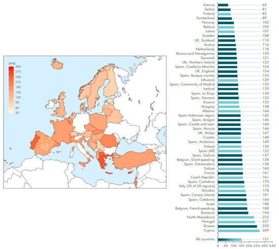
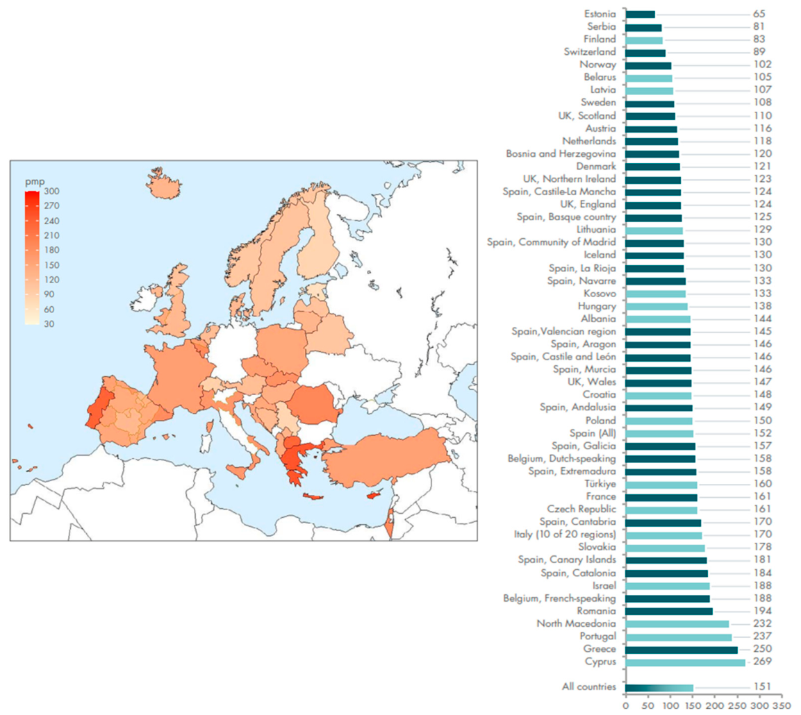
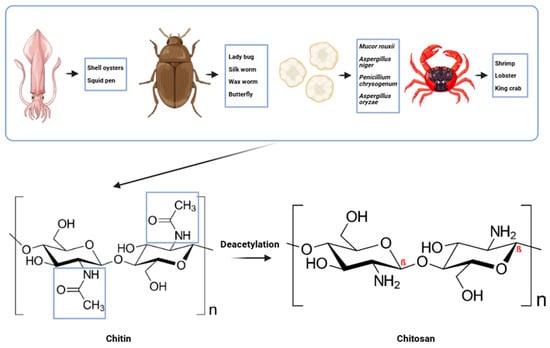
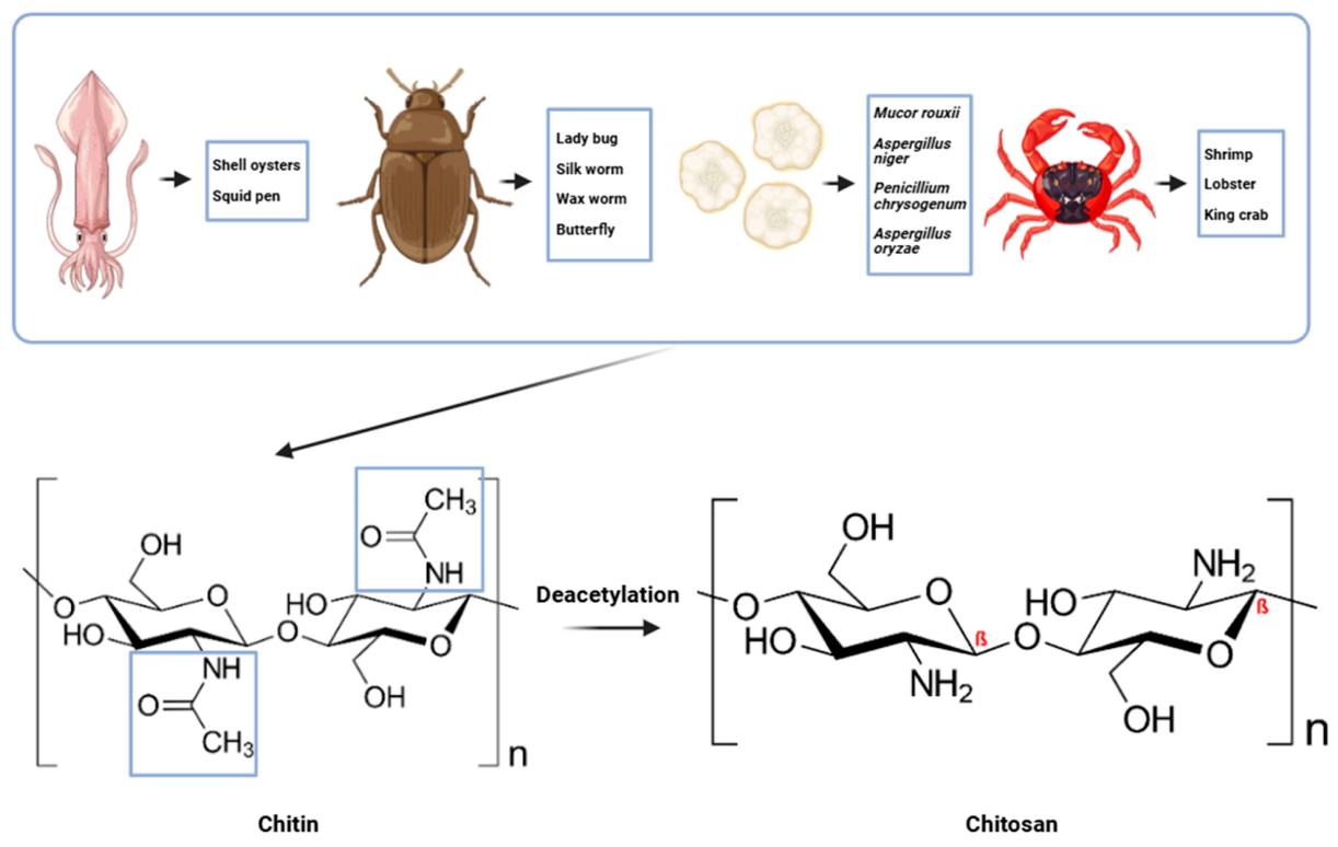
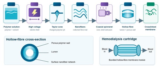
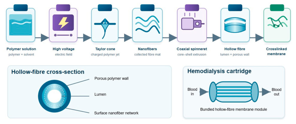
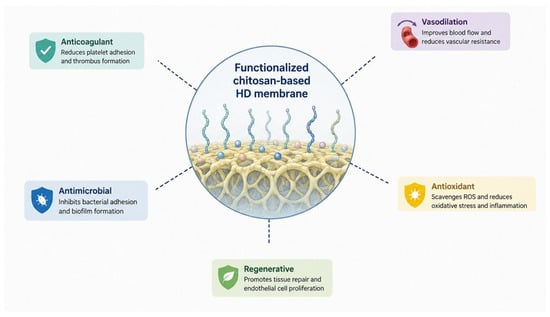
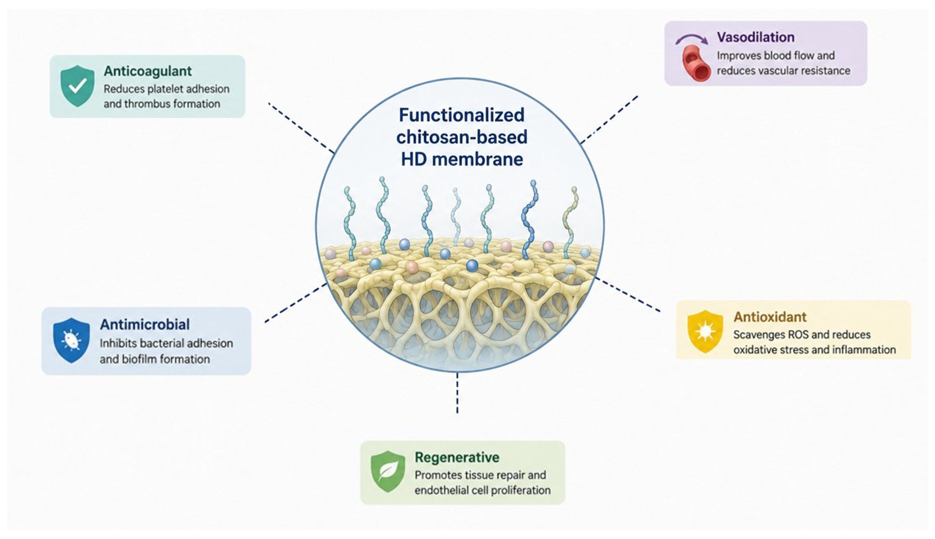

Disclaimer/Publisher’s Note: The statements, opinions and data contained in all publications are solely those of the individual author(s) and contributor(s) and not of MDPI and/or the editor(s). MDPI and/or the editor(s) disclaim responsibility for any injury to people or property resulting from any ideas, methods, instructions or products referred to in the content. |
© 2026 by the authors. Licensee MDPI, Basel, Switzerland. This article is an open access article distributed under the terms and conditions of the Creative Commons Attribution (CC BY) license.