


Abstract
Alzheimer’s disease (AD) is a progressive neurodegenerative disorder, and women are disproportionately affected. This review examines the evidence that estrogen signaling contributes to this disparity and asks what a molecularly targeted therapeutic would require. We first distinguish prevalence from incidence in the gender-difference literature and review the genetic, epigenetic, and ascertainment factors that structure female-specific risk, noting that the associations localize consistently to tau rather than to amyloid pathology. We then examine estrogen receptor (ER) biology in the brain, reconciling apparently contradictory accounts of ER and ER function by resolving them at the level of downstream pathway and cell type rather than receptor identity, and review evidence that DNA methylation at ER genes predicts cognitive decline and tangle density through mechanisms independent of receptor expression. The consistent failure of systemic hormone therapy in clinical trials is interpreted not as a refutation of this biology but as a constraint defining three design requirements: receptor engagement resolved by subtype, cell type, and tissue compartment; sufficient central nervous system exposure; and delivery timed to the perimenopausal window. Against these requirements we review AI-driven approaches to ER modulator design, spanning diffusion-based backbone generation and protein language models for peptides, and virtual screening and generative frameworks for small molecules. We argue that the principal barrier is data rather than model architecture: ER-selective binding data and peptide blood–brain barrier permeability data are both scarce, and no AI-designed ER modulator has been evaluated in an AD model. We conclude by identifying the epigenetic layer as a target class that current design pipelines do not address, and for which the necessary editing tools are technically available but validated against the wrong readout.
1. Introduction
Women account for approximately two-thirds of all Alzheimer’s disease cases [1]. The most widely supported mechanistic explanation for this is hormonal: the permanent decline in estrogen at menopause coincides with a well-documented period of elevated AD vulnerability [2,3]. In the brain, estrogen reduces amyloid-beta production, suppresses tau hyperphosphorylation, supports hippocampal neurogenesis, and modulates inflammatory cascades implicated in neurodegeneration [4,5,6].
Clinical trials of hormone therapy (HT) have largely failed to prevent cognitive decline and in some cases have worsened outcomes [7]. This does not, however, constitute evidence against estrogen’s neuroprotective role. Systemic HT lacks receptor subtype selectivity and cannot preferentially engage estrogen receptor beta (ER), which is well expressed in brain but minimally expressed in breast and uterus, over estrogen receptor alpha (ER), whose activation in peripheral tissue carries proliferative risk [8,9,10]. It cannot be timed to the critical perimenopausal window identified in imaging studies [11,12], nor does it address the epigenetic remodeling of ER genes, where DNA methylation patterns predict cognitive decline independently of receptor expression levels [13].
Two therapeutic modalities merit consideration for a molecularly targeted approach. Peptides can engage the ER coactivator-binding AF2 domain, a surface inaccessible to small hormones, enabling subtype-selective interference with ER signaling without systemic estrogenic effects [14,15,16]. Small molecules, on the other hand, offer the bioavailability and blood–brain barrier (BBB) penetration that peptides struggle to achieve [17]. Neither modality has been fully realized for AD, but recent advances in artificial intelligence now make both tractable.
Optimizing a therapeutic candidate requires satisfying constraints on binding affinity, receptor subtype selectivity, metabolic stability, and brain penetration across a chemical and sequence space too vast for direct experimental exploration. AI-driven methods address this problem through diffusion models for peptide backbone generation [18], protein language models for sequence optimization [19,20], deep learning virtual screening [21,22], and generative frameworks for de novo molecular design [23,24]. Structure-conditioned backbone generation, billion-compound virtual screening, and simultaneous multi-objective optimization represent strategies with no current experimental equivalent.
This review examines the neuropathology and female-specific epidemiology of AD, the mechanistic and epigenetic basis of estrogen’s neuroprotective role, and the current state of AI methods for peptide and small-molecule ER modulator design, synthesizing these into a rationale for AI-driven, ER-targeted therapeutics in AD drug development.To provide a comprehensive overview of the field, we conducted a literature search using PubMed, Web of Science, and Google Scholar. Search terms included combinations of “Alzheimer’s disease,” “estrogen,” “estrogen receptor,” “ER,” “ER,” “DNA methylation,” “epigenetics,” “artificial intelligence,” “machine learning,” “drug design,” and “peptide design.” We prioritized recent peer-reviewed studies, primary research articles, and highly relevant reviews, while also including seminal studies when necessary to provide historical and mechanistic context.
2. Alzheimer’s Disease
Dementia describes a group of progressive, irreversible brain disorders affecting memory and behavior. An estimated 55 million people worldwide currently live with dementia, projected to reach approximately 139 million by 2050 [25,26]. Alzheimer’s disease (AD) is the most common cause, and no disease-modifying cure exists.
Definitive diagnosis rests on pathologic examination of brain tissue. Macroscopically, AD presents with moderate cortical atrophy concentrated in multimodal association cortices and limbic structures, medial temporal atrophy involving the hippocampus and amygdala, enlarged lateral ventricles, and reduced brain weight. None of these features is specific to AD; each also occurs in healthy aging and in other neurodegenerative conditions [27].
The microscopic hallmarks are neurofibrillary tangles (NFTs) and neuritic plaques. NFTs are composed primarily of paired helical filaments of tau, a cytoskeletal protein that becomes abnormally phosphorylated and aggregates into filamentous structures [28,29]. Neuritic plaques consist principally of amyloid-beta (), a peptide derived from amyloid precursor protein (APP) that aggregates into beta-pleated sheets and accumulates extracellularly, where it interacts with microglia, reactive astrocytes, and complement proteins to drive further neuronal damage [27].
The relationship between these two pathologies is central to what follows. The amyloid hypothesis holds that production or deposition is the initiating event, but the correlation between amyloid burden and cognitive decline is weak, and therapeutic reduction of has not yielded effective treatments. Tau aggregation may precede amyloid accumulation, though fibrils appear to promote tau tangling [27]. This distinction between amyloid accumulation and the amyloid-to-tau transition recurs throughout this review: as Section 3.3 and Section 6.2 document, the gender differences and estrogen-related associations in AD localize consistently to tau rather than to amyloid.
Early-onset AD, defined by onset before age 60, is associated with autosomal dominant mutations driving excessive accumulation. The more common late-onset form is linked to the apolipoprotein E (APOE) allele, which increases AD risk and is associated with greater amyloid deposition; APOE functions in lipid transport and amyloid processing in brain. The magnitude of -associated risk differs by gender, a point developed in Section 3.2 [30]. The hippocampus and adjacent medial temporal regions are affected early, as are cholinergic neurons of the basal forebrain, and the resulting decline in acetylcholine is a further hallmark of the disease [27,31].
3. Female and Male Differences in AD Risk
3.1. Prevalence, Incidence, and the Survival Confound
Women account for approximately two-thirds of Alzheimer’s disease cases [1], but this figure describes prevalence and cannot by itself establish differential susceptibility. Because women live longer on average and age is the dominant risk factor for late-onset AD, a population can exhibit substantially skewed prevalence even under identical age-specific incidence rates [32]. Distinguishing prevalence from incidence is therefore prerequisite to any hormonal interpretation of the disparity.
The incidence evidence is mixed. Beam et al. found no consistent gender difference in age-specific incidence across most age bands, with elevated rates in women appearing in some cohorts above age 80 and not in others [1]. This heterogeneity is itself informative: it suggests that if a gender-specific biological risk exists, its effect size is modest relative to age and likely interacts with other modifiers rather than acting uniformly across the female population [32].
3.2. Genetic Modifiers of Female-Specific Risk
The clearest evidence for a sex-by-genotype interaction comes from APOE. In a meta-analysis spanning 27 datasets, Neu et al. found that the allele confers a stronger association with clinical AD in women than in men, with the disparity concentrated between ages 55 and 70 and attenuating outside that window [30].
This interaction is directly relevant to the hormonal account. The HT literature reviewed in Section 5.4 reports cognitive and volumetric benefit specifically in APOE4 carriers [33], implying that APOE genotype conditions the response to estrogen rather than acting on an independent axis.
Sex-chromosomal contributions provide a second, mechanistically distinct route. Yan et al. identified the X-linked deubiquitinase USP11 as a driver of tau aggregation: USP11 escapes complete X-inactivation and is expressed at higher levels in female mice and in women, and its genetic elimination preferentially protects female mice from acetylated tau accumulation and cognitive impairment [34]. This establishes a sex-specific pathway to tau pathology that does not require hormonal mediation, although USP11 has also been reported to interact with estrogen signaling [34].
3.3. Gender Differences in Neuropathology
Gender differences appear in the distribution of pathology, not only in disease occurrence. Buckley et al. found that among clinically normal older adults, women showed elevated entorhinal tau relative to men specifically within the amyloid-positive subgroup, with the difference scaling with amyloid burden; no consistent sex effect was present in the sample as a whole [35]. The conditional structure of this result matters, as it locates the gender difference at the amyloid-to-tau transition rather than in amyloid accumulation itself.
Coughlan et al. extended this to reproductive variables, reporting that earlier age at menopause and initiation of HT more than five years after menopause were each associated with higher regional tau, again predominantly in the context of elevated amyloid [36]. Together with the observation that HT use associates with lower tau-PET measures in postmenopausal women [37], these findings converge on tau as the pathological compartment most sensitive to estrogen exposure, which connects directly to Section 6, where methylation at ER genes predicts tangle density specifically [13].
3.4. Reproductive History as a Cumulative Exposure
Treating menopause as a single event obscures substantial variation in lifetime estrogen exposure. Reproductive history variables—age at menarche, parity, reproductive span, and whether menopause was natural or surgical—collectively index this exposure and associate with midlife neuroimaging markers of dementia risk [11,12]. Schelbaum et al. report associations between HT use and gray matter volume most pronounced in women aged 50–59 [11], Lee et al. find that initiation five or more years after menopause associates with increased white matter hyperintensity (WMH) volume [12], and Coughlan et al. report a concordant five-year threshold for tau [36]. That three independent cohorts using different outcome measures converge on approximately the same timing threshold is the strongest available support for the critical-window hypothesis, and it is the empirical basis for treating delivery timing as a design constraint in Section 9.
3.5. Non-Biological Contributors and Ascertainment
Not all of the observed disparity is biological. Historical differences in educational and occupational attainment between men and women in the cohorts now reaching AD-typical ages contribute to differences in cognitive reserve [32]. Ascertainment introduces a further distortion: women outperform men on verbal memory tests even at comparable levels of hippocampal atrophy [38], so sex-invariant diagnostic cutoffs will systematically under-detect early impairment in women.
Throughout this review we use sexto denote biological attributes and reserve gender for socially patterned exposures, using the latter only where the distinction is material.
4. Estrogen and Menopause
Estrogens and progesterone are produced cyclically by the ovaries during a woman’s reproductive years. During perimenopause, there are fluctuations in hormone levels, and after menopause, the depletion of ovarian follicles leads to a permanent decline in the circulating levels of these sex hormones. After menopause, estrone becomes the main estrogen in the body, derived from androstenedione produced in fat tissue and other sources. Estradiol, the main estrogen before menopause, is now mainly produced from estrone. While testosterone levels remain largely unchanged after natural menopause, circulating estrone levels exceed those of estradiol postmenopause. These hormonal changes can impact the brain, potentially influencing cognitive processes and the development of AD. However, it is difficult to assess the direct effects of menopause on AD risk, as menopause cannot be randomly assigned in studies. As a result, most research on AD has focused on the potential therapeutic role of estrogen-containing HT during the postmenopausal period [7].
5. Biological Role of Estrogen in the Brain
The effects of estrogen go beyond the regulation of reproductive behaviors. Estrogen plays a significant role in the brain because it could impact the brain’s cognitive functions and synaptic health through a complex interplay of receptors and signaling pathways. In addition, estrogen could also influence mood, motor coordination, pain sensitivity, and provide neuroprotection against conditions such as stroke, Parkinson’s, and Alzheimer’s diseases. Estrogen facilitates higher cognitive functions by acting on brain regions including the prefrontal cortex and hippocampus, which are crucial for learning and memory [4].
5.1. Estrogen Receptors
Estrogen receptors (ERs) can mediate different effects in nuclear and non-nuclear sites within the brain. In general, the classical genomic effects are primarily mediated through the activation of ER and ER, whereas rapid non-genomic effects are facilitated by membrane-bound ER, ER, and G protein-coupled estrogen receptor 1 (GPER1). These receptors are found in synapses of the hippocampus and prefrontal cortex. ER and ER exhibit differential expression across various brain regions and are regulated by estradiol [4]. For instance, ER mRNA is highly abundant in the amygdala and hypothalamus, while ER mRNA is found in the hippocampus, claustrum, thalamus, and cerebral cortex [39]. In humans, ER protein is first detected in the nuclei of pyramidal cells of the Ammon’s horn at 15 gestational weeks, whereas ER protein is first detected in proliferating zones and the cortical plate at 9 gestational weeks. Furthermore, ER is found in both the cytoplasm and nuclei of pyramidal neurons in adulthood. In all, ER and ER functions are tissue-, cell-type-, and context-dependent. It is not describing ER as exclusively harmful or ER as exclusively protective; it better explains their distinct CNS and peripheral effects.
The non-genomic actions of estrogen involve the activation of signaling pathways at extranuclear sites, resulting in rapid effects on cell function and gene expression. These actions occur via receptors located outside the nucleus, including in mitochondria, presynaptic terminals, postsynaptic spines, and glial cell processes. Caveolae function as reservoirs of ERs that are activated by estradiol near the cell surface, and GPER1 is associated with PSD-95 in the dendritic spine, coupling estrogen action to membrane progestin receptors, 5HT1A receptors, and corticotrophin releasing hormone (CRH1) receptors [40]. Furthermore, non-genomic actions are noted for other steroid hormones, such as progestins, androgens, glucocorticoids, mineralocorticoids, and vitamin D [4].
5.2. Estrogen Receptors in Alzheimer’s Disease
Estrogen signaling plays a complex role in AD, with both protective and potentially harmful effects influenced by various factors. A key observation is that the drop in estrogen levels during menopause is linked to an increased risk of AD in women. Beyond its role in reproduction, estrogen is involved in numerous processes, particularly in the central nervous system (CNS), where it supports neuronal health and influences APP processing. This is achieved through mechanisms involving brain-derived neurotrophic factor (BDNF), calcium signaling, and Daxx protein regulation [3]. Estrogen is considered a neuroprotective agent, reducing the risk of AD by countering oxidative stress, combating toxicity, and protecting neurons from glutamate-induced damage [6].
Estrogen’s effects are mediated through three main receptors: GPER1, ER, and ER. These receptors are found throughout the brain, where they contribute to cognitive function and nervous system protection. ER has been reported to associate with neuronal survival and cognitive support, with reduced levels linked to a higher risk of AD; lower ER expression has likewise been observed in AD patients [8]. Both ER and ER regulate gene transcription following estrogen binding, influencing critical cellular processes [8]. These subtype-specific attributions are not straightforwardly consistent with one another, and we return to reconciling them in Section 5.3.
Estrogen protects the brain through several convergent pathways, including regulation of calcium levels, APP metabolism, and glutamate toxicity. It helps maintain calcium balance in neurons, reduces harmful calcium surges caused by and glutamate, and promotes the non-amyloidogenic breakdown of APP to lower production. Estrogen additionally enhances clearance by microglia, reduces its aggregation, and protects neurons by inhibiting pathways such as apoptosis signal-regulating kinase 1 (Ask1). It also supports synapse formation, potentially through interactions with apolipoprotein E (ApoE) [6,41].
The relationship between estrogen and production is particularly well characterized: estrogen reduces levels, enhances APP transport, and minimizes aggregation. Estradiol has shown promise in reducing in animal studies. Estrogen also interacts with ApoE and with enzymes such as catechol-O-methyltransferase (COMT), potentially influencing AD risk depending on genetic factors including the APOE allele. A decline in estrogen’s protective effects may relate in part to reduced COMT activity in estrogen metabolism [9].
Therapeutic strategies targeting estrogen signaling include HT, selective estrogen receptor modulators (SERMs), ER-specific agents, and alternative estrogenic compounds such as genistein. Other approaches—transdermal estrogen delivery, selective ER activation, and compounds such as 2-methoxyestradiol (2ME)—aim to leverage estrogen’s neuroprotective effects while minimizing systemic risk [6,9]. These strategies share a common limitation: each modulates estrogen signaling with only coarse control over receptor subtype, cell type, and tissue compartment. That limitation is what motivates the computationally designed modalities reviewed in Section 7 and Section 8.
5.3. Reconciling Subtype-Specific Roles
The literature assigns apparently contradictory roles to ER, and the contradiction dissolves only when the relevant level of resolution is specified. Three observations structure the resolution.
First, neuroprotection is not subtype-exclusive. Selective agonists for either receptor—propyl pyrazole triol (PPT) for ER, diarylpropionitrile (DPN) for ER—protect hippocampal neurons against excitotoxic insult with comparable efficacy and comparable induction of Bcl-2, and protection against toxicity requires expression of either subtype together with mitogen-activated protein kinase (MAPK) activation [42]. Framing ER as the neuroprotective receptor and ER as merely proliferative is therefore not supported at the level of neuronal survival.
Second, the subtypes diverge on specific downstream pathways rather than on protection globally. Xiong et al. report opposing effects of the two receptors on tau phosphorylation, mediated through disparate effects on the miR-218/PTPA pathway [43], and ER and ER activation have been reported to exert opposing effects on hippocampal ApoE expression. Pathway-level divergence, not global divergence, is what makes subtype selectivity a coherent design objective.
Third, the relevant resolution is often cellular rather than regional. Spence et al. demonstrated that estrogen-mediated neuroprotection and anti-inflammatory effects operate through ER signaling on astrocytes, and not through ER signaling on either astrocytes or neurons [44,45]. A subtype-selective agent therefore acts on a specific cell population, and its net effect depends on the expression profile of that population rather than on receptor identity alone.
Against this background, the association between ER and neuronal survival noted in Section 5.2 [8] admits a mechanistic interpretation that does not conflict with ER’s proliferative role in peripheral tissue. Wang et al. found that ER co-localizes with neurofibrillary tangles and interacts with tau in vivo, with the interaction increased in AD brain and total ER expression unchanged relative to controls [46]. The functional deficit is thus consistent with sequestration of ER by tau pathology rather than with reduced receptor expression—placing loss of ER signaling downstream of tau accumulation and connecting this section to the tau-specific findings of Section 3.3 and Section 6.2 [13,35].
The design implication is that ER-preferential agonism remains the rational objective, though not for the reason usually given. ER is well expressed in brain and minimally expressed in breast and uterus [10], so ER-selective agents capture CNS benefit while avoiding peripheral proliferative liability—a compartmental argument rather than a claim about which subtype is neuroprotective. ER should not be abandoned as a target, given the astrocytic evidence above, but ER-directed strategies face the additional obstacle that receptor availability is compromised by tau sequestration in established disease, which argues for their use in prevention rather than treatment.
5.4. Estrogen Therapy and Brain Morphology in Alzheimer’s Disease
In non-imaging research on HT, the focus is predominantly on how the timing of therapy initiation and genetic influences, particularly the APOE genotype, affect cognitive scores and behavioral changes. This emphasizes the significance of understanding individual variability in response to HT. Concurrently, multiple brain imaging studies have explored these dynamics across various brain regions, using diverse neuroimaging modalities. A key advantage of this approach is its emphasis on biomarkers within brain imaging studies for AD. This methodology underscores the premise that changes in specific biomarkers can often precede noticeable cognitive impairments, offering potential early intervention strategies to mitigate AD progression.
In a recent study, Saleh et al. [33] examined the interplay between APOE genotype and age at HT initiation on the heterogeneity of cognitive response to HT, employing magnetic resonance imaging (MRI). They reported that APOE4 carriers who used HT had the highest Repeatable Battery for the Assessment of Neuropsychological Status (RBANS) delayed memory scores, and exhibited 6–10% larger left entorhinal and bilateral amygdala volumes. Notably, earlier initiation of HT was associated with greater right and left hippocampal volumes, but only among APOE4 carriers. Lord et al. [47] investigated the overall effect of HT on the aging brain by studying the influence of HT use and duration on gray matter (GM) density through voxel-based morphometry (VBM). In their study, HT users had higher GM density in the superior frontal gyrus and lower GM density in the posterior hippocampus, parahippocampal gyrus, posterior cingulate, and angular gyri relative to non-users. Wang et al. [37] used MRI and positron emission tomography (PET) to explore how HT affects AD biomarker-informed pathophysiology. Their findings suggest that HT use is linked to lower tau-PET measures in postmenopausal women. Schelbaum et al. [11] reported positive associations between HT use and MRI-based gray matter volume (GMV) in several regions, including the bilateral superior frontal and supramarginal gyri, right middle temporal gyrus and frontal pole, and left inferior temporal, fusiform, and medial frontal gyri. These associations were most pronounced among younger participants (50–59 years), reinforcing that initiating HT near the onset of menopause may confer supportive effects on brain aging. Lee et al. [12] examined the influence of HT use on WMH volume, a neuroimaging marker associated with cerebrovascular health and Alzheimer’s pathology. Their findings indicate that initiating HT five or more years after menopause is linked to increased WMH volume, which may have detrimental consequences for brain health.
6. DNA Methylation and Estrogen Receptor Genes
6.1. Mechanistic Background
DNA methylation is the covalent addition of a methyl group to cytosine, predominantly at CpG dinucleotides. The mark is established de novo by DNA methyltransferase 3A (DNMT3A) and DNMT3B, maintained through replication by DNMT1, and actively reversed through ten-eleven translocation (TET)-mediated oxidation of 5-methylcytosine [48]. Its functional consequence is context-dependent: promoter and CpG-island methylation is generally associated with transcriptional repression, whereas gene-body methylation shows a weaker and less consistent relationship with expression.
This context dependence is directly relevant to interpreting the findings below, because an association between methylation and a clinical outcome does not entail a corresponding change in receptor abundance.
6.2. Methylation at ER Genes and Alzheimer’s Traits
Oveisgharan et al. examined DNA methylation at all three estrogen receptor genes—ESR1, ESR2, and GPER1—in relation to cognitive trajectory and neuropathological burden in women [13]. Methylation at each of the three loci associated with the rate of cognitive decline and with tau tangle density, and GPER1 RNA expression showed independent associations with cognitive decline and with indices of AD pathology [13].
The specificity of the association to tau tangle density rather than to AD neuropathology generally [13] places this finding alongside the sex-specific tau results discussed in Section 3.3 [35,36], and reinforces tau as the pathological compartment most responsive to estrogen-related variation.
6.3. Dissociation Between Methylation and Receptor Expression
The most consequential result is a negative one: methylation at the ER genes and GPER1 expression were each associated with outcomes but not with one another [13]. This dissociation excludes the most intuitive causal model, that methylation drives cognitive decline by silencing receptor expression, and leaves several alternatives open. Methylation may act through isoform- or cell-type-specific effects that bulk expression measurement cannot resolve; it may index cumulative hormonal or inflammatory exposure history rather than functioning as a proximate regulator; or it may partly reflect shifts in cellular composition rather than regulatory state within a fixed population. Distinguishing these possibilities remains unresolved and constitutes a substantive gap in the field.
6.4. Methodological Constraints
Two limitations bound the interpretation. Methylation was assayed in bulk brain tissue, so the measured signal reflects both within-cell regulatory state and between-cell composition; without cell-type deconvolution or single-cell resolution these cannot be separated. Molecular measurement is also cross-sectional even where cognitive assessment is longitudinal, so temporal ordering between methylation change and disease process cannot be established.
6.5. Toward Epigenetically Directed Intervention
If ER gene methylation influences AD traits through mechanisms independent of receptor expression, then therapeutics directed at receptor activity do not engage this axis at all. The relevant target class would instead be the methylation machinery itself.
This is no longer purely hypothetical. Locus-specific methylation editing has been demonstrated using catalytically inactive Cas9 (dCas9) fused to TET1 or DNMT3A, which permits targeted demethylation or de novo methylation at defined genomic sites without altering DNA sequence [48]. Critically for CNS applications, Liu et al. showed that dCas9-TET1-mediated demethylation of BDNF promoter IV was sufficient to induce BDNF expression in post-mitotic cortical neurons, and that lentiviral delivery of the system in vivo produced targeted demethylation in mice [48], establishing that methylation editing is achievable in non-dividing neurons, the relevant cell population for AD. Independent groups have demonstrated in vivo demethylation using dCas9–scFv–TET1 architectures [49], improved methylation efficiency through DNMT3A–DNMT3L fusions [50], and durable “hit-and-run” silencing that persists after transient effector expression [51].
Delivery to brain has also been addressed. Kantor et al. engineered a compact dSaCas9–KRAB–MeCP2(TRD) repressor packaged in a single adeno-associated virus (AAV) vector, circumventing the packaging-capacity limit that ordinarily forces dCas9-effector systems to be split across two vectors, and demonstrated sustained in vivo repression of endogenous murine Apoe following stereotaxic injection [52]. That this platform was validated against APOE—the genetic modifier most strongly implicated in the sex-specific risk discussed in Section 3.2 [30]—is notable, since it establishes both feasibility and precedent within the same risk architecture.
Three obstacles nonetheless separate this from an ER-directed application. Delivery remains invasive: the demonstrations above rely on stereotaxic or direct viral administration rather than systemic CNS-penetrant delivery [52]. Durability and off-target specificity of induced methylation changes are incompletely characterized, and the transcriptional consequence of dCas9-directed methylation is frequently modest and not proportional to the methylation change achieved [48]. Most fundamentally, these tools are validated by their effect on gene expression, whereas the Oveisgharan finding is precisely that ER methylation acts through an expression-independent route [13]—so the available instruments are calibrated against the wrong readout for this target. Resolving that mismatch is prerequisite to designing an intervention here, and it is a question about the biology rather than about the technology.
7. AI-Driven Peptide Design for Estrogen Receptor Targeting
The discovery of peptides that bind to ERs represents an important strategy for developing therapeutics to modulate ER activity in AD [18,53]. Unlike small-molecule drugs that typically bind the estrogen-binding pocket, peptides can target alternative sites on the ER, such as the coactivator-binding AF2 domain, potentially enabling more selective modulation of ER signaling pathways [14,15]. This approach is particularly relevant in AD, where precise control of ER subtype activity could maximize neuroprotective benefits while minimizing peripheral proliferative risks [53]. Recent advances in artificial intelligence and computational modeling have further accelerated the design and optimization of ER-targeting peptides by integrating structural data, peptide–protein interaction prediction, and molecular-dynamics-based screening [18,53,54].
7.1. Experimental Foundations and Design Challenges
The AF2 site serves as the docking surface on the ER for coactivator proteins containing LXXLL motifs (leucine-X-X-leucine-leucine), where X represents any amino acid [14,55]. Upon estrogen binding and conformational change of the ER, the AF2 groove becomes accessible, allowing LXXLL-containing coactivators to bind and initiate transcription [14]. Peptides designed to occupy this site can have specific functions antagonizing ER activity, disrupting some coactivator interactions while preserving interactions necessary for neuroprotective signaling [15,16]. Additionally, different coactivator recruitment patterns between ER and ER suggest that AF2-targeted peptides could achieve ER subtype selectivity, which is relevant given the compartmental rationale for ER-preferential engagement set out in Section 5.3 [10,14,16].
Early peptide approaches were mainly empirical and constrained by experimental capabilities. In the 1990s, Chang et al. used phage display technology to screen combinatorial libraries exceeding over peptide variants for ER binding [14]. While this approach successfully identified the importance of the LXXLL motif and demonstrated that flanking sequence residues determine estrogen binding affinity and receptor subtype selectivity, the experiment was fundamentally limited by random sampling of sequence space and required extensive experimental validation for each candidate. Peptide design faced several intrinsic challenges: (i) achieving binding affinity sufficient to compete with endogenous coactivators while also maintaining selectivity for ER over other nuclear receptors that recognize similar LXXLL motifs; (ii) efficiently crossing cell membranes despite peptide hydrophilicity and size, even when binding affinity is strong; (iii) avoiding degradation by cellular proteases; and (iv) for AD applications, crossing the BBB. Achieving therapeutic efficacy therefore requires multi-parameter optimization [14,53].
7.2. Computational and AI-Assisted Peptide Optimization
Computational modeling began to address these bottlenecks by allowing visualization of peptide–receptor interactions. In a landmark study, Speltz et al. (2016) demonstrated that molecular dynamics (MD) simulations, computational methods that model atomic-level movements and interactions over time, could guide peptide design [15]. MD calculates the forces acting on each atom based on quantum mechanical principles and predicts how molecules move and interact in solution or when bound to proteins. The authors applied MD simulations to design stapled peptides that target the ER AF2 site by modeling arginine placement within the stapled helices. Arginine’s positive charge enables electrostatic interactions with the negatively charged phospholipid heads on cell membranes, which allows peptides to enter cells more easily. Their optimized peptide, R4K1, exhibited a 22-fold improvement in ER binding affinity (Kd = 19 nM) and achieved nearly 80% nuclear localization in MCF-7 cells (a breast cancer cell line with estrogen, progesterone, and glucocorticoid receptors). This study demonstrated that computationally guided peptide optimization can simultaneously enhance receptor affinity and cellular permeability—a substantial improvement over traditional screening methods that struggle to balance these properties, as modifications improving one property typically compromise the other [15].
Building on the work of Speltz et al. [15], Fujiwara et al. [16] used structure-guided and AI-assisted modeling to design cyclic peptides that integrated membrane-permeable arginine residues with the LXXLL recognition motif. Cyclization, or connecting the ends of the peptide in a circle, makes the peptide more resistant to being broken down by cellular enzymes. Their lead peptide bound ER, crossed cell membranes, and successfully blocked the interaction between ER and its coactivator SRC1 (IC50 = 7.1 M), providing a prototype for stable, cell-permeable AF2 antagonists [16].
In parallel to these discoveries, recent deep generative models and reinforcement-learning frameworks are redefining de novo peptide design [18]. Diffusion-based models, such as RFdiffusion and related methods, generate peptide backbones that conform to specific binding surfaces, which enables structure-conditioned sequence designs without any predefined motifs. In addition, transformer-based language models, trained on large protein–peptide interaction datasets (ProtT5, PepGPT), learn context and amino acid dependencies and can predict binding-competent sequences directly from target embeddings [56,57,58]. Together, these frameworks allow optimization of multiple parameters through feedback between prediction and simulation. These pipelines can evaluate millions of virtual peptides, filter top-scoring candidates, and output three-dimensional complexes [18].
Modern AI-driven peptide design employs diverse computational frameworks, each addressing different aspects of the design challenge. While these methods were originally developed for broader peptide and protein design applications, including antimicrobial peptides, enzyme design, and therapeutic antibodies, they can be readily adapted for ER-targeting peptides. Understanding these categories and how they complement each other is essential for applying them effectively to AD therapeutic development. Figure 1 illustrates how these components can be assembled into a single design pipeline.
Figure 1.
Integrated AI pipeline for ER-selective peptide design.
These approaches can be broadly categorized into sequence-based methods that learn patterns from protein databases, structure-based methods that generate geometrically precise backbones, and hybrid approaches that combine both [18,20]. Sequence-based language models like ESM-2 excel at rapid generation and capture implicit biochemical constraints but lack structural awareness [19]. Structure-based approaches like RFdiffusion provide exceptional geometric control for binding-interface design [18]. Hybrid methods integrate multiple information sources for comprehensive optimization. State-of-the-art peptide design pipelines typically integrate multiple frameworks in sequence, using diffusion models to generate backbones, language models to optimize sequences, and AlphaFold to validate binding modes before MD refinement [18,20,59].
Table 1 summarizes the major computational and AI framework categories used in modern peptide design. While these methods are general-purpose tools applicable across diverse peptide design challenges, each offers specific capabilities relevant to ER-targeting therapeutics for AD.
Table 1.
Categories of computational and AI frameworks used for modern peptide design.
These frameworks can work together in modern design pipelines. For example, to design an ER-selective peptide, RFdiffusion can generate backbones that fit the ER AF2 groove; ProteinMPNN can design sequences that fold into those backbones; ESM-2 can create variants around the LXXLL motif; AlphaFold-Multimer can predict how each variant binds to ER versus ER to identify selective candidates; and MD can test whether these complexes are actually stable.
For applications to ERs in AD, peptide therapeutics that selectively engage ER or modulate ER signaling could restore estrogen’s neuroprotective effects without systemic HT. Computationally optimized peptides capable of crossing the BBB and avoiding degradation have particular promise as next-generation SERMs tailored for the CNS. With more refinement and validation in AD models, AI-designed ER-targeting peptides can enable therapeutics that combine molecular precision with efficiency [18,53].
8. AI-Driven Small Molecule Design
8.1. Rationale for Small-Molecule ER Modulators in Alzheimer’s Disease
While peptides offer selectivity through their extended binding interfaces, small molecules remain the dominant modality for drug delivery because of their favorable properties, including bioavailability and metabolic stability, and—critically for AD applications, as discussed in Section 7—enhanced BBB penetration [21,22]. Small molecules are typically defined as organic compounds with molecular weights below 500 Da that obey Lipinski’s rule of five [17], enabling efficient absorption and distribution throughout the body [22]. For ER modulation, small molecules offer many practical advantages over peptides: established manufacturing processes, predictable dosing, and decades of clinical experience with SERMs like tamoxifen and raloxifene [21,55]. Recent advances in artificial intelligence have also dramatically accelerated the identification and optimization of small-molecule ER modulators by enabling virtual screening of billion-compound libraries, property prediction through machine learning, and de novo molecular generation [21,22,62].
8.2. AI-Enabled Virtual Screening and Structure-Based Design
The scale of chemical space—estimated at possible drug-like molecules—makes exhaustive experimental screening impossible [22]. AI-driven virtual screening addresses this challenge by using neural networks to predict which compounds merit experimental testing. Foo et al. (2024) demonstrated this approach by employing Deep Docking to virtually screen over 1.36 billion compounds from the ZINC15 database against the ER AF2 coactivator-binding site [55]. Deep Docking is an iterative neural network framework that learns to predict protein binding affinity without evaluating every compound: a random subset undergoes conventional docking, a neural network learns patterns from these results, and the model then prioritizes the most promising untested compounds for subsequent rounds. Using the crystal structure of ER bearing the clinically relevant Y537S mutation—one of the most common resistance mutations in metastatic breast cancer—Foo et al. reduced the 1.36 billion compound library to 9.2 million predicted AF2 binders, ultimately identifying VPC-260724 as a potent small-molecule ER modulator through cell-based validation [55].
VPC-260724 demonstrated multiple favorable properties: it inhibited ER transcriptional activity, directly bound the AF2 site displacing natural coactivator peptides, retained activity against both Y537S and D538G ER mutants, and showed selectivity for ER over other steroid hormone receptors [55]. This work illustrates how AI can compress years of traditional screening into months while specifically addressing drug resistance mechanisms.
Complementing high-throughput virtual screening, structure-based drug design (SBDD) employs detailed computational modeling to design molecules that fit target binding pockets. Samanta et al. (2024) applied this approach to identify novel ER binders from natural product scaffolds found in Datura species (a type of toxic flowering plant), then used knowledge-based de novo design to optimize these hits [22]. Their workflow began with molecular docking of 99 natural products against the ER ligand-binding domain (PDB: 3OS8), followed by 100-nanosecond MD simulations to assess binding stability [22]. Their top hit was a natural compound called NPC169485, which they used as a starting template for optimization. They then made several chemical modifications to improve its properties, simplifying the ring structure, changing a chemical bond from an ester to a more stable amide, and adding a chlorine atom to one of the rings. The resulting compound, DL-50, bound to the ER much more strongly ( kcal/mol) than either the original natural product ( kcal/mol) or the reference drug in the crystal structure ( kcal/mol) [22].
For AD applications, the most important finding was that DL-50 is predicted to cross the BBB. However, this needs to be tested further as it may be pumped back out by P-glycoprotein transporters, which could reduce how much drug actually accumulates in brain tissue [22].
8.3. Machine Learning and Generative Models for Small-Molecule Discovery
Beyond screening and structure-based design, machine learning models trained on experimental data can also predict whether novel compounds will bind ER, enabling rapid prioritization before synthesis [21,62]. Quantitative structure–activity relationship (QSAR) models correlate molecular descriptors with biological activity, and recent advances have substantially improved their accuracy [21,62].
Pusparini et al. (2023) developed MATH (Molecular Attention Transformer with Hierarchical features), a deep learning framework that integrates self-attention transformers with molecular graph information to predict ER inhibitors [62]. Training on 3065 compounds from ChEMBL, their model achieved strong classification performance (AUC-ROC > 0.85), demonstrating that transformer architectures—which were originally developed for natural language processing—can effectively learn chemical patterns relevant to ER binding [62].
The most transformative recent development is generative AI, which creates entirely novel molecules [23,24]. Three different learning schemes dominate the field: reinforcement learning, transformer-based language models, and diffusion models [23,24].
REINVENT 4, developed by AstraZeneca, is an example of reinforcement learning for molecular generation [24]. The system works by first learning chemical structures of valid drug molecules from millions of examples encoded as SMILES strings, which are text-based representations of chemical structures (for example, “CCO” represents ethanol). Once trained, the system generates new molecules and receives a score based on how well they meet desired criteria: Does the molecule bind the target? Is it drug-like? Can it be synthesized in a lab? The system then adjusts to produce better-scoring molecules in the next round, repeating this cycle thousands of times [24]. This approach is particularly powerful because it can optimize multiple properties at once, and for ER-targeting therapeutics, this could mean simultaneously maximizing receptor binding, ensuring selectivity over related receptors, achieving BBB penetration, and maintaining metabolic stability [24].
Diffusion models represent a different approach that has recently transformed image generation and is now being applied to molecular design [23]. These models work by learning to reverse a noise-addition process. During training, the model observes as valid drug molecules are gradually corrupted with noise until they become static, and then learns to run this process backward, starting from noise and iteratively reversing it to reveal a valid molecule [23]. The key advantage over text-based methods like REINVENT is that diffusion models can work directly with 3D molecular coordinates rather than 1D text strings. This means they can inherently consider the three-dimensional shape of a molecule and how it might physically fit into a protein’s binding pocket, potentially generating compounds that are geometrically optimized for their targets from the start [23].
Modern small-molecule discovery pipelines increasingly integrate multiple AI approaches [21,22,23,24]. A representative workflow might begin with generative models creating novel scaffolds, use QSAR models to predict ER activity, filter candidates through absorption, distribution, metabolism, excretion, and toxicity (ADMET) prediction, dock promising compounds to assess binding mode, and validate top hits through MD [22,23,24]. For AD applications specifically, BBB permeability prediction becomes an essential filter that can be incorporated into multi-objective optimization [22].
8.4. Analysis and Implications for Alzheimer’s Disease Therapeutics
Together, the peptide and small-molecule examples presented in Section 7 and Section 8 illustrate several key advantages of AI-driven therapeutic design. First, these approaches dramatically accelerate discovery timelines: Foo et al. screened 1.36 billion compounds computationally in a timeframe that would be impossible experimentally, while generative models like REINVENT can propose thousands of novel candidates in hours [24,55]. Second, AI enables true multi-parameter optimization, simultaneously balancing target affinity, selectivity, metabolic stability, and tissue penetration rather than optimizing each property sequentially [24,53,60]. Third, computational methods can explicitly address drug resistance by designing against mutant protein structures, as demonstrated by VPC-260724’s retained activity against Y537S and D538G ER variants [55].
Several cutting-edge platforms show particular promise for ER-targeting therapeutics in AD, even though they have not yet been specifically applied to this context. RFdiffusion and BindCraft enable de novo protein binder design requiring only target structure as input, potentially generating peptides with improved selectivity for ER over ER and other nuclear receptors [18]. AlphaFold3, with enhanced capabilities for modeling protein–ligand complexes, could enable design of compounds active against specific ER conformational states relevant to neuroprotection [59]. Additionally, integration of generative AI with automated synthesis platforms is beginning to close the loop between computational prediction and experimental validation [23,24].
Despite these advances, there are still significant challenges. Experimental validation is still essential; computational predictions of binding affinity, while increasingly accurate, cannot replace biochemical and cellular assays [21,22]. BBB penetration remains particularly difficult to predict and optimize, with many promising compounds failing to achieve therapeutic brain concentrations [22]. ER subtype selectivity presents an additional challenge: achieving preferential ER engagement in brain while minimizing peripheral ER agonism, for the compartmental reasons set out in Section 5.3, requires sophisticated multi-objective optimization that current methods are only beginning to address [10].
The translational gap between these proofs-of-concept and an AD-relevant candidate warrants explicit statement, because it is wider than the individual results suggest. Every compound and peptide discussed in Section 7 and Section 8 was developed and validated in an oncology context against ER. VPC-260724 was selected against breast cancer resistance mutations and validated in cell-based transcriptional assays [55]; R4K1 was characterized for affinity and nuclear localization in MCF-7 breast cancer cells [15]; the Fujiwara cyclic peptide was assayed for disruption of the ER–SRC1 interaction in vitro [16]; MATH was trained on ER inhibitor data from ChEMBL [62]; and DL-50 exists only computationally, with BBB penetration predicted rather than measured [22]. None has been administered to an animal, let alone evaluated in an AD model.
Four specific things are therefore missing, and none of them is addressed by improved computational methods. First, ER-selective binding data: selectivity is currently predicted rather than measured for every candidate discussed, and the assay data required to validate those predictions is sparse. Second, measured CNS exposure: no candidate has brain concentration data, and the computed permeability scores that stand in for it are least reliable in exactly the molecular weight range at issue. Third, efficacy in an AD model rather than a proliferation assay; the endpoints that established these compounds as active are endpoints of tumor biology, which bear no defined relationship to neuroprotection. Fourth, chronic safety data appropriate to a prevention indication: if the timing evidence in Section 3.4 is correct, the target population is healthy perimenopausal women receiving sustained administration, a use case with a substantially stricter safety threshold than oncology. Until these four are addressed, the AI methods reviewed here should be understood as having demonstrated capability for ER-directed design in general, and not for AD in particular.
9. Discussion
9.1. Hormone Therapy Failure and Therapeutic Design Requirements
Collectively, the evidence reviewed supports a mechanistic model in which estrogen loss accelerates AD pathology through convergent receptor-mediated and epigenetic mechanisms [4,6,13]. The central interpretive challenge is reconciling this with the consistent failure of HT in clinical trials [7]. These failures do not constitute evidence against estrogen as a therapeutic target. Hormone Therapy (HT) trials delivered estrogen systemically, without receptor subtype selectivity, at varied timepoints relative to menopause onset, using heterogeneous formulations, in heterogeneous patient populations [7]. These methodological variables are sufficient to account for inconsistent cognitive outcomes without requiring revision of the underlying biology, a point supported by studies showing benefit specifically in APOE4 carriers and when initiated closer to menopause onset [33,37].
The limitations of systemic HT define the design requirements for a targeted intervention: preferential engagement of ER in brain with peripheral sparing [8,10,39], delivery timed to the perimenopausal window [2], and avoidance of systemic estrogenic risk. The AI-driven approaches reviewed here are positioned to address these requirements through modality-specific strategies. ER-selective modulation represents a particularly attractive starting strategy for CNS/AD applications because it may retain neuroprotective estrogen signaling while reducing peripheral ER-associated proliferative effects. It could be emphasized that receptor activity is tissue- and cell-type-dependent, and future drug design should consider both subtype selectivity and tissue-specific exposure.
9.2. Computational Limitations: Peptide Modalities
For peptide-based modulators, the most critical unresolved obstacle is BBB penetration. Structural modifications that improve receptor affinity and cellular permeability—including cyclization [16,63], stapling [15], and arginine incorporation—do not reliably confer CNS penetration, and computational BBB permeability models offer limited guidance here because they are trained predominantly on small molecules [53]. Predictive accuracy degrades substantially for the molecular weight range relevant to ER-targeting peptides, a limitation that reflects genuine scarcity of experimental BBB permeability data for peptides rather than a gap that better model architectures alone can close.
Subtype selectivity between ER and ER presents a related challenge. The two receptors share highly homologous AF2 surfaces [14,55], meaning that meaningful selectivity depends on capturing subtle geometric and electrostatic differences between their binding grooves. Diffusion-based backbone generators [18] and inverse folding models [20,61] can address this in principle, but doing so reliably requires high-quality structural templates and extensive MD validation. AlphaFold’s confidence metrics for flexible peptide–protein interfaces are imperfect enough that false positives in selectivity prediction remain a practical concern [59,63].
9.3. Computational Limitations: Small Molecule Modalities
Small molecule modalities benefit from a more mature AI toolset, but carry a distinct set of limitations. Most AI-driven ER modulator work has been conducted in oncology contexts [21,55,62], where the compound properties that matter most differ substantially from those relevant to AD. CNS penetration, metabolic stability in brain tissue, and avoidance of CNS off-target effects have not been primary optimization targets in existing pipelines, and CNS toxicity prediction remains one of the weakest components of current ADMET modeling [23,53]. A generative model optimizing against an incomplete reward function can produce compounds that score well computationally but fail at experimental validation [24], a failure mode that is difficult to detect until late in the pipeline.
There is also a dataset problem specific to this target. ER-selective binding data is sparse relative to ER because ER drives the majority of published breast cancer assay data [21,62], meaning that models trained on available datasets will systematically underperform in the selectivity dimension that matters most for neuroprotective applications.
9.4. Future Directions
9.4.1. Improving Predictive Reliability
Improving predictive reliability for AD-relevant ER modulators will require both computational and experimental advances. On the computational side, multi-task learning frameworks that jointly optimize across ER binding, subtype selectivity, and CNS penetration are likely to outperform current architectures that treat these as independent sequential filters [23,53]. Integrating MD simulation earlier in generative pipelines, as an active scoring function rather than a terminal validation step, would better capture the conformational flexibility and binding thermodynamics relevant to selectivity prediction [54].
On the experimental side, the most pressing need is data generation specifically targeting ER selectivity and CNS penetration in AD-relevant models. This is the bottleneck that constrains computational progress regardless of model sophistication.
9.4.2. Sequencing In Vitro and Computational BBB Assessment
A practical question follows from the limitations described in Section 9.2: whether peptide candidates should be filtered by in vitro BBB assay before MD simulation, or the reverse. The argument favors in vitro screening first. Because computational permeability predictors are trained predominantly on small molecules and transfer poorly to the peptide molecular weight range [53], using simulation as the triage step means allocating substantial compute against a poorly calibrated objective, and candidates discarded at that stage are discarded on weak evidence. In vitro BBB models are comparatively inexpensive, higher in throughput, and empirical rather than inferential, which makes them better suited to coarse filtering of a structurally diverse candidate set.
The trade-off is real in both directions. In vitro models correlate imperfectly with in vivo brain exposure, particularly for efflux-transporter substrates and receptor-mediated transcytosis, so an in vitro-first strategy risks discarding candidates that would have performed adequately in vivo. Simulation-first avoids that particular loss but incurs cost per candidate high enough to constrain the number of designs evaluated, and there is no validated force-field-level criterion for peptide BBB transit against which to score. The more defensible sequence uses MD after empirical screening rather than before it—not as a filter but as an explanatory step, identifying which structural features of the permeable candidates drove permeability so that the next design round is informed rather than exploratory. This treats simulation as a hypothesis generator for iterative design, which is where its accuracy is adequate, rather than as a decision rule, which is where it is not.
9.4.3. Incorporating Genotype and Timing into Design Objectives
The evidence that HT efficacy depends on APOE genotype and on initiation timing [30,33,36] raises the question of whether this information can inform AI-driven design. The obstacle is categorical: genotype and timing are patient-stratification variables, not molecular properties. No scoring function can optimize a molecule for activity in carriers, because there is no structural or physicochemical feature to which that outcome maps.
The information can nonetheless enter design in three indirect ways. Most directly, timing determines the target product profile: if benefit requires perimenopausal initiation, the indication is prevention rather than treatment, and the target population is healthy women receiving chronic administration. That translates into concrete and encodable objectives—tighter off-target thresholds, a higher bar on metabolic liability, and reduced tolerance for CNS side effects—which multi-objective frameworks can accommodate directly [24]. Second, APOE genotype influences BBB integrity and cerebral lipid transport, so it bears on delivery and pharmacokinetics rather than on target engagement, and could in principle be represented as a modified permeability constraint. Third and most speculatively, if ER and ER activation exert opposing effects on ApoE expression, as noted in Section 5.3, then genotype could inform which subtype a campaign should target—the only route by which genotype touches the molecular design objective itself.
The binding constraint is data rather than formulation. Genotype- and timing-stratified evidence exists in clinical and imaging cohorts, but not in the binding, permeability, and efficacy datasets from which predictive models are trained. Until stratified molecular datasets exist, genotype-conditioned design remains a specification exercise rather than an optimization one.
9.4.4. The Epigenetic Layer as an Unaddressed Target Class
One mechanistic entry point remains largely unengaged by current AI-driven design approaches: therapeutics that target DNA methylation at ER loci rather than receptor activity directly. Because ER gene methylation predicts cognitive decline and tau pathology through mechanisms independent of receptor expression [13], it represents a distinct and underexplored node in the estrogen–AD relationship that existing peptide and small molecule pipelines do not address. As set out in Section 6.5, locus-specific methylation editing is technically feasible in post-mitotic neurons and has been delivered to brain in vivo [48,52]; what remains unresolved is whether such tools can be directed at an expression-independent mechanism.in the future, we might consider physiology associated with the causes of neurodegeneration to generate more hypotheses in Alzheimer’s study.
10. Conclusions
The evidence reviewed here converges on estrogen signaling as a mechanistically substantiated contributor to the sex disparity in Alzheimer’s disease. Estrogen acts on the brain through nuclear and membrane-initiated receptor pathways that regulate APP processing, tau phosphorylation, calcium homeostasis, and neuroinflammation [4,6], and the permanent decline in circulating estradiol at menopause coincides with a period of measurable change in brain structure and AD biomarkers [11,12]. A distinct epigenetic layer operates alongside this: DNA methylation at all three ER genes predicts cognitive decline and tau tangle density through mechanisms apparently independent of receptor expression [13]. The consistent failure of systemic HT to prevent cognitive decline [7] is best read not as a refutation of this biology but as a constraint on how it can be acted upon—a conclusion supported by the observation that benefit appears selectively in APOE4 carriers and with initiation near the menopausal transition [33,37].
What follows from this is a set of concrete requirements for any molecularly targeted alternative: receptor engagement resolved by subtype, by cell type, and by tissue compartment; sufficient CNS exposure to act on the relevant neural targets; and a delivery window aligned with the period of elevated vulnerability. Systemic HT satisfies none of these; peptide and small-molecule ER modulators can in principle satisfy all three, and the AI methods reviewed here are what make the associated design problem tractable at the scale it demands.
That tractability should not be mistaken for demonstrated success. The capabilities are real—billion-compound virtual screening [55], structure-conditioned backbone generation [18], and simultaneous multi-parameter optimization [24] have no experimental equivalent—but essentially all published AI-driven ER modulator work has been conducted in oncology contexts, optimizing against ER in peripheral tissue. None of the compounds or peptides highlighted in this review has been evaluated in an AD model, and the properties that matter most for a CNS indication, specifically BBB penetration, brain metabolic stability, and CNS off-target liability, are precisely those that current predictive models handle least reliably. The rate-limiting factor is data rather than model architecture: ER-selective binding data is sparse relative to ER, and experimental BBB permeability data for peptides is scarce enough that permeability predictors trained predominantly on small molecules cannot be expected to transfer.
A credible AD-relevant candidate would therefore need to demonstrate measured selectivity between ER subtypes rather than predicted selectivity, verified brain exposure in vivo rather than a computed permeability score, and efficacy in an AD model rather than in a proliferation assay. Progress toward that standard depends less on further methodological refinement than on the generation of targeted training data in AD-relevant systems. A further possibility remains largely unexplored: because ER gene methylation predicts AD traits independently of receptor expression [13], the epigenetic layer constitutes a target class that current peptide and small-molecule design pipelines do not address at all. The editing tools required are technically available and have been demonstrated in brain [48,52], but they are validated against gene expression rather than against the expression-independent mechanism at issue. Closing that gap is among the more interesting problems the field currently faces.
Author Contributions
Conceptualization, T.Z.; methodology, T.Z.; visualization, R.G.; writing—original draft preparation, R.G., P.L. and Z.W.; writing—review and editing, T.Z., R.G., Y.S. and L.S.; supervision, T.Z., Y.S. and L.S.; project administration, T.Z.; funding acquisition, L.S. All authors have read and agreed to the published version of the manuscript.
Funding
This work was supported in part by NIH Grants R01 AG071470, U01 AG068057, U19 AG074879, and U01 AG066833.
Data Availability Statement
No new data were generated or analyzed in this review. All data and information discussed are available in the cited literature.
Acknowledgments
During the preparation of this manuscript, the authors used ChatGPT (GPT-4 and GPT-5; OpenAI) and Claude Opus 4 (Anthropic) for assistance with manuscript organization, language refinement, and synthesis and presentation of the reviewed literature. The authors reviewed and edited all output, independently verified the scientific content and cited sources, and take full responsibility for the content of this publication. The authors also used ChatGPT (GPT-4 and GPT-5; OpenAI) to assist with the organization and visual layout of Figure 1 The scientific content and interpretation of the figure were determined by the authors.
Conflicts of Interest
The authors declare no conflicts of interest.
References
- Beam, C.R.; Kaneshiro, C.; Jang, J.Y.; Reynolds, C.A.; Pedersen, N.L.; Gatz, M. Differences between women and men in incidence rates of dementia and Alzheimer’s disease. J. Alzheimer’s Dis. 2018, 64, 1077–1083. [Google Scholar] [CrossRef] [Scilit] [PubMed]
- Jamshed, N.; Ozair, F.F.; Aggarwal, P.; Ekka, M. Alzheimer disease in post-menopausal women: Intervene in the critical window period. J. Mid-Life Health 2014, 5, 38–40. [Google Scholar] [CrossRef] [Scilit] [PubMed]
- Silva, G.B.; Pascucci, J.A.; Karim, H.; Kaur, G.; Lerma, R.O.; Mann, A.K.; Gnanasekaran, S.; Garcia, K.D.T.; Silva, G.D.B. Influence of the Onset of Menopause on the Risk of Developing Alzheimer’s Disease. Cureus 2024, 16, e69124. [Google Scholar] [CrossRef] [Scilit] [PubMed]
- Hara, Y.; Waters, E.M.; McEwen, B.S.; Morrison, J.H. Estrogen effects on cognitive and synaptic health over the lifecourse. Physiol. Rev. 2015, 95, 785–807. [Google Scholar] [CrossRef] [Scilit] [PubMed]
- Vegeto, E.; Benedusi, V.; Maggi, A. Estrogen anti-inflammatory activity in brain: A therapeutic opportunity for menopause and neurodegenerative diseases. Front. Neuroendocrinol. 2008, 29, 507–519. [Google Scholar] [CrossRef] [Scilit] [PubMed]
- Rishabh; Rohilla, M.; Bansal, S.; Bansal, N.; Chauhan, S.; Sharma, S.; Goyal, N.; Gupta, S. Estrogen signalling and Alzheimer’s disease: Decoding molecular mechanisms for therapeutic breakthrough. Eur. J. Neurosci. 2024, 60, 3466–3490. [Google Scholar] [CrossRef] [Scilit] [PubMed]
- Henderson, V.W. Alzheimer’s disease: Review of hormone therapy trials and implications for treatment and prevention after menopause. J. Steroid Biochem. Mol. Biol. 2014, 142, 99–106. [Google Scholar] [CrossRef] [Scilit] [PubMed]
- Almey, A.; Milner, T.A.; Brake, W.G. Estrogen receptors in the central nervous system and their implication for dopamine-dependent cognition in females. Horm. Behav. 2015, 74, 125–138. [Google Scholar] [CrossRef] [Scilit] [PubMed]
- Xiang, X.; Palasuberniam, P.; Pare, R. The role of estrogen across multiple disease mechanisms. Curr. Issues Mol. Biol. 2024, 46, 8170–8196. [Google Scholar] [CrossRef] [Scilit] [PubMed]
- Paterni, I.; Granchi, C.; Katzenellenbogen, J.A.; Minutolo, F. Estrogen Receptors Alpha (ERα) and Beta (ERβ): Subtype-Selective Ligands and Clinical Potential. Steroids 2014, 90, 13–29. [Google Scholar] [CrossRef] [Scilit] [PubMed]
- Schelbaum, E.; Loughlin, L.; Jett, S.; Zhang, C.; Jang, G.; Malviya, N.; Hristov, H.; Pahlajani, S.; Isaacson, R.; Dyke, J.P.; et al. Association of reproductive history with brain MRI biomarkers of dementia risk in midlife. Neurology 2021, 97, e2328–e2339. [Google Scholar] [CrossRef] [Scilit] [PubMed]
- Lee, J.K.; Raghavan, S.; Christenson, L.R.; Frank, R.D.; Kantarci, K.; Rocca, W.A.; Vemuri, P.; Mielke, M.M. Longitudinal associations of reproductive factors and exogeneous estrogens with neuroimaging biomarkers of Alzheimer’s disease and cerebrovascular disease. Alzheimer’s Dement. 2024, 20, 4613–4624. [Google Scholar] [CrossRef] [Scilit] [PubMed]
- Oveisgharan, S.; Yang, J.; Yu, L.; Burba, D.; Bang, W.; Tasaki, S.; Grodstein, F.; Wang, Y.; Zhao, J.; De Jager, P.L.; et al. Estrogen receptor genes, cognitive decline, and Alzheimer disease. Neurology 2023, 100, e1474–e1487. [Google Scholar] [CrossRef] [Scilit] [PubMed]
- Chang, C.Y.; Norris, J.D.; Grøn, H.; Paige, L.A.; Hamilton, P.T.; Kenan, D.J.; Fowlkes, D.; McDonnell, D.P. Dissection of the LXXLL Nuclear Receptor-Coactivator Interaction Motif Using Combinatorial Peptide Libraries: Discovery of Peptide Antagonists of Estrogen Receptors α and β. Mol. Cell. Biol. 1999, 19, 8226–8239. [Google Scholar] [CrossRef] [Scilit] [PubMed]
- Speltz, T.E.; Fanning, S.W.; Mayne, C.G.; Fowler, C.; Tajkhorshid, E.; Greene, G.L.; Moore, T.W. Stapled Peptides with γ-Methylated Hydrocarbon Chains for the Estrogen Receptor/Coactivator Interaction. Angew. Chem. Int. Ed. 2016, 55, 4252–4255. [Google Scholar] [CrossRef] [Scilit] [PubMed]
- Fujiwara, D.; Inaura, S.; Tanaka, Y.; Ito-Harashima, S.; Yamaguchi-Nomoto, A.; Kawanishi, M.; Yagi, T.; Nakase, I.; Fujii, I. A Cyclized Helix-Loop-Helix Peptide as a Molecular Scaffold for Cell-Membrane Permeable Inhibitors for the Interaction Between Estrogen Receptor α and Coactivator SRC1. ChemBioChem 2025, 26, e202500232. [Google Scholar] [CrossRef] [Scilit] [PubMed]
- Lipinski, C.A.; Lombardo, F.; Dominy, B.W.; Feeney, P.J. Experimental and Computational Approaches to Estimate Solubility and Permeability in Drug Discovery and Development Settings. Adv. Drug Deliv. Rev. 2001, 46, 3–26. [Google Scholar] [CrossRef] [Scilit] [PubMed]
- Watson, J.L.; Juergens, D.; Bennett, N.R.; Trippe, B.L.; Yim, J.; Eisenach, H.E.; Ahern, W.; Borst, A.J.; Ragotte, R.J.; Milles, L.F.; et al. De Novo Design of Protein Structure and Function with RF diffusion. Nature 2023, 620, 1089–1100. [Google Scholar] [CrossRef] [Scilit] [PubMed]
- Lin, Z.; Akin, H.; Rao, R.; Hie, B.; Zhu, Z.; Lu, W.; Smetanin, N.; Verkuil, R.; Kabeli, O.; Shmueli, Y.; et al. Evolutionary-scale prediction of atomic-level protein structure with a language model. Science 2023, 379, 1123–1130. [Google Scholar] [CrossRef] [Scilit] [PubMed]
- Dauparas, J.; Anishchenko, I.; Bennett, N.; Bai, H.; Ragotte, R.J.; Milles, L.F.; Wicky, B.I.M.; Courbet, A.; de Haas, R.J.; Bethel, N.; et al. Robust Deep Learning–Based Protein Sequence Design Using ProteinMPNN. Science 2022, 378, 49–56. [Google Scholar] [CrossRef] [Scilit] [PubMed]
- Ciallella, H.L.; Russo, D.P.; Aleksunes, L.M.; Grimm, F.A.; Zhu, H. Predictive Modeling of Estrogen Receptor Agonism, Antagonism, and Binding Activities Using Machine and Deep Learning Approaches. Lab. Investig. 2021, 101, 490–502. [Google Scholar] [CrossRef] [Scilit] [PubMed]
- Samanta, R.; Pradhan, K.K.; Sen, D.; Kar, S.; Ghosh, M. Structure-Based Drug Design-Guided Identification of Estrogen Receptor Binders. Mol. Divers. 2024, 28, 1291–1303. [Google Scholar] [CrossRef] [Scilit] [PubMed]
- Tang, X.; Dai, H.; Knight, E.; Wu, F.; Li, Y.; Li, T.; Gerstein, M. A Survey of Generative AI for De Novo Drug Design: New Frontiers in Molecule and Protein Generation. Brief. Bioinform. 2024, 25, bbae338. [Google Scholar] [CrossRef] [Scilit] [PubMed]
- Loeffler, H.H.; He, J.; Tibo, A.; Janet, J.P.; Voronov, A.; Mervin, L.H.; Engkvist, O. REINVENT 4: Modern AI-Driven Generative Molecule Design. J. Cheminformatics 2024, 16, 20. [Google Scholar] [CrossRef] [Scilit] [PubMed]
- Alzheimer’s Disease International. Dementia Facts and Figures. 2023. Available online: https://www.alzint.org/about/dementia-facts-figures (accessed on 10 November 2025).
- World Health Organization. Dementia Fact Sheet. 2023. Available online: https://www.who.int/news-room/fact-sheets/detail/dementia (accessed on 10 November 2025).
- DeTure, M.A.; Dickson, D.W. The neuropathological diagnosis of Alzheimer’s disease. Mol. Neurodegener. 2019, 14, 32. [Google Scholar] [CrossRef] [Scilit] [PubMed]
- Binder, L.I.; Guillozet-Bongaarts, A.L.; Garcia-Sierra, F.; Berry, R.W. Tau, tangles, and Alzheimer’s disease. Biochim. Biophys. Acta (BBA) -Mol. Basis Dis. 2005, 1739, 216–223. [Google Scholar] [CrossRef] [Scilit] [PubMed]
- Medeiros, R.; Baglietto-Vargas, D.; LaFerla, F.M. The role of tau in Alzheimer’s disease and related disorders. CNS Neurosci. Ther. 2011, 17, 514–524. [Google Scholar] [CrossRef] [Scilit] [PubMed]
- Neu, S.C.; Pa, J.; Kukull, W.; Beekly, D.; Kuzma, A.; Gangadharan, P.; Wang, L.S.; Romero, K.; Arneric, S.P.; Redolfi, A.; et al. Apolipoprotein E Genotype and Sex Risk Factors for Alzheimer Disease: A Meta-analysis. JAMA Neurol. 2017, 74, 1178–1189. [Google Scholar] [CrossRef] [Scilit] [PubMed]
- Chen, Z.R.; Huang, J.B.; Yang, S.L.; Hong, F.F. Role of cholinergic signaling in Alzheimer’s disease. Molecules 2022, 27, 1816. [Google Scholar] [CrossRef] [Scilit] [PubMed]
- Ferretti, M.T.; Iulita, M.F.; Cavedo, E.; Chiesa, P.A.; Dimech, A.S.; Chadha, A.S.; Baracchi, F.; Girouard, H.; Misoch, S.; Giacobini, E.; et al. Sex Differences in Alzheimer Disease: The Gateway to Precision Medicine. Nat. Rev. Neurol. 2018, 14, 457–469. [Google Scholar] [CrossRef] [Scilit] [PubMed]
- Saleh, R.N.; Hornberger, M.; Ritchie, C.W.; Minihane, A.M. Hormone replacement therapy is associated with improved cognition and larger brain volumes in at-risk APOE4 women: Results from the European Prevention of Alzheimer’s Disease (EPAD) cohort. Alzheimer’s Res. Ther. 2023, 15, 10. [Google Scholar] [CrossRef] [Scilit] [PubMed]
- Yan, Y.; Wang, X.; Chaput, D.; Shin, M.K.; Koh, Y.; Gan, L.; Pieper, A.A.; Woo, J.A.A.; Kang, D.E. X-Linked Ubiquitin-Specific Peptidase 11 Increases Tauopathy Vulnerability in Women. Cell 2022, 185, 3913–3930.e19. [Google Scholar] [CrossRef] [Scilit] [PubMed]
- Buckley, R.F.; Mormino, E.C.; Rabin, J.S.; Hohman, T.J.; Landau, S.; Hanseeuw, B.J.; Jacobs, H.I.L.; Papp, K.V.; Amariglio, R.E.; Properzi, M.J.; et al. Sex Differences in the Association of Global Amyloid and Regional Tau Deposition Measured by Positron Emission Tomography in Clinically Normal Older Adults. JAMA Neurol. 2019, 76, 542–551. [Google Scholar] [CrossRef] [Scilit] [PubMed]
- Coughlan, G.T.; Betthauser, T.J.; Boyle, R.; Koscik, R.L.; Klinger, H.M.; Chibnik, L.B.; Jonaitis, E.M.; Yau, W.Y.W.; Wenzel, A.; Christian, B.T.; et al. Association of Age at Menopause and Hormone Therapy Use with Tau and β-Amyloid Positron Emission Tomography. JAMA Neurol. 2023, 80, 462–473. [Google Scholar] [CrossRef] [Scilit] [PubMed]
- Wang, Y.T.; Therriault, J.; Tissot, C.; Servaes, S.; Rahmouni, N.; Macedo, A.C.; Fernandez-Arias, J.; Mathotaarachchi, S.S.; Stevenson, J.; Lussier, F.Z.; et al. Hormone therapy is associated with lower Alzheimer’s disease tau biomarkers in post-menopausal females-evidence from two independent cohorts. Alzheimer’s Res. Ther. 2024, 16, 162. [Google Scholar] [CrossRef] [Scilit] [PubMed]
- Sundermann, E.E.; Biegon, A.; Rubin, L.H.; Lipton, R.B.; Mowrey, W.; Landau, S.; Maki, P.M. Better Verbal Memory in Women than Men in MCI Despite Similar Levels of Hippocampal Atrophy. Neurology 2016, 86, 1368–1376. [Google Scholar] [CrossRef] [Scilit] [PubMed]
- Österlund, M.K.; Gustafsson, J.A.; Keller, E.; Hurd, Y.L. Estrogen Receptor β (ERβ) Messenger Ribonucleic Acid (mRNA) Expression within the Human Forebrain: Distinct Distribution Pattern to ERα mRNA. J. Clin. Endocrinol. Metab. 2000, 85, 3840–3846. [Google Scholar]
- Akama, K.T.; Thompson, L.I.; Milner, T.A.; McEwen, B.S. Post-synaptic density-95 (PSD-95) binding capacity of G-protein-coupled receptor 30 (GPR30), an estrogen receptor that can be identified in hippocampal dendritic spines. J. Biol. Chem. 2013, 288, 6438–6450. [Google Scholar] [CrossRef] [Scilit] [PubMed]
- Nomura, K.; Lee, M.; Banks, C.; Lee, G.; Morris, B.J. An ASK1-p38 signalling pathway mediates hydrogen peroxide-induced toxicity in NG108-15 neuronal cells. Neurosci. Lett. 2013, 549, 163–167. [Google Scholar] [CrossRef] [Scilit] [PubMed]
- Fitzpatrick, J.L.; Mize, A.L.; Wade, C.B.; Harris, J.A.; Shapiro, R.A.; Dorsa, D.M. Estrogen-Mediated Neuroprotection against Beta-Amyloid Toxicity Requires Expression of Estrogen Receptor Alpha or Beta and Activation of the MAPK Pathway. J. Neurochem. 2002, 82, 674–682. [Google Scholar] [PubMed]
- Xiong, Y.S.; Liu, F.F.; Liu, D.; Huang, H.Z.; Wei, N.; Tan, L.; Chen, J.G.; Man, H.Y.; Gong, C.X.; Lu, Y.; et al. Opposite Effects of Two Estrogen Receptors on Tau Phosphorylation through Disparate Effects on the miR-218/PTPA Pathway. Aging Cell 2015, 14, 867–877. [Google Scholar] [CrossRef] [Scilit] [PubMed]
- Spence, R.D.; Hamby, M.E.; Umeda, E.; Itoh, N.; Du, S.; Wisdom, A.J.; Cao, Y.; Bondar, G.; Lam, J.; Ao, Y.; et al. Neuroprotection Mediated through Estrogen Receptor-α Astrocytes. Proc. Natl. Acad. Sci. USA 2011, 108, 8867–8872. [Google Scholar] [CrossRef] [Scilit] [PubMed]
- Spence, R.D.; Wisdom, A.J.; Cao, Y.; Hill, H.M.; Mongerson, C.R.L.; Stapornkul, B.; Itoh, N.; Sofroniew, M.V.; Voskuhl, R.R. Estrogen Mediates Neuroprotection and Anti-Inflammatory Effects during EAE through ER. J. Neurosci. 2013, 33, 10924–10933. [Google Scholar] [CrossRef] [Scilit] [PubMed]
- Wang, C.; Zhang, F.; Jiang, S.; Siedlak, S.L.; Shen, L.; Perry, G.; Wang, X.; Tang, B.; Zhu, X. Estrogen Receptor-α Is Localized to Neurofibrillary Tangles in Alzheimer’s Disease. Sci. Rep. 2016, 6, 20352. [Google Scholar] [CrossRef] [Scilit] [PubMed]
- Lord, C.; Engert, V.; Lupien, S.J.; Pruessner, J.C. Effect of sex and estrogen therapy on the aging brain: A voxel-based morphometry study. Menopause 2010, 17, 846–851. [Google Scholar] [PubMed]
- Liu, X.S.; Wu, H.; Ji, X.; Stelzer, Y.; Wu, X.; Czauderna, S.; Shu, J.; Dadon, D.; Young, R.A.; Jaenisch, R. Editing DNA Methylation in the Mammalian Genome. Cell 2016, 167, 233–247.e17. [Google Scholar] [CrossRef] [Scilit] [PubMed]
- Morita, S.; Noguchi, H.; Horii, T.; Nakabayashi, K.; Kimura, M.; Okamura, K.; Sakai, A.; Nakashima, H.; Hata, K.; Nakashima, K.; et al. Targeted DNA Demethylation In Vivo Using dCas9-Peptide Repeat and scFv-TET1 Catalytic Domain Fusions. Nat. Biotechnol. 2016, 34, 1060–1065. [Google Scholar] [CrossRef] [Scilit] [PubMed]
- Stepper, P.; Kungulovski, G.; Jurkowska, R.Z.; Chandra, T.; Krueger, F.; Reinhardt, R.; Reik, W.; Jeltsch, A.; Jurkowski, T.P. Efficient Targeted DNA Methylation with Chimeric dCas9–Dnmt3a–Dnmt3L Methyltransferase. Nucleic Acids Res. 2017, 45, 1703–1713. [Google Scholar] [CrossRef] [Scilit] [PubMed]
- Amabile, A.; Migliara, A.; Capasso, P.; Biffi, M.; Cittaro, D.; Naldini, L.; Lombardo, A. Inheritable Silencing of Endogenous Genes by Hit-and-Run Targeted Epigenetic Editing. Cell 2016, 167, 219–232.e14. [Google Scholar] [CrossRef] [Scilit] [PubMed]
- Kantor, B.; O’Donovan, B.; Rittiner, J.; Hodgson, D.; Lindner, N.; Guerrero, S.; Dong, W.; Zhang, A.; Chiba-Falek, O. The Therapeutic Implications of All-in-One AAV-Delivered Epigenome-Editing Platform in Neurodegenerative Disorders. Nat. Commun. 2024, 15, 7259. [Google Scholar] [CrossRef] [Scilit] [PubMed]
- Zhai, S.; Liu, T.; Lin, S.; Li, D.; Liu, H.; Yao, X.; Hou, T. Artificial Intelligence in Peptide-Based Drug Design. Drug Discov. Today 2025, 30, 104300. [Google Scholar] [CrossRef] [Scilit] [PubMed]
- Chen, S.; Lin, T.; Basu, R.; Ritchey, J.; Wang, S.; Luo, Y.; Li, X.; Pei, D.; Kara, L.B.; Cheng, X. Design of Target-Specific Peptide Inhibitors Using Generative Deep Learning and Molecular Dynamics Simulations. Nat. Commun. 2024, 15, 1611. [Google Scholar] [CrossRef] [Scilit] [PubMed]
- Foo, J.; Gentile, F.; Massah, S.; Morin, H.; Singh, K.; Lee, J.; Smith, J.; Ban, F.; LeBlanc, E.; Young, R.; et al. Characterization of Novel Small Molecule Inhibitors of Estrogen Receptor-Activation Function 2 (ER-AF2). Breast Cancer Res. 2024, 26, 168. [Google Scholar] [CrossRef] [Scilit] [PubMed]
- Chen, L.T.; Quinn, Z.; Dumas, M.; Peng, C.; Hong, L.; Lopez-Gonzalez, M.; Mestre, A.; Watson, R.; Vincoff, S.; Zhao, L.; et al. Target Sequence-Conditioned Design of Peptide Binders Using Masked Language Modeling. Nat. Biotechnol. 2025, 44, 1002–1010. [Google Scholar] [CrossRef] [Scilit] [PubMed]
- Guan, C.; Fernandes, F.C.; Franco, O.L.; de la Fuente-nunez, C. Leveraging Large Language Models for Peptide Antibiotic Design. Commun. Biol. 2024, 7, 51. [Google Scholar]
- Li, T.; Ren, X.; Luo, X.; Wang, Z.; Li, Z.; Luo, X.; Shen, J.; Li, Y.; Yuan, D.; Nussinov, R.; et al. A Foundation Model Identifies Broad-Spectrum Antimicrobial Peptides against Drug-Resistant Bacterial Infection. Nat. Commun. 2024, 15, 7538. [Google Scholar] [CrossRef] [Scilit] [PubMed]
- Tsaban, T.; Varga, J.K.; Avraham, O.; Ben-Aharon, Z.; Khramushin, A.; Schueler-Furman, O. Harnessing Protein Folding Neural Networks for Peptide–Protein Docking. Nat. Commun. 2022, 13, 176. [Google Scholar] [CrossRef] [Scilit] [PubMed]
- Wang, Q.; Hu, X.; Wei, Z.; Lu, H.; Liu, H. Reinforcement Learning-Driven Exploration of Peptide Space: Accelerating Generation of Drug-Like Peptides. Brief. Bioinform. 2024, 25, bbae444. [Google Scholar] [CrossRef] [Scilit] [PubMed]
- Bryant, P.; Elofsson, A. Peptide Binder Design with Inverse Folding and Protein Structure Prediction. Commun. Chem. 2024, 7, 67. [Google Scholar] [CrossRef] [Scilit] [PubMed]
- Pusparini, R.T.; Krisnadhi, A.A.; Firdayani. MATH: A Deep Learning Approach in QSAR for Estrogen Receptor Alpha Inhibitors. Molecules 2023, 28, 5843. [Google Scholar] [CrossRef] [Scilit] [PubMed]
- Rettie, S.A.; Campbell, K.V.; Bera, A.K.; Kang, A.; Kozlov, S.; Bueso, Y.F.; De La Cruz, J.; Ahlrichs, M.; Cheng, S.; Gerben, S.R.; et al. Cyclic Peptide Structure Prediction and Design Using AlphaFold2. Nat. Commun. 2025, 15, 4730. [Google Scholar] [CrossRef] [Scilit] [PubMed]
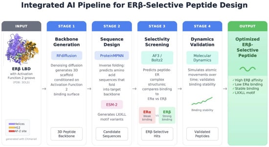
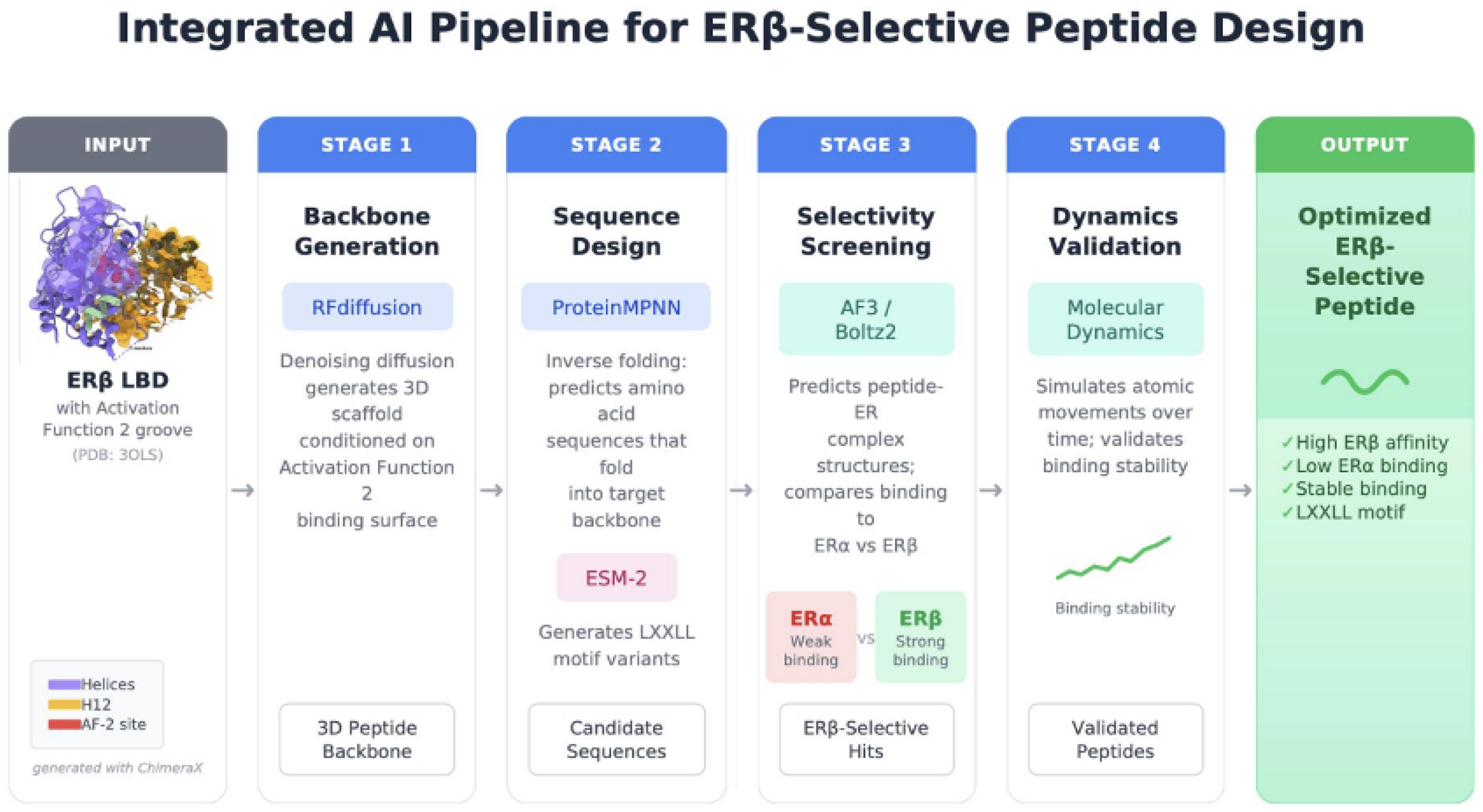

Disclaimer/Publisher’s Note: The statements, opinions and data contained in all publications are solely those of the individual author(s) and contributor(s) and not of MDPI and/or the editor(s). MDPI and/or the editor(s) disclaim responsibility for any injury to people or property resulting from any ideas, methods, instructions or products referred to in the content. |
© 2026 by the authors. Licensee MDPI, Basel, Switzerland. This article is an open access article distributed under the terms and conditions of the Creative Commons Attribution (CC BY) license.