


Abstract
Alzheimer’s and related dementias are not inevitable consequences of aging. This manuscript proposes that they arise when cumulative stress exceeds the repair capacity of three biological systems critical for maintaining brain health: proteostatic capacity, mitochondrial function, and neuroimmune regulation. In most cases, key influences such as genetics, environmental exposures, and health history are not primary determinants; they modify the baseline load borne by these pathways, influencing not only whether it is approaching a threshold, but how quickly it reaches the tipping point. To illustrate how diverse influences accumulate, the analysis maps 18 modifiable dementia risk factors onto the three convergent pathways. This approach reframes neurodegenerative risk as a dynamic, cumulative process and highlights opportunities for individualized assessment and targeted mitigation strategies long before dementia symptoms emerge.
1. Introduction
Alzheimer’s disease is defined biologically by the presence of amyloid-beta plaques and pathologic tau deposits, as detected by biomarkers or confirmed at autopsy [1]. The clinical syndrome most commonly associated with Alzheimer’s disease typically begins with early memory impairment and progresses to affect language, spatial navigation, executive function, and daily living, although presentations can vary [2]. Historically, because these symptoms often unfold in a predictable sequence, Alzheimer’s disease has been treated as a single, unified disorder. Lewy body dementia, a synucleinopathy that often overlaps with Parkinson’s disease, is marked by fluctuating cognition, visual hallucinations, rapid eye movement (REM) sleep behavior disorder, and parkinsonism—a less orderly and more variable constellation of symptoms [3]. Frontotemporal dementia (FTD) presents in several distinct variants—behavioral, language, and motor—reflecting the selective vulnerability of different frontal and temporal networks [4]. This variability has long made FTD particularly challenging to understand and difficult to unify under a single mechanism. Vascular insufficiency frequently co-occurs with all of these conditions and can accelerate or shape disease progression [5].
Despite these differences, Alzheimer’s disease and related dementias may represent distinct manifestations of a convergence of stress across three interdependent systems that maintain brain health: proteostatic capacity, mitochondrial function, and neuroimmune regulation. Although all three pathways ultimately lose resilience in progressive disease, different disorders show different points of initial vulnerability. In Alzheimer’s disease and related proteinopathies, proteostatic dysfunction often appears among the earliest detectable abnormalities [4,6,7]. Each system can absorb isolated insults, but chronic or repeated stress gradually erodes repair capacity, making the pathways increasingly vulnerable to failing together. This framework reflects a conceptual synthesis rather than a single established model, integrating evidence across these domains. In this view, progressive neurodegeneration emerges only when all three pathways are sufficiently compromised. This helps explain why individuals with similar exposures may experience different outcomes and why, in cases when at least one pathway is intact, substantial neuropathology can exist without significant impairment.
The rising global prevalence of Alzheimer’s disease suggests that modern exposures and habits may be imposing chronic, cumulative stress on these systems [8]. Large international studies have identified numerous modifiable lifestyle factors, reinforcing that dementia is not an inevitable consequence of aging. However, the diversity of identified risks—spanning vascular, metabolic, inflammatory, behavioral, and psychosocial domains—makes it difficult to design coherent strategies. Understanding neurodegeneration as the cumulative load on three interconnected pathways provides a unifying explanation for why such varied risk factors can produce similar clinical outcomes, why there are distinct dementias, and why no single biomarker or insult fully accounts for disease onset or progression. It also raises the possibility that strengthening the pathways that remain most resilient—rather than trying to rescue the one already under the greatest stress—may offer a more effective approach to pausing or slowing disease progression.
Differences in pathway load and resilience are evident in populations at the extremes of lifelong protection and risk. Some members of the School Sisters of Notre Dame show unusually low rates of cognitive decline despite Alzheimer’s-type neuropathology [9], reflecting a lifetime of stable routines and the absence of behaviors or exposures that would undermine these critical systems. In contrast, individuals with Down syndrome experience early and accelerated vulnerability due to trisomy 21, which increases APP expression, metabolic strain, and chronic immune dysregulation—stressing all three systems and producing Alzheimer’s neuropathology years earlier than in the general population [10]. These two populations demonstrate how lifelong variation in systems load shapes cognitive outcomes.
This paper argues that diverse modifiable lifestyle factors can be understood by their cumulative impact on three interconnected pathways.
2. Critical Pathways for Brain Health
The three major systems that sustain neurological function are:
- Proteostatic capacity: This system maintains healthy proteins by refolding those that are misfolded, tagging irreparable proteins for degradation, and clearing damaged or aggregated proteins through the ubiquitin–proteasome and autophagy–lysosomal systems. When refolding, degradation, or lysosomal clearance become overwhelmed, misfolded proteins accumulate—a process central to the pathophysiology of Alzheimer’s disease (Aβ and tau), Lewy body dementia (α-synuclein), and frontotemporal dementia (tau or TDP-43). Across these disorders, proteostatic strain appears early in the disease process and contributes to downstream mitochondrial and neuroimmune dysfunction [11].
- Mitochondrial function: Synaptic signaling is energy-intensive, which is why the brain—roughly two percent of one’s body weight—consumes about 20 percent of the body’s energy. Mitochondria supply this energy while also regulating calcium buffering, redox balance, and the initiation of repair processes. When mitochondrial output is chronically constrained—through impaired oxidative phosphorylation, reduced biogenesis, or increased reactive oxygen species (ROS)—neuronal resilience declines and vulnerability to downstream dysfunction increases. In Alzheimer’s disease, mitochondrial efficiency is reduced early through Aβ-related membrane disruption and tau-related transport deficits; in Lewy body dementia, α-synuclein interferes with complex I and mitochondrial membrane integrity; and in frontotemporal dementia, TDP-43 and tau pathology disrupt mitochondrial dynamics and axonal transport. Across these conditions, mitochondrial impairment is not the sole initiating event, but a consistent and amplifying feature that lowers the threshold at which other system failures lead to progressive neurodegeneration [12].
- Neuroimmune regulation: The brain’s support cells—particularly microglia and astrocytes—coordinate immune surveillance and tissue repair. When chronically overtaxed as in Alzheimer’s, they can shift into a pro-inflammatory state, leading to immune dysregulation and disruption of the blood–brain barrier (BBB) [13]. Similar patterns of chronic microglial activation and impaired immune resolution are implicated across neurodegenerative diseases, including Lewy body dementia and FTD.
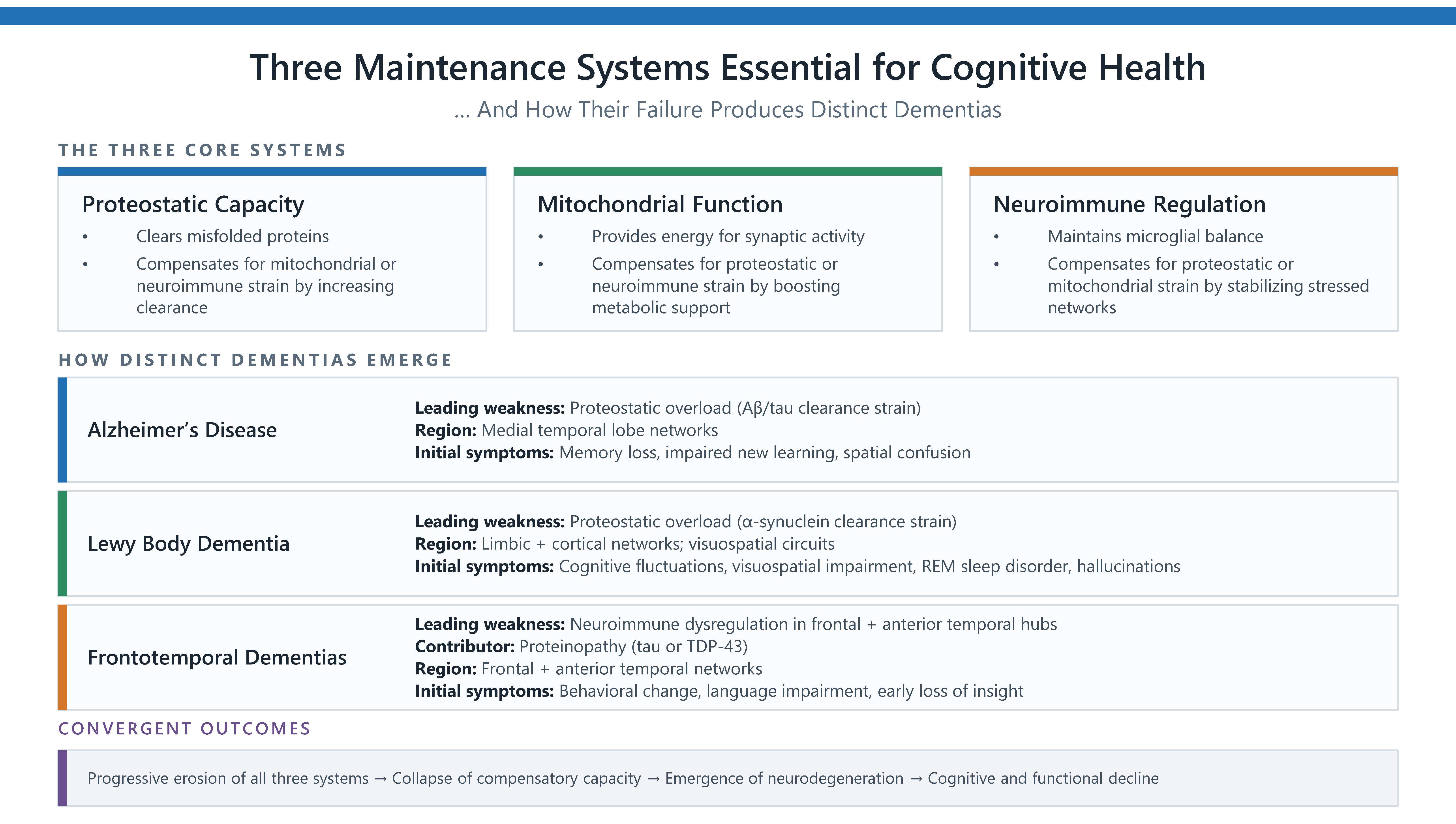
These distinctions allow each dementia to be understood as a different point of initial strain on the same three systems. Figure 1 summarizes how Alzheimer’s disease, Lewy body dementia, and frontotemporal dementias map onto these early vulnerabilities within the three-pathway framework.
Figure 1.
This diagram applies the three-pathway framework introduced in Rodgers (2026) to illustrate the proposed hypothesis that different forms of neurodegeneration arise from different points of earliest strain within proteostatic, mitochondrial, and neuroimmune systems [14]. The Lewy body dementia pathway reflects established α-synuclein clearance strain [3].
The framework presented here suggests that dementia only emerges when all three pathways are compromised beyond the point of repair and cross a threshold beyond which decline becomes progressive. The failure to identify a single cause for most neurodegenerative diseases—despite decades of effort—reflects this underlying architecture.
3. Genetics Do Not Equal Destiny
Although genetics can influence vulnerability, apart from FTD, inheritance is not the primary driver of most dementias, which is why the majority of cases are designated sporadic. Even when genetic predispositions are present, the variants involved do not introduce new disease mechanisms; instead, they weaken one or more of the same biological pathways necessary to maintain brain health [15]. These influences fall into two broad categories: autosomal-dominant mutations with near-complete penetrance, and common polymorphisms that modulate pathway resilience.
4. Autosomal-Dominant Mutations (Deterministic, Early-Onset)
A small subset of individuals carry mutations that almost invariably lead to early-onset dementia.
Alzheimer’s disease: Alzheimer’s disease offers the clearest example of how genetic risk maps onto the three-pathway framework, with both common variants (APOE ε4) and rare familial mutations (Pathogenic variants in APP, PSEN1, PSEN2) converging on lipid handling, proteostasis, and inflammatory regulation. Conversely, the rarer APOE ε2 variant is protective, improving lipid handling and dampening baseline neuroimmune activation, thereby reducing amyloid burden and slowing downstream proteostatic strain [16].
Lewy Body Dementia: Rare autosomal-dominant mutations in SNCA increase α-synuclein misfolding and aggregation, directly stressing proteostatic and lysosomal systems. These mutations produce early-onset Lewy body disorders with near-100% penetrance, reflecting deterministic overload of the same pathways implicated in sporadic Lewy body dementia [7].
FTD: These dementias have the highest proportion of identifiable genetic contributors, most commonly mutations in MAPT, GRN, or expansions in C9orf72. Each of these affects a different arm of the three-system framework: MAPT mutations disrupt microtubule stability and increase proteostatic burden; GRN haploinsufficiency impairs lysosomal function and heightens neuroimmune reactivity; and C9orf72 expansions generate toxic RNA and dipeptide repeats that strain mitochondrial and proteostatic capacity simultaneously [17]. By weakening these pathways from the start, these mutations reduce resilience long before age-related erosion occurs, resulting in earlier onset. Because these systems support distinct but interdependent frontotemporal networks, each clinical variant reflects the particular network under greatest strain—yet all impair insight early, as integrative hubs lose connective capacity first [18].
Together, these genetic examples illustrate that inherited risks do not create new disease pathways—they simply reduce the resilience of the existing ones, lowering the threshold at which lifelong stressors can tip the system into failure. Seen this way, genetics helps explain why people respond differently to similar exposures or age-related changes: some individuals begin life with less resilient protective systems, leaving them more vulnerable as stressors accumulate across decades. Genetic risk, then, does not contradict a convergence model of neurodegeneration—it reinforces it, showing how pathways that are weakened from the start can push the brain past a threshold from system compensation to progressive disease.
5. Vascular Insufficiency as an Upstream Systems Stressor
Vascular dysfunction provides one of the clearest bridges between lifestyle risk factors and neurodegeneration because even mild impairments in cerebral blood flow directly stress all three pathways required for brain maintenance. Impaired perivascular and glymphatic clearance slows the removal of misfolded proteins and metabolic waste, increasing proteostatic burden from the outset. Chronic hypoperfusion limits mitochondrial energy availability, reducing the metabolic reserve needed for synaptic upkeep. Disruption of the blood-brain barrier (BBB) exposes neural tissue to circulating inflammatory mediators and primes microglia toward a heightened immune tone, reducing their capacity for normal synaptic and neuronal support [19]. Together, these vascular changes act as upstream amplifiers that push vulnerable systems toward failure, regardless of the specific neurodegenerative diagnosis.
6. Mechanistic Mapping of Health and Lifestyle Factors to the Three Pathways
The 2024 Lancet Commission on dementia prevention, intervention, and care identified 14 modifiable dementia risk factors, representing a major synthesis of epidemiological evidence [8]. An additional four factors—healthy diet, sleep quality, cognitive engagement, and chronic stress—were identified in a 2025 international consensus statement on nutrition, metabolic health, and brain aging [20]. These 18 factors are combined here to illustrate how diverse lifestyle and environmental influences contribute to the preservation or erosion of the three core pathways that maintain brain health. Please note that the factors are not weighted, and their order does not imply relative impact.
6.1. Physical Activity
Supports:
- Proteostasis: Enhances autophagy and lysosomal clearance, preventing a backlog of damaged proteins.
- Mitochondria: Increases biogenesis and adenosine triphosphate (ATP) availability, expanding energetic reserves and reducing reactive oxygen species (ROS) load.
- Neuroimmune: Reduces inflammatory tone, allowing microglia to shift to balanced surveillance states.
When absent:
- Proteostasis: Impairs clearance of damaged proteins, increasing proteostatic strain.
- Mitochondria: Allows dysfunctional mitochondria to persist, reducing available energy.
- Neuroimmune: Inactivity causes immune cells to drift toward pro-inflammatory states.
6.2. Smoking
Supports (when stopped):
- Proteostasis: Lower toxin-induced misfolding and reduced burden on repair and clearance pathways.
- Mitochondria: Reduced oxidative injury allows more efficient ATP production and less ROS spillover.
- Neuroimmune: Decreased toxin exposure reduces chronic immune priming.
When ongoing:
- Proteostasis: Toxin exposure drives misfolding, overwhelms repair systems, and increases proteostatic strain.
- Mitochondria: Chronic oxidative stress damages mitochondrial membranes and impairs energy production.
- Neuroimmune: Persistent endothelial and tissue injury heightens inflammation.
6.3. Harmful Alcohol Use
Supports (when treated):
- Proteostasis: Less endoplasmic reticulum (ER) stress and reduced accumulation of misfolded or damaged proteins.
- Mitochondria: Lower toxic metabolite load allows more stable ATP production and less oxidative strain.
- Neuroimmune: Reduced glial signaling and lower baseline inflammatory signaling.
When untreated:
- Proteostasis: Impaired clearance and increased burden of damaged or misfolded proteins.
- Mitochondria: Direct toxicity disrupts membranes, increases ROS, and impairs energy production.
- Neuroimmune: Heightened glial activation and sustained inflammatory tone.
6.4. Obesity
Supports (when reduced):
- Proteostasis: Reduced ER stress improves folding capacity and lowers baseline clearance burden.
- Mitochondria: Lower metabolic overload decreases substrate pressure and reduces ROS generation.
- Neuroimmune: Less adipose-driven cytokine release reduces systemic and central inflammatory tone.
When present:
- Proteostasis: ER stress and misfolding increases metabolic load.
- Mitochondria: Excess substrate supply strains mitochondrial function and elevates oxidative stress.
- Neuroimmune: Adipose-derived inflammatory signals drive persistent systemic inflammation.
6.5. Hypertension
Supports (when controlled):
- Proteostasis: Controlled blood pressure maintains steady perfusion, reducing ischemic stress.
- Mitochondria: Protects microvasculature, stabilizing oxygen supply and preserving ATP production.
- Neuroimmune: Stable pressure prevents BBB leakage and downstream immune activation.
When uncontrolled:
- Proteostasis: Ischemic injury due to elevated pressure, damaging microvessels.
- Mitochondria: Disrupted oxygen delivery increases ROS, impairing ATP production.
- Neuroimmune: Endothelial injury triggers chronic vascular inflammation.
6.6. Diabetes (Including Insulin Resistance/Impaired Glucose Metabolism)
Supports (when controlled):
- Proteostasis: Stable glucose levels limit glycation stress, supporting proper folding and clearance.
- Mitochondria: Controlled insulin signaling allows efficient fuel use, sustaining ATP production.
- Neuroimmune: Balanced metabolic signaling reduces inflammation.
When uncontrolled:
- Proteostasis: Increased glycation, misfolding and strained clearance pathways.
- Mitochondria: Excess glucose impairs function, increasing oxidative stress and reducing energy.
- Neuroimmune: Metabolic dysregulation induces chronic inflammation.
6.7. Education Level
Supports (when high):
- Proteostasis: Enriched synaptic remodeling may support proteostatic health by increasing opportunities for clearance and reducing aggregation risk.
- Mitochondria: Lifelong cognitive load is associated with energetic resilience, potentially improving ATP efficiency and metabolic flexibility.
- Neuroimmune: Sustained cognitive engagement is linked to lower allostatic load, which may buffer against stress-induced immune activation.
When low:
- Proteostasis: Reduced remodeling, with fewer opportunities to clear damaged proteins, and greater risk of accumulation.
- Mitochondria: Lower reserve, decreasing metabolic “cushion” during systemic strain or aging stressors.
- Neuroimmune: Lower cognitive engagement increases vulnerability to stressors.
6.8. Social Engagement
Supports:
- Proteostasis: Social engagement is associated with healthier synaptic turnover and may help maintain adaptive remodeling.
- Mitochondria: Ongoing social interaction is linked to sustained synaptic activity and energetic demand, which may help keep complex neural circuits resilient.
- Neuroimmune: Reduces stress-related inflammation and lowers baseline inflammatory tone.
When absent:
- Proteostasis: Reduced synaptic remodeling and lower turnover.
- Mitochondria: Reduced synaptic activity and diminished energetic engagement.
- Neuroimmune: Social isolation is strongly linked to heightened stress signaling and increased inflammatory activation.
6.9. Depression
Supports (when treated):
- Proteostasis: Treatment improves sleep quality, supporting glymphatic clearance of misfolded proteins.
- Mitochondria: Lowers cortisol, improving energy metabolism and ATP efficiency.
- Neuroimmune: Reduced inflammatory tone, stabilizing microglial activity.
When untreated:
- Proteostasis: Impaired repair, leading to protein aggregation.
- Mitochondria: Cortisol-driven exposure impairs mitochondrial efficiency.
- Neuroimmune: Immune activation, with heightened inflammatory signaling.
6.10. Air Pollution (Including Second-Hand Smoke)
Supports (when reduced):
- Proteostasis: Reduced protein oxidation and misfolding, lowering aggregation risk.
- Mitochondria: Less oxidative stress from particulate exposure, preserving ATP production.
- Neuroimmune: Lower systemic and central nervous system inflammation, helping preserve BBB integrity.
When high:
- Proteostasis: Protein oxidation leads to misfolding and aggregation, overwhelming clearance systems.
- Mitochondria: Particulate-induced oxidative injury reduces energy production and mitochondrial resistance.
- Neuroimmune: Chronic inflammatory activation, disrupting the BBB, and increasing vulnerability to neurotoxic exposures.
6.11. Hearing Loss
Supports (when corrected):
- Proteostasis: Reduced chronic compensatory synaptic stress from degraded hearing input, reducing misfolding pressure.
- Mitochondria: Reduced compensatory auditory cortical energy demand, preserving metabolic reserve.
- Neuroimmune: Reduced isolation-driven inflammation and stress-related microglial activation, stabilizing immune tone.
When uncorrected:
- Proteostasis: Sustained strain leads to misfolding, impaired turnover, and aggregation.
- Mitochondria: Increased energetic load from compensatory processing, driving inefficiency and oxidative stress.
- Neuroimmune: Social withdrawal and chronic stress sustain inflammatory activation, eroding resilience.
6.12. Vision Loss
Supports (when corrected):
- Proteostasis: Reduced chronic compensatory synaptic stress from degraded visual input, reducing misfolding pressure.
- Mitochondria: Reduced compensatory cortical energy demand in visual processing networks, preserving metabolic reserves.
- Neuroimmune: Reduced isolation-driven inflammation and stress-related microglial activation, stabilizing immune tone.
When uncorrected:
- Proteostasis: Sustained strain leads to misfolding, impaired turnover and aggregation.
- Mitochondria: Increased energetic load from compensatory processing, driving inefficiency and oxidative stress.
- Neuroimmune: Social withdrawal and chronic stress sustain inflammatory activation, eroding resilience.
6.13. Cholesterol (Dyslipidemia/High LDL)
Supports:
- Proteostasis: Balanced lipid transport supports membrane integrity and synaptic vesicle dynamics, reducing misfolding pressure on protein-handling systems.
- Mitochondria: Healthy lipid profiles maintain mitochondrial membrane composition and reduce oxidized-lipid-driven energetic stress.
- Neuroimmune: Stable lipid metabolism limits endothelial inflammation and helps preserve BBB function, keeping microglial activation low and regulated.
When poor:
- Proteostasis: Oxidized LDL and disrupted lipid transport (including ApoE-related pathways) impair membrane stability and increase proteostatic burden.
- Mitochondria: Excess or oxidized lipids damage mitochondrial membranes, reduce ATP efficiency, and increase ROS production.
- Neuroimmune: Dyslipidemia promotes endothelial inflammation, microvascular injury, and BBB permeability, sustaining chronic neuroimmune activation.
6.14. Traumatic Brain Injury (TBI)
Supports (when preventable or mitigated):
- Proteostasis: Prevents tau hyperphosphorylation and misfolding cascades, maintaining clearance capacity.
- Mitochondria: Avoids acute energetic collapse and chronic fragmentation, preserving ATP production.
- Neuroimmune: Avoids microglial priming and chronic neuroinflammation, stabilizing immune tone.
When present:
- Proteostasis: Tauopathy-like misfolding, impaired clearance, and aggregation.
- Mitochondria: Acute dysfunction and oxidative stress, contributing to long-term instability.
- Neuroimmune: Chronic microglial activation, sustaining persistent inflammatory signaling, and BBB disruption.
6.15. Healthy Diet
Supports:
- Proteostasis: Lowers misfolded-protein burden via reduced glycation (sugar-induced protein damage) and oxidative injury, both of which promote misfolding and overwhelm clearance systems.
- Mitochondria: Stabilizes glucose supply, reduces oxidative load, and improves cellular energy efficiency.
- Neuroimmune: Reduces chronic inflammation through polyphenols and omega-3 fatty acids, supporting microglial stability and lowering baseline immune activation.
When poor:
- Proteostasis: Increased glycation and impaired clearance, driving protein aggregation.
- Mitochondria: Increased ROS, damaging membranes, and impairing energy production.
- Neuroimmune: Chronic low-grade inflammation, destabilizing baseline immune tone.
6.16. Sleep Quality
Supports:
- Proteostasis: Glymphatic clearance of misfolded proteins and nightly reduction in proteostatic load.
- Mitochondria: Nightly repair, redox restoration, and metabolic reset.
- Neuroimmune: Circadian regulation of immune tone and microglial activity.
When poor:
- Proteostasis: Backlog of misfolded proteins and reduced clearance capacity.
- Mitochondria: Impaired repair and disrupted metabolic recovery.
- Neuroimmune: Circadian disruption elevates inflammatory signaling.
6.17. Cognitive Engagement
Supports:
- Proteostasis: Promotes synaptic remodeling—constant turnover stimulates clearance machinery, reducing misfolded protein accumulation.
- Mitochondria: Sustained energetic demand builds resilience—frequent firing trains mitochondria for efficient ATP production and metabolic flexibility.
- Neuroimmune: Reduces stress-related activation by lowering allostatic load, dampening cortisol, and microglial priming.
When low:
- Proteostasis: Impaired synaptic turnover, providing fewer opportunities to clear damaged proteins.
- Mitochondria: Reduced reserve, as lowered demand signals lead to smaller, less efficient mitochondrial populations.
- Neuroimmune: Vulnerability to stress, as reduced cognitive buffering increases susceptibility to inflammatory activation.
6.18. Chronic Stress
Supports (when mitigated):
- Proteostasis: Improved repair, facilitating clearance of misfolded proteins through enhanced autophagy and glymphatic flow.
- Mitochondria: Reduced cortisol load, aiding energy production by lowering oxidative strain and preserving ATP efficiency.
- Neuroimmune: Reduced microglial priming, stabilizing immune tone, preventing exaggerated inflammatory responses.
When persistent:
- Proteostasis: Impaired repair, leading to protein aggregation and reduced clearance capacity.
- Mitochondria: Dysfunction caused by oxidative damage, impairing ATP production, and metabolic resilience.
- Neuroimmune: Microglial priming leads to chronic inflammation, causing a chronically harmful state that maintains inflammatory signaling beyond adaptive needs.
These 18 factors suggest that a structured, clinically usable checklist could help quantify an individual’s cumulative risk by mapping each factor onto the three convergent systems. Such a tool could help clinicians identify which pathways are most affected and why. Embedding targeted recommendations into the checklist would make the process actionable, transforming a broad set of lifestyle and medical variables into a coherent, system-level risk profile. Consistent with the Lancet Commission’s emphasis that it is never too early or too late to modify these risk factors, this approach creates opportunities for proactive changes that could slow or even prevent cognitive decline [8].
7. How Environmental Exposures and Infections Fit the Convergence Model
It has long been known that many environmental and physiological stressors erode the same three pathways necessary for brain health. Chronic exposure to pollutants, heavy metals, and other toxins increases oxidative load, impairs mitochondrial efficiency, and disrupts proteostatic and immune regulation. Recurrent or untreated infections similarly impose repeated neuroimmune activation [21], whereas vaccines targeting shingles and respiratory syncytial virus (RSV) have been found to reduce dementia risk [22]. Chronic infections such as herpesviruses [23] or periodontal disease [24] can maintain a low-grade activated immune state, incrementally taxing all three systems, and gradually diminishing the repair capacity that keeps them stable.
Taken together, genetic predispositions, environmental exposures, and lifestyle-linked physiological stressors do not represent competing explanations for neurodegenerative disease. They are overlapping contributors to the same cumulative system load. Genetic variants determine baseline resilience, environmental exposures add intermittent or chronic demands on those systems, and health history and everyday behaviors—such as sleep, substance use, diet and physical activity—shape how effectively these systems recover between stressors. Because these influences accumulate slowly, the total burden placed on these systems over decades explains why neurodegeneration typically emerges later in life.
8. Mapping Related Dementias and Mixed Pathology
Understanding Alzheimer’s disease within a three-system framework also clarifies why related dementias share overlapping features and why mixed pathology is so common in late life. Lewy body dementia, Parkinson’s disease dementia, frontotemporal dementia, and vascular contributions each stress the same three systems, but in different sequences and with different dominant pressures. Variations in timing and degree produce distinct clinical presentations, yet the underlying vulnerabilities are shared.
Mixed dementia pathology, often treated as a diagnostic complication, is better understood as the predictable outcome of cumulative system erosion. When proteostasis, mitochondrial strain, and impaired and chronic immune activation progress at different rates across brain regions, multiple pathological signatures can emerge simultaneously. Rather than representing separate diseases, these patterns reflect the brain’s attempt to compensate as multiple protective systems weaken together. This perspective explains why neuropathological boundaries blur in older adults and why clinical symptoms often reflect contributions from more than one system.
9. Discussion
This manuscript reframes neurodegeneration as the cumulative failure of three maintenance systems—proteostatic capacity, mitochondrial function, and neuroimmune regulation—and situates a wide range of environmental, physiological, and behavioral stressors within this convergence framework. Although Alzheimer’s and related dementias are not natural consequences of aging, lifetime habits and exposures, combined with a gradually declining repair capacity, can progressively weaken these systems. Within this model, diverse influences on dementia risk can be understood as modifiers of system load, shaping how quickly or slowly these systems erode over the lifespan.
Alzheimer’s disease now has an impressive biomarker ecosystem—plasma assays, cerebrospinal fluid signatures, positron emission tomography (PET) tracers, and staging frameworks—that is the envy of every adjacent field. Yet this success has revealed a structural limitation: these biomarkers track downstream artifacts of systems already under strain. They illuminate proteostatic overload, metabolic collapse, and neuroimmune drift with remarkable clarity, but they do not identify the upstream failure that made these cascades possible. As a result, even perfect detection of amyloid, tau, or other neurodegenerative signatures cannot yield a pharmaceutical “silver bullet” because no downstream therapy can substitute for repairing the mitochondrial, proteostatic, and neuroimmune systems whose erosion set the stage for pathology in the first place.
All the stressors associated with dementia identified by the Lancet Commission and consensus statement on brain aging exert their effects by impairing waste clearance, increasing energetic demand, or sustaining low-grade immune activation. However, the system stresses do not act in isolation—they may also interact. For example, sleep disruption becomes far more metabolically costly when it occurs against a backdrop of circadian misalignment, because the brain must maintain basic function while its time-sensitive repair programs are weakened [25].
Advancing age further shapes pathway resilience because all three maintenance systems show characteristic shifts over time. Neuroimmune regulation becomes harder to sustain as microglia adopt more reactive, pro-inflammatory states—a pattern described in the aging literature as “inflammaging,” a chronic, low-grade immune activation attributed to multiple systemic influences, including metabolic stress and changes in gut microbiota composition [26]. These broad age-related shifts reduce the system’s ability to maintain synaptic stability. Mitochondrial efficiency also declines with age, with cumulative metabolic stress and impaired quality-control mechanisms reducing the energy available for synaptic repair [27]. Proteostatic capacity weakens as autophagy and lysosomal clearance become less responsive [28]. These changes do not cause neurodegeneration on their own, but they narrow the compensatory reserve that protects against additional burdens. Individuals with greater immune resilience—the ability to sustain balanced, adaptive immune responses across the lifespan—show markedly better long-term outcomes [29], underscoring that aging increases vulnerability primarily by diminishing repair capacity rather than initiating disease.
The decades-long Nun Study provides a compelling illustration of how strong compensatory systems can preserve cognitive function even in the presence of substantial neuropathology. Participants who remained cognitively healthy in advanced age often showed extensive amyloid plaques and neurofibrillary tangles at autopsy—levels typically associated with dementia. Their resilience has been attributed to factors such as cognitive reserve and stable routines [9]. Yet equally important, their lives lacked many of the stressors that erode pathway resilience. In this light, the disconnect between neuropathology and cognitive outcomes is not paradoxical but predictable: when at least one system remains stable, the brain can tolerate substantial pathological burden.
Down syndrome illustrates the opposite extreme. Triplication of chromosome 21 increases amyloid precursor protein expression, placing a constant burden on protein management and waste-clearance systems. Overexpression of genes involved in oxidative stress contributes to chronic mitochondrial strain, while neuroimmune regulation is altered from early life [10]. Each of these factors alone might be manageable, but together they impose continuous stress across all three systems, leading to the near-universal early development of Alzheimer-type pathology. Down syndrome thus reinforces the central premise of this framework: neurodegeneration emerges not from a single cause, but from the convergence of multiple impaired systems.
Between these extremes lies the general population, where individuals differ widely in their baseline resilience and in the cumulative stress experienced over a lifetime. Early-life health, metabolic status, sleep quality, environmental exposures, infections, and lifestyle patterns—many of which have shifted dramatically in recent decades—all influence the compensatory capacity a person begins with and how quickly it erodes.
Within this broader landscape, several nuances within the risk-factor literature are important for interpretation. The 2020 Lancet Commission found that the protective effect of education is concentrated in the years up to age 20, underscoring the critical importance of early education [30]. In contrast, the 2024 Lancet Commission reported that midlife cognitive stimulation from work or hobbies confers protection even in individuals with low formal education. The latest Lancet report found even stronger evidence that depression is strongly associated with dementia risk—although, as noted in previous Lancet Commission reports, in some individuals it may be a prodromal symptom rather than an upstream causal factor. Whichever the case, depression should always be treated. Excessive alcohol use, although strongly associated with dementia risk [8], is notably the only risk factor linked to early-onset dementia (prior to age 65) that does not have a genetic basis [30].
Genetic variation further shapes baseline system load by influencing proteostatic capacity, mitochondrial efficiency, and neuroimmune responsiveness from early life. Some individuals inherit variants that confer above-average resilience, while others begin life with reduced reserve in one or more systems. These inherited differences do not determine outcomes, but they influence how much compensatory capacity a person starts with and how quickly it may erode under cumulative stress. In this framework, genetics functions as part of the initial conditions that interact with lifelong habits, exposures, and repair capacity.
Although longitudinal measures of cumulative system load remain limited, this framework provides a coherent structure for interpreting existing findings and identifying priorities for future research. It suggests that everyday clinical decisions may influence neurodegenerative vulnerability by altering cumulative stress on these systems. It also highlights the value of strengthening primary care capacity for routine screening of sleep disorders, sensory impairment, cardiometabolic, and other modifiable risk factors in older adults, supported by clear referral pathways when indicated. Ongoing management of conditions such as obesity, hypertension, and diabetes may be particularly important, given their well-established roles in accelerating vascular dysfunction, metabolic strain, and inflammatory activation.
10. Conclusions
This framework proposes that neurodegenerative diseases arise when the three core systems that sustain brain health can no longer compensate for cumulative stress. It raises the possibility that neurodegenerative risk could one day be estimated—and mitigated—by assessing the status of these systems on an individual basis and developing targeted interventions that restore resilience wherever possible. As tools for measuring proteostatic capacity, mitochondrial efficiency, and neuroimmune regulation continue to advance, a systems-level approach may allow pathway load to be quantified directly, creating opportunities for interventions that delay or prevent the emergence of dementia.
Funding
The author received no external funding for this work.
Institutional Review Board Statement
Not applicable.
Informed Consent Statement
Not applicable.
Data Availability Statement
No new data were created or analyzed in this study. Data sharing is not applicable to this article.
Acknowledgments
Bing Copilot (https://copilot.microsoft.com) was used during manuscript preparation for language-focused assistance and to help identify areas where additional literature review by the author might be useful. The free version of ChatGPT (GPT-5.6 Luna) was used occasionally for general editorial suggestions. Adobe Acrobat AI was used to assist with reviewing the placement and context of cited scientific text. PowerPoint with Bing Copilot was used to create the figure. All factual claims and citations were independently researched, verified, and confirmed by the author, who takes full responsibility for the scientific content of this publication.
Conflicts of Interest
The author declares no conflicts of interest.
Abbreviations
| BBB | Blood–brain barrier |
| ER | Endoplasmic reticulum |
| FTD | Frontotemporal dementia |
| PET | Positron emission tomography |
| REM | Rapid eye movement |
| ROS | Reactive oxygen species |
References
- Jack, C.R., Jr.; Bennett, D.A.; Blennow, K.; Carrillo, M.C.; Dunn, B.; Haeberlein, A.B.; Holtzman, D.M.; Jagust, W.; Jessen, F.; Karlawish, J.; et al. NIA-AA Research Framework: Toward a biological definition of Alzheimer’s disease. Alzheimers Dement. 2018, 14, 535–562. [Google Scholar] [CrossRef] [Scilit] [PubMed]
- McKhann, G.M.; Knopman, D.S.; Chertkow, H.; Hyman, B.T.; Jack, C.R., Jr.; Kawas, C.H.; Klunk, W.E.; Koroshetz, W.J.; Koroshetz, J.J.; Manly, J.J.; et al. The diagnosis of dementia due to Alzheimer’s disease: Recommendations from the National Institute on Aging–Alzheimer’s Association workgroups on diagnostic guidelines for Alzheimer’s disease. Alzheimers Dement. 2011, 7, 263–269. [Google Scholar] [CrossRef] [Scilit] [PubMed]
- McKeith, I.G.; Boeve, B.F.; Dickson, D.W.; Halliday, G.; Taylor, J.-P.; Weintraub, D.; Aarsland, D.; Galvin, J.; Attems, J.; Ballard, C.G.; et al. Diagnosis and management of dementia with Lewy bodies: Fourth consensus report of the DLB Consortium. Neurology 2017, 89, 88–100. [Google Scholar] [CrossRef] [Scilit] [PubMed]
- Bang, J.; Spina, S.; Miller, B.L. Frontotemporal dementia. Lancet 2015, 386, 1672–1682. [Google Scholar] [CrossRef] [Scilit] [PubMed]
- Jellinger, K.A. Pathology and pathogenesis of vascular cognitive impairment—A critical update. Front. Aging Neurosci. 2013, 5, 17. [Google Scholar] [CrossRef] [Scilit] [PubMed]
- De Strooper, B.; Karran, E. The cellular phase of Alzheimer’s disease. Cell 2016, 164, 603–615. [Google Scholar] [CrossRef] [Scilit] [PubMed]
- Surguchov, A.; Surgochov, A. Synucleins: New data on misfolding, aggregation and role in diseases. Biomedicines 2022, 10, 3241. [Google Scholar] [CrossRef] [Scilit] [PubMed]
- Livingston, G.; Huntley, J.; Liu, K.Y.; Costafreda, S.G.; Selbaek, G.; Alladi, S.; Ames, D.; Banerjee, S.; Burns, A.; Brayne, C.; et al. Dementia prevention, intervention, and care: 2024 report of the Lancet standing Commission. Lancet 2024, 404, 572–628. [Google Scholar] [CrossRef] [Scilit] [PubMed]
- Clarke, K.M.; Etemadmoghadam, S.; Danner, B.; Corbett, C.; Ghaseminejad-Bandpey, A.; Dopler, M.; Parker-Garza, J.; Alhneif, M.; Babu, S.; Ogunbona, O.B.; et al. The Nun Study: Insights from 30 years of aging and dementia research. Alzheimers Dement. 2025, 21, e14626. [Google Scholar] [CrossRef] [Scilit] [PubMed]
- Garcia, O.; Dominguez de la Cruz, E.; Luna-Martínez, I.G.; Garnett-Carbajal, J.A.; Villegas-Piña, J.A. Development of Alzheimer’s disease in Down syndrome. Explor. Neurosci. 2025, 4, 1006104. [Google Scholar] [CrossRef] [Scilit]
- Wilson, D.M., III; Cookson, M.R.; Van Den Bosch, L.; Zetterberg, H.; Holtzman, D.M.; Dewachter, I. Hallmarks of neurodegenerative diseases. Cell 2023, 186, 693–714. [Google Scholar] [CrossRef] [Scilit] [PubMed]
- Klemmensen, M.M.; Borrowman, S.H.; Pearce, C.; Pyles, B.; Chandra, B. Mitochondrial dysfunction in neurodegenerative disorders. Neurotherapeutics 2024, 21, e00292. [Google Scholar] [CrossRef] [Scilit] [PubMed]
- Heneka, M.T.; Carson, M.J.; El Khoury, J.; Landreth, G.E.; Brosseron, F.; Feinstein, D.L.; Jacobs, A.H.; Wyss-Coray, T.; Vitorica, J.; Ransohoff, R.M.; et al. Neuroinflammation in Alzheimer’s disease. Lancet Neurol. 2015, 14, 388–405. [Google Scholar] [CrossRef] [Scilit] [PubMed]
- Rodgers, C.C. How Three-Pathway Erosion Shapes Distinct Neurodegenerative Diseases. Qeios 2026. [Google Scholar] [CrossRef]
- Lambert, J.C.; Ramirez, A.; Grenier-Boley, B.; Bellenguez, C. Step by step: Towards a better understanding of the genetic architecture of Alzheimer’s disease. Mol. Psychiatry 2023, 28, 2716–2727. [Google Scholar] [CrossRef] [Scilit] [PubMed]
- Holtzman, D.M.; Morris, J.C.; Goate, A.M. Alzheimer’s disease: The challenge of the second century. Sci. Transl. Med. 2011, 3, 77sr1. [Google Scholar] [CrossRef] [Scilit] [PubMed]
- Greaves, C.V.; Rohrer, J.D. An update on genetic frontotemporal dementia. J. Neurol. 2019, 266, 2075–2086. [Google Scholar] [CrossRef] [Scilit] [PubMed]
- Muñoz-Neira, C.; Tedde, A.; Coulthard, E.; Thai, N.J.; Pennington, C. Neural correlates of altered insight in frontotemporal dementia: A systematic review. Neuroimage Clin. 2019, 24, 102066. [Google Scholar] [CrossRef] [Scilit] [PubMed]
- Iadecola, C. The neurovascular unit coming of age: A journey through neurovascular coupling in health and disease. Neuron 2017, 96, 17–42. [Google Scholar] [CrossRef] [Scilit] [PubMed]
- Johnstone, A.M.; Albanese, E.; Crabtree, D.R.; Dalile, B.; Grabucker, S.; Gregory, J.M.; Grosso, G.; Holliday, A.; Hughes, C.; Itsiopoulos, C.; et al. Consensus statement on exploring the nexus between nutrition, brain health and dementia prevention. Nutr. Metab. 2025, 22, 82. [Google Scholar] [CrossRef] [Scilit] [PubMed]
- Sipilä, P.N.; Heikkilä, N.; Lindbohm, J.V.; Hakulinen, C.; Vahtera, J.; Elovainio, M.; Suominen, S.; Väänänen, A.; Koskinen, A.; Nyberg, S.T.; et al. Hospital-treated infectious diseases and the risk of dementia: A large, multicohort observational study with a replication cohort. Lancet Infect. Dis. 2021, 21, 1557–1567. [Google Scholar] [CrossRef] [Scilit] [PubMed]
- Taquet, M.; Todd, J.A.; Harrison, P.J. Lower risk of dementia with AS01-adjuvanted vaccination against shingles and respiratory syncytial virus infections. npj Vaccines 2025, 10, 130. [Google Scholar] [CrossRef] [Scilit] [PubMed]
- Itzhaki, R.F. Herpes simplex virus type 1 and Alzheimer’s disease: Increasing evidence for a major role of the virus. Front. Aging Neurosci. 2014, 6, 202. [Google Scholar] [CrossRef] [Scilit] [PubMed]
- Chen, C.K.; Wu, Y.T.; Chang, Y.C. Association between chronic periodontitis and the risk of Alzheimer’s disease: A retrospective, population-based, matched-cohort study. Alzheimers Res. Ther. 2017, 9, 56. [Google Scholar] [CrossRef] [Scilit] [PubMed]
- Musiek, E.S.; Holtzman, D.M. Mechanisms linking circadian clocks, sleep, and neurodegeneration. Science 2016, 354, 1004–1008. [Google Scholar] [CrossRef] [Scilit] [PubMed]
- Franceschi, C.; Garagnani, P.; Parini, P.; Giuliani, C.; Santoro, A. Inflammaging: A new immune-metabolic viewpoint for age-related diseases. Nat. Rev. Endocrinol. 2018, 14, 576–590. [Google Scholar] [CrossRef] [Scilit] [PubMed]
- Sun, N.; Youle, R.J.; Finkel, T. The mitochondrial basis of aging. Mol. Cell 2016, 61, 654–666. [Google Scholar] [CrossRef] [Scilit] [PubMed]
- Kaushik, S.; Cuervo, A.M. Proteostasis and aging. Nat. Med. 2015, 21, 1406–1415. [Google Scholar] [CrossRef] [Scilit] [PubMed]
- Manoharan, M.S.; Lee, G.C.; Harper, N.; Meunier, J.A.; Restrepo, M.I.; Jimenez, F.; Karekatt, S.; Branum, A.P.; Gaitan, A.A.; Andampour, K.; et al. The 15-year survival advantage: Immune resilience as a salutogenic force in healthy aging. Aging Cell 2025, 24, e70063. [Google Scholar] [CrossRef] [Scilit] [PubMed]
- Livingston, G.; Huntley, J.; Sommerlad, A.; Ames, D.; Ballard, C.; Banerjee, S.; Brayne, C.; Burns, A.; Cohen-Mansfield, J.; Cooper, C.; et al. Dementia prevention, intervention, and care: 2020 report of the Lancet Commission. Lancet 2020, 396, 413–446, Erratum in Lancet 2023, 402, 1132. https://doi.org/10.1016/S0140-6736(23)02043-3.. [Google Scholar] [CrossRef] [Scilit] [PubMed]
Disclaimer/Publisher’s Note: The statements, opinions and data contained in all publications are solely those of the individual author(s) and contributor(s) and not of MDPI and/or the editor(s). MDPI and/or the editor(s) disclaim responsibility for any injury to people or property resulting from any ideas, methods, instructions or products referred to in the content. |
© 2026 by the author. Licensee MDPI, Basel, Switzerland. This article is an open access article distributed under the terms and conditions of the Creative Commons Attribution (CC BY) license.