


Abstract
Background/Objectives: Alzheimer’s disease (AD) disproportionately affects women, who account for nearly two-thirds of affected individuals worldwide. This sex imbalance cannot be explained by longevity alone and likely reflects complex interactions among biological sex, endocrine aging, genetic susceptibility, and brain-specific mechanisms of vulnerability. Neuroimaging has played a pivotal role in characterizing these sex-related differences in vivo, enabling the assessment of amyloid-β deposition, tau propagation, neurodegeneration, cerebral glucose metabolism, and network reorganization. This invited review examines AD through a rigorously sex-specific neuroimaging perspective, with particular emphasis on implications for women’s brain health. Methods: We integrated evidence from structural MRI, FDG-PET, amyloid-PET, tau-PET, estrogen receptor PET, diffusion MRI, and fluid biomarkers, together with epidemiological, molecular, genetic, and endocrine studies. The review focuses on female-specific trajectories of AD initiation and progression, highlighting the contribution of neuroendocrine aging, menopause, metabolic dysfunction, and sex-modulated genetic risk factors. Results: Available evidence indicates that women exhibit distinct biological and neuroimaging signatures across the AD continuum. Menopause emerges as a critical neuroendocrine transition associated with metabolic decline, altered brain connectivity, increased amyloid and tau vulnerability, and progressive neurodegeneration. Female-specific patterns of tau propagation and sex-dependent interactions with genetic risk factors further contribute to differential disease trajectories. Advanced multimodal neuroimaging approaches have substantially improved the characterization of these mechanisms and their relationship with cognitive decline and clinical progression. Conclusions: A sex-specific neuroimaging framework is essential to improve understanding of AD pathophysiology and to advance precision medicine approaches tailored to women’s brain health. Recognition of endocrine aging and female-specific biological vulnerability may inform earlier identification of at-risk individuals and the development of targeted prevention and treatment strategies. Future research should prioritize sex-aware longitudinal studies and multimodal biomarker integration to optimize personalized interventions in AD.
1. Introduction
Alzheimer’s disease (AD) is the leading cause of dementia worldwide and a major contributor to disability, institutionalization, and mortality in aging populations. A consistent and robust feature of AD epidemiology is its disproportionate impact on women, who account for approximately two-thirds of affected individuals globally [1,2]. Although women tend to live longer than men, survival differences explain only part of this disparity, indicating that sex-specific biological mechanisms significantly shape disease susceptibility and progression [1,2,3].
Over the past two decades, neuroimaging has transformed the conceptual framework of AD by enabling in vivo assessment of its core pathological hallmarks—amyloid-β (Aβ) plaques, tau neurofibrillary tangles, synaptic dysfunction, and neurodegeneration. However, many biomarker models, diagnostic thresholds, and clinical trial designs have historically been sex-neutral, despite mounting evidence that men and women follow distinct biological trajectories along the AD continuum [2,3,4]. Recognizing sex as a fundamental biological variable is therefore essential for accurate biomarker interpretation, early diagnosis, and the development of effective prevention and therapeutic strategies.
This review synthesizes multimodal neuroimaging and biomarker evidence to examine Alzheimer’s disease through a sex-specific lens, with an emphasis on mechanisms uniquely or disproportionately affecting women. We integrate data across imaging modalities, genetics, endocrine aging, and fluid biomarkers to provide a comprehensive and up-to-date framework for understanding women’s brain vulnerability to AD.
2. Sex Differences in Alzheimer’s Disease: Epidemiology and Biological Vulnerability
Epidemiological studies consistently demonstrate that women not only have a higher lifetime risk of developing AD but are also more likely to transition from preclinical pathology to symptomatic dementia [1,2,5]. Longitudinal cohort studies indicate that women often exhibit faster cognitive decline and functional deterioration once symptoms emerge, even when controlling for age, education, and baseline cognitive reserve [3,6].
Neuroimaging and neuropathological investigations reveal a dissociation between pathological burden and clinical expression across sexes. At comparable levels of cognitive impairment, men frequently show higher amyloid burden, whereas women manifest clinical symptoms at lower pathological load [3,7]. This pattern suggests impaired compensatory capacity or reduced metabolic resilience in the female brain. Sex differences are evident even during preclinical stages, with cognitively normal women demonstrating faster cognitive decline than men at similar levels of amyloid and tau pathology [7,8,9].
Genetic risk factors further amplify sex disparities in AD. The apolipoprotein E ε4 (APOE ε4) allele confers significantly greater risk in women than in men, with female carriers showing earlier disease onset, accelerated cognitive decline, and heightened tau accumulation [1,2,6]. These sex–genotype interactions underscore the necessity of incorporating biological sex into genetic and biomarker-based risk stratification models.
Literature Search Strategy
This narrative review was based on a comprehensive literature search conducted using PubMed, databases up to January 2026. Keywords included combinations of “Alzheimer’s disease”, “sex differences”, “women”, “menopause”, “APOE ε4”, “neuroimaging”, “MRI”, “FDG-PET”, “amyloid PET”, “tau PET”, and “biomarkers”. Priority was given to peer-reviewed human studies, longitudinal cohorts, multimodal neuroimaging investigations, and recent translational research focusing on sex-specific mechanisms in Alzheimer’s disease. Reference lists of relevant articles were also screened to identify additional studies.
3. Neuroimaging Biomarkers Through a Sex-Specific Lens
3.1. Structural MRI and Sex-Dependent Neurodegeneration
Structural MRI studies have consistently demonstrated sex-specific trajectories of brain atrophy across the AD continuum. Women often exhibit relatively preserved global and regional brain volumes during early or preclinical stages, followed by steeper longitudinal atrophy—particularly in the hippocampus, entorhinal cortex, and medial temporal lobe—once cognitive symptoms emerge [10,11,12]. These accelerated atrophy patterns persist after adjustment for age, education, and baseline cognition, supporting the presence of intrinsic biological vulnerability rather than purely sociocultural influences (Table 1).
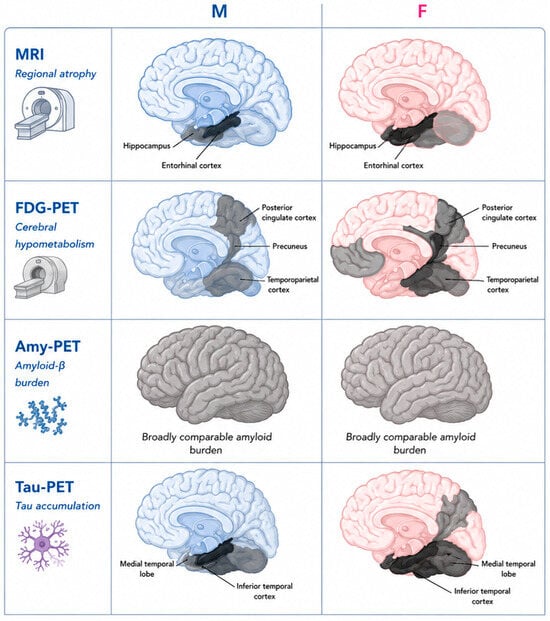
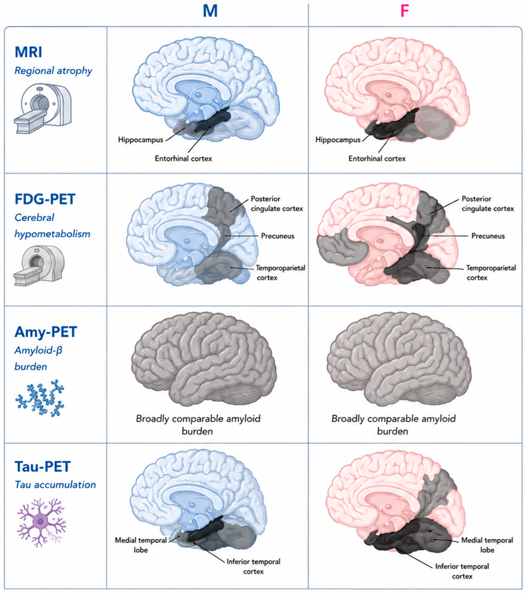
Multimodal neuroimaging reveals distinct biological pathways underlying Alzheimer’s disease in men and women. Structural MRI demonstrates accelerated medial temporal atrophy with symptom onset in AD women as compared to men. FDG-PET shows cerebral glucose hypometabolism especially in medial temporal regions in women, indicating heightened limbic metabolic vulnerability. Although amyloid-PET reveals comparable amyloid burden across sexes, the downstream impact on metabolism and cognition is stronger in women. Tau-PET imaging consistently demonstrates higher regional tau accumulation and steeper tau–cognition relationships in women, suggesting that tau-mediated neurodegeneration disproportionately drives disease severity and progression in female patients (Figure 1).
Figure 1.
Sex-specific neuroimaging patterns across Alzheimer’s disease biomarkers.
Schematic representation of sex-specific regional vulnerability patterns across multimodal neuroimaging biomarkers in Alzheimer’s disease. MRI panels represent regional brain atrophy; FDG-PET panels represent cerebral glucose hypometabolism; amyloid-PET panels represent amyloid-β burden; and tau-PET panels represent regional tau accumulation.
Dark gray/black regions indicate greater regional pathological involvement, whereas lighter gray regions indicate less pronounced abnormalities. Colored brain templates (blue for males and pink for females) are schematic and do not represent quantitative signal intensity.
Structural MRI highlights medial temporal atrophy involving the hippocampus and entorhinal cortex. FDG-PET illustrates hypometabolism within the posterior cingulate cortex, precuneus, temporoparietal cortex, and limbic regions, with relatively greater involvement in women. Amyloid-PET panels depict broadly comparable cortical amyloid burden between sexes, whereas tau-PET panels illustrate greater regional tau vulnerability in women, particularly within medial temporal and temporoparietal regions.
The figure represents a conceptual literature-based summary rather than direct quantitative imaging data.
3.2. FDG-PET and Cerebral Glucose Metabolism
[18F] FDG-PET provides a sensitive marker of synaptic function and neuronal integrity and is a cornerstone biomarker of AD-related neurodegeneration. In healthy aging, women typically exhibit higher global and regional cerebral glucose metabolism than men, particularly in frontal and temporal cortices [13,14,15]. While this pattern may confer functional reserve, it may also mask early pathological changes.
Along the AD continuum, women demonstrate faster and more extensive metabolic decline than men, particularly in temporoparietal regions, posterior cingulate cortex, and limbic networks [16,17,18,19]. Longitudinal FDG-PET studies reveal a stronger coupling between amyloid burden and metabolic dysfunction in women, especially among APOE ε4 carriers [16,18]. These findings indicate heightened vulnerability of neuronal energy metabolism in the female brain and provide a mechanistic link between pathology and accelerated clinical decline (Table 1).
3.3. Amyloid PET and Sex-Specific Pathological Effects
Amyloid-PET imaging has established Aβ deposition as an early and necessary component of AD pathophysiology. Although most studies report comparable global amyloid burden and positivity rates between women and men at equivalent clinical stages [2,7,9], sex significantly modifies the relationship between amyloid pathology and downstream neurodegeneration.
Women exhibit stronger associations between amyloid load, cerebral hypometabolism, and cognitive decline than men [7,9,20]. Multimodal imaging suggests that cognitively normal women may tolerate higher amyloid levels during compensatory stages, but once resilience mechanisms are exhausted, progression to neurodegeneration and clinical impairment accelerates (Table 1).
3.4. Tau PET and Female-Specific Vulnerability
Tau pathology is more closely associated with neurodegeneration and cognitive impairment than amyloid deposition. Tau-PET studies consistently demonstrate higher regional tau accumulation in women, particularly in the entorhinal cortex, inferior temporal regions, and parietal cortices, even at similar levels of cognitive impairment [7,21,22].
Longitudinal tau-PET analyses further reveal faster tau accumulation in women during preclinical and prodromal stages, particularly among amyloid-positive individuals [8]. Sex also modifies the tau–cognition relationship, with steeper associations between tau burden and memory decline observed in women [21]. These findings suggest that tau-mediated neurodegeneration disproportionately drives disease severity and progression in women (Table 1).
Table 1.
Sex Differences in AD Patients and AD Risk: Biomarker Evidence.
Interpretive summary. This table summarizes key sex-specific differences in Alzheimer’s disease risk and biomarker expression across epidemiological, genetic, and multimodal neuroimaging studies. Overall, the evidence indicates that while amyloid burden may be broadly comparable between sexes at similar clinical stages, women exhibit a disproportionate vulnerability to downstream neurodegenerative processes, including tau accumulation, cerebral hypometabolism, and cognitive decline. Across multimodal imaging studies, APOE ε4 status emerges as a critical modifier of these effects, with stronger associations between pathological biomarkers and clinical outcomes consistently observed in women. In addition, FDG-PET and tau-PET findings suggest enhanced metabolic and tau-related susceptibility in female cohorts, particularly at preclinical and prodromal stages. Collectively, these studies support a model in which sex interacts with genetic and neurobiological factors to shape differential trajectories of Alzheimer’s disease progression.
Overall, the studies summarized in Table 1 support the presence of sex-specific differences across multiple Alzheimer’s disease biomarkers and neuroimaging modalities. Although amyloid burden is often comparable between women and men, women tend to exhibit stronger associations between amyloid pathology, tau accumulation, cerebral hypometabolism, and cognitive decline. Structural MRI and FDG-PET findings further suggest accelerated neurodegenerative and metabolic trajectories in women, particularly following symptom onset. Importantly, several studies indicate that APOE ε4 status and disease stage may significantly modulate these associations, supporting the need for sex-aware biomarker interpretation across the Alzheimer’s disease continuum.
Nevertheless, the reported sex differences in the coupling among Alzheimer’s disease biomarkers are not entirely consistent across studies. The direction and magnitude of associations between amyloid, tau, and neurodegeneration may vary depending on cohort characteristics, disease stage, and clinical outcomes. Key moderators such as age, APOE ε4 genotype, vascular burden, hormone therapy exposure, and menopausal status likely contribute to this heterogeneity and should be systematically considered in future multimodal imaging studies.
4. Menopause, Neuroendocrine Aging, and Brain Vulnerability
4.1. Menopausal Transition as a Female-Specific Risk Window for Alzheimer’s Disease
Growing molecular and functional neuroimaging evidence supports the menopausal transition as a critical, female-specific window of vulnerability for Alzheimer’s disease (AD), during which endocrine aging intersects with bioenergetic, structural, and molecular brain changes relevant to AD pathophysiology [23,24,25,26,27]. During this transition, systemic and brain levels of 17β-estradiol decline, affecting synaptogenesis, mitochondrial function, glucose metabolism, and gene expression, and providing a biological basis for the predominantly neurological symptoms of perimenopause, including thermoregulatory instability, sleep disruption, mood changes, and subjective cognitive complaints [27,28,29].
Beyond chronological aging, menopausal status has emerged as one of the most consistent predictors of AD-related neuroimaging abnormalities in women, independently associated with alterations in cerebral glucose metabolism, amyloid-β (Aβ) burden, tau pathology, and brain structure [23,24,25,26,27,28,29]. Multiple FDG-PET studies demonstrate that perimenopausal and postmenopausal women exhibit progressive reductions in cerebral metabolic rate of glucose (CMRglc), particularly within AD-vulnerable regions such as the posterior cingulate cortex, precuneus, frontal, parietal, and temporal cortices [23,24,26]. Longitudinal evidence indicates that while premenopausal women maintain stable glucose metabolism over time, perimenopausal women already show emerging frontal hypometabolism, which becomes spatially extensive in postmenopause, with age-adjusted CMRglc reductions approaching 6% over three years [25].
At the molecular level, estrogen withdrawal during the menopausal transition disrupts cerebral bioenergetics by impairing glucose transport, aerobic glycolysis, and mitochondrial oxidative phosphorylation. Neuroimaging and biochemical studies demonstrate reductions in cytochrome c oxidase activity of approximately 30% in perimenopause and over 50% in postmenopause, tightly correlated with regional FDG-PET hypometabolism [23,26]. These changes are largely independent of APOE genotype and are accompanied by a compensatory metabolic shift toward lipid and ketone utilization, reflecting an adaptive yet potentially maladaptive response that may promote oxidative stress and amyloid accumulation [27,28,29].
A comprehensive conceptual framework for sex-specific vulnerability to Alzheimer’s disease (AD) can be proposed by integrating three converging biological axes: genetic susceptibility (particularly APOE ε4), neuroendocrine aging, and vascular/metabolic risk factors [1,12,24,30].
In this model, menopause represents a critical neuroendocrine transition characterized by a progressive decline in circulating and brain estrogen levels, which play a key role in maintaining synaptic plasticity, mitochondrial bioenergetics, glucose metabolism, and cerebrovascular integrity [18,22,27]. Estrogen depletion is therefore associated with reduced neuronal metabolic efficiency and increased vulnerability of AD-sensitive networks, particularly within the hippocampal–cortical circuitry, as demonstrated by structural MRI and FDG-PET studies in peri- and postmenopausal women [18,22,28].
APOE ε4 genotype interacts synergistically with this endocrine transition. In postmenopausal women, the loss of estrogen-mediated neuroprotection may exacerbate APOE ε4-associated impairments in lipid transport, amyloid clearance, synaptic repair, and mitochondrial function [1,12,24]. This interaction contributes to earlier and more pronounced amyloid accumulation, accelerated tau propagation, and reduced cerebral glucose metabolism across multimodal neuroimaging studies, including amyloid-PET, tau-PET, and FDG-PET investigations [14,19,21,26].
Vascular risk factors represent a further amplifying mechanism within this framework. Cerebrovascular dysfunction, including reduced cerebral perfusion, blood–brain barrier disruption, and metabolic syndrome-related processes, may converge with both APOE ε4-related and menopause-related alterations [22,25,29]. This convergence promotes chronic neuroinflammation, impaired neurovascular coupling, and enhanced susceptibility to neurodegeneration, particularly within temporoparietal and default mode network regions vulnerable to AD pathology [22,25].
Importantly, these three axes do not operate independently but interact dynamically across the Alzheimer’s disease continuum. The resulting phenotype in women—particularly APOE ε4 carriers after menopause—is characterized by reduced metabolic resilience, heightened vulnerability of limbic and association networks, and accelerated progression from preclinical pathology to clinical impairment [14,18,24,27].
This integrative model provides a mechanistic explanation for observed sex differences in neuroimaging biomarkers, including stronger coupling between amyloid burden, tau accumulation, and neurodegeneration in women compared with men [14,19,26], while also acknowledging that these associations may vary according to age, disease stage, APOE ε4 status, vascular burden, and hormonal exposure. Overall, this framework highlights key targets for sex-specific prevention and therapeutic strategies in Alzheimer’s disease.
4.2. Structural Brain Remodeling and Network Vulnerability
Structural MRI studies reveal a clear stage-dependent gradient of gray and white matter loss across the menopausal transition, with the most pronounced reductions observed in postmenopausal women, intermediate changes in perimenopause, and relative preservation in premenopause [31,32,33]. Regions affected by menopausal brain remodeling show substantial overlap with estrogen-regulated networks, including the hippocampus, amygdala, prefrontal cortex, posterior cingulate cortex, precuneus, and temporal association areas. Across cohorts, menopausal status consistently outperforms chronological age as a predictor of structural brain alterations, second only to biological sex itself [25].
Longitudinal analyses identify hippocampal atrophy as a particularly sensitive marker of menopausal stage, reliably discriminating menopausal women from premenopausal and perimenopausal women with classification accuracies exceeding 70% [24]. Comparisons with age-matched men further indicate that these structural changes reflect endocrine rather than chronological aging, as postmenopausal women exhibit greater gray and white matter loss in frontal and parietal regions than men of comparable age [31]. Notably, despite measurable structural decline, objective cognitive performance often remains within normal limits across menopausal stages, whereas subjective cognitive complaints are more prevalent, suggesting that brain vulnerability precedes clinically detectable impairment [24,25].
4.3. Amyloid-β Deposition and Tau Pathology Across Menopausal Stages
Amyloid-PET studies demonstrate that menopausal status robustly predicts regional Aβ accumulation, independent of age and APOE genotype in several cohorts [25,27,31]. Perimenopausal and postmenopausal women show increased amyloid uptake in posterior cingulate and frontal cortices compared with premenopausal women, with longitudinal increases of 6–9% over three years reported in menopausal women [27]. The spatial distribution of amyloid deposition also appears more widespread in postmenopause, extending into frontal and temporal regions and resembling early AD patterns [26].
Estrogen decline has been implicated in dysregulation of Aβ homeostasis through transcriptional upregulation of amyloidogenic pathways and downregulation of Aβ-degrading enzymes, linking endocrine aging to molecular AD mechanisms [32,33]. Importantly, earlier age at menopause is associated with higher amyloid burden, poorer cognitive performance, and increased AD risk, particularly when menopause occurs before age 50 [34].
Recent tau-PET studies further demonstrate menopause-dependent differences in tau accumulation. Postmenopausal women exhibit significantly higher tau-PET signal than age-matched men in inferior parietal, lateral occipital, and frontal regions, whereas premenopausal women do not differ from men [8]. Earlier age at menopause is associated with higher tau burden, an effect amplified in the presence of elevated amyloid, and interactions between age at menopause and amyloid—but not chronological age—moderate tau accumulation, underscoring the specificity of endocrine aging effects [8,35,36].
Collectively, PET, SPECT, and MRI findings converge on the menopausal transition as a biologically meaningful inflection point in female brain aging, during which metabolic dysfunction, protein aggregation, and network vulnerability emerge years to decades before clinical AD onset. These converging biomarkers support menopause as a critical window for risk stratification and early preventive intervention in women.
Beyond structural and metabolic changes, recent advances in molecular neuroimaging have provided direct in vivo evidence that menopause profoundly alters estrogen signaling within the female brain. Using 18F-fluoroestradiol (18F-FES) PET, studies have demonstrated progressively increased cerebral estrogen receptor (ER) density across the menopausal transition, particularly in estrogen-regulated limbic, frontal, and cingulate regions [28,29]. Importantly, these increases occur independently of chronological age and circulating estradiol levels, indicating compensatory upregulation of ER expression in response to declining estrogen availability rather than simple hormone deficiency.
Higher ER density has been associated with lower gray matter volume, altered cerebral perfusion, reduced mitochondrial ATP production, and the presence of neurological menopausal symptoms, including cognitive complaints, affective changes, and thermoregulatory instability [28,29,37]. These findings suggest that ER-PET imaging captures an adaptive—but potentially maladaptive—neuroendocrine response that signals emerging bioenergetic stress and increased vulnerability of estrogen-dependent neural networks.
In parallel, dysregulation of the hypothalamic–pituitary–gonadal axis has emerged as an additional, estrogen-independent contributor to menopause-related AD risk. Elevations in gonadotropins, particularly follicle-stimulating hormone (FSH) and luteinizing hormone (LH), which rise markedly during perimenopause and postmenopause, have been linked to increased amyloid-β burden and reduced gray matter volume in AD-vulnerable regions of midlife women at risk for AD [30]. These associations persist after adjustment for age, estradiol levels, APOE ε4 status, and hormone therapy use, supporting a direct role of gonadotropins in modulating brain aging trajectories.
Together, ER-PET and gonadotropin-related imaging findings expand the menopause–AD framework beyond estrogen depletion alone, highlighting menopause as a systems-level neuroendocrine transition characterized by altered receptor signaling, pituitary hormone dysregulation, metabolic stress, and increased susceptibility to downstream amyloid and tau pathology (Table 2).
Table 2.
Menopause, Neuroendocrine Aging, and Neuroimaging Biomarkers of Alzheimer’s Disease Risk.
Interpretive summary. This table synthesizes evidence linking menopausal and neuroendocrine aging processes to multimodal neuroimaging biomarkers of Alzheimer’s disease risk. Across studies, the menopausal transition emerges as a biologically meaningful period associated with progressive alterations in cerebral metabolism, structural integrity, and molecular imaging markers. FDG-PET and MRI findings consistently indicate early and regionally specific changes in glucose metabolism and gray matter volume, particularly within AD-vulnerable networks such as the posterior cingulate cortex, precuneus, frontal cortex, and hippocampal formation. Importantly, these alterations are observed independently of chronological age, supporting the role of endocrine aging as a distinct modulator of brain vulnerability.
Multimodal imaging studies further demonstrate that menopause is associated with increased amyloid burden, altered tau accumulation, and disruptions in mitochondrial and energetic function, suggesting convergence between hormonal decline and core AD-related pathological processes. Estrogen receptor imaging studies provide additional evidence of compensatory changes in neuroendocrine signaling, while gonadotropin-related findings suggest that hypothalamic–pituitary–gonadal axis dysregulation may further contribute to disease susceptibility. Across modalities, earlier age at menopause and postmenopausal status are consistently associated with more pronounced biomarker alterations and increased AD risk profiles. Collectively, these findings support menopause as a critical window of vulnerability in women, characterized by interacting metabolic, structural, and molecular changes that may accelerate Alzheimer’s disease–related trajectories.
4.4. APOE ε4, Menopause, and Sex-Specific Neuroimaging Vulnerability
Evidence from epidemiological and neuroimaging studies indicates that APOE ε4 is associated with increased Alzheimer’s disease risk and may exert stronger effects in women, particularly after menopause [6,11,21]. Female APOE ε4 carriers have been reported to show greater cognitive decline, higher tau burden, and more pronounced metabolic alterations compared with male carriers across different stages of the AD continuum [6,7,8,21].
Neuroimaging studies have shown that postmenopausal APOE ε4 carriers may exhibit reduced cerebral glucose metabolism on FDG-PET, particularly within temporoparietal and posterior cingulate regions, together with increased amyloid and tau deposition compared with non-carriers [21,27,29]. Several studies have proposed that estrogen decline during menopause may contribute to reduced metabolic efficiency and altered amyloid-related pathways in susceptible individuals [27,39,40].
Experimental and translational studies have also examined the interaction between APOE ε4 status and estrogen depletion in relation to brain metabolism and neuroinflammatory processes [39,40,41]. In tau-PET investigations, female APOE ε4 carriers have shown greater tau accumulation and stronger associations between tau burden and cognitive impairment compared with male carriers in some cohorts [7,8].
Overall, current evidence supports a relationship between APOE ε4 status, menopause-related endocrine aging, and sex-specific neuroimaging patterns associated with Alzheimer’s disease vulnerability [6,7,8,21,27,41].
5. Fluid and Blood-Based Biomarkers
Sex differences in AD extend beyond imaging to cerebrospinal fluid and blood-based biomarkers. Women with cognitive impairment or AD frequently exhibit higher levels of total tau, phosphorylated tau, and astrocytic activation markers such as glial fibrillary acidic protein compared with men at similar disease stages [21,22,33].
Plasma biomarkers, including the Aβ42/40 ratio and neurodegeneration markers, demonstrate stronger associations with cerebral hypometabolism and structural decline in women, indicating sex-specific biomarker trajectories that may necessitate distinct diagnostic thresholds [21,22,33].
6. Clinical and Translational Implications
Failure to account for sex-specific biomarker trajectories may result in underestimation of disease burden and delayed diagnosis in women [1,2,38]. Incorporating sex-aware interpretation of neuroimaging, genetic, hormonal, and fluid biomarkers could improve early detection, risk stratification, and clinical trial design [1,2,4].
From a precision medicine perspective, integrating sex, endocrine status, and genetic susceptibility is essential for developing targeted prevention and therapeutic strategies aimed at preserving women’s brain health.
7. Future Directions
Future research should prioritize longitudinal, life-course studies spanning midlife to late life with detailed characterization of hormonal transitions. Developing and validating sex-adjusted biomarker thresholds—particularly for tau-PET, FDG-PET, ER-PET, and plasma biomarkers—represents a critical step toward reducing diagnostic bias.
Clarifying the temporal sequence of biomarker changes in women, integrating genetic and endocrine mechanisms, and embedding sex-specific endpoints into clinical trials will be essential. A sex-informed framework offers a path toward improved prevention, diagnosis, and treatment of Alzheimer’s disease, with particular benefit for women.
Author Contributions
Conceptualization, M.N., E.M.A., V.B.; methodology, V.M., L.M., V.C., A.B.; writing—original draft preparation, V.M., L.M., V.C., A.B.; writing—review and editing, M.N., E.M.A., V.B.; visualization, M.N.; supervision, V.B.; project administration, V.B. All authors have read and agreed to the published version of the manuscript.
Funding
This research received no external funding.
Data Availability Statement
No new data were created or analyzed in this study.
Acknowledgments
During the preparation of this manuscript, the author(s) used BioRender for the purposes of producing figures. The authors have reviewed and edited the output and take full responsibility for the content of this publication.
Conflicts of Interest
The authors declare no conflicts of interest.
References
- Nebel, R.A.; Aggarwal, N.T.; Barnes, L.L.; Gallagher, A.; Goldstein, J.M.; Kantarci, K.; Mallampalli, M.P.; Mormino, E.C.; Scott, L.; Yu, W.H.; et al. Understanding the impact of sex and gender in Alzheimer’s disease: A call to action. Alzheimers Dement. 2018, 14, 1171–1183. [Google Scholar] [CrossRef] [Scilit]
- Ferretti, M.T.; Iulita, M.F.; Cavedo, E.; Chiesa, P.A.; Dimech, A.S.; Chadha, A.S.; Baracchi, F.; Girouard, H.; Misoch, S.; Giacobini, E.; et al. Sex differences in Alzheimer disease. Nat. Rev. Neurol. 2018, 14, 457–469. [Google Scholar] [CrossRef] [Scilit] [PubMed]
- Carter, C.L.; Resnick, E.M.; Mallampalli, M.; Kalbarczyk, A. Sex and gender differences in Alzheimer’s disease: Recommendations for future research. J. Womens Health 2012, 21, 1018–1023. [Google Scholar] [CrossRef] [Scilit]
- Snyder, H.M.; Asthana, S.; Bain, L.; Brinton, R.D.; Dubal, D.B.; Espeland, M.A.; Gatz, M.; Mielke, M.M.; Raber, J.; Rapp, P.R.; et al. Sex biology contributions to vulnerability to Alzheimer’s disease: A think tank convened by the Women’s Alzheimer’s Research Initiative. Alzheimers Dement. 2016, 12, 1186–1196. [Google Scholar] [CrossRef] [Scilit]
- Andersen, K.; Launer, L.J.; Dewey, M.E.; Letenneur, L.; Ott, A.; Copeland, J.R.; Dartigues, J.F.; Kragh-Sorensen, P.; Baldereschi, M.; Brayne, C.; et al. Gender differences in the incidence of AD and vascular dementia: The EURODEM Studies. Neurology 1999, 53, 1992–1997. [Google Scholar] [CrossRef] [Scilit] [PubMed]
- Altmann, A.; Tian, L.; Henderson, V.W.; Greicius, M.D. Sex modifies the APOE-related risk of developing Alzheimer disease. Ann. Neurol. 2014, 75, 563–573. [Google Scholar] [CrossRef] [Scilit]
- Buckley, R.F.; Mormino, E.C.; Rabin, J.S.; Hohman, T.J.; Landau, S.; Hanseeuw, B.J.; Jacobs, H.I.L.; Papp, K.V.; Amariglio, R.E.; Properzi, M.J.; et al. Sex differences in the association of global amyloid and regional tau deposition measured by PET in clinically normal older adults. Alzheimers Dement. 2019, 15, 1269–1280. [Google Scholar]
- Buckley, R.F.; O’Donnell, A.; McGrath, E.R.; Jacobs, H.I.L.; Lois, C.; Satizabal, C.L.; Ghosh, S.; Rubinstein, Z.B.; Murabito, J.M.; Sperling, R.A.; et al. Menopause status moderates sex differences in tau burden: A Framingham PET study. Ann. Neurol. 2022, 92, 11–22. [Google Scholar] [CrossRef] [Scilit]
- Cavedo, E.; Chiesa, P.A.; Houot, M.; Ferretti, M.T.; Grothe, M.J.; Teipel, S.J.; Lista, S.; Habert, M.O.; Poiter, M.C.; Dubois, B.; et al. Sex differences in functionaland molecular neuroimaging biomarkers of Alzheimer’s disease in cognitively normal older adults with subjective memory complaints. Alzheimers Dement. 2018, 14, 1204–1215. [Google Scholar]
- Inguanzo, A.; Poulakis, K.; Oltra, J.; Maioli, S.; Marseglia, A.; Ferreira, D.; Mohanty, R.; Westman, E. Atrophy trajectories in Alzheimer’s disease: How sex matters. Alzheimers Res. Ther. 2025, 17, 79. [Google Scholar] [CrossRef] [Scilit]
- Zeydan, B.; Tosakulwong, N.; Schwarz, C.G.; Weigand, S.D.; Lowe, V.J.; Senjem, M.L.; Gunter, J.L.; Reid, R.I.; Spychalla, A.J.; Petersen, R.C.; et al. Sex differences in brain imaging markers of neurodegeneration and Alzheimer disease. Neurology 2022, 98, e1911–e1921. [Google Scholar]
- Armstrong, N.M.; An, Y.; Beason-Held, L.; Resnick, S.M. Sex Differences in Brain Aging and Predictors of Neurodegeneration in Cognitively Healthy Older Adults. Neurobiol. Aging 2019, 81, 146–156. [Google Scholar] [CrossRef] [Scilit]
- Kim, J.H.; Lee, J.S.; Kang, E.S.; Lee, D.S.; Lee, M.C. Gender-related differences in regional cerebral glucose metabolism in normal aging brain. Nucl. Med. Mol. Imaging 2009, 43, 131–137. [Google Scholar]
- Nugent, S.; Castellano, C.A.; Bocti, C.; Dionne, I.; Fulop, T.; Cunnane, S.C. Evaluation of age and sex-related metabolic changes in the human brain. J. Cereb. Blood Flow Metab. 2014, 34, 450–458. [Google Scholar]
- Allocca, M.; Linguanti, F.; Calcagni, M.L.; Cistaro, A.; Gaudieri, V.; Guerra, U.P.; Morbelli, S.; Nobili, F.; Pappatà, S.; Sestini, S.; et al. Evaluation of age- and sex-related metabolic changes in healthy subjects using FDG-PET. J. Clin. Med. 2021, 10, 4932. [Google Scholar] [CrossRef] [Scilit]
- Park, J.-C.; Lim, H.; Byun, M.S.; Yi, D.; Byeon, G.; Jung, G.; Kim, Y.K.; Lee, D.Y.; Han, S.-H.; Mook-Jung, I. Sex differences in the progression of glucose metabolism dysfunction in Alzheimer’s disease. Exp. Mol. Med. 2023, 55, 1023–1032. [Google Scholar] [CrossRef] [Scilit] [PubMed]
- Nerattini, M.; Abenavoli, E.M.; Caramelli, F.; Giacomucci, G.; Nacmias, B.; Mossello, E.; Bessi, V.; Berti, V. Sex differences in brain glucose metabolism in Alzheimer’s disease: A voxel-based FDG-PET study. GeroScience 2025. Epub ahead of print. [Google Scholar] [CrossRef] [Scilit]
- Rahman, A.; Schelbaum, E.; Hoffman, K.; Diaz, I.; Hristov, H.; Andrews, R.; Jett, S.; Jackson, H.; Lee, A.; Sarva, H.; et al. Sex-driven modifiers of Alzheimer risk: A multimodality brain imaging study. Neurology 2020, 95, e166–e178. [Google Scholar] [CrossRef] [Scilit] [PubMed]
- Qian, S.; Ba, Y.; Xue, L.; Jin, C.; Zhou, R.; Liao, Y.; Zhong, Y.; Xu, Y.; Shi, F.; Wang, C.; et al. Age-related changes of human brain metabolism. Eur. J. Nucl. Med. Mol. Imaging 2025, 52, 3412–3423. [Google Scholar] [CrossRef] [Scilit] [PubMed]
- Luchsinger, J.A.; Palta, P.; Rippon, B.; Soto, L.; Ceballos, F.; Pardo, M.; Laing, K.; Igwe, K.; Johnson, A.; Tomljanovic, Z.; et al. Sex differences in in vivo Alzheimer’s disease neuropathology in late middle-aged Hispanics. Alzheimers Dement. 2021, 17, 1038–1049. [Google Scholar] [CrossRef] [Scilit]
- Boccalini, C.; Peretti, D.E.; Scheffler, M.; Mu, L.; Griffa, A.; Testart, N.; Allali, G.; Prior, J.O.; Ashton, N.J.; Zetterberg, H.; et al. Sex differences in the association of Alzheimer’s disease biomarkers and cognition in a multicenter memory clinic study. Alzheimers Res. Ther. 2025, 17, 46. [Google Scholar] [CrossRef] [Scilit]
- Mak, E.; Kantarci, K.; Arvanitakis, Z. Sex differences in in vivo biomarkers of neurodegenerative dementia. Front. Dement. 2025, 4, 1706177. [Google Scholar] [CrossRef] [Scilit]
- Mosconi, L.; Berti, V.; Quinn, C.; McHugh, P.; Petrongolo, G.; Varsavsky, I.; Osorio, R.S.; Pupi, A.; Vallabhajosula, S.; Isaacson, R.S.; et al. Sex differences in Alzheimer risk: Brain imaging of endocrine vs chronologic aging. Neurology 2017, 89, 1382–1390. [Google Scholar]
- Mosconi, L.; Berti, V.; Quinn, C.; McHugh, P.; Petrongolo, G.; Osorio, R.S.; Connaughty, C.; Pupi, A.; Vallabhajosula, S.; Isaacson, R.S.; et al. Perimenopause and emergence of an Alzheimer’s bioenergetic phenotype in brain and periphery. PLoS ONE 2017, 12, e0185926. [Google Scholar] [CrossRef] [Scilit]
- Mosconi, L.; Rahman, A.; Diaz, I.; Wu, X.; Scheyer, O.; Hristov, H.W.; Vallabhajosula, S.; Isaacson, R.S.; De Leon, M.J.; Brinton, R.D. Increased Alzheimer’s risk during the menopause transition: A 3-year longitudinal brain imaging study. PLoS ONE 2018, 13, e0207885. [Google Scholar] [CrossRef] [Scilit]
- Mosconi, L.; Berti, V.; Dyke, J.; Schelbaum, E.; Jett, S.; Loughlin, L.; Jang, G.; Rahman, A.; Hristov, H.; Pahlajani, S.; et al. Menopause impacts human brain structure, connectivity, energy metabolism, and amyloid-beta deposition. Sci. Rep. 2021, 11, 10867. [Google Scholar] [CrossRef] [Scilit] [PubMed]
- Schelbaum, E.; Jett, S.; Jang, G.; Boneu Yepez, C.; Dyke, J.P.; Pahlajani, S.; Diaz Brinton, R.; Mosconi, L. Ovarian steroid hormones: A long overlooked but critical contributor to brain aging and Alzheimer’s disease. Front. Aging Neurosci. 2022, 14, 948219. [Google Scholar] [CrossRef] [Scilit] [PubMed]
- Mosconi, L.; Nerattini, M.; Matthews, D.C.; Jett, S.; Andy, C.; Williams, S.; Yepez, C.B.; Zarate, C.; Carlton, C.; Fauci, F.; et al. In vivo brain estrogen receptor density by neuroendocrine aging and relationships with cognition and symptomatology. Sci. Rep. 2024, 14, 12680. [Google Scholar] [CrossRef] [Scilit] [PubMed]
- Mosconi, L.; Jett, S.; Nerattini, M.; Andy, C.; Yepez, C.B.; Zarate, C.; Carlton, C.; Kodancha, V.; Schelbaum, E.; Williams, S.; et al. In vivo Brain Estrogen Receptor Expression by Neuroendocrine Aging And Relationships with Gray Matter Volume, Bio-Energetics, and Clinical Symptomatology. Res. Sq. 2023, Preprint. [Google Scholar] [CrossRef] [Scilit]
- Nerattini, M.; Rubino, F.; Jett, S.; Andy, C.; Boneu, C.; Zarate, C.; Carlton, C.; Loeb-Zeitlin, S.; Havryliuk, Y.; Pahlajani, S.; et al. Elevated gonadotropin levels are associated with increased biomarker risk of Alzheimer’s disease in midlife women. Front. Dement. 2023, 2, 1303256. [Google Scholar] [CrossRef] [Scilit]
- Raikes, A.C.; Dyke, J.P.; Nerattini, M.; Boneu, C.; Ajila, T.; Fauci, F.; Battista, M.; Pahlajani, S.; Williams, S.; Brinton, R.D.; et al. White matter microstructural and macrostructural profiles during midlife reveal sex differences between men and women at different menopausal stages. Sci. Rep. 2025, 15, 40312. [Google Scholar] [CrossRef] [Scilit]
- Nerattini, M.; Jett, S.; Andy, C.; Carlton, C.; Zarate, C.; Boneu, C.; Battista, M.; Pahlajani, S.; Loeb-Zeitlin, S.; Havryulik, Y.; et al. Systematic review and meta-analysis of the effects of menopause hormone therapy on risk of Alzheimer’s disease and dementia. Front. Aging Neurosci. 2023, 15, 1260427. [Google Scholar] [CrossRef] [Scilit]
- Blennow, K.; Hampel, H.; Weiner, M.; Zetterberg, H. Cerebrospinal fluid and plasma biomarkers in Alzheimer disease. Nat. Rev. Neurol. 2010, 6, 131–144. [Google Scholar] [CrossRef] [Scilit] [PubMed]
- Boyle, C.P.; Raji, C.A.; Erickson, K.I.; Lopez, O.L.; Becker, J.T.; Gach, H.M.; Kuller, L.H.; Longstreth, W., Jr.; Carmichael, O.T.; Riedel, B.C.; et al. Estrogen, brain structure, and cognition in postmenopausal women. Hum. Brain Mapp. 2021, 42, 24–35. [Google Scholar] [CrossRef] [Scilit]
- Coughlan, G.T.; Betthauser, T.J.; Boyle, R.; Koscik, R.L.; Klinger, H.M.; Chibnik, L.B.; Jonaitis, E.M.; Yau, W.Y.W.; Wenzel, A.; Christian, B.T.; et al. Association of age at menopause and hormone therapy use with tau and β-amyloid positron emission tomography. JAMA Neurol. 2023, 80, 462–473. [Google Scholar] [CrossRef] [Scilit]
- Bove, R.; Secor, E.; Chibnik, L.B.; Barnes, L.L.; Schneider, J.A.; Bennett, D.A.; De Jager, P.L. Age at surgical menopause influences cognitive decline and Alzheimer pathology in older women. Neurology 2014, 82, 222–229. [Google Scholar] [CrossRef] [Scilit] [PubMed]
- Mosconi, L.; Williams, S.; Carlton, C.B.; Andy, C.; Fauci, F.B.; Zarate, C.B.; Boneu, C.M.; Ajila, T.B.; Nerattini, M.; Jett, S.M.; et al. Neurophysiological correlates of subjective cognitive decline in perimenopausal and postmenopausal midlife women at risk for Alzheimer’s disease. Menopause 2025, 32, 433–442. [Google Scholar] [CrossRef] [Scilit] [PubMed]
- Aggarwal, N.T.; Mielke, M.M. Sex Differences in Alzheimer’s Disease. Neurol. Clin. 2023, 41, 417–432. [Google Scholar] [CrossRef] [Scilit]
- Brinton, R.D.; Yao, J.; Yin, F.; Mack, W.J.; Cadenas, E. Perimenopause as a neurological transition state. Nat. Rev. Endocrinol. 2015, 11, 393–405. [Google Scholar] [CrossRef] [Scilit]
- Yao, J.; Brinton, R.D. Estrogen regulation of mitochondrial bioenergetics: Implications for prevention of Alzheimer’s disease. Adv. Pharmacol. 2012, 64, 327–371. [Google Scholar]
- Jett, S.; Dyke, J.P.; Yepez, C.B.; Zarate, C.; Carlton, C.; Schelbaum, E.; Jang, G.; Pahlajani, S.; Williams, S.; Brinton, R.D.; et al. Effects of sex and APOE ε4 genotype on brain mitochondrial high-energy phosphates in midlife individuals at risk for Alzheimer’s disease: A 31Phosphorus MR spectroscopy study. PLoS ONE 2023, 18, e0281302. [Google Scholar] [CrossRef] [Scilit] [PubMed]

Disclaimer/Publisher’s Note: The statements, opinions and data contained in all publications are solely those of the individual author(s) and contributor(s) and not of MDPI and/or the editor(s). MDPI and/or the editor(s) disclaim responsibility for any injury to people or property resulting from any ideas, methods, instructions or products referred to in the content. |
© 2026 by the authors. Licensee MDPI, Basel, Switzerland. This article is an open access article distributed under the terms and conditions of the Creative Commons Attribution (CC BY) license.