


Abstract
Periodontal disease (PD) is highly prevalent in dogs and is associated with oral biofilms that promote chronic inflammation and tissue damage. Disrupting biofilms may therefore support canine oral health. This study evaluated protease (subtilisin) and betaine using complementary in vitro and in vivo approaches. Porphyromonas gingivalis, Porphyromonas gulae, and Streptococcus mutans were used as model oral pathogens in biofilm inhibition, biofilm removal, and planktonic growth assays. An 8-day canine pilot study assessed the tolerability of a dental chew containing protease and betaine and its effects on the oral microbiome. In vitro data were analyzed using standard least-squares models, and microbiome responses using diversity and differential abundance analyses. Protease reduced biofilm formation by P. gingivalis and S. mutans (p < 0.0001) and removed established biofilms (p < 0.05), while showing limited effects on planktonic growth. Betaine responses varied by bacterial strain and assay type and did not significantly interact with protease. The chew showed good physiological tolerability, although not all dogs consumed it completely. Microbiome analysis revealed limited treatment effects, with no differences in alpha diversity between groups. Overall, protease was the main driver of biofilm-related responses, supporting further investigation in canine oral care products; however, clinical periodontal outcomes, inflammatory biomarkers, and direct evidence of betaine-mediated tissue protection were not assessed.
1. Introduction
Periodontal disease (PD) is the most diagnosed problem in small animal veterinary medicine [1]. With few to no visible signs, PD often goes untreated until later stages, affecting approximately 80–100% of dogs over three years of age [2,3,4]. This disease progresses from gingivitis to periodontitis and is influenced by the interaction between the host’s defense mechanism and plaque-building microorganisms. The action of virulent bacteria, when untreated, can lead to periodontitis [5], which is defined as infection and inflammation of the gums, bone, and other tissues surrounding and supporting the teeth [6,7].
Although canine dentition provides some natural self-cleaning through the shearing action of the teeth during mastication, plaque commonly accumulates along the gingival margin where the tooth meets the gingiva. Consequently, this region remains particularly susceptible to plaque retention, gingival inflammation, and progression of PD [3,5]. Periodontal disease is fundamentally a biofilm-mediated condition. Once established, oral biofilm protects resident bacteria from host defenses and environmental stressors, allowing pathogenic communities to persist and contribute to inflammation and tissue destruction [7]. Consequently, strategies that disrupt biofilm formation or destabilize established biofilm may offer an effective approach for supporting canine oral health [8]. The American Veterinary Medical Association (AVMA) has recommended professional dental cleaning annually, and daily brushing as the gold standard for maintenance [9,10]. However, maintaining daily brushing routines can be challenging for many pet owners [8]. As a result, passive strategies such as dental chews have been developed to support active home care. Although dental chews may improve oral health through mechanical abrasion, delivery of functional ingredients, or a combination of both mechanisms; their efficacy varies considerably among products [11,12,13].
Dental chews have emerged as a practical tool, offering a non-invasive method to manage PD by mechanically disrupting plaque and delivering anti-microbial agents whilst being safe and appealing to pets [9,13]. Studies have shown that daily feeding of oral chews to dogs can significantly improve oral health by positively shifting the oral microbiota, reducing the abundance of potentially pathogenic bacteria such as Porphyromonas, Tannerella, and Treponema, while increasing beneficial bacteria genera like Neisseria and Actinomyces [14,15]. In dogs, Porphyromonas species are among the most prevalent bacteria in the oral microbiome and have been consistently linked to the development and progression of PD [2,4]. Among them, Porphyromonas gulae is considered a major canine periodontal pathogen and exhibits virulence characteristics similar to those of Porphyromonas gingivalis, a well-characterized human periodontal pathogen [16]. In addition to these periodontal pathogens, Streptococcus mutans is a biofilm-forming bacterium frequently used as a model organism to study bacterial adhesion, extracellular matrix production, and plaque development due to its extensive production of extracellular polysaccharides [17,18,19,20]. As one of the most extensively studied oral bacteria, S. mutans has served as a key model for understanding the mechanisms underlying biofilm formation, stress adaptation, and microbial interactions within dental plaque [17]. Furthermore, as a facultative anaerobe, S. mutans can grow under both aerobic and anaerobic conditions and readily forms biofilms across a range of environmental conditions relevant to the oral cavity [21]. Therefore, S. mutans was included as a reference organism to evaluate the efficacy of the protease against a broader range of oral microbes and biofilm phenotypes beyond the periodontal pathogens P. gingivalis and P. gulae. Together, these organisms represent relevant models for investigating ingredient effects on oral bacterial growth and biofilm formation.
Subtilisin, a serine protease derived from Bacillus subtilis, has been proposed for managing oral biofilms in dogs due to its capacity to enzymatically disrupt protein-based matrices within dental plaque. Subtilisin’s ability to target and cleave structural proteins in the extracellular polymeric substance (EPS) of biofilm [22] weakens the biofilm structural integrity, making bacteria more susceptible to shear force, antimicrobials, or host immune response. This enzymatic activity also promotes oral hygiene by hydrolyzing proteins from food residues. In early stages of biofilm development, subtilisin can disrupt bacterial adhesion and coaggregation, interfering with plaque establishment, maturation and formation [20,23,24]. Research has shown that proteolytic enzymes, including subtilisin, can effectively disrupt biofilms and reduce interkingdom biofilm biomass, which contributes to tartar formation and persistent periodontal inflammation [1,25]. Given that PD is strongly associated with biofilm accumulation rather than bacterial presence alone, proteases represent a promising strategy for selectively targeting biofilm architecture while avoiding the use of conventional antimicrobials.
Betaine (trimethylglycine) is a naturally occurring zwitterionic compound with osmoprotective and anti-inflammatory properties. By helping to relieve dry mouth, maintain cellular hydration, and protein stability during stress, betaine may help support oral tissue resilience [26,27,28,29]. In addition, studies have shown that betaine can modulate inflammatory pathways, including reduction in IL-1β production, a cytokine associated with PD progression [30,31]. Although direct evidence for betaine in canine PD remains limited, these properties suggest a potential complementary role in supporting the gum tissues in the oral environment.
Based on its biofilm disrupting properties, protease was hypothesized to act as the primary functional ingredient responsible for reducing oral biofilm formation. Betaine was included as a complementary ingredient due to its osmoprotective and anti-inflammatory properties, which may support the oral environment and host associated responses. Therefore, the objective of this study was to characterize the effects of protease and betaine on bacterial growth and biofilm responses using in vitro oral pathogen models with detectable microbiome responses in dogs receiving a functional dental chew.
2. Materials and Methods
2.1. In Vitro Biofilm Growth—Biofilm Reactor
2.1.1. Bacteria Strain and Culture Conditions
Streptococcus mutans ATCC 25175 (S. mutans), Porphyromonas gingivalis ATCC 33277 (P. gingivalis), and Porphyromonas gulae ATCC 51700 (P. gulae) were obtained from the American Type Culture Collection (ATCC, Mamassas, VA, USA). S. mutans was grown on brain heart infusion agar plates supplemented with hemin, menadione and yeast extract (BHIS) at 37 °C under anerobic conditions (80% N2, 10% CO2, 10% H2) overnight and cultured in BHIS broth at 37 °C under anerobic conditions for 24 h. P. gingivalis and P. gulae were cultured on CDC blood agar (Anaerobe Systems, Morgan Hill, CA, USA) at 37 °C under anaerobic conditions for 7 days and cultured in yeast casitone fatty acids broth with carbohydrates (YCFAC; Anaerobe Systems) for 72 h.
2.1.2. Enzyme Extraction
Commercially available protease enzyme (ValoraseTM Pro, IFF, New York, NY, USA) granules were extracted using 1x PBS (pH = 7.4; Corning). Enzyme granules were mixed in 1x PBS, shaken at 37 °C intermittently at 200 rpm, then centrifuged at 4000× g for 10 min. The supernatant was then aspirated and filtered sterilized using 0.22 µm PES filter membrane (MilliporeSigma™ Steriflip™, Burlington, MA, USA), and the extracted enzyme was stored at 4 °C until further use.
2.1.3. Biofilm Formulation and Quantification
Culture Conditions
S. mutans were inoculated in BHIS broth for 24 h, while P. gingivalis and P. gulae were each inoculated in YCFAC broth for 72 h under anaerobic conditions. After incubation, cultures of each strain were diluted to optical density at 600 nm (OD600) of 0.1 in BHIS broth supplemented with 1% sucrose (BHIS-S) for S. mutans and YCFAC broth for P. gingivalis and P. gulae. A total volume of 200 µL was added to each well of a 96-well flat-bottom non-treated polystyrene plate and incubated anaerobically at 37 °C (24 h for S. mutans; 72 h for P. gingivalis and P. gulae).
Biomass Quantification
Total biomass (culture and biofilm) was quantified immediately after incubation to evaluate total growth. The plate was removed from the incubator, and the optical density was measured at 600 nm (OD600) using a Tecan Spark 10M. Biofilm biomass was quantified using a crystal violet assay. Briefly, the wells were rinsed with 200 µL of 1x PBS three times, then stained with 200 µL of 0.1% crystal violet solution (Sigma-Aldrich, St. Louis, MO, USA) and incubated at room temperature for 5 min. The wells were again rinsed with 200 µL of 1x PBS three times, and the crystal violet stained biofilms were resolubilized with 70% ethanol. The optical density was measured at 590 nm (OD590) using a Tecan Spark 10M to determine biofilm biomass.
2.1.4. Biofilm Inhibition and Removal Assay
Biofilm Inhibition
To evaluate the efficacy of protease to inhibit the growth and biofilm formation of oral strains, the extracted enzyme was added to the bacterial cultures at the start of incubation. Each strain was prepared as previously described. Then, protease was added to the well containing the culture to a final volume of 200 µL. The cultures were incubated for biofilm formation as described previously. The total biomass of the cultures and biofilm biomass were quantified as previously described.
Biofilm Removal
To evaluate the efficacy of protease to remove the biofilm of oral pathogens, the extracted enzyme was added to the biofilms after formation. Briefly, each strain was prepared for biofilm formation as previously described. After incubation (24 h for S. mutans and 72 h for P. gingivalis and P. gulae), the wells were decanted, and fresh media containing the protease was added to a final volume of 200 µL. The cultures were incubated anaerobically at 37 °C for 24 h. Afterwards, the wells were washed and quantified using a crystal violet assay as previously described.
2.1.5. Checkerboard Assay
The effect of combining commercially available betaine (Betafin® Pet, IFF, New York, NY, USA) and protease was evaluated via checkerboard biofilm assays to determine additive or deleterious effects of betaine on the efficacy of protease. A dilution series was created for protease and betaine across 96-well polystyrene plates using a Biomek i7 Automated Workstation. The concentrations of protease were 2% (w/v), 1%, and 0.5%, and the concentrations for betaine were 10% (w/v), 5%. 2%, 1%, and 0.5%. The protease extracted in 1x PBS was used for this assay, and both protease and betaine were diluted using 1x PBS. Plates of only protease and plates of only betaine were also evaluated. Total biomass and biofilm biomass were quantified as previously described.
2.1.6. Data Analysis
The in vitro dataset was analyzed in JMP 18 using standard least-squares models. Estimated group means were obtained as least-squares means (LS means), and pairwise comparisons among treatment groups were performed using the Tukey–Kramer adjustment to control the family-wise Type I error rate for multiple comparisons. The synergy assays were analyzed using full factorial model effects to determine which factors and interactions significantly impacted the response. Data were normalized relative to untreated control, which was set as 0.000 inhibition. Statistical significance was declared at p < 0.05. A positive least-squares mean correlated to the inhibition or removal ability of protease or betaine, whereas a negative least-squares mean indicated increased growth or biofilm formation of the target strain.
2.2. In Vivo Pilot Study in Dogs
2.2.1. Dental Chew Study
The pilot study was conducted at the High Quality Research (HQR) animal facility study site (Fort Collins, CO, USA). The protocol was approved by the Institutional Animal Care and Use Committee (IACUC) of HQR, Fort Collins, CO, USA (protocol number 210.001), following the Welfare Act Regulations (United States Department of Agriculture, Washington, DC, USA, 2019).
Animal and Housing Conditions
Eighteen adult male Beagle dogs (mean age = 1.17 ± 0.04 yr, mean body weight [BW] = 9.2 ± 1.2 kg) were included in the study and individually housed in kennels within a room located at HQR. The room was kept on a 12 h light schedule, with temperatures between 16.7 and 19 °C, and relative humidity ranging from 34% to 43%.
Diet and Treatment Design
All dogs were fed commercially available dry food (Adult Red Flannel, Red Flannel, PMI Nutrition, Arden Hills, MN, USA), once daily throughout the study. The amount of food provided was to reach their caloric maintenance requirements and remained consistent throughout the study. Dogs always had access to fresh water. This experiment followed a single-blind parallel comparative design.
Two types of pet dental chews were produced by Targeted Pet Treats LLC (Warren, PA, USA) using an identical base formulation. Both chews were manufactured via injection molding. The control product contained only the base recipe, while the test product included the same formulation with the addition of commercially available functional ingredients for betaine (1%, 0.3 g) and protease (0.1%, 0.03 g). The experimental chew was formulated with a concentrated, salt-coated protease preparation (80,000 U/g) designed to enhance enzyme stability and help withstand the high temperatures encountered during manufacturing. The enzyme was included at a target concentration of 0.1% (w/w) in the finished product. To compensate for anticipated processing losses and ensure a minimum final concentration of 0.1%, the protease was added at three times the target dose during manufacturing. Based on an average chew weight of approximately 30 g, the target activity was 2400 U per chew. An external laboratory, Eurofins (Des Moines, IA, USA), quantified protease activity in the finished chews and determined it to be 6000 U per chew, confirming enzyme activity 2.5-fold above the target specification and demonstrating effective retention of protease activity throughout processing.
Data Collection
The consumption of dental chews and the fecal conditions were monitored daily during the study. Physical exams were conducted on Day 0 (pre-chews) and Day 8.
Two oral swabs were collected from teeth and gums on Day 0 (pre-chews), Day 4 and Day 8 (post-chews). Gum swabs were collected by gently brushing a sterile dry swab over the upper and lower gums of both sides of the mouth while avoiding teeth. To retain a representative sample for teeth microbiome, one dry swab per dog was gently brushed on eight teeth according to the modified Triadan system [32]: Maxillary canine teeth (104/204), mandibular canine teeth (304/404), maxillary fourth premolars (108/208), and mandibular first molar teeth (309/409). Fresh feces were collected on Day 0 (pre-chews), Day 4, and Day 8 post-chews; these were then placed into a labeled whirl pack for each dog at each collection timepoint.
All the data collection samples were kept at −70 °C or colder freezer and shipped to the microbiology lab at Wilmington, Delaware, upon study completion.
2.2.2. Microbiome Sequencing
16S rRNA Amplicon Sequencing and Profiling
Microbial genomic DNA extraction and 16S rRNA amplicon sequencing were conducted according to the protocols detailed in the section “DNA extraction, 16S rRNA Gene amplification from Cecal Swab samples” in study of van der Klein et al. [33]. The amplicon data generated by the Illumina MiSeq sequencing were then processed by an in-house pipeline described as following: the raw paired-end Illumina 16S rRNA reads first had primers trimmed using cutadapt version 5.0 [34], then DADA2 version 1.26.0 [35] was used to denoise and remove chimeras. The sequences passing the above processing steps were rarefied to 20,000 sequences per sample. The classification assignments at genus and higher ranks of the sequences were performed by RDP Classifier [36] against the RDP Classifier 16S training set No. 19 [37].
Microbial Community Composition and Diversity Analysis
Individual variations were included as a blocking factor to reduce noise and increase the power to detect true treatment effect of this study. The Shannon diversity index was calculated using the diversity function in R vegan package [38]. Alpha diversity was determined by the Wilcoxon rank sum test. It was used for significance testing of the differences in Shannon diversity indices between groups, categorized by sample type (gum, teeth and feces), treatments (C and T groups), and sampling timepoints (D0, D4, and D8). Differences in microbial community compositions between groups (beta diversity) were assessed using multivariate permutational ANOVA tests (PERMANOVA) based on Bray–Curtis dissimilarity using the adonis2 function from vegan package (R version 4.2.3, R Foundation for Statistical Computing, Vienna, Austria). The p values were corrected using Benjamini–Hochberg false discovery correction for multiple comparisons. The microbial profile abundance matrices were compared and visualized using Principal coordinate analysis (PCoA) based on the Bray–Curtis dissimilarity matrix.
Differentially Abundant Taxa Analysis
The differential abundant taxa between treatment groups or sampling timepoints were detected by DESeq2 [39] using the abundance count data. Low-abundant taxa with less than 10 counts in at least one sample were filtered out prior to running DEseq2. For multiple comparisons, the p values were adjusted for false discovery rate using Benjamini–Hochberg correction. Taxa with adjusted p value < 0.05 were considered significant. DESeq2 outputs the Log2-fold changes in the relative abundance of taxa between groups. A positive value of Log2FoldChange (L2FC) indicates that the corresponding taxon is more abundant in the second group than the first group.
3. Results
3.1. In Vitro
3.1.1. Biofilm Inhibition
Inhibition Efficacy of Protease
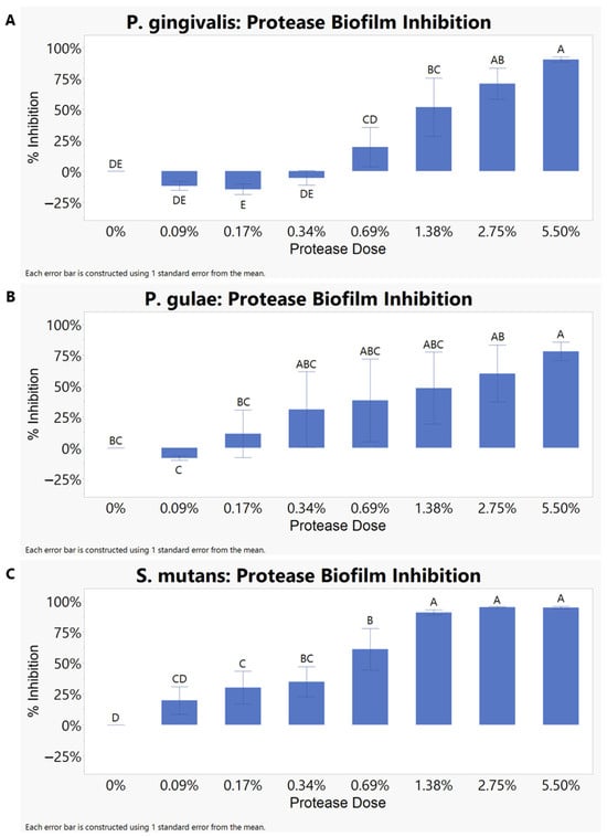
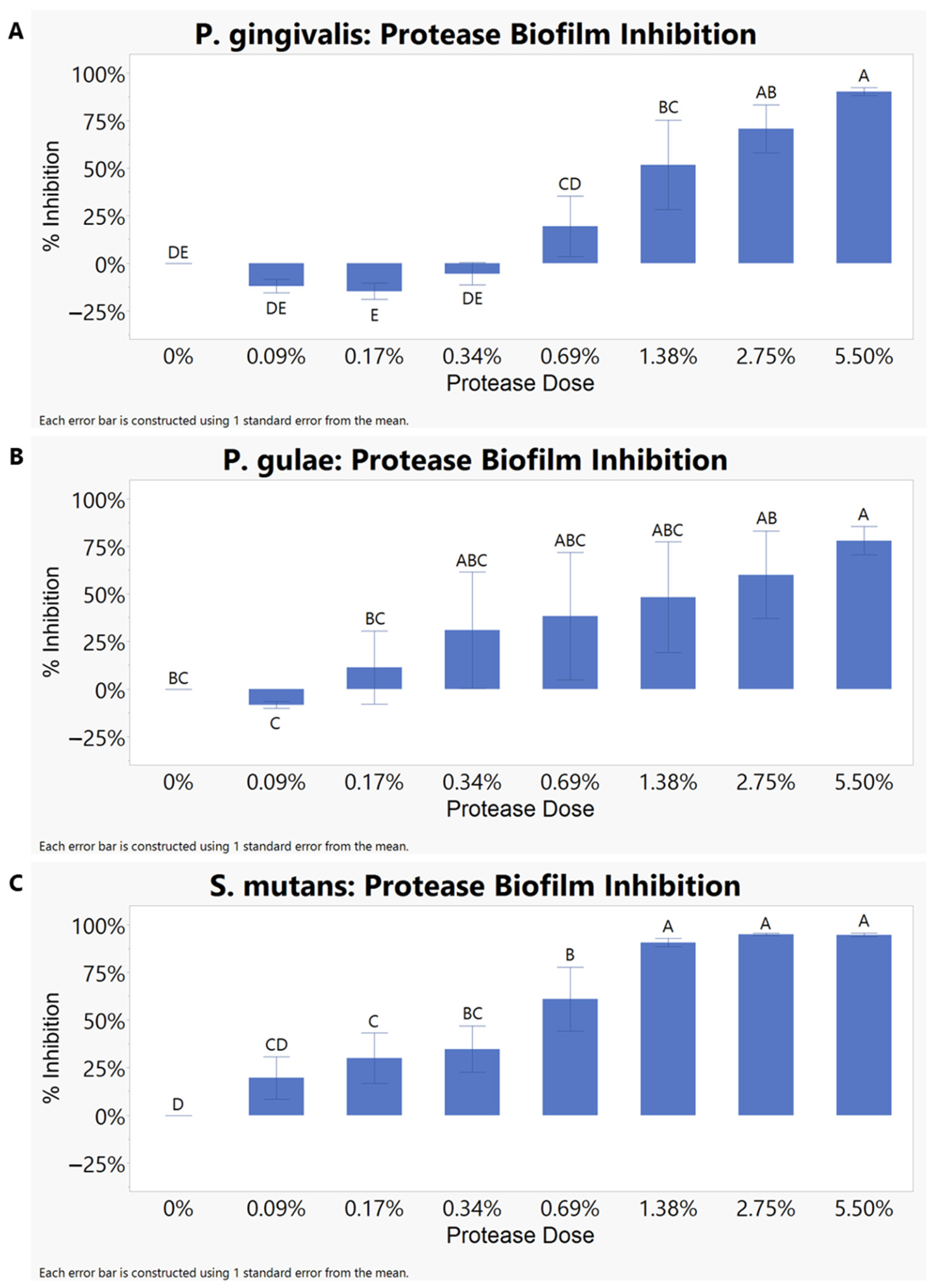
To evaluate the inhibition efficacy of protease, a dilution series from 5.50% to 0.09% of the protease in 1x PBS was added to the cultures prior to incubation. As the protease concentration increased, the biofilm biomass decreased (Figure 1). The overall least-squares statistical model indicates significance in P. gingivalis (p < 0.0001) and S. mutans (p < 0.0001). In P. gingivalis, biofilm least-squares means rose from 0.000 at the untreated control (0% protease) to 0.195 at 0.69% protease, 0.518 at 1.38% protease, 0.708 at 2.75% protease, and 0.903 at 5.50% protease, indicating a dose-dependent inhibitory response. Protease concentrations of 1.38%, 2.75%, and 5.50% were significantly different from the control. S. mutans displayed a similar pattern, with means increasing from 0.000 at the untreated control to 0.610 at 0.69% protease and reaching 0.907–0.950 at concentrations of 1.38–5.50%. This trend suggests that higher protease concentrations were associated with greater inhibition of S. mutans biofilm formation. In P. gulae, the overall model was nonsignificant (p = 0.1452) likely due to high variability. However, P. gulae also showed increasing biofilm inhibition with increasing protease concentrations, and the 5.50% protease concentration was significantly different from the untreated control.
Figure 1.
Estimated mean % inhibition ± SE of protease treatment on biofilm response relative to the untreated control in (A) Porphyromonas gingivalis ATCC 33277, (B) P. gulae ATCC 51700, and (C) Streptococcus mutans ATCC 25175. A positive least-squares mean correlates to the inhibition ability of protease, whereas a negative least-squares mean indicates increased biofilm formation of the target strain. Levels not connected by same letter are significantly different (p < 0.05).
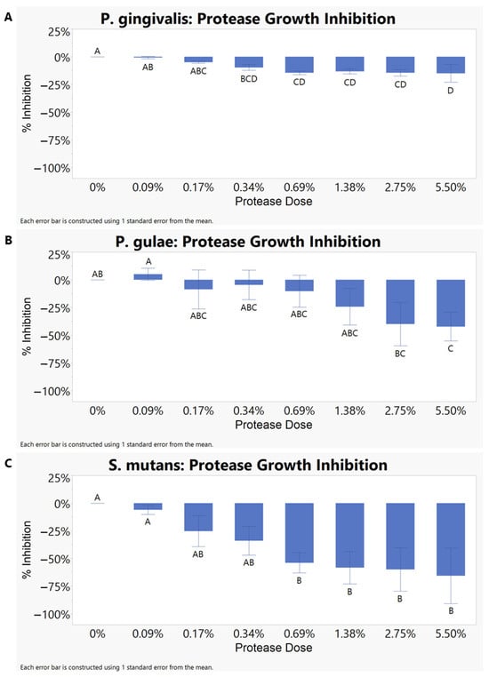
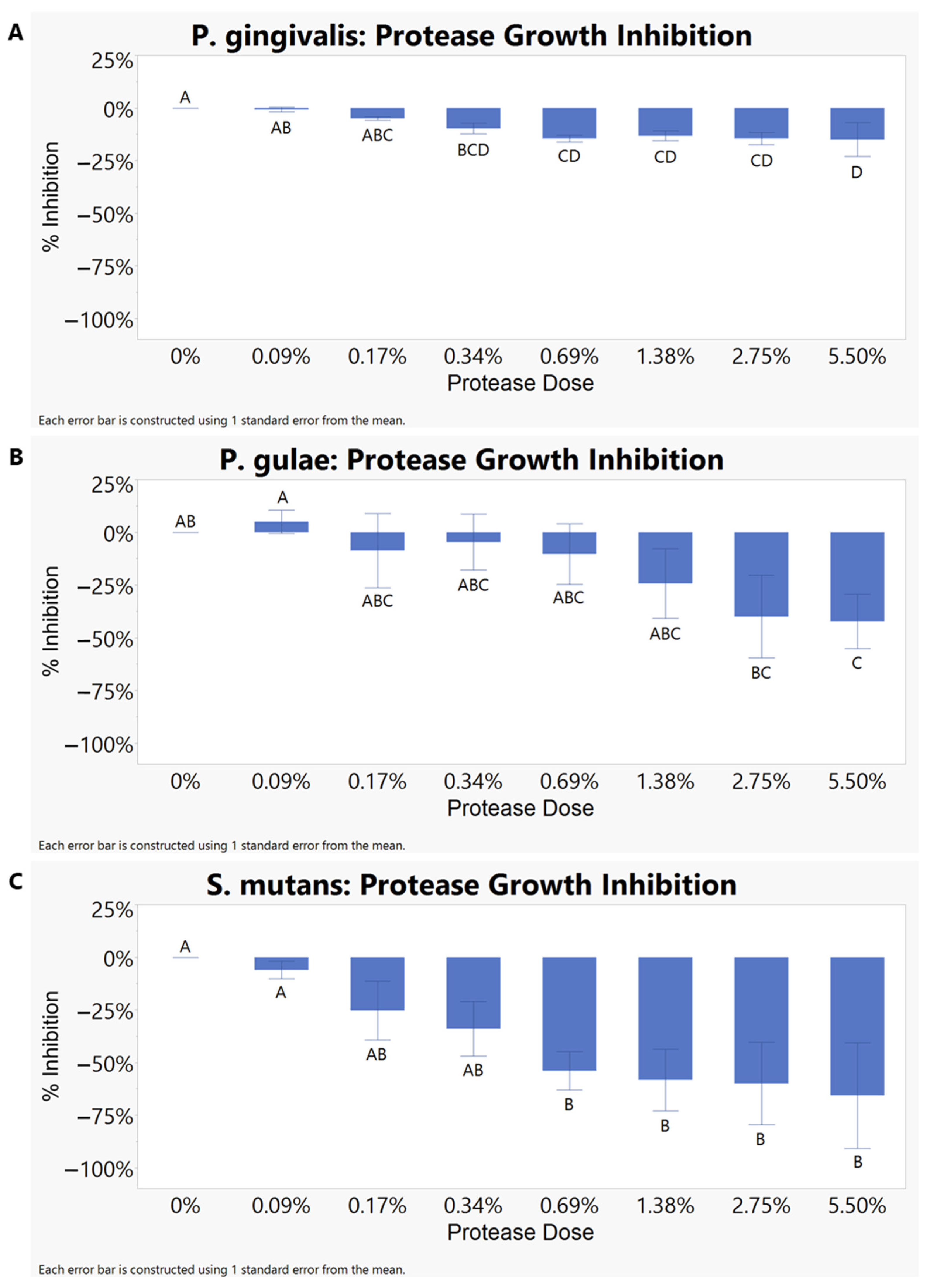
The planktonic growth was elevated compared to the untreated control, increasing growth proportionally as the protease concentration increased, for all three strains (Figure 2). The analysis of the data using the overall least-squares statistical model indicates significant change for P. gingivalis (p = 0.0088) and S. mutans (p = 0.0302) but again not for P. gulae (p = 0.1894). For P. gingivalis, the least-squares means declined from 0.000 in the untreated control (0% protease) to −0.145 and −0.150 at 0.69% and 5.50% protease, respectively. P. gulae showed a similar trend, increasing as protease concentration increased. S. mutans displayed a strong downward trend, with least-squares means decreasing from 0.000 at the untreated control to −0.540 to −0.657 across 0.69% to 5.50% protease, respectively. The progressively more negative least-squares means trend indicates increased growth at increasing protease concentrations.
Figure 2.
Estimated mean % inhibition ± SE of protease treatment on planktonic growth response relative to the untreated control (A) Porphyromonas gingivalis ATCC 33277, (B) P. gulae ATCC 51700, and (C) Streptococcus mutans ATCC 25175. A positive least-squares mean correlates to the inhibition ability of protease, whereas a negative least-squares mean indicates increased growth of the target strain. Levels not connected by same letter are significantly different (p < 0.05).
Inhibition Efficacy of Betaine
To determine if betaine had any impact on bacterial growth and biofilm formation, a dilution series from 0.13% to 5.0% of betaine in 1x PBS was added to the cultures prior to incubation. In the biofilm assays, the analysis of the data using the overall least-squares statistical model indicates significance for S. mutans (p = 0.0023) and P. gingivalis (p < 0.0001) but not for P. gulae (p = 0.5423). In P. gingivalis, a betaine concentration of 5% was the only positive least-squares mean at 0.370 and significantly different from the untreated control (0% betaine), indicating that the biofilm was not inhibited until 5% betaine was used. For S. mutans, betaine produced a progressively more negative least-squares means response with increasing dose, becoming significantly different from the untreated control with −0.138 at 2.5% betaine, −0.160 at 4% betaine, and −0.253 at 5% betaine, which indicates that increasing betaine doses increased biofilm production. In P. gulae, the least-squares means were numerically negative across the dose range (Supplemental Figure S1) but did not reach statistical significance.
In the planktonic growth assays, the analysis of the data using the overall least-squares statistical model indicates significance in P. gingivalis (p < 0.0001) and P. gulae (p = 0.0013) growth but not S. mutans (p = 0.3261). In P. gingivalis, the least-squares means increased from 0.000 at the untreated control (0% betaine) to 0.194 at 0.13% betaine, 0.286 at 4% betaine, and 0.446 at 5% betaine, indicating that betaine significantly decreased P. gingivalis growth at concentrations as low as 0.13%. The 5% betaine was the highest-response group, distinct from the lower doses and untreated conditions (Supplemental Figure S2). P. gulae also showed an inhibition response to betaine, with least-squares means increasing from 0.000 at the untreated control to 0.165 at 0.13% betaine, 0.173 at 4% betaine, and 0.400 at 5% betaine. As in P. gingivalis, the highest betaine dose (5%) formed the strongest-response group (Supplemental Figure S2), indicating a strong inhibitory effect at higher concentrations. Conversely, S. mutans’ growth was not impacted by betaine.
Checkerboard Assay
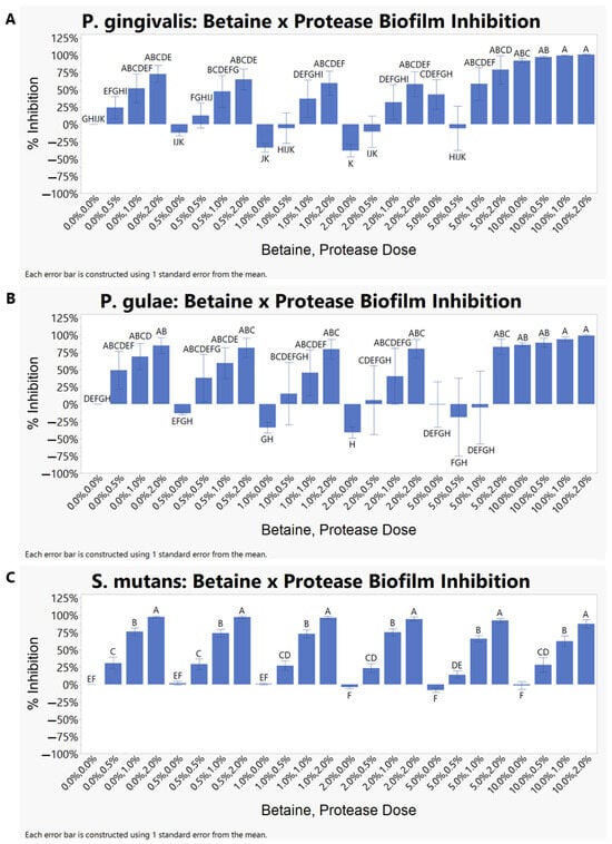
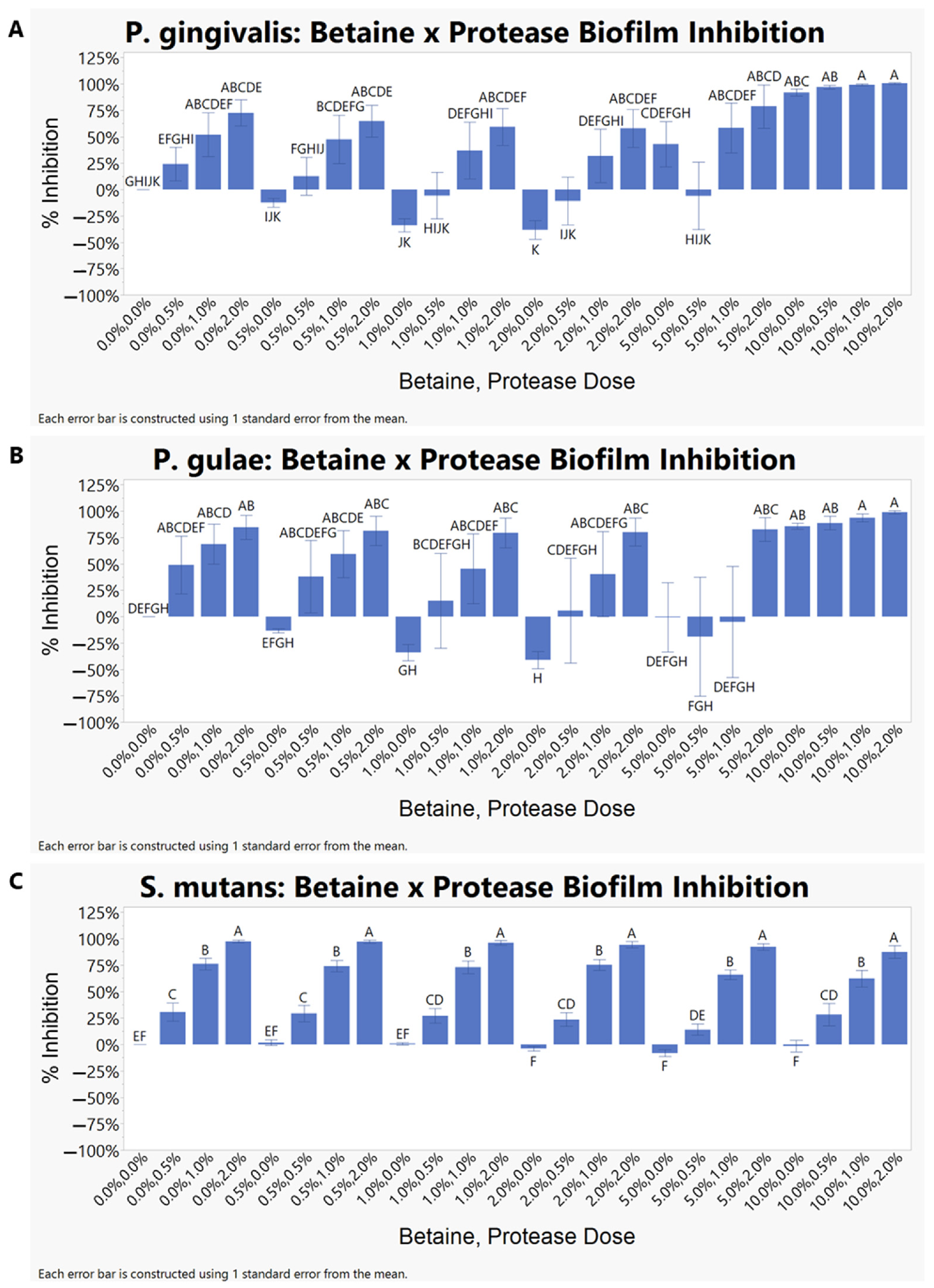
To assess the potential effects of combining betaine and protease, a checkerboard strategy was used. The analysis of the data using the overall least-squares statistical model indicates that all three combinations for biofilm inhibition were significant overall. In P. gingivalis, the biofilm model was highly significant (p < 0.0001), with significant main effects of betaine (p < 0.0001) and protease (p < 0.0001), while no significant interaction was observed (p = 0.3545). Similarly in P. gulae, the overall model was significant (p = 0.0020), with significant main effects of betaine (p = 0.0029) and protease (p < 0.0001), while no significant interaction was observed (p = 0.8198). Similarly in S. mutans, the overall model was significant (p < 0.0001), with significant main effects of betaine (p = 0.0450) and protease (p < 0.0001), and no interaction (p = 0.9850). Student’s t comparisons showed that treatment groups with the same protease concentration were generally grouped together regardless of betaine concentration (Figure 3).
Figure 3.
Estimated mean % inhibition ± SE of the combined effect of betaine and protease treatment on biofilm response relative to the untreated control in (A) Porphyromonas gingivalis ATCC 33277, (B) P. gulae ATCC 51700, and (C) Streptococcus mutans ATCC 25175. A positive least-squares mean correlates to the inhibition ability of the betaine and protease combination, whereas a negative least-squares mean indicates increased biofilm formation of the target strain. Levels not connected by same letter are significantly different (p < 0.05).
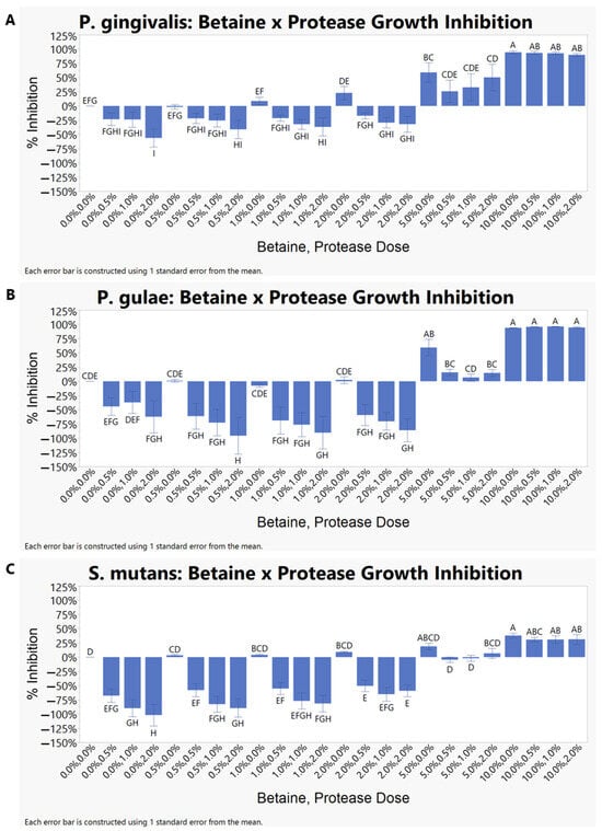
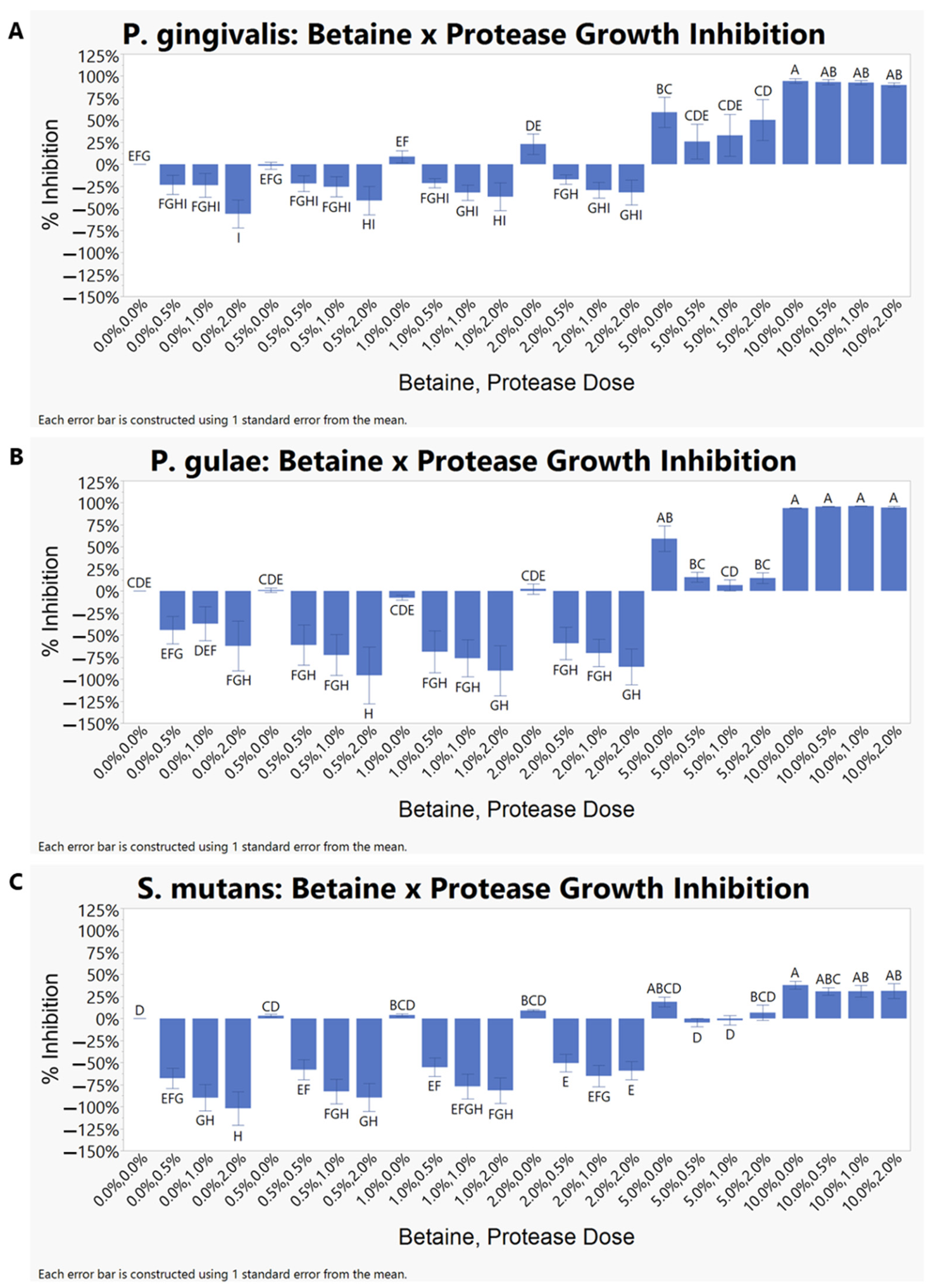
The planktonic growth measurements from the two-factor least-squares design were significant in all three strains (p < 0.0001). There was significant impact on the growth of P. gingivalis in the overall statistical model in a concentration-dependent manner (p < 0.0001). Betaine (p < 0.0001) and protease (p < 0.0001) main effects were significant but not their interaction (p = 0.5499). Similarly, in P. gulae, the overall model was significant (p < 0.0001), with main effects of betaine (p < 0.0001) and protease (p < 0.0001), but no interaction (p = 0.5151). P. gingivalis and P. gulae showed similar growth responses across the treatment matrix, but these varied across protease and betaine concentration (Figure 4). The strongest growth responses happened at 10% betaine across all protease doses (Figure 4). The S. mutans growth model differed in that all three terms were significant: the overall model (p < 0.0001), main effects of betaine (p < 0.0001) and protease (p < 0.0001), and their interaction (p = 0.0001). At 5% betaine, several treatment combinations did not differ from the untreated control, whereas responses remained positive at 10% betaine. That is, the 5% betaine combinations did not significantly differ from the untreated control, and all the 10% betaine combinations remained positive.
Figure 4.
Estimated mean % inhibition ± SE of combined betaine and protease treatment on planktonic growth response relative to the untreated control in (A) Porphyromonas gingivalis ATCC 33277, (B) P. gulae ATCC 51700, and (C) Streptococcus mutans ATCC 25175. A positive least-squares mean correlates to the inhibition ability of the betaine and protease combination, whereas a negative least-squares mean indicates increased growth of the target strain. Levels not connected by same letter are significantly different (p < 0.05).
3.1.2. Biofilm Removal
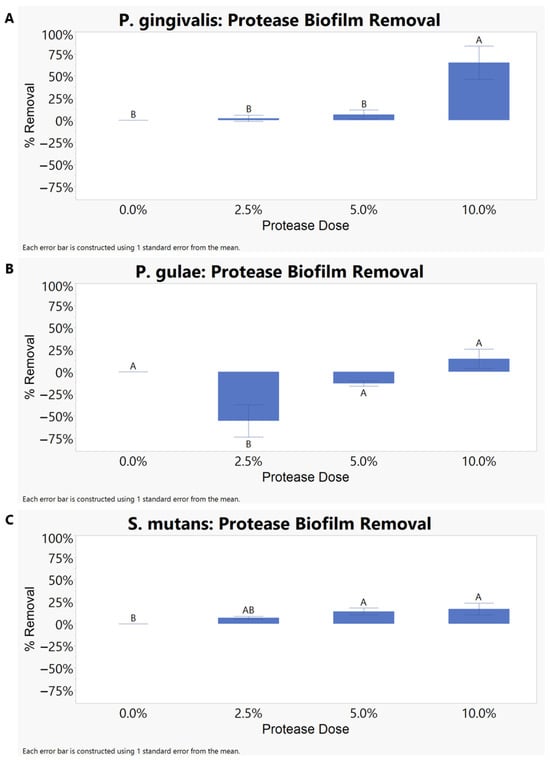
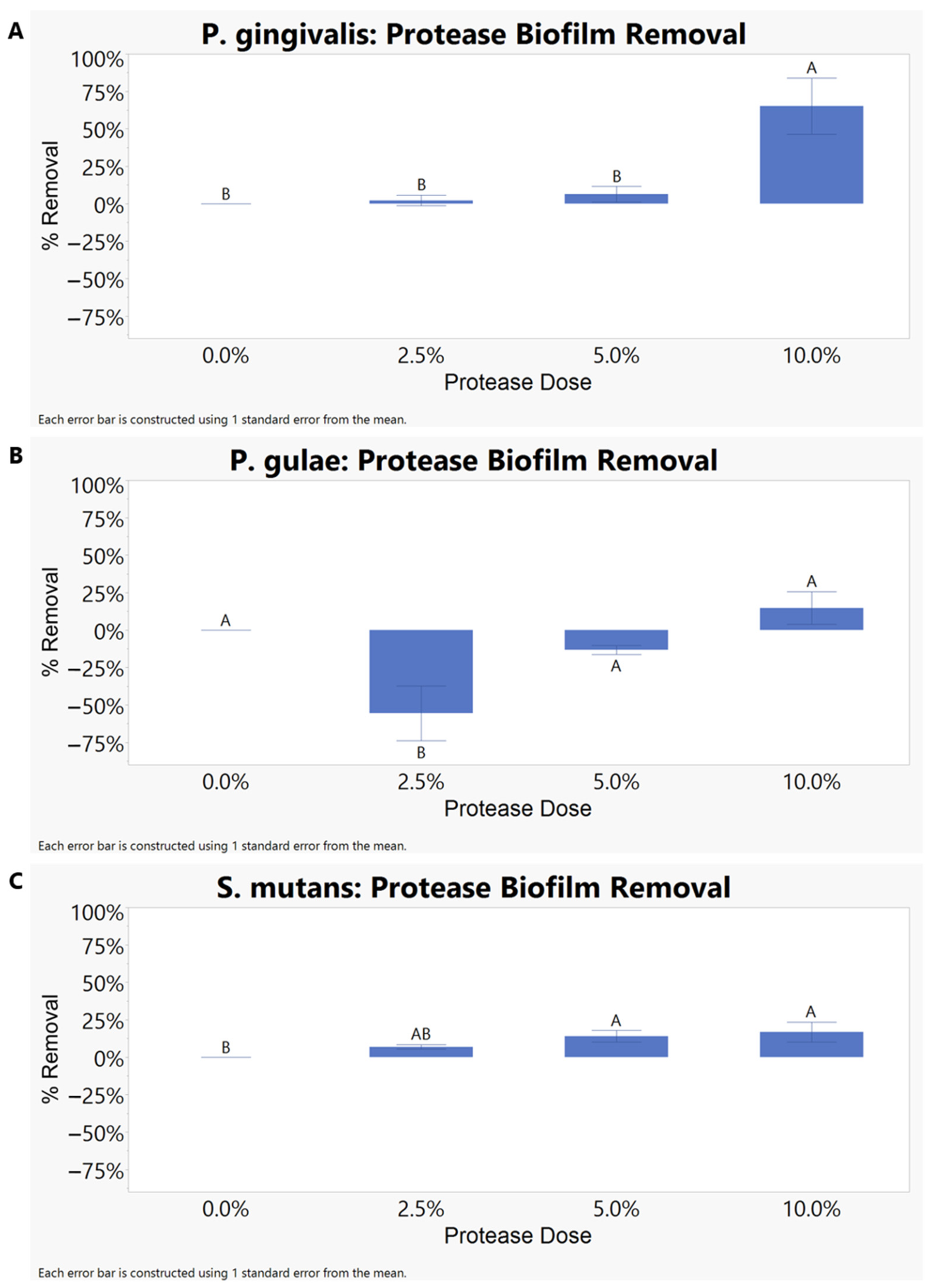
To evaluate the biofilm removal capabilities of protease, the protease was applied in a dilution series to the mature biofilm of P. gingivalis, P. gulae, and S. mutans. The results are displayed in Figure 5. The analysis of the data using the overall least-squares statistical model indicates that all three strains were significant. In P. gingivalis, treatment dose significantly influenced biofilm removal (p = 0.0002). Least-squares means were 0.000 in the untreated control (0%), 0.022 at 2.5% protease, 0.064 at 5.0% protease, and 0.653 at 10.0% protease, indicating an increase in removal with increasing dose and a notable increase at 10.0% protease. The 10.0% protease group differed significantly from the untreated control, 2.5% protease, and 5.0% protease groups, whereas the lower concentrations did not differ (p > 0.10) from one another. In P. gulae, the overall model was significant (p = 0.0036), although this significance was driven by a decrease in least-squares means from 0.000 in the untreated control to –0.555 at 2.5% protease. The 2.5% protease treatment grouped separately from the untreated control, 5.0% protease, and 10.0% protease groups, demonstrating increased biofilm production, whereas 5% and 10% protease were not significantly different from the untreated control. In S. mutans, treatment dose had a significant overall effect on biofilm removal (p = 0.0350). Least-squares means increased from 0.000 in the untreated control to 0.068, 0.140, and 0.168 at 2.5%, 5.0%, and 10.0% protease, respectively. The 5.0% and 10.0% protease groups were significantly different from the untreated control, which indicates a dose-dependent response for biofilm removal in S. mutans.
Figure 5.
Estimated mean % removal ± SE of protease treatment on biofilm removal relative to the untreated control in (A) Porphyromonas gingivalis ATCC 33277, (B) P. gulae ATCC 51700, and (C) Streptococcus mutans ATCC 25175. A positive least-squares mean correlates to the removal ability of protease, whereas a negative least-squares mean indicates increased biofilm formation of the target strain. Levels not connected by same letter are significantly different (p < 0.05).
3.2. In Vivo
3.2.1. Acceptability and Tolerability of Dental Chews
The average weight at the beginning of the experiment for C and T were both 9.2 kg and the average weights at the end of the experiment were 9 kg and 9.2 kg, respectively. No adverse events such as vomiting or soft stools were recorded during the study.
A total of 72 chews were administered to each group. The proportion of chews fully consumed by dogs in groups T and C was 79.2% (57/72) and 94.4% (68/72), respectively. Three dogs in the T group showed limited consumption of the assigned chews throughout the study. Two of these dogs consumed less than half of the assigned chews and were excluded from downstream microbiome analysis following Illumina sequencing. Physical examinations were performed to assess oral health, with particular attention to gingival inflammation, ulceration, and bleeding. All dogs received normal scores throughout the study period (D0 to D8), with no signs of oral abnormalities observed.
3.2.2. Microbiome Sequencing
We conducted microbiome analysis by sequencing the amplified 16S genes. The study generated a total of 162 samples across the three sampling timepoints (D0, D4, and D8) of both treatments (C and T) for each sample type (gum, teeth and feces). Of the three dogs that showed limited chew consumption, two consumed less than half of the assigned chews and were excluded prior to microbiome analysis. The final dataset contained 144 samples, including 48 gum, 48 teeth, and 48 fecal samples.
Illumina sequencing produced a total of 4,709,298 rRNA amplicon sequences, with an average of 29,433 sequences per sample after quality filtering. Analyses were conducted with all samples rarified to a level of 20,000 sequences per sample.
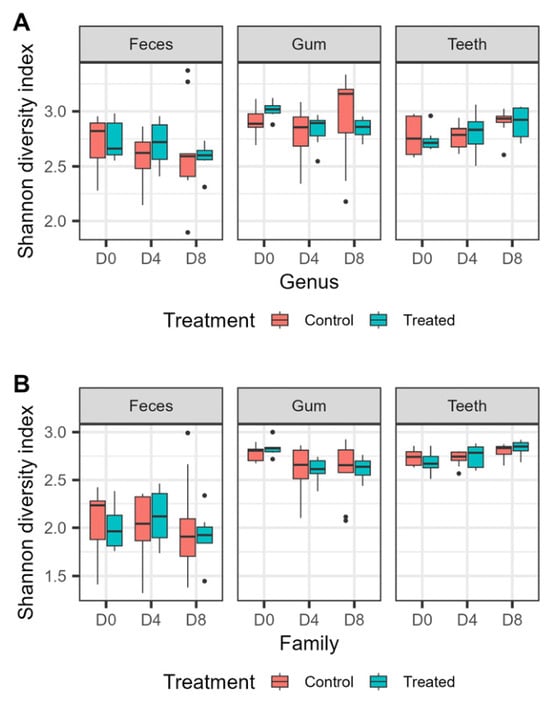
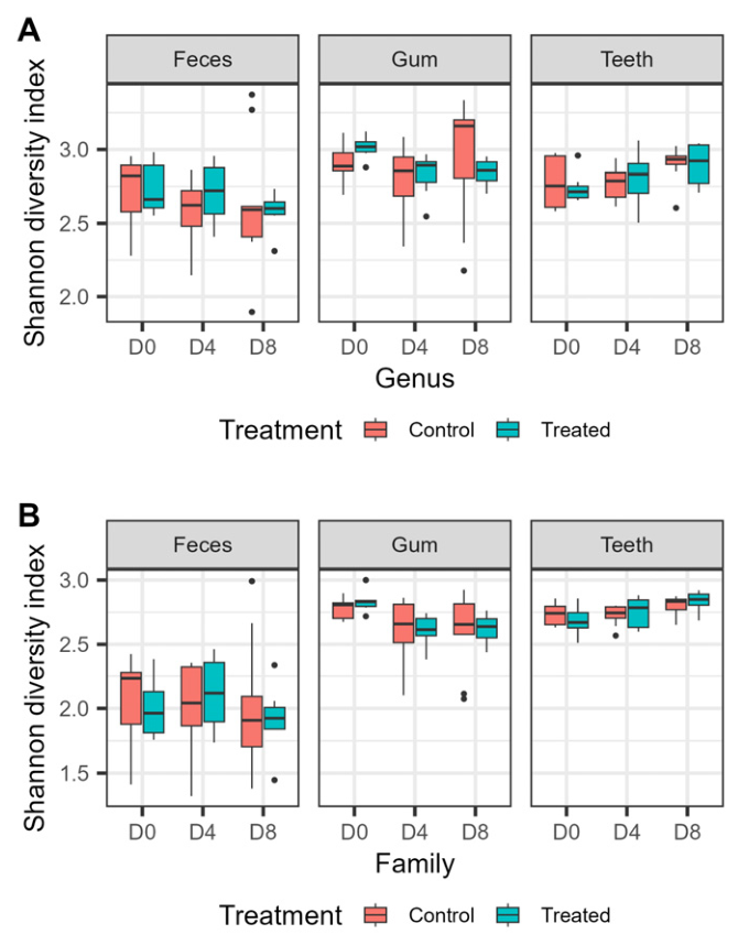
Analysis of alpha diversity revealed significant differences between sample types at both genus and family levels, with gum and teeth microbiota exhibiting higher Shannon diversity than fecal microbiota (Wilcoxon test, adjusted p < 0.05; Figure 6). No significant differences in genus-level alpha diversity were detected between treatment groups at any sampling timepoint. Significant differences at the family level were detected within gum samples between D0 and D4 and between D0 and D8 in the treatment group (Wilcoxon test, adjusted p < 0.05).
Figure 6.
Shannon diversity index of fecal, gum, and teeth microbiota at the (A) genus and (B) family levels for control and treated dogs at D0, D4, and D8.
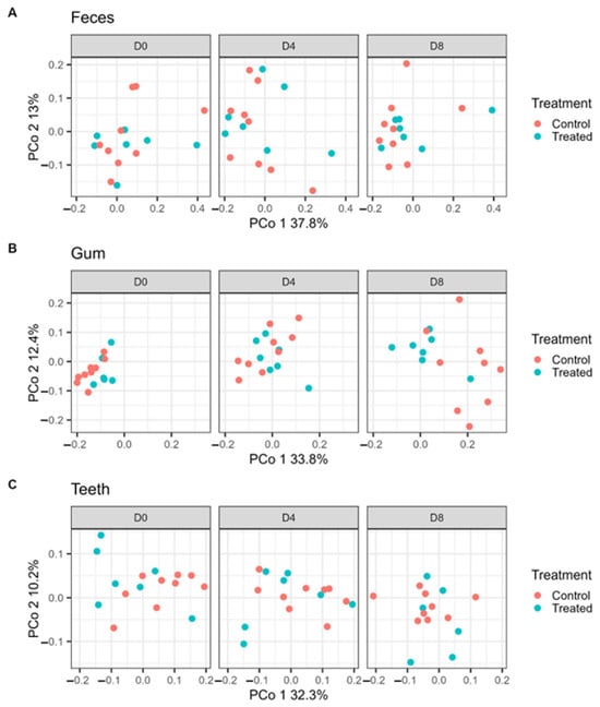
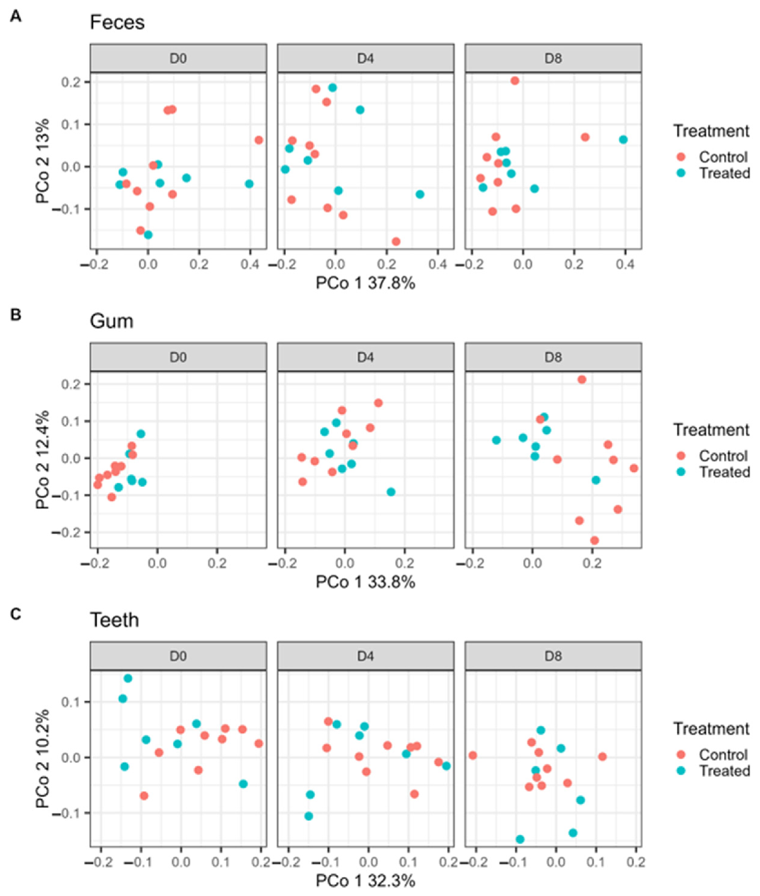
Significant differences in microbial community composition between treatment groups were detected in gum samples at D0 (adjusted p = 0.039) and D8 (adjusted p = 0.020) (Figure 7A). No significant differences were detected between treatment groups in gum samples at D4 (adjusted p = 0.260). No significant treatment-associated differences were detected in teeth or fecal samples (Figure 7B,C).
Figure 7.
Principal coordinates analysis (PCoA) based on Bray–Curtis dissimilarity of (A) feces, (B) gum, and (C) teeth microbiota at D0, D4 and D8 at genus. Colors indicate sample type and symbols indicate treatment group.
Within gum samples, differential abundance analysis detected multiple taxa that differed between D0 and D8 (adjusted p < 0.05; Figures S1 and S2).
4. Discussion
To our knowledge, limited information is available regarding the combined use of proteolytic enzymes and plant-derived betaine as functional ingredients targeted to canine oral health. While both compounds have been extensively studied in animal nutrition, their potential in targeting PD-associated bacteria and biofilm have received comparatively little attention. The present study investigated the complementary effects of these ingredients using both mechanistic in vitro assay and in vivo canine study. The results demonstrated distinct responses in planktonic growth and biofilm formation among PD-associated bacteria species, with protease mainly influencing the biofilm formation and removal, while betaine exhibited strain dependent effects on bacterial growth and biofilm responses. Together, these results support further investigation of protease and betaine as functional ingredients for canine oral health.
4.1. Protease—Biofilm Disrupting Ingredient
Biofilm is formed by the attachment of microorganisms to surfaces through surface proteins and has an extracellular matrix comprising excreted polysaccharides, lipids, proteins, and extracellular DNA and RNA [18,19,40]. Previous studies have found that proteases have reduced biofilm formation [41,42,43]. In the present study, protease reduced biofilm formation in P. gingivalis, P. gulae, and S. mutans as concentration increased. P. gingivalis and P. gulae were included because P. gulae is considered a major canine periodontal pathogen and shares virulence and immunological characteristics with P. gingivalis [16,44]. In addition, P. gingivalis has been detected within the canine oral microbiota [45,46]. S. mutans was included as a reference organism because of its well-characterized ability to form biofilms and its widespread use as a model for studying bacterial adhesion and plaque development [17,18,21]. Although S. mutans is not considered representative of the canine oral microbiota, its inclusion enabled evaluation of protease activity against a distinct biofilm-forming organism. The strongest biofilm inhibition responses were observed in P. gingivalis and S. mutans. In contrast, although biofilm formation in P. gulae decreased numerically with increasing protease concentration, the overall inhibition model was not significant. This suggests that the response to protease differed among the bacterial species evaluated, indicating variability in responses across the microorganisms tested.
Treatment with the protease displayed increased planktonic growth but reduced biofilm biomass in a dose-dependent manner. Proteases break down proteins, releasing essential amino acids and peptides into the environment, which then promote the growth of bacteria [47]. However, proteolytic enzymes can degrade extracellular bacterial proteins and biofilm attachment through targeting surface proteins, thus inhibiting early bacterial adhesion and interfering with plaque maturation without suppressing growth of the bacteria [20,22,23,24]. Importantly, since the protease does not inhibit the planktonic growth of these bacteria, the protease is not antimicrobial.
While inhibition of biofilm formation is necessary to prevent plaque buildup in the oral cavity, the ability to remove established biofilm would help support oral health more by clearing the established matrix structures to which pathogenic bacteria may attach to [48]. The protease was able to remove the biofilm formed by both P. gingivalis and S. mutans at 5% protease and 5% and 10% protease, respectively. Biofilm removal is a more effective mechanism in preventing disease because biofilm acts as a protective barrier for microorganisms, protecting them from pH changes, antimicrobials, host immune system, and shearing forces [49,50,51,52]. In removing the existing biofilm and preventing new biofilm attachment, there is greater ability to support oral health against the oral pathogens.
4.2. Understanding Betaine Response; Betaine as Supporting Tissue Ingredient
The in vitro portion examines the direct effect of applying betaine to the culture of oral pathogens P. gingivalis, P. gulae, and S. mutans. In this study, betaine decreased the planktonic growth of P. gingivalis and P. gulae with increasing concentrations while increasing their biofilm production. As betaine is an osmoprotectant, it promotes the survival of microorganisms under osmotic stress [53,54,55]. For example, in Kapfhammer et al. 2005 [56] the addition of betaine had been found to increase biofilm formation of Vibrio cholera. This beneficial effect, however, depends on taxa, as some studies have shown that while some bacterial groups may increase growth, others may decrease. Such was the case in Chen et al. [57] after supplementing the diet of specific pathogen-free mice with 0.8 g/kg betaine per day. While the populations of Bacteroidaceae, Bacteroides, Parabacteroides, and Prevotella were enriched, the populations of Coriobacteriaceae, Lachnospiraceae, Enterorhabdus, and Coriobacteriales were depleted. Similarly in Al-khalaifah et al. [58], after feeding laying hens a diet supplemented with 1.2 g/kg betaine for eight weeks, the abundance of Staphylococcus, Escherichia coli, Clostridium, and Pseudomonas significantly decreased when compared to the control group. However, there have not been studies found directly assessing the impact of betaine on the growth and biofilm formation of P. gingivalis, P. gulae, and S. mutans. As such, this study may be the first to analyze their interactions.
After assessing the impacts of betaine and the protease individually on the growth and biofilm formations of the oral pathogens, a combined assay was implemented to evaluate the interaction of the two components in one culture. For all three target strains, there was no interaction between the betaine and the protease. This was expected, as the purpose of betaine was to primarily support host health in terms of oral hydration and relieving gum irritation and inflammation [59,60,61], while the protease was expected to target oral pathogens.
4.3. In Vivo
Despite clear protease effects on biofilm formation and removal in vitro, the canine study showed only limited microbiome separation between treatment. Several study factors may have contributed, including the study length, incomplete chew consumption by a small number of dogs, and substantial baseline variation among individuals. Two of the nine dogs did not completely consume the chew during the study period. As no signs of oral irritation, gastrointestinal discomfort, or other adverse reactions were observed, the incomplete consumption was considered unlikely to be associated with product intolerance. Because the study dogs had no previous exposure to chew treats and the sample size was small, the reasons for incomplete consumption cannot be determined from the present study. Therefore, these findings should not be interpreted as evidence of either reduced acceptability or palatability. As a result, ingredient exposure was not uniform across all dogs in the treatment group. Two of these dogs consumed less than half of the assigned chews and were subsequently excluded from microbiome analyses. Similar challenges in translating microbiological responses into measurable oral health outcomes have been reported in canine dental chew studies, where treatment effects can be influenced by individual animal variation and pre-existing differences in oral microbial composition [14,15]. Previous studies evaluating dental chews in dogs have reported shifts in oral microbial communities together with improvements in oral health parameters such as plaque, calculus, gingivitis, and halitosis. Oba et al. [14,15] demonstrated reductions in several taxa associated with periodontal disease, including Porphyromonas and Treponema, following daily dental chew administration. More recently, studies characterizing the canine oral microbiome have further demonstrated that microbial composition differs across oral niches and disease states, supporting the use of microbiome profiling as a complementary tool for evaluating oral health interventions in dogs [14,15,62]. Furthermore, the experiment only lasted 8 days, which limited the opportunity and occurrence to sustain microbiome shifts. The use of a structurally identical chew may have also reduced the ability to detect ingredient specific effects because mechanical chewing alone has been associated with improvements in oral health parameters [9,11,13,14,15]. Together, these factors have likely reduced the ability to detect treatment-related differences drastically.
Although temporal changes were detected within gum microbial communities, these findings should be interpreted independently from the between-group comparisons. The control and treated groups differed in gum microbial composition at D0, indicating pre-existing baseline heterogeneity. Dogs were enrolled without professional dental cleaning prior to study initiation, allowing existing plaque and oral microbial communities to remain intact. While this non-invasive approach better reflects real-world conditions, it likely contributed to the baseline variability observed between animals. Consequently, the difference also detected at D8 cannot be attributed specifically to the functional ingredients without demonstrating that the D0-to-D8 change differed between groups. Within the control group, several genera previously associated with canine periodontal disease, including Porphyromonas, Treponema, Tannerella, Fretibacterium, and Filifactor, decreased between D0 and D8 (Figure S1). These findings are directionally consistent with previous reports showing that dental chews can positively influence the canine oral microbiota [14,15]. However, because similar changes were observed in dogs receiving the control chew, the results suggest that chew-associated mechanical effects and natural temporal variation likely contributed substantially to the observed microbial shifts.
Importantly, the in vivo part was designed primarily to assess the tolerability and microbiome responses rather than clinical PD outcomes. Measurements of gingival inflammation, index bleeding, wound healing, inflammatory biomarkers, and cytokines responses were not assessed. As a result, the proposed tissue supporting role of betaine could not be adequately evaluated in the scope of this study and does not rule out potential benefits on host tissues. This is relevant because betaine has been reported to function primarily as an osmoprotectant, helping maintain cellular hydration and cell stability during physiological stress [26,27,59,61]. In addition, betaine has shown to modulate inflammatory pathways, including reduction in IL-1β production, a cytokine strongly associated with PD progression [30,31]. Therefore, the contribution of betaine may be more accurately related to supporting the oral tissue environment rather than directly altering microbial composition. This is consistent with previous human studies showing betaine reduced sodium lauryl sulfate induced irritation of the oral mucosa and improved subjective symptoms of dry mouth, suggesting that its primary benefits may occurs at the tissue level and may not necessarily be reflected by measurable shift in microbial community composition [29,63]. Future studies incorporating clinical PD assessment, inflammatory endpoints, and longer experimental periods are needed to determine whether these host associated benefits translate into measurable improvement in canine oral health.
In summary, this study supports a two-level interpretation of the protease–betaine dental chew concept. Under controlled in vitro conditions, the ingredients showed distinct and biologically interpretable effects on growth and biofilm-associated responses of periodontal disease–associated bacteria. In dogs, the 8-day pilot study demonstrated acceptable short-term tolerability but only limited microbiome separation between treatments, likely reflecting the short study duration, baseline heterogeneity, and the confounding contribution of chew mechanics. These findings support further evaluation in longer-duration studies with designs better suited to resolving ingredient-specific oral health outcomes.
5. Conclusions
Protease reduced biofilm formation and disrupted established biofilms in vitro with the strongest and most consistent responses observed in P. gingivalis and S. mutans. Betaine had variable effects on growth and biofilm responses depending on the strain and did not affect protease activity when tested in combination. In the 8-day canine pilot study, the protease and betaine containing dental chew appeared physiologically well tolerated, with no adverse clinical observations reported. Although incomplete consumption was observed in a subset of dogs, the reasons for this response could not be determined from the present study because the dogs had limited prior exposure to chew treats and the number of affected animals was small. Therefore, additional studies specifically designed to evaluate voluntary acceptance and palatability are warranted. The chew was associated with only limited microbiome changes. As clinical periodontal outcomes, inflammatory biomarkers, and direct evidence of betaine-mediated tissue protection were not evaluated, the mechanisms underlying potential oral health benefits remain to be established. Longer-term studies including clinical oral health assessments are warranted to further evaluate the effects of protease-containing oral care products.
Supplementary Materials
The following supporting information can be downloaded at: https://www.mdpi.com/article/10.3390/pets3040049/s1. Figure S1: Betaine treatment effects on biofilm response; Figure S2: Betaine treatment effects on planktonic growth; Table S1: Differentially abundant genera in control gum samples between D0 and D8; Table S2: Differentially abundant genera in treated gum samples between D0 and D8.
Author Contributions
Conceptualization, I.C.A. and R.Y.; methodology, I.C.A., R.Y., M.M.Y. and Q.W.; investigation, M.M.Y. and Q.W.; formal analysis, M.M.Y. and Q.W.; writing—original draft preparation, S.Y.L. and M.M.Y.; writing—review and editing, S.Y.L., M.M.Y., Q.W., R.Y. and I.C.A.; project administration, S.Y.L. and I.C.A.; submission and revision management, S.Y.L. All authors have read and agreed to the published version of the manuscript.
Funding
This research received no external funding.
Institutional Review Board Statement
The animal study protocol was approved by the Institutional Animal Care and Use Committee of the contract research organization, High Quality Research, Fort Collins, CO, USA under protocol number 210.001 (approval date: 18 November 2024), and was conducted in accordance with the Welfare Act Regulations, United States Department of Agriculture, 2019.
Informed Consent Statement
Not applicable.
Data Availability Statement
The datasets generated and/or analyzed during the current study are available from the corresponding author upon reasonable request.
Acknowledgments
During the preparation of this manuscript, the authors used Microsoft Copilot for language editing and writing assistance. The authors reviewed and edited the output and take full responsibility for the content of this publication.
Conflicts of Interest
All authors are currently employees of IFF, the manufacturer of the product evaluated in this study. Shu Ying Lim was an intern at IFF and affiliated with Wageningen University & Research during this work and subsequently became an employee of IFF. IFF was involved in the study design, data collection, data analysis, interpretation of the results, preparation of the manuscript, and the decision to publish the results. The authors had full access to all the data in this study and take complete responsibility for the integrity of the data and the accuracy of the data analysis. The funder (IFF) imposed no restrictions on the authors’ ability to independently analyze, interpret, or publish findings that might be unfavorable to the product, and the authors retained the final authority to decide on publication.
Abbreviations
The following abbreviations are used in this manuscript:
| PD | Periodontal Disease |
| AVMA | American Veterinary Medical Association |
| IACUC | Institutional Animal Care and Use Committee |
| EPS | Extracellular Polymeric Substance(s) |
| PBS | Phosphate-Buffered Saline |
| ATCC | American Type Culture Collection |
| BHIS | Brain Heart Infusion supplemented with hemin, menadione and yeast extract |
| BHIS-S | BHIS supplemented with 1% sucrose |
| YCFAC | Yeast Casitone Fatty Acids broth with Carbohydrates |
| BW | Body Weight |
| PCoA | Principal Coordinates Analysis |
| PERMANOVA | Permutational Multivariate Analysis of Variance |
| L2FC | Log2 Fold Change |
| DESeq2 | Differential Expression Sequencing 2 (software package) |
| DADA2 | Divisive Amplicon Denoising Algorithm 2 (software name) |
| RDP | Ribosomal Database Project |
| SE | Standard Error |
References
- Niemiec, B.A. Periodontal Disease. Top. Companion Anim. Med. 2008, 23, 72–80. [Google Scholar] [CrossRef] [Scilit] [PubMed]
- Riggio, M.P.; Lennon, A.; Taylor, D.J.; Bennett, D. Molecular Identification of Bacteria Associated with Canine Periodontal Disease. Vet. Microbiol. 2011, 150, 394–400. [Google Scholar] [CrossRef] [Scilit] [PubMed]
- Wallis, C.; Holcombe, L.J. A Review of the Frequency and Impact of Periodontal Disease in Dogs. J. Small Anim. Pract. 2020, 61, 529–540. [Google Scholar] [CrossRef] [Scilit] [PubMed]
- Yasuda, J.; Yasuda, H.; Nomura, R.; Matayoshi, S.; Inaba, H.; Gongora, E.; Iwashita, N.; Shirahata, S.; Kaji, N.; Akitomo, T.; et al. Investigation of Periodontal Disease Development and Porphyromonas gulae FimA Genotype Distribution in Small Dogs. Sci. Rep. 2024, 14, 5360. [Google Scholar] [CrossRef] [Scilit] [PubMed]
- Albuquerque, C.; Morinha, F.; Requicha, J.; Martins, T.; Dias, I.; Guedes-Pinto, H.; Bastos, E.; Viegas, C. Canine Periodontitis: The Dog as an Important Model for Periodontal Studies. Vet. J. 2012, 191, 299–305. [Google Scholar] [CrossRef] [Scilit] [PubMed]
- Stella, J.L.; Bauer, A.E.; Croney, C.C. A Cross-Sectional Study to Estimate Prevalence of Periodontal Disease in a Population of Dogs (Canis familiaris) in Commercial Breeding Facilities in Indiana and Illinois. PLoS ONE 2018, 13, e0191395. [Google Scholar] [CrossRef] [Scilit] [PubMed]
- Cunha, E.; Tavares, L.; Oliveira, M. Revisiting Periodontal Disease in Dogs: How to Manage This New Old Problem? Antibiotics 2022, 11, 1729. [Google Scholar] [CrossRef] [Scilit] [PubMed]
- Enlund, K.B.; Brunius, C.; Hanson, J.; Hagman, R.; Höglund, O.V.; Gustås, P.; Pettersson, A. Dental Home Care in Dogs-A Questionnaire Study among Swedish Dog Owners, Veterinarians and Veterinary Nurses. BMC Vet. Res. 2020, 16, 90. [Google Scholar] [CrossRef] [Scilit] [PubMed]
- Rawlings, J.M.; Gorrel, C.; Markwell, P.J. Effect on Canine Oral Health of Adding Chlorhexidine to a Dental Hygiene Chew. J. Vet. Dent. 1998, 15, 129–134. [Google Scholar] [CrossRef] [Scilit] [PubMed]
- Carroll, M.Q.; Oba, P.M.; Sieja, K.M.; Alexander, C.; Lye, L.; De Godoy, M.R.C.; He, F.; Somrak, A.J.; Keating, S.C.J.; Sage, A.M.; et al. Effects of Novel Dental Chews on Oral Health Outcomes and Halitosis in Adult Dogs. J. Anim. Sci. 2020, 98, skaa274. [Google Scholar] [CrossRef] [Scilit] [PubMed]
- Gawor, J.; Jodkowska, K.; Klim, E.; Jank, M.; Nicolas, C.S. Comparison of a Vegetable-Based Dental Chew to 2 Other Chews for Oral Health Prevention. J. Vet. Dent. 2021, 38, 131–138. [Google Scholar] [CrossRef] [Scilit] [PubMed]
- Sieja, K.M.; Oba, P.M.; Schauwecker, A.; Hristova, T.; Keating, S.; Somrak, A.; Swanson, K.S. 109 Effects of a Novel Dental Chew on the Oral Health Outcomes and Malodor of Adult Dogs. J. Anim. Sci. 2022, 100, 48–49. [Google Scholar] [CrossRef] [Scilit]
- Brown, W.Y.; McGenity, P. Effective Periodontal Disease Control Using Dental Hygiene Chews. J. Vet. Dent. 2005, 22, 16–19. [Google Scholar] [CrossRef] [Scilit] [PubMed]
- Oba, P.M.; Carroll, M.Q.; Alexander, C.; Somrak, A.J.; Keating, S.C.J.; Sage, A.M.; Swanson, K.S. Dental Chews Positively Shift the Oral Microbiota of Adult Dogs. J. Anim. Sci. 2021, 99, skab100. [Google Scholar] [CrossRef] [Scilit] [PubMed]
- Oba, P.M.; Sieja, K.M.; Schauwecker, A.; Somrak, A.J.; Hristova, T.S.; Keating, S.C.J.; Swanson, K.S. Effects of a Novel Dental Chew on Oral Health Outcomes, Halitosis, and Microbiota of Adult Dogs. J. Anim. Sci. 2024, 102, skae071. [Google Scholar] [CrossRef] [Scilit] [PubMed]
- Lenzo, J.C.; O’Brien-Simpson, N.M.; Orth, R.K.; Mitchell, H.L.; Dashper, S.G.; Reynolds, E.C. Porphyromonas gulae Has Virulence and Immunological Characteristics Similar to Those of the Human Periodontal Pathogen Porphyromonas gingivalis. Infect. Immun. 2016, 84, 2575–2585. [Google Scholar] [CrossRef] [Scilit] [PubMed]
- Lemos, J.A.; Palmer, S.R.; Zeng, L.; Wen, Z.T.; Kajfasz, J.K.; Freires, I.A.; Abranches, J.; Brady, L.J. The Biology of Streptococcus mutans. Microbiol. Spectr. 2019, 7, 7.1.03. [Google Scholar] [CrossRef] [Scilit] [PubMed]
- Matsumoto-Nakano, M. Role of Streptococcus Mutans Surface Proteins for Biofilm Formation. Jpn. Dent. Sci. Rev. 2018, 54, 22–29. [Google Scholar] [CrossRef] [Scilit] [PubMed]
- Joo, H.-S.; Otto, M. Molecular Basis of In Vivo Biofilm Formation by Bacterial Pathogens. Chem. Biol. 2012, 19, 1503–1513. [Google Scholar] [CrossRef] [Scilit] [PubMed]
- Hahn Berg, I.C.; Kalfas, S.; Malmsten, M.; Arnebrant, T. Proteolytic Degradation of Oral Biofilms in vitro and in vivo: Potential of Proteases Originating from Euphausia superba for Plaque Control. Eur. J. Oral Sci. 2008, 109, 316–324. [Google Scholar] [CrossRef] [Scilit] [PubMed]
- Ahn, S.-J.; Wen, Z.T.; Burne, R.A. Effects of Oxygen on Virulence Traits of Streptococcus mutans. J. Bacteriol. 2007, 189, 8519–8527. [Google Scholar] [CrossRef] [Scilit] [PubMed]
- Leroy, C.; Delbarre, C.; Ghillebaert, F.; Compere, C.; Combes, D. Influence of Subtilisin on the Adhesion of a Marine Bacterium Which Produces Mainly Proteins as Extracellular Polymers. J. Appl. Microbiol. 2008, 105, 791–799. [Google Scholar] [CrossRef] [Scilit] [PubMed]
- Liu, J.; Madec, J.-Y.; Bousquet-Mélou, A.; Haenni, M.; Ferran, A.A. Destruction of Staphylococcus Aureus Biofilms by Combining an Antibiotic with Subtilisin A or Calcium Gluconate. Sci. Rep. 2021, 11, 6225. [Google Scholar] [CrossRef] [Scilit] [PubMed]
- Pleszczyńska, M.; Wiater, A.; Bachanek, T.; Szczodrak, J. Enzymes in Therapy of Biofilm-related Oral Diseases. Biotechnol. Appl. Biochem. 2017, 64, 337–346. [Google Scholar] [CrossRef] [Scilit] [PubMed]
- Ruiz-Sorribas, A.; Poilvache, H.; Kamarudin, N.H.N.; Braem, A.; Van Bambeke, F. Hydrolytic Enzymes as Potentiators of Antimicrobials against an Inter-Kingdom Biofilm Model. Microbiol. Spectr. 2022, 10, e02589-21, Correction in Microbiol. Spectr. 2022, 11, e04671-22. [Google Scholar] [CrossRef] [Scilit] [PubMed]
- Craig, S.A. Betaine in Human Nutrition. Am. J. Clin. Nutr. 2004, 80, 539–549. [Google Scholar] [CrossRef] [Scilit] [PubMed]
- Eklund, M.; Bauer, E.; Wamatu, J.; Mosenthin, R. Potential Nutritional and Physiological Functions of Betaine in Livestock. Nutr. Res. Rev. 2005, 18, 31–48. [Google Scholar] [CrossRef] [Scilit] [PubMed]
- Takis, P.G.; Papavasileiou, K.D.; Troganis, A.N.; Melissas, V.S. CHAPTER 4. The Chemistry of Betaine. In Food and Nutritional Components in Focus; Preedy, V.R., Ed.; Royal Society of Chemistry: Cambridge, UK, 2015; pp. 47–61. [Google Scholar]
- Söderling, E.; Le Bell, A.; Kirstilä, V.; Tenovuo, J. Betaine-Containing Toothpaste Relieves Subjective Symptoms of Dry Mouth. Acta Odontol. Scand. 1998, 56, 65–69. [Google Scholar] [CrossRef] [Scilit] [PubMed]
- Xia, Y.; Chen, S.; Zhu, G.; Huang, R.; Yin, Y.; Ren, W. Betaine Inhibits Interleukin-1β Production and Release: Potential Mechanisms. Front. Immunol. 2018, 9, 2670. [Google Scholar] [CrossRef] [Scilit] [PubMed]
- Cheng, R.; Wu, Z.; Li, M.; Shao, M.; Hu, T. Interleukin-1β Is a Potential Therapeutic Target for Periodontitis: A Narrative Review. Int. J. Oral Sci. 2020, 12, 2. [Google Scholar] [CrossRef] [Scilit] [PubMed]
- Floyd, M.R. The Modified Triadan System: Nomenclature for Veterinary Dentistry. J. Vet. Dent. 1991, 8, 18–19. [Google Scholar] [CrossRef] [Scilit]
- van der Klein, S.A.S.; Bernardeau, M.; Wang, Q.; Gibbs, K. A Cross-Study Analysis of the Effect of a Dual-Strain Probiotic Applied via the Waterline on the Growth Performance and Gut Health of Broilers under a Mild Necrotic Enteritis Challenge. Poult. Sci. 2025, 104, 104550. [Google Scholar] [CrossRef] [Scilit] [PubMed]
- Martin, M. Cutadapt Removes Adapter Sequences from High-Throughput Sequencing Reads. EMBnet.journal 2011, 17, 10. [Google Scholar] [CrossRef] [Scilit]
- Callahan, B.J.; McMurdie, P.J.; Rosen, M.J.; Han, A.W.; Johnson, A.J.A.; Holmes, S.P. DADA2: High-Resolution Sample Inference from Illumina Amplicon Data. Nat. Methods 2016, 13, 581–583. [Google Scholar] [CrossRef] [Scilit] [PubMed]
- Wang, Q.; Garrity, G.M.; Tiedje, J.M.; Cole, J.R. Naive Bayesian Classifier for Rapid Assignment of rRNA Sequences into the New Bacterial Taxonomy. Appl. Environ. Microbiol. 2007, 73, 5261–5267. [Google Scholar] [CrossRef] [Scilit] [PubMed]
- Wang, Q.; Cole, J.R. Updated RDP Taxonomy and RDP Classifier for More Accurate Taxonomic Classification. Microbiol. Resour. Announc. 2024, 13, e01063-23. [Google Scholar] [CrossRef] [Scilit] [PubMed]
- Oksanen, J.; Simpson, G.L.; Blanchet, F.G.; Kindt, R. Vegan: Community Ecology Package; R Package 2.6-4; The Comprehensive R Archive Network: Vienna, Austria, 2022. [Google Scholar]
- Love, M.I.; Huber, W.; Anders, S. Moderated Estimation of Fold Change and Dispersion for RNA-Seq Data with DESeq2. Genome Biol. 2014, 15, 550. [Google Scholar]
- Schilcher, K.; Horswill, A.R. Staphylococcal Biofilm Development: Structure, Regulation, and Treatment Strategies. Microbiol. Mol. Biol. Rev. 2020, 84, e00026-19. [Google Scholar] [CrossRef] [Scilit] [PubMed]
- Liu, Q.; Wang, X.; Qin, J.; Cheng, S.; Yeo, W.-S.; He, L.; Ma, X.; Liu, X.; Li, M.; Bae, T. The ATP-Dependent Protease ClpP Inhibits Biofilm Formation by Regulating Agr and Cell Wall Hydrolase Sle1 in Staphylococcus aureus. Front. Cell. Infect. Microbiol. 2017, 7, 181. [Google Scholar] [CrossRef] [Scilit] [PubMed]
- Mugita, N.; Nambu, T.; Takahashi, K.; Wang, P.-L.; Komasa, Y. Proteases, Actinidin, Papain and Trypsin Reduce Oral Biofilm on the Tongue in Elderly Subjects and In Vitro. Arch. Oral Biol. 2017, 82, 233–240. [Google Scholar] [CrossRef] [Scilit] [PubMed]
- Arenas, J.; Szabo, Z.; Van Der Wal, J.; Maas, C.; Riaz, T.; Tønjum, T.; Tommassen, J. Serum Proteases Prevent Bacterial Biofilm Formation: Role of Kallikrein and Plasmin. Virulence 2021, 12, 2902–2917. [Google Scholar] [CrossRef] [Scilit] [PubMed]
- Fournier, D.; Mouton, C.; Lapierre, P.; Kato, T.; Okuda, K.; Ménard, C. Porphyromonas gulae Sp. Nov., an Anaerobic, Gram-Negative Coccobacillus from the Gingival Sulcus of Various Animal Hosts. Int. J. Syst. Evol. Microbiol. 2001, 51, 1179–1189. [Google Scholar] [CrossRef] [Scilit] [PubMed]
- Di Bello, A.; Buonavoglia, A.; Franchini, D.; Valastro, C.; Ventrella, G.; Greco, M.F.; Corrente, M. Periodontal Disease Associated with Red Complex Bacteria in Dogs. J. Small Anim. Pract. 2014, 55, 160–163. [Google Scholar] [CrossRef] [Scilit] [PubMed]
- Özavci, V.; Erbas, G.; Parin, U.; Yüksel, H.T.; Kirkan, Ş. Molecular Detection of Feline and Canine Periodontal Pathogens. Vet. Anim. Sci. 2019, 8, 100069. [Google Scholar] [CrossRef] [Scilit] [PubMed]
- Lehman, M.K.; Nuxoll, A.S.; Yamada, K.J.; Kielian, T.; Carson, S.D.; Fey, P.D. Protease-Mediated Growth of Staphylococcus Aureus on Host Proteins Is Opp3 Dependent. mBio 2019, 10, e02553-18. [Google Scholar] [CrossRef] [Scilit] [PubMed]
- Mohanty, R.; Asopa, S.J.; Joseph, M.D.; Singh, B.; Rajguru, J.P.; Saidath, K.; Sharma, U. Red Complex: Polymicrobial Conglomerate in Oral Flora: A Review. J. Fam. Med. Prim. Care 2019, 8, 3480–3486. [Google Scholar] [CrossRef] [Scilit] [PubMed]
- Tsagkari, E.; Connelly, S.; Liu, Z.; McBride, A.; Sloan, W.T. The Role of Shear Dynamics in Biofilm Formation. npj Biofilms Microbiomes 2022, 8, 33. [Google Scholar] [CrossRef] [Scilit] [PubMed]
- Rupp, C.J.; Fux, C.A.; Stoodley, P. Viscoelasticity of Staphylococcus aureus Biofilms in Response to Fluid Shear Allows Resistance to Detachment and Facilitates Rolling Migration. Appl. Environ. Microbiol. 2005, 71, 2175–2178. [Google Scholar] [CrossRef] [Scilit] [PubMed]
- Hoštacká, A.; Čižnár, I.; Štefkovičová, M. Temperature and pH Affect the Production of Bacterial Biofilm. Folia Microbiol. 2010, 55, 75–78. [Google Scholar] [CrossRef] [Scilit] [PubMed]
- Müsken, M.; Pawar, V.; Schwebs, T.; Bähre, H.; Felgner, S.; Weiss, S.; Häussler, S. Breaking the Vicious Cycle of Antibiotic Killing and Regrowth of Biofilm-Residing Pseudomonas aeruginosa. Antimicrob. Agents Chemother. 2018, 62, e01635-18. [Google Scholar] [CrossRef] [Scilit] [PubMed]
- Liu, Y.; Li, Q.; Liu, R.; Wang, Z.; Zhao, S. Exploring the Multifunctional Roles of Betaine: Traditional Applications, Emerging Technologies, and Green Chemistry Innovations. Foods 2026, 15, 737. [Google Scholar] [CrossRef] [Scilit] [PubMed]
- Capp, M.W.; Pegram, L.M.; Saecker, R.M.; Kratz, M.; Riccardi, D.; Wendorff, T.; Cannon, J.G.; Record, M.T. Interactions of the Osmolyte Glycine Betaine with Molecular Surfaces in Water: Thermodynamics, Structural Interpretation, and Prediction of m-Values. Biochemistry 2009, 48, 10372–10379. [Google Scholar] [CrossRef] [Scilit] [PubMed]
- Xia, Y.; Jiang, X.; Guo, S.; Wang, Y.; Mu, Y.; Shen, J. Glycine Betaine Modulates Extracellular Polymeric Substances to Enhance Microbial Salinity Tolerance. Environ. Sci. Ecotechnol. 2024, 20, 100406. [Google Scholar] [CrossRef] [Scilit] [PubMed]
- Kapfhammer, D.; Karatan, E.; Pflughoeft, K.J.; Watnick, P.I. Role for Glycine Betaine Transport in Vibrio cholerae Osmoadaptation and Biofilm Formation within Microbial Communities. Appl. Environ. Microbiol. 2005, 71, 3840–3847. [Google Scholar] [CrossRef] [Scilit] [PubMed]
- Chen, Q.; Wang, Y.; Jiao, F.; Shi, C.; Pei, M.; Wang, L.; Gong, Z. Betaine Inhibits Toll-like Receptor 4 Responses and Restores Intestinal Microbiota in Acute Liver Failure Mice. Sci. Rep. 2020, 10, 21850. [Google Scholar] [CrossRef] [Scilit] [PubMed]
- Al-khalaifah, H.S.; Al-Ardhi, S.A.; Nafea, H.H.; Al-Nasser, A.; Khalil, H.A.; Hassan, M.I.S.; Ahmed, H.A. Impact of Betaine on Laying Hens’ Productive and Physiological Reactions at the Late Stage of Production. Front. Anim. Sci. 2025, 6, 1708362. [Google Scholar] [CrossRef] [Scilit]
- Zhao, G.; He, F.; Wu, C.; Li, P.; Li, N.; Deng, J.; Zhu, G.; Ren, W.; Peng, Y. Betaine in Inflammation: Mechanistic Aspects and Applications. Front. Immunol. 2018, 9, 1070. [Google Scholar] [CrossRef] [Scilit] [PubMed]
- Arumugam, M.K.; Paal, M.C.; Terrence, M.D.; Ganesan, M.; Osna, N.A.; Kharbanda, K.K. Beneficial Effects of Betaine: A Comprehensive Review. Biology 2021, 10, 456. [Google Scholar] [CrossRef] [Scilit] [PubMed]
- Rekima, A.; Yde Bak, S.; Christensen, N.; Shen, C. Plant-Derived Betaine Enhances Barrier Integrity and Immune Responses in Canine Intestinal Epithelial Cells and Macrophages under Endotoxin Challenge. J. Anim. Sci. 2026, 104, skag031. [Google Scholar] [CrossRef] [Scilit] [PubMed]
- Alessandri, G.; Fontana, F.; Mancabelli, L.; Tarracchini, C.; Lugli, G.A.; Argentini, C.; Longhi, G.; Rizzo, S.M.; Vergna, L.M.; Anzalone, R.; et al. Species-Level Characterization of Saliva and Dental Plaque Microbiota Reveals Putative Bacterial and Functional Biomarkers of Periodontal Diseases in Dogs. FEMS Microbiol. Ecol. 2024, 100, fiae082. [Google Scholar] [CrossRef] [Scilit] [PubMed]
- Rantanen, I.; Nicander, I.; Jutila, K.; Ollmar, S.; Tenovuo, J.; Söderling, E. Betaine Reduces the Irritating Effect of Sodium Lauryl Sulfate on Human Oral Mucosa In Vivo. Acta Odontol. Scand. 2002, 60, 306–310. [Google Scholar] [CrossRef] [Scilit] [PubMed]
Disclaimer/Publisher’s Note: The statements, opinions and data contained in all publications are solely those of the individual author(s) and contributor(s) and not of MDPI and/or the editor(s). MDPI and/or the editor(s) disclaim responsibility for any injury to people or property resulting from any ideas, methods, instructions or products referred to in the content. |
© 2026 by the authors. Licensee MDPI, Basel, Switzerland. This article is an open access article distributed under the terms and conditions of the Creative Commons Attribution (CC BY) license.