


Abstract
Tooth structure anomalies, though not frequently described, can be observed in the external appearance not only of humans but also of dogs. The emergence of an oversized set of teeth is not solely an esthetic and health problem, but often also leads to behavioral issues. In the commonly available medical literature, there are many cases describing the occurrence and treatment of double teeth, both in the incisors and in the cheek teeth. In this article, the authors describe a clinical case of a one-year-old female Golden Retriever dog diagnosed with a double tooth in her permanent dentition. The complex morphology of the affected tooth posed a considerable diagnostic and therapeutic challenge. Surgical separation of the tooth was performed, requiring an individualized treatment approach and the adaptation of protocols derived primarily from human dentistry. This case highlights the diagnostic and clinical decision-making difficulties associated with such anomalies. It also emphasizes the importance of tailored therapeutic strategies for managing rare dental developmental abnormalities in dogs. The teeth were successfully surgically separated, and thus the present report contributes to the limited body of literature on double teeth in veterinary patients and provides practical insight into their management. This description of the first case of this type in veterinary dentistry aims to encourage readers to explore the topic of double teeth and demonstrates a non-standard way of thinking and dealing with such a tooth. The presented case is unique from both a diagnostic and therapeutic perspective. Previous publications have focused on these abnormalities as separate entities, failing to consider their simultaneous occurrence within a single tooth. The therapeutic approach employed also deserves particular attention. The most commonly recommended approach involves extraction of the affected teeth; however, in this case, an attempt was made to separate and preserve them, representing a novel clinical approach.
1. Case Presentation
The patient, a one-year-old Golden Retriever female dog, was referred for a dental consultation due to behavioral therapy having limited effects. The owners were worried as the dog, despite the completed tooth exchange, was constantly biting surrounding objects. The dog was a sports dog; however, when playing with a ball or a rope toy, she let it go with clear signs of discomfort.
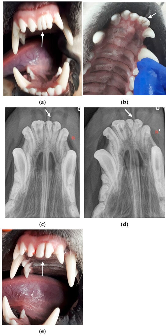
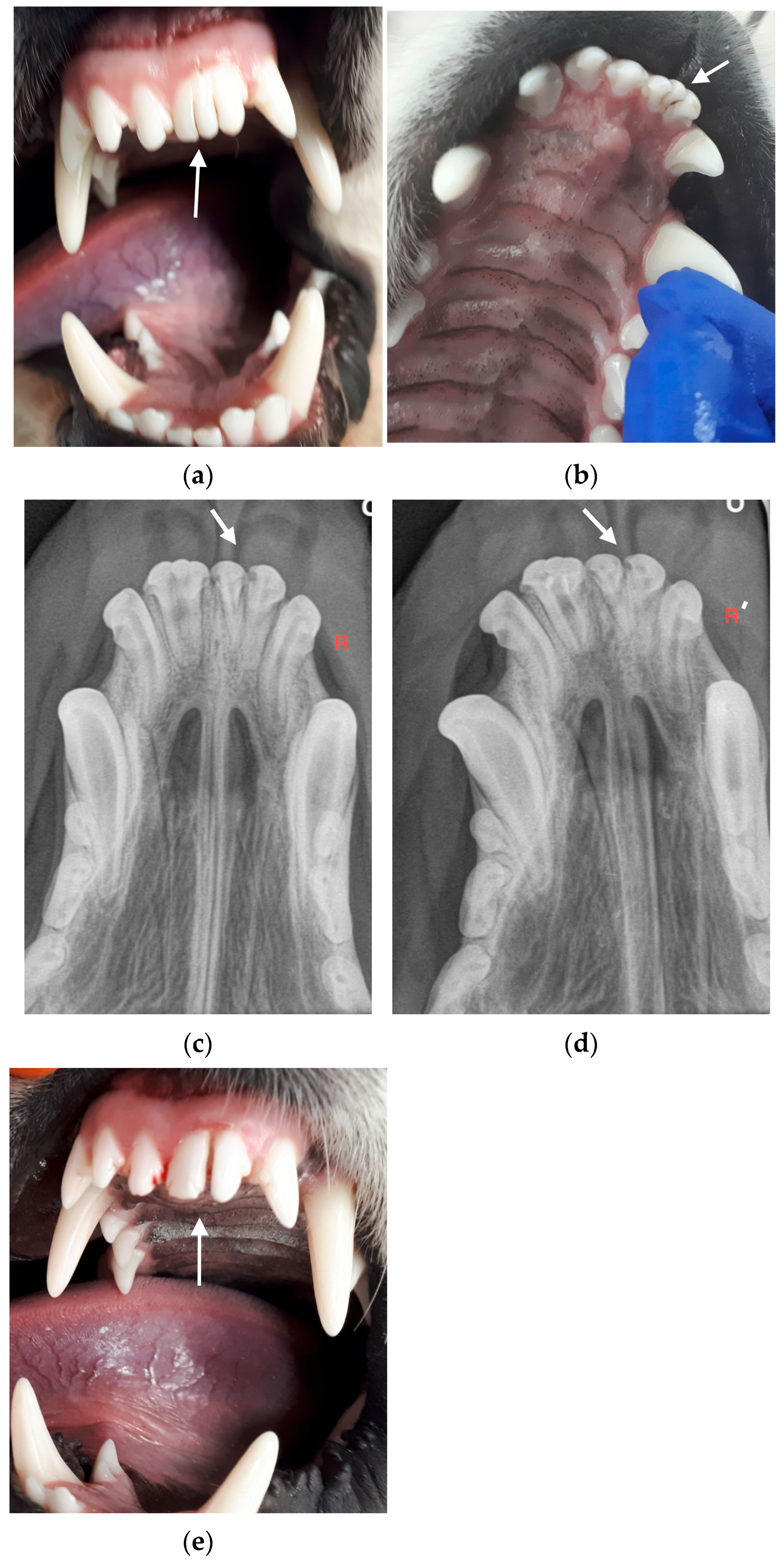
Clinical examination of the oral cavity revealed the connection of the crowns of teeth 201 and 202 into one large crown separated by three furrows reaching half the length of the tooth crown (Figure 1a). In the palatal examination, two furrows were found: one 1 mm deep furrow, located at about one-third of the length of the tooth, reaching the border of the palate’s mucous membrane, and a second furrow reaching about half the length of the tooth crown (Figure 1b). Radiological examination showed the presence of two tooth pulp canals, separated by dentine, leading into two tooth chambers (Figure 1c). After analyzing the clinical and radiological picture, the following diagnosis was made: fusion of gemination tooth 201 with tooth 202. After consultation with the owner, the decision was made to separate these teeth.
Figure 1.
(a) Tooth before treatment. Three connected tooth crowns can be observed; (b) Palatal view, clearly showing the difference between the two connected tooth units, including one with a split crown; (c) Tooth in an X-ray image –two separate canals can be observed, indicating the connection of two tooth units, which led to the decision to separate the teeth (R is the right side marker on the x-ray image); (d) Radiological control after root canal treatment; (e) Tooth after separation and composite protection during a control visit after 6 months (arrow).
The separation was planned in two stages. The first stage involved tooth pulp removal and root canal treatment. Lateral condensation of gutta-percha was performed and AH Plus Jet (Dentsply) was used to seal the canal (Figure 1d). The canal opening was marked with a pink Gradia flow composite (GC). After division, the tooth crowns were properly finished in terms of esthetics and function. Gradia Direct light-curing composite material in ODA1 and BW (GC) colors was used to rebuild the crowns. At each stage, the dog was premedicated with dexmedetomidine at a dose of 125 μg/m2, i.m. Propofol at a dose of 4 mg/kg was used to induce anesthesia, which was maintained with Isoflurane at a concentration adjusted to the patient’s condition. Analgesia was achieved using Mepivacaine infiltration at a dose of 2 mg/kg. In the second stage, the mucoperiosteal flap was cut and dissected from the labial side. Using a milling cutter, a piece of bone tissue was removed, revealing the border between the roots. The connected tooth roots were then separated using a diamond drill and the wound was stitched. Amoxicillin with clavulanic acid was administered as protection at a dose of 15 mg/kg BW 2× a day for seven consecutive days. Meloxicam at a dose of 0.1 mg/kg BW was used for postoperative analgesia for five consecutive days. The healing process progressed without complications and after seven days, the seams were removed.
After six months, a follow-up examination was performed (Figure 1e). The check-up without anesthesia revealed no dental pain and in the examination carried out under anesthesia, there were no signs of periodontitis. The alveolar bone was normal and there were no pathological pockets at the tooth separation site. Behavioral disorders in the dog had subsided.
2. Discussion
In veterinary dentistry, dental irregularities and anomalies in primary dentition account for only a small percentage of the described dental problems, which is why the available veterinary literature on recognizing and differentiating these issues is quite limited. Odontogenesis of dogs and people is based on similar mechanisms; therefore, answers to questions concerning these procedures and their possible consequences can be found in the literature on human dentistry.
The term “double teeth” refers to a type of structural anomaly in which the tooth is abnormally large [1]. Clinicians who encounter this type of abnormality are trying to create the perfect classification that would allow for the best procedure to be selected to deal with such teeth. The terms used for more accurate classification are shown in Table 1 [2,3]. The first type of double teeth is Gemination (dentes geminati), also known as schizodontism. Such teeth arise as a result of the splitting of one tooth bud during crown formation. In clinical examination, one tooth with one root and a wide crown separated by a furrow on the labial surface is observed [4]. Gemination most often occurs in Boxer breeds and occurs within the incisors [5,6]. To date, only three cases of bilateral gemination have been described, in Border Collie and Irish Setter dogs [7].
Table 1.
Classification of tooth developmental disorders.
The second type of double teeth is fusion (dentes confusi), where two separate developing tooth buds unite in the initial period of development, before calcification of tooth crowns. This can affect both normal teeth and supernumerary teeth [5,6]. The tooth crown is wide and there is vertical furrow on the vestibular surface of the tooth. Radiological examination shows one wide chamber with dentine protrusion into the lumen of the chamber and one wide root canal [8].
The third type is concrescence (dentes concreti), which occurs in the final stage of tooth development as a result of fusion within the root cementum. The teeth can be united on the whole surface of the root or only on part of it. X-ray pictures show two separate pulp chambers and separate root canals. Concrescence can occur between two normal, adjacent teeth or between normal and supernumerary teeth [9].
The exact etiology and pathogenesis of these anomalies are unknown—they can occur due to many genetic and environmental factors. Causal classifications are not easy to determine, especially for the clinicians to whom patients with abnormally formed/fused teeth or post-odontogenesis patients are most often referred. For this reason, all tooth unions are described as “double teeth” [10].
Double teeth mainly concern the joining of one of the central incisors with another central incisor or a lateral incisor. In veterinary dentistry, there is only residual data on the incidence of double teeth in individual sexes, species, and races. In humans, this problem largely affects males (77%) [11]. The presence of double teeth in primary dentition indicates a high probability of this anomaly appearing in permanent dentition [12].
The diagnosis of double teeth should be based on a radiological examination. However, due to anatomical conditions, overlapping anatomical structures may make it difficult to make a final diagnosis. In order to avoid complications, it is additionally advisable to extend the radiological diagnostics to Cone-Beam Computed Tomography (CBCT) [13].
Planning treatment for double teeth is challenge and involves fields of dentistry such as conservative dentistry, endodontics, dental surgery, orthodontics, and cosmetic dentistry.
Procedures can be divided into four categories: conservative treatment, hemisection, extraction, and leaving the tooth untreated. The former involves grinding the tooth in such a way as to obtain a tooth structure that is as close to normal as possible. Appropriate composite or prosthetic restoration can additionally improve esthetics [14,15]. Hemisection consists of separating two fused teeth. It can only be performed if there are two separate crowns and roots [16]. The extraction of a double tooth most often occurs in cases where conservative treatment does not have satisfactory esthetic results, and functional effects and prosthetic or implantological treatment are planned in place of extraction. In patients for whom esthetics are not important, double teeth are left untreated if they perform their function properly [17]. Although esthetics in the anterior segment are currently at the bottom of the list of indications for surgery in veterinary dentistry, every passing year, clients’ requirements are increasingly related not only to functionality but also to esthetics.
The consequences of leaving a double tooth without treatment include the crowding and displacement of adjacent teeth, irregularities in the replacement of milk teeth, caries, dental pulp necrosis, the formation of pathological cysts, the formation of an oro-nasal fistula, discomfort during food intake and pain [18].
When the furrow between the two crowns extends subgingivally, growing bacterial plaque can cause chronic gingivitis or periodontal disease [19].
3. Conclusions
For working dogs, tooth function, not esthetics, is paramount. Their bite plays a crucial role in their performance—from grasping and holding to precisely manipulating objects and reacting in work situations. Therefore, therapeutic approaches should focus on preserving the tooth function as much as possible, even if more complex and expensive treatment is required. From a medical perspective, the fight for each tooth is justified, as it directly impacts the dog’s ability to work.
It is not possible to determine one single appropriate way to deal with this rare anomaly. Success or failure depends on many factors, so each case should be considered individually—especially if the dental patient at the veterinarian clinic is a dog.
Author Contributions
Conceptualization A.M.-K.; methodology A.M.-K.; resources A.M.-K.; data curation S.D.; writing—original draft preparation A.M.-K.; writing—review and editing S.D.; visualization A.M.-K.; supervision S.D. All authors have read and agreed to the published version of the manuscript.
Funding
This research received no external funding.
Institutional Review Board Statement
This research does not require the consent of bioethics committee, number 15/2023.
Informed Consent Statement
Not applicable.
Data Availability Statement
The original contributions presented in this study are included in the article. Further inquiries can be directed to the corresponding authors.
Conflicts of Interest
The authors confirm that there are no commercial or financial relationships that could be construed as a potential conflict of interest. One of the authors is affiliated with a private veterinary clinic; however, this affiliation had no influence on the study design, data collection, analysis, interpretation, or the decision to publish the results.
References
- Schuurs, A.H.B.; Leveren, C.V. Double teeth: Review of the literature. ASDC J. Dent. Child. 2000, 67, 313–325. [Google Scholar] [PubMed]
- Pindborg, J.J. Pathology of the Dental Hard Tissues, 4th ed.; WB Saunders Co.: Philadelphia, PA, USA, 1970; pp. 30–64. [Google Scholar]
- Levitas, T.C. Gemination, fusion, twinning and concrescence. ASDC J. Dent. Child. 1965, 32, 93–100. [Google Scholar]
- Nandini, D.B.; Deepak, B.S.; Selvamani, M.; Puneeth, H.K. Diagnostic Dilemma of a Double tooth: A Rare Case Report and Review. J. Clin. Diagn. Res. 2014, 8, 271–272. [Google Scholar] [CrossRef] [Scilit] [PubMed]
- Pavlica, Z.; Erjavec, V.; Petelin, M. Teeth abnormalities in the dog. Acta Vet. Brno 2001, 70, 65–72. [Google Scholar] [CrossRef] [Scilit]
- Wiggs, R.B.; Lobprise, H.B. Clinical oral pathology. In Veterinary Dentistry Principles and Practice; Lippincott-Raven: Philadelphia, PA, USA, 1997; pp. 104–139. [Google Scholar]
- Rao, P.; Veena, K.; Chatra, L.; Shenai, P. Twin tooth on either side: A case report of bilateral gemination. An. Med. Health Sci. Res. 2013, 3, 271–273. [Google Scholar] [CrossRef] [Scilit] [PubMed]
- Ratajek-Gruda, M.; Grzesiak-Janas, G.; Białkowska-Głowacka, J. Zęby zrośnięte w bocznym odcinku szczęki [Double teeth in the lateral maxillary section]. Mag. Stomatol. 2001, 2, 2–43. [Google Scholar]
- Badełek-Mirek, B. Możliwości postępowania w przypadku zębów zrośniętych [Options for dealing with double teeth]. Mag. Stomatol. 1998, 5, 39–42. [Google Scholar]
- Wu, C.W.; Lin, Y.T. Double Primary teeth children under 17 years old and their correlation with permanent successors. Chang Gung Med. J. 2010, 33, 188–193. [Google Scholar] [PubMed]
- Hattab, F.N. Double talon cusps on supernumerary tooth Fused to maxillary central incisor: Review of literature and report case. J. Clin. Exp. Dent. 2014, 6, 400–407. [Google Scholar] [CrossRef] [Scilit] [PubMed]
- Mazumdar, P.; Das, K.; Rahaman, M. Endodontic Management of Geminated tooth: A case report. Int. J. Sci. Res. Publ. 2013, 3, 1–4. [Google Scholar]
- Castro, I.O.; Estrela, C.; Souza, V.R.; Lopes, L.G.; de Souza, J.B. Unilateral fusion of maxillary Lateran incisor: Diagnosis Rusing cone beam computer tomography. Case Rep. Dent. 2014, 2014, 934218. [Google Scholar] [PubMed]
- Turkaslan, S.; Gokce, H.S.; Dalkiz, M. Esthetic rehabilitation of bilateral geminated teeth: A case report. Eur. J. Dent. 2007, 1, 188–191. [Google Scholar] [CrossRef] [Scilit] [PubMed]
- Mader, C.L. Concrescence of teeth: A potential treatment hazard. Gen. Dent. 1984, 32, 52–55. [Google Scholar] [PubMed]
- Shapira, Y. Fused supernumerary central incisor. J. Dent. 1973, 2, 63–64. [Google Scholar] [CrossRef] [Scilit] [PubMed]
- Ramamurthy, S.; Satish, R.; Priya, K. Surgical and orthodontic management of Fusedmaxillary central and lateral incisors in Elary Mied dentition stage. Case Rep. Dent. 2014, 2014, 109301. [Google Scholar] [PubMed]
- Anthonappa, R.P.; Omer, R.S.; King, N.M. Characteristics of 283 supernumerary teeth in southern Chinese children. Oral Surg. Oral Med. Oral Pathol. Oral Radiol. Endod. 2008, 105, 48–54. [Google Scholar] [CrossRef] [Scilit] [PubMed]
- Lochib, S.; Indushekar, K.R.; Saraf, B.G.; Sheoran, N.; Sardana, D. Occlusal characteristics and prevalence of associated dental anomalies in the primary dentition. J. Epidemiol. Glob. Health 2015, 5, 151–157. [Google Scholar] [CrossRef] [Scilit] [PubMed]

Disclaimer/Publisher’s Note: The statements, opinions and data contained in all publications are solely those of the individual author(s) and contributor(s) and not of MDPI and/or the editor(s). MDPI and/or the editor(s) disclaim responsibility for any injury to people or property resulting from any ideas, methods, instructions or products referred to in the content. |
© 2026 by the authors. Licensee MDPI, Basel, Switzerland. This article is an open access article distributed under the terms and conditions of the Creative Commons Attribution (CC BY) license.