


Abstract
Viper envenomation in dogs represents a significant medical emergency in regions where vipers are endemic. Despite its clinical relevance, detailed data on the haematological and biochemical alterations in canine viper envenomation remain limited. This study aimed to evaluate the clinical presentation and haematological, biochemical and coagulative changes occurring in dogs following bites from the Vipera aspis species, and to assess their diagnostic and prognostic significance. Twelve dogs with suspected Vipera aspis envenomation were encompassed in the study. Clinical data were gathered and blood samples were collected at hospital admission (T1), 24 h (T2) and 48 h later (T3). Complete blood counts, biochemical profiles and coagulation parameters were analysed using standard automated systems. Common clinical signs included local pain and swelling, depression, fever, haematuria and melena. Haematological evaluation revealed progressive anaemia, leucocytosis and thrombocytopenia. Biochemical findings showed elevated alanine aminotransferase (ALT), aspartate aminotransferase (AST), alkaline phosphatase (ALP) and creatine kinas (CK), indicating hepatic and muscular injury; however, no consistent evidence of renal failure was found. Coagulation analysis revealed a significant shortening of activated partial thromboplastin time (aPTT) and prothrombin time (PT) over time, alongside marked increases in fibrinogen and antithrombin III. This indicates an inflammatory rather than consumptive coagulopathy. Viper envenomation in dogs induces complex haematological and biochemical alterations, reflecting both direct venom toxicity and systemic inflammatory responses. Early recognition, supportive care and continuous laboratory monitoring are essential for improving prognosis.
1. Introduction
Viper envenomation is a significant medical emergency in veterinary medicine, particularly in regions where vipers are endemic. This is due to the complex pathophysiological effects of envenomation and variable clinical outcomes associated with the condition. Of the estimated 3500 species of snake worldwide, only around 300 are venomous, belonging to five families: Elapidae, Colubridae, Hydrophidae, and Viperidae. In Europe, only the Viperidae family is present, except in some countries where vipers are absent, such as Ireland. In Italy, four species occur—Vipera aspis, Vipera berus, Vipera ammodytes, and Vipera ursinii—with Sardinia being the only region where they are absent [1]. Viper bites represent one of the most frequently reported types of envenomation in dogs, often resulting in severe local and systemic effects. The clinical presentation can vary significantly depending on the viper species, the amount of venom injected, the size of the bitten animal, the bite location, and the time elapsed between the bite and treatment [2].
Viper venom contains a mixture of proteases, phospholipases, and other bioactive components that can induce local tissue damage, systemic inflammation, haemolysis, and coagulopathies. Although several studies have described the epidemiological and clinical manifestations of viper bites in dogs, the mechanisms underlying haematological and biochemical alterations remain incompletely understood [3,4,5,6,7,8,9,10,11].
In veterinary practice, canine viper envenomation is characterised by a rapid onset of swelling at the bite site, haemorrhagic tendencies and alterations in haematological and biochemical parameters. Neurotoxic manifestations may also occur following envenomation by several viper species. Common laboratory abnormalities include thrombocytopenia, leucocytosis, elevated liver and muscle enzymes, and alterations in renal function markers, reflecting both the direct effects of venom and secondary systemic responses [12]. It is essential to understand the pathophysiological mechanisms underlying these clinical and laboratory changes to optimise diagnosis, prognostic evaluation and therapeutic management. Despite its recognised clinical relevance, detailed data on the biochemical and haematological consequences of Vipera aspis envenomation in dogs remain limited, particularly in cases observed in the field. In contrast, the effects of Vipera berus envenomation are better documented [11,12,13,14,15,16,17].
This study aimed to evaluate the clinical, haematological and biochemical alterations observed in dogs following Vipera aspis bites, shedding light on their diagnostic value and significance.
2. Materials and Methods
This prospective study was carried out in accordance with European veterinary good practice [18]. A written consent form was signed by each owner to include their animal in the study, and the form was completed properly and in full.
2.1. Study Group
The study looked at twelve dogs that had been bitten by the Italian viper (Vipera aspis), the most widespread venomous snake in Italy. The inclusion criteria required a well-founded suspicion of Vipera aspis envenomation at the time of presentation, based either on the owner’s account—such as direct observation of the bite or the presence of a viper near the dog—or on clinical manifestations consistent with snakebite. The time between being bitten by a viper and receiving medical care varied, ranging from less than 7 to 28 h afterwards.
These included lethargy and/or localised swelling at the presumed site of envenomation. Dogs were excluded if they were receiving glucocorticoid treatment for conditions other than viper bites, or if they had a history of known liver disease. For each dog, a detailed case history was collected, including signalment (breed, age, sex and body weight) and the time elapsed between the bite and clinical presentation. Clinical variables such as mental status, body temperature, and cardiovascular parameters (including mucous membrane colour, capillary refill time, and heart rate/rhythm) were documented, as were bite location and the extent of local swelling.
Blood samples were collected at the following time points: upon hospital admission (T1), before the initiation of any therapeutic procedures; and subsequently at 24 (T2) and after 48 (T3) hours following the onset of clinical manifestations.
Venous blood samples were collected from either the jugular or cephalic vein and allocated into three separate collection tubes.
The first tube, containing K3EDTA as an anticoagulant, was designated for haematological analysis to determine the complete blood count (CBC). These measurements were obtained with a haematology analyser that is founded on focused flow impedance and flow cytometry principles (ADVIA 2120, Siemens Healthineers—Lab Diagnostics, Tarrytown, NY, USA).
Blood smears were then prepared using an automated slide stainer (ADVIA S60, Siemens Healthineers—Lab Diagnostics, Tarrytown, NY, USA) and subjected to modified Wright staining (methylene blue and eosin) for microscopic examination, thereby completing the haematological evaluation.
The second tube, which contained no additives, was used for serum biochemical profiling. The following parameters were measured: aspartate aminotransferase (AST), alanine aminotransferase (ALT), alkaline phosphatase (ALP), urea (UREA), creatinine (CREA), bilirubin (BIL), total protein (TP) and creatine phosphokinase (CPK). Analyses were performed using a chemistry analyser (DADE RXL MAX, Siemens Healthineers—Lab Diagnostics, Tarrytown, NY, USA).
The third tube, which contained sodium citrate, was used for coagulation studies, including determining Partial Thromboplastin Time (PTT), Activated Partial Thromboplastin Time (aPTT), Prothrombin Time (PT), Fibrin Degradation Products (FDPs), D-Dimer (D-DIM), Fibrinogen (FIB) and Antithrombin III (AntiTromb III).
2.2. Statistical Analysis
Statistical analyses were performed using SPSS software (version 17.0, SPSS Inc., Chicago, IL, USA). The normality of the data distribution was assessed using the Shapiro–Wilk test. Categorical variables, such as laboratory and clinical signs, were expressed as absolute frequencies and percentages. Laboratory variables were also expressed as quantitative descriptive variables. Data were presented as mean ± standard deviation (SD) if they were normally distributed. One-way ANOVA was used to assess differences in clinical, haematological, and biochemical variables at different time points after the snakebite (<24 h, T1; >24–48 h, T2; >48 h, T3). No basal values were available as all examinations were performed after the snakebite. A p-value ≤ 0.05 was set as statistically significant.
3. Results
Of the 12 dogs examined, seven were male and five were female. They belonged to different breeds (six crossbreeds, two Jack Russells, one German Shepherd, one Pinscher and one Dachshund). The mean age was 5 years (range 2–7 years), with a mean body weight of 10.2 kg (range 3–25 kg). All dogs were referred between April and November. At the time of admission, the following clinical signs were observed: pain and swelling at the bite site (12/12; 100%), depression of mental status (8/12; 66%), fever (8/12; 66%), haematuria (8/12; 66%), melena (6/12; 50%), and subcutaneous and mucosal petechial and ecchymosis (5/12; 41.7%). Most dogs (9/12; 75%) were bitten on the front or hind limbs, while bites on other parts of the body, such as the head or nose, were less frequent. On average, it took 11 ± 6 h from being bitten by a viper to receiving medical care.
Haematological alterations recorded upon admission included anaemia (5/12; 41.6%), leucocytosis (9/12; 75%) and thrombocytopenia (4/12; 33.4%). Biochemical test alterations included increased BUN and CREA (3/12; 25%), and increased ALT, AST, ALP and CPK (9/12; 75%). An increase in aPTT and fibrinogen was also recorded in five dogs (41.6%). All dogs were given glucocorticoid treatment (methylprednisolone or prednisone, respectively in 5/12 and 7/12; 41.7% and 58.3%), in conjunction with antimicrobials (i.e., cefazolin in 3/12; 25% and enrofloxacin in 9/12; 75%), fluid therapy and opioids (tramadol in 8/12; 66% or methadone in 4/12; 33.4%). Of the seven dogs admitted, five were discharged after 3 ± 1 days, while seven (58.4%) died within 48 h due to serious clinical conditions (i.e., extensive necrosis, bleeding, shock).
A comparison of haematological variables at different time points following snakebite revealed a significant diminution in red blood cells (RBCs) count at T2 and T3 compared with baseline values (T1) (p = 0.01 and p = 0.02, respectively), which is consistent with the development of anaemia. The number of bands and neutrophils increased significantly at T2 and T3 relative to T1 (p = 0.02 and p = 0.02, respectively), indicating bone marrow activation and the release of immature neutrophils in response to inflammation or infection. A pronounced increase in monocyte count was also observed at T2 and T3 compared with T1 (p = 0.005 and p = 0.002, respectively). Platelet count (PLT) showed a significant decline at T2 compared to T1 (p = 0.03) (Table 1).
Table 1.
Haematological parameters expressed as mean and standard deviation (SD) recorded at T1, T2 and T3.
No statistically significant changes in serum biochemical parameters were detected at the different time points following envenomation (see Table 2).
Table 2.
Biochemical parameters are expressed as mean and SD, and were recorded at T1, T2 and T3.
Evaluation of coagulation parameters revealed a significant reduction in activated partial thromboplastin time (aPTT) at T2 and T3 compared to T1 (p = 0.002 and p = 0.001, respectively), as well as a decrease in prothrombin time (PT) (p = 0.006 and p = 0.02, respectively). Conversely, fibrinogen concentrations were markedly elevated at T2 and T3 (both p ≤ 0.001) and were accompanied by a significant increase in antithrombin III activity (p = 0.02 and p = 0.02, respectively) (see Table 3).
Table 3.
Coagulation parameters expressed as mean and standard deviation (SD) at T1, T2 and T3.
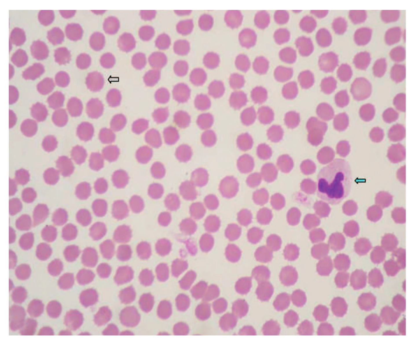
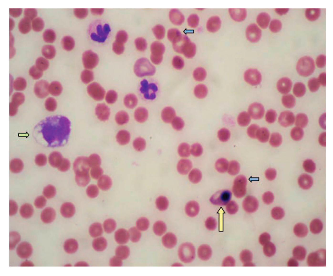
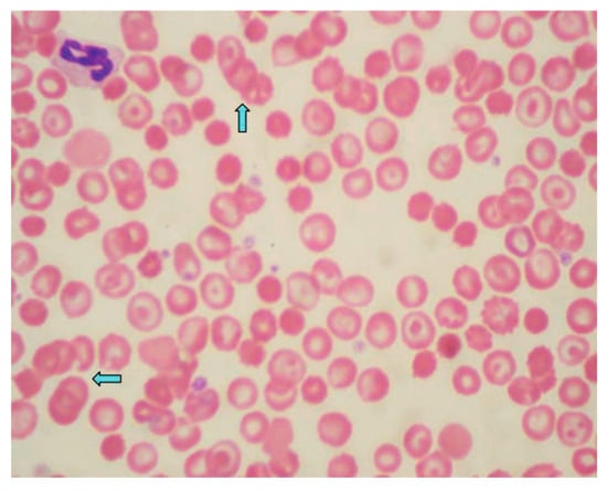
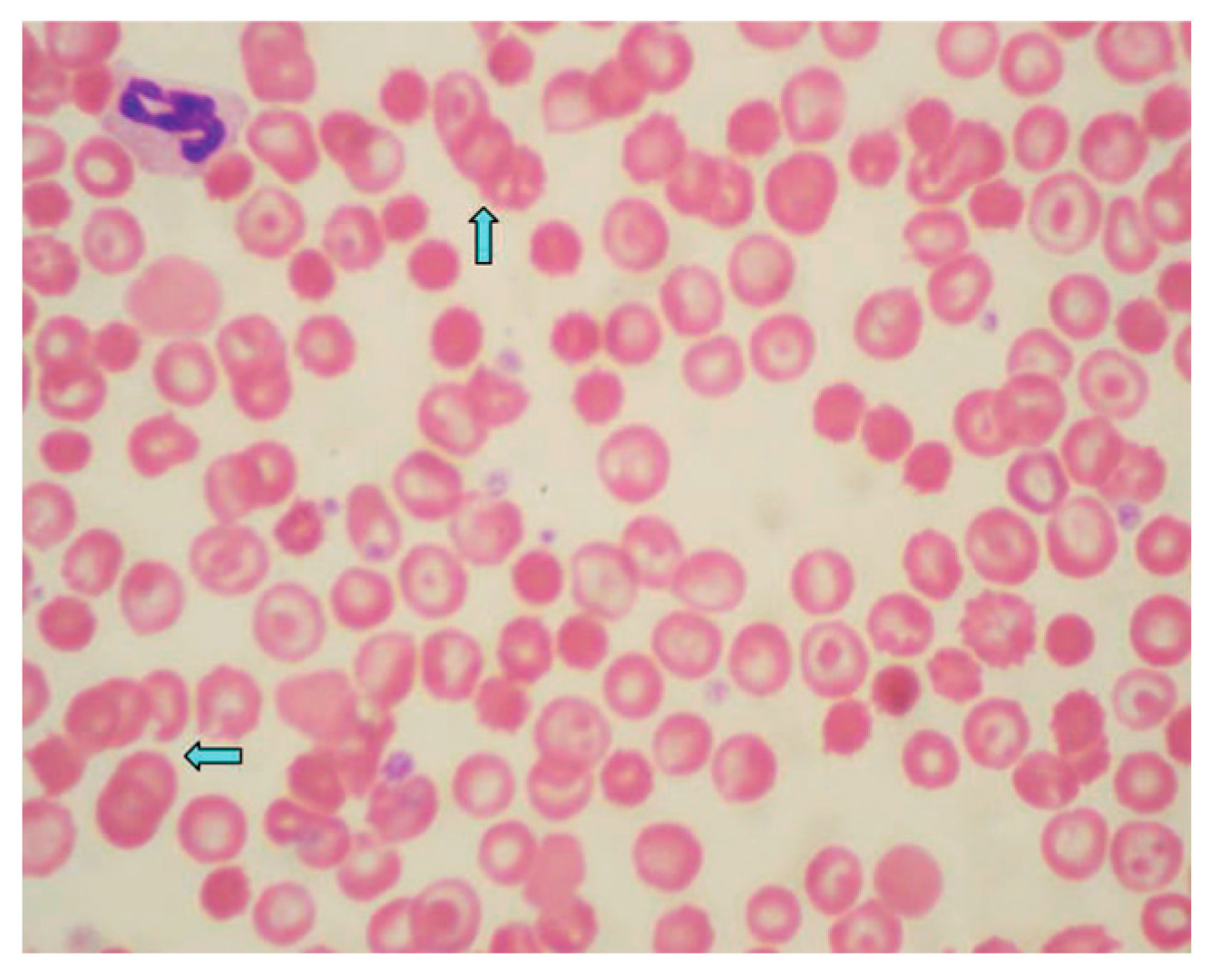
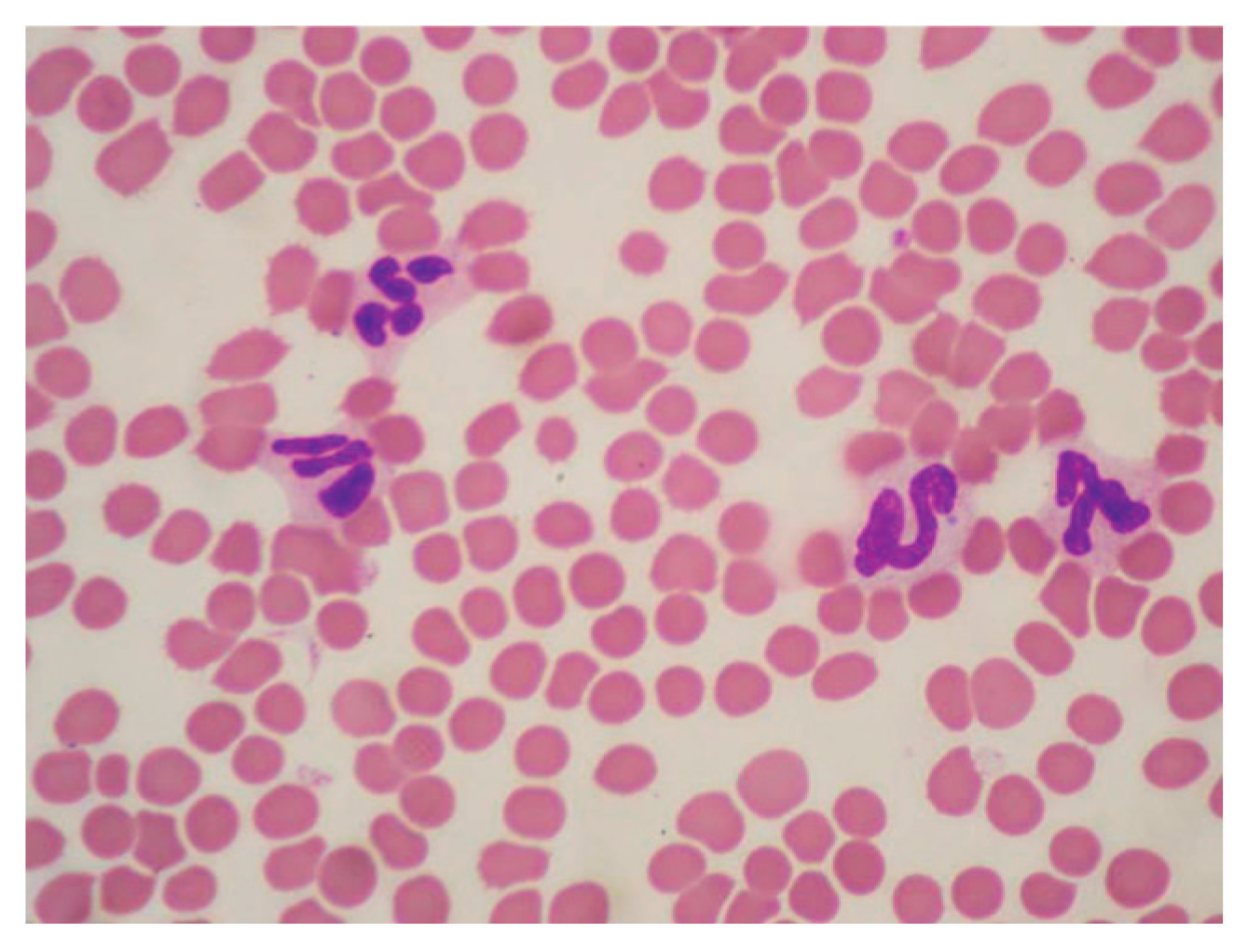
Other reported abnormalities included toxic changes to neutrophils, reticulocytes, spherocytes, sphero-echinocytes and rouleaux. Polychromasia and hypochromia were also observed, as were Howell-Jolly bodies and codocytes. These changes were evident in blood smears taken at various times after the snakebite (see Table 4 and Figure 1, Figure 2, Figure 3 and Figure 4).
Table 4.
Abnormalities in blood smears recorded at T1, T2 and T3.
Figure 1.
Blood smear of one of the dogs (ID1) included in the study. Venom within 24 h of the event. Sphero-echinocytes (erythrocytes lacking central pallor with a jagged outline and regular spicules along their entire circumference) (grey arrow). A neutrophil with a visible Döhle body inside (blue arrow). There is an almost total absence of platelets. Wright, 100× (immersion).
Figure 2.
Blood smear of one of the dogs (ID4) included in the study. Viper venom, 24 h after the event. Spherocytes and erythrocytes with central pallor. Also present are Howell-Jolly bodies (blue arrow), one NRBC (yellow arrow), a vacuolated monocyte (green arrow), and two neutrophils with cytoplasmic foam. Rare platelets. Wright, 100× (immersion).
Figure 3.
Blood smear of one of the dogs (ID3) included in the study. Viper venom, 24 h after the event. Rouleaux (stacking of erythrocytes) (blue arrows). Wright, 100× (immersion).
Figure 4.
Blood smear of one of the dogs (ID11) included in the study. Viper venom, 48 h after the event. There is a significant decrease in sphero-echinocytes, foamy leukocytes and Döhle bodies, as well as thrombocytopenia. Wright, 100× immersion.
4. Discussion
Dogs are particularly susceptible to envenomation due to their behaviour and exposure during outdoor activities. Young and curious individuals are at a higher risk, with the mean age distribution being ≤5 years, as reported.
The clinical presentation can vary greatly, ranging from mild local effects to severe systemic manifestations. Affected dogs can develop distributive shock, showing clinical indicators such as fast heart rate, reddened mucous membranes, shortened capillary refill time, and low blood pressure, as noted in the evaluated study group.
Other anomalies may be observed due to the activity of the venom, such as enhanced leakage of fluids from blood vessels, profound disturbances in coagulation processes, modified cardiac function, neurological impairment, depressed respiratory activity, and muscle tissue necrosis. Toxins may also sometimes impair cardiac function, potentially resulting in oedema and hypovolemic shock [7,11].
Treatment strategies typically include a combination of specific therapies, such as antivenom administration, alongside supportive care including fluid therapy, antibiotics and glucocorticoids. Glucocorticoids exert anti-inflammatory actions by limiting capillary widening and suppressing the release of vasoactive amines [21]. However, the use of glucocorticoids remains controversial due to conflicting evidence regarding their efficacy in modulating inflammatory responses and improving outcomes [22,23,24,25]. Due to their habit of walking and smelling with their noses near the ground, dogs are most commonly bitten in the head, nose and extremities [8,22]. Of the dogs included in the study, a large group (75%) were bitten in the front or hind limbs, and the majority (58.4%) died within 48 h. These differences may reflect variations in environmental exposure, behavioral responses, or circumstances surrounding the envenomation in our study population. In research conducted by Segev and colleagues involving 327 dogs bitten by Daboia palaestinae, fatalities were more likely when the bites occurred on the limb extremities [10]. Various studies have suggested that phospholipase A2 (PLA2) enzymes in snake venom play a key role in early morbidity and mortality following a snakebite [26,27]. PLA2 exerts its effects through the degradation of cell membranes, resulting in tissue destruction (necrosis and myonecrosis) and disruption of homeostatic mechanisms critical for the regulation of coagulation and oxygen transport [28,29,30,31].
These processes can determine the onset of extensive local and systemic damage, also contributing to life-threatening outcomes, such as cardiorespiratory arrest and paralysis. The severity of these effects varies depending on the specific PLA2 subtype and its concentration. The use of an inhibitor of secretory PLA2, such as varespladib, has been suggested as a potential treatment for severe snake envenomation in dogs [32].
The financial implications of treatment can play a significant role in clinical decision-making [33,34,35]. In our experience, these costs often affect an owner’s willingness or ability to pursue continued care, particularly when prolonged hospitalization, antivenom administration, or advanced supportive therapies are required. Economic considerations frequently become intertwined with discussions regarding prognosis and anticipated duration of critical care, and in some cases may contribute to decisions about euthanasia when the outlook is uncertain or guarded. Beyond individual cases in dogs, viper bites also impose broader economic consequences, associated with the financial burden of envenomation in humans, horses, and livestock. Incorporating economic factors, including medical expenses, loss of animal productivity, and the financial repercussions of morbidity or mortality, provides a more comprehensive understanding of envenomation severity [26,27,32].
Additionally, viper venom includes enzymes that promote the release of internal cytokines and other inflammatory agents. The frequency of secondary infections after Vipera berus bites in Swedish dogs has not been established, nor is it clear whether antibiotic treatment (such as cefazolin or ceftriaxone) provides any advantage [1]. Although viperid snakes carry substantial oral and fang contamination with numerous potentially pathogenic bacteria, reports indicate that human envenomation rarely leads to bacterial infections [36].
Our study also indicated that none of the dogs exhibited any clinical signs of infection, despite being treated with antibiotics. Secondary bacterial infections can significantly contribute to complications, particularly when extensive tissue necrosis occurs, potentially leading to severe soft tissue and systemic effects [37]. None of the dogs in the present study were treated with antivenom serum (immunoglobulin-based). Given that antivenom can still provide clinical benefit when administered late in animals with systemic signs, it is notable that the majority of our cohort presented 8–12 h after the bite—well beyond the optimal window for antivenom efficacy [38,39,40,41]—by which time systemic signs had already developed. This delay likely influenced the clinical management and outcomes observed in our population, and highlights the challenges associated with delayed presentation in this setting [42]. Treatment with antiserum is the only way to inactivate the enzymes in the venom by binding them to the antiserum’s antibodies [38]. Antiserum should be considered for severely affected dogs for whom supportive therapy is ineffective [22,24,43,44].
Haematological alterations recorded upon admission included anaemia, leucocytosis and thrombocytopenia. The decrease in platelet counts observed at T2, together with the concomitant rise in MPV, aligns with the thrombocytopenic effects typically described in viperid envenomation for the effects resulting from proteolytic enzymes and PLA2 capable of inducing platelet activation, aggregation, and consumption, as well as contributing to vascular endothelial damage, resulting in accelerated platelet removal from circulation [45,46]. The increase in MPV, although not significant, is consistent with two complementary processes such as platelet activation and a compensatory bone-marrow response (larger platelets are released following acute peripheral consumption, suggesting that consumption-mediated platelet loss predominates early, while regenerative mechanisms are already underway.
Biochemical test alterations included increased BUN and CREA, as well as increased ALT, AST, ALP, and CPK. The biochemical changes observed here are consistent with those reported by other researchers. Aroch and Harrus [23] described comparable alterations in dogs poisoned by Vipera xanthina palestinae, except for notable creatine kinas (CK) and glutamate dehydrogenase values. Elevation of ALT and AST is a frequent biochemical finding in dogs following viper envenomation. This increase could be correlated with the myotoxic effects associated with the action of myotoxic components such as phospholipase A2 isoenzymes, metalloproteinases, and other proteolytic factors [21,47,48]. These toxins induce disruption of myonecrosis and lead to substantial leakage of AST into the bloodstream and concurrent increase in CK. Also, systemic effects of viper envenomation—such as hypotension, coagulopathies, and microvascular disturbances—can compromise hepatic perfusion, contributing to increased ALT, more specific to liver parenchyma [13,49]. The high CK values recorded at T2 suggest the onset of renal and muscle damage, which was probably caused by a local muscle injury at the snakebite site, given that there was no evidence of renal failure [11,40]. A decrease in serum creatinine values was observed in 82% of the sampled dogs between the T1 and T2 examinations. This could be associated with a reduction in glomerular filtration rate after the administered fluid therapy [50,51].
Cytological alterations included Döhle bodies, toxic granulation, spherocytes, echinocytes, sphero-echinocytes, rouleaux formation, nucleated red blood cells (NRBCs) and hypochromia.
Cytoplasmic abnormalities observed in the neutrophils, such as Döhle bodies (pale blue inclusions) and toxic granulation (larger, darker granules), suggest accelerated neutrophil production and maturation, which is typically associated with inflammation, infection, stress, or cytokine therapy [52]. Therefore, these changes indicate the body’s increased response to viper venom, affecting the appearance of neutrophils as they are released from the bone marrow.
About erythrocytes, it is possible to observe the formation of spheroidal erythrocytes, also known as spherocytes. These are characterised by the loss of part of the outer membrane, a smaller diameter than normal erythrocytes, and an absence of central pallor. This anomaly is commonly seen to some degree in all haemolytic anaemias: for example, in cases of hereditary spherocytosis or autoimmune haemolytic anaemia, which are characterised by spherocytes being the only haematological variation present [53]. Considering the presence of other morphological alterations of erythrocytes in our cases, it is likely that spherocytes were formed due to the activity of phospholipases present in viper venom. Echinocytes, which are cells like erythrocytes, but characterised by a smaller size, an absence of central pallor, and numerous membrane projections arranged regularly and symmetrically, were also found alongside spherocytes. These cells appear transiently following venomous snake bites due to the action of phospholipase present in the venom. Echinocytes and spherocytes have also been found in combination as sphero-echinocytes. According to some studies, various types of venom, including bee (Apis mellifera), redback spider (Latrodectus mactans), blue-ringed octopus (Hapalochlaena maculosa), ten different snake venoms, phospholipase A2, and four snake venom components, cause sphero-echinocytosis of human red blood cells [54]. Therefore, the formation of sphero-echinocytes could be considered a general sign of venom activity in both humans and dogs. Studies have indicated that most venoms and toxins are unable to alter human red blood cells when molecules weighing less than 10,000 are present [54]. The absence or low number of sphero-echinocytes in these dogs could imply exposure to minimal amounts of viper venom. These erythrocyte membrane modifications cause erythrocytes to lose their ability to repel each other, resulting in the formation of persistent aggregates of cells known as rouleaux.
Rouleaux formation is typically associated with hyperglobulinaemia in dogs. Specifically, an increase in two types of globulins can result in rouleaux formation: fibrinogen (a protein released during the acute-phase response) and immunoglobulin (a complex glycoprotein produced by plasma cells that acts as an antibody and forms part of the immune system) [55]. These two types of globulins both increase in the event of inflammation or antigenic stimulation. Considering that the venom causes vascular damage, protein release and severe coagulation alterations (with the potential for destruction and consumption of factors, as well as the release of debris), the changes induced by the venom in the plasma environment may also affect the stability and aggregation of red blood cells. Direct haemolysis caused by viper venom, combined with indirect haemolysis due to the increased fragility and tendency towards aggregation of modified erythrocytes (echinocytes, spherocytes and sphero-echinocytes), results in an elevated presence of NRBCs in the peripheral blood. NRBCs are immature red blood cells containing a nucleus that are usually found in the bone marrow, but not in the circulating blood of healthy adults [56]. The presence of NRBCs in the peripheral blood, known as polychromasia, indicates a significant underlying medical condition such as anaemia, severe hypoxia, sepsis, leukaemia, or bone marrow stress. Their presence is often associated with increased mortality and adverse outcomes [57].
Hypochromia is a medical term used to describe red blood cells that appear paler than normal, indicating that they contain an insufficient amount of haemoglobin. This condition is often a sign of anaemia, the most common cause of which is iron deficiency. Under a microscope, hypochromic cells appear pale due to the reduced amount of haemoglobin, which carries oxygen, increasing central pallor [58]. Hypochromia is related to haemolytic anaemia caused by viper venom, which increases the production of erythrocytes. However, this increase is not balanced by an adequate iron intake, leading to the production of iron-deficient erythrocytes [59].
Finally, alterations to the coagulation cascade included elevated levels of aPTT, PT, antithrombin III, and fibrinogen.
The proteases contained in viper venom interfere with the normal activation of the coagulation cascade by degrading the molecules involved in the process. Following a viper bite, elevated aPTT and PT values are frequently found [60]. These values indicate prolonged blood clotting time, significantly increasing the risk of spontaneous haemorrhage. Viper venom can cause coagulation disorders such as disseminated intravascular coagulation (DIC), mucosal haemorrhages, and prolonged bleeding at the site of the bite. At time T1, the aPTT and PT values are higher because the enzymes in the venom act quickly. The subsequent decrease in these values after 24 and 48 h (T2 and T3, respectively) is due to treatment with dexamethasone. Fibrinogen, an acute-phase reactant, is a non-specific protein associated with inflammation. A decrease in fibrinogen levels, and therefore its consumption, is indicative of DIC [61]. However, 24 h after the viper bite (T2), fibrinogen levels were found to be increasing, indicating an inflammatory response rather than DIC. Also, the rise in fibrinogen concentration (rather than a drop) could reflect a compensatory acute-phase response consequent to envenomation inflammation, cytokine release (e.g., IL-6), and hepatic upregulation of acute-phase proteins. Although specific data in dogs are limited, this is a common mechanism in systemic inflammation [62,63,64].
Elevated antithrombin III levels 24 and 48 h after the viper bite (T2 and T3) indicate specific systemic inflammation. However, these increases remained within the physiological range for dogs [16,62].
Viper bites in dogs are a true medical emergency, with the severity of the clinical manifestations depending on various factors, such as the amount of venom injected, the location of the bite, the species of viper involved, and the size and health of the dog.
It is essential to promptly recognise local symptoms such as swelling, pain, and necrosis, as well as systemic signs such as lethargy, tachycardia or bleeding tendencies, to enable early therapeutic intervention.
The results demonstrate that systemic involvement is common and can progress rapidly from local tissue damage to life-threatening complications, such as coagulopathies, shock and multi-organ dysfunction. Haematological analysis consistently revealed evidence of haemoconcentration, thrombocytopenia and varying degrees of anaemia, reflecting both haemolytic and consumptive processes. Laboratory findings, particularly alterations in coagulation parameters and biochemical profiles, confirmed the systemic toxic effects of the venom. These effects include endothelial damage, activation of coagulation pathways and secondary inflammatory responses. These changes emphasise the need for continuous monitoring and serial laboratory evaluations during the acute and recovery phases of envenomation.
This study presents potential limitations that should be considered when interpreting the findings. First, the small sample size limited the statistical power of the analysis. It may have prevented the detection of significant changes in clinical status, biochemistry parameters, or coagulation profiles associated with the time elapsed between the bite and clinical evaluation. Similarly, the limited number of cases may have hindered the identification of more pronounced differences in the most severe presentations or in those that resulted in fatality. An additional consideration is the inclusion of animals that underwent euthanasia, which may have introduced a severity-related selection bias and influenced the distribution of clinical outcomes. Additionally, the study did not evaluate the potential effects of treatments, such as corticosteroid administration, on clinical outcomes, nor did it assess the impact of the timing of antivenom administration on clinical, biochemical, or haematological changes. Overall, these limitations reduce the generalizability of the results and highlight the need for future studies with larger sample sizes and more standardised data-collection protocols. The study did not evaluate the potential effects of treatments, such as corticosteroid administration, on clinical outcomes. Additionally, the impact of the timing of antivenom administration on clinical, biochemical, and haematological changes in dogs with Vipera aspis envenomation was not assessed.
5. Conclusions
In conclusion, a multidisciplinary approach is required for the diagnosis and treatment of canine viper envenomation, based on clinical assessment, haematological and biochemical monitoring, and evidence-based intervention. Further studies are needed to refine treatment protocols, identify prognostic biomarkers, and evaluate the long-term consequences of envenomation. Improving veterinary awareness and access to antivenom therapy is crucial to reducing the morbidity and mortality associated with viper bites in dogs.
Author Contributions
Conceptualization, F.M., G.M. and M.P.; methodology F.M., G.N., C.R. and M.P.; software, D.A.S. and P.G.; validation, G.M. and M.P.; formal analysis, D.A.S. and P.G.; investigation, F.M., G.N. and M.P.; data curation, F.M., G.M. and C.R.; writing—original draft preparation, F.M., G.M. and M.P.; writing—review and editing, M.P., D.A.S., P.G. and C.R.; visualization, G.M.; supervision, M.P.; funding acquisition, M.P. All authors have read and agreed to the published version of the manuscript.
Funding
This research received no external funding.
Institutional Review Board Statement
Ethical review was waived for this study because it was retrospective, meaning it analysed previously collected data and did not involve new interventions or risks to participants.
Informed Consent Statement
Informed consent was obtained from the owner of the dogs involved in the study.
Data Availability Statement
The original contributions presented in this study are included in the article. Further inquiries can be directed to the corresponding author.
Acknowledgments
The authors would like to thank Veterinary Laboratory EmatosVetLab (Rome, Italy) for their valuable collaboration and support in carrying out this study.
Conflicts of Interest
The authors declare no conflicts of interest.
References
- Tednes, M.; Slesinger, T.L. Evaluation and Treatment of Snake Envenomations. In StatPearls; StatPearls Publishing: Treasure Island, FL, USA, 2025. [Google Scholar]
- Gutiérrez, J.M.; Calvete, J.J.; Habib, A.G.; Harrison, R.A.; Williams, D.J.; Warrell, D.A. Snakebite envenoming. Nat. Rev. Dis. Primers 2017, 3, 17063. [Google Scholar] [CrossRef] [Scilit]
- Adhikari, R.B.; Gawarammana, I.B.; De Silva, D.D.N.; Dangolla, A.; Mallawa, C.; Premarathna, A.D.; Silva, I.D. Clinico-epidemiology and management of Russell’s viper (Daboia russelii) envenoming in dogs in Sri Lanka. Toxicol. Rep. 2019, 6, 809–818. [Google Scholar] [CrossRef] [Scilit]
- Adhikari, R.; Suriyagoda, L.; Premarathna, A.D.; Tuvikene, R.; Mallawa, C.; Niranjala De Silva, N.; Dangolla, A.; Silva, I.; Gawarammana, I. Clinico-epidemiology and management of hump-nosed pit viper (Hypnale spp.) bites in dogs. Sci. Rep. 2022, 12, 8232. [Google Scholar] [CrossRef] [Scilit]
- Aradilla, N.; Sánchez-Fortún, S. Retrospective analysis of the clinical approach practiced in dog bite cases caused by Vipera latastei, Vipera seoanei and Vipera aspis. Vet. J. 2025, 314, 106446. [Google Scholar] [CrossRef] [Scilit]
- Barr, S.C. Clinical features therapy and epidemiology of tiger snake bite in dogs and cats. Aust. Vet. J. 1984, 61, 208–212. [Google Scholar] [CrossRef] [Scilit]
- Kim, D.; Kim, S.; Kim, J.K.; Lim, J.H.; Choi, G.; Bae, S.; Kwon, Y.S.; Jang, M. Clinical features and management of snake bites in 70 dogs in Korea. J. Vet. Sci. 2022, 23, e81. [Google Scholar] [CrossRef] [Scilit] [PubMed]
- Leisewitz, A.L.; Blaylock, R.S.; Kettner, F.; Goodhead, A.; Goddard, A.; Schoeman, J.P. The diagnosis an management of snakebite in dogs—A southern African perspective. S. Afr. Vet. Assoc. 2004, 75, 7–13. [Google Scholar] [CrossRef] [Scilit] [PubMed]
- Karlsson-Stiber, C.; Salmonson, M.; Persson, H. A nationwide study of viperaberus bites during one year—Epidemiology and morbidity of 231 cases. Clin. Toxicol. 2006, 44, 25–30. [Google Scholar] [CrossRef] [Scilit]
- Segev, G.; Shipov, A.; Klement, E.; Harrus, S.; Kass, P.; Aroch, I. Vipera palaestinae envenomation in 327 dogs: A retrospective cohort study and analysis of risk factors for mortality. Toxicon 2004, 43, 691–699. [Google Scholar] [CrossRef] [Scilit] [PubMed]
- Lervik, J.B.; Lilliehöök, I.; Frendin, J.H. Clinical and biochemical changes in 53 Swedish dogs bitten by the European adder—Vipera berus. Acta Vet. Scand. 2010, 52, 26. [Google Scholar] [CrossRef] [Scilit]
- Monteiro, C.L.B.; da Silva, A.M.; Tavares, F.L.; de Oliveira, R. Clinical and laboratory evaluation of dogs experimentally envenomed by Bothrops alternatus venom. Toxicon 2018, 148, 91–94. [Google Scholar] [CrossRef] [Scilit]
- Nicolaysen, T.V.; Harjen, H.J.; Lund, H.S.; Sævik, B.K.; Rørtveit, R.; Zimmer, K.E. Clinicopathological biomarker patterns, venom detection and venom proteomics in canine Vipera berus envenomation. Top. Companion Anim. Med. 2024, 63, 100930. [Google Scholar] [CrossRef] [Scilit]
- Harjen, H.J.; Nicolaysen, T.V.; Negard, T.; Lund, H.; Sævik, B.K.; Anfinsen, K.P.; Moldal, E.R.; Zimmer, K.E.; Rørtveit, R. Serial serum creatinine, SDMA and urinary acute kidney injury biomarker measurements in dogs envenomated by the European adder (Vipera berus). BMC Vet. Res. 2021, 17, 154. [Google Scholar] [CrossRef] [Scilit]
- Harjen, H.J.; Anfinsen, K.P.; Hultman, J.; Moldal, E.R.; Szlosek, D.; Murphy, R.; Friis, H.; Peterson, S.; Rørtveit, R. Evaluation of Urinary Clusterin and Cystatin B as Biomarkers for Renal Injury in Dogs Envenomated by the European Adder (Vipera berus). Top. Companion Anim. Med. 2022, 46, 100586. [Google Scholar] [CrossRef] [Scilit]
- Harjen, H.J.; Hellum, M.; Rørtveit, R.; Oscarson, M.; Anfinsen, K.P.; Moldal, E.R.; Solbak, S.; Kanse, S.M.; Henriksson, C.E. Persistent hypercoagulability in dogs envenomated by the European adder (Vipera berus berus). PLoS ONE 2022, 17, e0263238. [Google Scholar] [CrossRef] [Scilit]
- Sutton, N.M.; Bates, N.; Campbell, A. Canine adder bites in the UK: A retrospective study of cases reported to the Veterinary Poisons Information Service. Vet. Rec. 2011, 169, 607. [Google Scholar] [CrossRef] [Scilit] [PubMed]
- International Cooperation on Harmonisation of Technical Requirements for Registration of Veterinary Medicinal Products Good Clinical Practices, Good Clinical Practices, VICH GL9 (GCP) June 2000—Implemented in July 2001. Available online: https://www.ema.europa.eu/en/vich-gl9-good-clinical-practices-scientific-guideline (accessed on 25 April 2025).
- Schalm, O.W.; Jane Wardrop, K.; Weiss, D.J. Schalm’s Veterinary Hematology, 6th ed.; Wiley-Blackwell: Ames, IA, USA, 2010. [Google Scholar]
- Kaneko, J.J.; Harvey, J.W.; Bruss, M.L. Clinical Biochemistry of Domestic Animals, 6th ed.; Elsevier: New York, NY, USA, 2008. [Google Scholar]
- Lee, J.M.; Song, J.H.; Song, K.H. A Retrospective Evaluation of Snake Envenomation in Dogs in South Korea (2004–2021). Toxins 2022, 14, 565. [Google Scholar] [CrossRef] [Scilit]
- Maddison, J. Corticosteroids—Friend or foe? In Proceedings of the 34th World Small Animal Veterinary Congress WSAVA, São Paulo, Brazil, 24 July 2009. [Google Scholar]
- Aroch, I.; Harrus, S. Retrospective study of the epidemiological, clinical, haematological and biochemical findings in 109 dogs poisoned by vipera xanthina palestinae. Vet. Rec. 1999, 144, 532–535. [Google Scholar] [CrossRef] [Scilit]
- Aroch, I.; Segev, G.; Klement, E.; Shipov, A.; Harrus, S. Fatal vipera xanthina palestinae envenomation in 16 dogs. Vet. Hum. Toxicol. 2004, 46, 268–272. [Google Scholar]
- Garland, T. Recognition and treatment for snakebites. In Proceedings of the 18th Annual American College of Veterinary Internal Medicine, Seattle, WA, USA, 25–28 May 2000; pp. 48–49. [Google Scholar]
- Lewin, M.; Samuel, S.; Merkel, J.; Bickler, P. Varespladib (LY315920) Appears to Be a Potent, Broad-Spectrum, Inhibitor of Snake Venom Phospholipase A2 and a Possible Pre-Referral Treatment for Envenomation. Toxins 2016, 8, 248. [Google Scholar] [CrossRef] [Scilit] [PubMed]
- Zinenko, O.; Tovstukha, I.; Korniyenko, Y. PLA2 Inhibitor Varespladib as an Alternative to the Antivenom Treatment for Bites from Nikolsky’s Viper Vipera berus nikolskii. Toxins 2020, 12, 356. [Google Scholar] [CrossRef] [Scilit]
- Isbister, G.K. Snakebite doesn’t cause disseminated intravascular coagulation: Coagulopathy and thrombotic microangiopathy in snake envenoming. Semin. Thromb. Hemost. 2010, 36, 444–451. [Google Scholar] [CrossRef] [Scilit] [PubMed]
- Herrera, M.; Fernández, J.; Vargas, M.; Villalta, M.; Segura, Á.; León, G.; Angulo, Y.; Paiva, O.; Matainaho, T.; Jensen, S.D.; et al. Comparative proteomic analysis of the venom of the taipan snake, Oxyuranus scutellatus, from Papua New Guinea and Australia: Role of neurotoxic and procoagulant effects in venom toxicity. J. Proteom. 2012, 75, 2128–2140. [Google Scholar] [CrossRef] [Scilit] [PubMed]
- Gulati, A.; Isbister, G.K.; Duffull, S.B. Effect of Australian elapid venoms on blood coagulation: Australian Snakebite Project (ASP-17). Toxicon 2013, 61, 94–104. [Google Scholar] [CrossRef] [Scilit] [PubMed]
- O’Rourke, K.M.; Correlje, E.; Martin, C.L.; Robertson, J.D.; Isbister, G.K. Point-of-care derived INR does not reliably detect significant coagulopathy following Australian snakebite. Thromb. Res. 2013, 132, 610–613. [Google Scholar] [CrossRef] [Scilit]
- Woliver, C.; Kastenholz, V.; Southern, C.; Odunayo, A.; Schaer, M.; Allen-Durrance, A. Phospholipase A2 inhibitor may shorten the duration of clinical signs in the treatment of neurotoxicity caused by eastern coral snake (Micrurus fulvius) envenomation in 3 dogs. J. Am. Vet. Med. Assoc. 2025, 30, 1–7. [Google Scholar] [CrossRef] [Scilit]
- Pérez, M.L.; Fox, K.; Schaer, M. A retrospective evaluation of coral snake envenomation in dogs and cats: 20 cases (1996–2011). J. Vet. Emerg. Crit. Care 2012, 22, 682–689. [Google Scholar] [CrossRef] [Scilit]
- Sullivan, J.M.; Aasen, T.L.; Fisher, C.J.; Schaer, M. Retrospective evaluation of clinical and clinicopathologic findings, case management, and outcome for dogs and cats exposed to Micrurus fulvius (eastern coral snake): 92 cases (2021–2022). Toxins 2024, 16, 246. [Google Scholar] [CrossRef] [Scilit]
- Campos, S.; Allen-Durrance, A.E.; Schaer, M.; Lynch, A. Retrospective evaluation of Micrurus fulvius (eastern coral snake) envenomation and the use of mechanical ventilation in dogs and a cat (2011–2016): 8 cases. J. Vet. Emerg. Crit. Care 2019, 29, 662–667. [Google Scholar] [CrossRef] [Scilit]
- Senthilkumaran, S.; Salim, A.; Almeida, J.R.; Williams, J.; Vijayakumar, P.; Thirunavukarasu, A.; Christopoulos, M.A.; Williams, H.F.; Thirumalaikolundusubramanian, P.; Patel, K.; et al. The Effectiveness of Antibiotics in Managing Bacterial Infections on Bite Sites following Snakebite Envenomation. Toxins 2023, 15, 190. [Google Scholar] [CrossRef] [Scilit]
- Barceloux, D.G. Terrestrial snakes. In Medical Toxicology of Natural Substances; Barceloux, D.G., Ed.; Wiley: Los Angeles, CA, USA, 2008; pp. 1032–1046. [Google Scholar]
- World Health Organization. Guidelines for the Management of Snakebites, 2nd ed.; WHO Regional Office for South-East Asia: New Delhi, India, 2016. [Google Scholar]
- Williams, D.J.; Faiz, M.A.; Abela-Ridder, B.; Ainsworth, S.; Bulfone, T.C.; Nickerson, A.D.; Habib, A.G.; Junghanss, T.; Fan, H.W.; Turner, M.; et al. Strategy for a Globally Coordinated Response to a Priority Neglected Tropical Disease: Snakebite Envenoming. PLoS Negl. Trop. Dis. 2019, 13, e0007059. [Google Scholar] [CrossRef] [Scilit] [PubMed]
- Gutiérrez, J.M.; León, G.; Bournouf, T. Antivenoms for the treatment of snakebite envenomings: The road ahead. Biologicals 2011, 39, 129–142. [Google Scholar] [CrossRef] [Scilit]
- Shah, R.; Sharma, R.; Yadav, S.; Singh, R.; Mishra, A. Impact of Delay in Antivenom Administration in Snakebite Envenomation: A Systematic Review. J. Trop. Med. 2021, 2021, 553428. [Google Scholar]
- Chippaux, J.P. Snakebite Envenomation Turns Again into a Neglected Tropical Disease! J. Venom. Anim. Toxins Incl. Trop. Dis. 2017, 23, 38. [Google Scholar] [CrossRef] [Scilit]
- Di Nicola, M.R.; Crevani, M.; Avella, I.; Cerullo, A.; Dorne, J.-L.C.M.; Paolino, G.; Zattera, C. A Guide to the Clinical Management of Vipera Snakebite in Italy. Toxins 2024, 16, 255. [Google Scholar] [CrossRef] [Scilit] [PubMed]
- Lobetti, R.G.; Joubert, K. Retrospective study of snake envenomation in 155 dogs from the Onderstepoort area of South Africa. S. Afr. Vet. Assoc. 2004, 75, 169–172. [Google Scholar] [CrossRef] [Scilit]
- Guillemin, M.; Chevillard, L.; Albouy-Laberty, J. Pathophysiology and clinical management of European viper envenomation in small animals. Vet. Clin. N. Am.-Small Anim. Pract. 2021, 51, 1237–1253. [Google Scholar]
- Isbister, G.K.; Hodgson, W.C. Mechanisms of venom-induced consumption coagulopathy. Toxicon 2015, 108, 72–82. [Google Scholar]
- Loan, N.V.T.H.; Chao, N.V.; Nhung, T.T. Clinical Features and Hematological and Biochemical Parameters in Envenomated Dogs. Vet. World 2025, 8, 986–993. [Google Scholar] [CrossRef] [Scilit]
- Sadhu, D.B.; Rao, N.M.; Shah, A.I.; Parmar, J.J.; Patel, B.R.; Hadiya, K.K.; Patel, O.; Pandya, D. Haemato-Biochemical Changes and Therapeutic Management of Snake Envenomation in Dogs. Int. J. Vet. Sci. Anim. Husb. 2024, 9, 131–134. [Google Scholar] [CrossRef] [Scilit]
- Pałgan, K.; Kuźmiński, A.; Janik, A.; Gotz-Żbikowska, M.; Bartuzi, Z. Snake (Vipera berus) Bite Causing Severe Systemic Manifestations, Including Hepatic Enzyme Elevation. Int. J. Environ. Health 2015, 28, 119–121. [Google Scholar] [CrossRef] [Scilit]
- Braun, J.P.; Lefebvre, H.P. Kidney Function and Markers of Glomerular Filtration. Compend. Cont. Educ. Pract. Vet. 2008, 30, 108–118. [Google Scholar]
- Mazzaferro, E.M. Complications of fluid therapy. Vet. Clin. N. Am. Small Anim. Pract. 2008, 38, 607–619. [Google Scholar] [CrossRef] [Scilit]
- Sjaastad, O.V.; Hove, K.; Sand, O. Physiology of Domestic Animals; Scandinavian Veterinary Press: Oslo, Norway, 2003; pp. 425–474. [Google Scholar]
- Boone, L.I. Disorders of White Blood Cells. In Handbook of Small Animal Practice; Elsevier: New York, NY, USA, 2008; pp. 641–655. [Google Scholar] [CrossRef] [Scilit]
- Flachsenberger, W.; Leigh, C.M.; Mirtschin, P.J. Sphero Echinocytosis of Human Red Blood Cells Caused by Snake, Red Back Spider, Bee and Blue Ringed Octopus Venoms and Its Inhibition by Snake Sera. Toxicon 1995, 33, 791–797. [Google Scholar] [CrossRef] [Scilit] [PubMed]
- Garden, O.A.; Kidd, L.; Mexas, A.M.; Chang, Y.M.; Jeffery, U.; Blois, S.L.; Fogle, J.E.; MacNeill, A.L.; Lubas, G.; Birkenheuer, A.; et al. ACVIM consensus statement on the diagnosis of immune-mediated hemolytic anemia in dogs and cats. J. Vet. Intern. Med. 2019, 33, 313–334. [Google Scholar] [CrossRef] [Scilit] [PubMed]
- Abramson, N. Rouleaux formation. Blood 2006, 107, 4205. [Google Scholar] [CrossRef] [Scilit]
- Hollmann, F.; Geisen, V.; Hartmann, K.; Doerfelt, R. Nucleated red blood cells as a prognostic indicator in dogs with anemia. Front. Vet. Sci. 2025, 12, 1585168. [Google Scholar] [CrossRef] [Scilit]
- Weiss, G.; Ganz, T.; Goodnough, L.T. Anemia of inflammation. Blood 2019, 133, 40–50. [Google Scholar] [CrossRef] [Scilit]
- Naigamwalla, D.Z.; Webb, J.A.; Giger, U. Iron deficiency anemia. Can. Vet. J. 2012, 53, 250–256. [Google Scholar]
- Latinović, Z.; Leonardi, A.; Koh, C.Y.; Kini, R.M.; Trampuš Bakija, A.; Pungerčar, J.; Križaj, I. The Procoagulant Snake Venom Serine Protease Potentially Having a Dual, Blood Coagulation Factor V and X-Activating Activity. Toxins 2020, 12, 358. [Google Scholar] [CrossRef] [Scilit]
- Bateman, S.W.; Mathews, K.A.; Abrams-Ogg, A.C.; Lumsden, J.H.; Johnstone, I.B.; Hillers, T.K.; Foster, R.A. Diagnosis of disseminated intravascular coagulation in dogs admitted to an intensive care unit. J. Am. Vet. Med. Assoc. 1999, 215, 798–804. [Google Scholar] [CrossRef] [Scilit] [PubMed]
- Gabay, C.; Kushner, I. Acute-Phase Proteins and Other Systemic Responses to Inflammation. N. Engl. J. Med. 1999, 340, 448–454. [Google Scholar] [CrossRef] [Scilit] [PubMed]
- Aroch, I.; Yas-Natan, E.; Segev, G. Haemostatic Abnormalities in Vipera palaestinae-Envenomed Dogs. Vet. Rec. 2009, 164, 562–568. [Google Scholar]
- Tizard, I. Veterinary Immunology: An Introduction, 11th ed.; Elsevier: New York, NY, USA, 2023; Chapter 7 on Sickness. [Google Scholar]


Disclaimer/Publisher’s Note: The statements, opinions and data contained in all publications are solely those of the individual author(s) and contributor(s) and not of MDPI and/or the editor(s). MDPI and/or the editor(s) disclaim responsibility for any injury to people or property resulting from any ideas, methods, instructions or products referred to in the content. |
© 2026 by the authors. Licensee MDPI, Basel, Switzerland. This article is an open access article distributed under the terms and conditions of the Creative Commons Attribution (CC BY) license.