


Abstract
Cryptococcus neoformans and Cryptococcus gatti are the two most common pathogenic species of the encapsulated yeast genus Cryptococcus and have been linked to fatal neurologic infections in both immunocompromised and healthy individuals. Cryptococcosis, which includes AIDS-defining diseases such as cryptococcal meningitis and meningoencephalitis, is the most frequent fungal infection of the central nervous system, with Africa having the greatest fatality rate. The goal of this study was to identify and characterize C. neoformans from pigeon and bat droppings, which are thought to be environmental reservoirs for the yeast and to determine their relatedness to clinical species. A total of 588 dried and 55 moist pigeon droppings, as well as 50 moist bat droppings, were collected from 18 sites in Ghana’s Greater Accra and Western regions, from sources which included markets, a church, households, and a public park. Seven clinical archived C. neoformans isolates were also retrieved from the Central Laboratory of the Korle-Bu Teaching Hospital (KBTH), Greater Accra. Using conventional methods and partial Multi-Locus Sequence Typing (MLST), 49 Cryptococcus neoformans var grubii were isolated from dried pigeon droppings only, yielding a prevalence of 7%. The sequence type (ST)/MLST group could not be defined based on the available data, although the allele types of the housekeeping genes in some of the isolates were identified. Some clinical strains had allele types that were found among the environmental strains, giving preliminary evidence of possible genetic relatedness, highlighting the need for more comprehensive genomic surveillance approaches, to better characterize environmental and clinical cryptococcal isolate transmission patterns and genetic relationships. This work also adds to the global literature supporting pigeon droppings as favorable ecological niches for Cryptococcus. This study was conducted within a One Health framework, recognizing the interconnected roles of environmental reservoirs, animal-associated sources, and human health in the epidemiology of Cryptococcus infections.
Keywords:
Cryptococcus neoformans; characterization; isolation; Southern Ghana; MLST; ITS; environmental 1. Introduction
Cryptococcus neoformans and C. gatti are the main pathogenic species of the yeast genus, Cryptococcus [1]. Their mucopolysaccharide capsules are used as antigenic determinants to classify them into serotypes A and D for C. neoformans and B and C for C. gatti. Serotypes A and D can recombine, to create hybrids [2,3]. Additionally, their mucopolysaccharide capsules are major virulence determinants capable of modulating host immune cells by impairing phagocytosis, suppressing the production of reactive molecules involved in antimicrobial functions, inhibiting inflammatory signaling, impairing antigen presentation, and altering cytokine production, thereby promoting fungal pathogenesis and survival [4,5]. Both healthy and immunocompromised individuals are affected by their pathogenic effects. C. neoformans causes disease in immunocompromised individuals, notably those with HIV, whereas C. gatti causes disease in otherwise healthy individuals [6]. Immunocompromised individuals are more vulnerable to the effects of cryptococcal infections, which typically affect the central nervous system in the form of cryptococcal meningitis or meningoencephalitis, whereas healthy people are usually able to clear the infections [7]. Cryptococcal meningitis is an AIDS-defining disease that kills over half a million HIV-positive people each year [8]. The highest mortality from HIV-associated cryptococcal meningitis occurs in Africa [9]. It is also the most common type of meningitis in adult HIV/AIDS patients in Africa, as well as the leading cause of community-acquired meningitis with a prevalence between 20 and 45% prevalence [10].
Cryptococcal meningitis is prevalent in 11.7% of Ghanaians with unknown HIV status [11]. Ghana’s national HIV prevalence is however 1.7%, with roughly 14,000 deaths [12,13]. Cryptococcosis has also been reported in other immunocompromised individuals visiting the Korle-Bu Teaching Hospital in Accra, Ghana’s capital [14]. The inhalation of cryptococcal spores or dehydrated yeast cells from the environment is thought to be the source of cryptococcal infection [7,8]. Further, environmental and clinical cryptococcal isolates from same geographical location show genetic similarities [15,16]. Avian (particularly pigeon droppings and pigeon roosting places), bat guano, and arboreal environments are among the species’ implicated niches [3,17].
In Ghana, pigeons are commonly found in the environment roaming motor parks, playgrounds, markets, churches and residential areas. Pigeons are also sold at different marketplaces where they are often displayed in open cages, thereby exposing many people to their droppings which may contain cryptococcal spores or dehydrated cells. Although the isolation of Cryptococcus species from environmental sources including pigeon and bat droppings have been reported from neighboring West African countries such as Nigeria [18,19,20,21], Cameroon [22] and Ivory coast [23], to the best of our knowledge, there are no reports on the prevalence of Cryptococcus species in pigeon droppings or in any other environmental niche in Ghana.
In this study, we used a combination of phenotypic and molecular methods [24], including approaches used for global strain genotyping [25], to characterize Cryptococcus isolates from environmental and clinical sources. We also assessed the potential relatedness between these environmental and clinical isolates. This study adopts a One Health approach, acknowledging the close interactions among environmental, animal, and human health systems in the transmission dynamics and public health impact of Cryptococcus species.
2. Materials and Methods
Between November 2016 and April 2017, we collected 588 dried and 55 moist pigeon droppings, and 55 moist bat droppings, from 15 locations in the Greater Accra and 3 locations in the Western regions of Ghana.
Pigeon and bat droppings were scooped with spatula and brushes, from roofs of buildings, cages and dovecotes, into sterile plastic bags, sealed and stored at room temperature for 1–2 days before processing and microbiological analysis. Additionally, seven clinical Cryptococcus species previously isolated from blood cultures between January and May 2017, were retrieved from the Central Laboratory of the Korle-Bu Teaching Hospital (KBTH), Greater Accra. These clinical isolates were characterized along with the environmental isolates obtained.
Culture and Characterization of Cryptococcal Isolates
For the environmental samples, an approximate amount of 1 g of pigeon/bat droppings was suspended in 20 mL of sterile distilled water, vortexed for two minutes and left to settle for about thirty minutes. Then, 100 μL of supernatant was inoculated onto Sabouraud dextrose agar (SDA) (Oxoid, Basingstoke, UK) plates supplemented with chloramphenicol (Melford Laboratories Ltd., Ipswich, UK) at a concentration of 0.05 mg/mL. The inoculated plates were then incubated for about 7 days at 37 °C and were monitored daily from day 2 for the growth of creamy colonies suggestive of Cryptococcus. Pure colonies were recovered and purified by subsequent streaking onto fresh SDA plates. Initial identification was done using standard microbiological techniques including wet preparation, Gram stain [26], urease test [27] and thermo-tolerance test [28]. Archived clinical Cryptococcus were recovered on SDA.
For molecular confirmation and genetic diversity of the isolates, genomic DNA was extracted using the Zymo Research Quick-DNA Fungal/Bacterial Miniprep Kit following the manufacturers’ instructions. DNA segments comprising the Internal transcribed spacer (ITS) region of the ribosomal DNA (rDNA) were amplified with primers ITS-1 and ITS-4 [24].
Species were identified by searching databases using the BLAST+ version 2.6.0 sequence analysis tool, an open source software developed by the National Center for Biotechnology Information (NCBI) (https://www.ncbi.nlm.nih.gov/, accessed on 30 May 2017), an agency of the United States federal government (https://blast.ncbi.nlm.nih.gov/Blast.cgi, accessed on 30 May 2017). The ITS-1 and ITS-4 sequences were compared using nucleotide–nucleotide BLAST (blastn) with default parameters except for the “Choose Search Set” option, which was set to Cryptococcus neoformans species complex (taxid:1897064) as the organism of choice. Species identification was determined from the lowest ‘expect’ value of the BLAST output with its corresponding percent identity.
Multi-locus sequence typing (MLST) was carried out according to the ISHAM consensus multi-locus sequence typing scheme for C. neoformans and C. gatti species complexes [17] with slight modifications. Four (CAP59, GPD1, LAC1, and PLB1) instead of seven unlinked genetic loci were used for genotyping. While the ISHAM consensus MLST scheme recommends the use of seven loci (CAP59, GDP1, LAC1, PLB1, IGS1, SOD1, and URA5) for optimal discriminatory power and sequence type assignment, the loci selected in this study were initially considered sufficient for preliminary genotypic classification as they encode important cryptococcal virulence factors and include some of the most genetically diverse loci within the MLST scheme [25,29]. The four loci were amplified and sequenced with the forward and reverse primers (Table 1) of each region using the ABI 3500XL Genetic Analyzer, POP7™, BrilliantDye™ Terminator v3.1 reagent kit via Inqaba Biotechnical Industries, Pretoria, South Africa.
Table 1.
Primers used in this study.
The four unlinked genetic loci were also used for species identification using nucleotide–nucleotide BLAST (blastn) with default settings (https://blast.ncbi.nlm.nih.gov/Blast.cgi, accessed on 30 May 2017). Species identification was then determined from the lowest ‘expect’ value of the BLAST output with its corresponding percent identity. Allele numbers were assigned by uploading sequences to the MLST database for the Cryptococcus neoformans/C. gattii species complex (http://mlst.mycologylab.org, accessed on 10 June 2017) an open-source software developed by the Medical Mycology Group of Westerdijk Fungal Biodiversity Institute of the KNAW 3584 CT Utrecht, Netherlands. Sampling and laboratory data were recorded manually and transferred to an Excel file template. Descriptive statistics to determine prevalence and distribution of species from the different sampling locations were done using MS Excel (Version 16.0).
3. Results
3.1. Distribution of Environmental Isolates
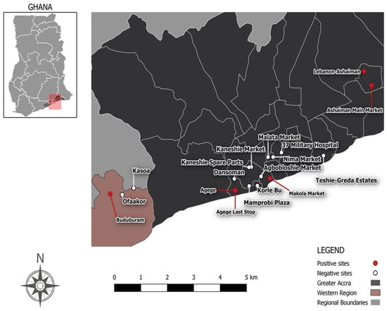
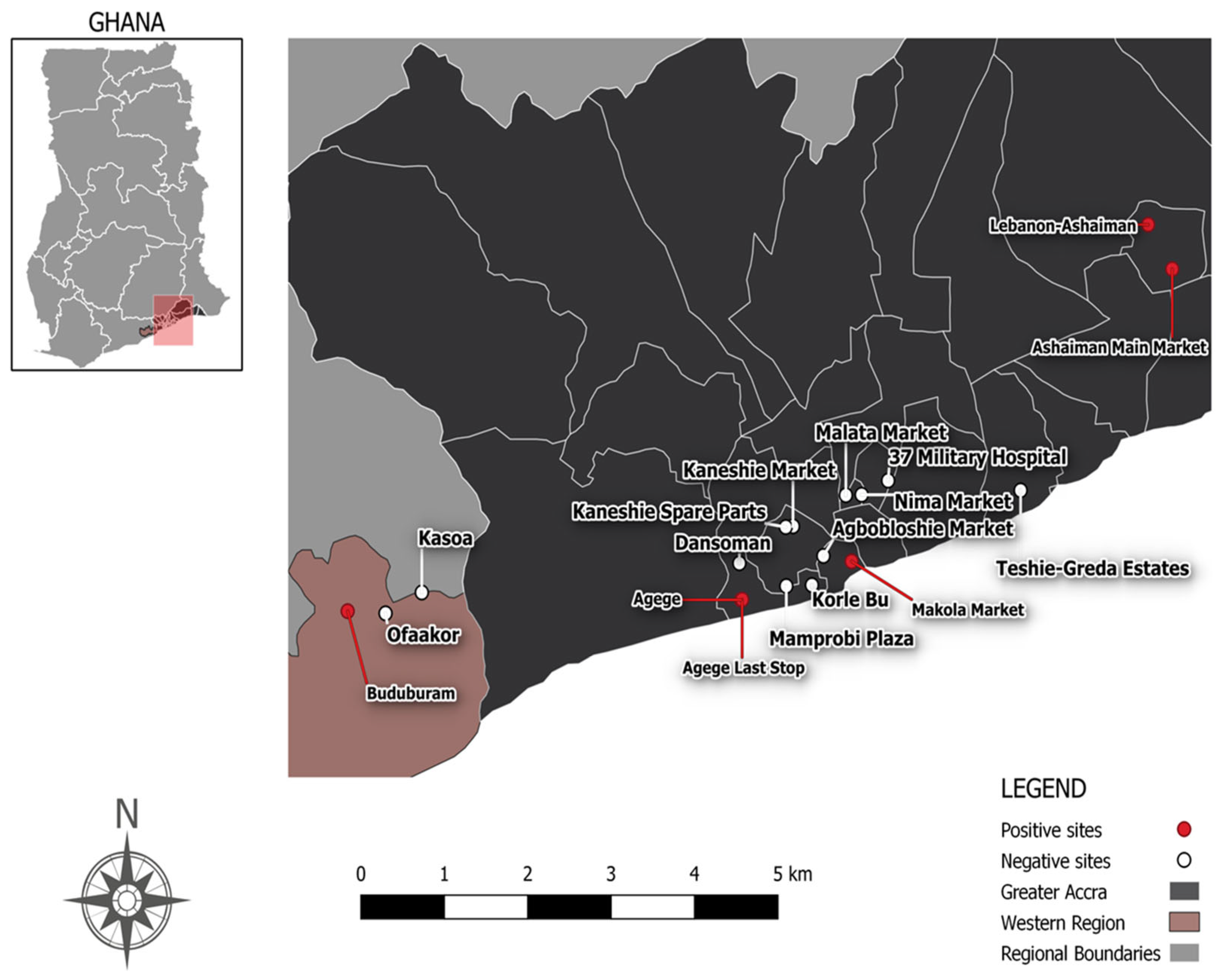
Figure 1 illustrates sampling localities where Cryptococcus isolates were retrieved from the environmental samples.
Figure 1.
A map of Ghana showing the locations where pigeon and bat droppings were sampled.
From the 698 pigeon and bat droppings cultured, 49 Cryptococcus species were obtained, a prevalence of 7%. All Cryptococcus species recovered were positive for Gram stain, urease test, and thermo-tolerance test, in addition to being round/oval cells (Supplementary Table S1). The distribution of the environmental Cryptococcus from the various locations investigated ranged from 0.0 to 26.3%. Cryptococcus was found in 12 of 243 (4.9%) market samples, 16 of 320 (5.0%) household samples, and 21 of 80 (26.3%) church samples (Table 2).
Table 2.
Sampling sites and isolation rate of environmental Cryptococcus.
3.2. Internal Transcribed Spacer Region (ITS) Region Analysis for Species Identification
ITS-1 was successfully amplified in all the environmental and clinical isolates. ITS-4 was, however, amplified in only five environmental and two clinical isolates. Both environmental and clinical isolates were identified as Cryptococcus neoformans var grubii using the ITS region of the rDNA as a barcode for fungal identification (Supplementary Table S2).
3.3. Housekeeping Genes Analysis for Species Identification
In 14 out of 49 environmental isolates, all four housekeeping genes (CAP59, GPD1, LAC1, and PLB1) were successfully amplified. Gene amplification was found in the remaining isolates as follows; three genes (CAP59, GPD1, PLB1) in five environmental and one clinical isolate, three genes (CAP59, GDP1, LAC1), two genes (CAP59, LAC1) and (GDP1, LAC1) and one gene (CAP59) in one environmental isolate each. Either none of the genes were amplified or no significant match was found in 28 of the isolates. Using default parameters, the BLAST sequence analysis tool recognized all isolates with at least one successfully amplified housekeeping gene as Cryptococcus neoformans var grubii (Supplementary Table S3).
3.4. Multi-Locus Sequence Typing (MLST) Analysis
The Multi-locus sequence typing (MLST) analysis carried out according to the ISHAM working group provided allele types for isolates for which the housekeeping genes were amplified. However, the CN Multi Loci Identification for these isolates to determine the sequence type (ST) did not yield any result. No records satisfying input parameters were found. The individual housekeeping genes provided allele types of the isolates for three housekeeping genes (GDP1, LAC1, PLB1). Analysis of the CAP59 gene sequences for all isolates yielded no records satisfying the submitted parameter. The allele types provided by the GDP1gene sequences included AT9, AT23, and AT1. The LAC1 gene sequence allele types were AT5, AT8, AT3, AT5, AT8, and AT1. The PLB1 gene sequence allele types were AT2, AT11 with a single isolate yielding two allele types AT44 and AT4. The clinical strains had allele types found among the environmental strains (Table 3 and Supplementary Table S3).
Table 3.
Allele types from the amplified housekeeping genes.
4. Discussion
This is the first report of environmental Cryptococcus isolates from Ghana and their recovery from pigeon and bat droppings collected from public settings such as marketplaces, a church, and households. Despite the low prevalence (7%) of Cryptococcus identified in the current study, our findings have public health implications for immunocompromised individuals especially those living with HIV/AIDS. Interestingly, Cryptococcus was recovered only from dry pigeon droppings, with none from moist pigeon droppings nor bat droppings. The rate of cryptococcal isolation from the environment varies around the globe, with pigeon dropping serving as a consistent niche [3]. Analogous studies carried out in other African countries, showed an isolation prevalence between 3% and 33% from environmental sources including soil, water, avian including pigeon droppings, bat droppings, wood, house-dust, eucalyptus, olive, almond trees, baobab, and mopane trees [18,19,20,21,22,23,29,30,31,32,33,34,35,36,37,38]. Studies that sampled pigeon or bat droppings generally used dried/weathered material. In Cameroon, Dongmo et al. sampled dried pigeon and bat droppings and isolated both C. neoformans and C. gattii [22]. Similar studies from other African countries including Nigeria [18,19,20,21], Ivory Coast [23], Libya [32], Egypt [30], Botswana [29], and South Africa [29] also reported the isolation of C. neoformans only from dried or weathered pigeon droppings.
As indicated, Cryptococcus was not isolated from any moist pigeon or bat droppings, supporting previous findings that dry droppings may provide a more suitable reservoir for the fungus [37]. This may be due to reduced competition from other microorganisms and favorable pH conditions [38]. In contrast, the high ammonia concentration in moist droppings may inhibit fungal growth [19]. However, the smaller number of moist-dropping samples may also have biased the isolation rate toward dry droppings.
All environmental and clinical isolates from the current study were identified as C. neoformans var grubii. This variety is widely reported in Africa with evidence indicating that they may be of African origin. Although the study did not identify specific molecular types, the prevalent molecular types in Africa are VNI and VNII [29].
The four housekeeping genes (CAP59, GDP1, LAC1, and PLB1) used in the current study were not discriminatory enough to provide any sequence type (ST) nor MLST groups. This may be because ST determination requires sequencing the complete set of seven housekeeping genes (CAP59, GDP1, LAC1, PLB1, IGS1, SOD1, and URA5), which generates unique allelic profiles for assigning sequence types [25,39]. Also, C. neoformans/C. gattii species complex MLST database (http://mlst.mycologylab.org, accessed on 10 June 2017) relies on all seven loci to generate a credible MLST typing and only 100% matches are considered acceptable and displayed. Additionally, the absence of CAP59 amplification across all isolates and the selective amplification of GDP1, LAC1, and PLB1 in some isolates remain unclear. Potential technical explanations, including variability in DNA quality and low fungal burden, were assessed and excluded. Therefore, the findings suggest sequence divergence within primer-binding regions, which could have reduced amplification efficiency for certain loci [40]. This observation may also indicate underlying genetic diversity or potentially novel polymorphisms among Ghanaian strains. Further studies using all seven housekeeping genes and larger sample size should give more meaningful conclusions.
Although the ST/MLST groups could not be determined in the current study, there were some overlaps in allele types between clinical and environmental isolates. However, the limited number of successfully amplified loci and incomplete allelic profiles restrict robust assessment of genetic relatedness. These findings likely serve as preliminary evidence of possible shared genetic characteristics, since environmental and clinical isolates from the same geographical location are usually genetically similar [15,16].
Our study has limitations. Only four of the seven recommended housekeeping genes were sequenced, which reduced the discriminatory power of the analysis and prevented assignment of sequence types (STs) and MLST groups. This limitation also restricted the ability to robustly assess the genetic relatedness between clinical and environmental isolates. Secondly, only few clinical isolates were available for comparison, further limiting the power of the study.
5. Conclusions
Our findings add to the growing body of evidence that dry pigeon droppings are a suitable ecological niche for Cryptococcus. The findings from this study highlight the public health importance of environmental reservoirs of Cryptococcus species and their potential role in the transmission of cryptococcal infections. Given the substantial morbidity and mortality associated with cryptococcosis, particularly among immunocompromised individuals, strengthening awareness, early detection, and preventive strategies remain essential. Targeted public health education should focus on increasing awareness of environmental exposure risks and promoting preventive measures, including avoiding direct contact with dry bird droppings and using appropriate protective equipment when cleaning dovecotes or handling potentially contaminated materials. Furthermore, these findings underscore the importance of environmental surveillance and strengthened clinical awareness for timely diagnosis, to reduce disease burden and improve public health outcomes.
While this study provides preliminary insights into potential genetic similarities between clinical and environmental isolates from Ghana, the findings from this work serve as an initial exploratory study and preliminary evidence, highlighting the need for more comprehensive genomic surveillance approaches, including using the complete set of loci for MLST and whole-genome sequencing to better characterize environmental and clinical cryptococcal isolates transmission patterns and genetic relationships.
Supplementary Materials
The following supporting information can be downloaded at https://www.mdpi.com/article/10.3390/amh71030027/s1, Table S1: Microbiological analysis, Table S2: ITS region analysis results, Table S3: Housekeeping gene BLAST analysis results, Table S4: MLST analysis results.
Author Contributions
Conceptualization, J.A.O.; methodology, N.E.A. and P.J.P.-A.; validation, J.A.O., N.E.A. and P.J.P.-A.; formal analysis, N.E.A., P.J.P.-A. and S.D.A.; investigation, N.E.A. and P.J.P.-A.; resources, J.A.O.; data curation, N.E.A. and J.A.O.; writing—original draft preparation, N.E.A.; writing—review and editing, N.E.A., J.A.O. and S.D.A.; supervision, J.A.O.; project administration, J.A.O.; funding acquisition, J.A.O. All authors have read and agreed to the published version of the manuscript.
Funding
This research was partly funded by ADMER project, grant number 09-099SS1 and the APC was funded by Medical Microbiology Department, University of Ghana Medical School.
Institutional Review Board Statement
Not applicable.
Informed Consent Statement
Not applicable.
Data Availability Statement
The original contributions presented in this study are included in the article/Supplementary Material. Further inquiries can be directed to the corresponding author.
Acknowledgments
We thank the staff of the Department of Medical Microbiology, University of Ghana Medical School and the Department of Microbiology, Korle-Bu Teaching Hospital for their support and encouragement.
Conflicts of Interest
The authors declare no conflicts of interest. The funders had no role in the design of the study; in the collection, analyses, or interpretation of data; in the writing of the manuscript; or in the decision to publish the results.
Abbreviations
The following abbreviations are used in this manuscript:
| KBTH | Korle-Bu Teaching Hospital |
| MLST | Multi-Locus Sequence Type |
| ST | Sequence Type |
| ITS | Internal Transcribed Spacer |
| SDA | Sabouraud Dextrose Agar |
| ISHAM | The International Society for Human and Animal Mycology |
| CN | Cryptococcus neoformans |
| HIV | Human Immuno-Deficiency Virus |
| AIDS | Acquired Immune Deficiency Virus |
| CAP59 | Capsule polysaccharide |
| GPD1 | Glycerol 3-Phosphate Dehydrogenase |
| LAC1 | Laccase |
| PLB1 | Phospholipase B1 |
| IGS1 | Intergenic spacer region of ribosomal DNA |
| SOD1 | Cu-Zn superoxide dismutase |
| URA51 | Orotate phosphoribosyltransferase |
References
- Nester, E.W.; Anderson, D.G.; Roberts, C.E., Jr.; Nester, M.T. Microbiology: A Human Perspective, 6th ed.; McGraw-Hill Higher Education: New York, NY, USA, 2009. [Google Scholar]
- Xu, J.; Mitchell, T.G. Comparative gene genealogical analyses of strains of sero-type AD identify recombination in populations of serotypes A and D in the human pathogenic yeast Cryptococcus neoformans. Microbiology 2003, 149, 2147–2154. [Google Scholar] [CrossRef] [Scilit]
- Cogliati, M. Global molecular epidemiology of Cryptococcus neoformans and Cryptococcus gattii: An atlas of the molecular types. Scientifica 2013, 2013, 675213. [Google Scholar] [CrossRef] [Scilit] [PubMed]
- LaRocque-de-Freitas, I.F.; da Silva-Junior, E.B.; Gemieski, L.P.; da Silva Dias Lima, B.; Diniz-Lima, I.; de Carvalho Vivarini, A.; Lopes, U.G.; Freire-de-Lima, L.; Morrot, A.; Decote-Ricardo, D. Inhibition of microbicidal activity of canine macrophages DH82 cell line by capsular polysaccharides from Cryptococcus neoformans. J. Fungi 2024, 10, 339. [Google Scholar] [CrossRef] [Scilit]
- Decote-Ricardo, D.; LaRocque-de-Freitas, I.F.; Rocha, J.D.B.; Nascimento, D.O.; Nunes, M.P.; Morrot, A.; Freire-De-Lima, L.; Previato, J.O.; Mendonça-Previato, L.; Freire-de-Lima, C.G. Immunomodulatory role of capsular polysaccharides constituents of Cryptococcus neoformans. Front. Med. 2019, 6, 129. [Google Scholar] [CrossRef] [Scilit]
- Idnurm, A.; Lin, X. Rising to the challenge of multiple Cryptococcus species and the diseases they cause. Fungal Genet. Biol. 2015, 78, 1–6. [Google Scholar] [CrossRef] [Scilit] [PubMed]
- Kwon-Chung, K.J.; Fraser, J.A.; Doering, T.L.; Wang, Z.A.; Janbon, G.; Idnurm, A.; Bahn, Y. Cryptococcus neoformans and Cryptococcus gattii, the Etiologic Agents of Cryptococcosis. Cold Spring Harb. Perspect. Med. 2014, 4, a019760. [Google Scholar] [CrossRef] [Scilit] [PubMed]
- Park, B.J.; Wannemuehler, K.A.; Marston, B.J.; Govender, N.; Pappas, P.G.; Chiller, T.M. Estimation of the current global burden of cryptococcal meningitis among persons living with HIV/AIDS. AIDS 2009, 23, 525–530. [Google Scholar] [CrossRef] [Scilit] [PubMed]
- Holmes, C.B.; Losina, E.; Walensky, R.P.; Yazdanpanah, Y.; Freedberg, K.A. Review of Human Immunodeficiency Virus Type 1—Related Opportunistic Infections in Sub-Saharan Africa. Clin. Infect. Dis. 2003, 36, 02114. [Google Scholar] [CrossRef] [Scilit]
- Hakim, J.G.; Gangaidzo, I.T.; Heyderman, R.S.; Mushangi, E.; Taziwa, A.; Robertson, V.J.; Musvaire, P.; Mason, P.R. Impact of HIV infection on meningitis in Harare, Zim-babwe: A prospective study of 406 predominantly adult patients. AIDS 2000, 14, 1401–1407. [Google Scholar] [CrossRef] [Scilit] [PubMed]
- Owusu, M.; Nguah, S.B.; Boaitey, Y.A.; Badu-boateng, E.; Abubakr, A.; Lartey, R.A.; Adu-Sarkodie, Y. Aetiological agents of cerebrospinal meningitis: A retrospective study from a teaching hospital in Ghana. Ann. Clin. Microbiol. Antimicrob. 2012, 11, 28. [Google Scholar] [CrossRef] [Scilit] [PubMed]
- UNAIDS. Country Progress Report-Ghana. Global AIDS Monitoring. 2019. Available online: https://www.unaids.org/sites/default/files/country/documents/GHA_2019_countryreport.pdf (accessed on 16 February 2020).
- Ali, H.; Amoyaw, F.; Baden, D.; Durand, L.; Bronson, M.; Kim, A.; Grant-Greene, Y.; Imtiaz, R.; Swaminathan, M. Ghana’s HIV epidemic and PEPFAR’s contribution towards epidemic control. Ghana Med. J. 2019, 53, 59–62. [Google Scholar] [CrossRef] [Scilit] [PubMed]
- Opintan, J.A.; Awadzi, B.K.; Biney, I.J.K.; Ganu, V.; Doe, R.; Kenu, E.; Adu, R.F.; Osei, M.M.; Akumwena, A.; Grigg, M.E.; et al. High rates of cerebral toxoplasmosis in HIV patients presenting with meningitis in Accra, Ghana. Trans. R. Soc. Trop. Med. Hyg. 2017, 111, 464–471. [Google Scholar] [CrossRef] [Scilit] [PubMed]
- Alves, G.S.B.; Freire, A.K.L.; Bentes, A.D.S.; Pinheiro, J.F.D.S.; Souza, J.V.B.; Wanke, B.; Matsuura, T.; Jackisch-Matsuura, A.B. Molecular typing of environmental Cryptococcus neoformans/C. gattii species complex isolates from Manaus, Amazonas, Brazil. Mycoses 2016, 59, 509–515. [Google Scholar] [CrossRef] [Scilit] [PubMed]
- Litvintseva, A.P.; Kestenbaum, L.; Vilgalys, R.; Mitchell, T.G. Comparative analysis of environmental and clinical populations of Cryptococcus neoformans. J. Clin. Microbiol. 2005, 43, 556–564. [Google Scholar] [CrossRef] [Scilit] [PubMed]
- Mitchell, T.; Castañeda, E.; Nielsen, K.; Wanke, B.; Lazera, M. Environmental ncihes for Cryptococcus neoformmans and Cryptococcus gatti. In Cryptococcus; American Society of Microbiology: Washington, DC, USA, 2011. [Google Scholar]
- Nnadi, N.E.; Enweani, I.B.; Cogliati, M.; Ayanbimpe, G.M.; Okolo, M.O.; Kim, E.; Sabitu, M.Z.; Criseo, G.; Romeo, O.; Scordino, F. Molecular characterization of environ-mental Cryptococcus neoformans VNII isolates in Jos, Plateau State, Nigeria. J. Mycol. Médicale 2016, 26, 306–311. [Google Scholar] [CrossRef] [Scilit]
- Ior, C.A.; Ayanbimpe, G.; Ior, L.D.; Okechalu, J.; Chris-Otubor, G.O. Environmental isolation of Cryptococcus neoformans IN JOS. Sci. Res. J. (SCIRJ) 2015, 3, 15–17. [Google Scholar]
- Chidebelu, P.E.; Nweze, E.I.; Meis, J.F.; Cogliati, M.; Hagen, F. Multi-locus sequence typing reveals genotypic similarity in Nigerian Cryptococcus neoformans AFLP1/VNI of environmental and clinical origin. J. Med. Microbiol. 2021, 70, 001440. [Google Scholar] [CrossRef] [Scilit]
- Nweze, E.I.; Kechia, F.A.; Dibua, U.E.; Eze, C.; Onoja, U.S. Isolation of Cryptococcus neoformans from environmental samples collected in Southeastern Nigeria. Rev. Inst. Med. Trop. São Paulo 2015, 57, 295–298. [Google Scholar] [CrossRef] [Scilit] [PubMed]
- Dongmo, W.; Kechia, F.; Tchuenguem, R.; Nangwat, C.; Yves, I.; Kuiate, J.R.; Dzoyem, J.P. In Vitro Antifungal Susceptibility of Environmental Isolates of Cryptococcus spp. from the West Region of Cameroon. Ethiop. J. Health Sci. 2016, 26, 555–560. [Google Scholar] [CrossRef] [Scilit] [PubMed]
- Kassi, F.K.; Bellet, V.; Drakulovski, P.; Krasteva, D.; Roger, F.; Valérie, B.T.A.; Aboubakar, T.; Doumbia, A.; Kouakou, G.A.; Bertout, S.; et al. Comparative typing analyses of clinical and environmental strains of the Cryptococcus neoformans/Cryptococcus gattii species complex from Ivory Coast. J. Med. Microbiol. 2018, 67, 87–96. [Google Scholar] [CrossRef] [Scilit] [PubMed]
- White, T.J.; Bruns, T.; Lee, S.; Taylor, J. Amplification and Direct Sequencing of Fungal Ribosomal RNA Genes for Phylogenetics. PCR Protoc. 1990, 1, 315–322. [Google Scholar] [CrossRef] [Scilit]
- Meyer, W.; Aanensen, D.M.; Boekhout, T.; Cogliati, M.; Diaz, M.R.; Esposto, M.C.; Fisher, M.; Gilgado, F.; Hagen, F.; Kaocharoen, S.; et al. Consensus multi-locus sequence typing scheme for Cryptococcus neoformans and Cryptococcus gattii. Med. Mycol. 2009, 47, 561–570. [Google Scholar] [CrossRef] [Scilit] [PubMed]
- Smith, A.C.; Hussey, M.A. Gram stain protocols. Am. Soc. Microbiol. 2005, 1, 113–144. [Google Scholar] [CrossRef] [Scilit]
- Brink, B. Urease Test Protocol. 2020. Available online: https://asm.org/getattachment/ac4fe214-106d-407c-b6c6-e3bb49ac6ffb/urease-test-protocol-3223.pdf (accessed on 11 June 2026).
- Oghomwenakhin, E.A.; Eboigbe, L. Screening and selection of Cryptococcus neoformans using urease activity and temperature range. Niger. J. Basic Appl. Sci. 2019, 27, 57–61. [Google Scholar]
- Litvintseva, A.P.; Carbone, I.; Rossouw, J.; Thakur, R.; Govender, N.P.; Mitchell, T.G. Evidence that the human pathogenic fungus Cryptococcus neoformans var. grubii may have evolved in Africa. PLoS ONE 2011, 6, e19688. [Google Scholar] [CrossRef] [Scilit] [PubMed]
- Mahmoud, Y.A.G. First environmental isolation of Cryptococcus neoformans var. neoformans and var. gatti from the Gharbia Governorate, Egypt. Mycopathologia 2000, 148, 83–86. [Google Scholar] [CrossRef] [Scilit] [PubMed]
- Mseddi, F.; Sellami, A.; Jarboui, M.A.; Sellami, H.; Makni, F.; Ayadi, A. First Environmental Isolations of Cryptococcus neoformans and Cryptococcus gattii in Tunisia and Review of Published Studies on Environmental Isolations in Africa. Mycopathologia 2011, 171, 355–360. [Google Scholar] [CrossRef] [Scilit] [PubMed]
- Ellabib, M.S.; Aboshkiwa, M.A.; Husien, W.M.; D’Amicis, R.; Cogliati, M. Isolation, identification and molecular typing of Cryptococcus neoformans from pigeon droppings and other environmental sources in Tripoli, Libya. Mycopathologia 2016, 181, 603–608. [Google Scholar] [CrossRef] [Scilit] [PubMed]
- Kangogo, M.; Bader, O.; Boga, H.; Wanyoike, W.; Folba, C.; Worasilchai, N.; Weig, M.; Groß, U.; Bii, C.C. Molecular types of C ryptococcus gattii/C ryptococcus neoformans species complex from clinical and environmental sources in Nairobi, Kenya. Mycoses 2015, 58, 665–670. [Google Scholar] [CrossRef] [Scilit] [PubMed]
- Boekhout, T.; Theelen, B.; Diaz, M.; Fell, J.W.; Hop, W.C.; Abeln, E.C.; Dromer, F.; Meyer, W. Hybrid genotypes in the pathogenic yeast Cryptococcus neoformans. Microbiology 2001, 147, 891–907. [Google Scholar] [CrossRef] [Scilit] [PubMed]
- Elhariri, M.; Hamza, D.; Elhelw, R.; Refai, M. Eucalyptus tree: A potential source of Cryptococcus neoformans in Egyptian environment. Int. J. Microbiol. 2016, 2016, 4080725. [Google Scholar] [CrossRef] [Scilit] [PubMed]
- Elfadaly, H.A.; Hassanain, N.A.; Hassanain, M.A.; Barakat, A.M.; Shaapan, R.M. Evaluation of primitive ground water supplies as a risk factor for the development of major waterborne zoonosis in Egyptian children living in rural areas. J. Infect. Public Health 2018, 11, 203–208. [Google Scholar] [CrossRef] [Scilit] [PubMed]
- Ruiz, A.; Fromtling, R.A.; Bulmer, G.S. Distribution of Cryptococcus neoformans in a natural site. Infect. Immun. 1981, 31, 560–563. [Google Scholar] [CrossRef] [Scilit] [PubMed]
- Zarrin, M.; Jorfi, M.; Amirrajab, N.; Rostami, M. Isolation of Cryptococcus neoformans from pigeon droppings in Ahwaz, Iran. Turk. J. Med. Sci. 2010, 40, 313–316. [Google Scholar] [CrossRef] [Scilit]
- Francisco, A.P.; Bugalho, M.; Ramirez, M.; Carriço, J.A. Global optimal eBURST analysis of multilocus typing data using a graphic matroid approach. BMC Bioinform. 2009, 10, 152. [Google Scholar] [CrossRef] [Scilit] [PubMed]
- Boyle, B.; Dallaire, N.; MacKay, J. Evaluation of the impact of single nucleotide polymorphisms and primer mismatches on quantitative PCR. BMC Biotechnol. 2009, 9, 75. [Google Scholar] [CrossRef] [Scilit] [PubMed]

Disclaimer/Publisher’s Note: The statements, opinions and data contained in all publications are solely those of the individual author(s) and contributor(s) and not of MDPI and/or the editor(s). MDPI and/or the editor(s) disclaim responsibility for any injury to people or property resulting from any ideas, methods, instructions or products referred to in the content. |
© 2026 by the authors. Published by MDPI on behalf of the Hellenic Society for Microbiology. Licensee MDPI, Basel, Switzerland. This article is an open access article distributed under the terms and conditions of the Creative Commons Attribution (CC BY) license.