


Abstract
Background/Objectives: Fluoroscopically guided cervical nerve root corticosteroid injections are used for the treatment and diagnosis of radicular pain. Including a local anesthetic with the injected corticosteroid may decrease the pain associated with the procedure and add immediate diagnostic value. However, little is known about the safety of including a local anesthetic with a corticosteroid in these injections. Methods: A total of 299 consecutive cervical nerve root injections, performed between 2016 and 2024, were reviewed. Demographic and injection information (level/laterality and inclusion/exclusion of 1% preservative-free lidocaine with dexamethasone injectate) were documented. Charts were reviewed for major complications and increased pain post-procedure. Categorical data were compared between groups using Fisher’s exact test or Chi-square testing. Results: Injections were performed with 10 mg of dexamethasone only in 263 cases and with a mixture of 10 mg of dexamethasone and 1 mL of 1% lidocaine in 36 cases. There was no statistically significant difference in the incidence of major complications (p ≈ 1) or immediately increased pain post-procedure (p = 0.799). Conclusions: With proper technique, there is no evidence from this case–control study or in the available literature to suggest that including lidocaine with corticosteroid increases risks associated with cervical nerve root injections. However, serious adverse events are theoretically possible with injection of local anesthetic into a radicular artery, the vertebral artery, or subdural space. Given that such risks are not associated with the use of non-particulate steroids alone, large multi-institutional studies are needed to draw confident conclusions on the risks and benefits of the inclusion of local anesthetics with non-particulate corticosteroids for cervical transforaminal epidural steroid injection to inform clinical practice.
1. Introduction
Fluoroscopically guided cervical transforaminal epidural corticosteroid injections (TFESIs) are commonly performed for the treatment of radicular pain. Pain relief is believed to be the result of decreased inflammation, a reduction in nociceptive activity in the dorsal root ganglia, and stabilization of neural membranes [1,2]. In many cases of radiculopathy caused by cervical disc herniations, improvement in pain and function occurs naturally over time, and surgery can be avoided if pain can be managed through more conservative measures, such as treatment with TFESI [2,3]. Evidence suggests TFESI to be an effective treatment in these patients, with ~50% of those treated experiencing more than a 50% improvement in pain and function in the short to intermediate term [2].
TFESI is generally considered safe, though catastrophic adverse events associated with these injections have been reported, including stroke, seizures, paralysis, and death [4,5]. A cohort study including 32,913 patients reported the risk of stroke and spinal cord injury to be 0.46% and 0.08%, respectively [6]. The etiology of these adverse events is not always clear, though many of these events are believed to be related to central nervous system ischemia due to either inadvertent intra-arterial injection of an embolic agent (i.e., particulate steroid or air) into a cervical radicular artery or the vertebral artery [5,7,8,9]. Few case reports have implicated local anesthetic (LA) as a cause of serious adverse events during cervical TFESI through inadvertent subdural or intra-arterial injection [9,10,11,12,13,14]. These occurrences have led to providers approaching these procedures with extreme caution. Though most of these serious complications do not seem to be related to the injection of LA, the risk of including LA with the TFESI injection is unknown [7]. This has led to some providers adopting an approach in which they do not include LA with their corticosteroid when they perform these injections [15,16].
Here, we retrospectively compare the rate of major and minor complications related to fluoroscopically guided cervical TFESI when performed with or without LA in a single academic medical center.
2. Materials and Methods
This study was approved by the Institutional Review Board at Massachusetts General Hospital. This single-center retrospective case–control study included all patients aged 18 and above who received transforaminal cervical nerve root injections for pain management between 2016 and 2024 in the Department of Radiology at Massachusetts General Hospital (n = 299). Patients were identified for inclusion through a query of the electronic medical record (EMR) system.
All injections were performed in an outpatient setting under fluoroscopic guidance using a C-arm. The corticosteroid used for all injections was dexamethasone 10 mg (either 2.5 mL of dexamethasone 4 mg/mL OR 1 mL of dexamethasone 10 mg/mL). LA, in the form of lidocaine 1% preservative-free 1 mL, was mixed with the dexamethasone in 36/299 (12%) cases.
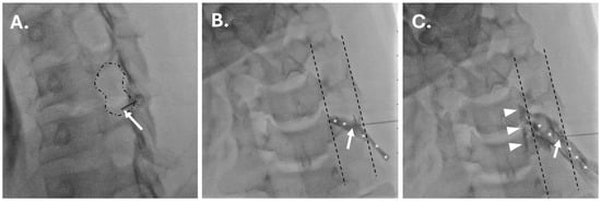
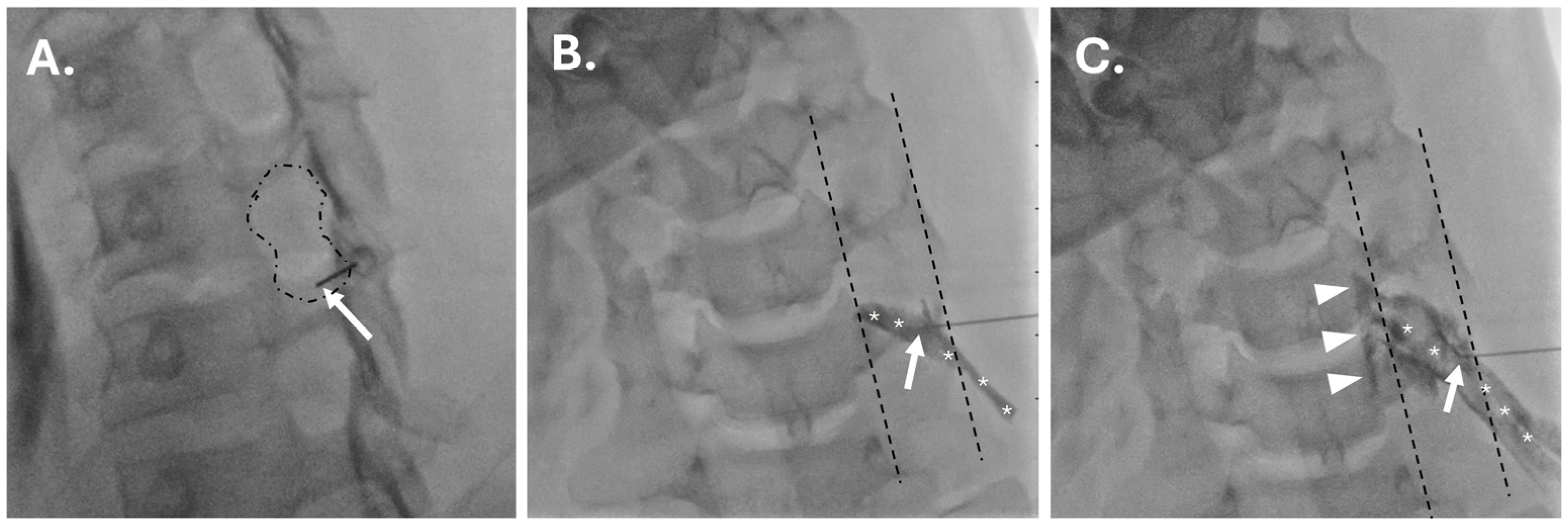
Prior to injection of perineural medications, iodinated contrast (Omnipaque™ 300, GE HealthCare, Chicago, IL, USA OR Isovue-M® 300, Bracco Diagnostic Inc., Milan, Italy) was injected through extension tubing under live fluoroscopic imaging to ensure extravascular needle placement. Live fluoroscopic imaging was also performed during the injection of medication to document the perineural and transforaminal spread of injectate and re-confirm that the injection was extravascular (Figure 1). In most cases, digital subtraction angiography (DSA) was performed.
Figure 1.
Representative images during a fluoroscopic cervical transforaminal epidural steroid injection. On the foraminal oblique projection (A), the needle (arrow) is positioned in the posteroinferior aspect of the foramen (outlined by dashed line). On the anteroposterior projection during injection of iodinated contrast (B,C), the needle is positioned just across the articular pillars (bracketed by dashes), and there is perineural flow (asterisks) and subsequently transforaminal epidural flow (arrowheads in C) of contrast without evidence of vascular uptake.
Demographic data was extracted from the EMR. Procedure-specific information, including the foraminal level and laterality of injection, as well as inclusion/exclusion of lidocaine with the corticosteroid injectate, was also retrieved from the EMR. Major complications—which we defined as immediate serious adverse events such as stroke, paralysis, death, or emergency department visit or hospital admission (within 72 h of procedure) for any neurologic presentation—were documented. Additionally, the most common minor complication, immediate post-procedural pain, was also recorded. The procedural reports consistently reported any complications (i.e., increased pain), but other qualitative or semi-quantitative information (i.e., improvement in pain, subjective procedural experience, etc.) was not consistently available for review in this retrospective study.
A Shapiro–Wilk test was performed to test for normality. Categorical variables were reported as counts (n) and percentages (%), and group comparisons were performed using Fisher’s exact test or Chi-square testing, depending on whether expected frequencies were greater than 5, as beyond this expected frequency value, a Chi-square would be preferred. Ultimately, the categorical demographic data for sex, immediate increased pain, and major complications were analyzed with Fisher’s exact test, while the laterality and level of procedure were analyzed using Chi-square testing. For the level of procedure, categories were collapsed due to small overall counts in C1–C3. Continuous variables were reported as means ± SD. A two-tailed, independent sample t-test was performed for comparison of continuous variables. A p < 0.05 was considered statistically significant. In addition to p-values, 95% confidence intervals were reported for dependent variable proportions (increased pain and major complications) and the between-group mean difference in age. Statistical analysis was performed using Stata software (v17.1, StataCorp LLC, College Station, TX, USA).
3. Results
3.1. Demographics and Sample Distribution
A total of 299 patients were included. Of these, 263/299 (88%) patients received dexamethasone without LA (controls), and 36/299 (12%) received combined dexamethasone + LA (cases). The average age at the time of injection was 55.2 ± 15.5 years for the control group and 58.9 ± 14.8 for the case group, without a statistically significant difference between the two groups (mean difference: 3.7 years; 95% CI: [−1.62, 9.02]; p = 0.079). Gender was almost evenly distributed between the two groups (controls: 47% female; cases: 52.8% females). There was a relatively even distribution of injection laterality between the two groups (controls: 58.6% right-side; cases: 44.4% right-side). Overall, there was no significant difference in the level at which the injections were performed in either group, and a majority of injections were performed between the C4–C5 and C6–C7 levels. Detailed data, including that pertaining to level-by-level injection sites, are found in Table 1.
Table 1.
Demographics and sample distribution.
3.2. Complications
There was no significant difference between the rate of increased pain immediately post-procedure or major complications between cases and controls (p = 0.799 and ≈1, respectively). Increased post-procedural pain was reported in 38/263 (14.5%; 95% CI: [10.4%, 19.3%]) of controls and 4/36 (11.1%; 95% CI: [3.1, 26.1]) of cases. Only 1 (0.4%, 95% CI: [0.01%, 2.10%]) subject in the control group met the criteria for a major complication, while no major complications were observed in the case group (0%, CI [0%, 9.74%]). The sinlge major complication manifested as severe neck pain and vague neurologic symptoms the evening after the injection. The patient was seen in the emergency department and evaluated for stroke. The work-up was negative for stroke, and the patient’s symptoms resolved (Table 2).
Table 2.
Complications.
4. Discussion
In this case–control study, we found no significant difference in the incidence of major complications or immediate post-procedural pain related to cervical TFESI performed with or without LA (1% lidocaine). To date, there is no literature to suggest an increased risk associated with the inclusion of LA with corticosteroid for cervical nerve root injections. However, there are theoretical risks associated with LA, and this has led some proceduralists to cease combining LA with corticosteroids when performing cervical TFESI. The theoretical risks of LA in TFESI are related to systemic toxicity, local neurotoxicity, or inadvertent intrathecal or subdural injection [7].
Lidocaine is often the preferred choice for cervical nerve root injections because it has a rapid onset, a relatively short duration of action, and a higher seizure threshold compared to bupivacaine [17]. To prevent direct neurotoxicity, only preservative-free LAs should be used, as common preservatives like methylparaben and sodium bisulfite are neurotoxic [18,19]. Direct neurotoxicity of lidocaine itself is a theoretical concern, though it is dependent on the dose and duration [7,17]. The exact mechanism of this neurotoxicity is not fully understood; however, with the small doses of lidocaine used (≤10 mg) for TFESI, significant nerve damage from local toxicity relating to a perineural injection is unlikely [20].
Systemic LA systemic toxicity is a well-known clinical phenomenon that can manifest as central nervous system (CNS) toxicity, cardiovascular toxicity, or both [21]. Serious CNS symptoms like seizures typically precede life-threatening cardiovascular issues, and a LA’s safety is often measured by the ratio of the dose needed to cause cardiovascular collapse versus the dose required for seizures [22]. However, this safety profile is less relevant for cervical nerve root injections due to the risk of direct injection into the vertebrobasilar arterial system, which can cause CNS toxicity at much lower doses [23]. While the exact toxic dose remains unclear and is influenced by factors like injection location and rate, studies and case reports suggest that even small amounts of lidocaine (as low as 5–10 mg) or bupivacaine (as low as 2.5 mg) injected into the vertebral artery can be enough to induce seizures [24,25,26,27,28].
Since LA doses used in cervical nerve root blocks may be close to the threshold for toxicity if the LA is inadvertently injected into the vertebrobasilar system, using lower concentrations, like 0.5–1% lidocaine, is presumably safer than using a 2% formulation. However, a study including almost 800 cervical nerve root blocks using 2% lidocaine as their LA reported no major complications [29]. Operators should always remain vigilant to stay in the posterior portion of the foramen to avoid the vertebral artery.
Even with proper needle placement, intra-arterial injection into a cervical radicular artery supplying the spinal cord may occur [10,30]. Cadaveric studies have identified such vessels supplying both the anterior and posterior spinal arteries within the posterior peripheral portion of cervical foramina [8,31]. A case of suspected radicular artery injection with 0.8 mL of 2% lidocaine resulted in transient quadriplegia, loss of pin-prick sensation, and preservation of proprioception and fine touch, suggesting anesthesia of the spinothalamic and corticospinal tracts, presumably through communication of the radicular artery with the anterior spinal artery [30]. Alternatively, if a radicular artery communicating with the posterior spinal artery is injected with LA, the neurologic deficits would be expected in proprioception and fine touch due to the effect on the posterior columns [10]. Using DSA as an adjunct to live fluoroscopic imaging may improve the detection of inadvertent injection of a radicular vessel [10].
Inadvertent subdural injection is possible when performing cervical TFESI [12]. This is because the dura mater extends laterally along the dorsal root ganglia (DRG), forming a dural sleeve [12,14,32]. The DRG is usually positioned anterosuperiorly within the foramen. Lower in the cervical spine, particularly at C6 and C7, the dural sleeve can be encountered further laterally and extra-foraminal about 50% of the time [32,33]. Generally, symptoms of subdural LA injection will present as transient sensory phenomena [7,32]. This is because the subdural space is widest dorsally and laterally, where sensory fibers predominate [12,34]. In larger volumes, beyond those that would be routinely used for a cervical TFESI, there is potential for pressure-related ischemia of a nerve root [35]. If subdural LA migrates intracranially, there is potential for serious side effects, including apnea, loss of consciousness, hypotension, bradycardia, and cardiac arrest [11,12,14,32,36].
Intrathecal injection of LA in the cervical spine can have catastrophic consequences. This will present rapid onset motor and sensory deficits and resulting respiratory/hemodynamic compromise [7]. This can only occur if the proceduralist advances the needle deep into the foramen. The needle should be placed no further centrally than 1/3 of the distance across the articular pillar on the anteroposterior projection.
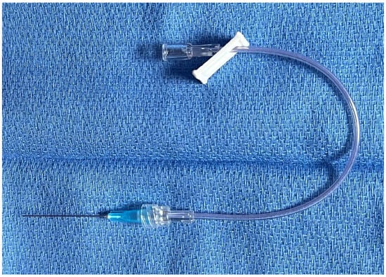
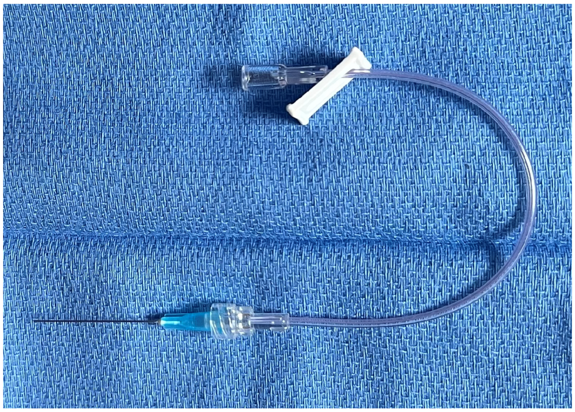
When performing cervical TFESI, risk mitigation strategies can and should be employed. In this case series, such strategies were employed for all injections. Such strategies include the use of a small gauge needle (≤25 gauge) primed with iodinated contrast; use of fluoroscopic guidance with a C-arm, allowing for orthogonal views to confirm appropriate needle positioning in the posteroinferior foramen, with the needle tip no further medial than the lateral third of the articular pillar on the anteroposterior projection; live-fluoroscopic injection of contrast prior to medication administration through extension tubing (Figure 2); and subsequent injection of therapeutic injectate through the primed extension tubing under live fluoroscopic guidance [7,10]. Consider using DSA, if available, to improve confidence of an extravascular location of the injection. When including LA as part of the injectate, one should inject slowly and ask patients if they are experiencing any sensory phenomena, such as a metallic taste or tinnitus [7,37]. Lastly, if injecting low in the cervical spine, risk of subdural injection can be minimized by accepting a more peripheral needle position on the anteroposterior projection (i.e., just lateral to the articular pillar), given the anatomic considerations of the DRG and dural sleeve at these levels [7,33].
Figure 2.
Extension tubing connected to a 25-gauge 1.5” needle.
This study is limited by operator selection bias, with operator preference determining whether or not LA was included in the injection. The decision to include LA by the operator was made almost exclusively based upon the background/training experience of the provider, with those providers who trained routinely using LA in cervical TFESI choosing to use it, and those operators that did not have such experience generally electing to withhold the LA. Other case-specific factors (i.e., patient allergies, technical considerations, injection indications, etc.) may have influenced the decision to include or exclude LA in a small minority of cases. The greatest limitation of this study is the small sample size. This limitation is exacerbated by the generally low incidence of major adverse events during these procedures [6,38]. Furthermore, only an immediate increase in pain was documented, and potential benefits of LA were not routinely available to report in this study. Similarly, the long-term efficacy of the injections in this cohort was not assessed, nor was the diagnostic value of the injection for surgical consideration.
Based on the current available literature, risks of serious adverse events with cervical TFESI are low [6,39]. Many of these risks have been posited to be related to the injection of an embolic agent, such as particulate steroid or air, or direct needle trauma to the vertebrobasilar system [7]. There are only a few case reports implicating LA as the cause of adverse events during TFESI [7]. However, these case reports do suggest that inadvertent injection of a small volume of LA into a radicular artery or into the subdural space may cause serious harm [10,12,31]. There is no existing evidence that injection of non-particulate steroid without the LA into these same spaces would cause significant injury.
Since there are potential risks with the inclusion of LA, albeit extraordinarily rare, future studies should explore the diagnostic and therapeutic benefits of including LA in these injections. Additionally, future investigations including large sample sizes are necessary to determine the true risk profile specific to the inclusion of LA with non-particulate corticosteroid for TFESI. Together, this information would allow for a more confident assessment of the risk-benefit profile of LA in cervical TFESI.
5. Conclusions
No evidence exists, including in this case–control study, to support the notion that perineural lidocaine substantially increases the risks associated with fluoroscopically guided cervical nerve root injections. However, serious adverse events are theoretically possible with the injection of LA into a radicular artery, the vertebral artery, or the subdural space. Given that such risks are not associated with the use of non-particulate steroids alone, large multi-institutional studies are needed to draw confident conclusions on the risks and benefits of the inclusion of LA with non-particulate corticosteroids for cervical TFESI to inform clinical practice.
Author Contributions
Conceptualization, Z.E.S., K.E.S., S.G., R.W.M. and J.Y.; methodology, Z.E.S. and K.E.S.; formal analysis, S.G.; investigation, Z.E.S., R.W.M., J.Y., K.E.S. and S.G.; writing—original draft, K.E.S. and Z.E.S.; writing—review & editing, K.E.S., Z.E.S., J.Y. and R.W.M.; visualization, Z.E.S.; supervision, Z.E.S.; project administration, K.E.S., S.G., J.Y., Z.E.S. and R.W.M. All authors have read and agreed to the published version of the manuscript.
Funding
This research received no external funding.
Institutional Review Board Statement
The study was conducted in accordance with the Declaration of Helsinki and approved by the Institutional Review Board of Massachusetts General Hospital (Protocol #: 2024P002293) on 23 August 2024.
Informed Consent Statement
Patient consent was waived because no identifying/personal health information was disclosed, and it would not be possible to obtain consent for all patients in this retrospective case series.
Data Availability Statement
The raw data supporting the conclusions of this article will be made available by the authors upon request.
Conflicts of Interest
The authors declare no conflicts of interest.
Abbreviations
The following abbreviations are used in this manuscript:
| TFESI | Transforaminal epidural corticosteroid injection |
| LA | Local anesthetic |
| EMR | Electronic medical record |
| CNS | Central nervous system |
| DRG | Dorsal root ganglion |
| DSA | Digital subtraction angiography |
References
- Conger, A.M.; Randall, D.J.; Sperry, B.P.; Kuo, K.T.; Petersen, R.; Henrie, A.M.; Kendall, R.W.; Bisson, E.F.; Teramoto, M.; Martin, B.I.; et al. The effectiveness of cervical transforaminal epidural steroid injections for the treatment of cervical radicular pain: A prospective cohort study reporting 12-month outcomes. Interv. Pain Med. 2024, 3, 100379. [Google Scholar] [CrossRef] [Scilit] [PubMed]
- Conger, A.; Cushman, D.M.; Speckman, R.A.; Burnham, T.; Teramoto, M.; McCormick, Z.L. The Effectiveness of Fluoroscopically Guided Cervical Transforaminal Epidural Steroid Injection for the Treatment of Radicular Pain; a Systematic Review and Meta-analysis. Pain Med. 2020, 21, 41–54. [Google Scholar] [CrossRef] [Scilit]
- Wong, J.J.; Côté, P.; Quesnele, J.J.; Stern, P.J.; Mior, S.A. The course and prognostic factors of symptomatic cervical disc herniation with radiculopathy: A systematic review of the literature. Spine J. 2014, 14, 1781–1789. [Google Scholar] [CrossRef] [Scilit]
- Scanlon, G.C.; Moeller-Bertram, T.; Romanowsky, S.M.; Wallace, M.S. Cervical transforaminal epidural steroid injections: More dangerous than we think? Spine 2007, 32, 1249–1256. [Google Scholar] [CrossRef] [Scilit]
- Hodler, J.; Boos, N.; Schubert, M. Must we discontinue selective cervical nerve root blocks? Report of two cases and review of the literature. Eur. Spine J. 2013, 22, S466–S470. [Google Scholar] [CrossRef] [Scilit]
- Stephens, A.R.; Rogerson, A.; Patel, R.K.; El-Hassan, R. Rates of complications after cervical transforaminal epidural steroid injections for patients with cervical radiculopathy utilizing a large national database. Interv. Pain Med. 2025, 4, 100606. [Google Scholar] [CrossRef] [Scilit]
- Stewart, Z.E. Safety of local anesthetics in cervical nerve root injections: A narrative review. Skelet. Radiol. 2023, 52, 1893–1900. [Google Scholar] [CrossRef] [Scilit]
- Hoeft, M.A.; Rathmell, J.P.; Monsey, R.D.; Fonda, B.J. Cervical transforaminal injection and the radicular artery: Variation in anatomical location within the cervical intervertebral foramina. Reg. Anesth. Pain Med. 2006, 31, 270–274. [Google Scholar] [CrossRef] [Scilit]
- Rathmell, J.P.; Benzon, H.T.; Dreyfuss, P.; Huntoon, M.; Wallace, M.; Baker, R.; Riew, K.D.; Rosenquist, R.W.; Aprill, C.; Rost, N.S.; et al. Safeguards to Prevent Neurologic Complications after Epidural Steroid Injections. Anesthesiology 2015, 122, 974–984. [Google Scholar] [CrossRef] [Scilit]
- Baker, R.; Dreyfuss, P.; Mercer, S.; Bogduk, N. Cervical transforaminal injection of corticosteroids into a radicular artery: A possible mechanism for spinal cord injury. Pain 2003, 103, 211–215. [Google Scholar] [CrossRef] [Scilit] [PubMed]
- Lehmann, L.; Pallares, V. Subdural Injection of a Local Anesthetic With Steroids. South. Med. J. 1995, 88, 467–469. [Google Scholar] [CrossRef] [Scilit]
- Sadacharam, K.; Petersohn, J.D.; Green, M.S. Inadvertent Subdural Injection during Cervical Transforaminal Epidural Steroid Injection. Case Rep. Anesthesiol. 2013, 2013, 847085. [Google Scholar] [CrossRef] [Scilit]
- Lubenow, T.; Keh-Wong, E.; Kristof, K.; Ivankovich, O.; Ivankovich, A.D. Inadvertent subdural injection: A complication of an epidural block. Anesth. Analg. 1988, 67, 175–179. [Google Scholar] [CrossRef] [Scilit]
- Tofuku, K.; Koga, H.; Komiya, S. Subdural spread of injected local anesthetic in a selective transforaminal cervical nerve root block: A case report. J. Med. Case Rep. 2012, 6, 142. [Google Scholar] [CrossRef] [Scilit]
- Palmer, W.E. Spinal Injections for Pain Management. Radiology 2016, 281, 669–688. [Google Scholar] [CrossRef] [Scilit] [PubMed]
- Bolger, M.P.; MacMahon, P.J.; Kavanagh, E.C. Is There a Need for Contrast and Local Anesthetic in Cervical Epidural Steroid Injections? Am. J. Neuroradiol. 2016, 37, E61. [Google Scholar] [CrossRef] [Scilit] [PubMed]
- Sakura, S.; Kirihara, Y.; Muguruma, T.; Kishimoto, T.; Saito, Y. The comparative neurotoxicity of intrathecal lidocaine and bupivacaine in rats. Anesth. Analg. 2005, 101, 541–547. [Google Scholar] [CrossRef] [Scilit] [PubMed]
- Hogan, Q. Pathophysiology of Peripheral Nerve Injury During Regional Anesthesia. Reg. Anesth. Pain Med. 2008, 33, 435–441. [Google Scholar] [CrossRef] [Scilit]
- Becker, D.E.; Reed, K.L. Local anesthetics: Review of pharmacological considerations. Anesth. Prog. 2012, 59, 90–102. [Google Scholar] [CrossRef] [Scilit]
- Guo, J.; Lv, N.; Su, Y.; Liu, Y.; Zhang, J.; Yang, D. Effects of intrathecal anesthesia with different concentrations and doses on spinal cord, nerve roots and cerebrospinal fluid in dogs. Int. J. Clin. Exp. Med. 2014, 7, 5376–5384. [Google Scholar]
- El-Boghdadly, K.; Pawa, A.; Chin, K.J. Local anesthetic systemic toxicity: Current perspectives. Local Reg. Anesth. 2018, 11, 35–44. [Google Scholar] [CrossRef] [Scilit]
- El-Boghdadly, K.; Chin, K.J. Local anesthetic systemic toxicity: Continuing Professional Development. Can. J. Anaesth. 2016, 63, 330–349. [Google Scholar] [CrossRef] [Scilit] [PubMed]
- Woo, J.H.; Park, H.S. Cervical Transforaminal Epidural Block Using Low-Dose Local Anesthetic: A Prospective, Randomized, Double-Blind Study. Pain Med. 2015, 16, 61–67. [Google Scholar] [CrossRef] [Scilit]
- Mahli, A.; Coskun, D.; Akcali, D.T. Aetiology of convulsions due to stellate ganglion block: A review and report of two cases. Eur. J. Anaesthesiol. 2002, 19, 376–380. [Google Scholar] [CrossRef] [Scilit] [PubMed]
- Guay, J. Adverse events associated with intravenous regional anesthesia (Bier block): A systematic review of complications. J. Clin. Anesth. 2009, 21, 585–594. [Google Scholar] [CrossRef] [Scilit] [PubMed]
- Lu, F.; Tian, J.; Dong, J.; Zhang, K. Tonic-clonic seizure during the ultrasound-guided stellate ganglion block because of an injection into an unrecognized variant vertebral artery: A case report. Medicine 2019, 98, e18168. [Google Scholar] [CrossRef] [Scilit]
- Kozody, R.; Ready, L.B.; Barsa, J.E.; Murphy, T.M. Dose requirement of local anaesthetic to produce grand mal seizure during stellate ganglion block. Can. Anaesth. Soc. J. 1982, 29, 489–491. [Google Scholar] [CrossRef] [Scilit]
- Digiovanni, A. Xylocaine convulsions: Analysis of two cases. Intoxication or sensitization? Anesth. Analg. 1963, 42, 355–359. [Google Scholar] [CrossRef] [Scilit]
- Pobiel, R.S.; Schellhas, K.P.; Eklund, J.A.; Golden, M.; Johnson, B.; Chopra, S.; Broadbent, P.; Myers, M.; Shrack, K. Selective cervical nerve root blockade: Prospective study of immediate and longer term complications. Am. J. Neuroradiol. 2009, 30, 507–511. [Google Scholar] [CrossRef] [Scilit]
- Karasek, M.; Bogduk, N. Temporary Neurologic Deficit After Cervical Transforaminal Injection of Local Anesthetic. Pain Med. 2004, 5, 202–205. [Google Scholar] [CrossRef] [Scilit]
- Huntoon, M.A. Anatomy of the cervical intervertebral foramina: Vulnerable arteries and ischemic neurologic injuries after transforaminal epidural injections. Pain 2005, 117, 104–111. [Google Scholar] [CrossRef] [Scilit]
- Agarwal, D.; Mohtaf, M.; Tyagi, A.; Sethi, A.K. Subdural Block and the Anaesthetist. Anaesth. Intensive Care 2010, 38, 20–25. [Google Scholar] [CrossRef] [Scilit]
- Haberberger, R.V.; Barry, C.; Dominguez, N.; Matusica, D. Human Dorsal Root Ganglia. Front. Cell. Neurosci. 2019, 13, 271. [Google Scholar] [CrossRef] [Scilit]
- Ajar, A.; Rathmell, J.; Mukherji, S. The subdural compartment. Reg. Anesth. Pain Med. 2002, 27, 72–76. [Google Scholar] [CrossRef] [Scilit]
- McMenemin, I.M.; Sissons, G.R.J.; Brownridge, P. Accidental Subdural Catheterization: Radiological Evidence of a Possible Mechanism for Spinal Cord Damage. Br. J. Anaesth. 1992, 69, 417–419. [Google Scholar] [CrossRef] [Scilit]
- Chua, K.J.; Cernadas, M. Atypical presentation of subdural block resulting in Horner’s syndrome and loss of consciousness. BMJ Case Rep. 2021, 14, e242622. [Google Scholar] [CrossRef] [Scilit] [PubMed]
- Scott, D.B. Toxic effects of local anaesthetic agents on the central nervous system. Br. J. Anaesth. 1986, 58, 732–735. [Google Scholar] [CrossRef] [Scilit]
- Wozniak, G.H.; Rana, A.A.; Stephens, A.R.; El-Hassan, R.; Laplante, B.L.; Patel, R.K. No major complications seen in a retrospective review of 1,018 cervical transforaminal epidural steroid injections. Interv. Pain Med. 2025, 4, 100622. [Google Scholar] [CrossRef] [Scilit] [PubMed]
- Beckworth, W.J.; Ghanbari, G.M.; Lamas-Basulto, E.; Taylor, B. Safety of cervical transforaminal epidural steroid injections. Interv. Pain Med. 2024, 3, 100420. [Google Scholar] [CrossRef] [Scilit] [PubMed]

Disclaimer/Publisher’s Note: The statements, opinions and data contained in all publications are solely those of the individual author(s) and contributor(s) and not of MDPI and/or the editor(s). MDPI and/or the editor(s) disclaim responsibility for any injury to people or property resulting from any ideas, methods, instructions or products referred to in the content. |
© 2026 by the authors. Licensee MDPI, Basel, Switzerland. This article is an open access article distributed under the terms and conditions of the Creative Commons Attribution (CC BY) license.