


Abstract
Background and Objectives: Despite evidence demonstrating the advantages of rheumatology-led point-of-care (POC) polarised light microscopy (PLM), it remains underutilised in many hospital settings. We aimed to identify barriers to its use and assess typical caseloads of crystal arthropathy, diagnostic methods and frequency of rheumatology-led POC PLM in practice in Ireland. We also assessed reliance on laboratory-led crystal analysis and the turnaround time for results to be reported. Solutions to improve implementation of rheumatologist-led POC PLM were also explored. Material and Methods: We surveyed 29 rheumatologists registered to the Irish Society of Rheumatology via SurveyMonkey, between May and October 2024. Textual responses were categorised into key themes, and word clouds were generated using Tagcrowd to visualise thematic content. Results: Fifty-five percent of respondents faced challenges using POC PLM for crystal analysis. Common barriers included lack of access to a microscope, lack of a polarising light function and lack of training in performing and interpreting of PLM. Fifty-two percent of rheumatologists highlighted the critical need for accessible and functional microscopes. Ninety-two percent of respondents relied on in-house laboratory crystal analysis. Thirty-two percent received inaccurate or incomplete results from laboratory reports and 28% of respondents reported delays in sample analysis from the laboratory. Laboratory turnaround was 1–2 days for 43% of rheumatologists and 3–5 days for 33%. Sixty-nine percent had not received training on PLM and 48% emphasised the need for improved education on POC PLM, including improved integration into the national rheumatology training. Conclusions: Rheumatology-led POC PLM remains underutilised despite its clinical advantage primarily due to equipment and training deficits. Addressing these barriers through improved infrastructure, education and equipment is essential to ensure effective and timely care for patients with crystal arthropathy.
1. Introduction
Calcium pyrophosphate dihydrate crystal deposition disease (CPPD) and gout are common causes of acute hot swollen joints. Accurate and timely diagnosis is critical to guide management and avoid unnecessary admissions [1]. Crystal arthropathies are important to diagnose and treat due to their impact on patient quality of life, association with cardiovascular and renal comorbidities and the significant burden they place on healthcare systems, which is expected to increase as our population ages [2,3,4].
Synovial fluid analysis using polarised light microscopy (PLM) is highly specific for the diagnosis of crystal arthropathy as outlined by both the American College of Rheumatology (ACR) and European League Against Rheumatism (EULAR) [5,6]. Gout and CPPD are both classed as crystal arthropathies; however, they are distinct diseases with unique pathophysiology and clinical courses. Accurate crystal identification, particularly for CPPD where research is rapidly evolving, is essential to support future research and improve patient care.
Point-of-care (POC) PLM enables real-time diagnosis and management decisions, facilitating safe discharge and outpatient follow up [1]. Laboratory-led crystal analysis can lead to delays and inaccuracies in crystal identification. POC PLM is recommended globally in rheumatology education frameworks and is a competency of the Irish rheumatology training scheme. Teaching on POC PLM is often offered at international rheumatology conferences [5,7]. However, the use of POC PLM by rheumatology trainees in Ireland often varies between centres. Some centres rely on laboratory-led crystal analysis while others lack access to microscopes which are required to develop and maintain proficiency in POC PLM.
Laboratories in Ireland are accredited through the Irish National Accreditation Board (INAB), which ensures compliance with internationally recognised quality and competency standards for medical testing. While accreditation ensures compliance with standard operating procedures (SOP), training and competency in PLM are often governed internally and not through formalised national training [8]. This may result in inconsistent crystal identification. While formal comparisons between rheumatologist and laboratory-led PLM are limited, the higher specialty training curriculum for rheumatology in Ireland includes PLM as a recommended competency, supported by the ACR/EULAR criteria which recommends that crystal analysis should be completed by an individual trained in the use of compensated light microscopy [9]. Rheumatologists are therefore well suited to undertake POC PLM, combining bedside fluid aspiration, microscopy and clinical interpretation for efficient patient care. Despite these advantages, uptake and practice of rheumatologist-led POC PLM remains inconsistent across Ireland.
There is limited data on the use of POC PLM, barriers to implementation and functional equipment for rheumatologists working in Ireland or elsewhere. Understanding these factors is important to improve the use of POC PLM and standardise high quality care for patients with crystal arthropathy.
2. Objectives
We aimed to identify key barriers to rheumatology-led POC PLM and assess typical caseloads of crystal arthropathy, diagnostic methods and the frequency of rheumatology-led POC PLM in practice in Ireland. We aimed to evaluate reliance on hospital-based clinical laboratories for crystal analysis and measure the typical turnaround time for results. We also aimed to explore solutions to improve the use of rheumatologist-led POC PLM in practice.
3. Methods
We surveyed 29 rheumatologists registered to the Irish Society of Rheumatology (ISR) via SurveyMonkey, a secure, GDPR compliant online platform, from May to October 2024. The survey was distributed via the ISR email list which includes approximately 150 members. A total of 29 responses were received, resulting in an estimated response rate of 19%. Question denominators varied because not all respondents completed each question.
Ethical approval was not required for this study as this was a voluntary and anonymised survey of rheumatologists. The survey consisted of 10 questions covering five domains: access to polarising microscopes, training and competency, current practice, perceived barriers to use and reliance on laboratory-led crystal analysis. Seven questions were multiple-choice and three were free-text responses. The survey was pre-tested by two rheumatology specialist registrars and two consultants to ensure validity.
Quantitative data were analysed descriptively using frequencies and proportions. The number of respondents per question ranged from 22 to 29 because some questions were not answered or contained uninterpretable responses. Analyses were conducted using the available responses for each question. All analyses were conducted in line with GDPR requirements, and no identifiable information was collected.
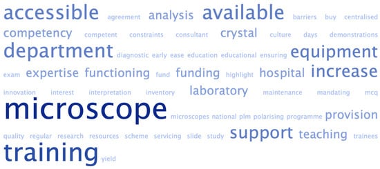
Free text responses to the question ‘What actions can be taken to encourage the use of point-of-care polarised light microscopy in clinical practice?’ were extracted, pre-processed and categorised using an inductive thematic approach. A word cloud was generated using Tagcrowd, a secure online tool, to provide a visual illustration of frequently occurring terms within the free-text responses.
4. Results
Respondents reported a mean caseload of 12.5% of patients with suspected crystal arthropathy (n = 27, range 5–70%). Fifty-five percent (16/29) of respondents faced challenges when attempting to use POC PLM for crystal analysis. Ninety-two percent (24/26) of respondents relied on in-house laboratory crystal analysis and 8% (2/26) relied on outsourced laboratory analysis. Thirty-two percent (7/22) of respondents reported receiving inaccurate or incomplete results from laboratory reports and 28% (7/25) reported delays in sample analysis from the laboratory. Forty-three percent (12/28) of rheumatologists waited 1–2 days and 33% (9/28) waited 3–5 days for laboratory results; the mean reported turnaround time for laboratory crystal analysis was 2.7 days (Table 1).
Table 1.
A summary table to demonstrate key survey findings on the use of point-of-care polarised light microscopy in the diagnosis of crystal arthropathy.
Respondents were able to select multiple barriers when asked ‘If you rarely use point-of-care polarised light microscopy, please specify the reasons why’. Twenty-three rheumatologists (79%, 23/29) answered the question, while 6 selected ‘not applicable’ (21%, 6/29); therefore, a denominator of 23 was used for this analysis. The most frequently reported barriers to rheumatologist-led POC PLM were a broken microscope (52%, 12/23), no access to a microscope (30%, 7/23) and a broken polarising light function (26%, 6/23). Training-related barriers were also selected including ‘lack of training in POC PLM’ (48%, 11/23) and ‘lack of training in interpretation of PLM results’ (39%, 9/23). Overall, 61% of respondents (14/23) identified at least one training-related barrier. Time constraints were also reported by 35% (8/23) and no respondents identified cost as a barrier (0/23, 0%).
When asked whether their department had received training in the use of POC PLM to diagnose crystal arthropathy, 69% (20/29) of rheumatologists reported that they had not received training, while 31% (9/29) reported they had. In response to the free-text question ‘What actions can be taken to encourage the use of point-of-care polarised light microscopy in clinical practice?’, 48% (12/25) of respondents identified the need for improved education on POC PLM, with one respondent highlighting the need for improved integration into the national rheumatology training programme (Figure 1).
Figure 1.
A word cloud demonstrating the frequency of themes identified in survey responses to ‘What actions can be taken to encourage the use of point-of-care polarised light microscopy in clinical practice?’. Larger words, such as ‘microscope’, ‘training’, ‘accessible’ and ‘available’ indicate the most frequently mentioned themes.
5. Discussion
This survey identified several barriers to the use of rheumatologist-led POC PLM. The most frequently reported barriers were a broken microscope (52%, 12/23) and training-related barriers (61%, 14/23), while only 31% (9/29) of rheumatologists reported having received formal training in POC PLM. These factors are closely interlinked; without access to a working polarising microscope, trainees cannot gain practical experience or teaching. Lack of a functioning microscope fosters a culture where rheumatologist-led POC PLM is deprioritised, increasing reliance on laboratory analysis and reducing opportunities for trainees to develop proficiency in POC PLM. Limited training, regulatory barriers and access to microscopy were also described by Pastor et al. who emphasised that systemic barriers limited the use of rheumatologist-led POC PLM despite it being the diagnostic gold standard. The ACR has acknowledged similar challenges, noting that regulatory issues and the costs of purchasing and maintaining microscopes often limit POC PLM, and is exploring whether rheumatologist-led use could be legitimised as a provider-performed microscopy procedure (PPMP) [10]. The presence of similar practical and regulatory barriers in the United States, where rheumatologist-led POC PLM is well established, suggests that restricted access is a global issue with implications for timely and accurate diagnoses of crystal arthropathy.
Training in PLM is an essential skill for rheumatologists, particularly for identifying CPPD crystals. CPPD crystals can be challenging to detect due to their small and scanty characteristics, weakly positive birefringence and often intracellular location, emphasising the need for training in analyses [2,9]. The literature reports poor inter-observer reliability and high false negative rates in the identification of both CPPD and MSU crystals, further highlighting the importance of skilled analysers [11,12]. The 2023 ACR/EULAR classification criteria also specifies that analysis should be completed by an individual trained in the use of PLM [9].
Our findings suggest a theory–practice gap between the curriculum expectations and trainee experience. This has been described previously by the Working Group on Training in Rheumatology across Europe, which reported that only 53% of trainees received formal education in PLM, 29% were formally assessed, and 61% reported limited experience in using PLM. This paper reported crystal analyses as one of the weakest areas of rheumatology training and associated this with poor trainee confidence [13]. In contrast, rheumatology training curriculums in Ireland, the UK and USA all emphasise the importance of learning PLM. The Royal College of Physicians Ireland (RCPI) requires trainees to ‘competently diagnose common and unusual crystals using polarising microscopy’ and lists POC PLM as a desirable skill, assessed through workplace-based assessments, ePortfolio evidence and end-of-year evaluations [7]. However, training is not standardised and inclusion in end of year exams is encouraged but not mandated.
Studies have shown that standardised training courses can improve the theory–practice gap. Berendsen et al. demonstrated that competency in crystal analysis is variable and argued that training and examination could improve identification, particularly for CPPD [11]. Lumbreras et al. reported improved inter-observer reliability, sensitivity and specificity following a focused training course [14]. The educational literature describes how repeated practice consolidates learning and competency, particularly in the medical field. Therefore, regular access to microscopes and training is essential for proficiency in PLM [15,16].
Rheumatologist-led POC PLM facilitates rapid access to crystal identification and enables same-day clinical decision making in patients presenting with crystal arthropathy [1]. In our survey, 92% (24/26) of respondents relied on the in-house laboratory, while 8% (2/26) outsourced samples to tertiary centres for crystal identification. Laboratory-led crystal analysis may be less reliable due to variable staff experience, lack of formalised training and absence of standardised accreditation in Ireland. Our survey reported a mean laboratory turnaround time of 2.7 days (n = 28); 32% (7/22) of rheumatologists reported inaccurate or incomplete laboratory results; and 28% (7/25) reported delays in laboratory sample analysis. The literature has described low inter-observer agreement, false negatives and misidentification, particularly for CPP crystals, before structured training for laboratory staff [12,14]. Laboratory analyses are further complicated by limited publicly available data on formalised training or accreditation for crystal identification in Ireland, as well as a lack of central data on technician competency or oversight into crystal identification [8]. ACR is beginning to formalise requirements of polarising light microscopes in certified laboratories; however, rheumatologist-led POC PLM often remains outside regulatory frameworks, creating further barriers to its use [10]. The high specificity of rheumatologist-led POC PLM further supports its role as a valuable tool to complement laboratory-led crystal analysis [1,17].
Limited access to microscopes (30%, 7/23), broken microscopes (52%, 12/23) and lack of polarising light function (26%, 6/23) suggest inadequate investment in the acquisition and maintenance of microscopes for rheumatologist-led POC PLM in Ireland. Although cost was not identified as a barrier by survey respondents, funding remains an important consideration for sustaining POC PLM as reflected by the frequency of equipment-related barriers. Similar concerns have been described in the United States where rheumatologist-led POC PLM is not uniformly recognised as a provider-performed microscopy procedure (PMPP), potentially limiting reimbursement, investment and training opportunities. In contrast, The College of American Pathologists has specified that laboratories performing crystal analysis must have a polarising light microscope with a compensator, reflecting an increased focus on standardisation and quality assurance in crystal identification [10]. The healthcare system in Ireland is primarily publicly funded and future investment in PLM will depend on clinical benefit as well as economic value. Statistically powered studies and cost analyses comparing rheumatologist-led, laboratory-led and dual approaches to crystal identification would be valuable to assess their impact on diagnostic accuracy and length of stay. Such data may help support the provision and maintenance of polarising light microscopes within the Irish rheumatology service.
Finally, a dual approach incorporating both rheumatologist-led POC PLM and laboratory-based crystal analysis may offer a practical solution to the barriers raised in this survey. Rheumatologist-led POC PLM facilitates rapid bedside diagnosis and provides essential hands-on exposure to trainees, directly addressing gaps in training. In contrast, laboratory-led crystal analysis may provide confirmatory testing and an additional layer of quality assurance, with potential for reciprocal audit to improve diagnostic accuracy. Rather than functioning as competing pathways, these approaches are complementary, combining the efficiency and educational value of POC PLM with the standardisation and governance of laboratory services. Implementation would require ongoing audit, structured diagnostic pathways and formalised competency frameworks.
Limitations
Sample size was limited to 29 respondents and statistical analyses were not performed, limiting generalisability. We were unable to determine the stage of training versus consultant status of rheumatologists completing the survey. Participation was voluntary; therefore response bias may have occurred. Future studies should aim to incorporate quantitative measures and larger sample sizes.
6. Conclusions
Crystal arthropathy is an important condition, projected to increasingly burden healthcare systems as the population ages [18]. Rheumatologist-led POC PLM is associated with improved diagnostic accuracy, decreased length of stay and admission avoidance for patients who present with crystal arthropathy in the ambulatory care setting [1]. Despite this, it remains underutilised due to barriers such as limited access to a polarising light microscope, broken microscopes and limited education on PLM.
To optimise patient care, a standardised approach is needed. Each rheumatology department should be equipped with a functional polarising light microscope. A standardised competency assessment, access to a working polarising light microscope and regular training for rheumatology trainees would bridge the theory–practice gap.
A collaborative training approach, involving both rheumatologists and laboratory technicians may help standardise crystal identification. Formalised targeted training for laboratory staff to recognise CPPD and MSU crystals may also improve consistency of reports. Routine audits of PLM competency would help establish barriers and guide interventions to improve laboratory-led crystal analysis. A dual approach where rheumatologist-led POC PLM is used for rapid diagnoses and laboratory analyses are used for quality assurance could improve diagnostic accuracy, support trainee education and enable continuous audits of both laboratory and clinician-led crystal analysis.
Author Contributions
Conceptualization, J.S. and G.M.; methodology, A.J.L.; software, A.J.L.; validation, A.J.L., J.S. and G.M.; formal analysis, A.J.L.; investigation, A.J.L.; resources, A.J.L.; data curation, A.J.L.; writing—original draft preparation, A.J.L.; writing—review and editing, A.J.L., J.S. and G.M.; visualization, A.J.L.; supervision, J.S. and G.M.; project administration, A.J.L. All authors have read and agreed to the published version of the manuscript.
Funding
This research received no external funding.
Data Availability Statement
Anonymised survey data supporting the findings of this study are available from the corresponding author upon reasonable request.
Conflicts of Interest
The authors declare no conflicts of interest.
References
- Lewis, A.; McCarthy, G.M.; Cowley, S.; Swift, C.; Corish, O.; Taha, K.; Abdalla, A.; Breslin, T.; Lyons, F.; Muldoon, E.; et al. Rapid access microscopy and real-time case discussion via a secure messaging app improves diagnostic accuracy and management of acute hot swollen joints. BMJ Open Qual. 2025, 14, e003417. [Google Scholar] [CrossRef] [Scilit] [PubMed]
- Pascart, T.; Filippou, G.; Liote, F.; Sirotti, S.; Jauffret, C.; Abhishek, A. Calcium pyrophosphate deposition disease. Lancet Rheumatol. 2024, 6, e791–e804. [Google Scholar] [CrossRef] [Scilit] [PubMed]
- Agudelo, C.A.; Wise, C.M. Crystal-associated arthritis in the elderly. Rheum. Dis. Clin. N. Am. 2000, 26, 527–546. [Google Scholar] [CrossRef] [Scilit]
- Yang, Y.; Liu, Z. The changing burden of gout in adults aged 70 and above based on the Global Burden of Disease 2019. Front. Public Health 2025, 13, 1455726. [Google Scholar] [CrossRef] [Scilit] [PubMed]
- Richette, P.; Doherty, M.; Pascual, E.; Barskova, V.; Becce, F.; Castaneda, J.; Coyfish, M.; Guillo, S.; Jansen, T.; Janssens, H.; et al. 2018 updated European League Against Rheumatism evidence-based recommendations for the diagnosis of gout. Ann. Rheum. Dis. 2020, 79, 31–38. [Google Scholar] [CrossRef] [Scilit] [PubMed]
- Neogi, T.; Jansen, T.L.T.A.; Dalbeth, N.; Fransen, J.; Schumacher, H.R.; Berendsen, D.; Brown, M.; Choi, H.; Edwards, N.L.; Janssens, H.J.E.M.; et al. 2015 gout classification criteria: An American College of Rheumatology/European League Against Rheumatism collaborative initiative. Arthritis Rheumatol. 2015, 67, 2557–2568. [Google Scholar] [CrossRef] [Scilit] [PubMed]
- Royal College of Physicians of Ireland. Higher Specialist Training in Rheumatology and General Internal Medicine: Outcome-Based Education Curriculum 2024; Royal College of Physicians of Ireland: Dublin, Ireland, 2024; Available online: https://www.rcpi.ie/Portals/0/Document%20Repository/Training/Higher%20Specialist%20Training/Curricula%202024/Institute%20of%20Medicine/Training_HST%20Rheumatology%20and%20General%20Internal%20Medicine%20OBE%20Curriculum%202024.pdf?ver=VrQraDL1JhUYOqtBmDpdzQ%3d%3d (accessed on 29 October 2025).
- Irish National Accreditation Board. Medical Testing Laboratories; Irish National Accreditation Board: Dublin, Ireland, 2022; Available online: https://www.inab.ie/inab-directory/laboratory-accreditation/medical-testing-laboratories/ (accessed on 29 October 2025).
- Abhishek, A.; Tedeschi, S.K.; Pascart, T.; Latourte, A.; Dalbeth, N.; Neogi, T.; Fuller, A.; Rosenthal, A.; Becce, F.; Bardin, T.; et al. The 2023 ACR/EULAR classification criteria for calcium pyrophosphate deposition disease. Ann. Rheum. Dis. 2023, 82, 1306–1314. [Google Scholar] [CrossRef] [Scilit]
- Wener, M. Considerations for Rheumatologists & ACR: Rheumatologists Performing Synovial Fluid Crystal Analysis; American College of Rheumatology: Atlanta, GA, USA, 2025; Available online: https://assets.contentstack.io/v3/assets/bltee37abb6b278ab2c/bltcc91c5767e8e4317/acr-position-statement-synovial-fluid.pdf (accessed on 29 October 2025).
- Berendsen, D.; Neogi, T.; Taylor, W.J.; Dalbeth, N.; Jansen, T.L. Crystal identification of synovial fluid aspiration by polarized light microscopy: An online test suggesting that our traditional rheumatologic competence needs renewed attention and training. Clin. Rheumatol. 2017, 36, 641–647. [Google Scholar] [PubMed]
- Schumacher, H.R., Jr.; Sieck, M.S.; Rothfuss, S.; Clayburne, G.M.; Baumgarten, D.F.; Mochan, B.S.; Kant, J.A. Reproducibility of synovial fluid analyses: A study among four laboratories. Arthritis Rheum. 1986, 29, 770–774. [Google Scholar] [CrossRef] [Scilit] [PubMed]
- Sivera, F.; Ramiro, S.; Cikes, N.; Cutolo, M.; Dougados, M.; Gossec, L.; Kvien, T.K.; Lundberg, I.E.; Mandl, P. the Working Group on Training in Rheumatology across Europe; et al. Rheumatology training experience across Europe: Analysis of core competences. Arthritis Res. Ther. 2016, 18, 213. [Google Scholar] [CrossRef] [Scilit] [PubMed]
- Lumbreras, B.; Pascual, E.; Frasquet, J.; Gonzalez-Salinas, J.; Rodriguez, E.; Hernandez-Aguado, I. Analysis for crystals in synovial fluid: Training of the analysts results in high consistency. Ann. Rheum. Dis. 2005, 64, 612–615. [Google Scholar] [CrossRef] [Scilit] [PubMed]
- Tambunan, E.H. Theory-practice gap during clinical learning: A descriptive qualitative study of nursing students’ experiences and perceptions. J. Caring Sci. 2024, 13, 74–81. [Google Scholar] [CrossRef] [Scilit] [PubMed]
- Garrood, T.; Iyer, A.; Gray, K.; Prentice, H.; Bamford, R.; Jenkin, R.; Shah, N.; Gray, R.; Mearns, B.; Ratoff, J. A structured course teaching junior doctors invasive medical procedures results in sustained improvements in self-reported confidence. Clin. Med. 2010, 10, 464–467. [Google Scholar] [CrossRef] [Scilit]
- Pascual, E.; Sivera, F.; Andres, M. Synovial fluid analysis for crystals. Curr. Opin. Rheumatol. 2011, 23, 161–169. [Google Scholar] [CrossRef] [Scilit] [PubMed]
- May, P.; Moriarty, F.; Hurley, E.; Matthews, S.; Nolan, A.; Ward, M.; Johnston, B.; Roe, L.; Normand, C.; Kenny, R.A.; et al. Formal health care costs among older people in Ireland: Methods and estimates using the Irish Longitudinal Study on Ageing (TILDA). HRB Open Res. 2023, 6, 16. [Google Scholar] [CrossRef] [Scilit] [PubMed]

Disclaimer/Publisher’s Note: The statements, opinions and data contained in all publications are solely those of the individual author(s) and contributor(s) and not of MDPI and/or the editor(s). MDPI and/or the editor(s) disclaim responsibility for any injury to people or property resulting from any ideas, methods, instructions or products referred to in the content. |
© 2026 by the authors. Published by MDPI on behalf of the Gout, Hyperuricemia and Crystal Associated Disease Network. Licensee MDPI, Basel, Switzerland. This article is an open access article distributed under the terms and conditions of the Creative Commons Attribution (CC BY) license.