


Abstract
The 1st Joint Ectopic Calcification Meeting (JECM) was held in Nancy, France on 24–26 September 2025. In response to the growing need for unified scientific dialogue on soft tissue ectopic calcification, the Joint Ectopic Calcification Meeting (JECM) brought together the communities of INTEC, ISSEC, BBC, iSCCa, and the PXE Budapest meeting. This initiative emerged from concerns over fragmentation in the field, with multiple smaller meetings diluting collaborative potential. By consolidating efforts, JECM aims to foster interdisciplinary exchange, highlight cutting-edge research, and build a flagship event for the ectopic calcification community. With over 100 participants, the inaugural meeting in Nancy marks a promising step toward a more integrated and dynamic future for the field. The abstracts of this year’s meeting oral and poster presentations are collected in this conference paper, with permission from the corresponding authors.
1. Introduction
The first Joint Ectopic Calcification Meeting (JECM) took place in Nancy, France, on 24–26 September 2025. We extend our sincere thanks to all attendees, abstract submitters, presenters, and sponsors for their active participation and support, which played a central role in the meeting’s success. The idea of JECM emerged in 2024, during the third annual INTEC meeting and the inaugural ISSEC meeting—two initiatives created to address the growing need for dedicated scientific forums on soft tissue ectopic calcification. From the outset, both meetings distinguished themselves through their high scientific quality and welcoming, interactive atmosphere. As the field expanded, however, concerns grew about increasing fragmentation with multiple small, separate meetings. In addition to INTEC and ISSEC, ectopic calcification featured at the BBC (Basic Research in Bone and Cardiovascular Biology) meeting, and more specialized gatherings such as iSCCa (International Symposium on Cardiovascular Calcification) and the Budapest Meeting on Pseudoxanthoma elasticum. Recognizing the benefits of a unified platform, INTEC and ISSEC initiated discussions to consolidate efforts. We found enthusiastic collaborators in the BBC, iSCCa, and PXE Budapest research communities, who shared our vision. The result is the first Joint Ectopic Calcification Meeting—an initiative we hope will evolve into a flagship event for the field.
The central theme of this year’s meeting was ‘Tracing Roots, Finding Remedies’, bringing together clinicians and researchers across the ectopic mineralization spectrum. This theme highlighted the dual need to deepen our understanding of the fundamental mechanisms driving rare and common forms of ectopic mineralization, while simultaneously advancing therapeutic strategies for patients.
This year, we received 77 abstracts: 22 from invited speakers, 14 selected for short oral presentations, and 41 for poster presentations. Eight ISSEC early-career researcher prizes were awarded across oral and poster categories (Figure 1).
Figure 1.
The 2025 Joint Ectopic Calcification Meeting. On site participants at the scientific meeting (top) and the winners of the presentation awards for early-career researchers (bottom).
The complete 2025 JECM program is available at www.itnintec.com. In addition to the scientific sessions, we continued the annual tradition of the INTEC workshop for early-career researchers. This year’s workshop focused on entrepreneurship in the biological and medical sciences, guiding participants through the path from discovery to start-up formation, including intellectual property, legal considerations and testimonials from scientists who have successfully founded companies or are in the process of creating them.
The meeting opened with the inaugural Pedro Valdivielso Keynote Lecture, established in memory of Pedro Valdivielso following his passing in 2024. This year’s lecture presented a European consensus on the diagnostic criteria for pseudoxanthoma elasticum (PXE). Scientific discussions throughout JECM underscored that vascular and soft-tissue calcification is a multifaceted, active process driven by disrupted phosphate–pyrophosphate balance, extracellular matrix (ECM) remodeling, inflammation, and metabolic reprogramming. Rare genetic disorders—including arterial calcification due to CD73 deficiency (ACDC), pseudoxanthoma elasticum (PXE), generalized arterial calcification of infancy (GACI), primary familial brain calcification (PFBC), Keutel syndrome and inactivating PTH/PTHrP signaling disorders (iPPSD) continue to illuminate core mechanisms, such as dysfunction of CD73, ABCC6, ENPP1, and MGP, that converge on reducing PPi availability leading to pathological mineral deposition. Imaging, particularly CT, remains central for detecting and quantifying ectopic calcification and guiding interventions. The discussed clinical implications included vascular disease, endovascular procedures, osteoarthritis, chronic kidney disease, and surgical outcomes. Advanced models such as hiPSC-derived vascular organoids and genetically engineered animals (mice, rats, Acomys, and zebrafish) are accelerating mechanistic insights and therapeutic discovery. Updates were provided on promising therapeutic avenues, including oral or systemic PPi supplementation, etidronate, ENPP1 enzyme replacement therapy (INZ-701), targeted inhibition of soluble epoxide hydrolase, antisense oligonucleotides correcting phosphate-transporter defects, and modulation of metabolic or inflammatory pathways. Patient-driven research networks and multidisciplinary collaborations are increasingly propelling translational progress. Collectively, these advances reinforce the view that calcification is not a passive by-product of disease but an active, targetable pathological process.
Our goal in establishing JECM was—and remains—to create a dynamic platform that showcases innovation, strengthens collaborations, and connects diverse expertise in this rapidly evolving area of research. With more than 100 participants, we are delighted with the success of the inaugural 2025 JECM (Figure 1). We look forward to continuing this trajectory at the 2026 JECM, which will be held in Stockholm, Sweden.
2. Pedro Valdivielso Keynote Lecture
Revisiting the Phenotype, Genotype and Diagnostic Criteria of PXE—A European Consensus
- Olivier Vanakker
- Ghent University, Ghent, Belgium
- Abstract: Pseudoxanthoma elasticum (PXE) is an autosomal recessive disorder described more than 150 years ago in which calcification and fragmentation of the elastic fibers result in a variety of symptoms which differ greatly in presentation and severity between patients. The diagnosis of PXE can, in many patients, still be made based on the clinical presentation. However, in the advent of genetic screening that followed upon the identification of the first causal gene, ABCC6, milder cases of PXE or patients with a less typical onset of disease surfaced. Moreover, it became clear in recent years that PXE has at least one allelic disorder which can also result from (identical) ABCC6 or (ENPP1) pathogenic variants, namely generalized arterial calcification of infancy (GACI). These recent evolutions lead to challenges in making the correct diagnosis in a given number of patients. As emerging possibilities to treat PXE require a definite diagnosis, a critical re-appraisal of the diagnostic criteria for PXE—taking into account new clinical, biochemical and molecular characteristics—appeared to be necessary. The REACT-PXE Consortium—gathering most of the European experts on PXE—has been established to take on this task as a first step in updating the management of PXE. In this paper, we present a European Consensus Statement on the definition and updated diagnostic criteria for PXE.
3. Invited Speaker Abstracts
3.1. Vascular Calcification and Extracellular Matrix Remodeling: A Chicken and Egg Paradox
- Cynthia ST. Hilaire
- University of Pittsburgh, Pittsburgh, USA
- Abstract: Our research into vascular calcification and extracellular matrix (ECM) remodeling began with the discovery of Arterial Calcification due to deficiency of CD73 (ACDC), a rare autosomal recessive genetic disease caused by loss-of-function mutations in the NT5E gene, which encodes the CD73 enzyme. Patients with ACDC exhibit extensive calcification in their lower-extremity arteries and small joint capsules. Histopathological examinations of affected arteries revealed fragmented and duplicated elastic fibers, strikingly similar to observations in pseudoxanthoma elasticum (PXE), a connective tissue disorder. This initial discovery revealed a critical relationship: medial arterial calcification (MAC) is characterized by the accumulation of calcium and phosphate along the elastic fibers within arterial walls. Mechanistically, CD73 deficiency leads to reduced pericellular adenosine levels, which in turn increases the activity of tissue-nonspecific alkaline phosphatase (TNAP). This elevated TNAP activity promotes calcification by reducing pyrophosphate (PPi), a crucial inhibitor of ECM calcification, into inorganic phosphate (Pi). The consistent observation of damaged and duplicated elastic lamina alongside calcification in ACDC vessels is mirrored in the Mgp-deficient mouse model where increased collagen accumulation and elastic fiber fragmentation occur where calcification is localized. These observations suggest a “chicken-and-egg” paradox: does the breakdown and remodeling of the ECM predispose the tissue to calcify, or does the calcification process actively induce and exacerbate ECM damage, or are these two processes mutually reinforcing drivers of MAC pathogenesis? This complex, often reciprocal interplay is crucial to understanding MAC, which is now recognized as a distinct and independent pathology contributing to adverse outcomes in peripheral artery disease (PAD), separate from atherosclerotic plaque.
3.2. Measuring Ectopic Calcification with Computed Tomography
- Pim A. De Jong
- University Medical Center Utrecht, Utrecht, The Netherlands
- Abstract: Ectopic calcification is common in the human body and can occur in nearly every organ or tissue. Understanding its causes and consequences, as well as improving patient diagnostics and outcomes through calcification-modifying interventions, requires reliable methods to measure ectopic calcification. This can be achieved using various in vivo non-invasive imaging techniques, among which computed tomography (CT) plays a central role. This lecture will focus on CT-based assessment of ectopic calcification. Different strategies and methods for quantification will be presented, with their respective strengths and limitations discussed. The simplest approach—visual assessment of the presence or absence of calcification—will be illustrated with examples from multiple organs. Particular attention will then be given to arterial wall and brain calcifications. Semi-quantitative visual scoring systems will be reviewed, followed by more advanced methods that combine visual/manual annotation with the computation of standardized scores, such as Agatston Units and volume scores. Finally, the presentation will highlight future directions, including fully automated calcium scoring and CT-based material decomposition, where Hounsfield Units are translated into tissue-specific compositions, similar to dual-energy X-ray absorptiometry (DEXA) of the spine or hips. While each method has advantages and limitations, for intervention studies quantitative calcium scores such as Agatston Units, volume, or mass are recommended.
3.3. Calcification and Femoropopliteal Restenosis
- Yann Gouëffic
- Vascular Center, Groupe Hospitalier Paris Saint Joseph, Paris, France
- Abstract: The most common cause of peripheral arterial disease is atherosclerosis. Despite being exposed to similar risk factors, peripheral arteries develop heterogeneous atherosclerotic lesions. Our previous work showed that carotid arteries develop predominantly lipid-rich lesions and microcalcifications, while femoral arteries develop fibrotic lesions, with extensive vascular calcification (VC) and frequent presence of osteoid tissue. These differences may have major clinical implications. Firstly, VC may destabilize atheromatous plaques and contribute to plaque rupture (5–7). Moreover, advanced and extensive VC contributes to arterial stiffness and hypertension, an important risk factor for plaque rupture. Secondly, the presence of VC may influence the technical success rate and outcomes of peripheral endovascular procedures. It is noteworthy that severe VC is often considered an exclusion criterion in femoropopliteal clinical study protocols. Moreover, balloon angioplasty of severe calcified lesions is limited by early elastic recoil and poor acute and long-term outcomes. Although nitinol stents are designed to prevent elastic recoil and constrictive remodeling, severe VC may prevent stent expansion, resulting in poorer outcomes when compared to fully expanded stents. Also, it seems that VC may influence the efficacy of drug-eluting balloons during revascularization of femoropopliteal lesions, mainly in cases of circumferential distribution. Nevertheless, few data are available to determine the influence of VC on femoropopliteal endovascular treatment outcomes.
3.4. Modeling Vascular Restructuration and Calcifications in Pediatric Chronic Kidney Disease with iPS-Derived Organoids
- Matthieu Rouleau
- Université Côte d’Azur, Nice, France
- Abstract: Chronic kidney disease (CKD) in pediatric patients is associated with a significantly elevated risk of cardiovascular disease (CVD), predominantly due to the development of vascular calcifications (VC). The vasculopathy and VC in pediatric CKD involve complex and poorly understood patho-mechanisms, including endothelial dysfunction and gene expression changes. To advance our understanding of these mechanisms and support the development of targeted therapies, there is a critical need for reliable in vitro models that mimic pediatric CKD-induced vascular pathology, at least partially enough to allow discerning single mechanisms out of a complex network on entire vessels level. We adapted, to a vascular CKD environment, a human-induced pluripotent stem cell (hiPSC)-based vascular organoid model that replicates key features of the human vasculature. To model CKD-related vascular changes, organoids were treated with pro-calcifying agents (inorganic phosphate), inflammatory cytokines (IL-1β, IL-6, TNF-α), and uremic toxins (indoxyl sulfate). Analyses included immunofluorescence for vascular markers (CD31, CD140b, αSMA), RT-qPCR to assess gene expression changes, and calcium deposits detection. The vascular organoids successfully developed key components of human vasculature, including endothelial cells surrounded by pericytes and smooth muscle cells, all embedded in a collagen rich extracellular matrix. Exposure to inflammatory cytokines (IL-6 and TNF-α) and uremic toxins (indoxyl sulfate) resulted in a marked reduction in vascular density, characterized by diminished endothelial and smooth muscle cell populations. Inorganic phosphate treatment induced additional pathological features, including changed in vessel diameter and significant calcium phosphate deposits. These findings underline molecular processes contributing to vascular remodeling and calcifications possibly reminiscent of those observed in CKD. Molecular analyses are currently ongoing to further highlight critical signaling pathways that could be considered as key players in the pediatric CKD-induced vascular changes. This study establishes a robust vascular organoid model replica/ng the vascular remodeling and calcification processes associated with pediatric CKD, representing a promising platform for exploring CKD-induced vascular molecular changes. It will allow validation/verification of advanced omics analyses (i.e., tissue transcriptomics and proteomics, as well as single-nuclei RNA sequencing) and provide mechanistic data, guiding the development of targeted therapeutic strategies for CKD-related vascular diseases.
3.5. Not All Abcc6-/- Animal Models Are Created Equal
- Koen Van De Wetering
- Thomas Jefferson University, Philadelphia, USA
- Abstract: Pseudoxanthoma elasticum (PXE) is an ectopic calcification disorder caused by the absence of ABCC6-mediated ATP release into the bloodstream. This deficiency reduces substrate availability for the ectonucleotidase ENPP1, leading to lower plasma levels of pyrophosphate (PPi), a key inhibitor of calcification. The resulting PPi deficiency drives abnormal calcium phosphate deposition in soft connective tissues. Oral PPi therapy has emerged as a potential treatment, but its efficacy remains uncertain. Most ingested PPi is hydrolyzed in the gut to inorganic phosphate, which may exacerbate calcification. Its effects on mineralized tissues like bone and teeth also remain poorly understood. Using Abcc6−/− mice, a well-established PXE model, we investigated the impact of oral PPi on calcification in skin and eyes. While PPi concentrations up to 90 mM in drinking water did not increase calcification, only extremely high doses—equivalent to an impractical 2.5 g/kg/day in humans—were effective in preventing it. These doses also led to PPi accumulation in bone, compromising bone structure and strength. Interestingly, other groups have reported efficacy at much lower doses. In my talk, I will explore possible reasons for these discrepancies and introduce more potent alternatives to oral PPi therapy with improved efficacy against ectopic calcification.
3.6. Pyrophosphate, from Bench to Bedside
- Flora Szeri
- HUN-REN RCNS, Budapest, Hungary
- Abstract: The inhibition of soft tissue mineralization is a tightly regulated process in vertebrates. Rare monogenic disorders such as pseudoxanthoma elasticum, generalized arterial calcification of infancy, cranial metaphyseal dysplasia, and hypophosphatasia highlight the profound consequences of impaired pyrophosphate (PPi) homeostasis, a key mechanism that ensures physiological mineralization in bone and teeth while preventing ectopic mineralization of soft tissues. Our recent work suggests that evolutionarily conserved transcriptional networks orchestrate the production and degradation of PPi, and that the kinetics of circulating phosphate (Pi), PPi, and alkaline phosphatase (AP) activity are central determinants of both physiological and pathological mineralization in mice and humans. Extending these mechanistic insights to cardiovascular disease, we show that systemic PPi levels and the Pi/PPi ratio are associated with vascular calcification. Importantly, altered PPi levels predispose to mineral deposition, while local increases in PPi release from calcified vascular tissue act as compensatory mechanisms limiting progression. We have also revealed that PPi levels decrease in healthy pregnancy, which may contribute to increased cardiovascular risk in multiparous women. In addition, using a novel mouse model, we found that the maternal ABCC6 knockout genotype and related decreases in PPi levels determine the extent of calcification in the offspring. These findings bridge genetic and evolutionary determinants of PPi biology with clinical observations in cardiovascular disorders. Disturbances in the kinetics of Pi, PPi, and AP activity drive not only rare calcification syndromes but also contribute to coronary artery and valvular calcification, positioning PPi metabolism as a promising focus for therapeutic innovation.
3.7. The State of Rare Disease Research Globally: Voices of the People
- Sharon Terry
- Genetic Alliance & PXE International, Washington, USA
- Abstract: Research into genetic diseases has accelerated dramatically over the past two decades, yet it remains unevenly distributed and often disconnected from the people most affected. This talk highlights the vital role of lay-led initiatives in shaping research agendas, driving discovery, and building sustainable systems of engagement. Drawing on PXE International’s experience, the presentation will trace how community-driven approaches can lead to leadership at every stage of research—engagement, recruitment, enrollment, registries, biobanks, and clinical trials. The global framework of the International Rare Disease Research Consortium (IRDiRC) has also been infused with this perspective. Case studies will include leadership of the KCNT1 clinical trial, the establishment and support of hundreds of registries and dozens of biobanks across the rare disease ecosystem, and large-scale collaborations such as the genomics study with Takeda in mucopolysaccharidosis type I (MPS1) and the Biogen study in Aicardi-Goutières syndrome (AGS). Together, these examples demonstrate how the voices of patients, families, and communities are indispensable to advancing scientific progress, ensuring equity in research, and delivering meaningful outcomes for those living with rare genetic conditions worldwide.
3.8. When Bones Form Where They Shouldn’t: The iPPSD Puzzle
- Alexandra Ertl
- Bicêtre Paris-Saclay University Hospital, Paris, France
- Abstract: Inactivating PTH/PTHrP Signaling Disorders (iPPSDs) represent a group of rare, heterogeneous but closely related conditions caused by complex genetic and epigenetic defects affecting the Gsα/cAMP/PKA signaling pathway. These disorders encompass various entities formerly known as pseudohypoparathyroidism, pseudopseudohypoparathyroidism, progressive osseous heteroplasia, and acrodysostosis. The underlying molecular alterations disrupt key components of the PTH/PTHrP signaling cascade, including the PTHR1 receptor and downstream effectors such as GNAS, PRKAR1A, PDE4D, and PDE3A. Patients with iPPSDs typically experience a wide spectrum of clinical manifestations, beginning in early childhood and often persisting throughout life. These include severe disturbances in mineral metabolism, multi-hormonal resistance, as well as growth impairment independent of hormonal status. Additional complications involve skeletal abnormalities, ectopic ossifications potentially causing significant limitations in mobility, and varying degrees of cognitive and/or psychomotor impairment. Molecular classification of iPPSDs is organized into six major subtypes (iPPSD1–6) based on the causative genetic or epigenetic alterations. The significant molecular and clinical overlap among these subtypes often complicates accurate and timely diagnosis. Therefore, a multidisciplinary approach involving endocrinology, genetics, orthopedics, neurology, and developmental specialists is essential for optimal diagnosis, management, and long-term follow-up.
3.9. Cartilage Calcification: What Role in Osteoarthritis?
- Augustin Latourte
- Lariboisière Hospital, Paris, France
- Abstract: Osteoarthritis (OA) is a chronic age-related disease characterized by progressive deterioration of joint tissue and a significant clinical burden. The epidemiological correlation between OA and the presence of calcium deposits in the cartilage (chondrocalcinosis) suggests a potential role for cartilage calcifications in the pathogenesis of OA. The presence of cartilage calcifications has been demonstrated to be associated with an increased risk of cartilage degradation; however, the most sensitive imaging techniques detect calcifications in almost all cartilage samples studied. The composition of the calcifications may be a contributing factor: while all end-stage OA cartilage contains basic calcium phosphate (BCP) calcifications at the time of knee replacement, a minority (approximately 20%) contains calcium pyrophosphate (CPP) crystals. The presence of CPP deposits may therefore constitute a specific endotype of OA. The mechanisms by which calcium crystals are thought to contribute to cartilage degradation are likely to differ depending on the type of crystal (BCP or CPP). These mechanisms are characterized by a pro-inflammatory and pro-catabolic response, along with phenotypic changes in the chondrocytes, as well as apoptosis and senescence. Consequently, the targeting of pathological calcifications in cartilage could represent a promising therapeutic approach for the treatment of OA.
3.10. Astrocyte-Modulated Phosphate Homeostasis and Antisense Oligonucleotide Therapy for Primary Familial Brain Calcification
- Xuewen Cheng
- Lin Gang Laboratory, Shangai, China
- Abstract: Primary familial brain calcification (PFBC) is a genetic-driven and aging-associated neurological disease, yet the underlying mechanism remains unclear, and no effective treatment is currently available. Here, based on the identification of two new PFBC-linked genes MYORG and CMPK2, we found that astrocytes show an enriched and polarized distribution pattern of inorganic phosphate (Pi) transporters, with importer PiT2 distributed over the entire astrocyte processes and exporter XPR1 localized to astrocyte end-feet on blood vessels. Astrocytes also exhibited higher Pi transport capacity than neurons via isotope uptake assay. These features endow astrocytes with Pi transport capacity competent for modulating brain Pi homeostasis. Moreover, in the SLC20A2 gene from six PFBC families, we identified novel intronic variants, which increased aberrant SLC20A2 pre-mRNA splicing inclusion of newly characterized cryptic exons, and ultimately caused premature termination of SLC20A2 translation. Inhibiting the cryptic-exon incorporation with splice-switching antisense oligonucleotides (ASOs) increased the expression levels of functional SLC20A2 in cells carrying SLC20A2 mutations. Moreover, intracerebroventricular administration of ASOs reduced CSF Pi levels and suppressed brain calcification in humanized SLC20A2 knockin mice. Together, our findings establish impairment of astrocyte-mediated Pi homeostasis as a key pathology of PFBC, and demonstrate ASO-mediated splice modulation as a potential therapy for PFBC patients with SLC20A2 haploinsufficiency.
3.11. Metabolic Rewiring in Vascular Calcification
- Claudia Goettsch
- TU Dresden, Dresden, Germany
- Abstract: Vascular smooth muscle cells (SMCs) play a crucial role in maintaining vascular homeostasis. In the context of fibro-calcific vascular remodeling, SMCs contribute to the formation of a collagen-rich extracellular matrix that incorporates mineral deposits, leading to the development of vulnerable atherosclerotic plaques when located in the tunica intima. SMCs exhibit a high degree of plasticity, particularly in the atherosclerotic plaque environment, where various SMC phenotypes have been identified, including osteogenic-like (calcifying) and fibroblast-like phenotypes. Metabolic reprogramming is a hallmark of cell transition and is essential for adopting different phenotypic states necessary for adaptation to the microenvironment. Our research has demonstrated that the energy metabolism and mitochondrial dynamics are altered in calcifying SMCs. Understanding the changes in cellular metabolism might be the basis of controlling or restoring the physiological phenotype of SMCs as a therapeutic avenue for preventing or reversing vascular calcification.
3.12. Musculoskeletal Pathologic Calcification: Mechanistic Insights and Emerging Therapies
- Sonia Nasi
- University Hospital of Lausanne, Lausanne, Switzerland
- Abstract: Musculoskeletal calcification affects millions of people worldwide and is a major cause of pain and disability. Calcification of cartilage and tendons shares common pathological features and underlying mechanisms. This talk will present various experimental models used to study cartilage and tendon calcification, including surgery-induced, genetic, and aging models. Furthermore, recent advances in understanding the molecular and cellular mechanisms driving these processes will be discussed. Finally, potential novel therapeutic strategies, including approaches targeting H2S pathway activation, will be highlighted.
3.13. Ectopic Calcification Associated with MGP Mutations
- Monzur Murshed
- McGill University, Montréal, Canada
- Abstract: Matrix Gla protein (MGP) is a potent inhibitor of extracellular matrix mineralization. Null mutations in MGP cause Keutel syndrome in humans, characterized by ectopic calcification of multiple cartilaginous tissues, including the trachea, nasal septum, and growth plates. In some cases, patients with Keutel syndrome also develop vascular calcification. Two sets of conserved residues—four γ-carboxylated glutamic acids and three phosphorylated serine residues—are critical for MGP’s anti-mineralization functions. Our recent work shows that these residues may have distinct roles in mediating MGP’s activity at different sites. The phosphate groups enable MGP to interact with calcium phosphate minerals in vivo. Importantly, we showed that it is the phosphate moieties themselves, rather than charge contribution from the residues or the surrounding amino acid sequence, that are required for MGP’s antimineralization function. More recently, we identified four individuals carrying dominant mutations in MGP that affect the same cysteine 19 residue. These mutations cause ectopic calcification in the growth plates of endochondral bones, but not in the nasal septum or vascular tissues. These findings suggest that the pathologies associated with the newly identified dominant MGP mutations arise through mechanisms distinct from those underlying Keutel syndrome. Collectively, our mechanistic studies shed light on MGP’s mode of action, identify the kinase responsible for its phosphorylation, and explain the skeletal anomalies associated with dominant MGP mutations.
3.14. Soluble Epoxide Hydrolase: A Janus-Faced Enzyme Regulating Cardiovascular Calcification
- Jeremy Bellien
- Rouen University Hospital, Rouen, France
- Abstract: Cardiovascular calcification is a major contributor to increased morbidity and mortality in aging and in various pathophysiological conditions, including chronic kidney disease (CKD). To date, no pharmacological treatment is available to prevent the development or progression of cardiovascular calcification. The mammalian soluble epoxide hydrolase (sEH) is a bifunctional enzyme composed of a C-terminal epoxide hydrolase domain (sEH-H) and a more recently identified N-terminal lipid phosphatase domain (sEH-P), each of which may influence cardiovascular calcification through distinct mechanisms. The sEH-H domain catabolizes biologically active epoxides of polyunsaturated fatty acids (PUFAs), including epoxyeicosatrienoic acids (EETs), which are known to promote calcification. In contrast, the biological role of the sEH-P domain remains poorly understood. However, our recent evidence suggests that its phosphatase activity may regulate cardiovascular calcification through the metabolism of pyrophosphate anions. Consistent with this, we demonstrated that genetic inactivation of sEH-P using CRISPR-Cas9 technology protects CKD rats from the development of vascular and valvular calcification, highlighting the potential of pharmacological sEH-P inhibitors as a novel therapeutic strategy for cardiovascular calcification.
3.15. Ectopic Calcifications in Digestive, Urologic, and Thoracic Surgery: Surgical Relevance Beyond the Stones
- Nicolas Berte
- CHU de Nancy—Hôpitaux de Brabois, Nancy, France
- Abstract: Ectopic calcifications encompass a broad spectrum of conditions encountered in digestive, urologic, and thoracic surgery. While frequently incidental radiological findings, they may hold important clinical significance by indicating chronic inflammation, underlying neoplasia, or potential surgical complications. This presentation will provide an overview of the most relevant entities across surgical practice. In the urologic field, renal and bladder lithiasis remain the most prevalent, whereas testicular microlithiasis continues to generate debate regarding its prognostic value. In digestive surgery, gallstones and porcelain gallbladder exemplify how calcifications can progress from common pathology to risk factor for malignancy. Appendicoliths, pancreatic calcifications related to chronic disease, and adrenal calcifications following hemorrhage or neoplasia will also be discussed. In thoracic surgery, pleural, pericardial, and mediastinal calcifications illustrate the long-term sequelae of infection, inflammation, or tumors, and highlight potential intraoperative challenges. Soft tissue and vascular calcifications further demonstrate the interplay between trauma, inflammation, and systemic metabolic disorders. By combining clinical cases and literature data, this lecture will emphasize the surgical relevance of ectopic calcifications. Far from being benign incidental findings, they frequently provide diagnostic clues, guide preoperative planning, and influence therapeutic decision-making.
4. Update on Ongoing Therapeutic Trials
4.1. The PROPHECI Trial: An Update on Oral Pyrophosphate
- Georges Leftheriotis
- Université Côte d’Azur, Nice, France
- Abstract: Pseudoxanthoma elasticum (PXE) is an incurable heritable rare disease which therapeutic options are limited to symptomatic treatments to stem the devastating effect of the ectopic calcifications in the cardiovascular, eye and dermal systems. PXE is characterized by a 50–60% decrease in the plasma level of inorganic pyrophosphate (PPi), a major physiological anti-calcifying factor. Encouraging proof of concept studies with animals PXE models and healthy volunteers have shown that the oral administration of PPi salts is able to increase PPi plasma levels. The PROPHECI trial (ie “PPI Supplementation to Fight ECtopIc Calcification in PXE”, NCT04868578) is the first phase II, double-blind, placebo-controlled and randomized clinical trial to evaluate the safety and efficacy of a daily and oral administration of PPi salts (Fe-2Na-PPi, n = 66) against placebo (n = 33) in clinically and genetically confirmed PXE adult patients (>18 and <65 yo). The trial started in 2019 and 65 patients have been included to date with 43 patients completed the full 12 months protocol. Plasma PPI as well as changes in vascular calcifications (primary endpoint assessed with non-contrast CT scan), ophthalmic, dermatologic, cardiovascular and QOL changes (=secondary endpoints) will be assessed before and after 12 months of treatment. Ancillary studies including functional imaging (i.e., PET-scans) will be performed in Angers, calcification propensity score (i.e T50, partner Ghent) and retinal calcification analysis (Bonn) will be specifically analyzed in a subgroup of these patients.
4.2. Etidronate for the Prevention of Arterial Calcification in Pseudoxanthoma elasticum
- Wilko Spiering
- University Medical Center Utrecht, Utrecht, The Netherlands
- Abstract: Pseudoxanthoma elasticum (PXE) is a genetic disease with autosomal recessive inheritance, characterized by slowly progressive ectopic calcification in the elastic fibers of the skin, Bruch’s membrane in the retina, and in the arteries. Etidronate was found to be effective to halt the progression of arterial calcification in an earlier trial including older patients with PXE with manifest arterial disease. It is uncertain if there are similar effects in younger patients with PXE. The TEMP-PREVENT study is a randomized, double-blind, placebo-controlled clinical trial, investigating the effect of 20 mg/kg cyclical etidronate on ectopic calcification in PXE patients >18 and <55 years old. The study is conducted at the University Medical Center, Utrecht, the Netherlands. The primary endpoint is the change in arterial calcification volume between baseline and 24 months in the leg arteries and carotid siphons, as measured on low-dose unenhanced Computed Tomography. Secondary endpoints are changes in dermatological, ophthalmological, vascular, laboratory, cognitive and quality of life outcomes/parameters. Recruiting started in April 2023, and results are expected in 2027.
4.3. GACI Trial Updates in ENPP1 and ABCC6 Deficient Infants
- Yves Sabbagh
- Inozyme Pharma, Boston, USA
- Abstract: Infants with ENPP1 deficiency are diagnosed with generalized arterial calcification of infancy (GACI) Type 1, while infants with ABCC6 deficiency are diagnosed with GACI Type 2. GACI Type 1 and 2 are rare and serious conditions that affect babies before or shortly after birth. In GACI, hydroxyapatite crystals build up in the walls of the arteries due to low levels of pyrophosphate (PPi) a potent inhibitor of calcification. This buildup stiffens and narrows arteries, leading to cardiovascular complications like heart failure, high blood pressure, breathing difficulties, or failure to thrive. Infants with GACI Type 1 have a poor prognosis, with approximately 50% mortality and in GACI Type 2 10% mortality before the age of 6 months despite supportive care. A strategy to increase the levels of PPi is by an enzyme replacement therapy approach with ENPP1-Fc (INZ-701). ENPP1 is the major enzyme in the body that makes PPi. We have previously shown that INZ-701 can increase plasma PPi in adults with ENPP1 or ABCC6 Deficiency. Interim data from our ongoing trials in infants with GACI Type 1 and 2 being treated with INZ-701 demonstrated improved survival, reduction in arterial calcifications, improved heart function, prevention of rickets in GACI Type 1 and a favorable safety profile.
4.4. A 12-Week PH2 Evaluation of the TNAP Inhibitor DS-1211
- Hubert Chou
- Daiichi Sankyo
- Abstract: DS-1211b is a potent small-molecule inhibitor of tissue-nonspecific alkaline phosphatase (TNAP) being developed by Daiichi Sankyo for the treatment of ectopic calcification diseases such as PXE. In this Phase 2 study, DS-1211b was evaluated in individuals with an established clinical diagnosis of PXE to assess the safety and tolerability of DS-1211b as well as the dose response by changes in the TNAP-related PD biomarkers over a 12-week treatment duration. Safety assessment included a careful evaluation on potential risks for HPP and nephrotoxicity. Extensive PK analysis was also conducted. In addition, assessments of DS-1211b on biomarkers of bone metabolism and mineralization as well as ABCC6 genotyping confirmation for PXE diagnosis were included. A total of 65 PXE participants were randomized in this study, including both younger and older individuals. These study participants have demographic and baseline characteristics reflective of the general PXE population. Adherence to study visits and protocol procedures, and medication compliance were adequate, with all participants completing the study except for 1 early withdrawal. Results on safety, tolerability, genotyping, PK assessment, and PD changes relating to TNAP inhibition will be discussed.
4.5. Effect of Lansoprazole on Inorganic Pyrophosphate Levels in Patients with Pseudoxanthoma elasticum
- Juan Luis Carillo Linaris
- University of Malaga, Malaga, Spain
- Abstract: Inorganic pyrophosphate is one of the main factors involved in ectopic calcification, and its levels are decreased in patients with pseudoxanthoma elasticum. Therefore, increasing its levels could favorably modify the natural course of the disease. In our study, we demonstrated that the administration of lansoprazole in PXE patients increases inorganic pyrophosphate levels.
5. Selected Oral Presentations
5.1. Cellular Dynamics and Heterogeneity of Cardiovascular Calcification in Zebrafish
- Anabela Bensimon-Brito
- Center for Integrative Biology, Toulouse, France
- Abstract: Cardiac valves are the structures inside the heart that ensure unidirectional blood flow. Due to the constant mechanical stress they experience, these structures are particularly prone to disease, making them a valuable model for studying cardiovascular tissue homeostasis, disease, and regeneration. A major contributor to cardiovascular dysfunction is the abnormal deposition of minerals, which leads to soft tissue calcification. While numerous factors can trigger this process, the specific cellular contributors and the signaling pathways driving it remain poorly understood. Using a genetic model for cardiovascular calcification, combined with single-cell RNA sequencing and tissue analyses, we investigated which cell populations and signaling pathways disrupt tissue homeostasis and drive soft tissue calcification. We also compared these findings with a model of cardiac valve regeneration, where cells coordinate to restore tissue structure and function, aiming to identify key differences between regenerative and pathological conditions. Prior to the onset of cardiovascular calcification, we observed increased apoptosis, immune cell infiltration, and valve thickening. We also detected a marked expansion of valve cell populations, suggesting overactivation of these cells. Single-cell analyses revealed that endothelial cells undergo apoptosis, while valve interstitial and immune cells expand and contribute to calcification through alterations in the extracellular matrix. Interestingly, immune cell populations displayed distinct transcriptional profiles between the disease and regeneration models, indicating that similar cell types can adopt divergent programs to either promote pathological calcification or support tissue repair. We are developing tools to selectively target key cell populations and assess their roles in cardiovascular calcification. Furthermore, we are leveraging the zebrafish model’s amenability to live imaging to directly visualize how distinct cellular populations coordinate to drive the calcification phenotype.
5.2. Physicochemical Characteristics of Cardiovascular Calcification Suggest Tissue- and Sex-Specific Pathological Mineralization Mechanisms
- Marta Cerruti
- McGill University, Montréal, Canada
- Abstract: During cardiovascular calcification (CC) calcium phosphate minerals deposit in heart valves, atherosclerotic plaques, or in the medial layer of blood vessels. The amount and type of minerals deposited depends on underlying disease, age, and sex of the patient. The reasons behind these differences are still poorly understood, also because most analyses rely on heavily calcified samples obtained during replacement surgery. Studying minerals in less calcified samples may help understanding calcification onset and progression, sex- and tissue-specific patterns, and the biological mechanisms underlying CC. We characterize 134 samples of aortic valve, internal carotid artery and descending thoracic aorta from 47 body donors (30 female, 17 male) using X-Ray Fluorescence (XRF), Raman, Near-Edge X-Ray Absorption Fine Structure (NEXAFS) spectroscopy, synchrotron X-Ray Diffraction (sXRD), and Scanning Electron Microscopy (SEM). All body donor samples exhibit some degree of CC because of old age. Clusters of the NEXAFS phase data correlate with the mineralization amount, showing that amorphous and poorly crystalline precursors often precede hydroxyapatite calcifications. Comparing tissues from the same donor through sXRD contrasts highly crystalline whitlockite in medial and early valvular and plaque calcifications with hydroxyapatite in late valvular and plaque calcifications. In the latter samples, Raman shows hydroxyapatite becomes more crystalline with calcification degree. XRF reveals zinc co-localizes with whitlockite, and in male aortic valves also with hydroxyapatite. We propose these mineral features are imprints from the calcification mechanism: whitlockite could be a universal byproduct of cell senescence, while hydroxyapatite in male valves might result from dystrophic calcification and in plaques and female valves from acellular precipitation. A multi-technical characterization of a large sample set from body donors paves the way to unveiling how minerals form in cardiovascular tissues and raises new questions whose answers will be key to developing patient-specific treatments.
5.3. Crosstalk Between ER Stress and the Nrf2 Pathway in Chronic Kidney Disease-Associated Vascular Calcification
- Enikő Balogh
- University of Debrecen, Debrecen, Hungary
- Abstract: Due to vascular calcification (VC), adverse cardiovascular events occur 10–20 years earlier in chronic kidney disease (CKD) patients than in the general population. Studies showed the causative role of reactive oxygen species (ROS) and endoplasmic reticulum (ER) stress in VC. Nrf2 (nuclear factor (erythroid-2 related) factor 2)-driven antioxidant signaling counteracts excessive ROS production and attenuates VC. Here, we investigated the crosstalk between ER stress and the Nrf2 pathway in VC. We triggered calcification of vascular smooth muscle cells (VSMCs) with high phosphate (Pi) and tunicamycin (Tun). VSMCs calcification was assessed by Alizarin red (AR) staining. In vivo, we induced CKD in mice by adenine + high Pi diet. To exacerbate ER stress, we injected the mice with Tun. VC was evaluated by OsteosenseTM staining. Osteogenic (alkaline phosphatase (ALP), RUNX2), ER stress (p-PERK, ATF4, CHOP, GRP78), and Nrf2 activation markers were evaluated by Western blot. Tun induced PERK phosphorylation and upregulated the expression of ATF4, CHOP, and GRP78 in VSMCs, exacerbated Pi-induced osteogenic transition and calcification of VSMCs and decreased the expression of Nrf2. Inhibition of ER stress restored Nrf2 expression. Inhibition of ER stress by 4-PBA, or knock-down of ATF4 attenuated Tun + Pi-induced VSMCs calcification. In CKD mice Tun increased the expression of ER stress and osteogenic markers, and remarkably decreased Nrf2 expression in the kidney. Tun largely increased aorta and kidney calcification in CKD mice. Our results suggest a crosstalk between ER stress and Nrf2 pathways, which plays a role in osteogenic transformation of VSMCs and VC.
5.4. Up-to-Date Knowledge of the Role of SLC20A1/PiT1 and SLC20A2/PiT2 in Mineralization and Ectopic Calcification
- Sarah Beck-Cormier
- Nantes Université, Nantes, France
- Abstract: The genes encoding the Slc20 family of transporters, Slc20a1/PiT1 and Slc20a2/PiT2, are expressed in all mammalian organs, which, for a long time, led to the proposal that they served a housekeeping function in supplying Pi to cells. However, with the exception of their very similar Pi transport function, the last 15 years have shown that these proteins are multifunctional, with cell-specific and non-redundant functions. In the field of skeletal mineralization and ectopic calcification, in vitro cell culture models have long suggested that PiT1 is a major contributor to tissue mineralization. However, with the advent of the use of genetically modified mouse models, and the identification of heterozygous pathogenic variants in SLC20A2 that cause autosomal dominantly-inherited primary familial brain calcification (PFBC), it is now recognized that SLC20A2 is the main contributor to tissue mineralization/calcification, albeit with yet unknown molecular mechanisms. The intention here is to provide an up-to-date overview of research into the roles of PiT proteins in skeletal mineralization and ectopic calcification.
5.5. Lipid Metabolism Alterations in Hereditary PPi Deficiency Syndromes: A Narrative Review of Insights and Controversies
- Robbe Derudder
- Ghent University, Ghent and Belgium
- Abstract: Soft tissue calcification is often driven by a deficiency in inorganic pyrophosphate (PPi), a key endogenous calcification inhibitor. PPi deficiency syndromes-pseudoxanthoma elasticum (PXE), generalized arterial calcification of infancy, arterial calcification due to CD73 deficiency, ankylosis, and Hutchinson-Gilford progeria syndrome (HGPS)—result from mutations in genes critical for PPi homeostasis. Though abnormalities in lipid metabolism have been implicated in all these conditions, common understanding of these abnormalities has yet to be established. We explored the evidence for a potential role of PPiregulating genes in lipid metabolic pathways, highlighting opportunities for future research and potential therapeutic interventions. A comprehensive review of published studies describing PPi deficiency syndromes was conducted, focusing on lipid metabolism alterations observed in patient samples, animal models, and in vitro cellular systems. Relevant findings were analyzed to identify shared patterns and mechanistic insights linking PPiregulating pathways to lipid metabolic processes. Altered lipid metabolism—including changes in cholesterol, triglycerides, HDL-c, and LDL-c—was observed in PXE, HGPS, and several murine models of PPi-related diseases. Despite some inconsistencies and species-specific differences, recurring evidence suggests that PPi-regulating proteins may have functional roles beyond mineralization, impacting key enzymes and signaling pathways in lipid metabolism. These lipid abnormalities appear to be syndrome-specific but share overlapping mechanistic themes, pointing toward convergent biological processes. In addition to the fact that dyslipidemia may exacerbate ectopic mineralization by promoting inflammation, oxidative stress, and endothelial dysfunction, it also suggests a broader metabolic burden beyond ectopic soft tissue calcification that may contribute to the overall disease phenotype and accelerate disease progression. As such, the connection between PPi deficiency and lipid metabolism alterations highlights a previously underappreciated dimension of ectopic calcification disorders and lipids should be taken into account in the validation of treatment options, as, e.g., demonstrated in HGPS. Irrespective of the many indications of the potential of PPi-regulating genes to disrupt lipid homeostasis, the exact mechanisms remain largely unknown, underscoring the need to further explore the mechanistic links between PPi regulation and lipid pathways. A better understanding of these interactions could open new therapeutic avenues that address both mineralization and metabolic dysfunctions in these complex diseases.
5.6. Plasma Pyrophosphate (PPi) Levels Correlate with Severity of Clinical Manifestations of ENPP1 Deficiency
- Hervé Husson
- Inozyme Pharma, Boston, USA
- Abstract: Pyrophosphate (PPi) has long been recognized as a key mediator of mineralization. ENPP1 deficiency, an autosomal recessive genetic disease, is characterized by low circulating PPi levels resulting in paradoxical mineralization consisting of extensive vascular and soft tissue calcification and hypomineralization of the bones. In some instances, heterozygous individuals, who display PPi levels 50% of normal also exhibit skeletal abnormalities such as ossification of the posterior longitudinal ligament (OPLL) and early onset osteoporosis. Other disorders characterized by low levels of PPi levels include ABCC6 deficiency, Chronic Kidney Disease, and calciphylaxis. In contrast, elevated PPi is observed in hypophosphatasia which also leads to severe skeletal disease. Despite the established connection between PPi dysregulation and pathological calcifications, there is a dearth of evidence showing a specific correlation between plasma PPi levels and clinical manifestations such as ectopic calcification and skeletal abnormalities. Previous preclinical investigations have focused on supratherapeutic doses of drugs to show overall efficacy rather than subtherapeutic doses that allow for correlation analysis between PPi and pathological mineralization. Herein, we describe correlation analysis between plasma PPi and various pathological measures in an ENPP1 deficient mouse model treated with ENPP1-Fc from 0.05 up to 1 mg/kg, a soluble form of ENPP1, or vehicle control. Increasing plasma PPi levels were inversely correlated with the level of calcification in the liver, kidney, and spleen. Increasing plasma PPi levels were positively correlated with both trabecular (Tb.N, Tb.Th.) and cortical (Ct Th.) bone parameters. Additionally, normalization of plasma PPi increased body weight. These data demonstrate that improvements in plasma PPi levels are reflective of reduction in vascular calcification and restoration of bone abnormalities. These data provide crucial evidence that maintaining PPi within the normal range is important and position PPi as a key pharmacodynamic marker of efficacy to assess enzyme replacement therapy.
5.7. Mechanisms of Age-Related Calcification in α-Klotho Mutants
- Nuno Valério-Silva
- Center for Integrative Biology, Toulouse, France
- Abstract: In this project we aim to elucidate the mechanisms of aging-related cardiovascular calcification (CVC), a hallmark of aging characterized by progressive dysfunction of arteries and cardiac valves. α-Klotho is an anti-aging hormone, and its loss leads to premature aging phenotypes. While valve calcification has been described in mouse α-klotho mutants, such has never been reported in zebrafish. Using CRISPR/Cas9, we generated a zebrafish α-klotho mutant. To investigate early triggers of ectopic calcification in the α-klotho mutants, we performed scRNA-seq and characterized tissue alterations at CVC onset. Immune infiltration was assessed by L-plastin staining, and apoptosis by TUNEL assay. Genetic celltracing experiments are being used to identify the specific cell types contributing to the calcific phenotype, and cell-specific ablation tools are being developed to test their function. The zebrafish α-klotho mutant shows progressive ectopic calcification of the outflow tract and cardiac valves, establishing, to our knowledge, the first zebrafish model of valve calcification. The scRNAseq revealed an increase in immune cells, which was further confirmed by L-plastin staining showing immune cell infiltration in the mutant valves at 4 mpf, prior to tissue calcification. An increase in apoptotic genes and a reduction in endothelial cells were also observed in our single-cell analysis, with TUNEL assays confirming apoptosis in valve endothelial cells also at 4 mpf. From 4 mpf onward, valves showed increased cell number and tissue thickening by 5 mpf, a phenotype described prior to human valve calcification. Using genetic cell-tracing tools, we identified a specific valve cell population of neural-crest origin that increases in number prior to calcification and may be driving the process. By establishing a zebrafish model of valve calcification, this work has the potential to uncover early cellular changes in α-klotho mutants, contributing to uncover key populations and mechanisms driving age-related CVC.
5.8. TNAP Dephosphorylates Phosphocholine and Phosphoethanolamine and Participates in Triglyceride Transport from the Liver to the Bloodstream
- David Magne
- Université de Lyon I, Lyon, France
- Abstract: Tissue-nonspecific alkaline phosphatase (TNAP), an extracellular enzyme, is primarily known for its role in skeletal mineralization, through the hydrolysis of inorganic pyrophosphate (PPi). It is also a promising therapeutic target in several diseases associated with ectopic calcification. We recently identified phosphocholine as a potential TNAP substrate in the liver, suggesting a participation in lipid metabolism. Here, we show that TNAP-knockout mice exhibit liver steatosis and reduced serum triglyceride levels, mirroring the effects of choline deficiency. In fasting WT mice, TNAP inhibition via SBI-425 administration leads to decreased choline levels in blood and liver. Incubating mouse or human serum with SBI-425 inhibits the dephosphorylation of phosphocholine. In human hepatocytes, TNAP inhibition blocks the hydrolysis of phosphocholine and impedes proliferation when the medium is supplemented with phosphocholine instead of choline. In vitro, TNAP hydrolyzes phosphocholine with similar efficiency than PPi. X-ray diffraction determination of TNAP’s 3D structure revealed the positioning of Pi within the active site, in which phosphocholine binds in an orientation opposite to that of PPi and interacts with different amino acids. Cryo-EM demonstrate that the TNAP inhibitor binds at the entry of the active site, preventing the dephosphorylation of both substrates. In summary, TNAP is the phosphatase enabling cellular choline uptake during fasting, participating in hepatic lipid metabolism. This newly discovered function will have to be taken into consideration in future preclinical studies and clinical trials.
5.9. Tracheal Calcification and Respiratory Dysfunction in MGP Deficiency: Molecular Insights from a Keutel Syndrome Mouse Model
- Romain Hugon
- Université de Lorraine, Nancy, France
- Abstract: Tracheal calcification is a rare condition mainly affecting elderly individuals and younger patients with Keutel Syndrome (KS), a genetic disorder caused by mutations in Matrix Gla Protein (MGP). In contrast with elderly individuals, KS patients develop respiratory complications such as airway infections and breathing difficulties. In wild-type (WT) mice, calcification is limited to cartilage rings and appears after 30 days (P30), whereas in MGP-deficient mice (Mgp-/-), it starts earlier (P14) and extends into the mucosa. We hypothesize that this mucosal calcification plays a central role in the respiratory manifestations of KS. The proposed study aims to determine how it contributes to airway pathology. Mucin levels were measured by qPCR and ELISA on bronchoalveolar lavage fluid. Mucociliary clearance was assessed using a phenol red dye velocity test. Bulk RNA sequencing was performed on tracheas from WT or Mgp-/- mice at P21 using polyA+ libraries. Differential expression analysis and gene ontology enrichment were used to identify pathways induced by MGP deficiency. qPCR validated transcriptomic findings across developmental stages. Mucosal calcifications disrupted the epithelium and were linked to increased mucin expression and reduced mucociliary clearance. Transcriptomic analysis revealed widespread gene expression changes. GO enrichment highlighted activation of inflammatory, mucin biosynthesis, lipoxygenase, ion transport, and extracellular matrix remodeling pathways. qPCR confirmed upregulation of key genes (Muc5ac, Mmp12, CtsS, Alox5, Alox5ap), linking mucosal calcification to epithelial stress and impaired function. This study identifies mucosal calcification in Mgp-/- tracheas as a possible trigger of epithelial function disruption including mucin expression and impaired mucociliary clearance. Transcriptomic and qPCR analyses highlight activation of inflammatory and lipoxygenase pathways linking MGP deficiency to epithelial stress. These findings provide new insight into KS respiratory manifestations. Future work will expand transcriptomic analysis across stages and use electron microscopy to assess ciliary ultrastructure and epithelial differentiation.
5.10. Tuning BMP-Regulated Cell Differentiation in the Aortic Media by Mutating MGP Introduction
- Kristina Boström
- University of California Los Angeles, Los Angeles, USA
- Abstract: Bone morphogenetic proteins (BMPs) are implicated in vascular development and disease. The BMPs are heavily regulated by inhibitors, including matrix Gla protein (MGP), which also protects elastin and inhibits vascular calcification (VC). We hypothesized that the BMP interference provided by MGP plays an important role in the guidance of progenitor cells in arterial wall assembly. We generated an Mgp-knockin (KI) mouse model with mutation of the conserved Pro64, essential for BMP binding. Computational analyses showed that mutation of Pro64Gly suppressed conformational changes required for BMP4 binding but did not disrupt calcium binding. The vascular phenotype of MgpKI mice was compared to wild-type and Mgp-knockout (KO) mice. Single cell RNA-sequencing (scRNA-seq) was used to identify cell composition in the vascular media. Vascular fibrosis, but not VC or premature deaths, was observed in Mgp-KI aortas. ScRNA-seq revealed enhanced myofibroblast trajectories in the Mgp-KI aortas compared to wild-type controls. Activation of both SMAD1/5/8 and SMAD2 was enhanced. BMP1/5/8 activation was located at the media-adventitia interface in Mgp-KI aortas, compared to endothelial location in Mgp-KO aortas. Furthermore, the Mgp-KI aortas did not show proteolysis of elastic laminae or endothelial-mesenchymal transitions observed in Mgp-KO aortas. High phosphate diet did not induce VC in Mgp-KI mice. However, Mgp-KI developed arteriovenous malformations in several organs. Our findings suggest that one of MGP’s function is guidance of progenitor cells as they undergo differentiation in the aortic wall, in part serving as an important BMP-trap ultimately aimed at preserving vascular integrity.
5.11. Arterial Stiffness Is Related to a Higher Risk of Cardiovascular Events in Patients with Pseudoxanthoma elasticum
- Melanie Haverkamp
- University Medical Center Utrecht, Utrecht, The Netherlands
- Abstract: Pseudoxanthoma elasticum (PXE) is a rare disease caused by pathogenic ABCC6 variants and results in arterial calcifications and increased cardiovascular risk. Validated intermediate cardiovascular endpoints are needed in clinical trials to evaluate treatments directed at reducing cardiovascular risk in PXE patients. This study aims to determine the relationship between arterial stiffness and cardiovascular events in a PXE cohort. This is a prospective cohort of patients from the Dutch UMC Utrecht Expertise Center for PXE. Arterial stiffness was measured with carotid-femoral pulse wave velocity (cfPWV) and the augmentation index (AIx). Cardiovascular endpoints of interest were cardiovascular death, cerebrovascular events, coronary events, and peripheral artery interventions. The relationship between arterial stiffness and the occurrence of cardiovascular events was analyzed with Cox proportional hazard models and adjusted for confounders. Among 390 patients (mean age 51 ± 15 years, 60% women), 45 cardiovascular events occurred during a median follow-up of 6.1 years (IQR 3.2;9.2). Cerebrovascular events were the most frequently observed (N = 32, 71%). A 1 m/s higher cfPWV was related to a higher risk of cardiovascular events (hazard ratio (HR): 1.24; 95%CI 1.03–1.49). The effect of cfPWV depended on age (p-value for interaction 0.03), with lower cardiovascular risk at older ages (HR at 40 years: 1.45; 95%CI 1.07–1.97, and HR at 60 years: 1.09; 95%CI 0.97–1.23). A 10% higher AIx at baseline was related to future cardiovascular events (HR 1.37; 95%CI 1.03–1.83). No significant interaction between the AIx and age was found. Arterial stiffness, measured by cfPWV and AIx, is independently related to a higher risk of cardiovascular events in patients with PXE. Measures of arterial stiffness may serve as an intermediate endpoint in clinical studies evaluating the effect of treatments on the risk of cardiovascular events in PXE patients.
5.12. Adenine Base Editing to Correct Pathogenic ABCC6 Variants Causing PXE
- Qiaoli Li
- Thomas Jefferson University, Philadelphia, USA
- Abstract: Pseudoxanthoma elasticum (PXE) is an autosomal recessive disorder caused by pathogenic variants in ABCC6, an efflux transporter primarily expressed in hepatocytes. The c.3490C > T (p.R1164X) variant is the second frequent nonsense variant in ABCC6 and is potentially amenable to adenine base editing. We aim to develop a liver-targeted adenine base editing therapeutic for PXE. To assess adenine base editing in vitro, we generated HuH-7 cells carrying the R1164X variant. We screened various adenine base editors (ABEs) combined with guide RNAs (gRNAs) in this cell line. The off-target editing was assessed by ONE-seq and targeted amplicon sequencing. To minimize off-target editing, we systematically screened hybrid gRNAs with DNA nucleotide substitutions. For in vivo evaluation, we formulated lipid nanoparticles (LNPs) with ABE mRNA and either standard or hybrid gRNA and administered them to humanized R1164X mice. We examined the corrective efficiency, ABCC6 expression in the liver, plasma PPi concentrations, and ectopic calcification in these mice. Screening various ABEs and gRNAs in vitro, we identified ABE8.8 with gRNA targeting R1164X adenine at position 4 of its spacer sequence as the most efficient editor. Hyb18, a hybrid gRNA with DNA substitutions at positions 5, 6, and 7 of the spacer sequence, reduced off-target and bystander editing while increasing on-target efficiency. Homozygous R1164X mice displayed features of the Abcc6 knockout mouse model of PXE, including low plasma PPi levels and progressive calcification in the muzzle skin. When ABE mRNA was formulated in LNPs for liver-directed correction, the hybrid gRNA restored ABCC6 expression in the liver, elevated plasma PPi levels, and almost completely prevented ectopic calcification in the humanized R1164X mice. Adenine base editing efficiently corrected the R1164X variant in HuH-7 cells and variant-humanized mice. Liver-targeted variant correction by genome editing may offer a one-and-done therapeutic approach for PXE.
6. Selected Poster Presentations
6.1. Daproductstat Promotes Soft Tissue Calcification Through the Interaction of HIF-1 Pathway and Er Stress
- Andrea Tóth
- University of Debrecen, Debrecen, Hungary
- Abstract: Daprodustat (DPD) is a prolyl hydroxylase inhibitor that enhances erythropoiesis through the activation of the HIF-1 pathway. DPD is an FDA-approved anemia treatment in chronic kidney disease (CKD) patients. Using adenine-induced CKD mice model we showed earlier that DPD corrects anemia but increases CKDassociated aorta calcification. HIF-1 activation interacts with endoplasmic reticulum (ER) stress, and because ER stress is a known trigger of vascular calcification our aim here was to investigate the potential contribution of ER stress to the pro-calcification effect of DPD. Calcification of human aortic smooth muscle cells (HAoSMCs) was triggered by inorganic phosphate (Pi) with or without DPD. CKD was induced with an adenine + high phosphate diet. Calcification was detected by Osteosense and Alizarin red staining. Protein expressions were detected by Western blot. Gene silencing was used to down-regulate HIF-1 and activating transcription 4 (ATF4) expression. DPD induced HIF-1 activation and ER stress and promoted osteo/chondrogenic differentiation of HAoSMCs. Inhibition of ER stress or silencing of HIF-1 or ATF4 resulted in decreased calcification. DPDinduced ATF4 expression was abolished in the absence of HIF-1α. However, knock-down of ATF4 did not influence the expression of HIF-1α. DPD increased the expression of hypoxia, ER stress and osteo/chondrogenic markers in the kidney and aorta, and accelerated aorta and kidney calcification in CKD-mice. DPD accelerates calcification of HAoSMCs through HIF-1/ATF4 axis and increases kidney and aorta calcification in mice with CKD. Targeting the HIF-1/ATF4 axis could be a novel approach to inhibit CKDassociated soft tissue calcification.
6.2. CALCIPATH: A Web-Based Variant Annotation Resource for Improved ABCC6 Classification in Pseudoxanthoma elasticum
- Bence Blaha
- Semmelweis University, Budapest, Hungary
- Abstract: Pseudoxanthoma elasticum (PXE) is a rare autosomal recessive disorder (prevalence 1:25,000–1:50,000) caused by mutations in the ABCC6 gene. It manifests as ectopic calcification in elastic fiber-rich tissues such as the skin, eyes, and cardiovascular system. Although numerous ABCC6 variants have been identified, distinguishing pathogenic from benign variants remains a major diagnostic challenge. To support variant classification, the Sherloc framework—a point-based decision tree categorizing variants from benign to pathogenic—was previously optimized for PXE in 2021, encompassing 286 ABCC6 variants obtained partially through literature and database screening. We now present an updated version of this classification using the curated genetic data from the largest European PXE cohort (590 patients; Szeri et al., 2022). The updated classification integrates recent evidence and refined criteria into the Sherloc framework. A key feature is a publicly accessible, searchable web server designed to host the updated classification system. The tool includes modified decision-tree steps and is intended to undergo regular data-driven updates. This resource enhances clinical interpretation of ABCC6 variants in PXE. By providing an accessible, standardized, and continuously updated platform, it supports both researchers and clinicians in understanding variant pathogenicity and improving PXE diagnostics.
6.3. Pomegranate Polyphenols Inhibit Extracellular Matrix Mineralization in a Model of Valvular Calcification
- Carme Ballester-Servera
- University of the Balearic Islands, Palma, Spain
- Abstract: Inflammation and oxidative stress are central drivers of cardiovascular calcification, a key predictor of cardiovascular morbidity and mortality. Pomegranate (Punica granatum L.) extracts, rich in polyphenolic compounds, exhibit antioxidant, and anti-inflammatory effects that may counteract these pathogenic processes. This study aimed to evaluate the effect of purified pomegranate extract (PPE) on in vitro valvular calcification. PPE was obtained from whole fruits using microwave-assisted hydrodiffusion and gravity extraction, followed by purification via AmberLiteTM XAD7HP resin chromatography. Its chemical profile was analyzed by HPLC-DAD. Primary human valvular interstitial cells (VICs) were cultured in osteogenic medium (OM) to induce calcification. After 10 days, extracellular matrix mineralization was quantified by ICP-OES and normalized to total protein. Gene expression of calcification, inflammation, and oxidative stress markers was evaluated by RT-qPCR at multiple time points (days 1, 3, 6, 8, and 10). VICs were also treated with PPE or individual compounds (cyanidin 3-glucoside, caffeic acid, catechin, and gallic acid) in OM to assess effects on calcium deposition. Chemical analysis of PPE identified high levels of ellagitannins (20.7 ± 0.4 g/100 g), anthocyanidins (10.1 ± 0.2 g/100 g), phenolic acids (6.7 ± 0.1 g/100 g), and flavan-3-ols (5.3 ± 0.2 g/100 g). VICs cultured in osteogenic medium (OM) showed markedly increased extracellular calcium deposition (~50 μg Ca/mg protein) compared to control (~5 μg Ca/mg protein). After 10 days, OM-treated VICs exhibited an upregulation of calcification markers (RUNX2, osteopontin, BMP2), inflammatory cytokine IL-6 and redoxrelated enzymes (SOD2, GPX). PPE treatment (30 μg/mL) significantly reduced calcium accumulation by 94%. Individual polyphenols—cyanidin 3-glucoside (10 μM), caffeic acid (3 μM), and gallic acid (10 μM)— also decreased mineralization, though to a lesser extent (35, 40, and 34%, respectively). PPE demonstrated potent inhibition of calcification in VICs. The superior efficacy of the whole extract over isolated components suggests synergistic interactions. These findings support further investigation of PPE as a candidate to modulate valvular calcification.
6.4. Refinement of a Rat Model of Chronic Kidney Disease for Testing Therapies Against Cardiovascular Calcifications
- Lucie Hénaut
- Université of Picardie Jules Verne, Amiens, France
- Abstract: Chronic kidney disease (CKD) induces phosphocalcic and metabolic disorders that promote vascular and valvular calcifications, as well as bone abnormalities, which together contribute to increased cardiovascular risk. Due to physiological differences between vascular and valvular cells, these calcifications are typically studied in separate models. Moreover, preclinical studies rarely assess how therapies targeting cardiovascular calcification affect bone, limiting their translation to clinical use due to potential bone toxicity. This study aimed to develop a reliable animal model that replicates CKD-associated valvular and vascular calcifications, as well as bone remodeling abnormalities, in order to evaluate the efficacy of new therapies on cardiovascular calcifications and their safety on bone. Thirty-eight male Sprague Dawley rats (8 weeks old) were divided into two groups: a control group receiving a standard diet (CT, n = 17), and a group receiving a diet enriched with 0.3% adenine to induce CKD and 1.2% phosphate to increase calcium-phosphate product (AP, n = 21) for 9 weeks. The diet included 20% lactose and 19% casein to enhance intestinal calcium and phosphate absorption. Blood samples and echocardiography were performed at baseline (week 0), and at weeks 5 and 9. Cardiovascular calcifications were assessed using Alizarin Red staining and O-cresolphthalein complexone assay. Bone microarchitecture was evaluated by micro-computed tomography. AP rats showed progressive increases in serum creatinine, calcium-phosphate product, and uremic toxins, along with marked aorta and aortic valve calcifications at week 9. By week 5, AP rats exhibited decreased cardiac output and increased isovolumic relaxation time, suggesting diastolic dysfunction. Tibias from AP rats showed reduced trabecular and cortical bone volume and trabecular number, along with increased trabecular spacing and cortical porosity. This model, which induces severe CKD associated with cardiovascular calcifications and major bone alterations, will soon serve as a platform for identifying novel drug candidates.
6.5. Cellular Senescence in Pseudoxanthoma elasticum
- Robbe Derudder
- Ghent University, Ghent, Belgium
- Abstract: Pseudoxanthoma elasticum (PXE) is a rare hereditary disorder caused by bi-allelic ABCC6 mutations, leading to pathological calcification in the skin, arteries, and eyes. Increasing evidence suggests that cellular senescence—a state of irreversible cell cycle arrest triggered by DNA damage, oxidative stress, and telomere shortening—plays a key role in PXE pathophysiology. Senescent cells contribute to tissue dysfunction through the senescence-associated secretory phenotype (SASP), which promotes chronic inflammation and extracellular matrix (ECM) remodeling. NF-κB, a central regulator of SASP, accelerates senescence, while IL-6 amplifies pro-inflammatory signaling and exacerbates disease progression. Furthermore, excessive activation of the DNA damage response (DDR) may further drive pathological changes in PXE. This study investigates the roles of NF-κB, IL-6, and DDR activation in PXE to identify potential therapeutic targets. Skin tissue (n = 4) and dermal fibroblasts (n = 9) from PXE patients and matched controls (n = 9) were analyzed. p-IκBα was assessed via immunofluorescence, and calcium deposits were visualized using Von Kossa staining. RT-qPCR was used to evaluate differential gene expression of NF-κB, IL6, DDR targets like PARP1 and downstream targets. p-IκBα was highly expressed in PXE skin, co-localizing with osteogenic markers and calcium deposits. PXE fibroblasts showed increased NF-κB, PIK3CA, AKT1, PARP1, ATM, p21, p53, RUNX2 and IL6 mRNA levels, while SIRT1 mRNA levels were decreased. Disrupted pathways in PXE, including DNA damage response, PARP1 activation, and NF- κB upregulation, link PXE to cellular senescence. Targeting senescence pathways and SASP may provide new therapeutic strategies to slow disease progression and improve patient outcomes.
6.6. Variations in Urate, Pyrophosphate, Phosphate and Calcium Ion Concentrations in Osteoarthritis and Crystal Deposition Diseases
- Christophe Duranton
- University Côte d’Azur, CNRS, LP2M, Nice, France
- Abstract: Gout (monosodium urate [MSU] crystal deposition), calcium pyrophosphate deposition disease (CPPD), and osteoarthritis (OA) are the primary adult arthropathies responsible for joint swelling and pain. Synovial fluid (SF) analysis plays a central role in diagnosing these diseases, though it is typically limited to visual crystal identification and assessment of cellularity. This study aimed to characterize the urate, pyrophosphate (PPi), phosphate, and calcium concentrations in SF samples from patients with MSU, CPPD, or OA, and to evaluate the diagnostic value of these measurements. A total of 74 SF samples were analyzed: 35 from OA patients, 17 from gout (MSU) patients, and 22 from CPPD patients. Samples were collected since 2020 by Prof. H.K. EA at the Rheumatology Department of Lariboisière, Hospital in Paris as part of routine care. Ion chromatography was used to quantify the concentration of each ion. Key findings include: • Urate: Significantly elevated in MSU patients (506 ± 79 µM; range: 76–1165 µM) compared to OA (265 ± 35 µM; range 42–705 µM) and CPPD (225 ± 53 µM; range 43–638 µM), consistent with MSU crystal presence in gout. • PPi: Highest in CPPD patients (5.2 ± 0.9 µM; range: 1.8–15.1 µM), aligning with the pathophysiology of chondrocalcinosis. OA patients showed unexpectedly variable and elevated PPi levels (6.2 ± 0.8 µM range: 0.2–13.2 µM), while MSU patients had the lowest concentrations (2.0 ± 0.4 µM, range 0.2–4.3 µM). • Phosphate: Concentrations varied across all groups, with means of 0.76 ± 0.05 mM (OA), 0.72 ± 0.06 mM (MSU), and 0.89 ± 0.05 mM (CPPD) with no significant intergroup differences. • Calcium: Levels were consistently lower than plasma values (~1.2 mM), averaging around 0.24 ± 0.05 mM across all groups, with no significant intergroup differences. This study demonstrates that ion chromatography can reveal distinct biochemical profiles in synovial fluid associated with different arthropathies. Elevated urate and PPi concentrations are particularly indicative of gout and CPPD, respectively, supporting their diagnostic relevance. The variability in PPi and phosphate levels in OA patients warrants further investigation. Overall, expanding SF analysis beyond crystal visualization to include ion profiling may enhance diagnostic accuracy and deepen understanding of joint disease pathophysiology.
6.7. Sex Differences in Warfarin-Induced Vascular Calcification and Extracellular Matrix Gene Expression in Rats
- Maria Del Mar Pérez-Ferrer
- University of the Balearic Islands, Palma, Spain
- Abstract: The warfarin-induced rat model is used to study ectopic calcification and bone pathophysiology. However, most studies focus exclusively on males, neglecting potential sex-specific responses. Given the influence of sex hormones on mineral metabolism, this study aimed to establish a warfarin induced vascular calcification model in Wistar rats, emphasizing sex- and time-dependent effects. Specifically, cardiovascular calcification progression and extracellular matrix gene expression were evaluated after 6 and 8 weeks of exposure. Thirty-six Wistar rats (18 males, 18 females; 225–250 g) were used. Control animals received a standard diet for 6 or 8 weeks (n = 3/sex/timepoint). The remaining 24 rats were fed a diet containing warfarin (0.32%) and low vitamin K1 (0.15%) for 6 or 8 weeks (n = 6/sex/timepoint). Calcium levels in the aorta, carotid arteries, and heart were quantified using inductively coupled plasma optical emission spectrometry. Expression of collagen type I alpha 1 (Col1a1), collagen type III alpha 1 (Col1a3), and plasminogen activator inhibitor-1 (PAI-1) in femoral arteries was analyzed by qRT PCR. Warfarin-treated males showed marked calcium accumulation in the aorta (5 to 10-fold), carotids (20-fold) and heart (3–4-fold), with no progression between weeks 6 and 8. Females showed only modest increases, mainly in the aorta at week 8 (2-fold). Col1a1 and Col1a3 were upregulated in both sexes but more strongly in males (50-fold and 25-fold, respectively). Female expression peaked at week 6 (around 20-fold) and declined by week 8 (10-fold). PAI-1 was upregulated only in males (28-fold at week 6, decreasing to 16-fold at week 8). This study revealed significant sex- and time-dependent differences in warfarin-induced vascular calcification. Males were more susceptible, with stronger and sustained gene expression responses, while females exhibited a milder, transient effect. These findings underscore the need to include both sexes in preclinical calcification studies.
6.8. Peripheral Arterial Disease in Pseudoxanthoma elasticum: Diagnostic Accuracy of the Six Minute Walking Test and WELCH Questionnaire
- Melanie Haverkamp
- Utrecht University, Utrecht, The Netherlands
- Abstract: Pseudoxanthoma elasticum (PXE) is a rare disease characterized by ectopic calcifications and increased risk of cardiovascular disease, including peripheral arterial disease (PAD). While PAD is prevalent in PXE, the diagnosis of PAD with current diagnostic tests is challenging, given extensive arterial calcifications. The WELCH questionnaire and the six-minute walking test (6MWT) may offer simple screening and diagnostic opportunities. This study evaluates walking capacity in PXE patients with the 6MWT compared to healthy references and assesses the diagnostic accuracy of the WELCH questionnaire and 6MWT compared with a resting ankle-brachial index (ABI) or treadmill test (TT) in a PXE population. Patients originate from the Dutch UMC Utrecht Expertise Center for Pseudoxanthoma elasticum. During regular examinations, patients underwent a TT, a 6MWT, and completed the WELCH questionnaire. The 6MWTs were compared with healthy reference values using equations that accounted for age, sex, and body mass index. The diagnostic accuracy of the 6MWT and WELCH was assessed by calculating diagnostic accuracy measures using the rest ABI (≤0.9) or a treadmill test (>20% decline in ABI after TT) as the reference standard. A total of 162 patients were included (mean age 50 ± 14 years and 52% females). Stratified analyses showed that for females and males, the observed walking distance was 533 ± 96 m and 558 ± 106 m, which is less than their expected walking distance for females (−32 m; 95%CI [−50;−13]) and males (−69 m; 95%CI [−93;−44]), respectively. The 6MWT had an area under the curve (AUC) of 0.50 (95%CI 0.37–0.63) and the WELCH questionnaire an AUC of 0.64 (95%CI 0.52–0.76) for discriminating between PXE patients. Patients with PXE have a lower walking capacity than healthy individuals. However, the 6MWT and WELCH are unreliable for diagnosing PAD. The treadmill test is recommended for diagnosing PAD in PXE patients.
6.9. Pseudoxanthoma elasticum: The Diagnostic Accuracy of Genetic Testing Compared to Clinical Criteria in a Large Cohort
- Melanie Haverkamp
- Utrecht University, Utrecht, The Netherlands
- Abstract: Pseudoxanthoma elasticum (PXE) is a rare disease caused by pathogenic ABCC6 variants, leading to ectopic calcifications in the retina, skin, and arteries. While current diagnostic criteria combine clinical and genetic findings, genetic testing alone could simplify the diagnostic process. This simplified method may miss patients due to false-negative results or rare genetic variants. The diagnostic accuracy of genetic testing alone has not been systematically evaluated, and its impact on PXE diagnoses remains unknown. This study assesses the diagnostic accuracy of solely a genetic test in a large nationwide population referred for suspected PXE. Patients suspected of having PXE were derived from the UMC Utrecht Expertise Center for PXE. Data on clinical and genetic involvement were collected from medical records. Diagnostic accuracy was evaluated using sensitivity, specificity, positive predictive value (PPV), and negative predictive value (NPV), with combined criteria as the reference standard. The combined criteria included skin, ophthalmological and genetic involvement. A total of 458 patients (median age 40 years [IQI 21;53], 62% women) were included. Patients experienced skin abnormalities (n = 350, 76%) and angioid streaks (n = 380, 83%). Of the 458 patients, 403 (88%) were diagnosed with PXE using the combined criteria, while 321 (70%) were diagnosed with PXE by genetic testing alone. The genetic test showed a sensitivity of 79% (95%CI 75–83), specificity of 98% (95%CI 90–100), a PPV of 100% (95%CI 98–100), and a NPV of 39% (95%CI 35–44), respectively. This study found that the diagnostic accuracy of a genetic test alone for diagnosing PXE is suboptimal, primarily due to its inability to reliably exclude PXE. Nearly 20% of patients lacked two pathogenic variants, but had clinical characteristics and cardiovascular history comparable to patients with confirmed genetic diagnoses. These findings support using diagnostic criteria that integrate both clinical and genetic information.
6.10. Tetracyclines Inhibit Ectopic Mineralization in a Murine Model of Neurogenic Heterotopic Ossification
- Eszter Kozak
- HUN-REN RCNS, Budapest, Hungary
- Abstract: Neurogenic heterotopic ossification (NHO) is a rare and difficult to treat complication of severe combined injuries affecting the central nervous system and peripheral (typically muscle or connective) tissue. This complex pathological process results in the formation of mature bone at the site of injury through a poorly understood mechanism. Tetracyclines are a class of antibiotics with diverse biologic effects. In addition to their antimicrobial properties, they have also been shown to inhibit ectopic mineralization through a mechanism involving the inhibition of poly-ADP-ribose polymerase (PARP). We have aimed to investigate the effectiveness of tetracycline treatment in a murine model of NHO, which consists of a central nervous system injury of concussion trauma, and additional peripheral muscle damage induced by cardiotoxin injection to the tibialis anterior. Tetracycline and minocycline were given as a single injection after combined trauma, and treatment effect was assessed after 5 days. Ectopic mineralization was quantified by extracting tissue calcium content, and in situ visualization with Alizarin Red staining. Quantitative PCR was performed to shed light on the mechanism of treatment effect. Tetracycline and minocycline both inhibited ectopic mineralization in a dose-dependent manner. Delayed treatment resembling application in a prophylactic setting did not decrease treatment effect. Gene expression of Runx2, which plays a central role in osteoblast differentiation, was shown to be decreased by tetracycline and minocycline treatment. Tetracyclines may provide an alternative to established preventive treatments of HO, which include radiotherapy and non-staroid anti-inflammatory drugs.
6.11. The Response of Pseudoxanthoma elasticum Fibroblasts to Pro-Osteogenic Stimuli
- Francesco Demetrio Lofaro
- University of Modena and Reggio Emilia, Modena, Italy
- Abstract: Ectopic calcification occurs in various physio-pathological conditions (e.g., aging, diabetes, and genetic diseases), often compromising both life quality and expectancy. Despite extensive research and the development of different in vivo and in vitro models, the molecular mechanisms driving pathological calcification are still unclear. This study investigates by a proteomic approach and functional tests the response to calcifying medium (CM) of human dermal fibroblasts isolated from patients affected by Pseudoxanthoma elasticum (PXE), a genetic disease characterized by ectopic calcification. Dermal fibroblasts from 5 PXE patients were cultured in standard medium (DMEM) or in CM (10 mM betaglycerophosphate, 50 μm/mL ascorbic acid and 100 nM dexamethasone) for 21 days. Proteins were extracted in RIPA buffer and processed using filter-aided sample preparation for LC-MS/MS analysis. Samples were injected in Ultimate 3000 system coupled to Exploris 480 and acquired in data-independent mode (DIA). A dedicated DIA library was built following gas-phase fractionation method. Mito stress test was applied to measure parameters of mitochondrial function. Exposure to CM led to significant changes in the expression of 623 out of 4212 identified proteins. Differentially expressed proteins were associated with mitochondrial structure and function, calcification process (e.g., phosphatases and tissue-nonspecific alkaline phosphatase), regulators of oxidative stress and several components of elastic fibers including those involved in the microfibrillar scaffold. Exposure to CM induces a significant proteomic shift in PXE fibroblasts, altering phenotypic traits and mitochondrial processes, along with extracellular matrix and elastic fibers remodeling. These findings deepen our knowledge on the pathways activated in a pro-calcifying environment and underline the importance of mesenchymal cell metabolic behavior and of altered matrix homeostasis in increasing the susceptibility to calcium deposition.
6.12. PXE Fibroblasts Are Highly Responsive to Pro-Osteogenic Stimuli: A Proteomic Approach
- Francesco Demetrio Lofaro
- University of Modena and Reggio Emilia, Modena, Italy
- Abstract: Ectopic calcification is an unsought complication of several acquired and genetic conditions; however, no effective treatments have been found so far due to complexity of pathogenic mechanisms and the lack of suitable identified targets. Cell cultures are frequently used to investigate the effect of pro-/antiosteogenic stimuli, and it has been demonstrated that mesenchymal cells (e.g., fibroblasts) undergo calcification only in the presence of specific media, although the most responsive pathways are not clear. By a proteomic approach, we have investigated the early response to pro-osteogenic stimuli in fibroblasts from healthy individuals (Crt) and from patients affected by the rare calcifying disease Pseudoxanthoma elasticum (PXE). Dermal fibroblasts from 5 Ctr and 5 PXE patients were cultured in standard (DMEM) or in calcifying (CM) media (supplemented with 10 mM β-glycerophosphate, 50 μm/mL ascorbic acid and 100 nM dexamethasone) for 24 h. Proteins were extracted in RIPA buffer and processed using filter-aided sample preparation for LC-MS/MS analysis. Samples were injected in Ultimate 3000 system coupled to Exploris 480 and acquired in data-independent mode (DIA). A dedicated DIA library was built following gas-phase fractionation method. CM induced the up- and down-regulation of 124 and 160 proteins, respectively, in Ctr fibroblasts, whereas PXE fibroblasts exhibited a stronger response, with 354 proteins upregulated and 482 downregulated. In both cell type, CM led to the downregulation of extracellular matrix components, including TGFβ1 and several collagens, which are crucial for creating a structural framework for mineral deposition. Interestingly, unlike Ctr cells, PXE fibroblasts displayed profound alterations in metabolic pathways and in the mitochondrial proteome. As expected, both Ctr and PXE cells respond to CM stimulation, although pathologic fibroblasts exhibit more pronounced proteomic alterations highlighting the link between mitochondrial alterations and metabolic reprogramming in ectopic calcification.
6.13. Direct Ink Writing of Innovative Bioactive Glass Scaffolds: Enhancing Osteoblast Response for Effective Bone Repair
- Alessia Mazzilli
- University of Modena and Reggio Emilia, Modena, Italy
- Abstract: Bone disorders and skeletal defects remain a significant clinical challenge, driving the development of advanced biomaterials for the production of scaffolds, one of the key components of tissue engineering. Among biomaterials, bioactive glasses (BGs) have emerged as particularly promising due to their osteoinductivity and osteoconductivity. Here, we investigated the effects of different BG scaffolds, produced via Direct Ink Writing (DIW)—an innovative extrusion-based additive manufacturing technology—on osteoblast proliferation and protein expression. For eluates preparation, scaffolds were sterilized and incubated for 48 h with minimum MEM; subsequently, eluates were filtered and used for cell treatment. Saos-2 osteoblast cells were cultured in standard condition (MEM) or treated with eluates from scaffolds made of two different BGs: the “gold standard” 45S5 Bioglass and S53P4_MSK, an experimental glass. Cell proliferation was assessed after 24 and 72 h. In parallel, proteins were extracted using RIPA buffer and processed via filter-aided sample preparation for subsequent LC-MS/MS analysis. Saos-2 cells exposed to S53P4_MSK scaffold eluates showed a proliferation rate comparable to that of cells cultured in MEM, whereas treatment with 45S5 eluates resulted in a significant reduction in cell proliferation at 72 h. Proteomic analysis revealed minimal differences in protein expression between cells treated with S53P4_MSK eluate and those in MEM. However, compared to cell cultured in MEM, exposure to 45S5 eluates induced differential expression of several proteins, many of which are linked to energy metabolism and mitochondrial regulation. While ions released by S53P4_MSK scaffolds maintain osteoblast proliferation with negligible protein changes, 45S5 scaffolds induce significant protein modulation, indicating a dynamic cellular metabolic response. These results highlight the potential of 45S5 scaffolds to actively modulate key processes involved in bone regeneration.
Work supported by PRIN2022 MUR #2022T4LPER and Next Generation EU—Project Title: “Multi Functional 3D printed Bio-Scaffolds—MULTIFUN3D”.
6.14. Mineral Stress Drives Loss of Heterochromatin: An Early Harbinger of Vascular Inflammaging and Calcification
- Meng-Ying Wu
- King’s College London, United Kingdom
- Abstract: Vascular calcification is a detrimental aging pathology markedly accelerated in patients with chronic kidney disease. PLA (prelamin A) is a biomarker of vascular smooth muscle cell aging that accelerates calcification; however, the mechanisms remain undefined. Vascular smooth muscle cells were transduced with PLA using an adenoviral vector and epigenetic modifications were monitored using immunofluorescence and targeted polymerase chain reaction array. Epigenetic findings were verified in vivo using immunohistochemistry in human vessels, in a mouse model of inducible prelamin A expression, and in a rat model of chronic kidney disease-induced calcification. Transcriptomic and chromatin immunoprecipitation followed by sequencing analyses were used to identify gene targets impacted by changes in the epigenetic landscape. Molecular tools and antibody arrays were used to monitor the effects of mineral dysregulation on heterochromatin, inflammation, aging, and calcification. We report that depletion of the repressive heterochromatin marks, H3K9me3 and H3K27me3, is an early hallmark of vascular aging induced by both nuclear lamina dysfunction and dysregulated mineral metabolism, which act to modulate the expression of key epigenetic writers and erasers. Global analysis of H3K9me3 and H3K27me3 marks and pathway analysis revealed deregulation of insulin signaling and autophagy pathways as well as cross-talking DNA damage and NF-κB (nuclear factor κB) inflammatory pathways consistent with early activation of the senescence-associated secretory phenotype. Expression of PLA in vivo induced loss of heterochromatin and promoted inflammation and osteogenic differentiation, which preceded aging indices, such as DNA damage and senescence. Vessels from children on dialysis and rats with chronic kidney disease showed prelamin A accumulation and accelerated loss of heterochromatin before the onset of calcification. Dysregulated mineral metabolism drives changes in the epigenetic landscape and nuclear lamina dysfunction that together promote early induction of inflammaging pathways priming the vasculature for downstream pathological change.
6.15. AAV-Based Gene Therapy for PXE
- Dora Obiri-Yeboah
- Thomas Jefferson University, Philadelphia, PA, USA
- Abstract: Pseudoxanthoma elasticum (PXE) is an autosomal recessive disorder attributed to loss-of-function mutations in the ABCC6 gene, primarily expressed in the liver. The ABCC6 deficiency leads to decreased plasma levels of PPi, a potent inhibitor of calcium crystal deposition. It is hypothesized that AAV-mediated liver-targeted human ABCC6 gene therapy normalizes plasma PPi levels and prevents ectopic calcification in an Abcc6-/- mouse model of PXE. Five AAV transgene plasmids containing the hABCC6 cDNA driven by liver-specific promoters were screened in transfected HepG2 cells and C57BL/6J mouse liver for transgene expression 96 h after administration. The top two performing plasmids, AB-PL-03 and AB-PL-04, were pseudo-typed into AAV serotype 8 and administered via a single intravenous injection to 4-week-old Abcc6-/- mice to compare transgene expression in the liver 9 weeks after administration. Blood samples were collected by retroorbital bleeds at 0, 2, 4, 6, 8, and 9 weeks. PPi levels were measured in platelet-free plasma. In addition, AAV8 viral vectors were administered in Abcc6-/- mice combined with dexamethasone every 4 weeks for 9 weeks to boost the transgene expression. ABCC6 expression in transfected HepG2 cells and mice hepatocytes treated with constructs AB-PL-03 and AB-PL-04 was higher than in other constructs. When administering AAV vectors for AB-PL-03 and ABPL-04, the latter construct showed higher expression of human ABCC6 in the liver of Abcc6-/- mice. Dexamethasone boosted transgene expression in mice receiving AB-PL-04. This led to a significant increase in plasma PPi levels 6 and 9 weeks after injection of both AAV and dexamethasone (p < 0.05). These findings suggest that liver-directed AAV8-mediated delivery of the hABCC6 gene elevates plasma PPi levels in Abcc6–/– mice, particularly when combined with dexamethasone to enhance transgene expression. Future studies will evaluate the effects of therapy on the prevention of ectopic calcification.
6.16. Extracellular Pyrophosphate Regulates the Transcriptome of Calcifying Vascular Smooth Muscle Cells
- Isabel R Orriss
- Royal Veterinary College, London, UK
- Abstract: Extracellular pyrophosphate (PPi) is a well-known physiochemical inhibitor of physiological and pathalogical calcification. This study investigated the direct effects of PPi on the transcriptome of calcifying vascular smooth muscle cells (VSMCs), the main cell type driving the development of arterial medial calcification (AMC). Human VSMCs were cultured for 7 days in normal or calcifying medium ± 100 μM PPi for the last 24 h. RNASeq (n = 5) was used to investigate the actions of PPi on the VSMC transcriptome. The effect of PPi on calcification and protein expression was assessed using cell-based assays and Western blotting. PPi for the final 24 h had no effect on calcification (versus ≥90% inhibition when present throughout, p < 0.001). Thus, when the RNA was collected, outwardly PPi-treated VSMCs appeared the same as calcifying cells. Compared to normal VSMCs, calcifying cells displayed 4827 differentially expressed genes (DEGs), of which, 2055 were upregulated and 2772 downregulated. Treatment with PPi caused a significant shift in the cell transcriptome. Of the DEGs in calcifying VSMCs, 1111 and 1973 were no longer significantly upregulated or downregulated, respectively, following one dose of PPi. A further 134 (up) and 139 (down) genes were directly regulated by PPi. Notably GO analysis revealed that, in calcifying cells, PPi reduced the expression of ER stress-associated genes including ATF4, DDIT3, and TRIB3. This effect on expression was confirmed using qPCR. Western blotting showed that PPi also decreased ATF4 protein levels in calcifying cells. Calcifying VSMCs exposed to one dose of PPi have a transcriptome that is intermediate between noncalcifying and calcifying cells. This suggests that PPi can rapidly regulate gene expression prior to any actions on calcification. Furthermore, since AMC is associated with increased ER stress and apoptosis these data suggest that PPi may exert its beneficial effects on calcifying VSMCs by reducing these processes.
6.17. The Effect of Methylene Disphosphonate, a Pyrophosphate Analogue, on Vascular Smooth Muscle Cell Function and Calcification
- Isabel R Orriss
- Royal Veterinary College, London, UK
- Abstract: Methylene diphosphonate (MDP), the simplest bisphosphonate, is the closest in chemical structure to the mineralisation inhibitor, pyrophosphate (PPi). This study investigated whether MDP can regulate vascular smooth muscle cell (VSMC) function, protein expression and calcification in a manner similar to PPi. VSMCs were cultured for 7 days in normal or calcifying media ± MDP (0.1–10 μM) or PPi (1–100 μM). The effect on cell function, calcification and protein expression was investigated using cell-based assays and Western blotting. Pertussis toxin and FR18204 (1 μM) were used to establish whether Gαi G-protein coupled receptor or Erk1/2 signaling, respectively, mediated the effects of PPi and MDP. PPi (≥10 μM) and MDP (≥0.1 μM) inhibited calcification by ≤90% and ≤85% (p < 0.01), respectively. Both compounds also reduced VSMC death (≤40%, p < 0.01) whereas MDP was without effect. PPi and MDP reduced controlled ATP release by ≤85% and ≤65%, respectively (p < 0.001). Both compounds increased the protein levels of SM22α, MGP and Runx2 by ≤2-fold (p < 0.001). MDP also promoted OPN (2-fold) but decreased TNAP expression (75%, p < 0.05). Pertussis toxin prevented or attenuated the functional effects of PPi but only reduced the actions of MDP on protein expression. FR18204 also blocked the actions of PPi and MDP on protein expression (e.g., MGP) but had no effect on calcification and VSMC survival. MDP exerted its actions at lower concentrations (≥0.1 μM) than PPi (≥10 μM), most likely reflecting increased resistance to hydrolysis. MDP exerts beneficial actions on VSMCs reducing calcification, increasing cell survival and promoting the expression of other mineralization inhibitors. Whilst many of these mimic the effects seen with PPi there are some functional differences. These data also suggest that whilst the molecular pathways mediating the effects of PPi and MDP show commonalities, there are some differences that require further characterization.
6.18. Calcinosis in Systemic Sclerosis: A Monocentric Retrospective Cohort Study
- Paul Decker
- CHRU Nancy, Vandœuvre-lès-Nancy, France
- Abstract: Systemic sclerosis (SSc) is a rare systemic autoimmune disease, characterized by tissue fibrosis and vasculopathy. Calcinosis—calcium deposits in soft tissues, mostly distally—affects up to 40% of patients and is a source of damage. To date, no effective medical treatments can prevent calcinosis, representing a significant unmet need. We aimed to describe the clinical and radiographic features of SSc patients with distal calcinosis in a single-center retrospective cohort. Patients with SSc according to the 2013 ACR/EULAR criteria were screened. Those with evidence of calcinosis on bilateral comparative X-rays of wrists and hands were included. Two independent experts reviewed the imaging. For each patient, they assessed the number, anatomical location, and maximal size of calcinosis lesions. Demographic data, SSc subtype, autoantibodies, and visceral complications during the disease course were recorded. Among 148 SSc patients with X-rays, 28 (19%) had at least one distal radiographic calcinosis lesion. These patients were mostly female (96%), with a median age of 66 [61–71] years; 13/19 (68%) also had at least one extra-site lesion. Most (86%) had limited cutaneous SSc. Anti-centromere antibodies were found in 54% of patients, while 18% were seronegative. Patients experienced digital ulcerations in 54%, interstitial lung disease in 25%, and pulmonary hypertension in 14%. The median number of calcinosis lesions per patient was 5 [2–13], most frequently located in fingers (100%), hands (18%), and wrists (14%). The median lesion size was 5.6 [3.1–11.5] mm. SSc patients with calcinosis were predominantly female, had limited cutaneous disease, and often presented with multiple concurrent infracentimetric distal radiographic lesions. Further detailing of clinically significant lesions, complications, and treatments applied is needed to complete the description of this under-studied manifestation.
6.19. Investigating Diabetes-Induced Vascular Calcification—Molecular Background and Potential Treatment
- Krisztina Fülöp
- HUN-REN RCNS, Budapest, Hungary
- Abstract: The predominant cause of death among diabetic patients comes from cardiovascular complications including vascular calcification. The objectives of this study were to investigate the molecular background of diabetes-related calcification and to test potential preventive therapies. We have investigated blood samples from diabetic patients and healthy controls and developed a mouse model to study diabetes-related ectopic calcification and its molecular background. Calcification was measured in mouse aorta and kidney tissues, and the level of pyrophosphate and the activity of alkaline phosphatase were determined from the plasma. We have found that levels of plasma pyrophosphate—a potent inhibitor of calcification—were decreased in type 1 and type 2 diabetic patients with cardiovascular symptoms. The diabetic mouse model we developed showed increased aorta and renal calcification compared to non-diabetic controls. Alkaline phosphatase activity was increased in the circulation, which resulted in a significant decrease in plasma pyrophosphate, as it was observed in diabetic patients. Inhibition of alkaline phosphatase restored plasma PPi in diabetic mice. Treating mice with orally delivered pyrophosphate prevented diabetes-induced calcification. The results obtained in this study show that this new mouse model holds potential to further study and test therapeutic interventions to mitigate soft tissue calcification in diabetic patients. Oral pyrophosphate treatment may have direct translational value for clinical applications.
6.20. Pyrophosphate Salts for Ectopic Calcification Inhibition
- Adriána Kutás
- HUN-REN RCNS, Budapest, Hungary
- Abstract: Pyrophosphate (PPi) is an endogenous inhibitor of soft tissue calcification. Oral PPi supplementation has demonstrated efficacy in preventing ectopic calcification in various animal models, leading to the initiation of clinical trials aimed at raising plasma PPi levels. Disodium pyrophosphate (Na2H2PPi) is the most commonly used form, but its high sodium content poses a potential risk for hypertension and related cardiovascular conditions. Our study aimed to evaluate the calcification inhibitory effects of alternative PPi salt forms, thereby avoiding the sodium load associated with NaPPi. We investigated the efficacy of four novel PPi salt forms alongside Na2H2PPi in Abcc6-/- and Enpp1-/- mice, well-established genetic models of calcification disorders. In Abcc6-/- mice, we induced acute cardiac calcification via cryoinjury, while chronic calcification was studied in Enpp1-/- mice, both with and without PPi treatment. Our findings demonstrate that pyrophosphate salt forms, other than disodium pyrophosphate, are effectively absorbed and prevent ectopic calcification in vivo. These novel pyrophosphate forms represent promising alternatives for future human applications to prevent ectopic calcification, mitigating the potential risks associated with increased sodium intake.
6.21. Towards the Interest of Measuring Plasmatic Pyrophosphate Concentrations for the Diagnosis of PXE
- Isabelle Rubera
- Université Côte d’Azur, Nice, France
- Abstract: Pseudoxanthoma elasticum (PXE; OMIM 264800) is a rare, autosomal recessive genetic disorder characterized by ectopic calcification. PXE diagnosis currently relies on a clinical evaluation (mainly dermal papules and angioid streaks) and/or conducting genetic testing. Interestingly, reduced plasma concentration of pyrophosphate (PPi), a physiological circulating anti-calcifying factor, has been linked to the pathogenesis of PXE. We have developed a novel and standardized method to assay plasma PPi that combines enzymatic and ion chromatography techniques (patent: WO2023036949A1). We have reported a 40% reduction in plasma PPi concentration in PXE patients (mean [SD] 0.92 [0.30] µmol/L), n = 153) when compared to nonPXE individuals (1.61 [0.33] µmol/L, n = 46, pA, p.Arg518Gln). Her plasma PPi concentration was 1.10 μmol/L (i.e., 40% lower than the mean value of 1.9 μmol/L observed in a control group of eight children under 12 y.o.), and below our cutoff defined for the adult PXE population. In a prospective approach, we also measured the plasma PPi level of siblings and parents, revealing a low PPi concentration (0.75 µmol/L) in one of her older sisters, aged 11, with previously undiagnosed PXE phenotype but further confirmed by the presence of angioid streak. Genetic analyses are in progress. These results support the hypothesis that quantification of plasma PPi represents the first biological blood biomarker for the diagnosis of PXE or GACI type 2. More cases are needed to prospectively confirm this hypothesis.
6.22. Evaluating Stimulation of the Nitric Oxide-cGMP Axis as a Potential Therapeutic Strategy for Arterial Media Calcification
- Saar Adriaensen
- University of Antwerp, Wilrijk, Belgium
- Abstract: Arterial media calcification (AMC) is an independent cardiovascular mortality predictor in chronic kidney disease (CKD). The role of endothelial cells (ECs) in AMC has recently gained attention. ECs maintain vascular homeostasis by releasing nitric oxide (NO), which stimulates cGMP production in vascular smooth muscle cells (VSMCs). Previous research has shown that inhibiting NO production significantly aggravates AMC. Therefore, the therapeutic potential of increasing VSMC cGMP-levels by phosphodiesterase-V inhibitor sildenafil, was investigated. AMC was induced in rats via warfarin administration for 2 weeks (higher dose, fast AMC development, n = 8) or 9 weeks (lower dose, gradual AMC development, n = 16). Rats received daily sildenafil- (50 mg/kg/day) or vehicle-treatment. Calcifications were evaluated using atomic absorption spectrometry and Von Kossa staining. Vascular reactivity of carotid segments was assessed using isometric organ baths. Blood pressure was measured with a tail-cuff system. Calcium deposition was numerically lower (not statistically significant) in all arterial regions (thoracic/abdominal aorta, femoral artery) of sildenafil- vs. vehicle-treated rats (1.905 ± 0.63 vs. 0.99 ± 0.87 mg Ca2+/g abdominal aorta of 2 weeks sildenafil- vs. vehicle-treated rats). This was most obvious after 2 weeks treatment. Carotids of 2 and 9 weeks sildenafil-treated rats exhibited significantly stronger relaxation in response to an NO donor vs. carotids of vehicle-treated rats, consistent with sildenafil’s presumed effects. Sildenafil’s blood pressure-lowering effects were only seen after 2 (not 9) weeks treatment, in line with the significantly lowered basal carotid NO levels after 9 (not 2) weeks sildenafil-treatment. In a warfarin-induced model of AMC, sildenafil showed moderate effects on AMC development. Prolonged Sildenafil-treatment during slower AMC-development did not provide additional benefits in preventing calcifications, which is in line with reduced sildenafil-functionality (via reduced basal NO availability) over time. Further research will clarify sildenafil’s therapeutic potential in CKD context.
6.23. From Fragmentation to Function: Building Coherent Narratives from Disparate Data in PXE
- Ian F. Terry
- Aretetic, San Diego, CA, USA
- Abstract: In rare diseases like pseudoxanthoma elasticum (PXE), data are often scattered and collected across different times, clinical specialties, and institutions, leading to an incomplete understanding of disease burden. This fragmentation affects both patient care and research, limiting insights into disease progression and lived experience. PXE International, in collaboration with Aretetic, developed a method to integrate diverse clinical and patient-reported data using a secure, person-centered data platform called Digital Cabinet. This tool enables individuals with PXE to collect, manage, and share their health information over time. It supports community-driven innovation, allowing individuals and communities to use their data to formulate research questions and collaborate with researchers. These integrated datasets now include data from 2500 individuals affected by PXE, constituting the largest cohort ever assembled for PXE-related research. Analyses of the integrated data reveal new findings by linking patient-reported outcomes in ophthalmology and gastroenterology. These insights suggest patterns in disease progression that are not evident in siloed data. Additionally, a community-led burden of disease study highlights outcomes prioritized by patients—such as pain, fatigue, and daily life impacts—that have historically been underreported and understudied. This approach outlines a practical model for rare disease communities to own their data and generate meaningful insights. By combining personal data with clinical and institutional sources, the community is constructing consistent longitudinal records, uncovering previously unrecognized symptoms or trajectories, and developing person-relevant endpoints for future clinical studies. This work demonstrates how a collaborative, patient-centered framework can drive innovation and transform fragmented data into functional knowledge.
6.24. Why ABCC6 Variants in Infants and Children Must Not Be Assumed to Indicate PXE: Clinical Urgency in Recognizing GACI
- Sharon F. Terry
- PXE International, Damascus, MD, USA
- Abstract: Biallelic pathogenic variants in ABCC6 are most often associated with pseudoxanthoma elasticum (PXE), a systemic calcification disorder that typically starts in adolescence or adulthood. However, ABCC6 variants can also cause generalized arterial calcification of infancy (GACI). This distinct and often fatal condition appears in the first months of life, leading to rapid vascular and cardiac deterioration. Despite this, infants with severe calcification and ABCC6 variants are often misdiagnosed with PXE, resulting in delayed recognition of GACI and missed opportunities for early treatment. The medical community must urgently address this diagnostic gap, especially as genomic sequencing becomes more common in neonatal care. These parents often call PXE International, a support organization for pseudoxanthoma elasticum (PXE). We examined two cases involving an infant and a child presenting with early-onset symptoms indicative of GACI, both found to have biallelic pathogenic variants in ABCC6. Clinical records, imaging studies, and genetic data were collected from the electronic health record (EHR). Parental interviews contributed to the process from beginning to end. Both children were initially misdiagnosed with PXE despite having symptoms more consistent with GACI. The delayed or missed GACI diagnosis led to a lack of early treatment with bisphosphonates or other supportive measures. An earlier diagnosis could have changed management, potentially improved clinical outcomes, and supported parental and family challenges sooner. Misclassifying ABCC6 variants in infants as PXE instead of GACI can have critical consequences. Clinicians and diagnostic labs must consider phenotypic features and new mechanistic evidence to tell these two apart. Early identification of GACI is not just clinically vital but also ethically necessary. The PXE and GACI communities should work to make these distinctions more straightforward. PXE International is working to support GACI parents and the community, helping them drive early diagnosis.
6.25. Increased Operculum Mineralization in mgpΔ10 Zebrafish Was Not Rescued in Double Mutant mgpΔ10/elnaSa17177
- Márcio Simão
- Universidade do Algarve, Faro, Portugal
- Abstract: Matrix Gla Protein (MGP) is very important in the regulation of endochondral ossification process and for the inhibition of ectopic calcifications in soft tissues. Recently it was described that elastin expression in soft tissues may contribute to a reduction in the ectopic calcification associated with loss of MGP function. Elastin is known to play a crucial role in soft tissue elasticity, and it is thought that elastin fibers in the extracellular matrix may serve as a scaffold for the deposition of mineralized matrix. This study aimed to characterize the impact of the mgpΔ10 mutation in the zebrafish (Danio rerio) as biological model for Mgp loss of function and analyze if double homozygous mutation mgpΔ10/elnaSa17177 can rescue the acceleration of early mineralization process observed in mgpΔ10 mutant zebrafish. Zebrafish double homozygous for mutations mgpΔ10 and elnaSa17177 and respective single mutant controls were characterized by analysis of early mineralization of the zebrafish operculum at 6 days postfertilization, in the presence and absence of vitamin D3 (10 pg/mL) as mineralization inductor, identified by alizarin red staining. In addition, to complement this data, a pool of 10 larvae at 6-dpf for each condition was used for RNA extraction and gene expression analysis by qPCR. The mgp mutation promoted a decrease in the length of mgpΔ10 fishes, an increase in operculum mineralization and upregulation of bglap and ucmaa expression. Homozygous elnaSa17177 mutants did not affect operculum mineralization and the presence of both mgpΔ10 and elnaSa17177 mutations was not sufficient to inhibit the development of an excess of early mineralization of the operculum in the double mutants when compared with mgpΔ10 group. With this study mgpΔ10 fish revealed a susceptibility for the acceleration of early mineralization but this phenotype cannot be rescued in the mgpΔ10/elnaSa17177 double mutants.
6.26. Correlated Raman and Electron Microscopy Highlights Three Distinct Mineralization Mechanisms in Aortic Heart Valve Calcification
- Raphaela Allgayer
- McGill University, Montreal, Canada
- Abstract: Aortic heart valve calcification is one of the most common cardiovascular diseases, yet the progression of the disease is not fully understood. The deposited minerals, which take on various morphologies and phases, hold yet unexplored information about the disease mechanism. However, their physicochemical properties have not been systematically investigated on the level of individual nano- to micrometer-sized calcifications. We first analyzed calcified aortic valves from male and female valve replacement patients with and without diabetes with bulk synchrotron-based X-ray absorption fine structure spectroscopy and X-ray diffraction to characterize the phases and crystallinities of the calcifications. We then developed correlative Raman spectroscopy-scanning electron microscopy (Raman-SEM) and lamella-free semicorrelative Raman-spectroscopy transmission electron microscopy (Raman-TEM) approaches, such that we could assign a morphology to the different mineral types found in bulk analysis. Irrespective of the sex or the diabetes status of the patient, aortic valve calcifications consist predominantly of hydroxyapatite with domain sizes of 10 s of nanometers, but they also contain whitlockite with domain sizes of 100 s of nanometers. Raman-SEM of sparsely calcified regions of the tissues showed up to 1 µm-sized particles, some of which consisted of aggregated hydroxyapatite platelets, while others exhibited a smooth surface and the characteristic Raman signal of whitlockite. More heavily calcified regions of the tissues showed millimeter-sized hydroxyapatite calcifications consisting of aggregated platelets with sub-structures of smaller hydroxyapatite particles, suggesting that the latter act as nuclei for more extensive mineralization. Raman-TEM further identified calcified fibers as made up of aligned hydroxyapatite platelets. Combining advanced characterization techniques generates mineral signatures of individual calcifications within the calcified heart valve. As the calcification mechanism affects the physicochemical properties of the minerals, we can identify at least three distinct calcification mechanisms occurring in aortic valves, leading to whitlockite and hydroxyapatite particles and hydroxyapatite fibers, respectively.
6.27. Culture-Based Modeling of RPE Mineralization/Calcification to Identify New Macular Degeneration Treatment Paradigms
- John Hulleman
- University of Minnesota, Minneapolis, USA
- Abstract: Drusen, which are extracellular protein and lipid deposits located in the retina, are the canonical hallmark of age-related macular degeneration (AMD). Interestingly, drusen also contain trace elements, including calcium and phosphate, which are underappreciated and potentially consequential mineral components that might promote drusen biogenesis. Herein, we used primary porcine RPE and immortalized human RPE cultures to study spontaneous and induced hydroxyapatite (HAP) formation, respectively. Live, autofluorescent (ex632/em692) imaging, Alizarin Red, and BoneTag-680 staining were used to confirm mineralized deposits and HAP presence. SEM-EDS was performed to provide elemental composition of deposits in cells as well as AMD patient tissue. Rationale-based methodology was used to identify mineralization-targeting small molecules. Spontaneous aged porcine RPE deposits stained positively with Alizarin Red and BoneTag-680, at ~4 weeks and progressing past 12 weeks in culture. Treatment of porcine RPE with two mineralization inhibitors halted HAP formation (p < 0.0001), but did not reverse existing HAP deposit formation. ARPE-19 cells cultured in DMEM/F12 (with or without osteogenic supplement) did not form HAP in culture. In contrast, cells treated with differentiation media containing osteogenic supplement deposited HAP as soon as 3 days (confirmed by BoneTag-680 and Alizarin Red staining), which continued to expand up to 14 days. SEM-EDS analysis of primary porcine HAP, immortalized RPE HAP, and AMD donor patients revealed that all deposits contained similarly high levels of calcium and phosphorus, consistent with mineralized structures. Our findings establish primary porcine RPE and ARPE-19 cells as important manipulatable models for studying HAP formation in vitro with relevance to early drusen development. These models provide relatively quick, feasible, and easy-to-use platforms for investigating the mechanisms of retinal mineralization, hopefully aiding in identification of new therapeutics for AMD intervention.
6.28. The Effect of Ectopic Calcification on the Functionality of the Retinal Pigment Epithelium
- Imre Lengyel
- Queen’s University Belfast, Northern Ireland, United Kingdom
- Abstract: Extracellular calcification is associated with age-related macular degeneration (AMD). Calcified deposits rich in the calcium phosphate mineral hydroxyapatite (HAP) accumulate under the retinal pigment epithelium (RPE), affecting the exchange of nutrients and the clearance of secreted molecules between the blood circulation and the RPE cells. This project aims to investigate the cellular and systemic factors contributing to extracellular (ectopic) calcification in the sub-RPE space. Long-term (>4 weeks) culture of Human RPE (hRPE) cells was used to investigate calcium deposition. Cells were exposed to conditions that promote calcification. In addition, we cultured cells in pre-seeded synthetic HAP. Calcification was assessed using Alizarin red and Osteosense labeling. Q-PCR and RNA sequencing were employed to investigate changes in gene expression. ELISA was utilized to explore the secretory phenotype of RPE cells. Cell layer integrity was assessed by TEER measurement using the EVOM 2 instrument. Finally, metabolic changes were assessed by the Seahorse Bioanalyzer. Cultured hRPE showed increased calcium deposition when grown under conditions that promote calcification. This affected cellular morphology and RPE pigmentation. The calcification microenvironment caused a loss of functionality, negatively impacting barrier capacity, as measured by TEER. Calcification conditions enhance the expression of genes involved in calcium phosphate metabolism, and HAP-seeding led to metabolic impairment, inducing a shift from mitochondrial respiration to glycolysis. We demonstrate that extracellular calcification influences numerous aspects of RPE cells: affecting cellular maturation, functionality, and metabolic capacity. These findings demonstrate that calcification is not only a byproduct of AMD but also directly affects retinal function, supporting the clinical observations that showed an association between calcification and the rapid progression to end-stage AMD.
6.29. 3D Immunolabelling of Human Calcific Aortic Valve by Optimized CUBIC Tissue Clearing and Its Application for Neural Mapping in Aortic Stenosis
- David Hupin
- University Hospital of Saint-Etienne, Saint-Étienne, France
- Abstract: Calcific aortic valve stenosis (CAVS) is characterized by significant structural alterations of the aortic valve. Yet, its neuroanatomical landscape remains poorly understood. This is why, in this study, we developed an optimized three-dimensional (3D) imaging approach that combined enhanced CUBIC tissue clearing and SWITCH Off/On to visualize the neuronal architecture within calcified human aortic valves derived from patients undergoing aortic valve replacement surgery. Standard CUBIC protocols have shown limited efficacy owing to poor clearing and antibody penetration in the mineralized tissues. By integrating SWITCH chemistry before decalcification steps, then refractive index matching, we achieved uniform delipidation, fluorescence preservation, and deep tissue labeling. Light-sheet fluorescence microscopy enabled high-resolution 3D reconstructions, revealing heterogeneous and spatially organized neuronal distributions across the aortic valves. This method offers a robust platform for studying valve innervation and its potential role in mechanosensing and disease progression, opening perspectives for neuro-targeted interventions in valvular heart disease.
6.30. Using Zebrafish Eyes as a Model for Ectopic Calcification Events in Association with Mgp Loss of Function
- Márcio Simão
- Universidade do Algarve, Faro, Portugal
- Abstract: The Matrix Gla protein gene (MGP) encodes a member of the family of Gla proteins that are mainly secreted by chondrocytes and vascular smooth muscle cells and functions as a physiological inhibitor of mineralization. Previous data have shown that MGP downregulation can lead to ectopic calcifications in different tissues, and we propose that this is the case for the eye, too. MGP was shown to be expressed in the eye structures like trabecular meshwork, the sclera, and the retinal ganglion cells and has also been linked to age-related macular degeneration. With this study, we aimed to promote a histopathological characterization of the eyes of a mgp mutant zebrafish (mgpΔ10). Using wt and mgpΔ10 zebrafish with 12 months subdivided into control groups and warfarine treated groups for 15 days aiming the exacerbation of ectopic calcifications on the fish. The histopathological analysis of the mgp_wt and mgp_Δ10 zebrafish eyes was made in Haematoxylin and Eosine for morphological analysis and with Dapi and Osteosense for confocal microscopy analysis. In addition, using wt and mgp_Δ10 zebrafish, we characterize the expression of Mgp in the eye of zebrafish. It was not possible to observe structural differences in the photoreceptors layers, but the identification of structures marked with osteosense in all the samples analyzed was allowed, suggesting possible mineralization evidence associated with aging. Also, the treatment of fish with warfarin promoted the formation of abnormal calcifications in the eyes. The results demonstrated clear expression of Mgp in the sclera, choroid, and trabecular meshwork. The main osteosense signal coincided mostly in structures identified to express Mgp. Due to the similarities between the structures of the zebrafish eyes and human, it could be a good model to understand the molecular pathways associated with pathological calcifications in the eyes.
6.31. Multimodal PET/CT Imaging of Valvular Inflammation and Mineralization in Murine Aortic Stenosis Induced by Wire Injury
- Fatiha Maskali
- Université de Lorraine, Nancy and France
- Abstract: Valvular inflammation is a key early driver of aortic stenosis, potentially preceding overt calcification. In this study, we investigated the temporal dynamics of inflammation and mineralization using multimodal PET/CT imaging in a murine model of aortic valve stenosis induced by mechanical wire injury (WI). Apolipoprotein E-deficient (ApoE−/−; WI n = 20; Sham n = 15) and C57BL/6 (WI n = 12; n = 3) were examined at 8 and 16 weeks post-intervention by static [18F] NaF PET to assess active microcalcification, and [18F] FDG PET to evaluate inflammatory metabolism, co-registered with high-resolution CT for anatomical correlation. Semi-quantitative metrics (SUVmax, SUVmean, SUVpeak) were extracted from manually delineated VOIs centered on the aortic valve. CT-based Hounsfield Units (HUmean, HUmax) were measured in the same valvular region to assess structural calcification. Valve-to-myocardium ratios were calculated to normalize tracer uptake. At 8 weeks, [18F] NaF SUVpeak was significantly higher in ApoE−/− WI mice compared to sham controls (0.416 ± 0.281 vs. 0.144 ± 0.234, p = 0.021), suggesting early metabolic activity. No difference was observed in C57BL/6 mice. At 16 weeks, [18F] NaF uptake remained detectable but no longer differed between groups. CT HUmean values in the aortic valve were consistently below the threshold typically associated with dense mineralized tissue. At 8 weeks, C57BL/6 WI mice showed elevated valve-to-myocardium [18F] FDG ratios compared to Sham, while ApoE−/− mice displayed high [18F] FDG uptake regardless of injury. By 16 weeks, [18F] FDG SUVpeak significantly increased in ApoE−/− WI mice (p = 0.0156), indicating ongoing inflammation in the absence of detectable calcification. Multimodal PET/CT imaging revealed that valvular inflammation can occur in the absence of detectable calcification in this WI model. [18F] FDG PET uncovered persistent inflammatory activation, particularly in ApoE−/− mice. These findings underscore the importance of combining molecular and functional imaging to characterize early disease processes that may precede structural mineralization.
6.32. Ectopic Calcification and Neurodegeneration in a Cellular Model of Optic Disc Drusen
- Yaping Joyce Liao
- Stanford University, Palo Alto, USA
- Abstract: Optic disc drusen (ODD) are calcified deposits in the anterior optic nerve present in ~2% of general population. ODD are associated with neurodegeneration, loss of retinal ganglion cells (RGCs), and visual field loss. ODD can also occur in association with calcifying diseases, most commonly in pseudoxanthoma elasticum, where 24.5–30.7% of patients have ODD. The pathogenesis of ODD is unknown. In this study, we investigated whether human RGCs can form calcified deposits and whether this process involves the mitochondria. We generate human RGCs using a 40-day differentiation protocol using human ESC reporter line (BRN3B–P2A-tdTomato-P2A-THY1.2). We purified tdTomato+ RGCs and induced ectopic calcification using two well established calcification conditions: inorganic monophosphate and organic phosphate (β-glycerophosphate). We analyzed cellular and molecular changes and mitochondrial structure and function. Calcification induction of cultured human RGCs for 3 days led to avid formation of OsteoSense+ and Alizarin red S+ deposits in the soma and axons. Induction for 5–7 days led to innumerable extracellular TdTomato+ OsteoSense+ deposits. Axonal calcified deposits were Mitotracker+ (contain mitochondria) and appeared to kink the axons. Calcification was associated with significantly decreased oxidative phosphorylation, decreased anterograde mitochondrial transport, and increased oxidative stress. Cryogenic electron tomography (Cryo-ET) of RGC axons revealed a significant increase in electron-dense granules in the mitochondria and abnormal cristae morphology. Elemental analysis using cryo-electron energy loss spectroscopy confirmed that the mitochondrial granules all contained calcium. Bulk RNA-seq and quantitative RT-PCR revealed that both inorganic and organic phosphate calcification conditions led to upregulation of genes important in mitochondrial calcium homeostasis, exocytic vesicles, extracellular matrix, oxidative stress, and NF-kB and TGF-β pathways. We show that human RGCs form calcified deposits in the mitochondria and the cytoplasm, which are readily expulsed from the soma and axons. The molecular and subcellular changes involved in neuronal calcification can serve as targets for future therapeutic intervention.
Author Contributions
All authors were involved in the conceptualization and writing of the draft and final version of the paper. All authors have read and agreed to the published version of the manuscript.
Funding
This research received no external funding.
Acknowledgments
We would like to thank the 2025 JECM sponsors: Université de Lorraine (France), Métropole du GrandNancy (France) and IMoPA (Nancy, France), Ghent University (Belgium), Inozyme Pharma (USA), PXE International (USA), Fondation Arthritis (France), BMG Labtech (France), Macherey-Nagel (France), Qualix Dot (Spain) and Association K20 (France).
Conflicts of Interest
The authors declare no conflicts of interest.
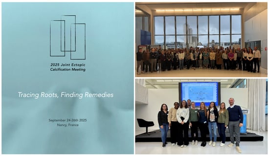
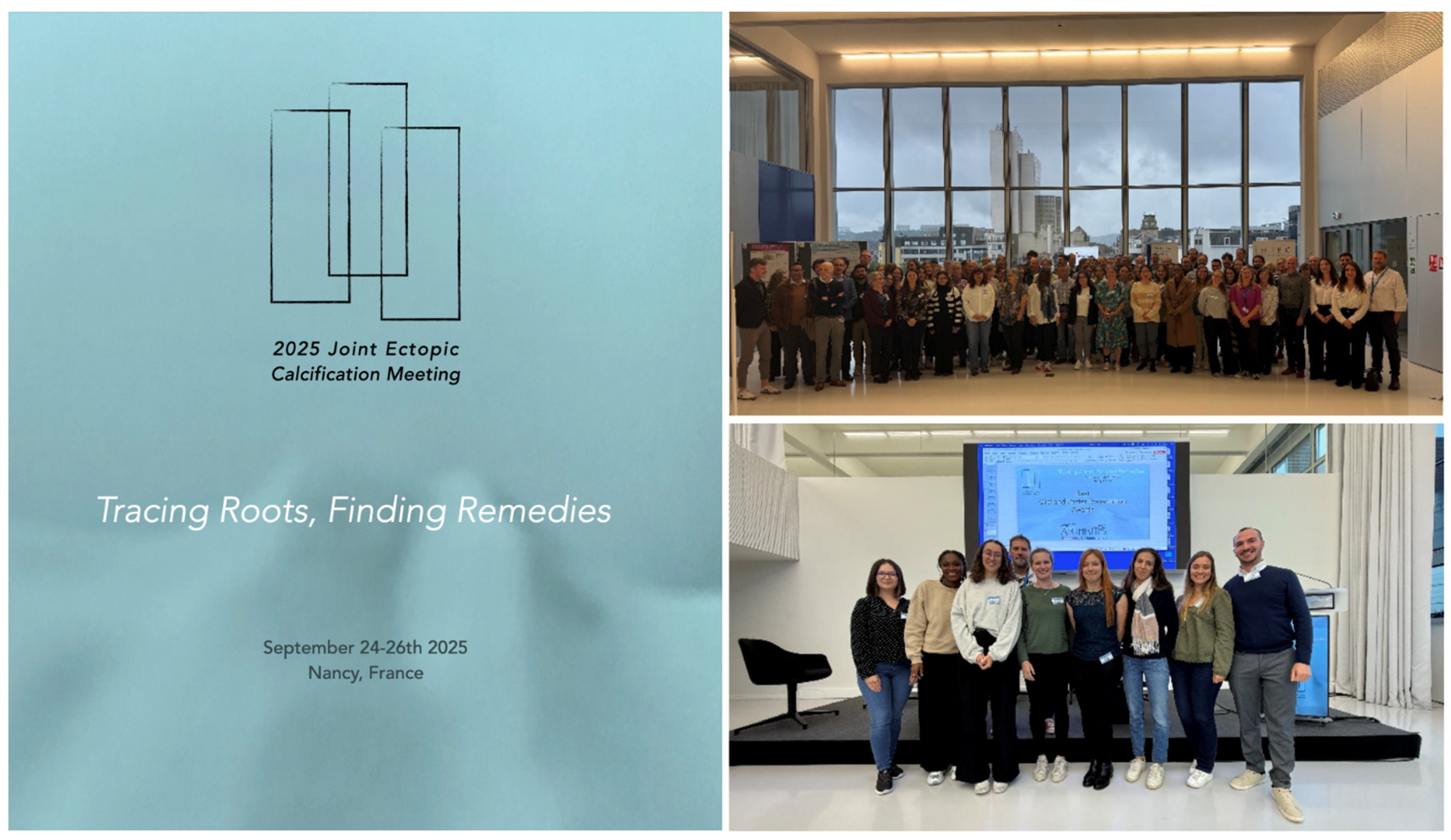
Disclaimer/Publisher’s Note: The statements, opinions and data contained in all publications are solely those of the individual author(s) and contributor(s) and not of MDPI and/or the editor(s). MDPI and/or the editor(s) disclaim responsibility for any injury to people or property resulting from any ideas, methods, instructions or products referred to in the content. |
© 2026 by the authors. Published by MDPI on behalf of the Gout, Hyperuricemia and Crystal Associated Disease Network. Licensee MDPI, Basel, Switzerland. This article is an open access article distributed under the terms and conditions of the Creative Commons Attribution (CC BY) license.