


Abstract
Cholesterol is increasingly recognized as a regulator of hematolymphoid malignancy biology, by influencing tumor proliferation, immune function, and therapeutic response. This review summarizes current evidence regarding the relationship between cholesterol metabolism and hematologic malignancies, integrating mechanistic studies with retrospective and emerging prospective clinical data. Cholesterol contributes to immune dysfunction by promoting CD8+ T-cell exhaustion within the tumor microenvironment. Clinical studies have reported associations between serum lipid profiles, statin use, and outcomes across multiple hematologic malignancies, although findings remain heterogeneous and are largely derived from retrospective analyses. Preclinical studies demonstrate that statins disrupt cholesterol-dependent signaling and inhibit tumor growth, whereas retrospective clinical studies generally support the safety of statin use and suggest possible therapeutic benefits in some lymphoma subtypes. Clinically, low high-density lipoprotein cholesterol (HDL) has been associated with advanced disease across multiple hematological malignancies. However, prospective, interventional data remain limited. Collectively, this review highlights the possible relationship between cholesterol metabolism and hematologic malignancy biology, with a focus on potential impacts on disease related outcomes.
1. Introduction
Cholesterol is an essential cellular component that plays an important role in membrane integrity, steroid synthesis, and immune regulation. Cholesterol is an essential structural component and the building block for cellular membranes, lipid rafts, and specialized domains for key oncogenic pathways, including B-cell receptor (BCR), phosphoinositide 3-kinase (PIK3), protein kinase B (AKT), mechanistic target of rapamycin (mTOR), and RAS/mitogen-activated protein kinase (MAPK).
The first step in cholesterol homeostasis is digestion and emulsification of dietary fat in the duodenum facilitated by bile acids and pancreatic enzymes. Small intestine enterocytes absorb free cholesterol and fatty acids and package cholesterol into chylomicrons with attached protein apolipoprotein B-48 (ApoB) in circulating blood. Next, lipolysis removes triglycerides via lipoprotein lipase (LPL), and the chylomicron remnants are endocytosed by hepatocytes through apolipoprotein E (ApoE) receptors. In hepatocytes, cholesterol is converted into very-low-density lipoprotein (VLDL) particles, which are secreted back to circulation and converted to low-density lipoprotein (LDL). High-density lipoprotein (HDL) facilitates mobilization of cholesterol from the tissue via apolipoprotein A-1 (ApoA) and delivers it to the liver for excretion or recycling.
For a deeper review on cholesterol biology, we recommend Xiao et al. [1] or Huang et al. [2]. In the past decade, growing data has suggested an association between cholesterol dysregulation within the tumor microenvironment (TME) and potential impacts on oncogenesis in cancer progression in hematologic malignancies.
Although outcomes in lymphoma have improved significantly with modern chemoimmunotherapy and targeted therapies, a clinically important subset of patients experience primary refractory disease or relapse. For example, in Diffuse Large B-Cell Lymphoma (DLBCL), frontline therapy is curative for many patients, yet outcomes remain poor in relapsed or refractory disease, even in the era of chimeric antigen receptor T-cell (CAR T) therapy [3]. Therapeutic limitations have prompted investigation into adjunctive therapeutic strategies that may enhance treatment response, overcome resistance, or improve long-term disease control. Cholesterol metabolism may represent one such potentially modifiable pathway.
A literature search was conducted using PubMed to identify relevant studies examining cholesterol metabolism in hematologic malignancies. Search terms included combinations of “cholesterol,” “statin,” “lipid metabolism,” “leukemia,” “lymphoma,” “multiple myeloma,” and individual lymphoma subtypes. Studies were selected based on relevance to cholesterol metabolism, serum cholesterol levels, statin use, clinical outcomes, and therapeutic implications in hematologic malignancies. Both preclinical and clinical studies were included, with particular attention to studies evaluating disease-specific mechanisms, treatment response, and patient outcomes. Reference lists of relevant articles and reviews were also examined to identify additional studies of interest.
This review provides an overview of the role of cholesterol metabolism in hematologic malignancies, beginning with its effects on malignant-cell signaling, the tumor microenvironment, and the prognostic significance of circulating cholesterol. We also examine the preclinical rationale and clinical evidence for cholesterol-targeted strategies, particularly statin use across lymphoma subtypes, followed by emerging applications in CAR T-cell therapy. Finally, we discuss prospective evidence, current limitations, and future directions for translating cholesterol modulation into therapeutic strategies for hematologic malignancies.
2. Cholesterol Biology in Hematological Malignancies
Cholesterol is a major structural component of plasma cell membranes and plays a vital role in maintaining membrane integrity, fluidity, and organization. Another important function is the formation of specialized membrane microdomains known as lipid rafts, which are cholesterol and sphingolipid-rich regions that serve as platforms for cellular signaling [4]. Lipid rafts facilitate the interaction of signaling molecules, receptors, and downstream effector proteins, allowing for the transduction of extracellular to intracellular signaling and cell-to-cell communication. In immune cells, these membrane domains are especially important for initiating activation pathways, including those involved in antigen recognition, the cytokine cascade, and immune synapse formation. In T-lymphocytes, these microdomains are important for initiation of T-Cell receptor (TCR) signaling, with key proteins such as lymphocyte-specific protein tyrosine kinase (Lck) localizing within lipid rafts as they participate in the earliest stages of T-Cell activation.
B-Cell receptor (BCR) signaling is also dependent on cholesterol-rich lipid rafts that concentrate signaling molecules within the plasma membrane. Following antigen engagement, the BCR translocates into the lipid rafts, where it interacts with proteins such as the Src-family of kinases, spleen tyrosine kinase (SYK), Bruton’s tyrosine kinase (BTK), and PI3K; the actions of these proteins lead to further downstream pathways responsible for B-cell proliferation, survival, and differentiation. Experimental studies have revealed that disruption of lipid raft integrity impairs proper BCR-mediated signaling; these observations are especially relevant in hematologic malignancies, as constitutive BCR signaling is a hallmark of several B-cell-related malignancies, such as Chronic Lymphocytic Leukemia (CLL), DLBCL, and Mantle Cell Lymphoma (MCL) [5].
Tumor cells have been shown to undergo metabolic reprogramming, enabling them to support rapid proliferation and survival [6]. Leukemia and lymphoma cells often upregulate pathways involved in cholesterol biosynthesis to meet the demands of membrane synthesis, cellular growth, and signaling. The Sterol Regulatory Element-Binding Proteins (SREBP) are a key family of transcription factors that promote the expression of enzymes involved in cholesterol and fatty acid synthesis [7]. Aberrant SREBP signaling has been associated with enhanced tumor growth, resistance to apoptosis, and poor clinical outcomes in several hematologic malignancies [8]. Collectively, increased cholesterol biosynthesis through the SREBP pathway activation and lipid raft-mediated signaling represent mechanisms through which tumor cells exploit cholesterol metabolism to drive disease progression and therapeutic resistance.
2.1. Malignancy Specific Mechanisms
2.1.1. Diffuse Large B-Cell Lymphoma
DLBCL, the most common subtype of non-Hodgkin lymphoma (NHL), frequently relies on aberrant BCR signaling for survival and proliferation [9]. Activation of the BCR initiates a downstream signaling cascade involving key proteins such as SYK, BTK, and PI3K, along with nuclear factor-kappa B (NF- κB). Experimental studies have shown that BCR activation drives receptor translocation into lipid rafts, and that disruption of these cholesterol-rich domains impairs early BCR signaling, creating a direct link between membrane cholesterol and malignant B-cell transformation. In Activated B-Cell Like (ABC) DLBCL, BCR is constitutively active, which serves as a central oncogenic driver and has led to the development of therapies such as BTK inhibitors [9]; this becomes particularly relevant because emerging evidence suggests that BCR signaling can also stimulate de novo cholesterol biosynthesis, creating a reinforcing loop in which BCR signaling promotes cholesterol production, while cholesterol-rich membrane domains help sustain the aberrantly functioning BCR signaling cascade [7]. Through these mechanisms, it is established that cholesterol homeostasis does not merely influence lymphoma growth in an abstract way, but it organizes the cellular architecture required for the continuation of the malignant cycle of tumors.
2.1.2. Chronic Lymphocytic Leukemia
CLL provides a distinct example of cholesterol dependence among the hematolymphoid malignancies. Unlike highly glycolytic tumors, CLL cells appear to rely substantially on lipid metabolism to support survival and proliferative signaling [10]. CLL cells has been shown to overexpress lipid-handling proteins, such as LPL and LDL receptors, which may facilitate uptake and mobilization of circulating lipoproteins as metabolic substrates. In experimental models, LDL exposure increased CLL cell membrane cholesterol, enhanced cytokine-mediated signaling such as phosphorylated STAT3, and increased the proportion of activated CLL cells, implying that lipoproteins can amplify pro-survival signaling rather than serving only as passive energy sources [11].
2.1.3. Acute Myeloid Leukemia
Acute Myeloid Leukemia (AML) cells demonstrate substantial dysregulation of cholesterol homeostasis, with increased cholesterol synthesis and uptake contributing to leukemic cell survival and therapeutic resistance [12]. Compared with normal hematopoietic cells, AML cells frequently exhibit increased expression of HMG-CoA reductase (HMGCR) and the LDL receptors. Importantly, exposure to cytarabine or daunorubicin can further increase HMGCR an LDL receptor expression and intracellular cholesterol, suggesting an adaptive response to chemotherapy [12]. Inhibition of this cholesterol response sensitizes AML cells to cytotoxic therapy, supporting cholesterol metabolism as a potential metabolic vulnerability in AML.
2.1.4. Acute Lymphoblastic Leukemia
Cholesterol metabolism may also contribute to the biology of Acute Lymphoblastic Leukemia (ALL), particularly early T-cell precursor ALL (ETP-ALL) [13]. Metabolomic profiling has demonstrated increased lipid biosynthesis in ETP-ALL and identified a selective dependence on the mevalonate pathway. Inhibition of the HMGCR reduced membrane cholesterol and oncogenic AKT signaling, suppressed MYC expression, and impaired leukemic cell growth and survival [13].
2.1.5. Multiple Myeloma
Cholesterol metabolism also represents a potential metabolic vulnerability in multiple myeloma (MM). MM cells regulate intracellular cholesterol through the SREBP2 pathway, which promotes both cholesterol biosynthesis and cholesterol uptake [14]. Notably, t(4:14)-positive MM cells demonstrate increased dependence on the mevalonate pathway and particular dependence on its downstream metabolite geranylgeranyl pyrophosphate (GGPP) [14].
2.2. Cholesterol, Immune Dysfunction, and Tumor Microenvironment
Beyond its direct effects on malignant cells, cholesterol dysregulation influences the immune microenvironment. Ma et al. demonstrated that cholesterol accumulates within tumor-infiltrating CD8+ T cells, where it induces endoplasmic reticulum (ER) stress and activation of the XBP1 arm of the unfolded protein response [15]. Activated XBP1 directly promotes transcription of multiple inhibitory immune checkpoint receptors, including PD-1 and 2B4, resulting in impaired cytokine production, diminished cytotoxicity, and acquisition of an exhausted T-cell phenotype. Importantly, in bone marrow samples from patients with multiple myeloma, increased cholesterol accumulation in CD8+ T cells was associated with greater expression PD-1 and 2B4, markers associated with T-cell exhaustion. Pharmacologic cholesterol reduction or inhibition of XBP1 restored T-cell effector function, identifying cholesterol accumulation as a reversible metabolic driver of immune exhaustion. These findings have important implications for hematolymphoid malignancies, where cholesterol-rich tumor microenvironments may similarly suppress antitumor immunity through ER stress-mediated T-cell dysfunction.
Emerging data has also identified CD36 as a cell receptor that mediates the uptake of long chain fatty acids metabolism and lipid taste in both human and rodent models [16]. Tcheng et al. showed that blockage of uptake of very long chain acyl-CoA dehydrogenase lipids was associated with cell death due to a combination of high leukemic mitochondrial mass and interruption of fatty acid oxidation essential to cell survival [16]. In a study of 1273 patients with AML, those with >20% CD36 expression had increased risk of increased extramedullary dissemination of leukemic blasts, increased risk of relapse (Sub-distribution hazard ratio (HR) 1.53; 95% Confidence Interval (CI): 1.07–2.19, p = 0.02) and reduced overall survival (OS) (HR: 1.69; 95% CI: 1.18–2.41, p = 0.005) [17]. A small molecule inhibitor of CD36 called SMS121 in a KG-1 cell model was shown to reduce uptake of fatty acid in AML cells; however, authors Abacka et al. note that downstream and off target effects of CD36 inhibition are currently unknown [18]. Strullu et al. [19] found that CD36 can be used on flow cytometry to identify high risk Philadelphia-like B-cell precursor ALL cases, specifically if they contain a ABL or JAK kinase fusion. This is not limited to the benchtop however; authors Gupta et al. [20] evaluated 1021 patients with ALL and found that CD36 expression predicts BCR:ABL1-like subtype ALL, and Newton et al. showed this was associated with inferior 5-year OS outcomes in 25 children (OS: 90% ± 9.5 vs. 100%, p = 0.019) [21].
Lipids and CD36 obviously play a role in cell survivability, and there is now some evidence that this translates to patient models; however, it is yet to be seen how to best manipulate these systems to improve cancer survival and outcomes. Future studies are needed to determine the role of CD36 as a possible routine measurement parameter, a target for therapy in conjunction with standard of care, or as a risk prognosticator.
2.3. Preclinical Rationale for Statins
Statins have emerged as possible adjunct therapeutic agents in hematologic malignancies because they target cholesterol biosynthesis through inhibition of HMGCR, the rate-limiting enzyme of the mevalonate pathway. In addition to reducing intracellular cholesterol levels, statins decrease production of the isoprenoid intermediates farnesyl pyrophosphate and GGPP, which are required for the post-translational prenylation of multiple signaling proteins, including Ras, Rho, Rac, and Rab GTPases. Disruption of these pathways has been shown to impair malignant cell proliferation, survival signaling, cytoskeletal organization, and vesicular trafficking, providing a biologic rationale for targeting cholesterol metabolism in hematologic cancers [22].
Preclinical studies across several lymphoma subtypes demonstrate that pharmacologic inhibition of cholesterol synthesis suppresses tumor growth and promotes cell death. In murine lymphoma models, Qi et al. showed that statins inhibited the proliferation of A20 and EL4 lymphoma cells in both a dose and time-dependent manner while largely sparing normal peripheral blood mononuclear cells at lower concentrations. Among the agents evaluated, fluvastatin exhibited the greatest anti-lymphoma activity, suggesting that inhibition of the mevalonate pathway may selectively impair malignant lymphoid cells [23].
Additional mechanistic studies have identified cholesterol biosynthetic signaling as a potential therapeutic target in DLBCL. Zhu et al. demonstrated that SREBP2 is highly expressed in human B-cell lymphoma specimens and that inhibition of the SREBP-HMGCR axis using either fatostatin or simvastatin suppressed proliferation of DLBCL cell lines in a dose-dependent manner, induced G2/M cell-cycle arrest, and reduced S-phase progression [24]. Interestingly, lymphoma cells activated the mTORC1-pS6 pathway as a compensatory survival mechanism following cholesterol depletion. Dual inhibition with low-dose statins and rapamycin produced synergistic suppression of proliferation, cell-cycle progression, and lipid raft formation, highlighting the potential benefit of combining cholesterol-targeting therapies with inhibitors of adaptive signaling pathways [24].
Similar findings have been reported in MCL. Santos et al. demonstrated that simvastatin reduced cellular viability, producing more than 50% inhibition of proliferation together with a two- to six-fold increase in lactate dehydrogenase release, consistent with enhanced cytotoxicity [25]. Santos et al. further demonstrated that simvastatin induces reactive oxygen species-mediated, caspase-independent cell death. In vivo, treatment resulted in approximately 55% reduction in tumor growth and was accompanied by decreased bone marrow and splenic metastatic involvement, providing evidence that cholesterol pathway inhibition may exert antitumor activity beyond in vitro models [25].
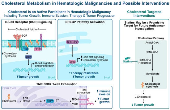
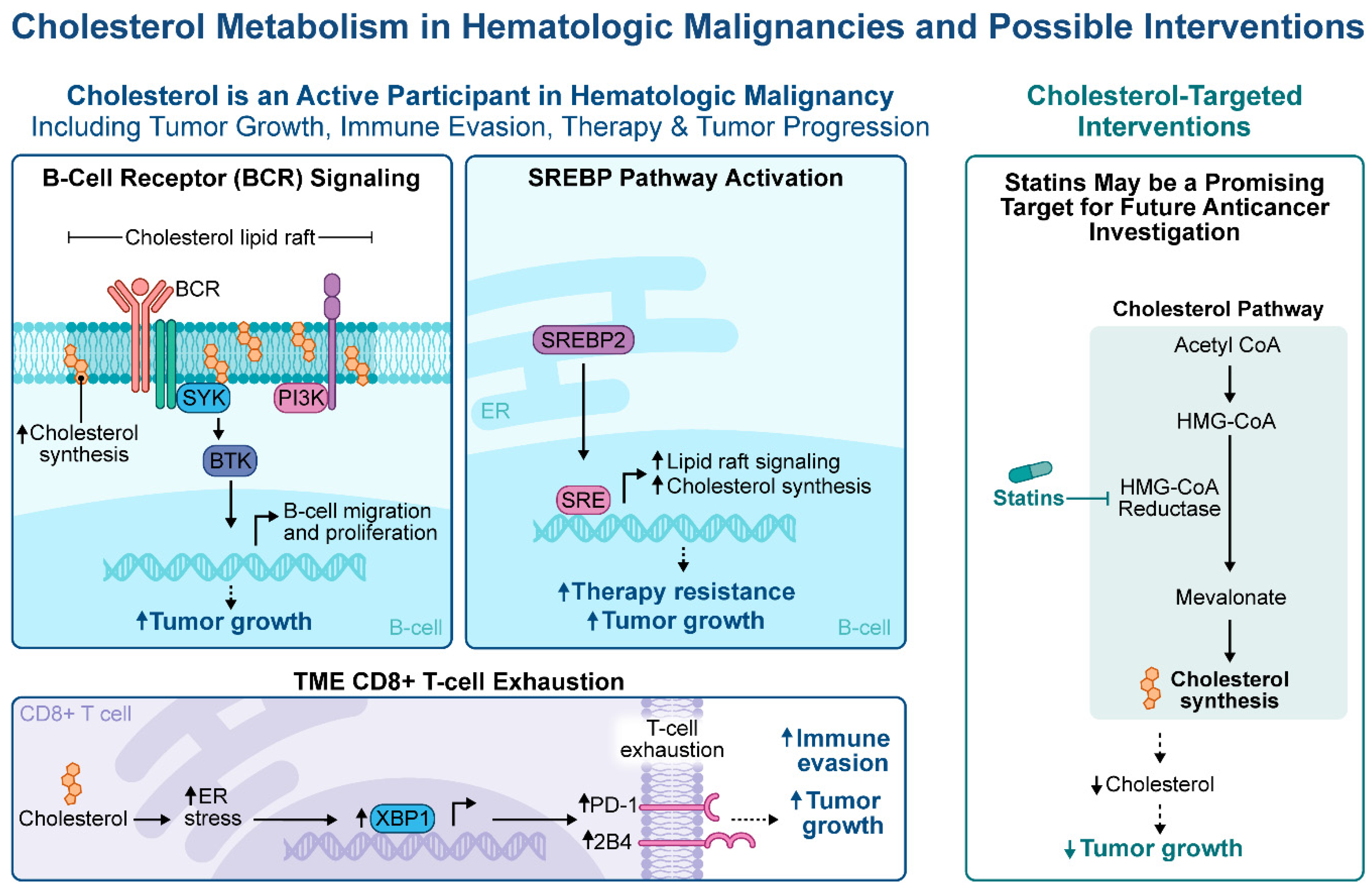
Collectively, these studies suggest that cholesterol metabolism represents a therapeutic vulnerability across multiple lymphoma subtypes. Beyond simply lowering cholesterol levels, statins interfere with critical oncogenic signaling pathways, disrupt lipid raft integrity, impair cell-cycle progression, and enhance tumor cell death. Although these findings remain largely preclinical, they provide a strong mechanistic foundation for future clinical studies evaluating cholesterol-targeting therapies. The major mechanisms linking cholesterol metabolism to hematologic malignancy biology and potential therapeutic interventions are summarized in Figure 1.
Figure 1.
Overview of cholesterol metabolism in hematologic malignancies and potential therapeutic interventions. Cholesterol contributes to malignant cell growth through B-cell receptor (BCR) signaling and SREBP pathway activation and may promote immune evasion through CD8+ T-cell exhaustion within the tumor microenvironment (TME). Statins inhibit HMG-CoA reductase and represent a potential strategy for targeting cholesterol metabolism.
2.4. Retrospective and Clinical Evidence
2.4.1. Dyslipidemia and Hematological Malignancy Risk
The relationship between abnormal cholesterol levels and cancer risk is complex and depends both on the type of cholesterol and malignancy. A UK study found that in people not taking cholesterol-lowering medications, higher cholesterol levels were generally linked to a lower risk of NHL and other hematological malignancies [26]. However, associations varied by subtype found that a higher ratio of LDL to HDL was consistently linked to a greater risk of CLL and small lymphocytic lymphoma (SLL). High LDL, apolipoprotein B (ApoB), and triglyceride levels were linked to a greater risk of MM after more than 10 years of follow-up [26]. A large Korean study of nearly 9.6 million people found that individuals with the low levels of HDL had a significantly higher risk of developing hematologic malignancies. The association was strongest for MM [27]. Also, decreased HDL-C (73.9% ± 4.6% vs. 95.3% ± 3.3%, p < 0.001) and ApoA (74.0% ± 4.6% vs. 95.2% ± 3.4%, p < 0.001) levels have also been associated with poorer 3-year OS in CLL [28]. These studies support a relationship between lipid metabolism and hematologic malignancy risk, although the associations differ by both lipid profile and disease subtype.
2.4.2. Association with Disease Presentation and Outcomes
Although elevated intracellular cholesterol supports malignant cell proliferation, lower circulating serum cholesterol at diagnosis has been associated with inferior clinical outcomes, suggesting that serum hypocholesterolemia reflects increased cholesterol utilization by metabolically active tumors.
In CLL, reduced HDL and ApoA predict poorer OS. Functional studies further demonstrate that low HDL is associated with diminished natural killer cell cytotoxicity, suggesting that alterations in cholesterol metabolism may contribute to immune dysfunction in addition to reflecting tumor burden [28].
The prognostic significance of hypocholesterolemia extends beyond B-cell malignancies. In peripheral T-cell lymphoma, not otherwise specified (PTCL-NOS), lower HDL and LDL are associated with inferior OS, and incorporation of cholesterol parameters into the Prognostic Index for PTCL (PITC) improves risk stratification compared with the conventional PIT score [29]. Likewise, patients with malignant lymphoma and adult T-cell leukemia/lymphoma (ATLL) in the lowest HDL tercile demonstrate significantly worse outcomes, with HDL showing an inverse relationship with soluble IL-2 receptor concentrations, a recognized marker of disease activity [30]. Similar observations have also been reported in myeloproliferative neoplasms (MPNs), where total cholesterol levels below 120 mg/dL are associated with a threefold increased risk of transformation to secondary myelofibrosis or acute leukemia (adjusted subdistribution HR, 3.00; 95% CI, 1.36–6.63) and all-cause death (adjusted HR, 1.87; 95% CI, 1.06–3.30) compared to cholesterol levels >150 mg/dL [31].
The role of LDL is more complex. While reduced circulating LDL correlates with inferior prognosis, experimental studies suggest that malignant cells actively depend upon LDL-derived cholesterol to support growth. Leukemic cells exhibit increased LDL receptor expression and HMGCR activity together with impaired feedback regulation of cholesterol uptake, indicating profound dysregulation of cholesterol homeostasis [32]. Consistent with these observations, increased LDL receptor expression in AML independently predicts higher relapse rates and worse survival beyond established European Leukemia Net (ELN) risk classification, while drug-resistant leukemic cell lines demonstrate enhanced LDL uptake and diminished feedback sensitivity [33,34].
Collectively, these findings suggest that reduced circulating cholesterol is not simply an epiphenomenon but instead reflects increased metabolic demand by proliferating malignant cells, linking serum cholesterol levels with both disease aggressiveness and treatment resistance [34]. Taken together, these clinical associations and findings suggest a complex and seemingly paradoxical relationship between cholesterol and hematologic malignancies, in which low circulating cholesterol may reflect underlying disease biology and tumor-driven metabolic alterations rather than a direct consequence of cholesterol reduction itself.
2.4.3. Hypocholesterolemia as a Marker of Tumor Burden and Treatment Response
Beyond its potential prognostic relevance, serum cholesterol levels may reflect changes in disease activity and treatment response. Rapidly dividing malignant cells increase cholesterol uptake and LDL receptor activity, which can lead to lower circulating cholesterol levels. In AML, higher leukemic burden and increased LDL receptor activity are associated with lower cholesterol, suggesting a relationship between disease activity and cholesterol metabolism.
Studies across multiple hematologic malignancies, including AML, ALL, DLBCL, and Hodgkin lymphoma (HL) show that cholesterol levels may rise with effective treatment and fall again with relapse. Some data also suggests cholesterol can decline years before a lymphoma diagnosis [35,36]. Overall, serum cholesterol may act as a simple, indirect marker of disease activity and treatment response, alongside established biomarkers like lactate dehydrogenase (LDH) and β2-microglobulin. Although prospective validation is needed, serial cholesterol measurements in conjunction with validated biomarkers, imaging, and circulating tumor DNA, may ultimately prove clinically useful.
3. Cholesterol as a Prognostic Indicator
Associations have been identified between serum cholesterol levels and clinical outcomes across multiple hematologic malignancies, raising the possibility that they could have prognostic relevance. Evidence from retrospective studies demonstrates that serum lipid profiles obtained at or near the time of diagnosis correlate with advanced disease stage, presence of extranodal sites, and poorer progression-free survival (PFS) and OS. In DLBCL, reduced HDL and LDL independently predict inferior PFS (HR = 3.420; 95% CI: 2.364–4.948, p < 0.001) and OS (HR = 2.323; 95% CI: 1.506–3.584, p < 0.001), and incorporation of serum cholesterol measurements into the National Comprehensive Cancer Network International Prognostic Index (NCCN-IPI) improves prognostic discrimination beyond established clinical risk factors [37].
Alterations in serum cholesterol appear to precede the clinical diagnosis of lymphoma. In an analysis of more than 13,000 lymphoma cases, HDL concentrations were significantly lower than those of matched controls, with the greatest differences observed during the three to four years preceding diagnosis [38]. The decline was even more pronounced among patients who ultimately presented with advanced-stage disease, suggesting that falling cholesterol levels may reflect tumor growth rather than representing an independent risk factor for lymphoma development. Whether hypocholesterolemia contributes directly to lymphomagenesis or instead represents an early manifestation of subclinical disease remains uncertain [26,38].
3.1. Statin Use and Clinical Outcomes
The potential anticancer effects of statins observed in preclinical models have prompted extensive investigation into whether statins impact outcomes in patients with hematological malignancies. The major clinical and observational studies evaluating the association between statin use and outcomes are summarized in Table 1.
Table 1.
Clinical and Observational Studies Evaluating Statin Therapy and Clinical Outcomes in Hematologic Malignancies.
3.1.1. Statins in Diffuse Large B-Cell Lymphoma
Most clinical evidence evaluating statin use in aggressive lymphomas has focused on DLBCL. Overall, most studies have shown either a neutral or favorable association between statin use and clinical outcomes in DLBCL, while consistently failing to support concerns that statins impair the efficacy of chemoimmunotherapy.
The largest population-based study in DLBCL was done by Smyth et al., who analyzed 4913 adults aged ≥ 66 years diagnosed with DLBCL or transformed lymphoma and treated with rituximab-based regimens in Ontario, Canada. Pretreatment statin use was associated with a duration-dependent improvement in survival, with longer exposure corresponding to progressively lower mortality risk and the greatest benefit observed among patients receiving statins for at least one year before diagnosis (HR 0.71; 95% CI 0.63–0.79) [44].
Subsequently, a meta-analysis of eight cohort studies involving 9927 patients found that statin use was associated with significantly improved OS and PFS in Western countries, but not in Asian countries, suggesting that the effect of statins may differ across patient populations or treatment settings [45]. This geographic heterogeneity may reflect differences in patient characteristics, cardiovascular risk profiles, statin exposure, lymphoma biology, or treatment patterns across study populations. However, the observational nature and heterogeneity of the included studies limit conclusions regarding the factors underlying these regional differences.
Additional observational data have suggested that statins may improve treatment response. In a cohort study of 271 treatment-naive advanced DLBCL patients, concurrent statin use during R-CHOP (rituximab, cyclophosphamide, doxorubicin, vincristine, and prednisone) therapy was associated with significantly higher complete response rates than R-CHOP alone [46]. Both hydrophilic and lipophilic statins demonstrated benefit, and patients receiving medium- or high-intensity statins experienced superior PFS during long-term follow-up, supporting a potential dose–response relationship between statin exposure and clinical outcomes [46]. Winiarska et al. demonstrated that in vitro, statin-mediated cholesterol depletion induced conformational changes in CD20, impairing rituximab binding and reducing both complement-mediated cytotoxicity (CDC) and antibody-dependent cellular cytotoxicity [48]. This finding raised the question of whether statins may undermine the efficacy of anti-CD20-based therapies.
Song et al. evaluated 409 patients with DLBCL and found that there was a difference in PFS and OS based on the germinal center B-cell-like (GCB) subtype of the patient [42]. For those with non-germinal center B-cell like (non-GCB) subtype and exposed to statin therapy, there was poorer PFS (HR: 1.55, 95% CI: 1.058–2.279, p = 0.024) and OS (HR = 1.532, 95% CI: 1.041–2.255, p = 0.02). However, there was no statistical significance in outcomes when the two groups were combined and exposed to statin compared to non-statin patients for PFS (63.7% vs. 68.4% p = 0.282) or OS (63% vs. 68.8%, p = 0.273) [42]. The authors hypothesized that statins may impair CD20 binding by rituximab. Other explanations include underlying high-risk mutations that affect cholesterol metabolism, immune mechanisms, or unaccounted confounding. Subsequent studies have not specifically evaluated outcomes by GCB versus non-GCB and generally have not reproduced this negative association between statin use and clinical outcomes. Benchtop research establishing a mechanism and incorporating this into a prospective trial is essential before widespread use can be safely attempted.
Several studies have specifically addressed concerns raised by preclinical data suggesting that statin-induced cholesterol depletion could impair CD20 conformation and reduce rituximab binding. Ennishi et al. retrospectively analyzed 256 newly diagnosed DLBCL patients treated with R-CHOP and found no impact of statin use on 3-year PFS or OS [40]. Similarly, Nowakowski et al. reported no association between statin use and response rate, event-free survival, or OS among patients receiving rituximab-containing therapy [39]. Together, these studies provide reassuring clinical evidence that statin therapy does not appear to diminish the effectiveness of anti-CD20-based treatment despite earlier mechanistic concerns.
Collectively, the evidence available suggests that statins are safe and may confer modest clinical benefits in select patients with DLBCL. While retrospective studies have reported improvements in survival and treatment response, these findings have not yet been consistently reproduced in prospective randomized trials, underscoring the need for larger studies to better define the role of statins as adjunctive therapy.
3.1.2. Statins in Mantle Cell Lymphoma
Clinical data for MCL is limited, and no clinical trials or retrospective studies have been conducted. Similarly to MCL, there are no reports of peripheral T-Cell Lymphomas and associated studies that focus on outcomes with cholesterol treatment. However, preclinical work suggests that simvastatin may have antitumor activity by promoting lymphoma cell death, inhibiting AKT/mTOR signaling, reducing tumor growth, and enhancing sensitivity to ibrutinib [25]. These findings provide a mechanistic rationale for future clinical investigations in MCL.
3.1.3. Statins in Follicular Lymphoma
There is little data evaluating the relationship between statin therapy and follicular lymphoma (FL). One study from Nowakowski et al. reported that statin use at diagnosis was associated with improved event-free survival among 294 patients with FL, including the subgroup of patients treated with a rituximab-containing regimen and in patients who were observed only [39].
However, these findings were not confirmed in the PRIMA trial, which investigated 1135 patients with de novo FL treated with immunochemotherapy. Among the 119 patients who were on statins, there were no significant differences in event-free survival, time to next lymphoma treatment, time to next chemotherapy, or OS [43]. In addition, the adverse events in the statin group were comparable to those who did not receive a statin [43]. It is important to note, however, that the PRIMA study enrolled patients with high-tumor-burden FL who required immediate treatment, representing a different population than the one evaluated by Nowakowski et al.
A Swedish population-based study of 16,098 patient with NHL and CLL patients similarly found that statin use did not appear to increase lymphoma-specific mortality in actively treated FL patients [49].
3.1.4. Statins in Other Types of Lymphoma
Evidence specific to Marginal Zone lymphoma (MZL), lymphoplasmacytic lymphoma (LPL/Waldenström macroglobulinemia), and other indolent B-cell lymphomas is limited to subgroup analyses of broader NHL groups [50]. A Cancer Prevention Study II Nutrition Cohort reported reduced risk for both DLBCL and MZL with prolonged statin use (≥5 years) [41]. Similarly, a population-based case–control study of 5541 NHL cases found that statin use was associated with lower risk of NHL overall, as well as DLBCL and plasma cell neoplasms, with the association limited to high-potency and lipophilic statins [50].
Overall, the available evidence regarding statin use in indolent B-cell lymphomas remains limited and inconsistent, largely due to retrospective study designs and heterogeneous patient populations. Prospective studies are needed to clarify whether statins influence disease biology, treatment response, and long-term clinical outcomes across individual indolent lymphoma subtypes.
3.1.5. Statins in Hodgkin Lymphoma (HL)
Preclinical work by Von Tresckow et al. demonstrated that simvastatin induced caspase-dependent apoptosis in Hodgkin lymphoma cell lines and inhibited tumor growth in vivo through disruption of the mevalonate pathway [51]. These findings support a potential therapeutic role for statins in HL, although both retrospective and prospective clinical outcome data in this disease remain limited.
3.2. Emerging Applications of Statins with CAR T Therapy
Emerging evidence suggests that statins may influence outcomes in patients receiving CD19-directed CAR T therapy. Ex vivo analysis cells exposed to cholesterol accumulation experienced CAR T cell exhaustion, which was reversed with fluvastatin. Furthermore, fluvastatin primed CAR T cells had superior in vivo expansion, and analysis showed fluvastatin enhances ERK1/2 phosphorylation which in turn pushes CAR T cells to switch from glycolysis to oxidative phosphorylation which boosts memory-like cells, namely CAR T [52]. Outside of the laboratory space, a preliminary abstract using 743 propensity matched patients in the TriNetX database who underwent CART therapy and were on statin therapy had an 8.6% improvement in 5-year OS (77.4% vs. 68.8%, 95% CI 0.041, 0.131; p = <0.001) compared to no statin exposure [53]. Because these TriNetX findings are currently available only in preliminary abstract form, they should be considered preliminary and interpreted with appropriate caution until validated in a full peer-reviewed publication. In a peer-reviewed retrospective study of 84 patients with relapsed or refractory DLBCL, statin exposure was associated with higher overall and complete response rates, prolonged PFS and OS, and no increase in CAR T-related toxicities such as cytokine release syndrome or immune effector cell-associated neurotoxicity syndrome [47]. Exploratory analyses further suggested improved outcomes among patients receiving lipophilic compared with hydrophilic statins. The impact of statins in the CAR T setting continues to be explored through ongoing prospective studies. Although these findings require prospective validation, they raise the possibility that statins may augment cellular immunotherapy through immunomodulatory effects, consistent with preclinical data demonstrating reduced T-cell exhaustion following cholesterol modulation [15]. There is existing interest using statin therapy prophylaxis prior to stem cell transplant, where atorvastatin was given 2–4 weeks before transplant to improve graft versus host burden [54]. However, this practice is not routine by most institutions. A single center phase 1 study (NCT04514029) from University of Minnesota recruited 37 patients to randomize intrathecal dexamethasone and simvastatin vs. placebo post axicabtagene ciloleucel for neurotoxicity prophylaxis; however, these anticipated results have not yet published.
4. Prospective and Interventional Evidence
Despite positive preclinical and retrospective data, prospective studies evaluating cholesterol-targeting strategies in hematologic malignancies remain limited. Most available studies have focused on establishing the safety of combining statins with standard therapies rather than showing improvements in cancer-specific outcomes. Thus, the current prospective evidence should be viewed as proof-of-concept supporting further investigation rather than confirmation of clinical benefit.
The only randomized data evaluating statins in lymphoma come from the STOP-CA trial [55] (NCT02943590). Although the parent trial was designed to assess the cardioprotective effects of atorvastatin during anthracycline-based chemotherapy, a prespecified subgroup analysis evaluated 102 patients with DLBCL receiving R-CHOP. Atorvastatin did not adversely affect treatment efficacy, with similar response rates and toxicity profiles compared with placebo, providing prospective evidence that concomitant statin therapy is safe in patients receiving rituximab-containing chemoimmunotherapy [56]. More recently, a phase I dose-escalation study (NCT04512105) investigated the addition of pitavastatin to venetoclax-based therapy in patients with newly diagnosed AML or CLL/small lymphocytic lymphoma (SLL). The combination was well tolerated; no unexpected dose-limiting toxicities were identified, and the study established the feasibility of combining HMGCR inhibition with BCL-2 inhibition in hematologic malignancies [57]. Although the trial was not designed to evaluate efficacy, it represents the first prospective interventional study directly testing statin therapy as an adjunctive anticancer strategy in hematologic malignancies and provides a foundation for future randomized trials evaluating whether cholesterol modulation can enhance treatment response or overcome therapeutic resistance.
5. Discussion
This review highlights the biological, mechanistic, prognostic, and theoretical relationship between cholesterol dysregulation in the tumor microenvironment and hematolymphoid oncogenesis. In addition, we review retrospective and early phase studies that may suggest a possible therapeutic benefit in statin use in the treatment of hematologic disease. While retrospective, these hypothesis generating studies suggest the need for prospective validation.
This review highlights cholesterol metabolism as a recognized component of hematologic malignancy biology that extends beyond its traditional role in cellular membrane structure. Accumulating evidence suggests that cholesterol dysregulation contributes to malignant transformation, tumor proliferation, immune evasion, and therapeutic resistance through multiple complementary mechanisms. At the cellular level, cholesterol plays an integral role in facilitating oncogenic signaling pathways such as BCR signaling, and aberrant SREBP pathway activation supports the metabolic demands of rapidly proliferating malignant cells [4,6]. In addition, emerging evidence demonstrates that cholesterol accumulation within the tumor microenvironment contributes to CD8 T-Cell exhaustion [15]. Together, these findings suggest that cholesterol metabolism may contribute to hematological malignancy progression.
Cholesterol as biomarkers may be an emerging topic of interest in hematolymphoid malignancies. Across multiple hematolymphoid subtypes such as CLL, AML, and MPNs, reduced serum cholesterol, particularly HDL, has repeatedly been associated with inferior survival [28,29,37]. The observation that serum cholesterol levels normalize following successful treatment and decline again with disease relapse suggests that changes in cholesterol may reflect underlying disease activity [35,36]. Given the widespread availability and low cost of lipid measurements, further studies are warranted to determine whether serial changes in cholesterol provide meaningful prognostic information alongside established biomarkers such as LDH and β2-microglobulin.
The therapeutic implications of cholesterol-lowering therapies need to be further established. While retrospective studies in DLBCL generally suggest neutral or favorable effects of statin use without compromising rituximab efficacy, evidence in FL and other indolent lymphomas has been less consistent. However, the role of statin therapy in high grade B-cell lymphomas and in conjunction with CAR T remain an area of active investigation.
The studies reviewed are subject to several sources of bias inherent to observational design. Most studies compare existing users with non-users, introducing prevalent-user bias whereby patients who discontinued therapy because of intolerance or early events are absent from the exposed group. Statin users also have regular healthcare contact and better adherence to treatment generally, producing healthy-user and healthy-adherer effects that can favor the exposed group. Statins are also commonly withheld or deprescribed in frail patients or those with multiple comorbidities, so confounding contraindication can categorize non-users to group with poorer baseline prognosis. Where studies defined statin exposure as continued use during lymphoma treatment, immortal-time bias is a further consideration, since patients classified as users must survive long enough to remain on therapy; by contrast, the studies by Nowakowski et al. [39] and Smyth et al. [44] ascertained exposure at or before the start of follow-up, hence, not affected by this bias. Finally, where overall rather than lymphoma-specific survival was reported, the established cardiovascular benefits of statins may account for part of the observed association and mortality benefit. These considerations do not negate the signal observed across independent cohorts and treatment settings, but they indicate that prospective evaluation will be required to establish a causal contribution.
Hence, several limitations of the current literature should be recognized. Much of the available evidence is obtained from retrospective observational studies that are inherently susceptible to selection bias, immortal time bias, healthy user bias, residual confounding, and differences in patient comorbidities and concomitant therapies. Significant heterogeneity exists between studies with respect to lymphoma subtype, statin class, dose and intensity of statin, duration of exposure, and outcomes measures, making direct comparisons difficult. In addition, it is worth noting that the heterogeneity of lipid-related exposures assessed across studies, including total cholesterol, LDL, HDL, ApoA1, ApoB, triglycerides, and statin exposure also limits their interpretation as manifestations of a single underlying phenomenon of cholesterol metabolism. Furthermore, cholesterol levels may be influenced by many other nutritional and inflammatory factors independent of malignancy, potentially limiting their specificity as biomarkers. Finally, despite strong mechanistic reasoning, prospective interventional studies evaluating cholesterol-lowering therapies as anticancer treatment remain remarkably limited.
6. Conclusions
Cholesterol metabolism is increasingly recognized as a component of hematologic malignancy biology with potential implications extending from tumor initiation and progression to immune regulation and therapeutic response. Experimental studies consistently demonstrate that malignant cells exploit cholesterol biosynthesis and aberrant lipid-raft-mediated signaling to support proliferation, survival, and resistance to therapy. While retrospective evidence suggests that statins are safe and may provide clinical benefit in certain populations, prospective evidence remains limited and insufficient to support their routine use as anti-neoplastic agents outside standard cardiovascular indications. However, the implications of experimental and retrospective data support cholesterol metabolism as a promising area for future investigation. Prospective clinical trials will be pivotal to determine whether cholesterol-targeted interventions can be translated into meaningful improvements in outcomes for patients with hematological malignancies.
Author Contributions
Conceptualization, F.A. and E.A.B.; Investigation, F.A., I.S., M.Z., G.T., N.G., M.S., H.P., C.-C.A.H., C.-H.A.T., Q.Y., S.R.P. and E.A.B.; Writing—original draft preparation, F.A.; Writing—review and editing, F.A., S.R.P. and E.A.B.; Supervision, E.A.B. All authors have read and agreed to the published version of the manuscript.
Funding
This project was partially funded by the Houston Methodist Academic Institute Presidential Cornerstone grant (S.R.P), for the National Cancer Institute (NCI), National Institutes of Health (NIH) grant R01CA269835 (C.-C.A.H. and S.R.P.).
Data Availability Statement
No new data was created or analyzed in this study. Data sharing is not applicable to this article.
Acknowledgments
The authors would like to thank all colleagues who provided valuable discussions and feedback during the preparation of this manuscript. We would also like to thank James Kasper and Rachael Whitehead for their invaluable assistance with figure preparation.
Conflicts of Interest
The authors declare no conflicts of interest.
References
- Xiao, M.; Xu, J.; Wang, W.; Zhang, B.; Liu, J.; Li, J.; Xu, H.; Zhao, Y.; Yu, X.; Shi, S. Functional Significance of Cholesterol Metabolism in Cancer: From Threat to Treatment. Exp. Mol. Med. 2023, 55, 1982–1995. [Google Scholar] [CrossRef] [Scilit] [PubMed]
- Huang, B.; Song, B.-L.; Xu, C. Cholesterol Metabolism in Cancer: Mechanisms and Therapeutic Opportunities. Nat. Metab. 2020, 2, 132–141. [Google Scholar] [CrossRef] [Scilit] [PubMed]
- Ghobadi, A.; Anagnostou, T.; Azzi, J.; Hamadani, M.; Logan, B.; Ahmed, S.; Bharadwaj, S.; Baird, J.H.; Jacobson, C.; Locke, F.L.; et al. Impact of Complete Response on Long-Term Survival in Patients with Relapsed or Refractory Large B-Cell Lymphoma: A Center for International Blood and Marrow Transplant Research Prospective Study and Systematic Literature Review/Meta-Analysis. Transplant. Cell. Ther. 2026, 32, 623.e1–623.e15. [Google Scholar] [CrossRef] [Scilit] [PubMed]
- Kim, S.; Han, J.; Lee, D.H.; Cho, K.H.; Kim, K.H.; Chung, J.H. Cholesterol, a Major Component of Caveolae, Down-Regulates Matrix Metalloproteinase-1 Expression through ERK/JNK Pathway in Cultured Human Dermal Fibroblasts. Ann. Dermatol. 2010, 22, 379–388. [Google Scholar] [CrossRef] [Scilit] [PubMed]
- Simons, K.; Toomre, D. Lipid Rafts and Signal Transduction. Nat. Rev. Mol. Cell Biol. 2000, 1, 31–39. [Google Scholar] [CrossRef] [Scilit] [PubMed]
- Mao, Y.; Xia, Z.; Xia, W.; Jiang, P. Metabolic Reprogramming, Sensing, and Cancer Therapy. Cell Rep. 2024, 43, 115064. [Google Scholar] [CrossRef] [Scilit] [PubMed]
- Li, Y.; Wu, S.; Zhao, X.; Hao, S.; Li, F.; Wang, Y.; Liu, B.; Zhang, D.; Wang, Y.; Zhou, H. Key Events in Cancer: Dysregulation of SREBPs. Front. Pharmacol. 2023, 14, 1130747. [Google Scholar] [CrossRef] [Scilit] [PubMed]
- Erkner, E.; Hentrich, T.; Schairer, R.; Fitzel, R.; Secker-Grob, K.-A.; Jeong, J.; Keppeler, H.; Korkmaz, F.; Schulze-Hentrich, J.M.; Lengerke, C.; et al. The RORɣ/SREBP2 Pathway Is a Master Regulator of Cholesterol Metabolism and Serves as Potential Therapeutic Target in t(4;11) Leukemia. Oncogene 2024, 43, 281–293. [Google Scholar] [CrossRef] [Scilit] [PubMed]
- Davis, R.E.; Ngo, V.N.; Lenz, G.; Tolar, P.; Young, R.M.; Romesser, P.B.; Kohlhammer, H.; Lamy, L.; Zhao, H.; Yang, Y.; et al. Chronic Active B-Cell-Receptor Signalling in Diffuse Large B-Cell Lymphoma. Nature 2010, 463, 88–92. [Google Scholar] [CrossRef] [Scilit] [PubMed]
- White, A.M.; Best, O.G.; Hotinski, A.K.; Kuss, B.J.; Thurgood, L.A. The Role of Cholesterol in Chronic Lymphocytic Leukemia Development and Pathogenesis. Metabolites 2023, 13, 799. [Google Scholar] [CrossRef] [Scilit] [PubMed]
- McCaw, L.; Shi, Y.; Wang, G.; Li, Y.-J.; Spaner, D.E. Low Density Lipoproteins Amplify Cytokine-Signaling in Chronic Lymphocytic Leukemia Cells. eBioMedicine 2017, 15, 24–35. [Google Scholar] [CrossRef] [Scilit] [PubMed]
- Banker, D.E.; Mayer, S.J.; Li, H.Y.; Willman, C.L.; Appelbaum, F.R.; Zager, R.A. Cholesterol Synthesis and Import Contribute to Protective Cholesterol Increments in Acute Myeloid Leukemia Cells. Blood 2004, 104, 1816–1824. [Google Scholar] [CrossRef] [Scilit] [PubMed]
- Rashkovan, M.; Albero, R.; Gianni, F.; Perez-Duran, P.; Miller, H.I.; Mackey, A.L.; Paietta, E.M.; Tallman, M.S.; Rowe, J.M.; Litzow, M.R.; et al. Intracellular Cholesterol Pools Regulate Oncogenic Signaling and Epigenetic Circuitries in Early T-Cell Precursor Acute Lymphoblastic Leukemia. Cancer Discov. 2022, 12, 856–871. [Google Scholar] [CrossRef] [Scilit] [PubMed]
- Torcasio, R.; Gallo Cantafio, M.E.; Ikeda, R.K.; Ganino, L.; Viglietto, G.; Amodio, N. Lipid Metabolic Vulnerabilities of Multiple Myeloma. Clin. Exp. Med. 2023, 23, 3373–3390. [Google Scholar] [CrossRef] [Scilit] [PubMed]
- Ma, X.; Bi, E.; Lu, Y.; Su, P.; Huang, C.; Liu, L.; Wang, Q.; Yang, M.; Kalady, M.F.; Qian, J.; et al. Cholesterol Induces CD8+ T Cell Exhaustion in the Tumor Microenvironment. Cell Metab. 2019, 30, 143–156.e5. [Google Scholar] [CrossRef] [Scilit] [PubMed]
- Tcheng, M.; Roma, A.; Ahmed, N.; Smith, R.W.; Jayanth, P.; Minden, M.D.; Schimmer, A.D.; Hess, D.A.; Hope, K.; Rea, K.A.; et al. Very Long Chain Fatty Acid Metabolism Is Required in Acute Myeloid Leukemia. Blood 2021, 137, 3518–3532. [Google Scholar] [CrossRef] [Scilit] [PubMed]
- Farge, T.; Nakhle, J.; Lagarde, D.; Cognet, G.; Polley, N.; Castellano, R.; Nicolau, M.-L.; Bosc, C.; Sabatier, M.; Sahal, A.; et al. CD36 Drives Metastasis and Relapse in Acute Myeloid Leukemia. Cancer Res. 2023, 83, 2824–2838. [Google Scholar] [CrossRef] [Scilit] [PubMed]
- Åbacka, H.; Masoni, S.; Poli, G.; Huang, P.; Gusso, F.; Granchi, C.; Minutolo, F.; Tuccinardi, T.; Hagström-Andersson, A.K.; Lindkvist-Petersson, K. SMS121, a New Inhibitor of CD36, Impairs Fatty Acid Uptake and Viability of Acute Myeloid Leukemia. Sci. Rep. 2024, 14, 9104. [Google Scholar] [CrossRef] [Scilit] [PubMed]
- Strullu, M.; Caye-Eude, A.; Robert, E.; Renard, J.-M.; Chaye, A.; Galimand, J.; Fenneteau, O.; Arfeuille, C.; Cuccuini, W.; Theron, A.; et al. CD36 Cell Surface Expression as a Surrogate Marker to Identify ABL/JAK-Class Kinase Fusions in Pediatric BCP-ALL. Leukemia 2025, 39, 64–74. [Google Scholar] [CrossRef] [Scilit] [PubMed]
- Gupta, D.G.; Varma, N.; Sharma, P.; Truica, M.I.; Abdulkadir, S.A.; Singh, P.; Singh Sachdeva, M.U.; Naseem, S.; Siddiqui, M.R.; Bose, P.; et al. Hematological, Clinical, Immunophenotypic Characterization, and Treatment Outcomes of Prognostically Significant Genetic Subtypes of B-lineage Acute Lymphoblastic Leukemia: A Report of 1021 Patients from India. Cancer 2023, 129, 3390–3404. [Google Scholar] [CrossRef] [Scilit] [PubMed]
- Newton, J.G.; Horan, J.T.; Newman, S.; Rossi, M.R.; Ketterling, R.P.; Park, S.I. CD36-Positive B-Lymphoblasts Predict Poor Outcome in Children with B-Lymphoblastic Leukemia. Pediatr. Dev. Pathol. 2017, 20, 224–231. [Google Scholar] [CrossRef] [Scilit] [PubMed]
- Juarez, D.; Fruman, D.A. Targeting the Mevalonate Pathway in Cancer. Trends Cancer 2021, 7, 525–540. [Google Scholar] [CrossRef] [Scilit] [PubMed]
- Qi, X.-F.; Zheng, L.; Lee, K.-J.; Kim, D.-H.; Kim, C.-S.; Cai, D.-Q.; Wu, Z.; Qin, J.-W.; Yu, Y.-H.; Kim, S.-K. HMG-CoA Reductase Inhibitors Induce Apoptosis of Lymphoma Cells by Promoting ROS Generation and Regulating Akt, Erk and P38 Signals via Suppression of Mevalonate Pathway. Cell Death Dis. 2013, 4, e518. [Google Scholar] [CrossRef] [Scilit] [PubMed]
- Zhu, Z.; Jiang, W.; Zhou, J.; Maldeney, A.R.; Liang, J.; Yang, J.; Luo, W. The Combined Inhibition of SREBP and mTORC1 Signaling Synergistically Inhibits B-Cell Lymphoma. Cancer Med. 2024, 13, e70342. [Google Scholar] [CrossRef] [Scilit] [PubMed]
- Santos, J.C.; Profitós-Pelejà, N.; Ribeiro, M.L.; Roué, G. Antitumor Activity of Simvastatin in Preclinical Models of Mantle Cell Lymphoma. Cancers 2022, 14, 5601. [Google Scholar] [CrossRef] [Scilit] [PubMed]
- Hermosa, S.; Benavente, Y.; Cabezudo, E.; Sainz, J.; Farràs, M.; Alemany, L.; Birmann, B.M.; Casabonne, D. Serum Lipid Levels and Risk of Lymphoid Malignancies in the UK Biobank Study. Br. J. Cancer 2025, 133, 1686–1694. [Google Scholar] [CrossRef] [Scilit] [PubMed]
- Jeong, S.-M.; Choi, T.; Kim, D.; Han, K.; Kim, S.J.; Rhee, S.Y.; Giovannucci, E.L.; Shin, D.W. Association between High-Density Lipoprotein Cholesterol Level and Risk of Hematologic Malignancy. Leukemia 2021, 35, 1356–1364. [Google Scholar] [CrossRef] [Scilit] [PubMed]
- Lv, W.; Liu, S.; Song, J.; Liang, Z.; Liao, H.; Huang, Q.; Zhu, Z.; Mo, Z.; Wang, Y.; Liang, Y. Adverse Effects of Low Serum HDL-C Levels on Prognosis and Tumor Immune Microenvironment in Patients with Newly Diagnosed Chronic Lymphocytic Leukemia. Cancer Med. 2026, 15, e71791. [Google Scholar] [CrossRef] [Scilit] [PubMed]
- Tang, J.; Yin, H.; Wu, J.-Z.; Xia, Y.; Liang, J.-H.; Chen, R.-Z.; Zhu, H.-Y.; Fan, L.; Li, J.-Y.; Wang, L.; et al. Low Serum Cholesterol Levels Predict Inferior Prognosis and Improve Prognostic Index Scoring for Peripheral T-Cell Lymphoma, Unspecified. Leuk. Res. 2021, 103, 106534. [Google Scholar] [CrossRef] [Scilit] [PubMed]
- Komiya, I.; Tomoyose, T.; Ouchi, G.; Yara, T.; Higa, S. Low Level of Serum HDL-Cholesterol with Increased sIL-2R Predicts a Poor Clinical Outcome for Patients with Malignant Lymphoma and Adult T-Cell Leukemia-Lymphoma. Cytokine 2018, 105, 57–62. [Google Scholar] [CrossRef] [Scilit] [PubMed]
- Leiva, O.; Soo, S.; Liu, O.C.; You, V.; Palmer, A.; Kahla, J.; Murtaza, Y.; Odenike, O.; Patel, A.; DeCara, J.; et al. Low Serum Cholesterol Is a Biomarker of Advanced Disease and Poor Outcomes in Myeloproliferative Neoplasms. Blood Adv. 2026, 10, 3746–3755. [Google Scholar] [CrossRef] [Scilit] [PubMed]
- Vitols, S.; Norgren, S.; Juliusson, G.; Tatidis, L.; Luthman, H. Multilevel Regulation of Low-Density Lipoprotein Receptor and 3-Hydroxy-3-Methylglutaryl Coenzyme A Reductase Gene Expression in Normal and Leukemic Cells. Blood 1994, 84, 2689–2698. [Google Scholar] [CrossRef] [Scilit]
- Floeth, M.; Elges, S.; Gerss, J.; Schwöppe, C.; Kessler, T.; Herold, T.; Wardelmann, E.; Berdel, W.E.; Lenz, G.; Mikesch, J.-H.; et al. Low-Density Lipoprotein Receptor (LDLR) Is an Independent Adverse Prognostic Factor in Acute Myeloid Leukaemia. Br. J. Haematol. 2021, 192, 494–503. [Google Scholar] [CrossRef] [Scilit] [PubMed]
- Tatidis, L.; Masquelier, M.; Vitols, S. Elevated Uptake of Low Density Lipoprotein by Drug Resistant Human Leukemic Cell Lines. Biochem. Pharmacol. 2002, 63, 2169–2180. [Google Scholar] [CrossRef] [Scilit] [PubMed]
- Budd, D.; Ginsberg, H. Hypocholesterolemia and Acute Myelogenous Leukemia. Association between Disease Activity and Plasma Low-Density Lipoprotein Cholesterol Concentrations. Cancer 1986, 58, 1361–1365. [Google Scholar] [CrossRef] [Scilit]
- Muller, C.P.; Trilling, B.; Steinke, B. The Prognostic Significance of Total Serum Cholesterol in Patients with Hodgkin’s Disease. Cancer 1992, 69, 1042–1046. [Google Scholar] [CrossRef] [Scilit]
- Gao, R.; Liang, J.-H.; Wang, L.; Zhu, H.-Y.; Wu, W.; Cao, L.; Fan, L.; Li, J.-Y.; Yang, T.; Xu, W. Low Serum Cholesterol Levels Predict Inferior Prognosis and Improve NCCN-IPI Scoring in Diffuse Large B Cell Lymphoma. Int. J. Cancer 2018, 143, 1884–1895. [Google Scholar] [CrossRef] [Scilit] [PubMed]
- Alford, S.H.; Divine, G.; Chao, C.; Habel, L.A.; Janakiraman, N.; Wang, Y.; Feigelson, H.S.; Scholes, D.; Roblin, D.; Epstein, M.M.; et al. Serum Cholesterol Trajectories in the 10 Years Prior to Lymphoma Diagnosis. Cancer Causes Control 2018, 29, 143–156. [Google Scholar] [CrossRef] [Scilit] [PubMed]
- Nowakowski, G.S.; Maurer, M.J.; Habermann, T.M.; Ansell, S.M.; Macon, W.R.; Ristow, K.M.; Allmer, C.; Slager, S.L.; Witzig, T.E.; Cerhan, J.R. Statin Use and Prognosis in Patients with Diffuse Large B-Cell Lymphoma and Follicular Lymphoma in the Rituximab Era. J. Clin. Oncol. 2010, 28, 412–417. [Google Scholar] [CrossRef] [Scilit] [PubMed]
- Ennishi, D.; Asai, H.; Maeda, Y.; Shinagawa, K.; Ikeda, K.; Yokoyama, M.; Terui, Y.; Takeuchi, K.; Yoshino, T.; Matsuo, K.; et al. Statin-Independent Prognosis of Patients with Diffuse Large B-Cell Lymphoma Receiving Rituximab plus CHOP Therapy. Ann. Oncol. 2010, 21, 1217–1221. [Google Scholar] [CrossRef] [Scilit] [PubMed]
- Jacobs, E.J.; Newton, C.C.; Thun, M.J.; Gapstur, S.M. Long-Term Use of Cholesterol-Lowering Drugs and Cancer Incidence in a Large United States Cohort. Cancer Res. 2011, 71, 1763–1771. [Google Scholar] [CrossRef] [Scilit] [PubMed]
- Song, M.-K.; Chung, J.-S.; Lee, G.-W.; Cho, S.-H.; Hong, J.; Shin, D.-Y.; Shin, H.-J. Statin Use Has Negative Clinical Impact on Non-Germinal Center in Patients with Diffuse Large B Cell Lymphoma in Rituximab Era. Leuk. Res. 2015, 39, 211–215. [Google Scholar] [CrossRef] [Scilit] [PubMed]
- Bachy, E.; Estell, J.A.; Van de Neste, E.; Bouabdallah, R.; Bargay, J.; Delmer, A.; Gelas-Dore, B.; Gomes da Silva, M.; Fitoussi, O.; Belada, D.; et al. Statin Use Is Safe and Does Not Impact Prognosis in Patient with de Novo Follicular Lymphoma Treated with Immunochemotherapy: An Exploratory Analysis of the PRIMA Cohort Study. Am. J. Hematol. 2016, 91, 410–415. [Google Scholar] [CrossRef] [Scilit] [PubMed]
- Smyth, L.; Blunt, D.N.; Gatov, E.; Nagamuthu, C.; Croxford, R.; Mozessohn, L.; Cheung, M.C. Statin and Cyclooxygenase-2 Inhibitors Improve Survival in Newly Diagnosed Diffuse Large B-Cell Lymphoma: A Large Population-Based Study of 4913 Subjects. Br. J. Haematol. 2020, 191, 396–404. [Google Scholar] [CrossRef] [Scilit] [PubMed]
- Li, Y.; Zhou, H.; Zou, L. Influence of Statins on the Survival Outcomes of Patients with Diffuse Large B Cell Lymphoma: A Systematic Review and Meta-Analysis. Int. J. Clin. Pract. 2022, 2022, 5618290. [Google Scholar] [CrossRef] [Scilit] [PubMed]
- Gouni, S.; Strati, P.; Toruner, G.; Aradhya, A.; Landgraf, R.; Bilbao, D.; Vega, F.; Agarwal, N.K. Statins Enhance the Chemosensitivity of R-CHOP in Diffuse Large B-Cell Lymphoma. Leuk. Lymphoma 2022, 63, 1302–1313. [Google Scholar] [CrossRef] [Scilit] [PubMed]
- Burns, E.A.; Giese, N.; Mathur, S.; Khosla, M.; Adroja, S.; Hassanain, H.; Rios, J.; Umoru, G.; Shah, S.; Mai, H.; et al. Statin Therapy with CD19-Directed CAR-T Therapy Improves Outcomes in Relapsed or Refractory Large B-Cell Lymphoma. Blood Immunol. Cell. Ther. 2026, 2, 100021. [Google Scholar] [CrossRef] [Scilit]
- Winiarska, M.; Bil, J.; Wilczek, E.; Wilczynski, G.M.; Lekka, M.; Engelberts, P.J.; Mackus, W.J.M.; Gorska, E.; Bojarski, L.; Stoklosa, T.; et al. Statins Impair Antitumor Effects of Rituximab by Inducing Conformational Changes of CD20. PLoS Med. 2008, 5, e64. [Google Scholar] [CrossRef] [Scilit] [PubMed]
- Brånvall, E.; Ekberg, S.; Eloranta, S.; Wästerlid, T.; Birmann, B.M.; Smedby, K.E. Statin Use and Survival in 16 098 Patients with Non-Hodgkin Lymphoma or Chronic Lymphocytic Leukaemia Treated in the Rituximab Era. Br. J. Haematol. 2021, 195, 552–560. [Google Scholar] [CrossRef] [Scilit] [PubMed]
- Ye, X.; Zhang, G.; Righolt, C.; Johnston, J.B.; Banerji, V.; Gibson, S.B.; Mahmud, S.M. Associations between Statin Use and Risk of Non-Hodgkin Lymphomas by Subtype. Int. J. Cancer 2018, 143, 971–979. [Google Scholar] [CrossRef] [Scilit] [PubMed]
- Von Tresckow, B.; Von Strandmann, E.P.; Sasse, S.; Tawadros, S.; Engert, A.; Hansen, H.P. Simvastatin-Dependent Apoptosis in Hodgkin’s Lymphoma Cells and Growth Impairment of Human Hodgkin’s Tumors In Vivo. Haematologica 2007, 92, 682–685. [Google Scholar] [CrossRef] [Scilit] [PubMed][Green Version]
- Zhu, Y.; Tu, J.; Li, S.; Li, S.; Xiao, B.; Zhou, X.; Zhang, G.; Li, X.; He, Y.; Wu, N.; et al. Redirecting Cholesterol Metabolism During Ex Vivo Biomanufacturing Enhances the Metabolic Fitness and Antitumor Efficacy of CAR-T Cells. Int. J. Biol. Sci. 2026, 22, 5228–5245. [Google Scholar] [CrossRef] [Scilit] [PubMed]
- Yu, S.L.; Abdulhaleem, M.; Ul Abedin, M.S.; Holley, N.; Al-saeed, N.; Anaum, S.; Saifuddin, A.; Safi, S. The Effect of Statin Use on Outcome in Patients Receiving Chimeric Antigen Receptor T-Cell Therapy. Transplant. Cell. Ther. 2025, 31, S247. [Google Scholar] [CrossRef] [Scilit]
- Hamadani, M.; Gibson, L.F.; Remick, S.C.; Wen, S.; Petros, W.; Tse, W.; Brundage, K.M.; Vos, J.A.; Cumpston, A.; Bunner, P.; et al. Sibling Donor and Recipient Immune Modulation with Atorvastatin for the Prophylaxis of Acute Graft-versus-Host Disease. J. Clin. Oncol. 2013, 31, 4416–4423. [Google Scholar] [CrossRef] [Scilit] [PubMed]
- Neilan, T.G.; Quinaglia, T.; Onoue, T.; Mahmood, S.S.; Drobni, Z.D.; Gilman, H.K.; Smith, A.; Heemelaar, J.C.; Brahmbhatt, P.; Ho, J.S.; et al. Atorvastatin for Anthracycline-Associated Cardiac Dysfunction: The STOP-CA Randomized Clinical Trial. JAMA 2023, 330, 528. [Google Scholar] [CrossRef] [Scilit] [PubMed]
- Nemec, R.; Scherrer-Crosbie, M.; Abramson, J.S.; Redd, R.; Gilman, H.K.; Ho, T.; Wu, J.; Heemelaar, J.; Neuberg, D.; Hochberg, E.P.; et al. Effect of Atorvastatin versus Placebo on Efficacy in Patients with Diffuse Large B-Cell Lymphoma Receiving R-CHOP. Leuk. Lymphoma 2024, 65, 783–788. [Google Scholar] [CrossRef] [Scilit] [PubMed]
- Brem, E.A.; Shieh, K.; Juarez, D.; Buono, R.; Jeyakumar, D.; O’Brien, S.; Taylor, T.H.; Fruman, D.A. A Phase 1 Study Adding Pitavastatin to Venetoclax Therapy in AML and CLL/SLL: A Mechanism-Based Drug Repurposing Strategy. Blood Neoplasia 2024, 1, 100036, Erratum in Blood Neoplasia 2026, 3, 100258. https://doi.org/10.1016/j.bneo.2026.100258. [Google Scholar] [CrossRef] [Scilit] [PubMed]

Disclaimer/Publisher’s Note: The statements, opinions and data contained in all publications are solely those of the individual author(s) and contributor(s) and not of MDPI and/or the editor(s). MDPI and/or the editor(s) disclaim responsibility for any injury to people or property resulting from any ideas, methods, instructions or products referred to in the content. |
© 2026 by the authors. Licensee MDPI, Basel, Switzerland. This article is an open access article distributed under the terms and conditions of the Creative Commons Attribution (CC BY) license.