1. Introduction
Modern radiotherapy planning is built on a simple ethical principle, which is to deliver a tumouricidal dose while protecting normal tissues that determine survival, function, and quality of life. Over the past two decades, this principle has transformed treatment planning. The spinal cord, brainstem, optic pathway, parotid glands, heart, lungs, bowel, bladder, rectum, femoral heads, brachial plexus, and other normal tissues are routinely delineated and evaluated during radiotherapy planning because the dose–volume exposure to these structures is linked to clinically recognisable toxicity and has informed organ-at-risk contouring, nomenclature, reporting, and dose-constraint guidance [
1,
2,
3]. The lymphatic system, however, remains largely absent from this planning culture. It is irradiated, surgically disrupted, inflamed by systemic therapy, and later rehabilitated when swelling becomes clinically visible; yet, it is rarely identified prospectively as a structure whose preservation might matter.
This omission is increasingly difficult to justify. Cancer-related lymphedema is not a cosmetic inconvenience. It is a chronic toxicity that may cause limb swelling, facial and neck oedema, fibrosis, heaviness, pain, cellulitis, impaired mobility, dysphagia, airway symptoms, body-image disturbance, and long-term quality-of-life loss [
4]. In head and neck cancer, lymphatic injury may manifest as external, internal, or combined lymphedema, affecting not only the appearance but also swallowing, speech and voice, secretion-related symptoms, and breathing or airway function [
5,
6]. In breast, pelvic, and inguinal cancer pathways, secondary lymphedema may involve the upper limb, breast or trunk, lower limb, genital region, or other tissues within the affected lymphatic drainage territory [
7,
8]. The clinical phenotype varies by tumour site, but the underlying problem is shared: lymphatic drainage pathways are damaged by treatments that are often planned with exquisite attention to other normal tissues but limited attention to lymphatic reserve.
The traditional explanation is understandable. Unlike the spinal cord or parotid glands, fine lymphatic vessels and functional drainage pathways are not directly or consistently visualised on routine, non-lymphographic planning computed tomography (CT); their assessment generally requires dedicated techniques such as lymphoscintigraphy, indocyanine-green lymphography, magnetic resonance lymphangiography, or contrast-enhanced CT lymphangiography [
9,
10]. Lymphatic drainage is organised into territories that converge on regional nodal basins, with interindividual variation and collateral pathways that may provide partial compensation after disruption, although injury to important collecting vessels or nodal basins can substantially impair drainage [
11]. In several cancers, lymphatic basins are not only organs at risk, but also potential routes of microscopic disease spread [
10]. This creates a legitimate planning dilemma, which is sparing lymphatic structures must never compromise oncological coverage. Yet, this difficulty should not be mistaken for irrelevance. Many accepted organs at risk entered routine planning only after dose–volume reporting, contouring consensus, and normal tissue complication modelling matured over time [
1]. The lymphatic system may be at an earlier stage of that trajectory.
This commentary argues for a measured change in practice and research culture. The lymphatic system is not ready for universal dose constraints analogous to the spinal cord, rectum, or parotid gland. The evidence base remains heterogeneous, largely retrospective in places, and limited by inconsistent lymphedema definitions, variable follow-up, confounding by surgery and systemic therapy, and the poor visualisation of lymphatic anatomy on routine planning scans. Nevertheless, the current evidence is sufficient to justify a shift from passive recognition to prospective attention. Lymphedema should not remain a toxicity discovered only in survivorship clinics after the treatment plan has already done its work.
The more immediate goal is not mandatory lymphatic dose limitation, but lymphatic-aware radiotherapy where there is a baseline risk assessment, identification of candidate drainage regions where feasible, exploratory dose–volume reporting, prospective toxicity measurement, and selective sparing of lymphatic bottlenecks or corridors when this does not compromise target coverage. Thus, the question is not whether the lymphatic system has already earned the status of a mature organ at risk (OAR). The more urgent question is whether modern radiotherapy can continue to treat it as invisible.
2. Why the Lymphatic System Is Missing from Organ-At-Risk Planning
The absence of the lymphatic system from routine organ-at-risk practice is not simply an oversight but reflects several real methodological and anatomical difficulties. First, lymphatics are difficult to visualise on standard planning CT. Most radiotherapy organs at risk are either discrete anatomical structures or reproducible functional units that can be contoured with acceptable interobserver consistency. By contrast, lymphatic vessels are thin, discontinuous, partly microscopic, and often inferred from nodal basins or vascular landmarks rather than directly seen. This makes them less amenable to conventional contouring workflows [
2].
Second, lymphatic anatomy is both anatomical and functional. Two patients with apparently similar surgery and radiotherapy fields may have different lymphatic reserve, collateral drainage, and susceptibility to oedema. This matters because lymphedema is not determined by dose alone. It reflects the balance between the remaining drainage capacity and cumulative injury from surgery, radiotherapy, systemic therapy, infection, obesity, inflammation, and tissue fibrosis [
12]. A patient who has undergone extensive lymphadenectomy may have little residual lymphatic reserve, whereas another patient receiving a similar radiation dose may remain asymptomatic because collateral pathways are preserved.
Third, lymphatic regions often overlap with clinical target volumes. In breast, head and neck, pelvic, and inguinal malignancies, the same nodal basins that support lymphatic drainage may also harbour microscopic disease. This creates a fundamental difference between lymphatics and many established organs at risk. The parotid gland, rectum, or femoral head may be dose-limited without directly undermining target coverage in many cases. Lymphatic basins, however, may be targets and normal tissues at the same time. This dual identity is probably the strongest reason why the lymphatic system cannot simply be imported into planning as a conventional OAR.
Fourth, the endpoint itself is inconsistently measured. Lymphedema may be clinician-rated, patient-reported, measured by limb volume, assessed by bioimpedance, detected by imaging, or inferred from symptoms such as heaviness, tightness, dysphagia, or swelling. In head and neck cancer, internal lymphedema may be more clinically relevant than visible external swelling, but it is less consistently reported [
5,
6]. In pelvic cancers, lower-limb and genital lymphedema may be underreported unless specifically assessed [
8]. This endpoint heterogeneity weakens the dose–response modelling and makes it difficult to define broadly applicable constraints.
These barriers are important, but they should not be used to justify continued neglect. The history of radiotherapy planning shows that OAR practice often matures gradually—first, through clinical recognition of toxicity, then through structured contouring, dose–volume reporting, normal tissue complication modelling, and, eventually, constraint development [
1]. The lymphatic system may currently sit at the earliest stages of that pathway. The immediate need is not to impose universal lymphatic dose constraints, but to make lymphatic injury visible in planning, reporting, and prospective toxicity assessment.
Important Evidence but Not Yet Ready for Dose Constraints
The strongest evidence that lymphatic injury is planning-relevant comes from breast cancer. Regional nodal irradiation has a clear oncological rationale in selected patients, but it also increases the lymphatic toxicity risk. In the MA.20 trial, regional nodal irradiation was associated with a near doubling of lymphedema, corresponding to an absolute increase of approximately 5% points [
13]. This trial further showed that nodal irradiation did not improve overall survival but reduced the recurrence of breast cancer. Prospective cohort data similarly show that supraclavicular fields and posterior axillary boosts increase the lymphedema risk compared with breast or chest wall irradiation alone [
14]. However, these findings do not imply that regional nodal irradiation should be avoided. Rather, they show that nodal irradiation carries a measurable lymphatic cost that should be acknowledged, quantified, and weighed against disease-control benefit.
Premature OAR declarations can be problematic in breast cancer irradiation. The axillary-lateral thoracic vessel junction (ALTJ) has been proposed as a candidate anatomic substructure because arm lymphatics often pass through this region. Early modelling suggested that a dose to this region may help predict breast cancer-related lymphedema [
15,
16]. However, subsequent studies have questioned whether this structure is sufficiently validated to function as a true OAR, and warning against modifying axillary target coverage based on the ALTJ dose alone [
17,
18]. This disagreement is scientifically useful. It demonstrates that the field is no longer ignoring the lymphatic dose, but it also confirms that candidate lymphatic OARs require prospective validation before being used to change target design.
Head and neck cancer provides a different but equally important model. Unlike breast cancer, where upper-limb swelling is the dominant phenotype, head and neck lymphatic toxicity may be external, internal, or mixed. Patients may develop facial swelling, neck fibrosis, mucosal oedema, pharyngeal or laryngeal oedema, dysphagia, secretion difficulty, and voice or airway symptoms. In nasopharyngeal carcinoma, dose–volume parameters have been associated with facial lymphedema after concurrent chemoradiation. One study proposed that keeping the mean dose to level IV and levels I–VII below approximately 58.7 Gy and 58.6 Gy, respectively, may reduce moderate or severe facial lymphedema risk [
19]. Other work has linked internal head and neck lymphedema to laryngeal dose, with larynx V45 ≥ 50% identified as a predictor of mucosal oedema [
20]. These findings are hypothesis-generating rather than definitive, but they show that lymphatic toxicity can be approached with the same dose–volume logic used for more established radiotherapy toxicities.
Pelvic and inguinal malignancies broaden the argument. In cervical cancer, a systematic review and meta-analysis identified radiotherapy, lymph-node dissection, number of nodes removed, body mass index, age, and FIGO stage as risk factors for lower-extremity lymphedema [
20]. A broader systematic review across gynaecological cancers reported wide incidence ranges, including 7.4–55.9% in cervical cancer, 1.2–47% in endometrial cancer, 5.6–30.4% in ovarian cancer, and 10.1–43% in vulvar cancer, with lymphadenectomy, radiotherapy, number of nodes removed, and body mass index (BMI) repeatedly emerging as risk factors [
21]. These ranges are too heterogeneous to define dose constraints, but they reinforce the central point: lymphatic toxicity is not confined to breast cancer and should not be considered a niche survivorship issue.
Extremity soft-tissue sarcoma offers perhaps the most conceptually direct example of lymph-sparing planning. The “lymph-sparing quotient” was proposed to quantify spared lymphatic volume during adjuvant extremity radiotherapy, with the authors suggesting that the preservation of a longitudinal lymphatic corridor may help reduce high-grade lymphedema risk [
22]. Although based on limited retrospective data, this approach is important because it shifts the discussion from naming a single lymphatic OAR to preserving functional drainage geometry. For lymphatics, this may be more biologically plausible than focusing only on one anatomical point. The evidence is strong enough to justify attention but not strong enough to mandate universal constraints. The available studies are mostly of retrospective design, inconsistent lymphedema definitions, variable imaging or measurement methods, and confounding from surgery, chemotherapy, nodal burden, and baseline patient risk. The conclusion should therefore be deliberately balanced; meaning lymphatic structures should enter radiotherapy planning as candidate OARs for reporting, modelling, and selective sparing, but not yet as rigid dose-limited structures whose protection overrides established oncological target coverage.
Table 1 provides an overview of some of the evidence and rationale surrounding lymphatic-aware radiotherapy planning.
3. From Lymphedema Management to Lymphatic-Aware Radiotherapy Planning
The practical question is not whether every lymphatic vessel should be contoured. That would be unrealistic, poorly reproducible, and currently unsupported by evidence. The more useful question is whether radiation oncology should begin to identify situations in which lymphatic injury is foreseeable and potentially modifiable. This distinction matters. A mature OAR has validated contouring guidance, clinically accepted dose constraints, and a reasonably established dose–toxicity relationship. However, it may already warrant recognition as a candidate toxicity structure that should be considered in treatment planning, reported consistently, and selectively spared when oncologically safe [
1].
Lymphatic-aware radiotherapy would begin before contouring. It would require clinicians to ask whether the patient already has reduced lymphatic reserve. Prior lymph-node dissection, sentinel-node surgery, immediate lymphatic reconstruction, bulky nodal disease, high BMI, diabetes, recurrent infection, baseline swelling, taxane or platinum exposure, and planned regional nodal irradiation may all increase vulnerability to treatment-related lymphatic failure [
4]. Such patients may not need a fundamentally different cancer treatment, but they may need a more careful documentation of the lymphatic risk, earlier referral to lymphedema services, and more deliberate scrutiny of the avoidable dose to drainage bottlenecks or residual lymphatic corridors.
The second step is anatomical humility. Lymphatic structures cannot be treated as if they were all visible, fixed, and equivalent across patients. In some settings, atlas-based nodal levels may be the only practical surrogate. In others, vascular landmarks may help define candidate drainage junctions, as in the ALTJ in breast radiotherapy. In selected high-risk or research settings, functional imaging with lymphoscintigraphy, indocyanine green lymphography, near-infrared fluorescence lymphatic imaging, or magnetic resonance lymphangiography may help identify patient-specific drainage pathways [
23,
24]. These tools are not ready for routine radiotherapy planning in most departments, but they offer a route toward moving lymphatic OAR research from anatomical guesswork to functional mapping.
The third step is dose reporting rather than premature dose restriction. For candidate lymphatic structures, future plans should report the mean dose, V5, V10, V20, V30, V40, V45, V50, circumferential dose metrics where relevant, and overlap with elective or gross nodal target volumes. Reporting is not the same as constraining. The aim is first to generate interpretable dose–toxicity datasets. This is how normal tissue protection has matured in other areas of radiotherapy, progressing from consistent contouring and prospective toxicity assessment to dose–volume modelling, and, ultimately, the cautious development of clinically validated constraints [
1]. For lymphatic structures, skipping directly to constraints would risk underdosing nodal targets without adequate evidence of toxicity reduction.
The fourth step is selective sparing. Lymphatic-aware planning should apply only where tumour control is not compromised. Gross nodal disease and high-risk clinical target volumes must remain adequately covered. The most plausible opportunities for lymphatic sparing therefore lie outside mandatory target volumes, including low-risk elective regions, drainage bottlenecks adjacent to treatment fields, superficial or longitudinal lymphatic pathways, sites of reconstructed lymphovenous anastomosis, and contralateral or otherwise uninvolved tissues where dose reduction would not compromise the disease-control intent. In this sense, lymph-sparing radiotherapy should not be understood as de-escalation by default. It is better framed as risk-adapted normal-tissue preservation.
3.1. When Should Clinicians Think About Lymphatic Organs at Risk?
Lymphatic-aware planning is unlikely to be equally relevant in every patient. Its greatest value is probably in settings where the baseline lymphatic reserve is already compromised or where the treatment intentionally exposes major drainage pathways. In breast cancer, this includes patients receiving regional nodal irradiation after axillary surgery, particularly when supraclavicular or posterior axillary fields are used [
12]. In head and neck cancer, it includes patients receiving bilateral neck irradiation, postoperative chemoradiation after nodal dissection, or high-dose treatment to nodal levels associated with facial, pharyngeal, or laryngeal oedema [
19,
20]. In pelvic and inguinal malignancies, it includes patients receiving pelvic plus inguinal nodal irradiation, or those treated after pelvic lymphadenectomy, particularly when lower-limb or genital lymphedema would substantially affect function and quality of life [
21]. In extremity sarcoma, it includes patients whose treatment volume risks circumferential irradiation or the interruption of longitudinal lymphatic drainage corridors [
22].
These examples suggest that lymphatic-aware planning should be triggered by risk, not applied indiscriminately. A pragmatic approach would classify patients into low-, intermediate-, and high-lymphatic-risk groups before planning. Low-risk patients may need no change beyond routine toxicity counselling. Intermediate-risk patients may benefit from baseline limb or regional measurements, patient-reported swelling assessment, and exploratory lymphatic dose reporting. High-risk patients may warrant formal lymphedema referral, a baseline objective assessment, the documentation of surgical lymphatic disruption, and a plan review to determine whether the avoidable dose to candidate drainage pathways can be reduced.
This risk-based approach has two advantages. First, it avoids overstating the current evidence. The field does not yet have enough validated dose constraints to justify universal lymphatic OAR protocols. Second, it aligns lymphatic sparing with the ethical principles of radiotherapy planning, whereby normal tissues should be protected when the risk of injury is clinically meaningful, the structure is functionally relevant, and dose reduction can be achieved without compromising the target coverage or disease control. The lymphatic system may not yet be a universal OAR, but, in selected patients, it is already a clinically important normal tissue at risk of cumulative treatment injury.
3.2. What Must Happen Before Lymphatic OARs Become Standard?
Three developments are needed before lymphatic OARs can move from commentary to protocol. The first is the standardised definition. At present, “lymphedema” may refer to visible swelling, patient-reported heaviness, limb-volume change, bioimpedance abnormality, imaging-detected dermal backflow, internal mucosal oedema, or late fibrotic restriction. These are related but not identical endpoints. Future radiotherapy studies should distinguish between external lymphedema, internal lymphedema, mixed lymphatic-fibrotic toxicity, and functional consequences such as dysphagia, airway symptoms, cellulitis, limb mobility, and quality-of-life impairment [
4]. Bioimpedance spectroscopy offers promise as a non-invasive, objective surveillance tool for detecting changes in extracellular fluid, particularly in breast-cancer-related upper-limb lymphedema [
25]. However, it measures the fluid imbalance rather than directly visualising hte lymphatic anatomy or quantifying fibrosis, and its diagnostic performance varies according to the threshold and population studied. False-negative results may occur, especially in early-stage disease, while conventional interlimb ratios are less informative when both limbs are affected or no reliable unaffected comparator is available. Measurements also require standardised patient positioning and electrode placement, ideally with pretreatment baseline values, and the need for specialised equipment may limit accessibility [
26]. Its role should therefore be considered complementary to clinical assessment, patient-reported outcomes, limb-volume measurements, and lymphatic imaging rather than as a stand-alone diagnostic test.
The second requirement is the contouring discipline. Candidate lymphatic structures must be defined in ways that can be reproduced across centres. This may involve atlas-based nodal basin contours, vascular-landmark-based junctions, functional lymphatic imaging, or hybrid approaches. The goal is not to create an impossibly detailed map of the entire lymphatic system, but to identify reproducible structures that plausibly explain the clinically important toxicity. Without such definitions, dose–volume studies will remain difficult to compare and meta-analyse.
The third requirement is prospective dose–toxicity modelling. Future trials and registries should collect baseline risk factors, surgical details, systemic therapy exposure, lymphedema measurements, patient-reported outcomes, imaging where available, and lymphatic dose–volume parameters. This should occur at baseline, end of treatment, 3 months, 6 months, 12 months, and during longer-term survivorship follow-up. This schedule should be regarded as a pragmatic framework rather than a universally validated timetable, because the onset and trajectory of lymphatic toxicity vary across anatomical sites, treatment modalities, and individual patients [
6]. Nevertheless, fixed study visits may lack toxicity developing between scheduled assessments; therefore, the symptom-triggered evaluation and longer-term follow-up should complement predefined time points.
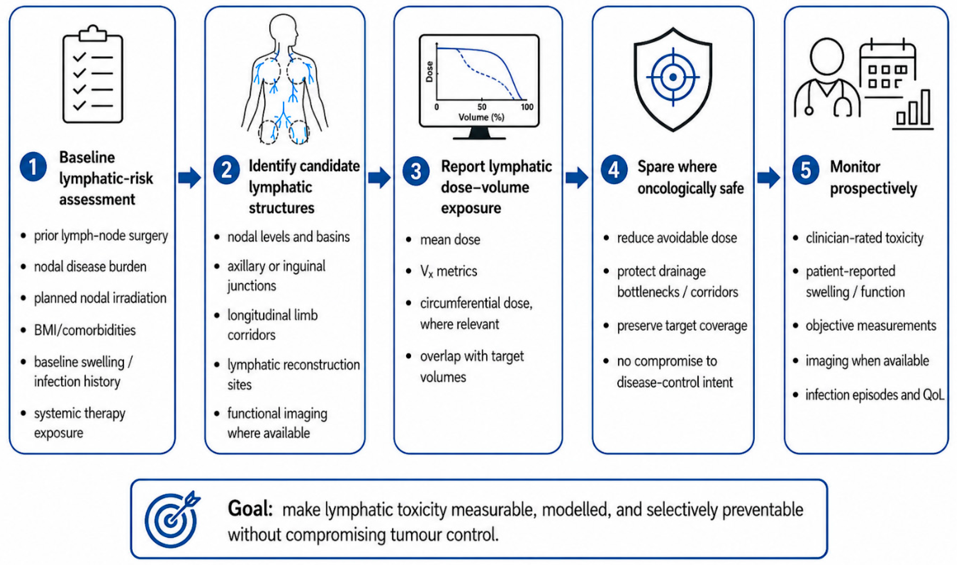
Figure 1 illustrates a pragmatic stepwise approach incorporating lymphatic toxicity into radiotherapy planning without compromising the disease-control intent.
Importantly, lymphatic OAR research should not be limited to identifying constraints. It should also test whether lymphatic-aware planning changes outcomes. A candidate structure may show a dose–toxicity association, but that does not automatically mean it can be spared without sacrificing the target coverage or increasing the recurrence risk. Feasibility planning studies, prospective observational cohorts, and, eventually, interventional trials will be needed to determine whether lymph-sparing approaches reduce the clinically meaningful lymphedema. Until then, the safest position is neither dismissal nor overadoption, but structured evaluation.
4. A Pragmatic Research and Practice Agenda
If the lymphatic system is to enter radiotherapy-planning culture, the first step should be standardisation rather than constraint-setting. The current evidence is sufficient to justify prospective attention, but not sufficient to justify universal dose limits. The field therefore needs a staged research agenda that separates three questions that are often conflated: (a) which lymphatic structures can be reproducibly identified, (b) which of these structures are associated with clinically meaningful toxicity, and (c) whether sparing them improves outcomes without compromising tumour control.
The priority is a common endpoint language. Lymphedema is frequently reported as if it were a single toxicity; yet, clinical syndromes differ substantially across tumour sites. Upper-limb swelling after breast cancer treatment, facial oedema after nasopharyngeal chemoradiation, mucosal and laryngeal oedema after head and neck radiotherapy, genital or lower-limb swelling after pelvic and inguinal irradiation, and limb lymphedema after extremity sarcoma treatment are not interchangeable endpoints. They differ in anatomy, measurement, functional consequences, and likely dose–response relationships. Future studies should therefore specify whether the endpoint is external lymphedema, internal lymphedema, mixed lymphatic–fibrotic toxicity, cellulitis risk, patient-reported swelling, objective volume change, imaging-detected lymphatic dysfunction, or functional impairment.
The second priority is reproducible contouring. Candidate lymphatic structures should be defined in ways that can be applied across institutions. In breast cancer, this may include the axillary–lateral thoracic vessel junction, supraclavicular region, or lymphatic reconstruction sites, while acknowledging that the ALTJ debate remains unresolved. In head and neck cancer, candidate structures may include nodal levels, anterior neck soft tissues, laryngeal and pharyngeal structures, and regions associated with internal or external lymphedema. In pelvic and inguinal cancers, pelvic nodal basins, inguinal-femoral drainage regions, and surgical disruption sites may be more practical starting points than attempting to contour microscopic lymphatic vessels. In extremity sarcoma, longitudinal lymphatic corridors may be more relevant than isolated nodal points. These candidate structures should initially be treated as research contours, not mandatory constraints.
The third priority is routine dose reporting. For candidate lymphatic structures, investigators should report the mean dose, maximum dose, Vx metrics, low-dose bath, circumferential dose where relevant, and overlap with target volumes. This reporting should be accompanied by surgical details, nodal burden, chemotherapy exposure, baseline swelling, body mass index, infection history, and follow-up duration. Without these variables, dose–response models will remain confounded by the same factors that already limit the interpretation of the literature. The aim is not to prove that the dose alone causes lymphedema, but to determine whether the dose contributes meaningfully to cumulative lymphatic failure in clinically identifiable subgroups.
The fourth priority is prospective risk stratification. Not every patient receiving radiotherapy requires lymphatic-aware planning. The concept is most relevant where the lymphatic reserve is already compromised or where the treatment intentionally exposes major drainage pathways. High-risk examples include regional nodal irradiation after axillary surgery, bilateral neck chemoradiation, postoperative head and neck radiotherapy after nodal dissection, pelvic plus inguinal nodal irradiation, treatment after lymphatic reconstruction, and extremity radiotherapy volumes that threaten circumferential dose or longitudinal drainage corridors. These are the settings in which the baseline assessment, dose reporting, early lymphedema referral, and selective sparing are most likely to be clinically meaningful.
The fifth priority is careful language around “sparing.” Lymphatic-aware planning should not be interpreted as reducing target coverage, omitting indicated nodal treatment, or treating lymphatic preservation as superior to disease control. Gross nodal disease and high-risk clinical target volumes must remain adequately treated. The practical opportunity lies in reducing the avoidable dose outside essential targets, preserving drainage corridors where feasible, avoiding unnecessary circumferential irradiation, and protecting surgically reconstructed or functionally important drainage pathways when this can be done without changing oncological intent. This distinction is critical.
Finally, future research should test whether lymphatic-aware planning changes outcomes that matter to patients. Demonstrating an association between dose and swelling is not enough. The next step is to show that candidate lymphatic structures can be identified reproducibly that the dose can be reduced without an unacceptable compromise to the target coverage, and that such a reduction translates into less clinically meaningful lymphedema, better function, fewer infections, or improved quality of life. This will require prospective cohorts, feasibility planning studies, and, eventually, interventional trials. Until then, the most defensible position is not to declare the lymphatic system a mature OAR, but to stop treating it as invisible.
5. Conclusions
Lymphedema remains a late-recognised toxicity in oncology, often addressed only after treatment-related lymphatic injury has occurred. This downstream model is increasingly inadequate. Although modern radiotherapy routinely protects established organs at risk, the lymphatic system is still largely absent from planning, despite evidence that radiation dose, surgery, systemic therapy, and patient-level vulnerability can interact to produce clinically meaningful lymphatic toxicity.
Lymphatic-aware radiotherapy should therefore be considered, but cautiously. Current evidence does not support universal lymphatic dose constraints, and nodal target coverage must not be compromised for unvalidated sparing. However, in selected high-risk settings, lymphatic structures should be treated as candidate toxicity structures for the baseline risk assessment, exploratory contouring, dose–volume reporting, prospective monitoring, and selective sparing where oncologically safe.
The proposed lymphatic-aware radiotherapy framework offers a practical first step: assess the baseline lymphatic risk, identify candidate lymphatic structures, report the dose–volume exposure, spare where oncologically safe, and monitor the toxicity prospectively. The immediate goal is not to contour every lymphatic vessel, but to make lymphatic toxicity visible, measurable, and modelled within radiotherapy planning. If future studies validate the reproducible lymphatic structures, dose–toxicity relationships, and patient-centred benefits of sparing, lymphatic organs at risk may eventually enter routine practice. Until then, radiation oncology should no longer treat the lymphatic system as invisible.