


Abstract
IgG4-related disease is a chronic, immune-mediated fibro-inflammatory condition that causes swelling, scarring (fibrosis), and tumour-like masses in multiple organs, and this case report aims to highlight its potential to mimic lymphoma and the resulting diagnostic challenge. We report a diagnostically challenging case of a 79-year-old man who presented with mediastinal lymphadenopathy, hepatosplenomegaly, and renal enlargement, raising suspicion for clinical lymphoma. However, the histological evaluation of a submandibular gland excision revealed fibrosis, a dense IgG4-positive plasma cell infiltrate (>100/HPF), and an IgG4:IgG ratio > 40%, supportive of IgG4-related disease (IgG4-RD) in the appropriate clinicopathologic context. This case illustrates an important but well-recognised diagnostic pitfall in which IgG4-RD may clinically and radiologically mimic lymphoma. PET-CT demonstrated multiorgan involvement with diffuse FDG uptake, but definitive diagnosis required the integration of clinical, radiologic, serologic, and pathologic findings. The patient’s laboratory profile, including hypocomplementemia and elevated inflammatory markers, supported the proliferative phenotype of IgG4-RD—recently proposed in the literature as a clinically distinct subgroup with systemic involvement and steroid responsiveness. Rather than representing a novel presentation, this case reinforces the importance of integrated assessment in distinguishing IgG4-RD from haematolymphoid malignancy. PET-CT served as a useful adjunct for identifying multiorgan disease and guiding diagnostic evaluation, but tissue evaluation remained essential to avoid misdiagnosis and inappropriate treatment. Recognition of this entity is vital to avoid misdiagnosis and inappropriate treatment.
1. Introduction
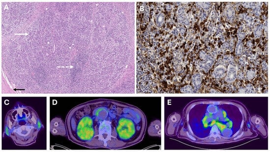
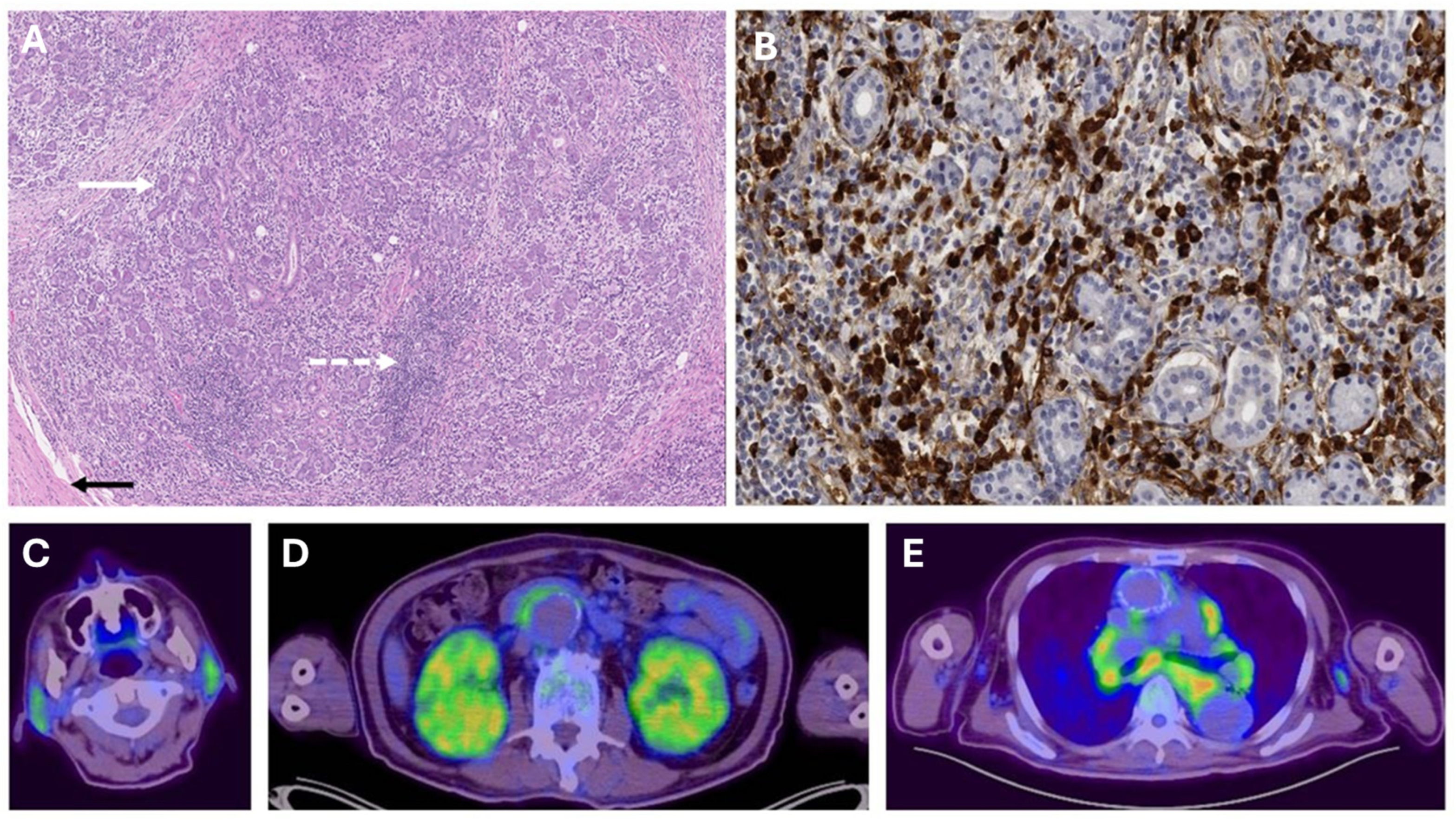
IgG4-related disease is a chronic, immune-mediated fibro-inflammatory condition that causes swelling, scarring (fibrosis), and tumour-like masses in multiple organs. This case highlights a rare systemic presentation of IgG4-related disease with multi-organ FDG uptake closely mimicking advanced-stage lymphoproliferative malignancy on PET/CT, underscoring the diagnostic challenge and the necessity of histopathological confirmation. We reported a 79-year-old man presented with a one-year history of significant weight loss. Computed tomography (CT) imaging revealed mediastinal lymphadenopathy, mild hepatosplenomegaly, and bilateral renal hypertrophy, raising clinical suspicion for lymphoma. An 18F-fluorodeoxyglucose (FDG) PET-CT scan demonstrated mild FDG-avid periaortic soft tissue thickening, diffuse splenic uptake, bilateral parotid and submandibular gland involvement, and enlarged kidneys with heterogeneous FDG uptake (Figure 1). Given these findings, a lymphoproliferative disorder was considered an important diagnostic possibility.
Figure 1.
(A) Biopsy of submandibular gland showing moderately dense inflammatory infiltrate (dotted arrow) within and surrounding the lobules (solid arrow), and bands of fibrosis within the gland (solid black arrow). (B) IgG4 immunohistochemical stain demonstrating abundance of IgG4+ plasma cells. (C) Whole-body 18F-FDG PET/CT shows bilateral parotid and submandibular gland FDG uptake. (D) Heterogeneous mild FDG-avid periaortic soft tissue thickening anterior to distant abdominal aorta and increased FDG uptake in bilateral enlarged kidneys, and (E) multiple FDG-avid non-bulky mediastinal lymphadenopathy.
2. Detailed Case Description
The histological evaluation of a left submandibular gland excision biopsy ruled out malignancy and instead revealed moderately dense lymphoplasmacytic infiltrate, fibrosis with over 100 IgG4-positive plasma cells per high-power field, and an IgG4:IgG ratio exceeding 40%—findings supportive of IgG4-related disease (IgG4-RD). The patient had associated raised ESR of 94 mm/Hr (Reference Range (RR) 1–10 mm/Hr), C-reactive protein of 20.7 mg/L (RR: 0.0–5.0 mg/L), and hypocomplementemia with C3 0.33 g/L (RR: 0.70–1.60 g/L) and C4 0.15 g/L (RR: 0.17–0.48 g/L). Despite these histopathological findings, the patient’s serum IgG4 levels were normal at 15.7 mg/dL (RR: 2.4–121.0 mg/dL). Transbronchial needle aspiration of mediastinal lymph nodes showed granulomatous inflammation and negative for mycobacterium tuberculosis. Given his advanced age and comorbidities, the patient declined further invasive investigations. Clinical diagnosis was revised to IgG4-RD (Mikulicz/systemic phenotype) after integration of the salivary gland histology, multiorgan imaging pattern, inflammatory and complement abnormalities, exclusion of malignancy, and subsequent steroid responsiveness. He was subsequently started on oral prednisolone at 0.6 mg/kg (40 mg daily), which resulted in a sustained clinical response with regression of lymphadenopathy on pan-CT scan performed 6 weeks after prednisolone and weight gain. He remained clinically stable thereafter, with steroid-responsive regression of lymphadenopathy, improvement in hepatosplenomegaly and bilateral renal involvement, and survived a further 4 years following initiation of steroid therapy, before passing away from an unrelated cardiovascular event.
3. Discussion
IgG4-RD is a heterogeneous clinical entity encompassing a wide variety of diseases [1], with several existing guidelines proposing clinical, radiological, and pathological criteria [2,3,4]. Lymphoma is a well-recognised and important differential diagnosis of IgG4-RD, particularly in patients presenting with systemic lymphadenopathy, salivary gland enlargement, constitutional symptoms, and multiorgan FDG-avid disease. Accordingly, the present case is not intended to represent a rare or previously unreported diagnostic dilemma, but rather an instructive example of a clinically important and well-established pitfall. The recent fifth edition of the WHO Classification of Haematolymphoid Tumours [5] is relevant, as it reinforces the recognition of IgG4-RD as a distinct fibroinflammatory disorder within haematopathology differential diagnosis and acknowledges the spectrum of IgG4-related lymphadenopathy. The defining histological characteristics of IgG4-RD include storiform fibrosis, a dense IgG4-positive plasma cell infiltrate, and obliterative phlebitis. However, there are histological variations depending on organ systems. For example, storiform fibrosis and obliterative phlebitis may be inconspicuous or absent in some organs, like the salivary gland [6]. Histopathology is highly informative in IgG4-RD, but no single morphologic or immunostaining feature is diagnostic in isolation. Characteristic findings include a lymphoplasmacytic infiltrate, fibrosis, increased IgG4-positive plasma cells, storiform fibrosis, and obliterative phlebitis, although their prominence varies by organ. In salivary gland disease, storiform fibrosis and obliterative phlebitis may be inconspicuous or absent [6]. In the present case, re-review of the submandibular gland biopsy confirmed a moderately dense lymphoplasmacytic infiltrate, fibrosis, abundant IgG4-positive plasma cells, and an IgG4:IgG ratio > 40%, while obliterative phlebitis was not identified and storiform fibrosis was not apparent. These findings were supportive, but not pathognomonic, and therefore had to be interpreted together with the clinical, radiologic, and laboratory context.
Unlike lymphomas, the lymphoplasmacytic infiltrate does not form sheets and does not show atypia. Furthermore, unlike lymphomas, IgG4-RD maintains a polyclonal plasma cell infiltrate and does not demonstrate aberrant light-chain restriction. A major refinement in the fifth edition is the recognition of IgG4-related lymphadenopathy as a distinct entity, acknowledging that lymph node involvement may present in several histological patterns, including Castleman disease-like changes, progressive germinal centre transformation, and interfollicular expansion. An increase in IgG4-positive plasma cells alone is now recognised as insufficient for diagnosis without accompanying architectural alterations and stromal fibrosis. Additionally, while serum IgG4 cutoff of 135 mg/dL has been widely used in the literature and in diagnostic criteria for IgG4-RD, up to 30% of patients with histologically confirmed disease may have normal or even low IgG4 titres, which may be related to a prozone effect [7]. This is relevant to the present case, in which the histopathological findings were supportive despite a normal serum IgG4 level, but required interpretation within the wider clinicoradiologic context. The 2019 ACR/EULAR classification criteria [4], in contrast, adopt a structured, weighted scoring system that integrates histopathology, serology, and imaging. Entry into the classification process requires typical organ involvement or a biopsy demonstrating a lymphoplasmacytic infiltrate of uncertain aetiology. A point-based scoring system is then applied, assigning the highest weight to storiform fibrosis and obliterative phlebitis (up to +13 points) and immunostaining for IgG4+ plasma cells (up to +16 points). Other features, such as serum IgG4 elevation, glandular involvement, and characteristic imaging findings, contribute additional points. Cases that reach a threshold of ≥20 points are classified as IgG4-RD, provided they do not meet exclusion criteria for malignancy, granulomatous inflammation, or autoimmune disease. While the 2019 ACR/EULAR classification criteria provide a quantitative, standardised approach, the guideline also emphasises that IgG4-RD is best approached through the integration of clinicopathologic, serologic, and radiologic findings rather than by reliance on any single feature.
Recent conceptual advances have further refined the understanding of IgG4-related disease (IgG4-RD). Stone [8] has argued that IgG4-RD may reasonably be considered a form of variable-vessel vasculitis because it can involve small-, medium-, and large-sized vessels, with recognised manifestations including phlebitis, aortitis/periaortitis, and coronary periarterial disease. At the same time, no histopathologic or immunostaining feature is diagnostic in isolation. Although lymphoplasmacytic infiltrates, fibrosis, increased IgG4-positive plasma cells, storiform fibrosis, and phlebitis are characteristic findings, diagnosis requires the integration of clinical, radiologic, serologic, and pathologic information, together with the exclusion of important mimics such as malignancy and infection. This framework is particularly relevant to the present case, in which the final diagnosis rested on integrated assessment rather than on any single histologic feature.
Although PET-CT is not part of the formal diagnostic criteria, it plays a supportive role in differentiating IgG4-RD from lymphoma. IgG4-RD lesions typically demonstrate diffuse, mild to moderate FDG uptake, a pattern distinct from the intense, focal hypermetabolism often seen in aggressive lymphoma. This case adds to the existing literature supporting 18F-FDG PET/CT as a useful adjunct in the evaluation of IgG4-RD. Compared to other conventional imaging modalities, it is more sensitive in detecting lesions/multiorgan involvement [9,10,11]. Increased 18F-FDG uptake according to specific image patterns (e.g., submandibular/salivary glands, retroperitoneal, pancreatic, vascular wall activity) allow differentiation between IgG4-RD versus other diseases [12,13], as seen in this case. Furthermore, PET-CT findings in IgG4-RD often involve multiple organ systems, including characteristic sites such as the pancreas, salivary glands, periaortic regions, and kidneys, further suggesting a systemic inflammatory rather than neoplastic process. PET-CT has also been used to select biopsy sites in patients with multi-organ disease [9,11] and to monitor treatment response [9], as demonstrated by the interval regression of lymphadenopathy in this patient following corticosteroid therapy. However, while PET-CT can help guide clinical decision-making, it should not replace histopathologic confirmation.
4. Conclusions
In the present case, this integrative approach was particularly important because the salivary gland biopsy showed supportive but not pathognomonic findings, with abundant IgG4-positive plasma cells and fibrosis, while obliterative phlebitis was not identified and storiform fibrosis was not apparent on re-review. The value of this report lies not in describing a rare presentation, but in illustrating a well-recognised and clinically important diagnostic pitfall in which IgG4-RD may mimic lymphoma. In this setting, accurate diagnosis depends on the careful integration of histopathology with organ distribution on imaging, laboratory abnormalities, exclusion of mimics, and treatment response. The WHO fifth edition provides a useful contemporary classificatory context. It supports the recognition of IgG4-RD and its lymphadenopathic manifestations within haematopathology practice, while also reinforcing the fact that serum IgG4 levels are neither sufficiently sensitive nor specific for diagnosis in isolation. PET-CT is as a useful adjunct in identifying multiorgan involvement, guiding biopsy site selection, and monitoring disease activity, particularly in proliferative forms of IgG4-RD [14]. In cases where IgG4-RD mimics lymphoma, prompt tissue-based evaluation remains the gold standard to ensure accurate diagnosis and avoid unnecessary oncologic treatment when interpreted within the broader clinical context.
Author Contributions
B.E.F. and L.X.A.T. wrote the initial draft and Y.L.T. provided histopathology expertise and images. All authors contributed to the critical revision of the manuscript. All authors have read and agreed to the published version of the manuscript.
Funding
This research received no external funding.
Institutional Review Board Statement
No ethical board review was required by the National Healthcare Group Domain Specific Review Board (https://ethics.gri.nhg.com.sg/investigator-manual/ accessed on 1 March 2026) as this is a single case report.
Informed Consent Statement
Informed consent was obtained for publication.
Data Availability Statement
The data that support the findings of this study are available from the corresponding author upon reasonable request.
Conflicts of Interest
The authors declare no conflicts of interest.
References
- Chen, L.Y.C. IgG4-related disease for the hematologist. Hematology 2024, 2024, 594–603. [Google Scholar] [CrossRef] [Scilit] [PubMed]
- Umehara, H.; Okazaki, K.; Kawano, M.; Mimori, T.; Chiba, T. How to diagnose IgG4-related disease. Ann. Rheum. Dis. 2017, 76, e46–e47. Available online: https://ard.bmj.com/content/76/11/e46 (accessed on 6 December 2019). [CrossRef] [Scilit] [PubMed]
- Umehara, H.; Okazaki, K.; Kawa, S.; Takahashi, H.; Goto, H.; Matsui, S.; Ishizaka, N.; Akamizu, T.; Sato, Y.; Kawano, M.; et al. The 2020 revised comprehensive diagnostic (RCD) criteria for IgG4-RD. Mod. Rheumatol. 2021, 31, 529–533. [Google Scholar] [CrossRef] [Scilit] [PubMed]
- Wallace, Z.S.; Naden, R.P.; Chari, S.; Choi, H.K.; Della-Torre, E.; Dicaire, J.F.; Hart, P.A.; Inoue, D.; Kawano, M.; Khosroshahi, A.; et al. The 2019 American College of Rheumatology/European League Against Rheumatism Classification Criteria for IgG4-Related Disease. Arthritis Rheumatol. 2019, 79, 77–87. [Google Scholar]
- Alaggio, R.; Amador, C.; Anagnostopoulos, I.; Attygalle, A.D.; Araujo, I.B.D.O.; Berti, E.; Bhagat, G.; Borges, A.M.; Boyer, D.; Calaminici, M.; et al. The 5th edition of the World Health Organization Classification of Haematolymphoid Tumours: Lymphoid Neoplasms. Leukemia 2022, 36, 1720–1748, Correction in Leukemia 2023, 37, 1944–1951. https://doi.org/10.1038/s41375-023-01962-5. [Google Scholar] [CrossRef] [Scilit] [PubMed]
- Deshpande, V.; Zen, Y.; Chan, J.K.; E Yi, E.; Sato, Y.; Yoshino, T.; Klöppel, G.; Heathcote, J.G.; Khosroshahi, A.; A Ferry, J.; et al. Consensus statement on the pathology of IgG4-related disease. Mod. Pathol. 2012, 25, 1181–1192. [Google Scholar] [CrossRef] [Scilit] [PubMed]
- Khosroshahi, A.; Cheryk, L.A.; Carruthers, M.N.; Edwards, J.A.; Bloch, D.B.; Stone, J.H. Brief Report: Spuriously low serum IgG4 concentrations caused by the prozone phenomenon in patients with IgG4-related disease. Arthritis Rheumatol. 2014, 66, 213–217. [Google Scholar] [CrossRef] [Scilit] [PubMed]
- Stone, J.H. IgG4-related disease: Lessons from the first 20 years. Rheumatology 2025, 64, i24–i27. [Google Scholar] [CrossRef] [Scilit] [PubMed]
- Tang, C.Y.L.; Chua, W.M.; Cheng, L.T.J.; Fong, W.; Zaheer, S.; Lam, W.W.-C. 18F-FDG PET/CT Manifestations of IgG4-related Disease. Br. J. Radiol. 2021, 94, 0210105. [Google Scholar] [CrossRef] [Scilit] [PubMed]
- Chen, J.; Li, X.L.; Huang, M. Utility of 18F-FDG PET/CT for differential diagnosis between IgG4-related lymphadenopathy and angioimmunoblastic T-cell lymphoma. Clin. Radiol. 2024, 79, 205–212. [Google Scholar] [CrossRef] [Scilit] [PubMed]
- Pucar, D.; Hinchcliff, M. FDG PET vascular imaging in IgG4-RD: Potential and challenges. J. Nucl. Cardiol. 2022, 29, 2934–2937. [Google Scholar] [CrossRef] [Scilit] [PubMed]
- Zhang, J.; Chen, H.; Ma, Y.; Xiao, Y.; Niu, N.; Lin, W.; Wang, X.; Liang, Z.; Zhang, F.; Li, F.; et al. Characterizing IgG4-related disease with 18F-FDG PET/CT: A prospective cohort study. Eur. J. Nucl. Med. Mol. Imaging 2014, 41, 1624–1634. [Google Scholar] [CrossRef] [Scilit] [PubMed]
- Lee, J.; Hyun, S.H.; Kim, S.; Kim, D.-K.; Lee, J.K.; Moon, S.H.; Cho, Y.S.; Choe, Y.S.; Kim, B.-T.; Lee, K.-H. Utility of FDG PET/CT for Differential Diagnosis of Patients Clinically Suspected of IgG4-Related Disease. Clin. Nucl. Med. 2016, 41, e237–e243. [Google Scholar] [CrossRef] [Scilit] [PubMed]
- Katz, G.; Hernandez-Barco, Y.; Palumbo, D.; Guy, T.V.; Dong, L.; Perugino, C.A. Proliferative features of IgG4-related disease. Lancet Rheumatol. 2024, 6, e481–e492. [Google Scholar] [CrossRef] [Scilit] [PubMed]
Disclaimer/Publisher’s Note: The statements, opinions and data contained in all publications are solely those of the individual author(s) and contributor(s) and not of MDPI and/or the editor(s). MDPI and/or the editor(s) disclaim responsibility for any injury to people or property resulting from any ideas, methods, instructions or products referred to in the content. |
© 2026 by the authors. Licensee MDPI, Basel, Switzerland. This article is an open access article distributed under the terms and conditions of the Creative Commons Attribution (CC BY) license.