


Abstract
The hepatic lymphatic system, long underappreciated, plays a critical role in liver physiology by maintaining interstitial fluid balance, removing metabolic waste, and facilitating immune surveillance. Emerging evidence indicates that lymphatic dysfunction contributes to the pathogenesis and progression of multiple liver diseases, including non-alcoholic fatty liver disease/non-alcoholic steatohepatitis (NAFLD/NASH), hepatitis, fibrosis, cirrhosis, and hepatocellular carcinoma. This review summarizes current knowledge on hepatic lymphatic anatomy, physiology, and molecular regulation, highlights pathological alterations, and discusses potential therapeutic implications. A better understanding of the hepatic lymphatic system may enable the development of novel lymphatic-targeted strategies to improve liver health and treat liver disease.
1. Introduction
The liver is a central metabolic organ essential for maintaining systemic physiological balance. Its diverse functions include the synthesis of plasma proteins, detoxification of endogenous metabolites and xenobiotics, regulation of glucose and lipid metabolism, storage of vitamins and glycogen, and secretion of bile necessary for digestion and nutrient absorption [1]. These processes collectively support metabolic homeostasis across multiple organ systems. The liver’s functional complexity is underpinned by its heterogeneous cellular composition, which includes hepatocytes, hepatic stellate cells, Kupffer cells, liver sinusoidal endothelial cells (LSECs), and cholangiocytes. Each cell type contributes to the unique function of the liver through nutrient processing, immune regulation, extracellular matrix remodeling, and bile-duct maintenance [2].
Beyond well-characterized cellular and molecular mechanisms, recent research highlights the hepatic lymphatic system as an essential yet historically neglected component of liver biology. The liver is one of the largest contributors of lymph or lymphatic fluid in the body, producing approximately 25–50% of the lymph entering the thoracic duct [3,4]. This significant volumetric contribution underscores the critical role of hepatic lymph as a major conduit for transporting liver-derived antigens and immune cells to the systemic circulation. Hepatic lymph originates primarily from plasma filtrate passing through highly permeable hepatic sinusoids into the perisinusoidal interstitial space (Space of Disse) [5,6]. This lymph consists of proteins, lipids, cholesterol, immune cells, and metabolic waste products, which are transported via lymphatic vessels to draining lymph nodes for filtration and immunological surveillance. Through this process, the hepatic lymphatic system maintains interstitial fluid balance, clearing excess macromolecules, as well as regulates immune cell trafficking, supporting hepatic homeostasis [7].
Despite its physiological importance, the hepatic lymphatic system has long been underappreciated in both research and clinical contexts. Only recently has its relevance to liver health and disease begun to be systematically explored. Pathological conditions, including hepatitis, non-alcoholic fatty liver disease (NAFLD/NASH), fibrosis, cirrhosis, portal hypertension, and hepatocellular carcinoma, significantly affect hepatic lymphatic function [8]. Key alterations include: increased lymph production, lymphatic vessel remodeling (lymphangiogenesis), impaired lymph drainage, disrupted immune trafficking, and accumulation of proteins and lipids. Therefore, understanding its function and dysfunction may reveal novel strategies for intervention. Therapeutic targeting of the hepatic lymphatics could enhance fluid clearance and resolve inflammation, modulating the pathological immune responses, to offer potential treatment avenues for a wide range of liver diseases [9]. Ongoing research into the molecular regulation of lymphangiogenesis and lymphatic remodeling is critical for translating these insights into clinical applications [10,11].
In this review, we aim to synthesize the current knowledge on the physiological and pathological roles of the hepatic lymphatic system. We examine its anatomical organization, molecular regulation, and contributions to liver function in health. We also explore the mechanisms of lymphatic dysfunction across various liver diseases and highlight emerging therapeutic opportunities that leverage lymphatic-targeted strategies. By consolidating current evidence and identifying key knowledge gaps, this review provides a comprehensive foundation for future research and supports the development of innovative clinical approaches that harness the hepatic lymphatic system in the management of liver diseases.
2. Anatomy and Drainage of the Hepatic Lymphatic System
2.1. Overview of Hepatic Lymph Formation
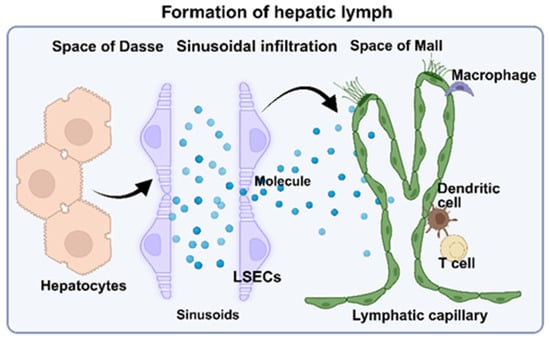
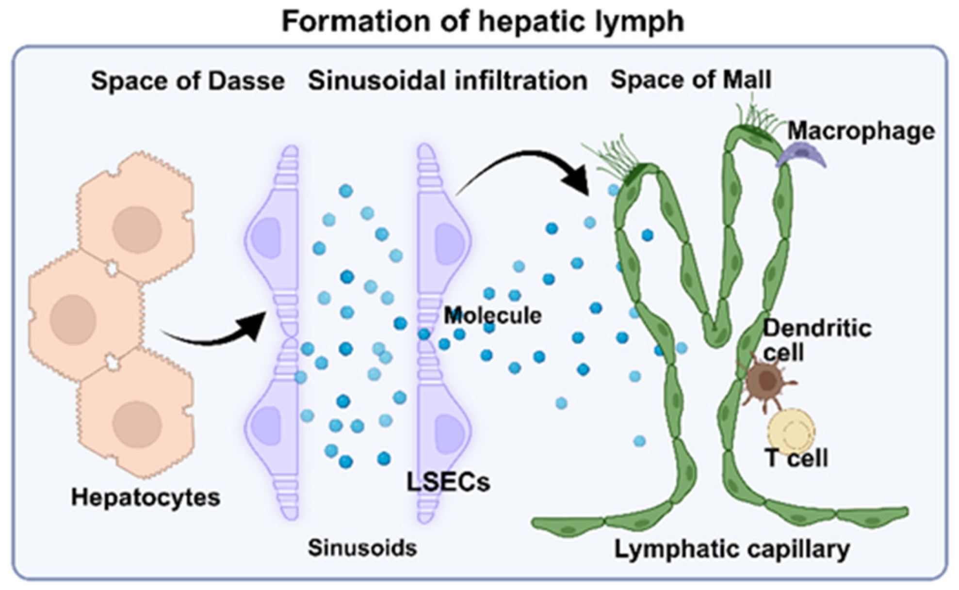
The liver produces a substantial volume of lymph. Estimates suggest that between 25% and 50% of the lymph entering the thoracic duct originates from the liver. Hepatic lymph stems from plasma filtrate that accumulates in the space of Disse (Figure 1), the narrow extracellular compartment situated between hepatocytes and liver sinusoidal endothelial cells (LSECs). Hepatic sinusoids have a uniquely fenestrated, discontinuous endothelium lacking a basement membrane, to permit extensive bidirectional exchange of fluid and macromolecules. Under normal hydrostatic and oncotic pressure gradients, fluid exits the sinusoidal plasma, enters the space of Disse, and subsequently drains into the hepatic lymphatic system [12,13].
Figure 1.
Overview of Hepatic Lymph Formation. The space of Disse is a perisinusoidal compartment between hepatocytes and fenestrated liver sinusoidal endothelial cells that facilitates plasma filtration, metabolic exchange, and lipoprotein transport. Sinusoidal endothelial fenestrae regulate fluid and macromolecule exchange between the sinusoidal lumen and the space of Disse. Interstitial fluid is subsequently collected by initial lymphatic capillaries in the space of Mall and drained into periportal lymphatic vessels, which also interact with immune cells (i.e., macrophage, dendritic cell, and T cell) and contribute to hepatic immune regulation. Created in BioRender. Do, L. (2025) https://BioRender.com/9wkyxw3 (accessed on 22 December 2025).
2.2. Lymphatic Subsystems and Drainage Pathways
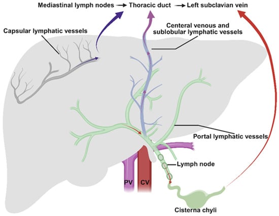
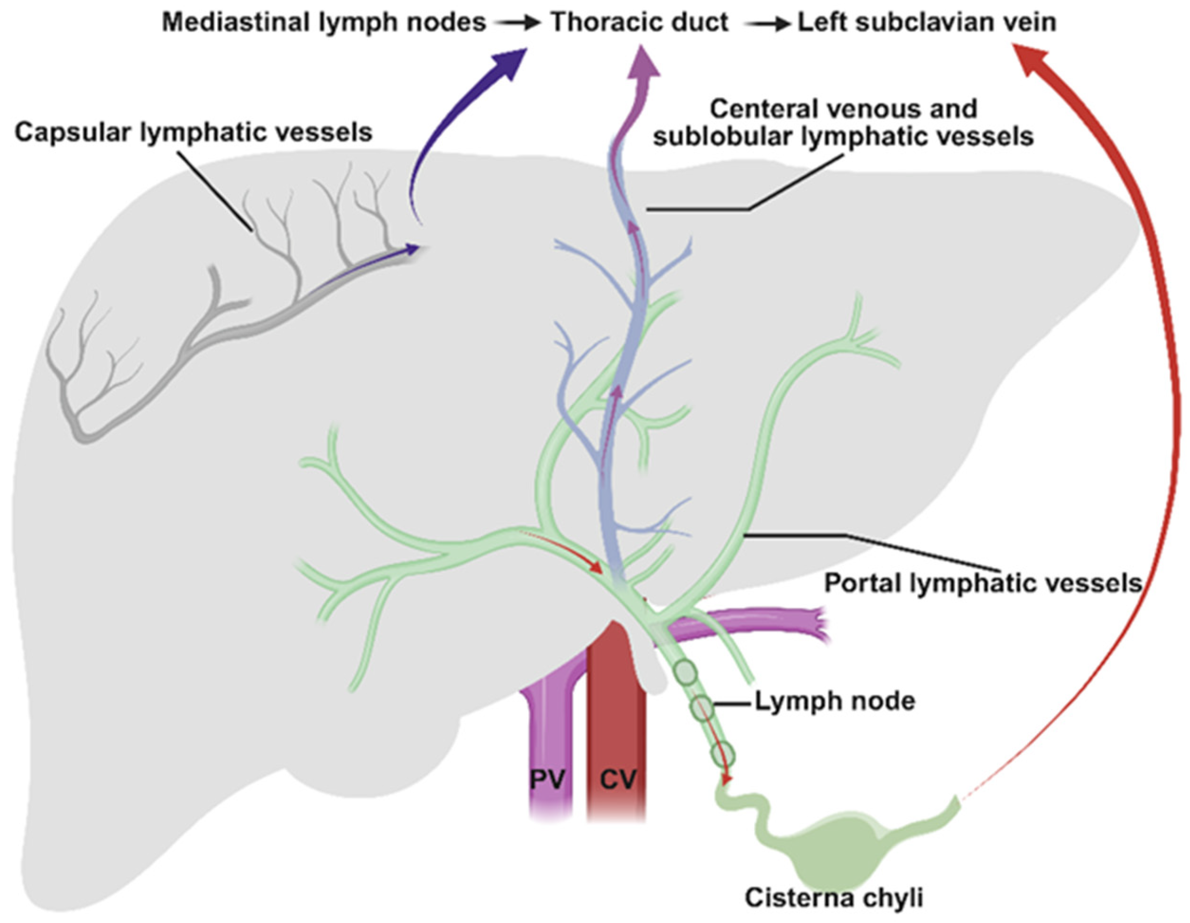
The hepatic lymphatic system is composed of several interconnected subsystems that together regulate interstitial fluid homeostasis, macromolecule clearance, and immune trafficking. These subsystems follow the anatomical compartments of the liver and can be broadly divided into: portal lymphatics, sublobular and capsular lymphatics, and deep parenchymal drainage pathways (Figure 2), each contributing individually to lymph formation and transport [3,5,14].
Figure 2.
Hepatic Lymph Drainage Pathway. Hepatic lymph drainage includes three pathways. The portal lymphatic subsystem accounts for ~80% of hepatic lymph drainage. Interstitial fluid from the space of Disse enters blind-ended lymphatic capillaries in the space of Mall, then drains into collecting lymphatic vessels within the portal tract that convey lymph toward the hilar lymphatics, celiac lymph nodes, cisterna chyli, and thoracic duct (red arrows). Approximately 20% of hepatic lymph originates from central venous and sublobular regions, where lymphatic channels adjacent to hepatic veins collect perivenular interstitial fluid, particularly under elevated sinusoidal pressure (purple arrows). Capsular lymphatics drain the liver surface to diaphragmatic and peritoneal lymph nodes, becoming prominent during inflammation or increased intrahepatic pressure (deep blue arrows). PV: Portal Vein; CV: Central Vein. Created in BioRender. Do, L. (2025) https://BioRender.com/h383ur8 (accessed on 22 December 2025).
The portal lymphatic subsystem represents the principal route of hepatic lymph drainage (~80%) under physiological conditions. Initial lymphatic capillaries arise in the space of Mall at the periportal interface, where interstitial fluid emerging from the space of Disse converges. These blind-ended lymphatic capillaries exhibit hallmark “button-like” junctions that support efficient uptake of fluid, proteins, and immune cells. As lymph enters the portal tract, it transitions into larger collecting lymphatic vessels that run alongside the portal vein and hepatic artery. These vessels possess valves and a layered smooth muscle structure that promote unidirectional flow toward the hilar (peri-hepatic) lymphatics. Lymph gets transported through celiac lymph nodes and cisterna chyli, ultimately draining into mediastinal lymph nodes and the thoracic duct [15,16,17,18].
A smaller yet functionally relevant proportion of hepatic lymph (~20%) is derived from central venous and sublobular regions. Lymphatic channels adjacent to hepatic veins collect fluid from perivenular interstitium, especially when sinusoidal pressures are elevated. Although less prominent during basal states, this subsystem becomes more active in pathological conditions like hepatic congestion, venous outflow obstruction, or right-sided heart failure. These lymphatics drain toward deeper hilar channels distinct from the portal system, emphasizing the liver’s compartmentalized lymphatic architecture [15,16,17,18].
The capsular or superficial lymphatic network, located beneath Glisson’s capsule, drains fluid from the liver surface and subcapsular region. These vessels are densely connected to extrahepatic lymphatics of the diaphragm and anterior abdominal wall. Although they play a minor role in basal hepatic lymph transport, capsular lymphatics are crucial during inflammatory processes that preferentially affect the liver surface (e.g., peritonitis, capsular fibrosis) or in states of elevated intrahepatic pressure that redirect fluid toward superficial channels. They also provide a route for immune cell trafficking to diaphragmatic and peritoneal lymph nodes [3,5,19].
Traditionally, hepatic lymphatic vessels have been studied using histological, imaging, molecular, and functional approaches, which are largely limited to two-dimensional analysis. More recently, optimized tissue-clearing and co-immunolabeling protocols have been developed that enable efficient three-dimensional visualization and quantitative assessment of lymphatic vessels in normal and diseased livers across species [15,16]. Using these approaches, hepatic lymphatic networks have been identified along portal tracts, representing the deep lymphatic system, as well as within Glisson’s capsule, corresponding to the superficial lymphatic system. Moreover, near-infrared lymphatic imaging, intravital microscopy, genetic lineage tracing using lymphatic-specific Cre mouse models, and single-cell transcriptomic approaches will provide promising methods for exploring hepatic lymphatic structure and function.
3. Pathophysiological Roles of Hepatic Lymphatic System
3.1. Interstitial Fluid Homeostasis and Waste Clearance
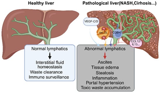
The hepatic lymphatic system maintains interstitial fluid balance and removes metabolic waste from the liver’s highly permeable sinusoidal environment. Because efficient lymphatic drainage is required to prevent increased interstitial pressure and preserve sinusoidal perfusion due to the large volumes of plasma filtrate enter the space of Disse. Initial lymphatics in the space of Mall rapidly clear excess fluid, transporting it through portal collecting vessels to the thoracic duct, to prevent perisinusoidal edema and sustaining nutrient and oxygen exchange. They also remove macromolecules—albumin, immunoglobulins, complement proteins, and lipoprotein remnants (Figure 1 and Figure 3)—that accumulate in the absence of a sinusoidal basement membrane. Specialized lymphatic endothelial cells mediate this uptake, limiting protein-rich edema and maintaining osmotic balance [7,20].
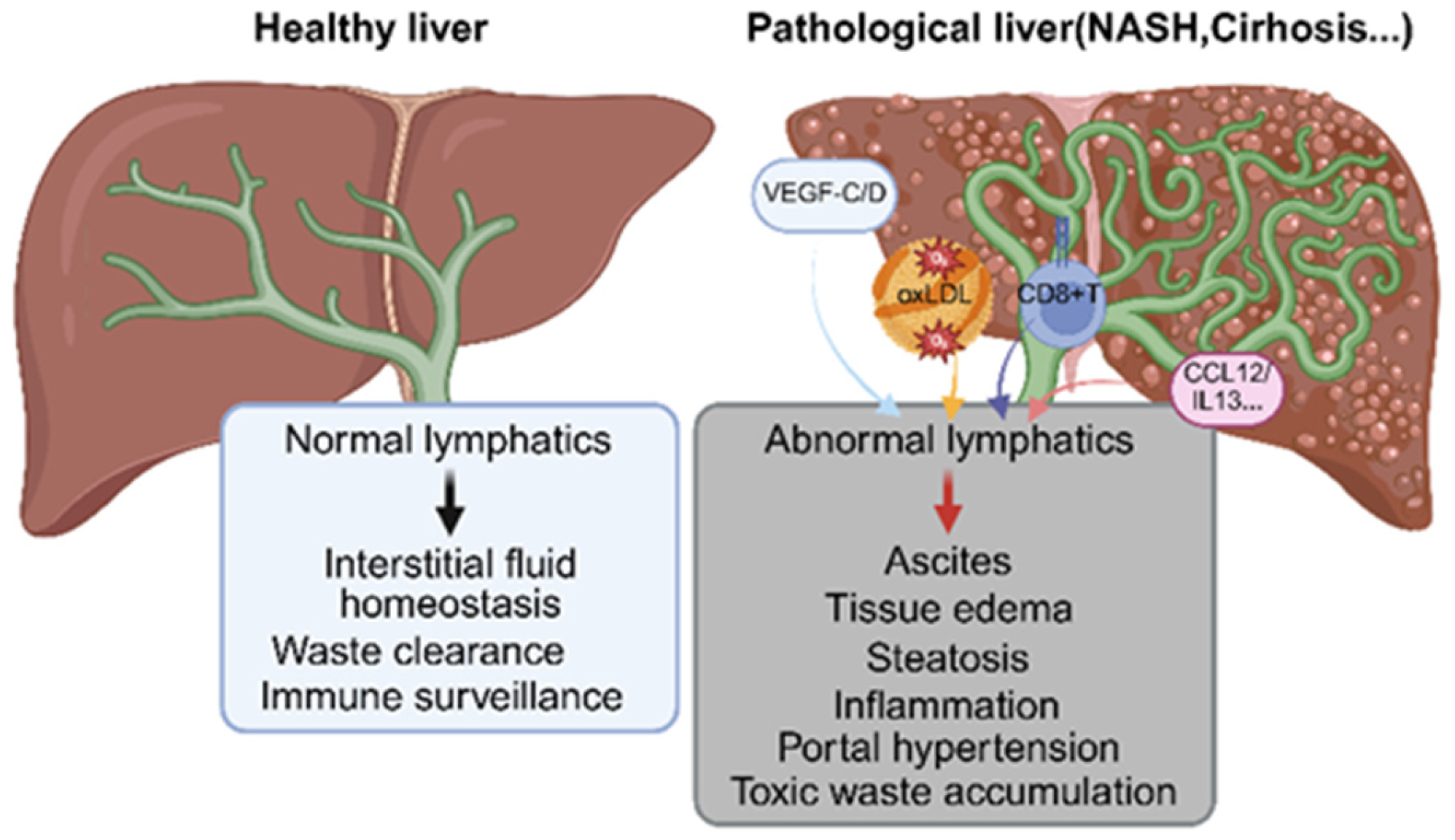
Figure 3.
Pathophysiology of Hepatic Lymphatics. Healthy hepatic lymphatics maintain fluid balance, waste clearance, and immune regulation. Dysfunctional lymphatics (i.e., NASH and cirhosis) with elevated oxLDL (yellow arrow), VEGF-C/D (blue arrow), CCL12, IL-13 (pink arrow), and active CD8+ T cells (dark blue arrow) impair fluid transport, causing edema, steatosis, ascites, inflammation, and portal hypertension. Created in BioRender. Do, L. (2025) https://BioRender.com/x0kdmd2 (accessed on 22 December 2025).
Hepatic lymph further clears metabolic waste such as oxidized proteins, apoptotic debris, extracellular vesicles, and excess lipids, thereby restraining sterile inflammation. It also carries antigens, cytokines, and metabolites to draining lymph nodes, linking hepatic metabolic activity to immune regulation [21].
When lymphatic drainage becomes insufficient—due to inflammation, fibrosis, sinusoidal hypertension, or lymphatic endothelial dysfunction—interstitial fluid accumulates, leading to perisinusoidal edema, impaired molecular exchange, and altered mechanical signaling to stellate cells. This promotes further fibrosis, sinusoidal capillarization, and progressive architectural distortion. The buildup of metabolic waste also amplifies local immune activation, contributing to chronic inflammation and exacerbating disease progression. Conversely, experimental models demonstrate that enhancing lymphangiogenesis or lymphatic function can improve interstitial clearance, reduce inflammation, and mitigate portal hypertension [3,22].
3.2. Dynamic Redistribution in Pathological Condition
Disruption of hepatic microcirculation profoundly alters lymphatic drainage patterns. Portal hypertension increases sinusoidal hydrostatic pressure, resulting in excessive filtration and lymph overproduction. When periportal lymphatics become overwhelmed, excess fluid is redirected toward capsular or perivenular pathways, contributing to hepatic edema and ascites. Conversely, conditions marked by impaired sinusoidal perfusion—such as steatosis or microvascular inflammation—reduce the driving forces for lymph formation, potentially leading to retention of metabolic waste in the interstitial space. Lymphangiogenesis observed in cirrhosis (Figure 3), steatohepatitis, and cholestatic disease reflects an adaptive response to these microcirculatory stresses, aiming to restore efficient interstitial drainage [23,24].
3.3. Bidirectional Signaling Between Blood and Lymph Systems
Beyond physical fluid exchange, the microcirculation and lymphatic system engage in biochemical cross-talk. Shear stress within sinusoids modulates endothelial expression of VEGF-C and VEGF-D, promoting lymphangiogenesis [25]. Conversely, lymphatic endothelial cells influence vascular tone and permeability through cytokine release and immune cell recruitment [26]. This reciprocal signaling creates a dynamic homeostatic loop that adjusts both vascular and lymphatic behavior in response to metabolic demand, inflammation, and injury.
3.4. Lymphangiogenesis
Persistent injury promotes structural remodeling of liver lymphatics. Chronic inflammation, ligand stimulation (e.g., VEGF-C/VEGFR-3 signaling) (Figure 3), and extracellular matrix reorganization collectively foster lymphangiogenesis within both portal tracts and, in advanced disease, fibrotic septa [27,28]. Newly formed lymphatic vessels are often dilated and twist, reflecting high flow demand and impaired mechanical integrity [29,30]. In cirrhosis, this proliferative lymphatic response becomes pronounced, with increased vessel density correlating with disease severity and portal pressure [31]. Similar lymphangiogenic signatures have been observed in nonalcoholic steatohepatitis (NASH), cholestatic disorders, and chronic viral hepatitis, suggesting a shared compensatory mechanism across diverse etiologies [13,21,32].
Although lymphangiogenesis enhances clearance of excess interstitial fluid, its functional consequences remain complex. Expansion of the lymphatic network may improve immune cell trafficking and antigen transport, yet dysfunctional or leaky neo-lymphatics can exacerbate inflammation by facilitating aberrant dendritic cell movement, antigen dispersal, or impaired lymph propulsion [30,33,34]. Moreover, increased lymph flow from the liver contributes to systemic lymphatic overload and may play a role in the formation of ascites, particularly when thoracic duct drainage becomes insufficient [35]. Understanding the balance between adaptive and maladaptive lymphatic remodeling is therefore critical for designing targeted interventions. Emerging therapeutic approaches—including VEGF-C modulation, enhancing lymphatic contractility, and normalizing lymphatic endothelial junctions—offer promising routes to restore fluid homeostasis and immune regulation in chronic liver disease [36,37,38].
3.5. Immune Surveillance and Cell Trafficking
The hepatic lymphatic system serves as a critical interface between the liver’s unique immune environment and the broader adaptive immune system. As the liver continuously encounters blood-borne antigens from the gastrointestinal tract, effective immune surveillance requires mechanisms that can sample interstitial antigens, transport immune cells, and relay contextual information to draining lymph nodes [39,40,41]. Lymphatic vessels within the portal tracts, capsule, and perivenular regions collectively facilitate the movement of immune cells and antigen-rich fluid from the liver parenchyma to regional lymph nodes, thereby orchestrating both tolerance and immunity.
Hepatic lymph carries antigens from hepatocytes, cholangiocytes, Kupffer cells, and infiltrating leukocytes, including microbial fragments, damage-associated molecules, and processed peptides. Initial lymphatics in the space of Mall sample interstitial fluid from the space of Disse and transport antigenic material to hepatic and celiac lymph nodes, where dendritic cells and macrophages interpret signals reflecting hepatic metabolic and inflammatory status. Immune-cell trafficking depends on hepatic lymphatic endothelial cells (LECs), which express chemokines and adhesion molecules enabling leukocyte entry. Dendritic cells migrate along CCL21 gradients after antigen uptake, while macrophage-derived vesicles and hepatocyte stress mediators further shape downstream immune responses [5,30,42,43].
Lymph-borne antigens normally promote tolerance to gut-derived products via regulatory dendritic cells and cytokines, but infection or metabolic stress shifts lymph signals toward immunogenicity and effector T-cell activation (Figure 3). Chronic liver diseases drive VEGF-C/VEGF-D-mediated lymphangiogenesis, enhancing immune-cell recruitment and inflammation, whereas dysfunctional lymphatics impair antigen clearance and lead to sustained macrophage activation [32,44].
Through coordinated antigen transport, immune-cell egress, and lymphatic remodeling, hepatic lymphatics regulate liver–immune communication and represent a potential therapeutic target for restoring immune balance in chronic liver disease.
4. Lymphatic System in Liver Diseases
4.1. Hepatic Lymphatic Vessels in Non-Alcoholic Fatty Liver Disease (NAFLD)
The prevalence of non-alcoholic fatty liver disease (NAFLD) has surged from 8.2% in 1990 to 30.2% in 2023, affecting roughly one-quarter of adults worldwide and reflecting the global rise in metabolic syndrome. Recognizing limitations in the exclusion-based definition and the need to highlight its metabolic basis, NAFLD was redefined as metabolic-associated fatty liver disease (MAFLD) in 2020, and more recently as metabolic dysfunction-associated steatotic liver disease (MASLD) in 2023. These updates emphasize the central role of metabolic dysregulation in disease pathogenesis and facilitate more precise diagnosis and epidemiological assessment [45,46].
In patients with NAFLD, lymphatic density increases during the progression of NAFLD and nonalcoholic steatohepatitis (NASH) [47]. Also, mice fed with a high-fat, high-cholesterol (HFHC) diet to mimic human NASH shows an increase in lymphatic density, an increased proportion of LECs in active cell cycle (S/G2/M) and increased Ki67 staining after 24-week feeding [48]. Both patients and mice with NASH have high levels of oxidized low-density lipoprotein (oxLDL), which leads to decreased permeability in hepatic lymphatic endothelial cells, but has no effect on hepatic sinusoidal endothelial cells [49]. oxLDL modulates intercellular junctions by regulating VE-Cadherin and VEGFR-3, thereby impeding lymphatic protein transport. In addition, ox-LDL downregulates Prox1 expression and ultimately reduces lymphatic stability [48,50]. All of these damage in LECs ultimately develop an obvious damage in lymphatic drainage from the intrahepatic parenchyma to liver draining lymph node. Furthermore, impairment of the lymphatic system compromises its ability to transport immune cells to lymph nodes, affecting the initiation of the adaptive immune response and subsequently amplifying inflammatory damage.
Transcriptional profiles of the LECs isolated from livers of control and HFHC-fed mice identify a more active cell cycle and higher CCL21 expression in HFHC-fed mice [48]. However, comparing LECs between NASH and hepatitis C virus (HCV) patient samples provides an interesting outcome: NASH specifically induced the activation of the IL13 pathway [47,50,51]. IL13 is a major inducer of fibrosis in many chronic infectious and autoimmune diseases, suggesting that lymphatic-targeted intervention may represent a promising therapeutic strategy [52,53]. In addition, single-cell sequencing revealed a significant downregulation of Lyve1, Pdpn, Flt4, and Prox1 but upregulation of Vegfc in HFHC-fed mice, and these gene profile changes were specific in LECs [47].
Promisingly, rVEGF-C containing the Cys156Ser mutation selectively binds VEGFR3 homodimers in lymphatic endothelial cells, rescuing hepatic lymphatic drainage function by increasing lymphangiogenesis in LECs but not LSECs or cholangiocytes [48,54,55]. Meanwhile, rVEGF-C stabilized lymphatic gene expression such as Lyve1, Prox1, and Flt4. However, rVEGF-C (Cys156Ser) treatment did not improve hepatic function as ALT, AST, and cholesterol levels were not significantly changed, but liver inflammation was decreased moderately [48].
In recent years, growing studies have demonstrated that the relationship between lymphatic dysfunction and obesity. For example, Prox1 haploinsufficient mice developed late-onset obesity, accompanied with lipid deposition in liver, with restoration of Prox1 in LECs rescued the adult-onset obesity phenotype [56,57]. Prox1 deficiency caused lymphatic defects, leading to lymph leakiness, and lipid-rich lymph stimulated adipogenesis. Conversely, obesity impaired normal lymphatic function. Multiple mouse models of obesity illustrated lymphatic dysfunction characterized by lymphatic leakiness and decreased pumping capacities of collecting lymphatic vessels. Therefore, interfering with lymphatic function at different stages of NAFLD might be a meaningful exploration for treatment.
4.2. Lymphatic Changes in Hepatitis
Hepatitis, a globally prevalent cause of liver injury, arises from viral, alcoholic, or other etiologies such as autoimmune and drug-induced forms. Hepatitis B virus (HBV) and HCV are the main drivers of chronic hepatitis, while hepatitis E virus (HEV) is a key cause of acute infection. Alcoholic hepatitis results from prolonged excessive alcohol intake, involving hepatocyte inflammation, oxidative stress, and immune activation. Effective management relies on vaccination, screening, antiviral therapy, alcohol cessation, and targeted treatments, with strengthened global surveillance and healthcare access essential to reducing disparities in disease burden [58].
During hepatitis progression, elevated portal pressure accelerates sinusoidal blood flow and increases lymph production, leading to dilation and hyperplasia of intrahepatic lymphatic vessels. Initially, this serves a compensatory function, expanding lymphatic capacity to drain excess fluid and alleviate portal hypertension [3,22]. Persistent pressure, however, overwhelms these mechanisms, potentially resulting in ascites and immune dysregulation. Lymphangiographic studies reveal abnormal intrahepatic lymphatic connections, while histological analyses of chronic viral hepatitis (e.g., HBV or HCV) show increased lymphatic vessel density around fibrotic regions, often with immune cell infiltration, highlighting the lymphatic system’s role in regulation of inflammation [59]. In alcoholic liver disease, inflammatory mediators such as oxidized low-density lipoprotein (oxLDL) impair hepatic lymphatic function, producing “leaky” vessels that exacerbate fibrosis and hinder lymphatic clearance, as validated in chronic alcohol-exposure mouse models [60]. Impaired lymphatic function in viral hepatitis may reduce viral particle clearance, facilitating persistent infection and liver damage. In fulminant (the rapid onset of) hepatitis, hyperactive inflammatory signaling, including TLR4-mediated TNF-α and IL-6 production, drives lymphocyte hyperactivation and functional exhaustion, worsening hepatocyte injury; protein tyrosine phosphatase receptor type O (PTPRO), which modulates TLR4/NF-κB signaling, has emerged as a potential therapeutic target in acute hepatitis models [61,62,63]. Similarly, in cholestatic or ischemia–reperfusion injury, interstitial pressure changes and growth factors such as VEGF-C/D drive lymphatic hyperplasia, while cytokines like TNF-α and IL-1β directly alter lymphatic endothelial cell transcriptomes, impairing immune regulation and promoting fibrogenesis [60,64]. Although the hepatic lymphatic system is central to antigen presentation and T-cell activation, its dysfunction may contribute to immune imbalance. Non-invasive imaging techniques, such as gadoxetic acid-enhanced MRI, allow periportal lymphatic and fibrosis assessment but lack the ability to dynamically monitor function [60,65,66]. Emerging lymph-targeted antiviral delivery systems, including lipid nanoparticles, hold promise for enhancing viral clearance, though their clinical efficacy remains to be validated [67].
4.3. Hepatic Lymphatics in Cirrhosis and Portal Hypertension
Cirrhosis, the advanced stage of liver fibrosis, is characterized by structural distortion, extensive fibrosis, and increased intrahepatic vascular resistance, often leading to portal hypertension. These pathophysiological changes profoundly affect the hepatic lymphatic system, contributing to clinical manifestations such as ascites and peripheral edema [68].
Lymph production and composition changes greatly in cirrhosis. Elevated sinusoidal hydrostatic pressure in cirrhosis enhances lymph formation, particularly in the protein-rich hepatic lymph compartment [69]. Lymph analyses reveal that hepatic lymph is protein-dense, thoracic duct lymph is intermediate, and intestinal lymph remains low in protein. As portal hypertension progresses, high-protein hepatic lymph is increasingly diluted by low-protein intestinal lymph. Correspondingly, in cirrhotic patients, thoracic duct diameters are two- to four-fold larger than normal, with lymph often grossly hemorrhagic and flow rates elevated three- to six-fold [70].
Lymphatic remodeling and lymphangiogenesis vary at different stages of cirrhosis. In early cirrhosis, lymphatic vessel density remains relatively stable. However, during compensatory stages, lymphatic vessels expand in number and size in attempt to enhance interstitial fluid drainage [32]. Lymphangiogenesis is primarily driven by vascular endothelial growth factors VEGF-C and VEGF-D, whose expression increases with fibrosis progression. Single-cell sequencing in partial portal vein ligation (PPVL) models identifies Schwann cells of sympathetic nerves as a major VEGF-C source [28]. Experimental interventions, including VEGF-C neutralization or sympathetic denervation, reduce hepatic lymphangiogenesis and accelerate fibrosis in PPVL and bile duct ligation (BDL) models. Clinical specimens from cirrhotic patients show a positive correlation between sympathetic nerve/Schwann cell areas and lymphatic vessel numbers, suggesting that sympathetic innervation promotes hepatic lymphangiogenesis in both cirrhotic and non-cirrhotic livers [71]. Enhanced lymphangiogenesis may exert protective effects against fibrosis, highlighting the critical communication between lymphatic endothelial cells (LECs) and surrounding hepatic cells.
Gene expression analyses reveal that LECs in cirrhotic livers actively contribute to lymphangiogenesis, fibrogenesis, and inflammation [72]. LECs modulate cell–cell interactions via upregulation of adherens junction proteins and influence intrahepatic vascular tone through downregulation of vasodilatory genes. These findings underscore that lymphatic vessels are not passive conduits but active regulators of cirrhosis pathophysiology.
Ascites formation and the “weeping liver” phenomenon could be seen very often as portal hypertension drives these phenotypes. Increased sinusoidal hydrostatic pressure and structural distortion cause rupture of capsular or superficial hepatic lymphatics, leading to the “weeping liver” phenomenon, in which lymph leaks directly into the peritoneal cavity [23,35]. High-protein hepatic lymph entering the peritoneal space creates an osmotic gradient that draws additional water from the mesenteric circulation, initiating a self-perpetuating cycle of fluid accumulation. Portal hypertension–induced splanchnic arterial vasodilation causes relative hypovolemia, triggering renal sodium and water retention. While initially compensatory, the chronic fluid retention exacerbates peritoneal fluid leakage. Hypoalbuminemia and reduced plasma oncotic pressure further facilitate fluid accumulation, eventually overwhelming peritoneal lymphatic drainage capacity [73,74,75].
Interventions that reduce portal pressure, such as portacaval shunting [69], effectively alleviate intrahepatic hypertension, and suppress excessive lymph formation, to mitigate ascites in both experimental models and clinical settings. Understanding the interplay between hepatic lymphatic remodeling, lymphangiogenesis, and portal hypertension may guide novel therapeutic strategies to prevent or reverse fluid retention and limit fibrosis progression.
4.4. Hepatic Encephalopathy
Hepatic encephalopathy (HE) is a neurologic complication of cirrhosis, characterized by a spectrum of neuropsychiatric abnormalities. Patients with HE exhibited elevated levels of metabolic waste products and neurotoxins—particularly ammonia—in the brain interstitial fluid, resulting from impaired hepatic detoxification [76].
Recent studies in rat models of HE demonstrated structural damage to brain lymphatic pathways, including the olfactory bulb, prefrontal cortex, and hippocampus. Neuroinflammation has emerged as a central pathogenic factor, contributing to microglial activation and neuronal dysfunction. Importantly, interventions that enhance meningeal lymphangiogenesis—such as adeno-associated virus 8-mediated VEGF-C delivery to the cisterna magna—improve lymphatic drainage, reduce neuroinflammation, and alleviate HE symptoms [77].
These findings underscore the critical, yet underexplored, role of brain lymphatics in clearing neurotoxic substances and suggest potential therapeutic strategies targeting the glymphatic and meningeal lymphatic systems for the treatment of HE [78,79,80].
4.5. Lymphatics in Liver Cancers
Liver cancers encompass several histological types, with hepatocellular carcinoma (HCC) representing the most prevalent, accounting for approximately 90% of cases [81]. Although lymph node metastasis (LNM) is relatively uncommon in HCC, its occurrence is associated with aggressive disease progression and poor survival outcomes [82]. Autopsy studies have reported that 27–42% of HCC patients exhibit lymphatic metastasis [83]. Furthermore, increased lymphangiogenesis in HCC correlates with significantly lower overall survival. Key prognostic factors for HCC, including localized tumor growth, vascular invasion, and LNM, suggesting that hepatic resection combined with lymphadenectomy may confer enhanced patient benefit [84].
In cholangiocarcinoma (CCA), high lymphatic vessel density is associated with extensive lymphatic dissemination of tumor cells and an elevated recurrence rate [85]. Overall, current evidence indicates that increased tumor-associated lymphangiogenesis and lymph node metastasis are critical determinants of the aggressive potential of liver malignancies [86]. Clinically, LNM frequently occurs in patients undergoing hepatic resection for liver cancer, underscoring the importance of understanding lymphatic dysfunction in precancerous liver diseases and its prognostic implications [87]. Nevertheless, the precise mechanisms that underly lymphatic involvement in liver tumor progression remain incompletely defined.
At the molecular level, growth factors such as VEGF-C and fibroblast growth factor (FGF) contribute to liver cancer metastasis by promoting tumor-associated lymphangiogenesis [88,89]. Inflammatory chemokines secreted by both cancer cells and components of the tumor microenvironment (TME) interact with chemokines expressed by lymphatic endothelial cells (LECs), facilitating lymphocyte homing and dendritic cell migration to lymphatic vessels [90]. These pathways enhance tumor cell migration and LNM which drives disease progression.
Despite these insights, therapeutic targeting of tumor lymphangiogenesis in mouse models has shown limited efficacy, highlighting the need for deeper mechanistic studies [91]. Historically, tumor-associated lymphatic remodeling was viewed primarily as a passive conduit for regional metastasis. Recent research, however, reveals that lymphatic vessels actively regulate tumor-associated inflammation and systemic immune surveillance, influencing the outcomes of cancer immunotherapy. Collectively, these findings underscore the critical role of lymphatic vessels in liver pathology, not only in facilitating metastasis but also as modulators of immune response and potential therapeutic targets.
4.6. Hepatic Lymphatic System in Other Liver Diseases
Biliary atresia (BA) is a pediatric cholangiopathy characterized by progressive obstruction of the extrahepatic bile ducts, leading to cholestasis, inflammation, and rapid progression to liver fibrosis and cirrhosis. In BA, hepatic lymphatic function is disrupted due to unresolved portal tract inflammation, bile duct obstruction, and periportal fibrosis, which results in impaired drainage of interstitial fluid and accumulation of pro-inflammatory mediators. Lymphangiogenesis is often observed around fibrotic portal areas, likely as a compensatory response to increased interstitial pressure and inflammatory stimuli, mediated in part by VEGF-C and VEGF-D signaling. However, chronic lymphatic remodeling may become maladaptive, contributing to persistent inflammation, fibrosis, and portal hypertension. Experimental and clinical studies suggest that altered hepatic lymphatic dynamics in BA may influence disease progression, modulate immune responses, impacting the development of complications such as ascites and portal hypertension [92,93].
Post-surgical lymphorrhea is a complication that can occur following hepatobiliary or liver transplantation surgeries. It is characterized by the leakage of lymphatic fluid from disrupted hepatic lymphatic vessels with surgical manipulation of the liver or portal structures damaging intrahepatic and periportal lymphatic networks, leading to excessive lymph formation and impaired drainage. The resulting lymph accumulation may contribute to ascites, delayed wound healing, and an increased risk of infection. Compensatory lymphangiogenesis may occur in response to injury, mediated by growth factors such as VEGF-C and VEGF-D, but may be insufficient to restore normal lymphatic function. Management typically involves careful drainage, optimization of fluid balance, and in some cases, interventional lymphatic repair or embolization [94,95,96].
The pathophysiology of hepatic lymphatics and liver diseases are summarized in Table 1.
Table 1.
Lymphatic Dysfunction in Liver Diseases.
5. Therapeutic Implications and Future Directions
Given the central role of the hepatic lymphatic system in fluid balance, waste clearance, and immune surveillance—and given its dysfunction in diverse liver diseases—the lymphatic system represents an underexplored but promising therapeutic target.
Potential interventions include stimulating lymphangiogenesis or remodeling lymphatics, for example, using pro-lymphangiogenic factors to restore drainage in chronic liver disease. At the same time, modulation of lymphatic endothelial-cell behavior (e.g., via targeting TGF-β or other signaling pathways) to improve vessel function or prevent maladaptive remodeling. Additionally, strategies that improve immune cell movement or antigen clearance through better lymphatic flow could greatly help in managing chronic inflammation, fibrosis, or liver cancer treatment. To support these clinical advancements, developing reliable imaging methods, like lymphangiography and MRI-based lymphatic mapping, is crucial for assessing lymphatic structure, spotting early dysfunction, and tracking treatment responses in patients. However, there are still significant gaps in our understanding of the precise molecular control of hepatic lymphangiogenesis, the complex links between lymphatic problems, blood flow, and fibrosis, and how to apply findings from animal studies to human disease. Tackling these challenges will need a strong team effort that brings together anatomy, molecular biology, imaging, pathology, and clinical research.
At present, no marketed drugs specifically target hepatic lymphatic vessels. However, several approved therapies may indirectly modulate hepatic lymphatic structure and function by affecting lymphangiogenesis, inflammation, fibrosis, and interstitial fluid homeostasis. Agents targeting the VEGF–VEGFR signaling pathway, such as multikinase inhibitors including sorafenib and lenvatinib, are widely used in hepatocellular carcinoma and have been shown to influence both angiogenic and lymphangiogenic processes due to shared molecular pathways between blood and lymphatic endothelial cells [97,98]. Anti-inflammatory agents, including corticosteroids, may indirectly affect lymphatic permeability and immune cell trafficking by suppressing inflammatory cytokines known to regulate lymphatic remodeling [99]. Antifibrotic therapies used in chronic liver disease may improve lymphatic drainage by reducing extracellular matrix accumulation and mechanical compression of lymphatic vessels [100]. In addition, metabolic and lipid-lowering agents such as statins and PPAR agonists have been reported to modulate endothelial function, inflammation, and oxidative stress, which may secondarily influence lymphatic endothelial biology [101,102]. Together, these observations suggest that although direct pharmacological targeting of hepatic lymphatics is not yet available, several clinically approved drugs may exert indirect effects on the hepatic lymphatic system, highlighting opportunities for future lymphatic-targeted therapeutic development.
6. Conclusions
The hepatic lymphatic system is a fundamental yet underrecognized component of liver biology. It plays indispensable roles in maintaining interstitial fluid balance, removing metabolic waste, and facilitating immune surveillance. In pathological conditions—such as chronic inflammation, steatosis, fibrosis, cirrhosis, and cancer—hepatic lymphatic function is disrupted, contributing to fluid overload, immune dysregulation, and disease progression.
Given its significant involvement in liver health and disease, the hepatic lymphatic system offers a promising but therapeutic axis. Advances in understanding of its anatomy, molecular regulation, and functional dynamics may enable the development of novel lymphatic-targeted therapies, imaging diagnostics, and biomarkers. We encourage further research to elucidate the mechanisms linking lymphatic dysfunction to liver pathologies and to translate these insights into clinical practice.
Author Contributions
Conceptualization, J.P.; literature search and data curation, J.P. and J.Z.; writing—original draft, J.P.; writing—review and editing, J.Z., L.F. and J.W.; visualization, J.P.; software, L.N.H.D. and E.D.; supervision, X.L. All authors have read and agreed to the published version of the manuscript.
Funding
This research received no external funding.
Data Availability Statement
No new data were created or analyzed in this study. Data sharing is not applicable to this article.
Conflicts of Interest
The authors declare no conflicts of interest.
References
- Ma, X.; Huang, T.; Chen, X.; Li, Q.; Liao, M.; Fu, L.; Huang, J.; Yuan, K.; Wang, Z.; Zeng, Y. Molecular mechanisms in liver repair and regeneration: From physiology to therapeutics. Signal Transduct. Target. Ther. 2025, 10, 63. [Google Scholar] [CrossRef] [Scilit]
- Huppert, S.S.; Schwartz, R.E. Multiple Facets of Cellular Homeostasis and Regeneration of the Mammalian Liver. Annu. Rev. Physiol. 2023, 85, 469–493. [Google Scholar] [CrossRef] [Scilit]
- Tanaka, M.; Iwakiri, Y. The Hepatic Lymphatic Vascular System: Structure, Function, Markers, and Lymphangiogenesis. Cell. Mol. Gastroenterol. Hepatol. 2016, 2, 733–749. [Google Scholar] [CrossRef] [Scilit]
- Pupulim, L.F.; Vilgrain, V.; Ronot, M.; Becker, C.D.; Breguet, R.; Terraz, S. Hepatic lymphatics: Anatomy and related diseases. Abdom. Imaging 2015, 40, 1997–2011. [Google Scholar] [CrossRef] [Scilit]
- Jeong, J.; Tanaka, M.; Iwakiri, Y. Hepatic lymphatic vascular system in health and disease. J. Hepatol. 2022, 77, 206–218. [Google Scholar] [CrossRef] [Scilit]
- Frenkel, N.C.; Poghosyan, S.; Verheem, A.; Padera, T.P.; Rinkes, I.H.M.B.; Kranenburg, O.; Hagendoorn, J. Liver lymphatic drainage patterns follow segmental anatomy in a murine model. Sci. Rep. 2020, 10, 21808. [Google Scholar] [CrossRef] [Scilit]
- Ohtani, O.; Ohtani, Y. Lymph Circulation in the Liver. Anat. Rec. 2008, 291, 643–652. [Google Scholar] [CrossRef] [Scilit]
- Iwakiri, Y. The lymphatic system: A new frontier in hepatology. Hepatology 2016, 64, 706–707. [Google Scholar] [CrossRef] [Scilit]
- Wu, Z.; Luo, G.; Li, H.; Chen, M.; Wu, G.; Fan, Q.; Zhang, C.; Yu, J.; Lin, J.; Zhao, J.; et al. Therapeutic potential of formononetin in cirrhotic portal hypertension: Modulating hepatic fibrosis, macrophage polarization, and lymphangiogenesis. Front. Immunol. 2025, 16, 1571007. [Google Scholar] [CrossRef] [Scilit]
- Chen, M.; Zhao, J.-B.; Wu, G.-B.; Wu, Z.-H.; Luo, G.-Q.; Zhao, Z.-F.; Zhang, C.-H.; Lin, J.-Y.; Li, H.-J.; Qi, X.-L.; et al. Platelet activation relieves liver portal hypertension via the lymphatic system though the classical vascular endothelial growth factor receptor 3 signaling pathway. World J. Gastroenterol. 2025, 31, 100194. [Google Scholar] [CrossRef] [Scilit]
- Luo, G.; Wu, Z.; Fan, Q.; Zhang, C.; Lin, J.; Li, H.; Zhao, J.; Huo, H.; Qi, X.; Wu, G.; et al. S-Allyl-Cysteine Ameliorates Cirrhotic Portal Hypertension by Enhancing Lymphangiogenesis via a VEGF-C-Independent Manner. Liver Int. 2025, 45, e70024. [Google Scholar] [CrossRef] [Scilit]
- Courtice, F.; Woolley, G.; Garlick, D. The transference of macromolecules from plasma to lymph in the liver. Aust. J. Exp. Biol. Med. Sci. 1962, 40, 111–119. [Google Scholar] [CrossRef] [Scilit]
- Burchill, M.A.; Goldberg, A.R.; Tamburini, B.A.J. Emerging Roles for Lymphatics in Chronic Liver Disease. Front. Physiol. 2020, 10, 1579. [Google Scholar] [CrossRef] [Scilit]
- Umemura, K.; Shimoda, H.; Ishido, K.; Kimura, N.; Wakiya, T.; Kagiya, T.; Sato, K.; Mitsuhashi, Y.; Watanabe, S.; Narita, H.; et al. Microanatomical organization of hepatic venous lymphatic system in humans. PLoS ONE 2023, 18, e0286316. [Google Scholar] [CrossRef] [Scilit]
- Huang, S.; Li, B.; Liu, Z.; Xu, M.; Lin, D.; Hu, J.; Cao, D.; Pan, Q.; Zhang, J.; Yuan, J.; et al. Three-dimensional mapping of hepatic lymphatic vessels and transcriptome profiling of lymphatic endothelial cells in healthy and diseased livers. Theranostics 2023, 13, 639–658. [Google Scholar] [CrossRef] [Scilit]
- Jeong, J.; Tanaka, M.; Yang, Y.; Arefyev, N.; DiRito, J.; Tietjen, G.; Zhang, X.; McConnell, M.J.; Utsumi, T.; Iwakiri, Y. An optimized visualization and quantitative protocol for in-depth evaluation of lymphatic vessel architecture in the liver. Am. J. Physiol. Gastrointest. Liver Physiol. 2023, 325, G379–G390. [Google Scholar] [CrossRef] [Scilit]
- Poonkhum, R.; Pisetpaisan, K.; Wang, B.-J.; Anupunpisit, V.; Ohtani, Y.; Ohtani, O. Origins and pathways of fluid entering sublobular lymphatic vessels in cat livers. Arch. Histol. Cytol. 2003, 66, 317–326. [Google Scholar] [CrossRef] [Scilit]
- Ohtani, Y.; Wang, B.-J.; Poonkhum, R.; Ohtani, O. Pathways for movement of fluid and cells from hepatic sinusoids to the portal lymphatic vessels and subcapsular region in rat livers. Arch. Histol. Cytol. 2003, 66, 239–252. [Google Scholar] [CrossRef] [Scilit]
- Szabó, G.; Magyar, Z.; Jakab, F. The lymphatic drainage of the liver capsula and hepatic parenchyma. Res. Exp. Med. 1975, 166, 193–200. [Google Scholar] [CrossRef] [Scilit]
- Smith, C.L.; Liu, M.; Saravanan, M.; Dewitt, A.G.; Biko, D.M.; Pinto, E.M.; Escobar, F.A.; Krishnamurthy, G.; Brownell, J.N.; Mamula, P.; et al. Liver lymphatic anatomy and role in systemic lymphatic disease. Eur. Radiol. 2022, 32, 112–121. [Google Scholar] [CrossRef] [Scilit]
- Tanaka, M.; Iwakiri, Y. Lymphatics in the liver. Curr. Opin. Immunol. 2018, 53, 137–142. [Google Scholar] [CrossRef] [Scilit]
- Juneja, P.; Yadav, R.; Tripathi, D.M.; Kaur, S. The interplay between lymphatic system and portal hypertension: A comprehensive review. Hepatol. Int. 2025, 19, 720–731, Erratum in Hepatol. Int. 2025, 19, 991–993. https://doi.org/10.1007/s12072-025-10830-6. [Google Scholar] [CrossRef] [Scilit] [PubMed]
- Gracia-Sancho, J.; Marrone, G.; Fernández-Iglesias, A. Hepatic microcirculation and mechanisms of portal hypertension. Nat. Rev. Gastroenterol. Hepatol. 2019, 16, 221–234. [Google Scholar] [CrossRef] [Scilit] [PubMed]
- Tammela, T.; Alitalo, K. Lymphangiogenesis: Molecular Mechanisms and Future Promise. Cell 2010, 140, 460–476. [Google Scholar] [CrossRef] [Scilit] [PubMed]
- McCord, T.J.; Kontos, C.D. VEGF-C and VE-cadherin: Balancing sinusoidal and lymphatic angiogenesis. Nat. Cardiovasc. Res. 2022, 1, 976–977. [Google Scholar] [CrossRef] [Scilit]
- Card, C.M.; Yu, S.S.; Swartz, M.A. Emerging roles of lymphatic endothelium in regulating adaptive immunity. J. Clin. Investig. 2014, 124, 943–952. [Google Scholar] [CrossRef] [Scilit]
- Kinashi, H.; Ito, Y.; Sun, T.; Katsuno, T.; Takei, Y. Roles of the TGF-β–VEGF-C Pathway in Fibrosis-Related Lymphangiogenesis. Int. J. Mol. Sci. 2018, 19, 2487. [Google Scholar] [CrossRef] [Scilit]
- Yokomori, H.; Oda, M.; Kaneko, F.; Kawachi, S.; Tanabe, M.; Yoshimura, K.; Kitagawa, Y.; Hibi, T. Lymphatic marker podoplanin/D2-40 in human advanced cirrhotic liver- Re-evaluations of microlymphatic abnormalities. BMC Gastroenterol. 2010, 10, 131. [Google Scholar] [CrossRef] [Scilit]
- Smood, B.; Smith, C.; Dori, Y.; Mavroudis, C.D.; Fuller, S.; Gaynor, J.W.; Maeda, K. Lymphatic failure and lymphatic interventions: Knowledge gaps and future directions for a new frontier in congenital heart disease. Semin. Pediatr. Surg. 2024, 33, 151426. [Google Scholar] [CrossRef] [Scilit]
- Schwager, S.; Detmar, M. Inflammation and Lymphatic Function. Front. Immunol. 2019, 10, 308. [Google Scholar] [CrossRef] [Scilit]
- Kumar, R.; Anand, U.; Priyadarshi, R.N. Lymphatic dysfunction in advanced cirrhosis: Contextual perspective and clinical implications. World J. Hepatol. 2021, 13, 300–314. [Google Scholar] [CrossRef] [Scilit] [PubMed]
- Zheng, W.; Aspelund, A.; Alitalo, K. Lymphangiogenic factors, mechanisms, and applications. J. Clin. Investig. 2014, 124, 878–887. [Google Scholar] [CrossRef] [Scilit] [PubMed]
- Liao, S.; von der Weid, P.Y. Inflammation-induced lymphangiogenesis and lymphatic dysfunction. Angiogenesis 2014, 17, 325–334. [Google Scholar] [CrossRef] [Scilit] [PubMed]
- Herrada, A.A.; Olate-Briones, A.; Lazo-Amador, R.; Liu, C.; Hernández-Rojas, B.; Riadi, G.; Escobedo, N. Lymph Leakage Promotes Immunosuppression by Enhancing Anti-Inflammatory Macrophage Polarization. Front. Immunol. 2022, 13, 841641. [Google Scholar] [CrossRef] [Scilit]
- Chung, C.; Iwakiri, Y. The lymphatic vascular system in liver diseases: Its role in ascites formation. Clin. Mol. Hepatol. 2013, 19, 99–104. [Google Scholar] [CrossRef] [Scilit]
- Juneja, P.; Rahman, S.N.R.; Jakhar, D.; Mourya, A.K.; Tripathi, D.M.; Kaur, I.; Tiwari, V.; Rohilla, S.; Gupta, A.; Rawal, P.; et al. Recombinant VEGF-C (Cys156Ser) improves mesenteric lymphatic drainage and gut immune surveillance in experimental cirrhosis. JHEP Rep. 2023, 5, 100816. [Google Scholar] [CrossRef] [Scilit]
- Scallan, J.P.; Zawieja, S.D.; Castorena-Gonzalez, J.A.; Davis, M.J. Lymphatic pumping: Mechanics, mechanisms and malfunction. J. Physiol. 2016, 594, 5749–5768. [Google Scholar] [CrossRef] [Scilit]
- Zhang, F.; Zarkada, G.; Yi, S.; Eichmann, A. Lymphatic Endothelial Cell Junctions: Molecular Regulation in Physiology and Diseases. Front. Physiol. 2020, 11, 509. [Google Scholar] [CrossRef] [Scilit]
- Jenne, C.N.; Kubes, P. Immune surveillance by the liver. Nat. Immunol. 2013, 14, 996–1006. [Google Scholar] [CrossRef] [Scilit]
- Thomas, S.N.; Rohner, N.A.; Edwards, E.E. Implications of Lymphatic Transport to Lymph Nodes in Immunity and Immunotherapy. Annu. Rev. Biomed. Eng. 2016, 18, 207–233. [Google Scholar] [CrossRef] [Scilit]
- Albillos, A.; de Gottardi, A.; Rescigno, M. The gut-liver axis in liver disease: Pathophysiological basis for therapy. J. Hepatol. 2020, 72, 558–577. [Google Scholar] [CrossRef] [Scilit]
- Crispe, I.N. Liver antigen-presenting cells. J. Hepatol. 2011, 54, 357–365. [Google Scholar] [CrossRef] [Scilit] [PubMed]
- Johnson, L.A.; Jackson, D.G. Inflammation-induced secretion of CCL21 in lymphatic endothelium is a key regulator of integrin-mediated dendritic cell transmigration. Int. Immunol. 2010, 22, 839–849. [Google Scholar] [CrossRef] [Scilit] [PubMed]
- Takeda, A.; Hollmén, M.; Dermadi, D.; Pan, J.; Brulois, K.F.; Kaukonen, R.; Lönnberg, T.; Boström, P.; Koskivuo, I.; Irjala, H.; et al. Single-Cell Survey of Human Lymphatics Unveils Marked Endothelial Cell Heterogeneity and Mechanisms of Homing for Neutrophils. Immunity 2019, 51, 561–572.e5. [Google Scholar] [CrossRef] [Scilit] [PubMed]
- Teng, M.L.; Ng, C.H.; Huang, D.Q.; Chan, K.E.; Tan, D.J.; Lim, W.H.; Yang, J.D.; Tan, E.; Muthiah, M.D. Global incidence and prevalence of nonalcoholic fatty liver disease. Clin. Mol. Hepatol. 2023, 29, S32–S42. [Google Scholar] [CrossRef] [Scilit]
- Portincasa, P.; Baffy, G. Metabolic dysfunction-associated steatotic liver disease: Evolution of the final terminology. Eur. J. Intern. Med. 2024, 124, 35–39. [Google Scholar] [CrossRef] [Scilit]
- Sanyal, A.J.; Banas, C.; Sargeant, C.; Luketic, V.A.; Sterling, R.K.; Stravitz, R.T.; Shiffman, M.L.; Heuman, D.; Coterrell, A.; Fisher, R.A.; et al. Similarities and differences in outcomes of cirrhosis due to nonalcoholic steatohepatitis and hepatitis C. Hepatology 2006, 43, 682–689. [Google Scholar] [CrossRef] [Scilit]
- Burchill, M.A.; Finlon, J.M.; Goldberg, A.R.; Gillen, A.E.; Dahms, P.A.; McMahan, R.H.; Tye, A.; Winter, A.B.; Reisz, J.A.; Bohrnsen, E.; et al. Oxidized Low-Density Lipoprotein Drives Dysfunction of the Liver Lymphatic System. Cell. Mol. Gastroenterol. Hepatol. 2021, 11, 573–595. [Google Scholar] [CrossRef] [Scilit]
- Goldberg, A.R.; Ferguson, M.; Pal, S.; Cohen, R.; Orlicky, D.J.; McCullough, R.L.; Rutkowski, J.M.; Burchill, M.A.; Tamburini, B.A.J. Oxidized low density lipoprotein in the liver causes decreased permeability of liver lymphatic- but not liver sinusoidal-endothelial cells via VEGFR-3 regulation of VE-Cadherin. Front. Physiol. 2022, 13, 1021038. [Google Scholar] [CrossRef] [Scilit]
- Tamburini, B.A.J.; Finlon, J.M.; Gillen, A.E.; Kriss, M.S.; Riemondy, K.A.; Fu, R.; Schuyler, R.P.; Hesselberth, J.R.; Rosen, H.R.; Burchill, M.A. Chronic Liver Disease in Humans Causes Expansion and Differentiation of Liver Lymphatic Endothelial Cells. Front. Immunol. 2019, 10, 1036. [Google Scholar] [CrossRef] [Scilit]
- Chiaramonte, M.G.; Cheever, A.W.; Malley, J.D.; Donaldson, D.D.; Wynn, T.A. Studies of Murine Schistosomiasis Reveal Interleukin–13 Blockade As A Treatment for Established and Progressive Liver Fibrosis. Hepatology 2001, 34, 273–282. [Google Scholar] [CrossRef] [Scilit] [PubMed]
- Fichtner-Feigl, S.; Strober, W.; Kawakami, K.; Puri, R.K.; Kitani, A. IL-13 signaling through the IL-13α2 receptor is involved in induction of TGF-β1 production and fibrosis. Nat. Med. 2006, 12, 99–106. [Google Scholar] [CrossRef] [Scilit] [PubMed]
- Liu, Y.; Munker, S.; Müllenbach, R.; Weng, H.-L. IL-13 Signaling in Liver Fibrogenesis. Front. Immunol. 2012, 3, 116. [Google Scholar] [CrossRef] [Scilit] [PubMed]
- Shimamura, T.; Fujisawa, T.; Husain, S.R.; Kioi, M.; Nakajima, A.; Puri, R.K. Novel Role of IL-13 in Fibrosis Induced by Nonalcoholic Steatohepatitis and Its Amelioration by IL-13R-Directed Cytotoxin in a Rat Model. J. Immunol. 2008, 181, 4656–4665. [Google Scholar] [CrossRef] [Scilit]
- Veikkola, T.; Jussila, L.; Makinen, T.; Karpanen, T.; Jeltsch, M.; Petrova, T.V.; Kubo, H.; Thurston, G.; McDonald, D.M.; Achen, M.G.; et al. Signalling via vascular endothelial growth factor receptor-3 is sufficient for lymphangiogenesis in transgenic mice. EMBO J. 2001, 20, 1223–1231. [Google Scholar] [CrossRef] [Scilit]
- Harvey, N.L.; Srinivasan, R.S.; E Dillard, M.; Johnson, N.C.; Witte, M.H.; Boyd, K.; Sleeman, M.W.; Oliver, G. Lymphatic vascular defects promoted by Prox1 haploinsufficiency cause adult-onset obesity. Nat. Genet. 2005, 37, 1072–1081. [Google Scholar] [CrossRef] [Scilit]
- Escobedo, N.; Proulx, S.T.; Karaman, S.; Dillard, M.E.; Johnson, N.; Detmar, M.; Oliver, G. Restoration of lymphatic function rescues obesity in Prox1-haploinsufficient mice. JCI Insight 2016, 1, e85096. [Google Scholar] [CrossRef] [Scilit]
- eClinicalMedicine. Global hepatitis elimination 2030: Immediate action is the need of the hour. eClinicalMedicine 2024, 73, 102763. [Google Scholar] [CrossRef] [Scilit]
- Wang, X.; Yan, S.; Xu, D.; Li, J.; Xie, Y.; Hou, J.; Jiang, R.; Zhang, C.; Sun, B. Aggravated Liver Injury but Attenuated Inflammation in PTPRO-Deficient Mice Following LPS/D-GaIN Induced Fulminant Hepatitis. Cell. Physiol. Biochem. 2015, 37, 214–224. [Google Scholar] [CrossRef] [Scilit]
- Osawa, Y.; Hoshi, M.; Yasuda, I.; Saibara, T.; Moriwaki, H.; Kozawa, O. Tumor Necrosis Factor-α Promotes Cholestasis-Induced Liver Fibrosis in the Mouse through Tissue Inhibitor of Metalloproteinase-1 Production in Hepatic Stellate Cells. PLoS ONE 2013, 8, e65251. [Google Scholar] [CrossRef] [Scilit]
- Zhang, P.; Li, H.; Zhou, C.; Liu, K.; Peng, B.; She, X.; Cheng, K.; Liu, H.; Ming, Y. Single-Cell RNA Transcriptomics Reveals the State of Hepatic Lymphatic Endothelial Cells in Hepatitis B Virus-Related Acute-on-Chronic Liver Failure. J. Clin. Med. 2022, 11, 2910. [Google Scholar] [CrossRef] [Scilit]
- Kondo, R.; Iwakiri, Y. The lymphatic system in alcohol-associated liver disease. Clin. Mol. Hepatol. 2020, 26, 633–638. [Google Scholar] [CrossRef] [Scilit] [PubMed]
- Guo, J.; Friedman, S.L. Toll-like receptor 4 signaling in liver injury and hepatic fibrogenesis. Fibrogenesis Tissue Repair 2010, 3, 21. [Google Scholar] [CrossRef] [Scilit] [PubMed]
- Nakamoto, S.; Ito, Y.; Nishizawa, N.; Goto, T.; Kojo, K.; Kumamoto, Y.; Watanabe, M.; Majima, M. Lymphangiogenesis and accumulation of reparative macrophages contribute to liver repair after hepatic ischemia–reperfusion injury. Angiogenesis 2020, 23, 395–410. [Google Scholar] [CrossRef] [Scilit] [PubMed]
- Yang, D.; Li, D.; Li, J.; Yang, Z.; Wang, Z. Systematic review: The diagnostic efficacy of gadoxetic acid-enhanced MRI for liver fibrosis staging. Eur. J. Radiol. 2020, 125, 108857. [Google Scholar] [CrossRef] [Scilit]
- Gonen, K.A.; Inecikli, M.F.; Mete, R.; Oznur, M. Optimal Segment Selection on Gadoxetic Acid-Enhanced MRI to Improve Diagnostic Accuracy in the Histological Grading of Liver Inflammation and Fibrosis in Patients with Chronic Hepatitis B. J. Clin. Med. 2025, 14, 8025. [Google Scholar] [CrossRef] [Scilit]
- Fofana, J.; Zahara, S.; Chan, T.; Zang, H.; Reinhard, B.M.; Gummuluru, S. Enhancing antiretroviral delivery to secondary lymph nodes by targeting CD169 + macrophages with HIV-mimicking nanoparticles. Sci. Rep. 2025, 15, 45302. [Google Scholar] [CrossRef] [Scilit]
- Vollmar, B.; Wolf, B.; Siegmund, S.; Katsen, A.D.; Menger, M.D. Lymph vessel expansion and function in the development of hepatic fibrosis and cirrhosis. Am. J. Pathol. 1997, 151, 169–175. [Google Scholar]
- Witte, M.H.; Dumont, A.E.; Cole, W.R.; Witte, C.L.; Kintner, K. Lymph circulation in hepatic cirrhosis: Effect of portacaval shunt. Ann. Intern. Med. 1969, 70, 303–310. [Google Scholar] [CrossRef] [Scilit]
- Dumont, A.E.; Mulholland, J.H. Flow Rate and Composition of Thoracic-Duct Lymph in Patients with Cirrhosis. N. Engl. J. Med. 1960, 263, 471–474. [Google Scholar] [CrossRef] [Scilit]
- Tanaka, M.; Jeong, J.; Thomas, C.; Zhang, X.; Zhang, P.; Saruwatari, J.; Kondo, R.; McConnell, M.J.; Utsumi, T.; Iwakiri, Y. The Sympathetic Nervous System Promotes Hepatic Lymphangiogenesis, which Is Protective Against Liver Fibrosis. Am. J. Pathol. 2023, 193, 2182–2202. [Google Scholar] [CrossRef] [Scilit] [PubMed]
- Tugues, S.; Morales–Ruiz, M.; Fernandez–Varo, G.; Ros, J.; Arteta, D.; Muñoz–Luque, J.; Arroyo, V.; Rodés, J.; Jiménez, W. Microarray Analysis of Endothelial Differentially Expressed Genes in Liver of Cirrhotic Rats. Gastroenterology 2005, 129, 1686–1695. [Google Scholar] [CrossRef] [Scilit] [PubMed]
- DE Franchis, R.; Salerno, F. Pathogenesis of ascites and predictors of resistance to therapy. J. Gastroenterol. Hepatol. 2002, 17, S242–S247. [Google Scholar] [CrossRef] [Scilit] [PubMed]
- Arroyo, V.; Colmenero, J. Ascites and hepatorenal syndrome in cirrhosis: Pathophysiological basis of therapy and current management. J. Hepatol. 2003, 38, 69–89. [Google Scholar] [CrossRef] [Scilit]
- Mankin, H.; Lowell, A. Osmotic factors influencing the formation of ascites in patients with cirrhosis of the liver. J. Clin. Investig. 1948, 27, 145–153. [Google Scholar] [CrossRef] [Scilit]
- Häussinger, D.; Dhiman, R.K.; Felipo, V.; Görg, B.; Jalan, R.; Kircheis, G.; Merli, M.; Montagnese, S.; Romero-Gomez, M.; Schnitzler, A.; et al. Hepatic encephalopathy. Nat. Rev. Dis. Primers 2022, 8, 43. [Google Scholar] [CrossRef] [Scilit]
- Hadjihambi, A.; Harrison, I.F.; Costas-Rodríguez, M.; Vanhaecke, F.; Arias, N.; Gallego-Durán, R.; Mastitskaya, S.; Hosford, P.S.; Damink, S.W.O.; Davies, N.; et al. Impaired brain glymphatic flow in experimental hepatic encephalopathy. J. Hepatol. 2019, 70, 40–49, Erratum in J. Hepatol. 2019, 70, 582. https://doi.org/10.1016/j.jhep.2018.12.012.. [Google Scholar] [CrossRef] [Scilit]
- Sepehrinezhad, A.; Larsen, F.S.; Ahmadabad, R.A.; Shahbazi, A.; Negah, S.S. The Glymphatic System May Play a Vital Role in the Pathogenesis of Hepatic Encephalopathy: A Narrative Review. Cells 2023, 12, 979. [Google Scholar] [CrossRef] [Scilit]
- Gallina, P.; Gallo, O.; Nicoletti, C.; Romanelli, R.G. A hydrodynamic hypothesis for the pathogenesis of glymphatic system impairment in hepatic encephalopathy. J. Hepatol. 2019, 71, 228–229. [Google Scholar] [CrossRef] [Scilit]
- Hsu, S.-J.; Zhang, C.; Jeong, J.; Lee, S.-I.; McConnell, M.; Utsumi, T.; Iwakiri, Y. Enhanced Meningeal Lymphatic Drainage Ameliorates Neuroinflammation and Hepatic Encephalopathy in Cirrhotic Rats. Gastroenterology 2021, 160, 1315–1329.e13. [Google Scholar] [CrossRef] [Scilit]
- Llovet, J.M.; Kelley, R.K.; Villanueva, A.; Singal, A.G.; Pikarsky, E.; Roayaie, S.; Lencioni, R.; Koike, K.; Zucman-Rossi, J.; Finn, R.S. Hepa-tocellular carcinoma. Nat. Rev. Dis. Primers. 2021, 7, 6. [Google Scholar] [PubMed]
- Xia, F.; Wu, L.; Lau, W.-Y.; Li, G.; Huan, H.; Qian, C.; Ma, K.; Bie, P. Positive Lymph Node Metastasis Has a Marked Impact on the Long-Term Survival of Patients with Hepatocellular Carcinoma with Extrahepatic Metastasis. PLoS ONE 2014, 9, e95889. [Google Scholar] [CrossRef] [Scilit] [PubMed]
- Ganesan, P.; Kulik, L.M. Hepatocellular Carcinoma: New Developments. Clin. Liver Dis. 2023, 27, 85–102. [Google Scholar] [CrossRef] [Scilit]
- Qin, L.-X.; Tang, Z.-Y. The prognostic molecular markers in hepatocellular carcinoma. World J. Gastroenterol. 2002, 8, 385–392. [Google Scholar] [CrossRef] [Scilit] [PubMed]
- Thelen, A.; Scholz, A.; Benckert, C.; Schröder, M.; Weichert, W.; Wiedenmann, B.; Neuhaus, P.; Jonas, S. Microvessel density correlates with lymph node metastases and prognosis in hilar cholangiocarcinoma. J. Gastroenterol. 2008, 43, 959–966. [Google Scholar] [CrossRef] [Scilit]
- Roy, S.; Banerjee, P.; Ekser, B.; Bayless, K.; Zawieja, D.; Alpini, G.; Glaser, S.S.; Chakraborty, S. Targeting Lymphangiogenesis and Lymph Node Metastasis in Liver Cancer. Am. J. Pathol. 2021, 191, 2052–2063. [Google Scholar] [CrossRef] [Scilit]
- Amini, N.; Ejaz, A.; Spolverato, G.; Maithel, S.K.; Kim, Y.; Pawlik, T.M. Management of Lymph Nodes During Resection of Hepatocellular Carcinoma and Intrahepatic Cholangiocarcinoma: A Systematic Review. J. Gastrointest. Surg. 2014, 18, 2136–2148. [Google Scholar] [CrossRef] [Scilit]
- Alitalo, A.; Detmar, M. Interaction of tumor cells and lymphatic vessels in cancer progression. Oncogene 2012, 31, 4499–4508. [Google Scholar] [CrossRef] [Scilit]
- Matsuo, M.; Yamada, S.; Koizumi, K.; Sakurai, H.; Saiki, I. Tumour-derived fibroblast growth factor-2 exerts lymphangiogenic effects through Akt/mTOR/p70S6kinase pathway in rat lymphatic endothelial cells. Eur. J. Cancer 2007, 43, 1748–1754. [Google Scholar] [CrossRef] [Scilit]
- Farnsworth, R.H.; Karnezis, T.; Maciburko, S.J.; Mueller, S.N.; Stacker, S.A. The Interplay Between Lymphatic Vessels and Chemokines. Front. Immunol. 2019, 10, 518. [Google Scholar] [CrossRef] [Scilit]
- Karakousi, T.; Mudianto, T.; Lund, A.W. Lymphatic vessels in the age of cancer immunotherapy. Nat. Rev. Cancer 2024, 24, 363–381. [Google Scholar] [CrossRef] [Scilit] [PubMed]
- Kosaka, S.; Muraji, T.; Ohtani, H.; Harumatsu, T.; Shimizu, S.; Toma, M.; Yanai, T.; Ieiri, S. Lymphangiogenesis in the liver of biliary atresia. BMC Gastroenterol. 2024, 24, 266. [Google Scholar] [CrossRef] [Scilit] [PubMed]
- Tsuruno, Y.; Sugita, K.; Muraji, T.; Masuya, R.; Harumatsu, T.; Yano, K.; Onishi, S.; Kawano, T.; Ichikawa, C.; Ohtani, H.; et al. Enhanced lymphangiogenesis in the left lateral segment of a biopsied liver during portoenterostomy for biliary atresia. Pediatr. Surg. Int. 2024, 40, 261. [Google Scholar] [CrossRef] [Scilit] [PubMed]
- Hasegawa, T.; Inoue, M.; Tsuboi, M.; Takase, K. Diagnosis and Treatment of Postoperative Hepatic Lymphorrhea. Interv. Radiol. 2024, 10, e20230042. [Google Scholar] [CrossRef] [Scilit]
- Liu, X.; Liu, Z.; Shen, W.; Xia, S.; Sun, Y.; Chang, K.; Xin, J.; An, R.; Liang, C.; Zhou, C. Diagnostic imaging, therapeutic interventions and suggestions for thoracic duct congestion in postoperative hepatic lymphorrhea: A retrospective analysis of 20 cases. BMC Surg. 2024, 24, 352. [Google Scholar] [CrossRef] [Scilit]
- Majdalany, B.S.; El-Haddad, G. Contemporary lymphatic interventions for post-operative lymphatic leaks. Transl. Androl. Urol. 2020, 9, S104–S113. [Google Scholar] [CrossRef] [Scilit]
- Carmeliet, P.; Jain, R.K. Molecular mechanisms and clinical applications of angiogenesis. Nature 2011, 473, 298–307. [Google Scholar] [CrossRef] [Scilit]
- Kudo, M. Lenvatinib May Drastically Change the Treatment Landscape of Hepatocellular Carcinoma. Liver Cancer 2018, 7, 1–19. [Google Scholar] [CrossRef] [Scilit]
- Oliver, G.; Detmar, M. The rediscovery of the lymphatic system: Old and new insights into the development and biological function of the lymphatic vasculature. Genes Dev. 2002, 16, 773–783. [Google Scholar] [CrossRef] [Scilit]
- Schuppan, D.; Kim, Y.O. Evolving therapies for liver fibrosis. J. Clin. Investig. 2013, 123, 1887–1901. [Google Scholar] [CrossRef] [Scilit]
- Jain, M.K.; Ridker, P.M. Anti-Inflammatory Effects of Statins: Clinical Evidence and Basic Mechanisms. Nat. Rev. Drug Discov. 2005, 4, 977–987. [Google Scholar] [CrossRef] [Scilit]
- Escobedo, N.; Oliver, G. The Lymphatic Vasculature: Its Role in Adipose Metabolism and Obesity. Cell Metab. 2017, 26, 598–609. [Google Scholar] [CrossRef] [Scilit]

Disclaimer/Publisher’s Note: The statements, opinions and data contained in all publications are solely those of the individual author(s) and contributor(s) and not of MDPI and/or the editor(s). MDPI and/or the editor(s) disclaim responsibility for any injury to people or property resulting from any ideas, methods, instructions or products referred to in the content. |
© 2026 by the authors. Licensee MDPI, Basel, Switzerland. This article is an open access article distributed under the terms and conditions of the Creative Commons Attribution (CC BY) license.