


Abstract
Gastrointestinal indolent B-cell lymphomas (GI-iBCLs) are a group of low-grade, slowly progressive malignancies, accounting for approximately 1–4% of all gastrointestinal tumors. They represent the most common type of extranodal indolent B-cell lymphoma. Their clinical presentation often overlaps with that of benign inflammatory conditions, posing diagnostic challenges. In recent years, the incidence of GI-iBCL has been increasing in Asia and Europe, while advances in molecular pathology have facilitated more precise classification. This review systematically summarizes recent progress in understanding the epidemiology, clinical features, pathogenesis, pathological characteristics, treatment, and prognosis of GI-iBCLs, with a specific focus on mucosa-associated lymphoid tissue (MALT) lymphoma and duodenal-type follicular lymphoma (DTFL). We also discuss critical issues such as the risk of histological transformation, treatment optimization for refractory cases, the potential of molecular markers, and the evolving landscape of precision medicine.
1. Introduction
Indolent B-cell lymphomas account for 10–20% of all non-Hodgkin lymphomas (NHL), with the gastrointestinal tract being one of the most common sites of extranodal involvement [1]. Approximately 90% of primary gastrointestinal lymphomas are of B-cell origin, while the remainder include T-cell and Hodgkin lymphomas [2]. Although Gastrointestinal indolent B-cell lymphomas (GI-iBCLs) typically progress more slowly than aggressive lymphomas, such as diffuse large B-cell lymphoma, their diagnosis remains challenging due to clinical and histological overlap with benign inflammatory conditions [3]. GI-iBCLs are defined as low-grade malignancies with slow progression, constituting about 1–4% of all gastrointestinal tumors [4].
The incidence and molecular profile of these lymphomas exhibit notable geographical variations, garnering increasing clinical attention in recent years, particularly in Asia and Europe. Advances in molecular pathology have also refined their classification according to the World Health Organization guidelines, highlighting distinct entities such as mucosa-associated lymphoid tissue (MALT) lymphoma and duodenal-type follicular lymphoma (DTFL).
A thorough understanding of GI-iBCLs is critical for optimal clinical management. Although most cases follow an indolent course, a subset may undergo histological transformation into aggressive lymphomas, underscoring the need for continuous follow-up to avoid diagnostic delay. Pathologically, accurate subtyping is essential to guide treatment. At the molecular level, recent studies have identified disease-specific genetic alterations that shed light on pathogenesis and offer potential diagnostic and prognostic biomarkers.
GI-iBCLs are characterized by a slow clinical course, in which lesions may remain stable for years, and a generally favorable prognosis. However, they may also transform into more aggressive lymphomas, underscoring the necessity for close monitoring. Pathologically, accurate and timely diagnosis and classification are critical for guiding treatment. The overall therapeutic principle combines “watchful waiting” with “treatment on demand,” heavily guided by etiology and anatomical site. For example, gastric MALT lymphoma is closely associated with Helicobacter pylori (Hp) infection, and its first-line treatment is Hp eradication. In contrast, DTFL is almost always confined to the duodenum, progresses extremely slowly, and its core management strategy involves watchful waiting, endoscopic surveillance, and biopsy. When initial treatment fails, or when histological transformation or systemic progression occurs, further radiotherapy, chemotherapy, and other systemic treatments may be considered. Local surgical resection is also a common therapeutic approach, followed by active surveillance. Many patients experience no recurrence for extended periods and achieve long-term survival [5,6].
In this review, we provide a comprehensive and up-to-date overview of GI-iBCLs. Given that MALT lymphoma and DTFL are the most common and representative gastrointestinal indolent B-cell lymphomas, with the most extensive research into their pathogenesis and treatment, this review will focus primarily on these two entities. We will systematically explore the latest advances in their epidemiology, clinical features, molecular pathogenesis, key pathological diagnostic points, therapeutic strategies, and prognosis. Additionally, we briefly introduce other rare but important types of indolent B-cell lymphomas in the gastrointestinal tract, such as mantle cell lymphoma (MCL) and lymphoplasmacytic lymphoma (LPL)/Waldenström macroglobulinemia (WM), outlining their key distinguishing characteristics and therapeutic principles. Their biological behavior and clinical management differ significantly from those of MALT lymphoma and DTFL, necessitating clear differentiation. By integrating the latest clinical and translational research findings and discussing unresolved challenges, we aim for this review to provide valuable insights for hematologists, oncologists, pathologists, and researchers, while also offering directions and novel perspectives for future investigations.
A systematic search was conducted in the PubMed, Embase, and Web of Science databases for articles published from their inception until December 2023. The search strategy utilized a combination of the following key terms and their variants: “gastrointestinal indolent B-cell lymphoma,” “MALT lymphoma,” “duodenal type follicular lymphoma,” “Helicobacter pylori,” “pathogenesis,” “molecular mechanism,” “treatment,” and “prognosis.” The Boolean operators “AND” and “OR” were employed to refine the search. The initial search results were screened by title and abstract, followed by a full-text review of potentially relevant articles. Studies were included if they focused on the pathogenesis, diagnosis, management, or prognosis of GI-iBCLs in humans.
2. MALT Lymphoma
2.1. Epidemiology and Clinical Presentation
MALT lymphoma is a low-grade marginal zone B-cell lymphoma. According to the World Health Organization (WHO) classification, it is recognized as a distinct entity termed “extranodal marginal zone lymphoma of mucosa-associated lymphoid tissue” [7]. While MALT lymphoma can arise in various extranodal sites, the gastrointestinal tract is the most commonly involved region, with the stomach being the predominant site [8]. Gastric MALT lymphoma accounts for up to 50% of all primary gastric lymphomas. Other frequently affected sites include the ocular adnexa, lungs, salivary glands, colorectum, and small intestine [9].
MALT lymphoma primarily affects middle-aged and older adults, with a median age at diagnosis between 50 and 70 years [10]. Recent epidemiological studies indicate a higher incidence in males than in females [11,12]. A well-established etiological factor for gastric MALT lymphoma is chronic Hp infection, which is implicated in over 90% of cases [13].
Clinically, gastric MALT lymphoma often presents with abdominal pain, nonspecific dyspepsia, or gastrointestinal bleeding, leading to frequent misdiagnosis as benign conditions such as gastritis or peptic ulcer disease. Approximately 23% of patients present with involvement of multiple mucosal sites at diagnosis; however, those with disease confined to the mucosa generally maintain a favorable prognosis. Notably, 3–5% of cases may undergo histological transformation to aggressive lymphomas, such as diffuse large B-cell lymphoma, which is associated with poorer outcomes.
Endoscopically, gastric MALT lymphoma is categorized into four main types: superficial, mass-forming, protuberant, and diffuse infiltrative. The superficial type is the most common, seen in approximately 70–80% of cases [14,15,16]. Endoscopic findings may include irregular erosions, ulcerations, thickened gastric folds, nodular lesions, or wall thickening [17]. These features often mimic gastric adenocarcinoma or chronic gastritis, making endoscopic differentiation challenging.
Small intestinal MALT lymphoma is relatively uncommon and may present with nonspecific symptoms such as abdominal pain, bloating, constipation, or diarrhea [18,19]. Endoscopic evaluation often reveals single or multiple ulcerative lesions or villous atrophy with polypoid changes [20,21]. In some cases, lesion morphology may evolve rapidly, underscoring the importance of close follow-up and repeated examination for accurate diagnosis [22,23].
Colorectal MALT lymphoma typically occurs in patients aged 50–70 years and shows a female predominance [24,25]. Tumor distribution varies, with one study reporting 74.0% of cases in the rectum, 13.6% in the right colon, 4.1% in the transverse colon, and 8.2% in the sigmoid colon [26]. Endoscopic appearances are heterogeneous, and patients may present with abdominal discomfort, altered bowel habits, or positive fecal occult blood tests, further complicating diagnosis. Nevertheless, colorectal MALT lymphoma generally carries a favorable prognosis, with 5-year progression-free and overall survival rates of 92% and 94%, respectively [24]. Importantly, intestinal MALT lymphoma must be distinguished from reactive lymphoid hyperplasia, which remains a key diagnostic consideration [27].
2.2. Pathogenesis and Risk Factors
A well-established body of evidence confirms the close association between gastric MALT lymphoma and Hp infection [11]. Studies indicate that over 90% of gastric MALT lymphoma cases are linked to Hp, which primarily colonizes the gastric antrum [28,29]. Corresponding to the site of infection, the severity of gastritis correlates with the density of lymphoid follicles. Notably, approximately 80% of patients with early-stage gastric MALT lymphoma achieve complete histological regression following Hp eradication therapy alone [30,31].
The pathogenesis of gastric MALT lymphoma is closely tied to specific chromosomal abnormalities. During chronic Hp infection, antigen-driven B-cell proliferation can lead to genetic alterations, including the translocations t(11;18)(q21;q21), t(1;14)(p22;q32), and t(14;18)(q32;q21). These aberrations result in constitutive activation of the nuclear factor-κB (NF-κB) pathway, a key regulator of immunity, inflammation, and cell survival. For instance, the t(11;18) translocation generates a BIRC3-MALT1 fusion protein that robustly activates NF-κB signaling [32], promoting sustained B-cell proliferation and survival. This chronic, immune-stimulated microenvironment thus provides a critical foundation for lymphomagenesis [33].
Beyond genetic mechanisms, the bacterial effector protein CagA plays a direct oncogenic role. Hp can translocate CagA into gastric epithelial cells and B lymphocytes. Inside B cells, CagA activates signaling pathways such as ERK and p38 MAPK, and upregulates anti-apoptotic proteins like Bcl-2 and Bcl-X(L), thereby inhibiting programmed cell death [34,35]. These findings suggest that CagA is directly delivered to B cells by Hp and contributes to MALT lymphomagenesis. Other infectious agents, including hepatitis B virus (HBV), human immunodeficiency virus (HIV), Epstein–Barr virus (EBV), and human T-cell leukemia virus type 1 (HTLV-1), have also been implicated as potential risk factors for primary gastric lymphoma [36,37,38,39,40,41].
Beyond gastric involvement, Campylobacter jejuni infection has been definitively linked to immunoproliferative small intestinal disease (IPSID), a variant of intestinal MALT lymphoma. IPSID is characterized by prominent plasmacytic differentiation and often responds favorably to antibiotic therapy [42,43,44]. Its most common clinical manifestations include malabsorption, intermittent diarrhea, and abdominal pain [42]. Serologically, approximately half of IPSID patients exhibit elevated immunoglobulin A (IgA) levels, which are mostly monoclonal, and 20–90% have detectable abnormal α-heavy chain proteins. Although elevated serum IgA is a suggestive finding, the definitive diagnosis of the disease requires correlation with the infiltration of monoclonal plasma cells in pathological tissues (or evidence of monoclonality from immunoglobulin gene rearrangement) [45,46]. The major molecular characteristic of IPSID is the deletion of the VH and CH1 regions in the immunoglobulin heavy-chain (IGH) locus, which abrogates the ability of α chains to bind to light chains and is accompanied by specific chromosomal rearrangements (e.g., t(9;14)). These genetic alterations directly lead to the aberrant production of monoclonal IgA heavy chains in the serum and serve as a key basis for the diagnosis and subclassification of the disease [47].
Colorectal MALT lymphoma is exceedingly rare, comprising only about 2.5% of all MALT lymphomas, and its pathogenesis remains poorly understood [26]. Hp positivity is reported in approximately 20% of these cases. A documented case of rectal MALT lymphoma was associated with primary biliary cholangitis and secondary Sjögren’s syndrome [48], although reports linking it to other chronic inflammatory conditions are scarce. It has been hypothesized that microbial infections other than Hp may contribute to its development [49], but this remains unconfirmed. While rectal MALT lymphoma generally carries a favorable prognosis, a lack of long-term follow-up data necessitates regular clinical monitoring for affected patients [50].
2.3. Pathological Diagnosis and Differential Diagnosis
2.3.1. Morphological Characteristics
Clonal B-cell proliferation constitutes the fundamental histological hallmark of MALT lymphoma. This process effaces the normal architecture of the involved tissue, which is replaced by a diffuse infiltrate of small to medium-sized atypical lymphocytes. The neoplastic population is cytologically heterogeneous, primarily comprising centrocyte-like cells, monocytoid B cells, small lymphocytes, and scattered large cells, such as immunoblasts and centroblast-like cells. Centrocyte-like cells typically exhibit slightly irregular nuclei, while their variant, the so-called “clear cells,” are characterized by abundant pale cytoplasm. Plasmacytic differentiation is observed in approximately 30% of cases; in these instances, clonality should be confirmed by demonstrating immunoglobulin light chain restriction (κ/λ) via immunohistochemistry or in situ hybridization [51,52].
A characteristic, though not pathognomonic, feature is the invasion of epithelial structures by tumor cells, leading to the formation of lymphoepithelial lesions (LEL). This is histologically evident as infiltration and disruption of glandular or crypt epithelium, often producing a “moth-eaten” appearance [53]. While highly suggestive of MALT lymphoma, the presence of LEL is not an absolute diagnostic requirement, as they can occasionally be seen in other B-cell lymphomas. Critically, similar lesions may also be observed in reactive inflammatory conditions. Therefore, accurate diagnosis necessitates a comprehensive assessment that integrates morphology with clonality analysis and immunohistochemical findings [54,55] (Figure 1).
Figure 1.
Gastric MALT lymphoma. Male, 62 years old, gastric antrum biopsy tissue. (A) The normal tissue architecture of the gastric mucosa is disrupted. H&E ×4. (B) Diffuse infiltration of small to medium-sized atypical lymphoid cells. H&E ×20. (C,D) Tumor cells show small to medium-sized cells with mildly irregular nuclear shapes, some of which exhibit plasmacytic differentiation. H&E ×40. (E–H) LEL: Tumor cells infiltrate and destroy glandular or pit epithelial cells, forming a “moth-eaten” appearance. (E,F): H&E ×20; (G,H): H&E ×40.
2.3.2. Immunohistochemistry and Molecular Characterization
No single immunohistochemical marker is pathognomonic for MALT lymphoma. Tumor cells typically express pan-B-cell markers, such as CD20 and CD79a. However, CD20 expression may be lost in cases with prominent plasmacytic differentiation; in such instances, clonality is reliably demonstrated by immunoglobulin light chain restriction (i.e., a marked skewing toward either kappa or lambda light chain expression). While a subset of cases may aberrantly express CD5, the neoplastic cells are generally negative for CD10, CD23, and CyclinD1. Accurate diagnosis requires differentiation from several other B-cell lymphomas: Follicular lymphoma (FL) (typically CD10+, BCL6+), MCL (typically CD5+, CyclinD1+, SOX11+), Chronic lymphocytic leukemia (CLL) (typically CD5+, CD23+, LEF1+). Current guidelines recommend a standardized diagnostic workup for gastric MALT lymphoma, including assessment of Hp status, comprehensive immunohistochemical profiling, and molecular clonality analysis (e.g., IGH gene rearrangement). Molecular testing is particularly emphasized as crucial for confirming the diagnosis when morphological features are atypical.
The molecular pathogenesis of gastrointestinal MALT lymphoma involves multiple key mechanisms. Chronic inflammation driven by Hp infection leads to constitutive activation of the NF-κB signaling pathway, which promotes B-cell proliferation and survival, thereby driving lymphomagenesis [56].
A hallmark genetic alteration is the t(11;18)(q21;q21) translocation, which fuses the API2 and MALT1 genes, resulting in an API2-MALT1 fusion protein that activates NF-κB signaling [57,58,59]. Although strongly associated with Hp-related pathogenesis, this translocation is not universally present.
In Hp-negative gastric MALT lymphomas, mutations in the ARID2 gene, which encodes a component of the SWI/SNF chromatin remodeling complex, play a pivotal role and are linked to poorer prognosis. ARID2 is a member of the AT-rich interaction domain (ARID) family of DNA-binding proteins and is involved in various biological processes, including transcriptional regulation, cell cycle control, embryonic development, and DNA damage repair. This gene has not yet been extensively studied in hematological malignancies. The high mutation rate of the ARID2 gene in Hp-negative gastric MALT lymphoma has drawn increasing attention to its role in this disease. Through Gene Set Variation Analysis (GSVA) and Gene Set Enrichment Analysis (GSEA), studies have revealed that ARID2-related mutations are primarily enriched in signaling pathways including IL-2/STAT5, NOTCH, P53, PI3K/AKT/mTOR, TGF-β, and Wnt/β-catenin. Therefore, it can be inferred that the activation of these signaling pathways may serve as a key driver in the pathogenesis of Hp-negative gastric MALT lymphoma. ARID2 likely plays a critical role in Hp-negative gastric MALT lymphomagenesis by contributing to the activation of these pathways. MTOR mutations present in approximately 16% of cases, these mutations are associated with an increased risk of recurrence and metastasis, serving as a biomarker for adverse outcomes. The mammalian target of MTOR is a serine/threonine kinase responsible for regulating cell growth and metabolism, cell proliferation, cell motility, cell survival, protein synthesis, autophagy, and transcription. It can be directly activated through stimulation of the PI3K/AKT pathway or indirectly activated via inactivation of DEPTOR. Bioinformatics analyses have shown that the MTOR gene is predominantly enriched in signaling pathways such as PI3K/AKT/MTOR, MTORC1, TGF-β, and the G2/M checkpoint within lymphoma databases, and it plays roles in the negative regulation of gene expression and the binding of misfolded proteins. Studies have identified the PI3K/AKT/MTOR pathway as the primary signaling pathway associated with this gene, and activation of this pathway has been recognized as a major driver of poor prognosis in lymphoma [60]. FOXP1 rearrangements correlate with specific clinical and immunophenotypic features [57]. TP53 mutations were detected in a subset of cases, contributing to dysregulated apoptosis [61]. Nuclear expression of BCL10 and BCL2 were observed in some patients and associated with disease progression [59]. Other gene recurrent mutations such as KMT2C, ITPKB, FAT1, and EP300 are frequently observed, implicating disruptions in epigenetic regulation, tumor suppression, and signal transduction pathways [60]. Recent studies also report alterations involving A20 (TNFAIP3) inactivation and aberrant Notch signaling [62].
2.3.3. Differential Diagnosis
Distinguishing MALT lymphoma from localized lymphoid hyperplasia (LLH) can be challenging. In contrast to MALT lymphoma, LLH typically demonstrates preserved tissue architecture with reactive follicular structures and intact mantle zones, lacking infiltrative or destructive growth. The presence of LEL and a diffuse infiltrate of atypical lymphocytes are hallmark features of MALT lymphoma. Furthermore, LLH generally follows a benign clinical course and may regress spontaneously [63,64].
Distinguishing severe gastritis from early-stage MALT lymphoma is particularly difficult in the absence of definitive endoscopic abnormalities (e.g., distinct masses or ulcers) [65]. The detection of clonal B-cell populations, evidenced by IGH gene rearrangement, is a critical diagnostic indicator for MALT lymphoma. Similarly, light chain restriction (monoclonal expression of either κ or λ) in plasma cells or lymphocytes strongly supports a diagnosis of lymphoma over inflammation. In contrast, gastritis is characterized by polyclonal B-cell and plasma cell infiltrates without light chain restriction [66]. Therefore, immunohistochemistry and molecular studies (e.g., IGH gene rearrangement analysis) are essential for accurate differentiation [52].
MALT lymphoma can occasionally be confused with FL. Both entities may show neoplastic follicles that are BCL2-positive and express germinal center markers such as CD10 and BCL6 [67]. Diagnostic challenges arise when MALT lymphoma undergoes transformation to diffuse large B-cell lymphoma, which may be associated with BCL6 rearrangement or aberrant CD10 expression; in such scenarios, a comprehensive assessment integrating morphology and molecular findings is required [68]. In FL, despite the characteristic t(14;18), BCL2 protein expression can be heterogeneous. Moreover, mutations in the translocated BCL2 gene may lead to false-negative immunohistochemical results. Fluorescence in situ hybridization (FISH) is often necessary to confirm the diagnosis and exclude MALT lymphoma [69].
MALT lymphoma must also be differentiated from MCL. The mantle zone growth pattern sometimes seen in MCL can histologically resemble MALT lymphoma. However, MCL tumor cells are typically positive for CD5, cyclin D1, and SOX11, an immunophenotypic profile that is not observed in MALT lymphoma and is crucial for this distinction [70].
2.4. Treatment Strategies and Prognostic Factors
The established link between gastric MALT lymphoma and Hp infection is well-documented [71,72,73]. For localized gastric MALT lymphoma, first-line management consists of Hp eradication therapy, which achieves complete remission (CR) in 70–80% of cases [74,75,76,77,78,79]. For patients unresponsive to antibiotic therapy, reassessment is critical and should include re-evaluation of Hp status, analysis of molecular features (e.g., the presence of t(11;18) translocation), and exclusion of high-grade transformation. These patients are candidates for second-line options, such as radiotherapy or rituximab [80]. In cases of localized recurrent gastric MALT lymphoma, involved-site radiotherapy (ISRT) combined with rituximab is the preferred regimen, yielding CR rates of up to 85% [81].
In contrast, the management of extra-gastric MALT lymphomas differs. Small intestinal MALT lymphoma has been associated with Campylobacter jejuni infection [73,82]; however, standardized guidelines for testing or antibiotic treatment are lacking and warrant further investigation [83]. Hp eradication is generally ineffective for extra-gastric MALT lymphomas, indicating a distinct pathogenesis, and is therefore not recommended for these patients [49,84]. For localized small intestinal MALT lymphoma, surgical resection is the primary treatment, though radiotherapy has been used successfully in select cases [71,85,86,87].
The optimal management for colonic MALT lymphoma remains controversial, with no universally standardized approach. While surgery or chemotherapy are most frequently employed as first-line treatments, complete remission with Hp therapy has been rarely reported [88]. Effective chemotherapeutic regimens have included mitoxantrone, chlorambucil, and prednisone [89,90], as well as chlorambucil monotherapy or rituximab combined with cyclophosphamide, vincristine, and prednisone (R-CVP) [85,86]. Other reported modalities include surgical resection, radiotherapy, and endoscopic resection [88,91].
MALT lymphoma typically follows an indolent clinical course [92]. The 10-year overall survival for gastric MALT lymphoma is approximately 90%, with a disease-free survival rate nearing 70% [93]. However, histological transformation to aggressive B-cell lymphomas (e.g., diffuse large B-cell lymphoma) occurs in about 2% of cases, portending a poorer prognosis and reducing the 10-year survival rate to approximately 42% [94].
Current research is evolving beyond Hp-positive disease to address the rising incidence and unclear drivers of Hp-negative gastric MALT lymphoma, which may demand distinct therapeutic strategies [95,96]. Recent discoveries have unveiled promising therapeutic targets. These include MALT1 inhibitors (e.g., the small molecule MI-2) that exhibit antitumor activity against API2-MALT1 fusion-driven lymphomas in preclinical models [59]. Additionally, newly identified genetic alterations such as “TBL1XR1-TP63” fusions and mutations in ARID2 and MTOR provide a foundation for personalized treatment approaches [60,97].
Future research should leverage multi-omics technologies (genomics, transcriptomics, microbiome) to elucidate the interplay between the tumor microenvironment and molecular drivers. Key priorities include investigating how gut microbiome dysbiosis and chronic inflammatory factor dysregulation contribute to tumorigenesis, and further delineating the molecular pathways—such as those involving ARID2 and MTOR—that regulate tumor cell proliferation and immune evasion. Such efforts will be crucial for advancing the clinical translation of targeted therapies and immunotherapies.
3. Duodenal-Type Follicular Lymphoma (DTFL)
3.1. Epidemiology and Clinical Presentation
Gastrointestinal follicular lymphoma (GI-FL) is rare, accounting for only 1% to 4% of all primary non-Hodgkin lymphomas involving the gastrointestinal tract. DTFL represents an even smaller subset, comprising approximately 10% of GI-FL cases. Recognized as a distinct entity in the 2016 WHO classification [98], DTFL exhibits unique clinicopathological characteristics. While it shares low-grade histological features with nodal FL, its biological behavior is considered intermediate between that of conventional FL and MALT lymphoma [99,100], displaying overlapping features with both entities [101].
DTFL is an indolent B-cell lymphoma derived from germinal center B cells. It predominantly affects middle-aged and elderly individuals, with no significant gender predilection [102], and demonstrates a higher incidence in Western populations compared to Asian countries [101,103]. Patients often present with non-specific gastrointestinal symptoms, underscoring the critical role of endoscopic and pathological examination for diagnosis [104,105].
DTFL is typically a localized and asymptomatic disease. Schmatz et al. [106] reported that it is frequently identified incidentally during endoscopic evaluation for unrelated upper gastrointestinal symptoms or routine health examinations. Similarly, Takata et al. [107] observed that the vast majority of patients are asymptomatic, with only a minority exhibiting abdominal complaints. However, recent studies have documented DTFL cases presenting with varied gastrointestinal manifestations, including jaundice [108].
Endoscopically, DTFL most commonly involves the descending duodenum or the duodenal bulb-descending junction. It typically appears as multiple white, granular, or polypoid lesions. The disease is generally confined to the mucosa and submucosa, with no evidence of lymph node or distant organ involvement at diagnosis. Abdominal contrast-enhanced CT scans often appear unremarkable, though mild wall thickening or mesenteric fat stranding may occasionally be observed. The diagnosis of DTFL is established after excluding systemic lymphoma involvement and reactive follicular hyperplasia secondary to infection.
3.2. Pathogenesis and Risk Factors
The pathogenesis of DTFL arises from a complex interplay of genetic alterations, chronic inflammation, and unique immune microenvironment interactions. The central genetic event in DTFL mirrors that of classic FL—the t(14;18)(q32;q21) translocation. This translocation fuses the IGH and BCL2 genes, leading to constitutive overexpression of the BCL2 protein, which inhibits apoptosis and serves as a key oncogenic driver in DTFL [109,110].
Supporting this similarity, research by Manessier et al. [111] demonstrated that DTFL shares recurrent cytogenetic abnormalities and gene mutations with its nodal counterpart, including 1p deletions and mutations in TNFRSF14. However, DTFL is distinguished by a unique chemokine profile and mucosal homing properties, suggesting a distinct, intestine-specific regulatory mechanism. Notably, CCL20 is significantly upregulated in DTFL, potentially recruiting Th17 cells and fostering a chronic inflammatory microenvironment conducive to lymphomagenesis. Its sole known receptor, CCR6, was initially identified to be expressed in dendritic cells, spleen, thymus, small intestine, and appendix. Studies have shown that CCL20 and CCR6 are frequently co-expressed in tumor cells of both DTFL and MALT lymphoma. The abundance of Th17 cells is higher in DTFL compared to FL, and genes associated with chronic inflammation are also more highly expressed in DTFL. CCL20 forms a chemotactic gradient from the epithelium toward the lamina propria. CCR6-expressing DTFL cells migrate along this gradient, ultimately accumulating in the follicular areas and subepithelial regions of the duodenal mucosa, which represent the typical pathological sites of DTFL. Additionally, CCL20 can recruit CCR6-expressing regulatory T cells and dendritic cells, potentially shaping an immunosuppressive or supportive tumor microenvironment that promotes lymphoma development. MAdCAM-1 is expressed on high endothelial venules in gut-associated lymphoid tissues, and studies have shown its overexpression in both DTFL and MALT lymphoma. MAdCAM-1 serves as the “postal code” receptor for lymphocyte homing to the intestinal mucosa. Lymphocytes express the corresponding homing receptor, α4β7 integrin, on their surface. Precursor or early tumor cells in DTFL, by persistently overexpressing α4β7 integrin, bind specifically to MAdCAM-1 on vascular endothelium, become selectively “captured,” and undergo rolling and extravasation into the lamina propria of the duodenal mucosa. After extravasation, the interaction between MAdCAM-1 and α4β7 not only provides adhesive signals but may also transmit pro-survival signals via downstream pathways, such as PI3K-Akt, aiding tumor cell colonization, proliferation, and resistance to apoptosis within the mucosal microenvironment. This explains why DTFL exhibits such high focality and indolence, as tumor cells are effectively “locked” into specific anatomical sites expressing MAdCAM-1. The observed transcriptional similarity between DTFL and MALT lymphoma is a crucial finding, which may be explained by shared mucosal homing programs. Both entities originate from mucosa-associated lymphoid tissue, and although their initiating triggers differ, they must each adapt to and exploit the gastrointestinal microenvironment. Consequently, at the transcriptomic level, both may exhibit gene signatures related to mucosal homing (e.g., high expression of α4β7/CCR6), B-cell receptor signaling, and immune responses to microbes, forming the background of their similarity. To survive and proliferate within the unique intestinal niche, both may convergently select clones capable of utilizing key homing molecules such as MAdCAM-1 and CCL20. Thus, the adaptive pressure imposed by the microenvironment drives the expression of similar homing receptors and associated signaling pathway genes. To date, research in this area remains limited. Future studies should perform detailed analyses to disentangle gene expression features related to homing programs from those associated with cellular origin and driver mutations, thereby enabling clearer distinction between the two entities. A deeper understanding of these homing axes is not only biologically significant but also suggests potential therapeutic relevance, as the MAdCAM-1/α4β7 and CCL20/CCR6 axes may represent promising therapeutic targets [112] (Figure 2).
Figure 2.
Schematic comparison of pathogenic mechanisms in gastric MALT lymphoma and DTFL.
Environmental factors, such as Hp infection or chemical exposures, are hypothesized to contribute indirectly by driving chronic inflammation. Some studies posit a potential role for Hp in promoting DTFL development through this mechanism [113]. However, the inconsistent efficacy of Hp eradication therapy in this context indicates that its precise pathogenic role remains unclear and warrants further investigation.
3.3. Pathological Diagnosis and Differential Diagnosis
3.3.1. Morphological Characteristics
The morphological hallmarks of DTFL include a reduction in native intestinal glands accompanied by lymphoid hyperplasia primarily confined to the mucosal layer. The lesions exhibit well-defined or sometimes subtle expansile borders and are composed of variably sized, follicular structures that predominantly occupy the lamina propria. While submucosal extension may occur, it generally does not extend beyond this layer. The tumor is composed predominantly of centrocyte-like cells with an admixture of scattered centroblasts, corresponding histologically to grade 1–2 FL. Characteristically, DTFL typically lacks both a “starry sky” pattern and well-formed mantle zones. In most cases, small lymphoid cells with round, dark nuclei are present outside the neoplastic follicles and often occupy at least a portion of the duodenal villi [99,114] (Figure 3).
Figure 3.
DTFL. Female, 59 years old, biopsy tissue from the duodenal mucosa. (A) Reduced number of intrinsic glands in the intestinal mucosa, with lymphoid tissue proliferation within the mucosa, showing an “expansile” growth border. H&E ×4. (B,C) No significant “starry sky” phenomenon or mantle zone observed, with small lymphoid cells featuring round dark nuclei present outside the follicular structure. H&E ×10. (D) The tumor cells consist of two types: the majority are centrocyte-like cells, with scattered centroblast-like cells visible. H&E ×40.
Takata et al. [109] identified several unique pathological features that distinguish DTFL from conventional FL. Although both entities exhibit somatic hypermutation, DTFL notably lacks expression of activation-induced cytidine deaminase (AID)—an enzyme whose expression in conventional FL is associated with ongoing mutagenesis. Additionally, the follicular dendritic cell (FDC) network is typically disrupted in DTFL, in contrast to the preserved FDC interactions seen in conventional FL.
The immunogenetic profile of DTFL reveals further distinctions: its response to antigen stimulation resembles that of MALT lymphoma, characterized by limited use of immunoglobulin heavy chain variable region (VH) genes. In contrast, conventional FL, originating from germinal centers, typically demonstrates isotype switching and extensive somatic hypermutation. Unlike some cases of conventional FL, DTFL does not exhibit marginal zone differentiation features such as plasmacytoid or monocytoid B-cell differentiation. Notably, a subset of DTFL cases may undergo transformation to diffuse large B-cell lymphoma (DLBCL).
3.3.2. Immunohistochemical Features
The tumor cells of DTFL consistently express pan-B-cell markers, including CD20, CD79a, CD19, CD22, and PAX5. They are negative for T-cell markers (CD3, CD5, CD43) as well as CyclinD1 and SOX11, aiding in the exclusion of MCL.
Neoplastic cells within the follicular structures typically exhibit strong positivity for CD10 and BCL6. However, this expression often diminishes—appearing partial, weak, or negative—in tumor cells that have migrated outside the follicles. A hallmark immunophenotypic feature is the strong, diffuse positivity for BCL2 protein, localizing predominantly to the cytoplasm of both centrocytes and centroblasts within the follicles, aligning with the profile of conventional FL.
The Ki-67 proliferation index in DTFL is characteristically low, generally ranging from 5% to 20%, which correlates with its low-grade histology (grade 1–2) and indolent clinical behavior. Notably, the Ki-67 index in DTFL is often even lower than that of nodal FL, with some studies reporting averages below 10%, suggesting an exceptionally slow proliferation rate and a favorable prognosis [105,106,115].
Staining with follicular dendritic cell (FDC) markers CD21 and CD23 reveals a distinctive “duodenal-type pattern.” This pattern is characterized by a densely stained, concentrated FDC network at the periphery of the neoplastic follicles, contrasting with a sparse and lightly stained center [106]. While a valuable diagnostic clue, this pattern is not universally observed in all DTFL cases. Immunohistochemistry is indispensable for determining cell lineage and distinguishing DTFL from other entities. The most valuable markers in this context include BCL2 (dense positivity within germinal center-like structures is mutational, highly suggestive of FL, primarily used to distinguish malignant from benign follicular hyperplasia), CD21 (its characteristic disrupted/displaced FDC pattern in DTFL serves as a relatively specific marker), BCL6 and CD10 (their expression helps differentiate DTFL from marginal zone lymphoma, which is typically negative for these markers). In addition, a low proliferation index (<30%, often much lower) supports the diagnosis of an indolent lymphoma like DTFL, in contrast to aggressive lymphomas where Ki-67 typically exceeds 50%. Beyond diagnosis, Ki-67 also has prognostic utility, prompting closer follow-up for patients with higher indices.
Furthermore, DTFL has been reported to express IgA, CD27, and BACH2, while consistently lacking expression of Activation-Induced Cytidine Deaminase (AID) [116]. The absence of AID, a key enzyme in class-switch recombination and somatic hypermutation, represents a significant difference from conventional FL. Notably, AID expression in other B-cell malignancies like Chronic Lymphocytic Leukemia/Small Lymphocytic Lymphoma (CLL/SLL) is associated with poorer outcomes [117].
3.3.3. Molecular and Genetic Features
DTFL shares substantial genetic similarities with conventional FL [118,119]. Genetically, a hallmark finding is the t(14;18)(q32;q21) (IGH::BCL2) translocation, present in approximately 83% of DTFL cases. This translocation drives constitutive overexpression of the BCL2 protein, inhibiting apoptosis and serving as a fundamental oncogenic event [120,121]. The mutational landscape of DTFL involves frequently altered genes such as CREBBP, TNFRSF14, EZH2, HVCN1, EEF1A1, and TBL1XR1. However, notable distinctions from nodal FL exist. DTFL is characterized by a higher prevalence of chromosomal loss at 1p (harboring the TNFRSF14 gene) and fewer mutations in the histone methyltransferase gene KMT2D (MLL2). A pivotal difference lies in the tumor immune microenvironment. The DTFL microenvironment is dominated by CD4+ T cells, with a notably enriched population of T follicular helper (Tfh) cells compared to nodal FL. This is associated with a unique cytokine profile: upregulation of pro-inflammatory cytokines IL-21 and IFN-γ, alongside downregulation of the anti-inflammatory cytokine IL-10. Gene expression analyses reveal a highly distinct chemokine signature, with CCL20 being the most significantly overexpressed gene [122]. Furthermore, DTFL shares transcriptional similarities with MALT lymphoma, including co-overexpression of CCL20 and MADCAM1—features not observed in conventional FL [112]. These findings suggest that the unique clinical-pathological features of DTFL are driven more by its specific immune context than by its core genetic lesions.
DTFL tumor cells undergo somatic hypermutation in IGH genes and typically express CD27, indicating a probable origin from memory B-cells [102]. It is hypothesized that DTFL cells, arrested at a memory B-cell stage, rely on BACH2 rather than AID for ongoing mutation [123,124]. The expression of BACH2, a regulator of B-cell differentiation, may also have prognostic implications, as high BACH2 expression has been associated with a more favorable outcome in diffuse large B-cell lymphoma [125].
3.3.4. Main Differential Diagnosis
The differential diagnoses for DTFL primarily include reactive follicular hyperplasia and other small B-cell lymphomas [106]. Patients with reactive lymphoid follicular hyperplasia often present with systemic symptoms such as fever and fatigue, accompanied by abnormal laboratory findings including elevated erythrocyte sedimentation rate (ESR), globulin, and albumin. This condition typically arises from immune responses driving B-cell proliferation and is frequently associated with autoimmune or infectious diseases such as rheumatoid arthritis and acquired immunodeficiency syndrome (AIDS). Pathological examination reveals polyclonal lymphocyte proliferation, characterized by variably sized follicles with well-defined borders. LEL may be present, often showing prominent superficial involvement with minimal basal infiltration. A “starry sky” pattern may be observed, along with markedly enlarged germinal centers surrounded by intact mantle zones. Follicular dendritic cell (FDC) networks are preserved, Bcl-2 is negative in germinal centers, and Ki-67 expression is high. In contrast, DTFL generally lacks systemic symptoms and underlying systemic diseases. Pathologically, it demonstrates monoclonal lymphocyte proliferation, absence of a “starry sky” pattern or zonation within follicular structures, and densely concentrated FDC networks encircling neoplastic follicles. Bcl-2 is positive in germinal centers, and Ki-67 expression is low.
Systemic FL with Duodenal Involvement is another significant differentiate diagnosis. While morphological and immunophenotypic features may resemble those of DTFL, systemic FL often presents with widespread lymphadenopathy and extranodal involvement (e.g., bone marrow). The central follicular regions lack the characteristic mottling or depletion seen in DTFL, and the dendritic cell network appears diffuse and structurally intact.
MALT lymphoma frequently involves the gastrointestinal tract, particularly the stomach. Common symptoms include dyspepsia, acid reflux, abdominal pain, and weight loss. Approximately 90% of gastric cases are associated with Hp infection. Endoscopic findings may include mucosal erythema, erosions, or ulcerations. Histologically, the tumor is composed of monocytoid B cells, small lymphocytes, and plasmacytoid cells, often with LEL. The neoplastic cells are not of germinal center origin and thus do not express CD10 or Bcl-6. Immunohistochemistry often shows light chain restriction (kappa or lambda), and some cases may exhibit aberrant CD43 expression. The t(11;18) translocation is the most common genetic abnormality.
MCL may involve the gastrointestinal tract, endoscopically appearing as multiple polypoid lesions. It typically follows an aggressive clinical course. Histologically, tumor cells are small to medium-sized with centrocyte-like morphology, forming nodular aggregates that infiltrate between glands. The genetic hallmark is t(11;14), leading to cyclin D1 overexpression. Immunophenotypically, the tumor cells are positive for CD20, CD5, SOX11, and cyclin D1, with a high Ki-67 proliferation index. In contrast, DTFL follows an indolent course, exhibits low Ki-67 expression, is positive for Bcl-2, and is negative for cyclin D1, CD5, and SOX11.
CLL/SLL also follows an indolent course but is typically associated with lymphadenopathy and/or splenomegaly. Gastrointestinal involvement usually reflects systemic disease. Peripheral blood often shows lymphocytosis with increased B-cells. Histologically, small lymphocytes form pale-staining nodules that may contain “proliferation centers,” a diagnostically significant feature. The tumor cells are positive for CD20, PAX5, CD5, CD23, and LEF1, but negative for CD10 and Bcl-6, aiding distinction from DTFL.
3.4. Treatment Strategies and Prognostic Factors
As a subtype of FL, DTFL exhibits a more indolent biological behavior and generally favorable prognosis compared to FL at other sites. Current management options include active surveillance, rituximab monotherapy, and radiotherapy [126]. Treatment selection depends on the extent of disease progression and symptom burden. Some patients may experience spontaneous regression, and those with asymptomatic, early-stage disease often require no immediate intervention, with overall survival rates estimated at 92–100% [127]. Patients with low tumor burden seldom require chemotherapy and may be managed with observation alone [102].
For more advanced or symptomatic cases, treatment may include biologic therapy, immunotherapy, chemotherapy, and/or radiotherapy. Certain biomarkers, such as robust T-cell–mediated immune responses, have been associated with improved prognosis in DTFL [128]. Although the overall prognosis is favorable, reliance on clinical surveillance may lead to underestimation of disease severity in a subset of patients. Some cases exhibit aggressive behavior, including systemic relapse or transformation to diffuse large B-cell lymphoma (DLBCL), resulting in poor outcomes [129]. Despite multi-agent chemotherapy, these patients may succumb within five years, highlighting the need for accurate prognostic stratification.
Considering efficacy, cost-effectiveness, and quality of life, asymptomatic patients or those with stable disease may be managed with active monitoring and periodic endoscopic surveillance. Upon signs of disease progression or poor prognosis, intervention with rituximab with or without chemoradiotherapy may be initiated. While Hp eradication has not been established as beneficial in DTFL, transformation to DLBCL often necessitates intensive regimens, including stem cell transplantation [130].
Targeted therapies such as erlotinib, cetuximab, pembrolizumab, osimertinib, and ramucirumab may be considered in cases with corresponding driver mutations. Agents targeting B-cell receptor signaling pathways—including BTK, PI3K, AKT, NF-κB, and BCL-2 inhibitors—are under active investigation [131]. Molecular features of DTFL, such as t(14;18) translocation and CCL20 expression, may inform targeted therapy selection. However, due to the rarity of DTFL and limited clinical data, no standardized targeted treatment protocol currently exists.
The interplay between DTFL and the intestinal immune microenvironment remains incompletely understood. Future studies should aim to validate the functional impact of genetic alterations such as the *TBL1XR1-TP63* fusion on follicular dendritic cells and B-cell differentiation, and to clarify the role of cytokines including IL-2 and IL-10 in modulating tumor progression—ultimately providing a mechanistic foundation for precision immunotherapy.
4. Summary
GI-iBCLs represent a heterogeneous group of tumors with distinct biological behaviors, clinical courses, and treatment approaches. This review systematically synthesizes the latest advancements in this field to provide a clear conceptual framework for clinical practice and future research. The core findings are summarized as follows: (1). Two Core Driving Models: Gastric MALT lymphoma exemplifies the classical “antigen-driven” model. Its development is closely associated with Hp infection. Chronic antigen stimulation drives clonal B-cell proliferation by directly activating the NF-κB signaling pathway, often through specific chromosomal translocations such as t(11;18). In Hp-negative cases, molecular alterations such as ARID2 mutations and mTOR activation serve as alternative oncogenic drivers. DTFL illustrates the “mucosal homing and microenvironment-driven” model. While its genetic core—the t(14;18)/BCL2 translocation—is shared with nodal FL, its unique clinicopathological features stem from an active mucosal homing program (via the α4β7/MAdCAM-1 axis) and adaptation to the local intestinal inflammatory microenvironment (mediated by the CCL20/CCR6 axis). This leads to the specific “homing” and sequestration of tumor cells in the duodenum, resulting in a highly indolent clinical course. (2). Evolution of Diagnostic Strategies: Accurate diagnosis relies on an integrated assessment of clinical presentation, endoscopic findings, histomorphology, immunophenotype, and molecular genetics. Molecular markers such as API2-MALT1 fusion, BCL2 rearrangement, and MYD88 L265P mutation are increasingly valuable in distinguishing challenging cases, predicting prognosis, and guiding targeted therapy. It remains crucial to differentiate MALT lymphoma and DTFL from reactive hyperplasia, other small B-cell lymphomas (e.g., MCL, CLL/SLL), and systemic FL involvement. (3). Shift in Treatment Paradigms: Management has shifted from a traditional “one-size-fits-all” approach to personalized strategies based on etiology, anatomical site, molecular profile, and disease burden. For gastric MALT lymphoma, Hp eradication remains the first-line cornerstone. In cases of treatment failure or Hp-negative disease, options include local radiotherapy, rituximab, or investigational targeted therapies (e.g., MALT1 inhibitors), guided by molecular features. For asymptomatic DTFL, watchful waiting is the standard approach. When treatment is indicated, local radiotherapy or rituximab may be employed. Targeting homing pathways or the microenvironment represents a potential future direction. (4). Other Important Subtypes: In MCL, the identification of molecular diagnostic markers (e.g., SOX11, MNDA) has improved diagnostic precision. The use of BTK inhibitors has significantly improved outcomes, although drug resistance remains a challenge. In LPL/WM, the discovery of MYD88 and CXCR4 mutations has clarified its unique pathogenesis, paving the way for novel therapies targeting these molecular pathways (Table 1 and Table 2).
Table 1.
Core Characteristics Comparison between MALT Lymphoma and DTFL.
Table 2.
Selected Targeted Therapies in Clinical Development for GI-iBCLs.
5. Discussion and Prospects
As a subgroup of non-Hodgkin lymphoma with distinct clinical phenotypes and biological behavior—primarily represented by MALT lymphoma and DTFL—GI-iBCLs have seen a foundational diagnostic and therapeutic framework established in recent years. Nevertheless, significant challenges remain, particularly in understanding pathogenic mechanisms and advancing precision diagnostics.
Although classified under the same category of GI-iBCL, these lymphomas exhibit considerable heterogeneity, leading to inevitable differences in pathogenic drivers, clinical behavior, pathology, molecular mechanisms, and treatment approaches. The “infection-driven” model of MALT lymphoma is relatively well characterized: Helicobacter pylori infection triggers chronic immune activation in the gastric mucosa, leading to the formation of acquired lymphoid tissue, which may then undergo malignant transformation through chromosomal translocations such as t(11;18)(q21;q21) and t(1;14)(p22;q32), as well as recurrent gene mutations (e.g., in ARID2 and MTOR).
In contrast, the pathogenesis of DTFL appears to depend more on “microenvironmental factors” in concert with “genetic alterations.” Tumor cells in DTFL are typically confined to the duodenal mucosa and submucosa, relying on the intestinal follicular microenvironment for survival. Most cases harbor the t(14;18)(q32;q21) translocation, resulting in BCL2 overexpression, yet no clear infectious trigger such as Hp has been established. Recent studies have identified the TBL1XR1::TP63 fusion gene, further illuminating a distinct molecular pathway in DTFL pathogenesis.
These molecular distinctions not only offer biomarkers for refined diagnosis but also provide a rationale for targeted therapeutic strategies. It is important to emphasize that an “indolent” clinical course does not imply “no risk of progression.” Each subtype retains the potential to transform into aggressive lymphoma, such as diffuse large B-cell lymphoma. Moreover, certain subtypes continue to pose diagnostic challenges, often requiring differentiation from benign inflammatory conditions based largely on clinical experience.
These observations underscore that future research must prioritize “precision” as a central direction for breakthroughs. This will require not only deeper investigation into molecular mechanisms but also continuous optimization of diagnostic and therapeutic protocols—ultimately aiming to prolong patient survival while preserving quality of life.
Author Contributions
Conceptualization, Y.Z. and X.W.; data curation, Y.Z.; writing—original draft preparation, Y.Z.; writing—review and editing, X.W. and W.Y.; supervision, X.W. and W.Y.; project administration, X.W. All authors have read and agreed to the published version of the manuscript.
Funding
This research received no external funding.
Institutional Review Board Statement
This review article requires no IRB approval for the purposes of literature review and the use of figures derived from the author’s own teaching materials.
Data Availability Statement
The original contributions presented in this study are included in the review article material. Further inquiries can be directed to the corresponding author.
Conflicts of Interest
The authors declare no conflicts of interest.
References
- Herlevic, V.; Reynolds, S.B.; Morris, J.D. StatPearls; StatPearls Publishing LLC.: Treasure Island, FL, USA, 2025. [Google Scholar]
- Ghimire, P.; Wu, G.Y.; Zhu, L. Primary gastrointestinal lymphoma. World J. Gastroenterol. 2011, 17, 697–707. [Google Scholar] [CrossRef] [Scilit]
- Olszewska-Szopa, M.; Wróbel, T. Gastrointestinal non-Hodgkin lymphomas. Adv. Clin. Exp. Med. 2019, 28, 1119–1124. [Google Scholar] [CrossRef] [Scilit]
- Oka, P.; Sidhu, R. Small bowel lymphoma: Clinical update and challenges for the gastroenterologist. Curr. Opin. Gastroenterol. 2022, 38, 270–278. [Google Scholar] [CrossRef] [Scilit] [PubMed]
- Luttwak, E.; Kumar, A.; Salles, G. Advances in the treatment of high burden Follicular lymphoma: A Comprehensive review. Leuk. Lymphoma 2025, 66, 818–829. [Google Scholar] [CrossRef] [Scilit] [PubMed]
- Board PDQATE. PDQ Cancer Information Summaries; National Cancer Institute (US): Bethesda, MD, USA, 2002. [Google Scholar]
- Khalil, M.O.; Morton, L.M.; Devesa, S.S.; Check, D.P.; Curtis, R.E.; Weisenburger, D.D.; Dores, G.M. Incidence of marginal zone lymphoma in the United States, 2001-2009 with a focus on primary anatomic site. Br. J. Haematol. 2014, 165, 67–77. [Google Scholar] [CrossRef] [Scilit] [PubMed]
- Raderer, M.; Kiesewetter, B.; Ferreri, A.J. Clinicopathologic characteristics and treatment of marginal zone lymphoma of mucosa-associated lymphoid tissue (MALT lymphoma). CA A Cancer J. Clin. 2016, 66, 153–171. [Google Scholar] [CrossRef] [Scilit]
- Abboud, Y.; Pirquet, C.; Timmons, K.; Abboud, I.; Awadallah, M.; Al-Khazraji, A.; Hajifathalian, K. The National Landscapes of Gastric Mucosa-Associated Lymphoid Tissue Lymphoma: Stable Trends in Black Populations and Late-Stage Tumors. Cancers 2024, 16, 2024. [Google Scholar] [CrossRef] [Scilit]
- Yang, Q.P.; Zhang, W.Y.; Yu, J.B.; Zhao, S.; Xu, H.; Wang, W.Y.; Bi, C.F.; Zuo, Z.; Wang, X.Q.; Huang, J.; et al. Subtype distribution of lymphomas in Southwest China: Analysis of 6382 cases using WHO classification in a single institution. Diagn. Pathol. 2011, 6, 77. [Google Scholar] [CrossRef] [Scilit]
- Violeta Filip, P.; Cuciureanu, D.; Sorina Diaconu, L.; Maria Vladareanu, A.; Silvia Pop, C. MALT lymphoma: Epidemiology, clinical diagnosis and treatment. J. Med. Life 2018, 11, 187–193. [Google Scholar] [CrossRef] [Scilit]
- Fischbach, W.; Neubauer, A.; Reinartz, G. Gastrointestinal mucosa-associated lymphoma. Inn. Med. 2024, 65, 690–700. [Google Scholar]
- Bautista-Quach, M.A.; Ake, C.D.; Chen, M.; Wang, J. Gastrointestinal lymphomas: Morphology, immunophenotype and molecular features. J. Gastrointest. Oncol. 2012, 3, 209–225. [Google Scholar]
- Nakamura, S.; Sugiyama, T.; Matsumoto, T.; Iijima, K.; Ono, S.; Tajika, M.; Tari, A.; Kitadai, Y.; Matsumoto, H.; Nagaya, T.; et al. Long-term clinical outcome of gastric MALT lymphoma after eradication of Helicobacter pylori: A multicentre cohort follow-up study of 420 patients in Japan. Gut 2012, 61, 507–513. [Google Scholar] [CrossRef] [Scilit] [PubMed]
- Gong, E.J.; Ahn, J.Y.; Jung, H.Y.; Park, H.; Ko, Y.B.; Na, H.K.; Jung, K.W.; Kim, D.H.; Lee, J.H.; Choi, K.D.; et al. Helicobacter pylori Eradication Therapy Is Effective as the Initial Treatment for Patients with Hp-Negative and Disseminated Gastric Mucosa-Associated Lymphoid Tissue Lymphoma. Gut Liver 2016, 10, 706–713. [Google Scholar] [CrossRef] [Scilit]
- Choi, Y.J.; Kim, N.; Paik, J.H.; Kim, J.M.; Lee, S.H.; Park, Y.S.; Hwang, J.H.; Kim, J.W.; Jeong, S.H.; Lee, D.H.; et al. Characteristics of Helicobacter pylori-positive and Helicobacter pylori-negative gastric mucosa-associated lymphoid tissue lymphoma and their influence on clinical outcome. Helicobacter 2013, 18, 197–205. [Google Scholar] [CrossRef] [Scilit]
- Zullo, A.; Hassan, C.; Ridola, L.; Repici, A.; Manta, R.; Andriani, A. Gastric MALT lymphoma: Old and new insights. Ann. Gastroenterol. 2014, 27, 27–33. [Google Scholar] [PubMed]
- Markopoulos, K.; Bührer, E.; Banz, Y.; Dawson, H.; Engelbrecht, S.; Essig, M.; Misselwitz, B. Challenges in the diagnosis of marginal zone lymphoma with symptoms of small intestinal disease: A case report and scoping review of the literature. J. Gastrointest. Oncol. 2022, 13, 2583–2607. [Google Scholar] [CrossRef] [Scilit] [PubMed]
- Chim, C.S.; Loong, F.; Ooi, G.C. Mucosa-associated lymphoid tissue (MALT) lymphoma of the jejunum: An elusive cause of recurrent upper gastrointestinal bleeding. Leuk. Lymphom 2004, 45, 405–407. [Google Scholar] [CrossRef] [Scilit]
- Terada, T. Extranodal marginal zone lymphoma of mucosa-associated lymphoid tissue (MALT lymphoma) of the ileum in a 35-year-old Japanese woman. Int. J. Clin. Exp. Pathol. 2013, 6, 951–956. [Google Scholar]
- Ohashi, S.; Yazumi, S.; Watanabe, N.; Matsumoto, S.; Fukui, T.; Nishio, A.; Chiba, T. Education and imaging. Gastrointestinal: MALT lymphoma of the terminal ileum. J. Gastroenterol. Hepatol. 2006, 21, 1495. [Google Scholar] [CrossRef] [Scilit]
- Yoneda, K.; Takahashi, H.; Abe, Y.; Inamori, M.; Kato, S.; Uchiyama, T.; Iida, H.; Mawatari, H.; Hosono, K.; Endo, H.; et al. A mucosa-associated lymphoid tissue (MALT) lymphoma of the small intestine that was difficult to diagnose endoscopically. Endoscopy 2010, 42, E175. [Google Scholar] [CrossRef] [Scilit] [PubMed]
- Kinkade, Z.; Esan, O.A.; Rosado, F.G.; Craig, M.; Vos, J.A. Ileal mucosa-associated lymphoid tissue lymphoma presenting with small bowel obstruction: A case report. Diagn. Pathol. 2015, 10, 105. [Google Scholar] [CrossRef] [Scilit] [PubMed]
- Jeon, M.K.; So, H.; Huh, J.; Hwang, H.S.; Hwang, S.W.; Park, S.H.; Yang, D.H.; Choi, K.D.; Ye, B.D.; Myung, S.J.; et al. Endoscopic features and clinical outcomes of colorectal mucosa-associated lymphoid tissue lymphoma. Gastrointest. Endosc. 2018, 87, 529–539. [Google Scholar] [CrossRef] [Scilit]
- Kelley, S.R. Mucosa-associated lymphoid tissue (MALT) variant of primary rectal lymphoma: A review of the English literature. Int. J. Color. Dis. 2017, 32, 295–304. [Google Scholar] [CrossRef] [Scilit]
- Won, J.H.; Kim, S.M.; Kim, J.W.; Park, J.H.; Kim, J.Y. Clinical features, treatment and outcomes of colorectal mucosa-associated lymphoid tissue (MALT) lymphoma: Literature reviews published in English between 1993 and 2017. Cancer Manag. Res. 2019, 11, 8577–8587. [Google Scholar] [CrossRef] [Scilit]
- Foukas, P.G.; de Leval, L. Recent advances in intestinal lymphomas. Histopathology 2015, 66, 112–136. [Google Scholar] [CrossRef] [Scilit]
- Vlăduţ, C.; Ciocîrlan, M.; Costache, R.S.; Jinga, M.; Balaban, V.D.; Costache, D.O.; Diculescu, M. Is mucosa-associated lymphoid tissue lymphoma an infectious disease? Role of Helicobacter pylori and eradication antibiotic therapy (Review). Exp. Ther. Med. 2020, 20, 3546–3553. [Google Scholar] [CrossRef] [Scilit]
- Zenzri, Y.; Charfi, L.; Sahraoui, G.; Yahyaoui, Y.; Mrad, K.; Boujelbene, N.; Doghri, R. Gastric mucosa-associated lymphoid tissue (MALT) lymphoma: Clinicopathological study and treatment outcome in 50 patients. Pan Afr. Med. J. 2020, 37, 372. [Google Scholar] [CrossRef] [Scilit]
- Lemos, F.F.B.; de Castro, C.T.; Calmon, M.S.; Silva Luz, M.; Pinheiro, S.L.R.; Faria Souza Mendes Dos Santos, C.; Correa Santos, G.L.; Marques, H.S.; Delgado, H.A.; Teixeira, K.N.; et al. Effectiveness of Helicobacter pylori eradication in the treatment of early-stage gastric mucosa-associated lymphoid tissue lymphoma: An up-to-date meta-analysis. World J. Gastroenterol. 2023, 29, 2202–2221. [Google Scholar] [CrossRef] [Scilit] [PubMed]
- Min, G.J.; Kang, D.; Lee, H.H.; Kim, S.J.; Kim, T.Y.; Jeon, Y.W.; O, J.H.; Choi, B.O.; Park, G.; Cho, S.G. Long-term clinical outcomes of gastric mucosa-associated lymphoid tissue lymphoma in real-world experience. Ann. Hematol. 2023, 102, 877–888. [Google Scholar] [CrossRef] [Scilit] [PubMed]
- Toracchio, S.; Ota, H.; de Jong, D.; Wotherspoon, A.; Rugge, M.; Graham, D.Y.; Samani, A.; El-Zimaity, H.M. Translocation t(11;18)(q21;q21) in gastric B-cell lymphomas. Cancer Sci. 2009, 100, 881–887. [Google Scholar] [CrossRef] [Scilit] [PubMed]
- Sagaert, X.; Van Cutsem, E.; De Hertogh, G.; Geboes, K.; Tousseyn, T. Gastric MALT lymphoma: A model of chronic inflammation-induced tumor development. Nat. Rev. Gastroenterol. Hepatol. 2010, 7, 336–346. [Google Scholar] [CrossRef] [Scilit]
- Lin, W.C.; Tsai, H.F.; Kuo, S.H.; Wu, M.S.; Lin, C.W.; Hsu, P.I.; Cheng, A.L.; Hsu, P.N. Translocation of Helicobacter pylori CagA into Human B lymphocytes, the origin of mucosa-associated lymphoid tissue lymphoma. Cancer Res. 2010, 70, 5740–5748. [Google Scholar] [CrossRef] [Scilit]
- Umehara, S.; Higashi, H.; Ohnishi, N.; Asaka, M.; Hatakeyama, M. Effects of Helicobacter pylori CagA protein on the growth and survival of B lymphocytes, the origin of MALT lymphoma. Oncogene 2003, 22, 8337–8342. [Google Scholar] [CrossRef] [Scilit]
- Bazarbachi, A.; Ghez, D.; Lepelletier, Y.; Nasr, R.; de Thé, H.; El-Sabban, M.E.; Hermine, O. New therapeutic approaches for adult T-cell leukaemia. Lancet Oncol. 2004, 5, 664–672. [Google Scholar] [CrossRef] [Scilit]
- Sakata, H.; Iwakiri, R.; Koyama, T.; Yoshida, T.; Okamoto, K.; Miyazaki, K.; Mizuguchi, T.M.; Kudo, S.; Tokunaga, O.; Fujimoto, K. Human T-cell lymphotropic virus-associated primary gastric lymphoma. Dig. Dis. Sci. 2001, 46, 1381–1386. [Google Scholar] [CrossRef] [Scilit] [PubMed]
- Carbone, A. Emerging pathways in the development of AIDS-related lymphomas. Lancet Oncol. 2003, 4, 22–29. [Google Scholar] [CrossRef] [Scilit] [PubMed]
- Suarez, F.; Lortholary, O.; Hermine, O.; Lecuit, M. Infection-associated lymphomas derived from marginal zone B cells: A model of antigen-driven lymphoproliferation. Blood 2006, 107, 3034–3044. [Google Scholar] [CrossRef] [Scilit]
- Konturek, P.C.; Konturek, S.J.; Starzyska, T.; Marlicz, K.; Bielanski, W.; Pierzchalski, P.; Karczewska, E.; Hartwich, A.; Rembiasz, K.; Lawniczak, M.; et al. Helicobacter pylori-gastrin link in MALT lymphoma. Aliment. Pharmacol. Ther. 2000, 14, 1311–1318. [Google Scholar] [CrossRef] [Scilit] [PubMed]
- Okano, M. Haematological associations of Epstein-Barr virus infection. Baillière’s Best Pract. Res. Clin. Haematol. 2000, 13, 199–214. [Google Scholar] [CrossRef] [Scilit]
- Al-Saleem, T.; Al-Mondhiry, H. Immunoproliferative small intestinal disease (IPSID): A model for mature B-cell neoplasms. Blood 2005, 105, 2274–2280. [Google Scholar] [CrossRef] [Scilit] [PubMed]
- Ismail, M.; Nasir, U.M.; Elaskandrany, M.A.; Kapila, R.; Wang, W. Treatment-Resistant Immunoproliferative Small Intestinal Disease (IPSID) Leading to Lymphoma. Cureus 2024, 16, e62302. [Google Scholar] [CrossRef] [Scilit]
- Mesnard, B.; De Vroey, B.; Maunoury, V.; Lecuit, M. Immunoproliferative small intestinal disease associated with Campylobacter jejuni. Dig. Liver Dis. 2012, 44, 799–800. [Google Scholar] [CrossRef] [Scilit]
- Evangelista-Leite, D.; Affonso Madaloso, B.; Shouta Yamashita, B.; Enrico Aloise, F.; Polito Verdasca, L.; Lopes de Mello, M.; Murata Hayashi, R.; Zimberg Chehter, E. Treating chronic diarrhea: A systematic review on Immunoproliferative Small Intestinal Disease (IPSID). PLoS ONE 2021, 16, e0253695. [Google Scholar] [CrossRef] [Scilit]
- Yang, J.; Chen, S.; Chen, L.; Ouyang, M.; Li, F. Chronic diarrhea associated with high serum level of immunoglobulin A and diffuse infiltration of plasma cell in small intestine: A case report. Medicine 2017, 96, e6057. [Google Scholar] [CrossRef] [Scilit] [PubMed]
- Nath, P.; Bhattacharya, S.; Bharadwaj, R.; Chatterjee, T. Immunoproliferative small intestinal disease-a rare extranodal marginal zone lymphoma of mucosa associated lymphoid tissue in the Indian subcontinent. Med. J. Armed Forces India 2011, 67, 277–279. [Google Scholar] [CrossRef] [Scilit] [PubMed]
- Kawashima, K.; Katakura, K.; Takahashi, Y.; Asama, H.; Fujiwara, T.; Kumakawa, H.; Ohira, H. Primary rectal mucosa-associated lymphoid tissue lymphoma in a patient with previously identified primary biliary cirrhosis and secondary Sjögren’s syndrome. Clin. J. Gastroenterol. 2016, 9, 124–128. [Google Scholar] [CrossRef] [Scilit]
- Nakase, H.; Okazaki, K.; Ohana, M.; Ikeda, K.; Uchida, K.; Uose, S.; Itoh, T.; Iwano, M.; Watanabe, N.; Yazumi, S.; et al. The possible involvement of micro-organisms other than Helicobacter pylori in the development of rectal MALT lymphoma in Hp-negative patients. Endoscopy 2002, 34, 343–346. [Google Scholar] [CrossRef] [Scilit] [PubMed]
- Ahlawat, S.; Kanber, Y.; Charabaty-Pishvaian, A.; Ozdemirli, M.; Cohen, P.; Benjamin, S.; Haddad, N. Primary mucosa-associated lymphoid tissue (MALT) lymphoma occurring in the rectum: A case report and review of the literature. South. Med. J. 2006, 99, 1378–1384. [Google Scholar] [CrossRef] [Scilit]
- Nakamura, S.; Ponzoni, M. Marginal zone B-cell lymphoma: Lessons from Western and Eastern diagnostic approaches. Pathology 2020, 52, 15–29. [Google Scholar] [CrossRef] [Scilit]
- Zucca, E.; Arcaini, L.; Buske, C.; Johnson, P.W.; Ponzoni, M.; Raderer, M.; Ricardi, U.; Salar, A.; Stamatopoulos, K.; Thieblemont, C.; et al. Marginal zone lymphomas: ESMO Clinical Practice Guidelines for diagnosis, treatment and follow-up. Ann. Oncol. 2020, 31, 17–29, Erratum in Ann. Oncol. 2023, 34, 325. [Google Scholar] [CrossRef] [Scilit]
- Ge, D.F.; Wang, Y.K.; Li, S.L.; Zou, X.F.; Kong, L.C.; Deng, W.Y.; Wang, S.N. Histopathological staging and differential diagnosis of marginal zone lymphoma of gastric mucosa-associated lymphoid tissue. Eur. J. Gastroenterol. Hepatol. 2024, 36, 720–727. [Google Scholar] [CrossRef] [Scilit]
- Cheah, C.Y.; Seymour, J.F. Marginal zone lymphoma: 2023 update on diagnosis and management. Am. J. Hematol. 2023, 98, 1645–1657. [Google Scholar] [CrossRef] [Scilit] [PubMed]
- Grønbaek, K.; Møller, P.H.; Nedergaard, T.; Thomsen, K.; Baadsgaard, O.; Hou-Jensen, K.; Zeuthen, J.; Guldberg, P.; Ralfkiaer, E. Primary cutaneous B-cell lymphoma: A clinical, histological, phenotypic and genotypic study of 21 cases. Br. J. Dermatol. 2000, 142, 913–923. [Google Scholar] [CrossRef] [Scilit]
- Ishikawa, E.; Nakamura, M.; Satou, A.; Shimada, K.; Nakamura, S. Mucosa-Associated Lymphoid Tissue (MALT) Lymphoma in the Gastrointestinal Tract in the Modern Era. Cancers 2022, 14, 446. [Google Scholar] [CrossRef] [Scilit] [PubMed]
- Nakamura, S.; Ye, H.; Bacon, C.M.; Goatly, A.; Liu, H.; Banham, A.H.; Ventura, R.; Matsumoto, T.; Iida, M.; Ohji, Y.; et al. Clinical impact of genetic aberrations in gastric MALT lymphoma: A comprehensive analysis using interphase fluorescence in situ hybridisation. Gut 2007, 56, 1358–1363. [Google Scholar] [CrossRef] [Scilit] [PubMed]
- Nakamura, T.; Inagaki, H.; Seto, M.; Nakamura, S. Gastric low-grade B-cell MALT lymphoma: Treatment, response, and genetic alteration. J. Gastroenterol. 2003, 38, 921–929. [Google Scholar] [CrossRef] [Scilit] [PubMed]
- Raderer, M.; Kiesewetter, B. What you always wanted to know about gastric MALT-lymphoma: A focus on recent developments. Ther. Adv. Med. Oncol. 2021, 13, 17588359211033825. [Google Scholar] [CrossRef] [Scilit]
- Huang, X.; Zeng, J.; Luo, Y.; Luo, S.; Li, Y.; Wang, J. Revealing the clinical impact of MTOR and ARID2 gene mutations on MALT lymphoma of the alimentary canal using targeted sequencing. Diagn. Pathol. 2024, 19, 102. [Google Scholar] [CrossRef] [Scilit]
- Kiesewetter, B.; Lamm, W.; Dolak, W.; Lukas, J.; Mayerhoefer, M.E.; Weber, M.; Schiefer, A.I.; Kornauth, C.; Bayer, G.; Simonitsch-Klupp, I.; et al. Transformed mucosa-associated lymphoid tissue lymphomas: A single institution retrospective study including polymerase chain reaction-based clonality analysis. Br. J. Haematol. 2019, 186, 448–459. [Google Scholar] [CrossRef] [Scilit]
- Chanudet, E.; Ye, H.; Ferry, J.; Bacon, C.M.; Adam, P.; Müller-Hermelink, H.K.; Radford, J.; Pileri, S.A.; Ichimura, K.; Collins, V.P.; et al. A20 deletion is associated with copy number gain at the TNFA/B/C locus and occurs preferentially in translocation-negative MALT lymphoma of the ocular adnexa and salivary glands. J. Pathol. 2009, 217, 420–430. [Google Scholar] [CrossRef] [Scilit]
- Troppan, K.; Wenzl, K.; Neumeister, P.; Deutsch, A. Molecular Pathogenesis of MALT Lymphoma. Gastroenterol. Res. Pract. 2015, 2015, 102656. [Google Scholar] [CrossRef] [Scilit]
- Wotherspoon, A.C.; Dogan, A.; Du, M.Q. Mucosa-associated lymphoid tissue lymphoma. Curr. Opin. Hematol. 2002, 9, 50–55. [Google Scholar] [CrossRef] [Scilit]
- Park, B.S.; Lee, S.H. Endoscopic features aiding the diagnosis of gastric mucosa-associated lymphoid tissue lymphoma. Yeungnam Univ. J. Med. 2019, 36, 85–91. [Google Scholar] [CrossRef] [Scilit]
- Owens, S.R.; Smith, L.B. Molecular Aspects of Hp-Related MALT Lymphoma. Pathol. Res. Int. 2011, 2011, 193149. [Google Scholar] [CrossRef] [Scilit]
- Dogan, A.; Bagdi, E.; Munson, P.; Isaacson, P.G. CD10 and BCL-6 expression in paraffin sections of normal lymphoid tissue and B-cell lymphomas. Am. J. Surg. Pathol. 2000, 24, 846–852. [Google Scholar] [CrossRef] [Scilit] [PubMed]
- Zucca, E.; Bertoni, F. Another Piece of the MALT Lymphomas Jigsaw. J. Clin. Oncol. 2005, 23, 4832–4834. [Google Scholar] [CrossRef] [Scilit] [PubMed]
- Masir, N.; Campbell, L.J.; Goff, L.K.; Jones, M.; Marafioti, T.; Cordell, J.; Clear, A.J.; Lister, T.A.; Mason, D.Y.; Lee, A.M. BCL2 protein expression in follicular lymphomas with t(14;18) chromosomal translocations. Br. J. Haematol. 2009, 144, 716–725. [Google Scholar] [CrossRef] [Scilit]
- Yuan, J.; Li, S.; Liu, X.; Su, R.J.; Chen, M.; Wu, X.; Zheng, G.; Smith, L.M.; Wang, L.; Li, Y.; et al. Mantle Cell Lymphoma With Mantle Zone Growth Pattern. Am. J. Clin. Pathol. 2019, 152, 132–145. [Google Scholar] [CrossRef] [Scilit]
- Aleman, B.M.; Haas, R.L.; van der Maazen, R.W. Role of radiotherapy in the treatment of lymphomas of the gastrointestinal tract. Best Pract. Res. Clin. Gastroenterol. 2010, 24, 27–34. [Google Scholar] [CrossRef] [Scilit]
- Nakamura, S.; Yao, T.; Aoyagi, K.; Iida, M.; Fujishima, M.; Tsuneyoshi, M. Helicobacter pylori and primary gastric lymphoma. A histopathologic and immunohistochemical analysis of 237 patients. Cancer 1997, 79, 3–11. [Google Scholar] [CrossRef]
- Piotrowski, R.; Kramer, R.; Kamal, A. Image of the month. Extranodal marginal zone B-cell (mucosa-associated lymphoid tissue) lymphoma of the colon presenting as an obstructing mass. Clin. Gastroenterol. Hepatol. 2008, 6, e18–19. [Google Scholar] [CrossRef] [Scilit]
- Zucca, E.; Copie-Bergman, C.; Ricardi, U.; Thieblemont, C.; Raderer, M.; Ladetto, M. Gastric marginal zone lymphoma of MALT type: ESMO Clinical Practice Guidelines for diagnosis, treatment and follow-up. Ann. Oncol. 2013, 24, vi144–vi148. [Google Scholar] [CrossRef] [Scilit]
- Ruskoné-Fourmestraux, A.; Delmer, A.; Lavergne, A.; Molina, T.; Brousse, N.; Audouin, J.; Rambaud, J.C. Multiple lymphomatous polyposis of the gastrointestinal tract: Prospective clinicopathologic study of 31 cases. Groupe D’étude des Lymphomes Digestifs. Gastroenterology 1997, 112, 7–16. [Google Scholar] [CrossRef] [Scilit] [PubMed]
- Moleiro, J.; Ferreira, S.; Lage, P.; Dias Pereira, A. Gastric malt lymphoma: Analysis of a series of consecutive patients over 20 years. United Eur. Gastroenterol. J. 2016, 4, 395–402. [Google Scholar] [CrossRef] [Scilit]
- Guo, Q.; Guo, S.; Zhang, Y. Treatment of gastric MALT lymphoma with a focus on Helicobacter pylori eradication. Int. J. Hematol. 2013, 97, 735–742. [Google Scholar] [CrossRef] [Scilit] [PubMed]
- Wündisch, T.; Thiede, C.; Morgner, A.; Dempfle, A.; Günther, A.; Liu, H.; Ye, H.; Du, M.Q.; Kim, T.D.; Bayerdörffer, E.; et al. Long-term follow-up of gastric MALT lymphoma after Helicobacter pylori eradication. J. Clin. Oncol. 2005, 23, 8018–8024. [Google Scholar] [CrossRef] [Scilit]
- Bayerdörffer, E.; Neubauer, A.; Rudolph, B.; Thiede, C.; Lehn, N.; Eidt, S.; Stolte, M. Regression of primary gastric lymphoma of mucosa-associated lymphoid tissue type after cure of Helicobacter pylori infection. MALT Lymphoma Study Group. Lancet 1995, 345, 1591–1594. [Google Scholar] [CrossRef] [Scilit]
- White, B.; Winte, M.; DeSipio, J.; Phadtare, S. Clinical Factors Implicated in Antibiotic Resistance in Helicobacter pylori Patients. Microorganisms 2022, 10, 322. [Google Scholar] [CrossRef] [Scilit] [PubMed]
- Ochi, M.; Murakami, Y.; Nishibuchi, I.; Imano, N.; Katsuta, T.; Takahashi, I. Outcome of Hypofractionated Radiotherapy for Localized Gastric Mucosa-associated Lymphoid Tissue Lymphoma. Anticancer Res. 2023, 43, 3673–3678. [Google Scholar] [CrossRef] [Scilit]
- Shaye, O.S.; Levine, A.M. Marginal zone lymphoma. J. Natl. Compr. Cancer Netw. 2006, 4, 311–318. [Google Scholar] [CrossRef] [Scilit]
- Lecuit, M.; Abachin, E.; Martin, A.; Poyart, C.; Pochart, P.; Suarez, F.; Bengoufa, D.; Feuillard, J.; Lavergne, A.; Gordon, J.I.; et al. Immunoproliferative small intestinal disease associated with Campylobacter jejuni. N. Engl. J. Med. 2004, 350, 239–248. [Google Scholar] [CrossRef] [Scilit] [PubMed]
- Grünberger, B.; Wöhrer, S.; Streubel, B.; Formanek, M.; Petkov, V.; Puespoek, A.; Haefner, M.; Hejna, M.; Jaeger, U.; Chott, A.; et al. Antibiotic treatment is not effective in patients infected with Helicobacter pylori suffering from extragastric MALT lymphoma. J. Clin. Oncol. 2006, 24, 1370–1375. [Google Scholar] [CrossRef] [Scilit] [PubMed]
- Amouri, A.; Chtourou, L.; Mnif, L.; Mdhaffar, M.; Abid, M.; Ayedi, L.; Daoud, J.; Elloumi, M.; Boudawara, T.; Tahri, N. MALT lymphoma of the rectum: A case report treated by radiotherapy. Cancer Radiother. 2009, 13, 61–64. [Google Scholar] [CrossRef] [Scilit]
- Yamashita, H.; Nakagawa, K.; Asari, T.; Murakami, N.; Igaki, H.; Ohtomo, K. Radiotherapy for 41 patients with stages I and II MALT lymphoma: A retrospective study. Radiother. Oncol. 2008, 87, 412–417. [Google Scholar] [CrossRef] [Scilit]
- Ha, C.S.; Cho, M.J.; Allen, P.K.; Fuller, L.M.; Cabanillas, F.; Cox, J.D. Primary non-Hodgkin lymphoma of the small bowel. Radiology 1999, 211, 183–187. [Google Scholar] [CrossRef] [Scilit]
- Matsuo, S.; Mizuta, Y.; Hayashi, T.; Susumu, S.; Tsutsumi, R.; Azuma, T.; Yamaguchi, S. Mucosa-associated lymphoid tissue lymphoma of the transverse colon: A case report. World J. Gastroenterol. 2006, 12, 5573–5576. [Google Scholar] [CrossRef] [Scilit]
- Gezen, C.; Kement, M.; Oncel, M.; Tuncay, E.; Sahlepci, T.; Alkan, S. Mucosa associated lymphoid tissue lymphoma of the colon: A case report. Cases J. 2009, 2, 9316. [Google Scholar] [CrossRef] [Scilit]
- Sallach, S.; Schmidt, T.; Pehl, C.; Gallenberger, S.; Keiditsch, E.; Starostik, P.; Ott, G.; Schepp, W. Primary low-grade B cell non-Hodgkin’s lymphoma of MALT type simultaneously arising in the colon and in the lung: Report of a case. Dis. Colon Rectum 2001, 44, 448–452. [Google Scholar] [CrossRef] [Scilit]
- Li, B.; Shi, Y.K.; He, X.H.; Zou, S.M.; Zhou, S.Y.; Dong, M.; Yang, J.L.; Liu, P.; Xue, L.Y. Primary non-Hodgkin lymphomas in the small and large intestine: Clinicopathological characteristics and management of 40 patients. Int. J. Hematol. 2008, 87, 375–381. [Google Scholar] [CrossRef] [Scilit]
- Teckie, S.; Qi, S.; Chelius, M.; Lovie, S.; Hsu, M.; Noy, A.; Portlock, C.; Yahalom, J. Long-term outcome of 487 patients with early-stage extra-nodal marginal zone lymphoma. Ann. Oncol. 2017, 28, 1064–1069. [Google Scholar] [CrossRef] [Scilit] [PubMed]
- Thieblemont, C.; Berger, F.; Dumontet, C.; Moullet, I.; Bouafia, F.; Felman, P.; Salles, G.; Coiffier, B. Mucosa-associated lymphoid tissue lymphoma is a disseminated disease in one third of 158 patients analyzed. Blood 2000, 95, 802–806. [Google Scholar] [CrossRef] [Scilit]
- Cogliatti, S.B.; Schmid, U.; Schumacher, U.; Eckert, F.; Hansmann, M.L.; Hedderich, J.; Takahashi, H.; Lennert, K. Primary B-cell gastric lymphoma: A clinicopathological study of 145 patients. Gastroenterology 1991, 101, 1159–1170. [Google Scholar] [CrossRef] [Scilit] [PubMed]
- Matysiak-Budnik, T.; Jamet, P.; Ruskoné-Fourmestraux, A.; de Mascarel, A.; Velten, M.; Maynadié, M.; Woronoff, A.S.; Trétarre, B.; Marrer, E.; Delafosse, P.; et al. Gastric MALT lymphoma in a population-based study in France: Clinical features, treatments and survival. Aliment. Pharmacol. Ther. 2019, 50, 654–663. [Google Scholar] [CrossRef] [Scilit]
- Kiesewetter, B.; Copie-Bergman, C.; Levy, M.; Wu, F.; Dupuis, J.; Barau, C.; Arcaini, L.; Paulli, M.; Lucioni, M.; Bonometti, A.; et al. Genetic Characterization and Clinical Features of Helicobacter pylori Negative Gastric Mucosa-Associated Lymphoid Tissue Lymphoma. Cancers 2021, 13, 2993. [Google Scholar] [CrossRef] [Scilit]
- Wu, G.; Yoshida, N.; Liu, J.; Zhang, X.; Xiong, Y.; Heavican-Foral, T.B.; Mandato, E.; Liu, H.; Nelson, G.M.; Yang, L.; et al. TP63 fusions drive multicomplex enhancer rewiring, lymphomagenesis, and EZH2 dependence. Sci. Transl. Med. 2023, 15, eadi7244. [Google Scholar] [CrossRef] [Scilit]
- Swerdlow, S.H.; Campo, E.; Pileri, S.A.; Harris, N.L.; Stein, H.; Siebert, R.; Advani, R.; Ghielmini, M.; Salles, G.A.; Zelenetz, A.D.; et al. The 2016 revision of the World Health Organization classification of lymphoid neoplasms. Blood 2016, 127, 2375–2390. [Google Scholar] [CrossRef] [Scilit]
- Sato, Y.; Ichimura, K.; Tanaka, T.; Takata, K.; Morito, T.; Sato, H.; Kondo, E.; Yanai, H.; Ohara, N.; Oka, T.; et al. Duodenal follicular lymphomas share common characteristics with mucosa-associated lymphoid tissue lymphomas. J. Clin. Pathol. 2008, 61, 377–381. [Google Scholar] [CrossRef] [Scilit]
- Shia, J.; Teruya-Feldstein, J.; Pan, D.; Hegde, A.; Klimstra, D.S.; Chaganti, R.S.; Qin, J.; Portlock, C.S.; Filippa, D.A. Primary follicular lymphoma of the gastrointestinal tract: A clinical and pathologic study of 26 cases. Am. J. Surg. Pathol. 2002, 26, 216–224. [Google Scholar] [CrossRef] [Scilit] [PubMed]
- Iwamuro, M.; Tanaka, T.; Ennishi, D.; Matsueda, K.; Yoshioka, M.; Miyahara, K.; Sakaguchi, C.; Nishimura, M.; Nagahara, T.; Mannami, T.; et al. Long-term outcomes of patients with primary intestinal follicular lymphoma managed with watch-and-wait strategy. Sci. Rep. 2023, 13, 5858. [Google Scholar] [CrossRef] [Scilit]
- Mori, M.; Kobayashi, Y.; Maeshima, A.M.; Gotoda, T.; Oda, I.; Kagami, Y.; Bennett, S.; Nomoto, J.; Azuma, T.; Yokoyama, H.; et al. The indolent course and high incidence of t(14;18) in primary duodenal follicular lymphoma. Ann. Oncol. 2010, 21, 1500–1505. [Google Scholar] [CrossRef] [Scilit] [PubMed]
- Iwamuro, M.; Okada, H.; Takata, K.; Shinagawa, K.; Fujiki, S.; Shiode, J.; Imagawa, A.; Araki, M.; Morito, T.; Nishimura, M.; et al. Diagnostic role of 18F-fluorodeoxyglucose positron emission tomography for follicular lymphoma with gastrointestinal involvement. World J. Gastroenterol. 2012, 18, 6427–6436; discussion 6434. [Google Scholar] [CrossRef] [Scilit]
- Fernández Suárez, B.; Seoane Blanco, L.; Gómez Rivas, M.; García Gómez, A.; González Otero, L.; Roibás Veiga, A.; Fernández Molina, J. Primary follicular lymphoma of the duodenum as an incidental finding on upper gastrointestinal endoscopy. Rev. Esp. Enferm. Dig. 2025, 117, 156–157. [Google Scholar] [CrossRef] [Scilit]
- Maeshima, A.M.; Taniguchi, H.; Suzuki, T.; Yuda, S.; Toyoda, K.; Yamauchi, N.; Makita, S.; Fukuhara, S.; Munakata, W.; Maruyama, D.; et al. Comparison of clinicopathologic characteristics of gastric follicular lymphomas and duodenal follicular lymphomas. Hum. Pathol. 2017, 65, 201–208. [Google Scholar] [CrossRef] [Scilit]
- Schmatz, A.I.; Streubel, B.; Kretschmer-Chott, E.; Püspök, A.; Jäger, U.; Mannhalter, C.; Tiemann, M.; Ott, G.; Fischbach, W.; Herzog, P.; et al. Primary follicular lymphoma of the duodenum is a distinct mucosal/submucosal variant of follicular lymphoma: A retrospective study of 63 cases. J. Clin. Oncol. 2011, 29, 1445–1451. [Google Scholar] [CrossRef] [Scilit] [PubMed]
- Takata, K.; Okada, H.; Ohmiya, N.; Nakamura, S.; Kitadai, Y.; Tari, A.; Akamatsu, T.; Kawai, H.; Tanaka, S.; Araki, H.; et al. Primary gastrointestinal follicular lymphoma involving the duodenal second portion is a distinct entity: A multicenter, retrospective analysis in Japan. Cancer Sci. 2011, 102, 1532–1536. [Google Scholar] [CrossRef] [Scilit] [PubMed]
- Moreta Rodríguez, M.; Fernández Prada, S.J.; Maroto Martin, C.; Martínez Ortega, A.; Madrigal Rubiales, B.; de la Serna Higuera, C. Biliary obstruction secondary to duodenal follicular lymphoma with papillary involvement. Rev. Esp. Enferm. Dig. 2023, 115, 663–665. [Google Scholar] [CrossRef] [Scilit]
- Takata, K.; Sato, Y.; Nakamura, N.; Kikuti, Y.Y.; Ichimura, K.; Tanaka, T.; Morito, T.; Tamura, M.; Oka, T.; Kondo, E.; et al. Duodenal and nodal follicular lymphomas are distinct: The former lacks activation-induced cytidine deaminase and follicular dendritic cells despite ongoing somatic hypermutations. Mod. Pathol. 2009, 22, 940–949. [Google Scholar] [CrossRef] [Scilit]
- Yoshino, T.; Takata, K.; Tanaka, T.; Sato, Y.; Tari, A.; Okada, H. Recent progress in follicular lymphoma in Japan and characteristics of the duodenal type. Pathol. Int. 2018, 68, 665–676. [Google Scholar] [CrossRef] [Scilit] [PubMed]
- Mamessier, E.; Song, J.Y.; Eberle, F.C.; Pack, S.; Drevet, C.; Chetaille, B.; Abdullaev, Z.; Adelaïde, J.; Birnbaum, D.; Chaffanet, M.; et al. Early lesions of follicular lymphoma: A genetic perspective. Haematologica 2014, 99, 481–488. [Google Scholar] [CrossRef] [Scilit]
- Takata, K.; Tanino, M.; Ennishi, D.; Tari, A.; Sato, Y.; Okada, H.; Maeda, Y.; Goto, N.; Araki, H.; Harada, M.; et al. Duodenal follicular lymphoma: Comprehensive gene expression analysis with insights into pathogenesis. Cancer Sci. 2014, 105, 608–615. [Google Scholar] [CrossRef] [Scilit]
- Hayashi, H.; Onishi, Y.; Mitsuoka, H.; Ogura, T.; Maeda, M.; Nishigami, T.; Harada, M. Regression of follicular lymphoma of the duodenum following eradication of Hp infection. Intern. Med. 2013, 52, 2611–2614. [Google Scholar] [CrossRef] [Scilit]
- Tanigawa, T.; Abe, R.; Kato, J.; Hosoe, N.; Ogata, H.; Kameyama, K.; Okamoto, S.; Mori, T. Histological transformation in duodenal-type follicular lymphoma: A case report and review of the literature. Oncotarget 2019, 10, 3424–3429. [Google Scholar] [CrossRef] [Scilit]
- Kiess, A.P.; Yahalom, J. Primary follicular lymphoma of the gastrointestinal tract: Effect of stage, symptoms and treatment choice on outcome. Leuk. Lymphoma 2013, 54, 177–180. [Google Scholar] [CrossRef] [Scilit]
- Muramatsu, M.; Kinoshita, K.; Fagarasan, S.; Yamada, S.; Shinkai, Y.; Honjo, T. Class switch recombination and hypermutation require activation-induced cytidine deaminase (AID), a potential RNA editing enzyme. Cell 2000, 102, 553–563. [Google Scholar] [CrossRef] [Scilit]
- Leuenberger, M.; Frigerio, S.; Wild, P.J.; Noetzli, F.; Korol, D.; Zimmermann, D.R.; Gengler, C.; Probst-Hensch, N.M.; Moch, H.; Tinguely, M. AID protein expression in chronic lymphocytic leukemia/small lymphocytic lymphoma is associated with poor prognosis and complex genetic alterations. Mod. Pathol. 2010, 23, 177–186. [Google Scholar] [CrossRef] [Scilit] [PubMed]
- Jaffe, E.S.; Quintanilla-Martinez, L. t(14;18)-positive B cells: Is it seed or soil? Blood 2018, 132, 1631–1632. [Google Scholar] [CrossRef] [Scilit] [PubMed]
- Iwamuro, M.; Tanaka, T.; Okada, H. Review of lymphoma in the duodenum: An update of diagnosis and management. World J. Gastroenterol. 2023, 29, 1852–1862. [Google Scholar] [CrossRef] [Scilit] [PubMed]
- Foukas, P.G.; Bisig, B.; de Leval, L. Recent advances upper gastrointestinal lymphomas: Molecular updates and diagnostic implications. Histopathology 2021, 78, 187–214. [Google Scholar] [CrossRef] [Scilit] [PubMed]
- Akiyama, S.; Izutsu, K.; Ota, Y.; Imamura, T.; Ogawa, O.; Wake, A.; Takeuchi, K. A case report of the histologic transformation of primary follicular lymphoma of the duodenum. Medicine 2014, 93, e165. [Google Scholar] [CrossRef] [Scilit]
- Hellmuth, J.C.; Louissaint, A., Jr.; Szczepanowski, M.; Haebe, S.; Pastore, A.; Alig, S.; Staiger, A.M.; Hartmann, S.; Kridel, R.; Ducar, M.D.; et al. Duodenal-type and nodal follicular lymphomas differ by their immune microenvironment rather than their mutation profiles. Blood 2018, 132, 1695–1702. [Google Scholar] [CrossRef] [Scilit] [PubMed]
- Takata, K.; Sato, Y.; Nakamura, N.; Tokunaka, M.; Miki, Y.; Yukie Kikuti, Y.; Igarashi, K.; Ito, E.; Harigae, H.; Kato, S.; et al. Duodenal follicular lymphoma lacks AID but expresses BACH2 and has memory B-cell characteristics. Mod. Pathol. 2013, 26, 22–31, Erratum in Mod. Pathol. 2013, 26, 1152. [Google Scholar] [CrossRef] [Scilit]
- Muto, A.; Tashiro, S.; Nakajima, O.; Hoshino, H.; Takahashi, S.; Sakoda, E.; Ikebe, D.; Yamamoto, M.; Igarashi, K. The transcriptional programme of antibody class switching involves the repressor Bach2. Nature 2004, 429, 566–571. [Google Scholar] [CrossRef] [Scilit] [PubMed]
- Sakane-Ishikawa, E.; Nakatsuka, S.; Tomita, Y.; Fujita, S.; Nakamichi, I.; Takakuwa, T.; Sugiyama, H.; Fukuhara, S.; Hino, M.; Kanamaru, A.; et al. Prognostic significance of BACH2 expression in diffuse large B-cell lymphoma: A study of the Osaka Lymphoma Study Group. J. Clin. Oncol. 2005, 23, 8012–8017. [Google Scholar] [CrossRef] [Scilit]
- Wei, D.H.; Peng, Y.K.; Liu, W. Duodenal-type follicular lymphoma. Am. J. Med. Sci. 2023, 366, e5. [Google Scholar] [CrossRef] [Scilit]
- Duffles Amarante, G.; Collins, G.; Rocha, V. What do we know about duodenal-type follicular lymphoma? From pathological definition to treatment options. Br. J. Haematol. 2020, 188, 831–837. [Google Scholar] [CrossRef] [Scilit]
- Inoue, H.; Rai, S.; Tanaka, H.; Espinoza, J.L.; Watatani, Y.; Kumode, T.; Serizawa, K.; Nakayama, S.; Taniguchi, Y.; Morita, Y.; et al. Tumour-immune microenvironment in duodenal-type follicular lymphoma. Br. J. Haematol. 2020, 191, 243–252. [Google Scholar] [CrossRef] [Scilit]
- Saito, M.; Mori, A.; Tsukamoto, S.; Ishio, T.; Yokoyama, E.; Izumiyama, K.; Morioka, M.; Kondo, T.; Sugino, H. Duodenal-type follicular lymphoma more than 10 years after treatment intervention: A retrospective single-center analysis. World J. Gastrointest. Oncol. 2022, 14, 1552–1561. [Google Scholar] [CrossRef] [Scilit] [PubMed]
- Saburi, M.; Kondo, Y.; Ogata, M.; Soga, Y.; Abe, M.; Takano, K.; Kohno, K.; Nagai, T.; Nakayama, T. Development of diffuse large B-cell lymphoma from duodenal type follicular lymphoma: A retrospective study of 23 cases. Int. J. Hematol. 2020, 112, 658–665. [Google Scholar] [CrossRef] [Scilit] [PubMed]
- Chen, C.J.; Choi, M.Y.; Heyman, B.M. Targeted Therapy in Follicular Lymphoma: Towards a Chemotherapy-Free Approach. Cancers 2023, 15, 4483. [Google Scholar] [CrossRef] [Scilit]
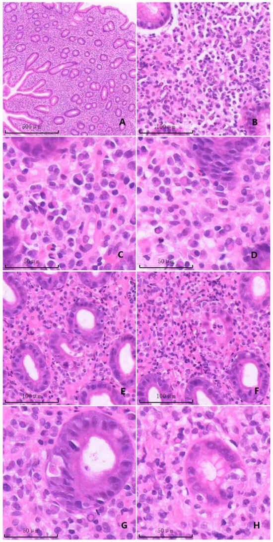
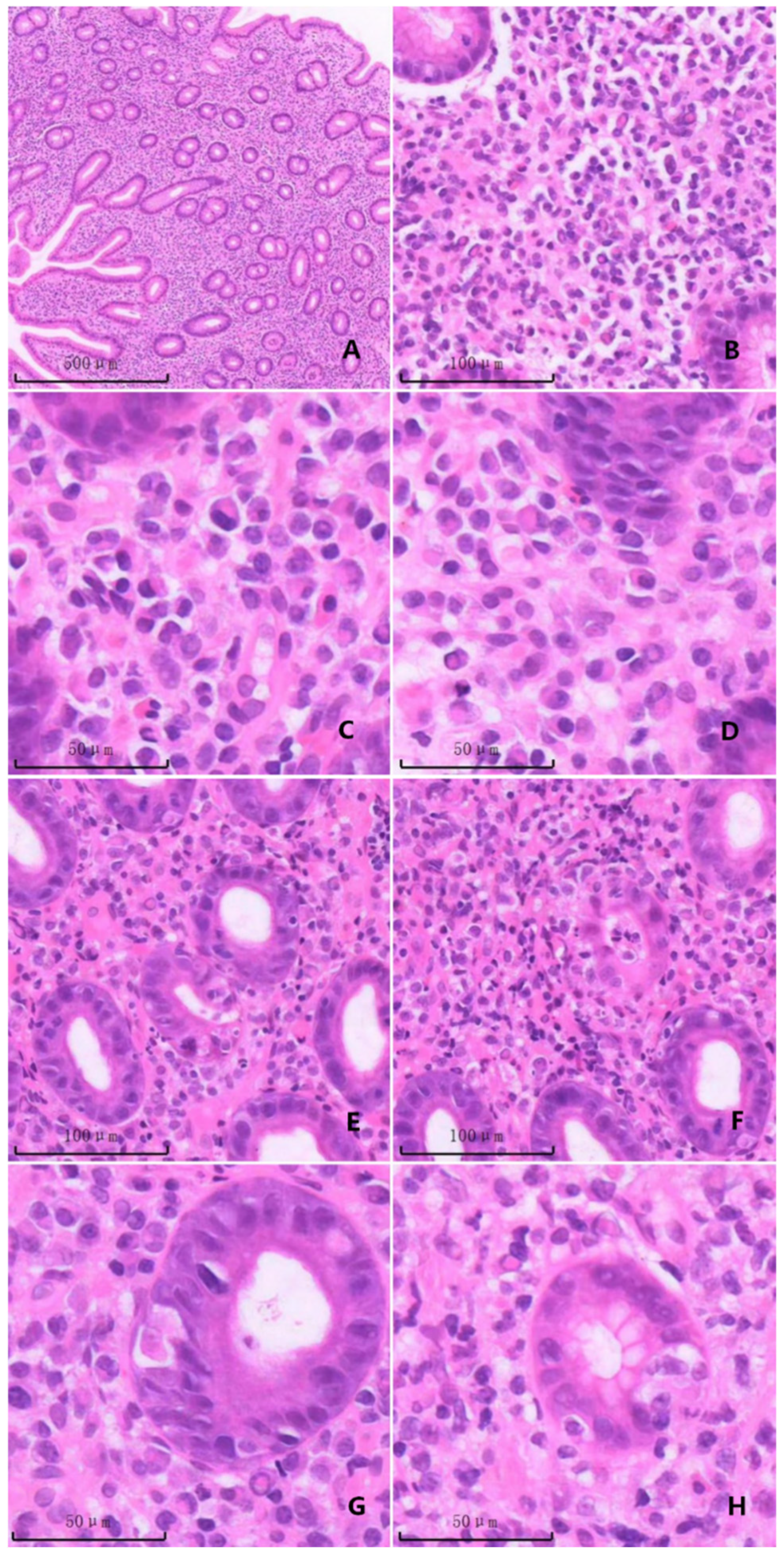
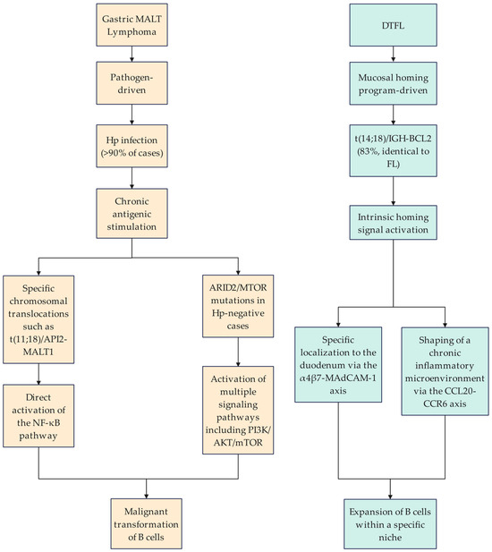
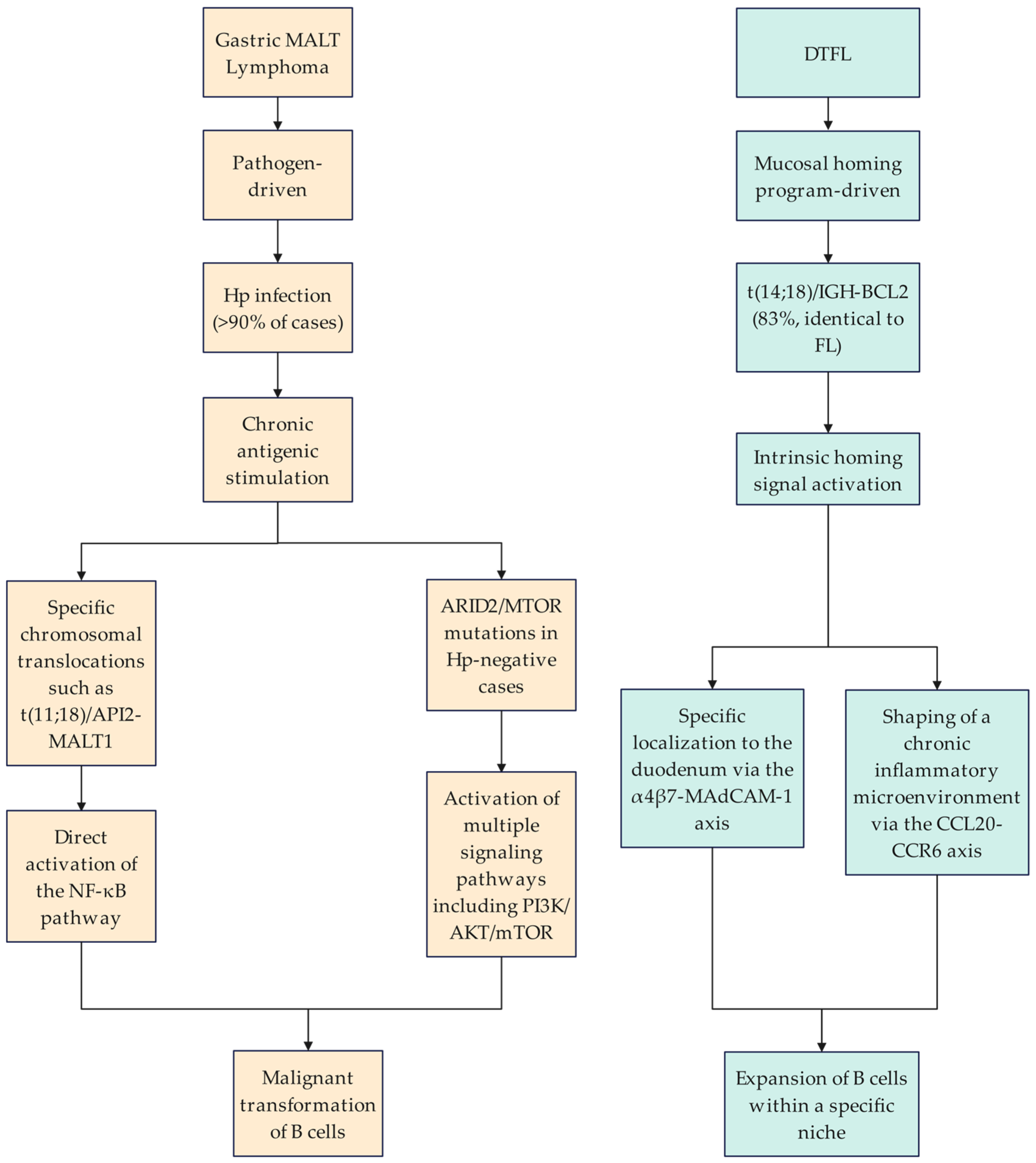
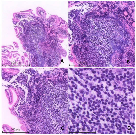
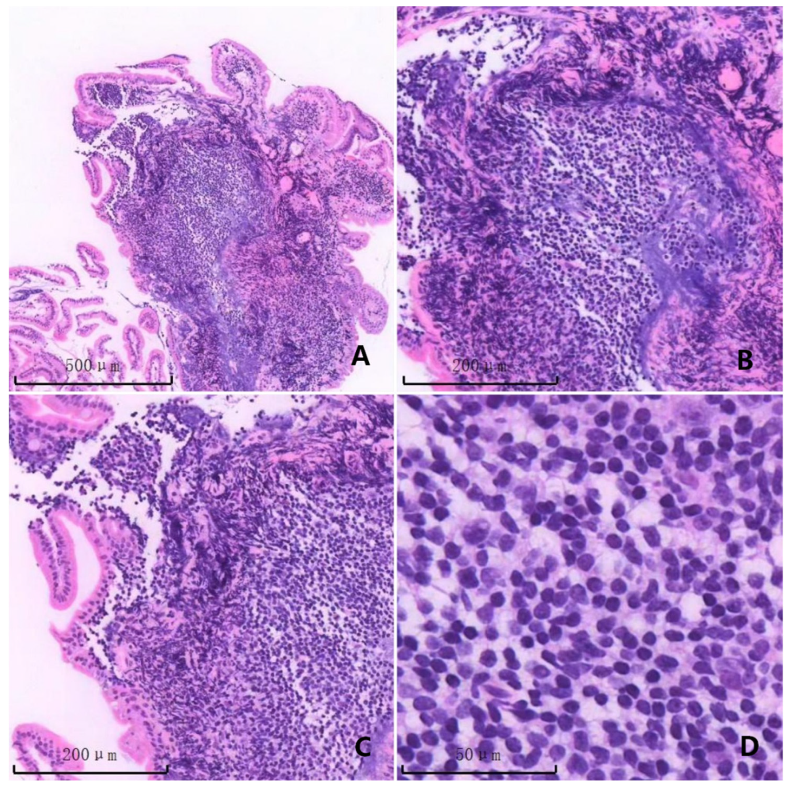

Disclaimer/Publisher’s Note: The statements, opinions and data contained in all publications are solely those of the individual author(s) and contributor(s) and not of MDPI and/or the editor(s). MDPI and/or the editor(s) disclaim responsibility for any injury to people or property resulting from any ideas, methods, instructions or products referred to in the content. |
© 2026 by the authors. Licensee MDPI, Basel, Switzerland. This article is an open access article distributed under the terms and conditions of the Creative Commons Attribution (CC BY) license.