1. Introduction
The treatment of chronic pain places a significant burden on the United States healthcare system, with estimated annual costs of approximately USD 723 billion (or about USD 2200 per person). Musculoskeletal pain, as the leading cause of disability in the United States, is a substantial contributor to these costs [
1,
2].
One novel, non-pharmacological intervention that is increasingly utilized in the clinical management of musculoskeletal, visceral, and vascular dysfunction is
Fascial Counterstrain (FCS). FCS, which evolved from the osteopathic technique
Strain and Counterstrain, is a multisystem, anatomically based manual therapy, proposed to reduce peripheral nociceptor activation through the modulation and normalization of regional lymphatic flow (for review, see [
3]). As an
indirect manual therapy technique, FCS treatments employ gentle, non-force positioning and/or tissue decompression to improve circulation and reflexively correct dysfunction [
4,
5]. This contrasts with
direct manual therapy techniques that apply force directly against restrictive barriers to improve mobility and/or reduce pain [
5].
1.1. Background
The goal of FCS treatments is to identify and remove regions of interstitial inflammatory stasis (IIS), theorized to create and maintain chronic somatic, visceral, and vascular dysfunction through the persistent chemical excitation of peripheral nociceptors. Once inflamed, deep tissue nociceptive sources are theorized to produce loci of cutaneous tender points (TPs) by activating dorsal root reflexes and promoting central sensitization (for review, see [
6]). This physiological process has led to the theoretical association of FCS TPs with specific underlying (inflamed)
anatomical source tissues, including muscles, ligaments, nerves, vessels, and organs. A correlational process that purportedly involved the palpatory assessment of all segmentally related tissues that could neurogenically produce the observed TP, based on previously established anatomical and segmental correlations [
3,
7,
8].
The described
anatomical correlation of TPs enables FCS to serve a dual role in both the diagnosis and treatment of multi-system fascial dysfunction. The process of TP palpation identifies the inflamed tissue (e.g., makes the diagnosis), leading to TP-specific treatments. Notably, a recent study provided direct clinical support for the IIS hypothesis in complex regional pain syndrome patients by demonstrating that these individuals have impaired lymphatic drainage and autonomic dysfunction from the accumulation of interstitial pro-inflammatory cytokines [
9]. Additionally, Counterstrain techniques have also been investigated at the cellular level and found to reduce pro-inflammatory cytokine production following trauma. Specifically, an experimentally induced repetitive strain model demonstrated a 46% reduction in fibroblast interleukin-6 production in the Counterstrain treatment group as compared to controls [
10].
1.2. FCS and the Lymphatic Pump Mechanism
Manual tissue decompression (performed during FCS treatments) is believed to create an interstitial
vacuum that enhances the lymphatic system’s own pressure-driven “suction mechanism”, an essential component in the transportation of pro-inflammatory cytokines from the interstitial space to initial lymphatic vessels [
11]. This supports the contention that FCS treatments have the potential to restore the function of the local lymphatic pump mechanism, known to be inhibited by the presence of specific, interstitial pro-inflammatory cytokines, including interleukin-1β, tumor necrosis factor alpha, and interleukin-6 [
12]. Once lymphatic pump function has been restored, nociceptive signaling to the dorsal horn can be anticipated to reduce as regions of IIS gradually dissipate. This may, in turn, alleviate all associated muscle guarding and/or somato-sympathetic reflexes [
13,
14] that can alter segmental mobility and impair regional vascular perfusion.
1.3. Research Gaps and Study Objectives
Recent randomized controlled trials and meta-analyses of indirect manual therapies, including Counterstrain and the Positional Release Technique, demonstrate short-term pain (VAS) reductions of 20–68% (e.g., in patients with low back myofascial pain syndrome) when compared to controls or when combined with exercise [
15,
16,
17]. Although preclinical evidence also supports Counterstrain’s modulation of pro-inflammatory cytokines (as previously cited), quantitative validation of FCS’s diagnostic accuracy and immediate post-treatment therapeutic efficacy is lacking. This case report addresses this gap using TSP and PPT; however, it cannot directly confirm the IIS hypothesis, as post-treatment cytokine levels and lymphatic flow were unmeasured.
TSP testing is a non-invasive method of assessing sensory perception by measuring an individual’s response to specific stimuli, including pinpricks. More specifically, it aims to quantify sensory thresholds and detect abnormalities such as hypoesthesia or hyperalgesia. With regard to the central nervous system, it can reveal enhanced sensory processing, including “wind-up” or temporal summation [
18]. Wind-up is a neuroadaptive phenomenon that shares several physiological manifestations with central sensitization, including the expression of hyperalgesia and receptive field expansion. More specifically, it can be defined as a frequency-dependent increase in neuron excitability, characterized by a progressive acceleration in (neuron) response to a series of repetitive stimuli delivered between 0.5 and 3 Hz [
19]. PPT assessment differs from TSP testing in that it uses a pressure algometer to determine the minimum force necessary to elicit the sensation of pain at a given anatomical location. PPT is therefore an indirect measure of pain sensitivity, used to identify regions with altered nociceptive processing, by combining deep tissue pressure stimulation with real-time patient feedback [
20].
In this quantitative case study,
FCS’s diagnostic validity was demonstrated by correspondence between the FCS clinical diagnosis and pre-treatment TSP sum scores (elevated wind-up from 10 repetitive pinprick stimuli), indicating central sensitization [
21].
Immediate
FCS therapeutic efficacy was determined by a >1.5 kg/cm
2 post-treatment increase in PPT measures (exceeding reported minimum detectable changes) [
22] and a >50% reduction in TSP wind-up sum scores, aligning with the Baeumler et al. (2019) criteria for clinically meaningful pain reduction in patients with high temporal summation [
23].
2. Detailed Case Description
The subject was a 22-year-old, non-smoking, 6’5’, Caucasian lacrosse player who presented with central lumbosacral pain that radiated intermittently into the left sciatic notch and hamstring region. Pain onset was insidious and estimated to begin on 1 May 2023. At the time of treatment, 16 July 2023, the subject reported taking no medication and rated his pain at 6/10 (VAS score). His past medical history was remarkable for several minor sport-related injuries, including a left mid-shaft fibula fracture. Previous treatment for this episode consisted of over-the-counter anti-inflammatory medication (short-term) and one session of physical therapy where he was prescribed core stabilization and hip flexibility exercises. The patient reported performing the prescribed exercises for several weeks; however, he eventually stopped due to a lack of symptom improvement. A Modified Oswestry Low Back Pain Scale was also administered on 16 July 2023, indicating 10% disability, including limitations in prolonged walking, standing, traveling, and athletic participation.
2.1. Materials and Methods
The FCS diagnostic process resulted in a clinical diagnosis of S1, S2 radicular pain or radiculitis. The identified TPs included those related to the L4-S2 spinal veins; S1, S2 epidural adipose tissue; S1, S2 epidural (meningovertebral) ligaments; and a left extended fibular dysfunction (subluxation), which may have contributed to the patient’s symptoms via its known attachments to the common fibular nerve [
24]. The patient underwent no additional diagnostic testing prior to the FCS treatment session. All mobility testing and TP palpation procedures were performed by the primary author, a certified FCS practitioner and originator of the FCS technique.
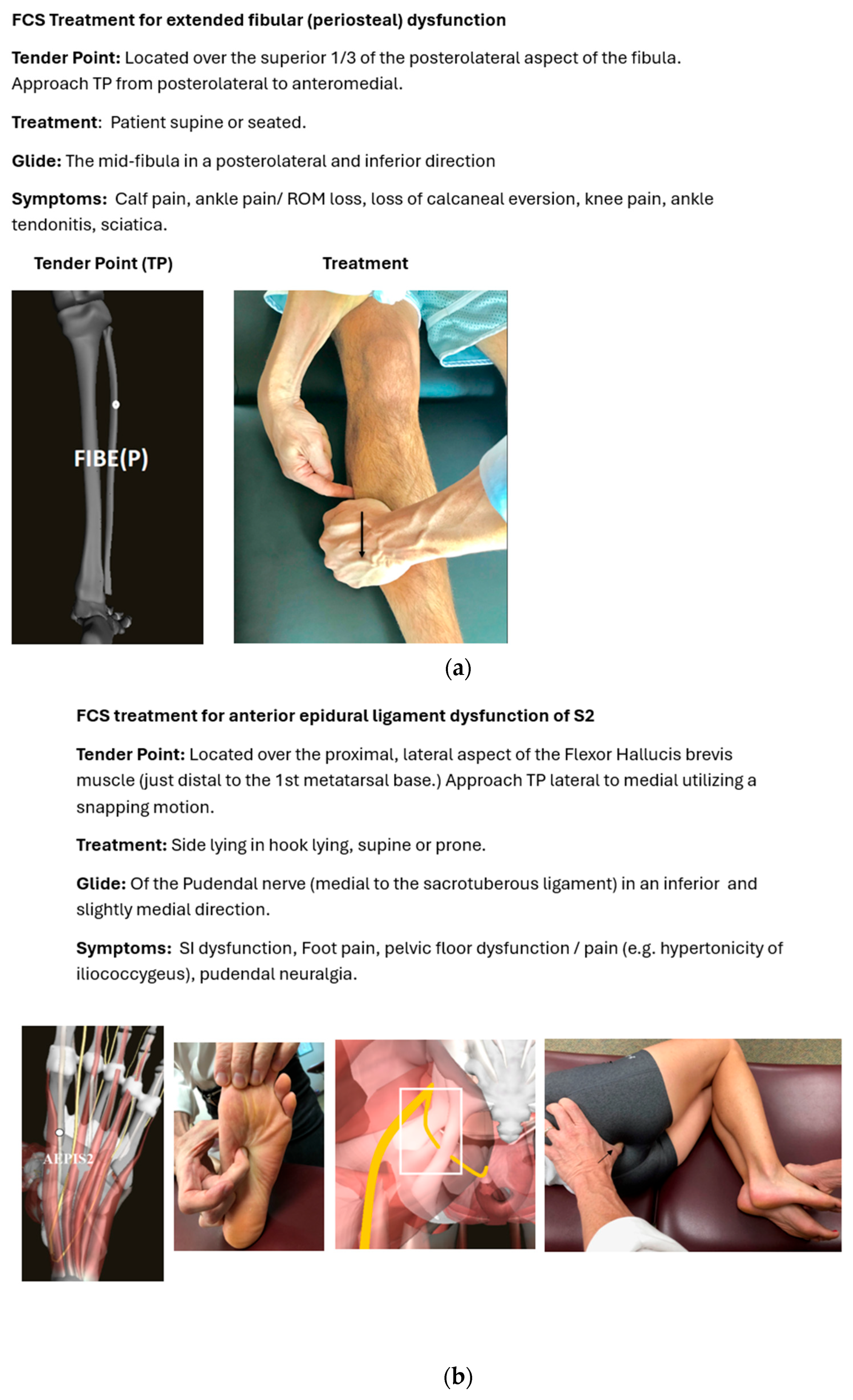
Following the FCS evaluation process, pre-treatment PPT and TSP measurements were taken by a separate practitioner (physician) trained in both evaluation tools. This included PPT measurements of the bilateral flexor hallucis brevis muscles (S1 and S2 myotome) and TSP testing of multiple nerve distributions corresponding to the bilateral S1 and S2 dermatomes. A singular, 40 min FCS treatment session was then performed to address the identified dysfunctions, including treatment of the bilateral S1, S2 epidural ligaments [
25]; bilateral S1–S3 epidural adipose [
26]; bilateral L4-S2 spinal veins [
27]; and left extended fibula dysfunction [
28]. See the example treatment in
Figure 1a,b.
Immediately following the 40 min treatment session, post-treatment PPT and TSP measurements were repeated for the previously tested myotomes and dermatomes. A comparative summary of the pre- and post-treatment PPT and TSP measurements is listed in
Table 1.
Pressure pain threshold (PPT) measurements (a) of the involved (left) lower extremity S1 and S2 myotomes increased from a mean of 2.4 kg/cm
2 pre-treatment to 6.1 kg/cm
2 post-treatment. Temporal summation of pain (TSP) sum scores (b) for the S1 and S2 dermatomes decreased from 75/10 pre-treatment to 18/10 post-treatment (76% reduction), meeting the threshold for clinically meaningful pain relief as defined by Baeumler et al. (2019) [
23].
2.2. Clinician-Assessed Outcomes
The observed elevation in wind-up within the left lateral plantar nerve distribution (S1–S2 dermatomes on TSP testing) aligned with the clinical FCS diagnosis of S1–S2 radiculitis. We then applied a TSP sum score outcome measure to quantify the total wind-up across the train of 10 stimuli, as previously published [
21]. Results demonstrated the normalization of all post-treatment wind-up scores, except for the left S1 and S2 dermatomes, which failed to fully normalize; however, they did improve by 76%, from a sum score of 75/10 to 18/10 [
29]. The results lend support to the validity of the FCS diagnostic process through its alignment with pre-treatment TSP indicators of central sensitization and by exceeding the study’s > 50% threshold for diagnostic validity and clinically meaningful pain relief as established by Baeumler et al. [
23].
Baseline PPT measurements obtained over the bilateral flexor hallucis brevis muscles (S1 and S2 myotomes) demonstrated pre-treatment thresholds to be 2.4 kg/cm2 left and 4.3 kg/cm2 right. Following treatment, muscle sensitivity decreased substantially to a pressure tolerance of 6.1 kg/cm2 left and 6.2 kg/cm2 right. These post-intervention PPT increases of 3.7 kg/cm2 (left) and 1.9 kg/cm2 (right) also exceed the previously defined minimum detectable change threshold of >1.5 kg/cm2 for clinical efficacy and meaningful post-treatment hypoalgesia.
Reduced pre-treatment PPTs on the patient’s right (asymptomatic) side suggested some degree of bilateral S1 and S2 central sensitization. As documented in
Table 1, right-sided muscle sensitivity was also reduced post-treatment, as all treatment techniques were performed bilaterally, as indicated by the FCS diagnostic process.
2.3. Patient Perspective Statement
Patient correspondence at six weeks post-treatment (via text message) subjectively confirmed the above improvements, which are described in the following patient perspective statement:
“After one session of treatment, I felt the tightness in my lower back which extended to my hamstrings reduced tremendously. The pain was certainly more manageable, and I could perform my usual activities without having to take breaks, and though it was only one session, there has been lasting improvement for me.”
2.4. Changes in Therapeutic Intervention
This study’s intent was to assess FCS’s diagnostic validity and immediate post-treatment effects using QST, without intervention changes or comparisons to other therapies. Randomized controlled trials are needed to evaluate relative efficacy and long-term outcomes.
2.5. Adverse Reactions and/or Events
There were no adverse reactions reported during or after the singular FCS treatment.
3. Follow-Up Diagnostic Tests and/or Results
As a single-session case report employing QST, this study assessed only the immediate post-treatment effect of FCS; therefore, no follow-up evaluations were conducted leaving long-term therapeutic outcomes unquantified.
4. Conclusions and Case Report “Take-Away”
The strength of this case report is that it utilized immediate, post-treatment QST measurements to evaluate treatment outcomes, removing the possibility of symptom reduction over time.
The exploratory nature of this investigation is limited by the inherent constraints of the case study design, including the absence of a placebo-controlled treatment, longitudinal quantitative outcome measurements, and the inability of case studies to be generalized to broader patient populations. To substantiate these preliminary findings, future research should include adequately powered randomized controlled trials across a diverse range of musculoskeletal and non-musculoskeletal conditions (>30 participants per group) to achieve sufficient statistical power.
The “take-away” from this case report is that it represents the first quantitative study providing preliminary evidence for both the diagnostic accuracy and immediate therapeutic efficacy of FCS. The results demonstrate substantial post-treatment improvements in TSP wind-up sum scores and PPTs (within the predicted S1–S2 segmental distributions), supporting both processes. Considering FCS’s non-pharmacological and non-invasive nature, it may offer a promising treatment option for chronic idiopathic musculoskeletal, lymphatic, visceral, and vascular conditions.
Author Contributions
Conceptualization, B.T., J.S. (Jay Shah), and J.S. (John Srbely); methodology, B.T., J.S. (Jay Shah), and J.S. (John Srbely); formal analysis, J.S. (Jay Shah); investigation, B.T., J.S. (Jay Shah), and J.S. (John Srbely); resources, J.S. (Jay Shah) provided QST and PPT equipment and performed the pre- and post-treatment measurements and data curation; writing of the original draft, B.T.; reviewing and editing, B.T., J.S. (Jay Shah), and J.S. (John Srbely); visualization, B.T. and J.S. (Jay Shah); supervision, J.S. (John Srbely); project administration, J.S. (John Srbely). All authors have read and agreed to the published version of the manuscript.
Funding
This research received no external funding.
Institutional Review Board Statement
Ethical approval was waived for this report because it describes a single clinical case and was not designed or conducted as a systematic research project and therefore does not meet the regulatory definition of human-subjects research.
Informed Consent Statement
Written informed consent was obtained from the participating patient to publish this paper.
Data Availability Statement
The original contributions presented in this study are included in the article material. Further inquiries can be directed to the corresponding authors.
Acknowledgments
We are grateful to Kristin Murner for her assistance and expertise with the manuscript preparation and submission process.
Conflicts of Interest
Author, Brian Tuckey PT, OCS is the developer of Fascial Counterstrain and teaches FCS internationally through several different intermediary companies including the Jones Institute in the United States. The remaining authors declare that the research was conducted in the absence of any commercial or financial relationships that could be construed as a potential conflict of interest.
Abbreviations
The following abbreviations are used in this manuscript:
| QST | Quantitative Sensory Testing |
| PPT | Pain Pressure Threshold |
| TSP | Temporal Summation of Pain |
| FCS | Fascial Counterstrain |
| TPs | Tender Points |
References
- Guy, G.P., Jr.; Miller, G.F.; Legha, J.K.; Rikard, S.M.; Strahan, A.E.; Mikosz, C.; Florence, C.S. Economic Costs of Chronic Pain—United States, 2021. Med. Care 2025, 63, 679–685. [Google Scholar] [CrossRef] [Scilit]
- Johannes, C.B.; Le, T.K.; Zhou, X.; Johnston, J.A.; Dworkin, R.H. The prevalence of chronic pain in United States adults: Results of an internet-based survey. J. Pain 2010, 11, 1230–1239. [Google Scholar] [CrossRef] [Scilit]
- Tuckey, B. Fascial Counterstrain: A methodological advancement in indirect osteopathic manipulation. Int. J. Osteopath. Med. 2025, 58, 100789. [Google Scholar] [CrossRef] [Scilit]
- Speicher, T.E.; Selkow, N.M.; Warren, A.J. Manual Therapy Improves Immediate Blood Flow and Tissue Fiber Orientation of the Forearm Extensors. J. Phys. Med. Rehabil. 2022, 4, 28–36. [Google Scholar] [CrossRef] [Scilit]
- Greenman, P.E. Principles of Manual Medicine, 5th ed.; Lippincott Williams & Wilkins: Philadelphia, PA, USA, 2016. [Google Scholar]
- Tuckey, B.; Srbely, J.; Rigney, G.; Vythilingam, M.; Shah, J. Impaired lymphatic drainage and interstitial inflammatory stasis in chronic musculoskeletal and idiopathic pain syndromes: Exploring a novel mechanism. Front. Pain Res. 2021, 2, 691740. [Google Scholar] [CrossRef] [Scilit] [PubMed]
- ISAFSCI Working Group. International Standards to document remaining Autonomic Function after Spinal Cord Injury (ISAFSCI), First Edition 2012. Top. Spinal Cord Inj. Rehabil. 2012, 18, 282–296. [Google Scholar] [CrossRef] [Scilit]
- Cramer, G.D.; Darby, S.A. Clinical Anatomy of the Spine, Spinal Cord, and ANS, 3rd ed.; Elsevier: St. Louis, MO, USA, 2014. [Google Scholar]
- Melin, M.M.; Rasmussen, J.C.; Aldrich, M.B.; Karni, R.J.; Fife, C.E.; Eckert, K.A. Additional evidence from a case report supports a novel hypothesis on the association between complex regional pain syndrome and lymphedema. Front. Pain Res. 2025, 6, 1540930. [Google Scholar] [CrossRef] [Scilit]
- Meltzer, K.R.; Standley, P.R. Modeled repetitive motion strain and indirect osteopathic manipulative techniques in regulation of human fibroblast proliferation and interleukin secretion. J. Am Osteopath Assoc. 2007, 107, 527–536. [Google Scholar]
- Jamalian, S.; Jafarnejad, M.; Zawieja, S.D.; Bertram, C.D.; Gashev, A.A.; Zawieja, D.C.; Davis, M.J.; Moore, J.E., Jr. Demonstration and Analysis of the Suction Effect for Pumping Lymph from Tissue Beds at Subatmospheric Pressure. Sci. Rep. 2017, 7, 12080. [Google Scholar] [CrossRef] [Scilit] [PubMed]
- Aldrich, M.B.; Sevick-Muraca, E.M. Cytokines are systemic effectors of lymphatic function in acute inflammation. Cytokine 2013, 64, 362–369. [Google Scholar] [CrossRef] [Scilit]
- Häbler, H.-J.; Wasner, G.; Jänig, W. Interaction of sympathetic vasoconstriction and antidromic vasodilatation in the control of skin blood flow. Exp. Brain Res. 1997, 113, 402–410. [Google Scholar] [CrossRef] [Scilit]
- He, X.; Proske, U.; Schaible, H.G.; Schmidt, R.F. Acute inflammation of the knee joint in the cat alters responses of flexor motoneurons to leg movements. J. Neurophysiol. 1988, 59, 326–340. [Google Scholar] [CrossRef] [Scilit]
- Koura, G.M.R.; Elshiwi, A.M.F.; Selim, N.M.; Asiri, A.A.M.; Alqahtani, R.H.J.; Elimy, D.A.; Alshehri, M.A.; Alnakhli, H.H.; Assiri, S.M.; Ahmad, F.; et al. Comparing the therapeutic impact of strain-counterstrain and exercise on low back myofascial pain syndrome: A randomized trial. J. Multidiscip. Healthc. 2025, 18, 1–12. [Google Scholar] [CrossRef] [Scilit]
- El-Khateeb, Y.S.; Mahmoud, A.G.; Mohamed, M.H.; El-Azeim, A.S.A. Influence of adding strain-counterstrain to standard therapy on axioscapular muscles amplitude and fatigue in mechanical neck pain: A single-blind, randomized trial. Eur. J. Phys. Rehabil. Med. 2022, 58, 621–629. [Google Scholar] [CrossRef] [Scilit] [PubMed]
- Bethers, A.H.; Swanson, D.C.; Sponbeck, J.K.; Mitchell, U.H.; Draper, D.O.; Feland, J.B.; Johnson, A.W. Positional release therapy and therapeutic massage reduce muscle trigger and tender points. J. Bodyw. Mov. Ther. 2021, 28, 264–270. [Google Scholar] [CrossRef] [Scilit] [PubMed]
- Rolke, R.; Magerl, W.; Campbell, K.A.; Schalber, C.; Caspari, S.; Birklein, F.; Treede, R.D. Quantitative sensory testing: A comprehensive protocol for clinical trials. Eur. J. Pain 2006, 10, 77–88. [Google Scholar] [CrossRef] [Scilit]
- Herrero, J.F.; Laird, J.M.; López-García, J.A. Wind-up of spinal cord neurones and pain sensation: Much ado about something? Prog. Neurobiol. 2000, 61, 169–203. [Google Scholar] [CrossRef] [Scilit]
- Park, G.; Kim, C.W.; Park, S.B.; Kim, M.J.; Jang, S.H. Reliability and usefulness of the pressure pain threshold measurement in patients with myofascial pain. Ann. Rehabil. Med. 2011, 35, 412–417. [Google Scholar] [CrossRef] [Scilit] [PubMed]
- Allison, C.; Loi, K.; Srbely, J.Z. A novel computational technique for the quantification of temporal summation in healthy individuals. Musculoskelet. Sci. Pract. 2021, 54, 102400. [Google Scholar] [CrossRef] [Scilit]
- Walton, D.M.; Levesque, L.; Payne, M.; Schick, J. Clinical pressure pain threshold testing in neck pain: Comparing protocols, responsiveness, and association with psychological variables. Phys. Ther. 2014, 94, 827–837. [Google Scholar] [CrossRef] [Scilit]
- Baeumler, P.I.; Conzen, P.; Irnich, D. High temporal summation of pain predicts immediate analgesic effect of acupuncture in chronic pain patients—A prospective cohort study. Front. Neurosci. 2019, 13, 498. [Google Scholar] [CrossRef] [Scilit] [PubMed]
- Ryan, W.; Mahony, N.; Delaney, M.; O’Brien, M.; Murray, P. Relationship of the common peroneal nerve and its branches to the head and neck of the fibula. Clin. Anat. 2003, 16, 501–505. [Google Scholar] [CrossRef] [Scilit] [PubMed]
- Tuckey, B. Fascial Counterstrain for Epidural Ligaments; Tuckey Continuing Educational Services, LLC: Frederick, MD, USA, 2021. [Google Scholar]
- Tuckey, B. Fascial Counterstrain for Adipose Tissue; Tuckey Continuing Educational Services, LLC: Frederick, MD, USA, 2023. [Google Scholar]
- Tuckey, B. Fascial Counterstrain for the Venous and Lymphatic System Part 2; Tuckey Continuing Educational Services, LLC: Frederick, MD, USA, 2013. [Google Scholar]
- Tuckey, B. Fascial Counterstrain for the Periosteum; Tuckey Continuing Educational Services, LLC: Frederick, MD, USA, 2018. [Google Scholar]
- Nothnagel, H.; Puta, C.; Lehmann, T.; Baumbach, P.; Menard, M.B.; Gabriel, B.; Gabriel, H.H.W.; Weiss, T.; Musial, F. How stable are quantitative sensory testing measurements over time? Report on 10-week reliability and agreement of results in healthy volunteers. J. Pain Res. 2017, 10, 2067–2078. [Google Scholar] [CrossRef] [Scilit] [PubMed]
| Disclaimer/Publisher’s Note: The statements, opinions and data contained in all publications are solely those of the individual author(s) and contributor(s) and not of MDPI and/or the editor(s). MDPI and/or the editor(s) disclaim responsibility for any injury to people or property resulting from any ideas, methods, instructions or products referred to in the content. |