


Abstract
Salicylic acid (SA) is a key regulator of plant immunity and contributes to defence against Plasmodiophora brassicae, the causal agent of clubroot disease in canola (Brassica napus) and other crucifers. Exogenous SA applications have reduced clubroot severity in some Brassica pathosystems, yet the effectiveness of foliar SA treatment against the predominant resistance-breaking pathotype 3A in western Canada remains unclear. This study evaluated the effects of weekly foliar applications of 0, 1, 5, or 10 mM SA on clubroot development in two B. napus var. napobrassica cultivars under greenhouse and growth chamber conditions. Plants inoculated with pathotype 3A were assessed for disease severity, pathogen resting spore load, plant height, and transcript accumulation of SA-responsive genes. Overall, SA treatments resulted in modest reductions in disease severity and resting spore concentrations; however, treatment effects did not reach statistical significance in most cases. Collectively, foliar SA applications provided limited suppression of clubroot caused by pathotype 3A. Further optimization of SA concentration, timing, and delivery, particularly when targeting the root zone, may be required before SA can be considered a complementary tool in integrated clubroot management.
1. Introduction
Plants activate and regulate defence responses to biotic and abiotic stresses through hormones such as jasmonic acid (JA), ethylene (ET), and salicylic acid (SA) [1]. SA, a phenolic compound linked to immunity against biotrophic or hemi-biotrophic pathogens [2], plays a key role in systemic acquired resistance (SAR) and functions as a signaling molecule in both pathogen-associated molecular pattern (PAMP)-triggered immunity (PTI) and effector-triggered immunity (ETI) [3]. It also participates in crosstalk with other key phytohormones, including JA, ET, abscisic acid (ABA), auxins, gibberellins (GA), brassinosteroids, and cytokinins (CK) [4]. Exogenous SA application has been shown to enhance defence responses under both biotic and abiotic stresses [5]. Its application has influenced root responses when applied to aboveground tissues [6,7,8], suggesting a potential strategy for managing root-infecting pathogens.
The soilborne microbe Plasmodiophora brassicae, a causal agent of clubroot disease, is a major root-infecting pathogen of the Brassicaceae family. P. brassicae first infects root hairs during the primary infection stage and later colonizes the cortical root tissue in the secondary stage, where hypertrophy and hyperplasia lead to characteristic gall formation [9,10,11]. In canola (Brassica napus), the primary tool for clubroot management is the use of clubroot-resistant (CR) cultivars, ideally complemented by sanitization of field equipment, chemical treatments (e.g., lime, fungicides, and fumigants), and crop rotation (reviewed in [12]). With the continued spread of clubroot, new management strategies are being explored, including the identification of biocontrol agents [12,13,14,15] and advanced technologies such as CRISPR/Cas9 and RNA interference [16,17,18,19]. Recent studies indicate that SA plays a key role in host defence against P. brassicae [20], presenting a potential avenue for disease control.
Multiple lines of evidence support an SA-mediated defence response against P. brassicae infection. Transcript levels of genes associated with SA biosynthesis and signaling are elevated following inoculation compared with non-inoculated controls [7,20,21,22,23,24,25,26,27,28,29]. Phytohormone analyses have also revealed increased levels of endogenous SA in both susceptible and resistant host roots in response to P. brassicae infection [7,30,31,32,33]. Comparisons between susceptible and resistant interactions indicate that SA levels, as well as the expression of SA-related genes, are typically sustained for a longer duration in resistant hosts.
To counteract the SA-mediated defence response, P. brassicae secretes at least one known effector that directly manipulates host SA levels to enhance susceptibility [34]. Specifically, the benzoic acid (BA)/SA methyltransferase (PbBSMT) effector methylates SA to form an inactive derivative, MeSA [34]. Expression of PbBSMT is notably upregulated during infection in A. thaliana, B. napus, and B. oleracea, and the gene appears to be conserved amongst pathotypes [21,34,35,36].
Several studies have examined the effects of exogenous SA applications on P. brassicae infection. Repeated SA treatments have been shown to reduce clubroot severity in susceptible A. thaliana [30,37], susceptible B. rapa (B. campestris) subsp. chinensis [38], partially resistant B. napus [7], and both susceptible and resistant B. napus [33]. The timing and method of SA application (root dip, soil drench, or foliar spray) varied among studies. Exogenous SA was also readily metabolized in B. napus, indicating effective utilization of the applied compound by the plant [7]. However, the plant’s ability to take up and quickly metabolize SA to maintain homeostasis may explain why single pre-inoculation applications were ineffective in reducing disease [31,39,40]. When applied optimally during infection, however, exogenous SA may suppress clubroot symptoms by enhancing host defence responses.
In western Canada, the resistance-breaking P. brassicae pathotype 3A is predominant in annual field surveys [41]. Newly developed CR canola cultivars with resistance to pathotype 3A and other virulent isolates have not fully controlled disease spread or severity [41,42]. Transcriptomic and proteomic analyses of resistant B. napus inoculated with pathotype 3A indicate an SA-mediated defence response [28,32,33]. In susceptible B. napus, clubroot severity was reduced when SA was applied near the root base [33]. However, the effect of foliar SA application during infection by pathotype 3A remains unexplored and may enhance the plant’s defences. Therefore, the objective of this study was to evaluate the impact of foliar SA applications on disease development in two B. napus cultivars inoculated with pathotype 3A under controlled conditions.
2. Materials and Methods
2.1. Plant Material
The rutabaga (B. napus var. napobrassica) cultivars ‘Laurentian’ and ‘Wilhelmsburger’ (European Clubroot Differential [ECD] 10) were used in this study, along with the Chinese cabbage (B. rapa var. pekinensis) cultivar ‘Granaat’ (ECD 05), which was included as a susceptible check.
2.2. Resting Spore Extraction and Inoculation
Resting spores of a P. brassicae field isolate classified as pathotype 3A were extracted from 5 g of frozen root galls following previously outlined methods [43]. The spore suspension was passed through eight layers of cheesecloth to filter out soil and plant debris, and the spore concentration was estimated using a haemocytometer (VWR, Mississauga, ON, Canada). The suspension was then adjusted with sterile distilled water (dH2O) to final concentrations of 1 × 10⁵ (low inoculum) and 1 × 10⁷ resting spores mL−1 (high inoculum). Spore viability was not assessed.
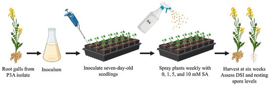
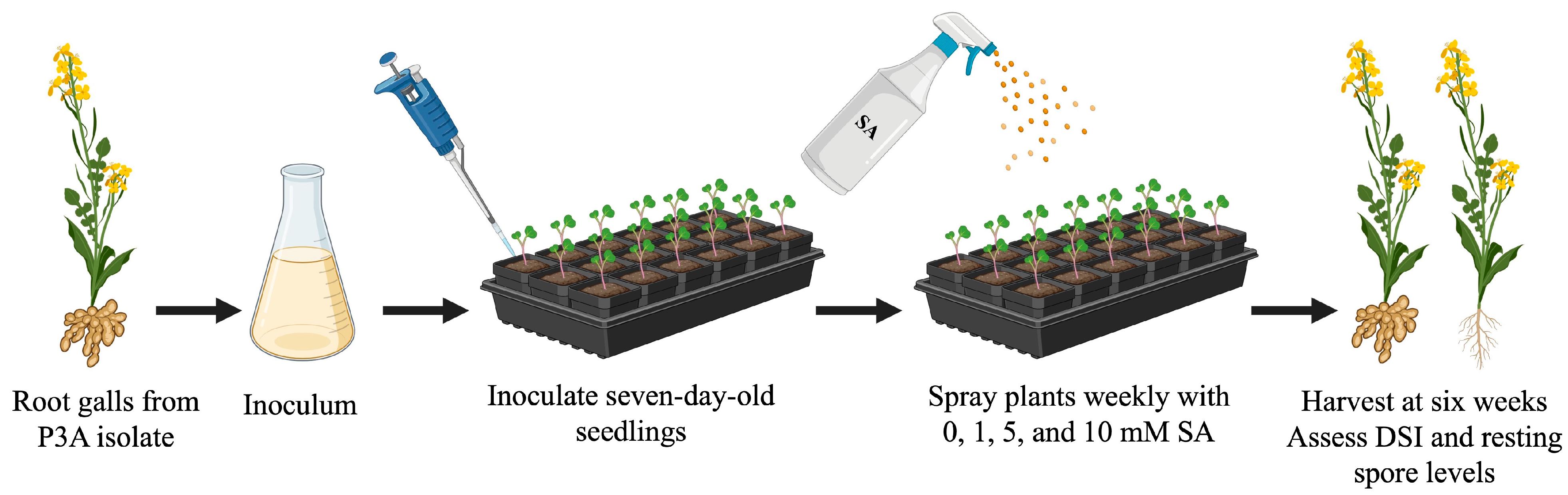
Seeds of each host genotype were directly sown in plastic trays containing 72 cells (3.75 × 3.75 × 5.7 cm) filled with Sunshine LA4 potting mixture (Sunshine Growers, Vancouver, BC, Canada). Seedlings were planted at a density of one plant per cell. Seven-day-old seedlings were inoculated with 1 mL of inoculum suspension pipetted directly to the base of each plant. The potting mix was kept moist for the first week following inoculation and subsequently watered as needed with tap water. Plants were fertilized with 20 N:20 P:20 K fertilizer as required and grown for six weeks in either a greenhouse or growth chamber at 20 ± 2 °C with a 16 h photoperiod. Greenhouse trials were replicated twice under low inoculum conditions and three times under high inoculum conditions, with each experimental unit comprising 12–18 plants per cultivar and SA treatment. Growth chamber trials were also replicated three times, with each experimental unit comprising eight plants per cultivar and SA treatment. The general methodology used for the greenhouse and growth chamber trials is illustrated in Figure 1.
Figure 1.
General experimental workflow used to assess the effect of salicylic acid (SA) on clubroot disease severity index (DSI) and resting spore accumulation in Plasmodiophora brassicae-inoculated plants. The study included B. rapa var. pekinensis ‘Granaat’ and B. napus var. napobrassica ‘Laurentian’ and ‘Wilhelmsburger’ in greenhouse and growth chamber trials. Created in BioRender. Storfie, E. (2025) https://app.biorender.com/illustrations/69c844c1f9524d459bbca5e8?slideId=d8d42b48-b620-46c4-b0af-479b9e2818c1, accessed on 19 December 2025.
2.3. Application of Exogenous Salicylic Acid
Solutions of SA (Sigma Aldrich, St. Louis, MO, USA) were prepared in dH2O at final concentrations of 1, 5, and 10 mM. One week after inoculation, plant leaves were sprayed weekly with the respective SA treatments using a hand-held bottle sprayer, continuing until harvest.
2.4. Disease Assessment
At six weeks post-inoculation, plants were harvested, and roots were washed and rated for clubroot severity using a standardized 0 to 3 scale [44], where: 0 = no galling; 1 = a few small galls (small galls on less than one-third of the primary root); 2 = moderate galling (small to medium galls on one-third to two-thirds of the primary root); and 3 = severe galling (medium to large galls on more than two-thirds of the primary root). Disease rating focused on gall severity on the primary root, as damage to this tissue can significantly impair plant growth and serves as a major source of resting spore inoculum [9].
A disease severity index (DSI) was calculated according to [45], as modified in [43], using the formula DSI (%) = {[∑ (n × 0) + (n × 1) + (n × 2) + (n × 3)]/N × 3} × 100, where n is the number of plants in each symptom severity class (0–3) and N is the total number of plants.
Prior to harvesting in each growth chamber trial, plant height was measured from the base of the plant to the apex, and the mean height was calculated for each biological replicate. Clubroot DSI and plant height data are presented in Tables S1 and S2.
2.5. DNA and RNA Isolation
After disease assessment, all roots within each experimental unit were pooled, frozen in liquid nitrogen, and stored at −80 °C until processing. Root galls were ground to a fine powder using a mortar and pestle. The NucleoSpin Plant II kit (Macherey-Nagel, Düren, Germany) was used to extract genomic DNA from ~100 mg of ground tissue according to the manufacturer’s instructions. Total RNA was extracted from ~100 mg of ground tissue following a combined protocol using TRIzol reagent (Ambion-Life Technologies, Carlsbad, CA, USA) and the RNeasy kit (QIAGEN, Hilden, Germany) [28]. The quantity and purity of the genomic DNA and RNA were assessed using a NanoDrop 2000c spectrophotometer (ThermoFisher Scientific, Waltham, MA, USA), and samples were stored at −20 °C and −80 °C, respectively.
2.6. Resting Spore Quantification
The amount of P. brassicae resting spores was measured by quantitative PCR (qPCR) analysis following the method in [46]. Genomic DNA was diluted to 1:10 with nuclease-free water. A standard curve was generated using genomic DNA extracted from a suspension containing 1 × 10⁷ resting spores mL−1, which was serially diluted 10-fold with nuclease-free water to a final concentration of 1 × 10² resting spores mL−1. Each qPCR contained 5 μL of an in-house SYBR Green reagent mix (Molecular Biology Services Unit [MBSU], University of Alberta, Edmonton, AB, Canada), 2.5 μL of the mixed primers DR1F and DR1R (3.2 μM; Table S3), and 2.5 μL of DNA template, for a total reaction volume of 10 μL. Amplification was performed using a QuantStudio 3 Real-Time PCR System (Applied Biosystems, Waltham, MA, USA) under the following cycling conditions: 95 °C for 10 min; 40 cycles of 95 °C for 30 s, 60 °C for 1 min; followed by a melt curve analysis of 95 °C for 15 s, 60 °C for 1 min, 95 °C for 15 s. Technical replicates were run in triplicate. The standard curves for each qPCR run had coefficients of determination (R2) between 0.99–1.0, with PCR efficiency ranges of 90% to 99%. Data are presented in Table S4.
2.7. Transcript Analysis of SA-Related Genes
To remove residual genomic DNA contamination from the extracted RNA, 500 ng of total RNA was treated with the RNase-Free DNase Set (ThermoFisher Scientific, Waltham, MA, USA) for 30 min at 37 °C. To terminate the reaction, 1 μL of 50 mM EDTA was added, followed by incubation at 65 °C for 10 min. Reverse transcription was performed using the RevertAid H Minus First Strand cDNA Synthesis Kit with an Oligo(dT)18 primer (Thermo Fisher Scientific, Waltham, MA, USA) according to the manufacturer’s instructions. The resulting cDNA was diluted with nuclease-free water to a final volume of 80 μL.
To confirm successful cDNA synthesis and the absence of genomic DNA contamination, end-point PCR was performed using primer sets targeting BnCAC (B. napus clathrin adaptor complex) and PbEFL (P. brassicae elongation factor-like) genes (Table S3). Each reaction contained 4 μL of cDNA, 0.75 μL of 50 mM MgCl2, 0.5 μL of 10 mM dNTPs, 2.5 μL of 10× PCR Buffer (without magnesium), 0.1 μL of Platinum Taq DNA polymerase (500 U μL−1; Thermo Fisher Scientific), 0.5 μL of each 10 μM primer, and nuclease-free water to a final volume of 25 μL. PCR amplification was carried out under the following cycling conditions: 94°C for 2 min; 35 cycles of 94 °C for 30 s, 55 °C for 30 s, and 72 °C for 30 s; followed by a final hold at 4 °C. PCR products were resolved on a 1% agarose gel (1× TAE) stained with SYBR Safe at 90 V for 1 h.
Quantitative PCR was conducted using the QuantStudio 3 Real-Time PCR system (Applied Biosystems) under the same cycling conditions described above. Reactions contained 5 μL of an in-house SYBR Green reagent mix (MBSU, University of Alberta), 2.5 μL of mixed primers (3.2 μM), and 2.5 μL of cDNA in a total volume of 10 μL. Primer sequences are listed in Table S3. Relative quantification of gene expression was performed using the 2−∆∆Ct method [47]. Expression levels of plant and pathogen target genes were normalized to BnCAC and PbEFL, respectively, and mean fold changes were calculated relative to transcript levels in the 0 mM SA treatment.
2.8. Statistical Analysis
Data analysis was conducted using GraphPad Prism version 10.5.0 (GraphPad Software, Boston, MA, USA). Assumptions of normality and heteroscedasticity were assessed based on model residuals using the Shapiro–Wilk Test (p > 0.05) and Spearman’s test (p > 0.05), respectively. When assumptions were met, statistical significance was determined by two-way analysis of variance (ANOVA) followed by a multiple comparison test. If assumptions were not met, a nonparametric multiple Mann–Whitney test was used. Nonparametric Spearman correlations were calculated between the SA treatments and both DSI and resting spore counts. Data points and error bars represent the means ± standard errors of the biological replicates.
3. Results
3.1. Effect of SA Treatments on Clubroot Severity During Greenhouse Trials
Greenhouse trials were conducted to determine whether weekly exogenous foliar applications of 0, 1, 5, or 10 mM SA affected clubroot development in seedlings inoculated with P. brassicae pathotype 3A. Trials were replicated twice under low inoculum conditions (1 × 10⁵ resting spores mL−1) and three times under high inoculum conditions (1 × 10⁷ resting spores mL−1). Across all SA treatments following inoculation with the pathogen, the universally susceptible Chinese cabbage cultivar ‘Granaat’ exhibited a DSI of 100%.
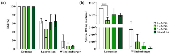
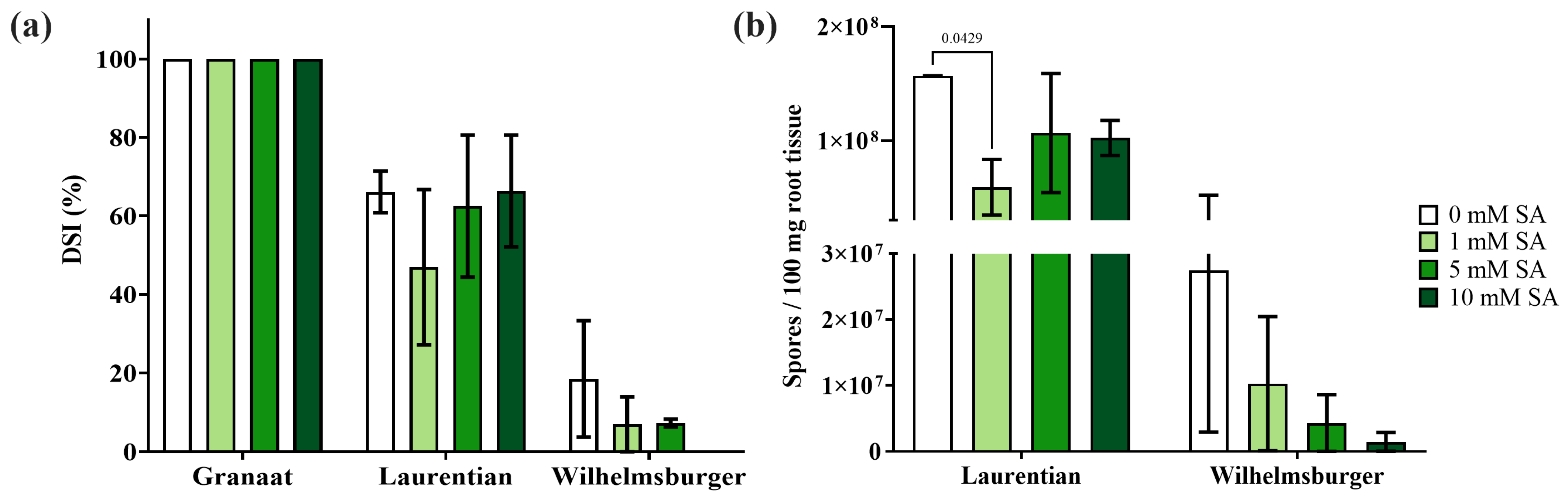
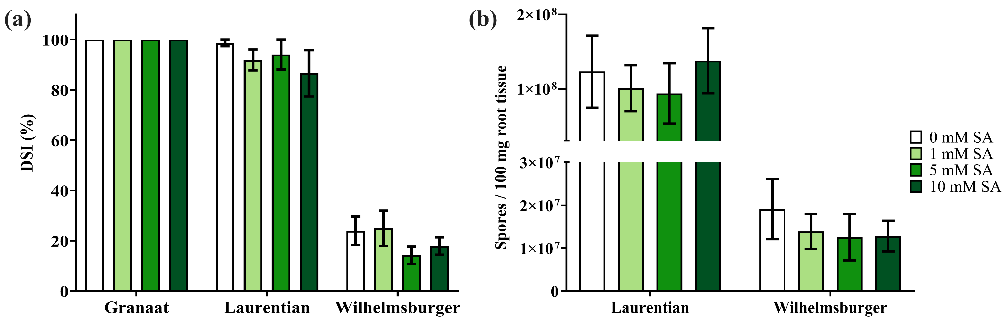
In the susceptible B. napus var. napobrassica cultivar ‘Laurentian’, DSI values were 66.1%, 46.9%, 62.5%, and 66.4% under low inoculum conditions and 98.7%, 91.9%, 94.1%, and 86.6% under high inoculum conditions following treatment with 0, 1, 5, or 10 mM SA, respectively (Figure 2a, Figure 3a and Figure S1). Inoculated ‘Laurentian’ plants under high inoculum conditions showed a negative correlation (r = −1, p = 0.083) between SA treatments and DSI. Under the same treatments, the partially resistant B. napus var. napobrassica cultivar ‘Wilhelmsburger’ exhibited DSI values of 18.5%, 7.0%, 7.3%, and 0% under low inoculum conditions and 24.0%, 25.0%, 13.4%, and 17.9% under high inoculum conditions. Although some treatments appeared to reduce DSI, multiple Mann–Whitney tests revealed no significant differences compared with the 0 mM SA control across both cultivars at either inoculum concentration. No clubroot symptoms were observed in the mock-inoculated control plants.
Figure 2.
Effect of salicylic acid (SA) on clubroot disease severity index (DSI) and Plasmodiophora brassicae resting spore accumulation in Brassica napus var. napobrassica ‘Laurentian’ and ‘Wilhelmsburger’ inoculated with a low inoculum of pathotype 3A under greenhouse conditions. B. rapa subsp. pekinensis ‘Granaat’ served as a susceptible check. (a) Mean DSI across two independent experiments at six weeks post-inoculation. (b) Mean resting spore quantification by qPCR using P. brassicae-specific primers and genomic DNA extracted from root galls at six weeks post-inoculation. Error bars indicate the standard error of the mean (n = 2). Compared with the 0 mM SA control, the 1 mM SA treatment in ‘Laurentian’ differed significantly (p = 0.0429) based on Dunnett’s multiple comparison test.
Figure 3.
Effect of salicylic acid (SA) on clubroot disease severity index (DSI) and Plasmodiophora brassicae resting spore accumulation in Brassica napus var. napobrassica ‘Laurentian’ and ‘Wilhelmsburger’ inoculated with a high inoculum of pathotype 3A under greenhouse conditions. B. rapa var. pekinensis ‘Granaat’ served as a susceptibility check. (a) Mean DSI across three independent experiments at six weeks post-inoculation. (b) Mean resting spore quantification by qPCR using P. brassicae-specific primers and genomic DNA extracted from root galls at six weeks post-inoculation. Error bars indicate the standard error of the mean (n = 3).
To evaluate the effect of SA treatments on in planta pathogen proliferation, resting spores were quantified from ground root tissue. In ‘Laurentian’ plants inoculated with low inoculum and treated with 0, 1, 5, or 10 mM SA, mean resting spore counts were 1.57 × 10⁸, 5.91 × 10⁷, 1.07 × 10⁸, and 1.02 × 10⁸ spores per 100 mg of root tissue, respectively (Figure 2b). Under high inoculum conditions, mean resting spore counts were 1.23 × 10⁸, 1.01 × 10⁸, 9.35 × 10⁷, and 1.38 × 10⁸ spores per 100 mg of ‘Laurentian’ root tissue, respectively (Figure 3b).
Under the same SA treatments, the partially resistant cultivar ‘Wilhelmsburger’ exhibited mean resting spore counts of 2.75 × 10⁷, 1.03 × 10⁷, 4.31 × 10⁶, and 1.46 × 10⁶ spores per 100 mg of root tissue under low inoculum conditions, and 1.91 × 10⁷, 1.39 × 10⁷, 1.26 × 10⁷, and 1.28 × 10⁷ spores per 100 mg of root tissue under high inoculum conditions. Similar to the DSI results, multiple Mann–Whitney tests revealed no significant differences among SA treatments relative to 0 mM SA under high inoculum conditions. However, under low inoculum conditions, a significant difference was detected for the 1 mM SA treatment of ‘Laurentian’ (p = 0.0429) when compared with the 0 mM SA control using Dunnett’s multiple comparison test.
3.2. Effect of SA Treatments on Clubroot Severity During Growth Chamber Trials
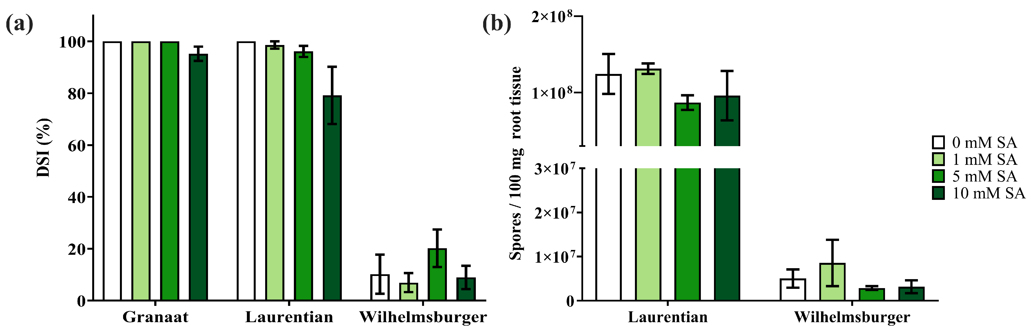
Growth chamber trials were conducted using the same procedures as the high inoculum greenhouse experiments and were replicated three times. The universally susceptible cultivar ‘Granaat’ maintained a DSI of 100% following treatment with 0, 1, and 5 mM SA, and a DSI of 95.2% with 10 mM SA (Figure 4a). In the inoculated susceptible cultivar ‘Laurentian’, mean DSI values were 100%, 98.6%, 96.1%, and 79.2% following application of 0, 1, 5, and 10 mM SA, respectively (Figure 4a; Figure S2). Under the same treatments, the partially resistant cultivar ‘Wilhelmsburger’ exhibited DSI values of 10.2%, 6.9%, 20.2%, and 8.9%. Overall, ‘Wilhelmsburger’ exhibited stronger resistance to pathotype 3A under growth chamber conditions compared with greenhouse trials. No clubroot symptoms were detected in any of the mock-inoculated control plants under growth chamber conditions.
Figure 4.
Effect of salicylic acid (SA) on clubroot disease severity index (DSI) and Plasmodiophora brassicae resting spore accumulation in Brassica napus var. napobrassica ‘Laurentian’ and ‘Wilhelmsburger’ inoculated with a high inoculum of pathotype 3A under growth chamber conditions. B. rapa var. pekinensis ‘Granaat’ served as a susceptibility check. (a) Mean DSI across three experiments at six weeks post-inoculation. (b) Mean resting spore quantification by qPCR with P. brassicae-specific primers and genomic DNA extracted from root galls at six weeks post-inoculation. Error bars indicate standard error of the mean (n = 3).
Resting spores were quantified in inoculated ‘Laurentian’ and ‘Wilhelmsburger’ plants across the different SA treatments. In ‘Laurentian’, mean resting spore counts were 1.24 × 10⁸, 1.31 × 10⁸, 8.69 × 10⁷, and 9.60 × 10⁷ spores per 100 mg of root tissue following application of 0, 1, 5, and 10 mM SA, respectively (Figure 4b). In ‘Wilhelmsburger’, mean resting spore counts were 5.03 × 10⁶, 8.55 × 10⁶, 2.86 × 10⁶, and 3.15 × 10⁶ spores per 100 mg of root tissue under the same SA treatments. Multiple Mann–Whitney tests revealed no significant differences among SA treatments for either DSI or resting spore counts.
3.3. Effect of SA Treatments on Plant Height During Growth Chamber Trials
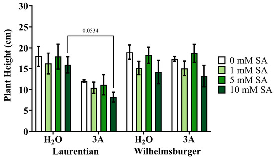
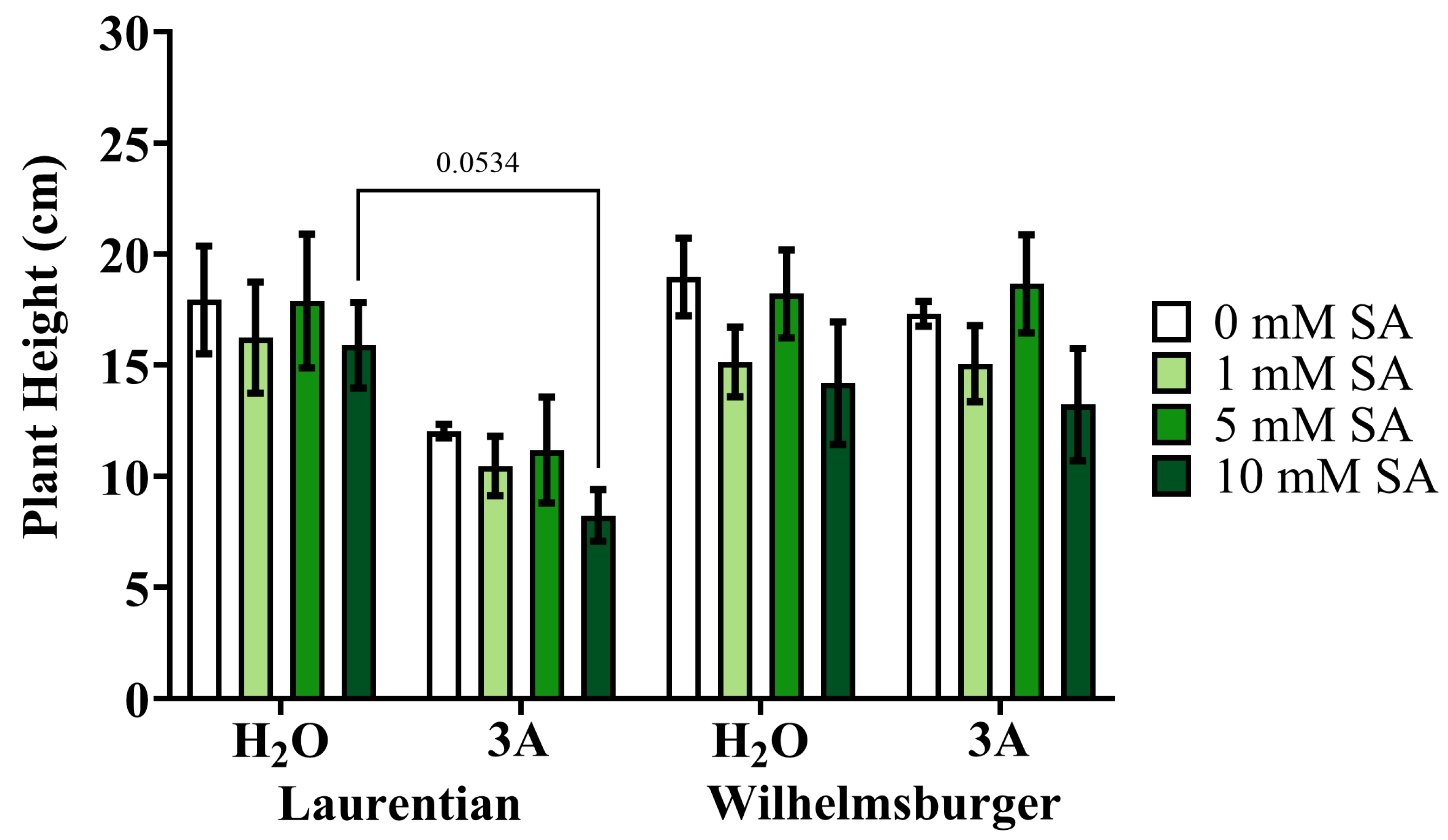
Plant height was measured across all SA treatments in both mock-inoculated and inoculated plants to assess potential effects on growth. No significant differences in mean plant height were observed between the 0 mM SA control and the other SA treatments. In ‘Laurentian’, a reduction in height was observed in inoculated plants compared with mock-inoculated controls at 10 mM (p = 0.0534) SA, based on Tukey’s multiple comparison test (Figure 5). Height differences between mock-inoculated and inoculated plants of ‘Wilhelmsburger’ were comparable across treatments. Foliar lesions developed, with severity increasing proportionally with SA concentration, suggesting some phytotoxic effects (Figure S3).
Figure 5.
Effect of salicylic acid (SA) on plant height in Brassica napus var. napobrassica ‘Laurentian’ and ‘Wilhelmsburger’ mock-inoculated with water or inoculated with Plasmodiophora brassicae pathotype 3A (high inoculum) under growth chamber conditions. Mean plant height (cm) across all three growth chamber experiments at six weeks post-inoculation. Error bars indicate the standard error of the mean (n = 3). A significant difference between mock-inoculated and inoculated ‘Laurentian’ plants was observed at 10 mM (p = 0.0534) SA, based on Tukey’s multiple comparison test.
3.4. Effect of SA Treatments on Expression of Plant SA Markers and P. brassicae Effectors
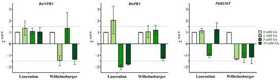
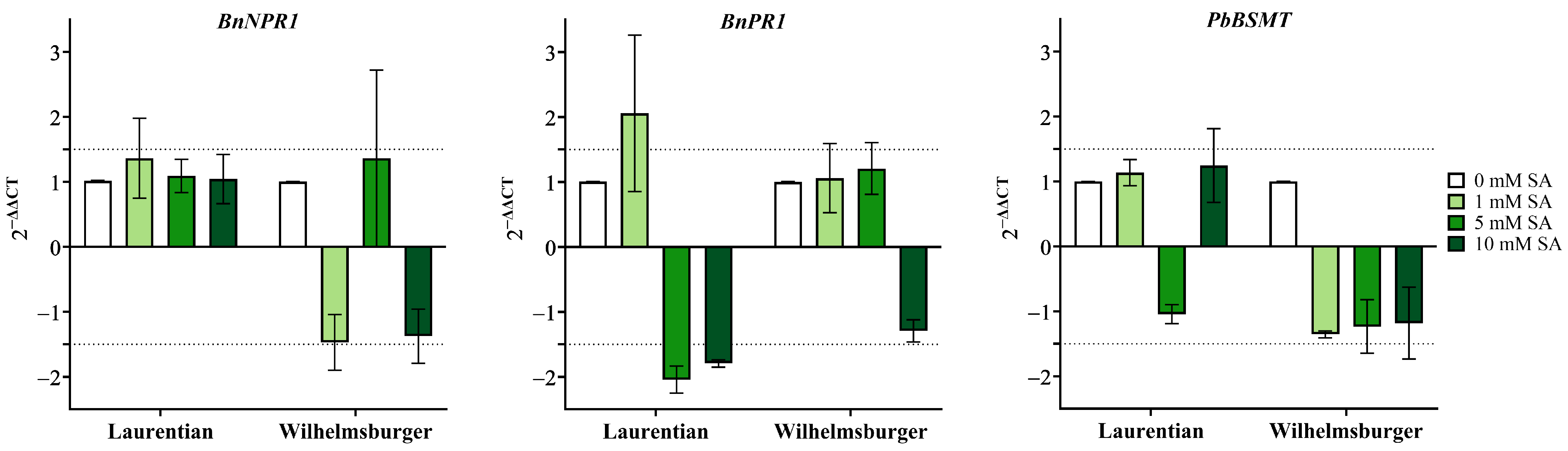
To determine whether foliar SA treatments activated downstream SA responses in B. napus roots, transcript levels of two well-characterized SA markers were measured. Overall, BnNPR1 was not differentially expressed relative to 0 mM SA across all treatments in either of the inoculated cultivars (Figure 6). In the inoculated ‘Laurentian’, BnPR1 transcript levels were reduced by 2.04 ± 0.36 and 1.79 ± 0.10-fold in the 5 and 10 mM SA treatments, respectively, relative to 0 mM SA. However, Dunnett’s multiple comparison test revealed no significant differences.
Figure 6.
Effect of salicylic acid (SA) on gene expression in Brassica napus var. napobrassica ‘Laurentian’ and ‘Wilhelmsburger’ inoculated with Plasmodiophora brassicae pathotype 3A under growth chamber conditions. Expression levels of plant SA marker genes (BnNPR1 and BnPR1) and the P. brassicae secreted protein gene (PbBSMT) were measured. Mean gene expression across three growth chamber experiments at six weeks post-inoculation is shown. Error bars represent the standard deviation of the mean (n = 3). Dashed lines at 1.5 and −1.5 indicate the threshold for differential expression.
To assess how P. brassicae responded to the SA treatments, the transcript levels of the PbBSMT effector were measured and were not differentially expressed relative to the 0 mM SA treatment (Figure 6).
4. Discussion
Transcriptome and proteome studies have shown that SA-mediated defence responses are activated during P. brassicae infection [7,27,28,32,33,48]. During early infection, the SA response is induced in both susceptible and resistant plants. However, as the disease progresses, SA biosynthesis and/or signaling is typically sustained in resistant plants, whereas it declines in susceptible plants.
Exogenous application of SA has shown some success in enhancing plant immunity against P. brassicae, although the effect varies with cultivar, SA concentration, and the timing and frequency of applications [7,30,33,38,39,49]. While P. brassicae is a soilborne pathogen that infects the roots, we applied SA as a foliar spray. This approach was chosen because foliar SA applications have previously reduced clubroot disease severity [7] and are more feasible than a root dip method [31,39] or soil-applied treatment at the base of the plant [33]. Repeated applications were used because SA is rapidly metabolized by both plant and pathogen [7,35,49], and multiple treatments have been shown to be more effective in reducing disease severity [7,30,33,38].
Although P. brassicae colonizes roots, foliar SA can still induce systemic defense responses, as evidence suggests that SA-induced signals are translocated within the plant [3,7,8,49]. We opted to spray one week after inoculation in an attempt to sustain the SA-mediated defence response already known to be activated in both ‘Laurentian’ and ‘Wilhelmsburger’ during early secondary infection [28]. At this time, infection would have been either late in the primary stage, early in the secondary stage, or both, as P. brassicae has an asynchronous life cycle [10]. However, as suggested by our results, this delay in spraying may have limited the plant’s ability to respond effectively to SA, and an application immediately after inoculation might have elicited a stronger defence response.
In this study, exogenous foliar application of SA had a greater effect under greenhouse conditions than under the more controlled growth chamber environment. Similarly, stronger effects of SA on clubroot infection were observed under field vs. greenhouse conditions in an earlier report [49]. In susceptible interactions under low inoculum pressure, evaluated only in the greenhouse, 1 mM SA significantly reduced resting spore quantities compared with the 0 mM SA control. Under high inoculum pressure, DSI and resting spore quantities did not differ significantly following SA application in either greenhouse or growth chamber conditions.
Growth reductions have been reported in other studies following high SA applications in both inoculated and mock-inoculated plants [38,39,49]. For example, treatment of B. oleracea var. italica seedlings with high SA concentrations (10 and 25 mM) significantly reduced survival, although surviving plants exhibited lower disease severity [49]. In the present study, SA-induced leaf lesions were observed, which likely impaired photosynthesis. Previous studies have reported phytotoxic effects of high SA concentrations, including reduced seedling survival and plant growth [31,49]. Nevertheless, optimized SA applications can reduce clubroot severity while supporting plant growth. For instance, treating clubroot-infested soil with 0.6 mM SA for four consecutive days after seedling emergence reduced disease incidence and severity and enhanced plant growth in B. rapa (B. campestris) compared with untreated controls [38].
To assess whether the plants responded transcriptionally to the SA treatments, transcript levels of two well-characterized SA markers, NPR1 and PR1, were quantified. During P. brassicae infection, increased transcripts of NPR1 and PR1 have been reported in both resistant and susceptible interactions in A. thaliana, B. napus, B. rapa, and B. oleracea [23,25,27,50,51,52], as well as in the B. napus ‘Laurentian’ and ‘Wilhelmsburger’ when inoculated with pathotype 3A [28].
In our study, PR1 showed nonsignificant differential expression only in ‘Laurentian’ treated with 1, 5, or 10 mM SA relative to 0 mM SA, whereas NPR1 did not exhibit differential expression in either inoculated cultivar. The downregulation of PR1 at 5 and 10 mM SA may reflect the plant’s attempt to negatively regulate SA and downstream pathways in response to high levels of SA [53]. The absence of differential expression of NPR1, an early key SA receptor, and the subsequent downregulation of PR1 may be related to the timing of sampling, one week after the final SA treatment, which could have missed transient transcriptional responses as the plant attempted to maintain homeostasis [7,53]. Sampling within the first 24–72 h after spraying would likely have captured any transcriptional changes [49]. Alternatively, the SA treatments may not have been sufficient to elicit a detectable transcriptional response in the roots.
The ability of P. brassicae to inactivate SA and suppress downstream plant responses may also explain why DSI and resting spore quantities were not substantially reduced by foliar SA treatment. One mechanism involves the secretion of the effector PbBSMT, which methylates SA more efficiently than the plant’s own BSMT [35]. In our study, however, PbBSMT transcripts were not differentially expressed, suggesting that either the sampling time-point did not capture the pathogen’s response to SA, or the treatments were insufficient to elicit activation of the pathogen’s effector repertoire.
In this study, foliar SA application did not result in a significant reduction in DSI or resting spore quantity. These results suggest that further optimization of SA concentration and application timing could improve its consistency and efficacy against clubroot. Delivering SA via soil application or directly to the base of the plant, closer to the root system, may enhance effectiveness, although such approaches are less practical for large-scale crop management. Future optimization of foliar SA application should also consider the use of a surfactant, such as Tween 20, to improve uptake [54], as well as make adjustments to the number and timing of applications.
Evaluation across a wider range of inoculum levels could reveal whether SA application benefits the host more under mild or moderate disease pressure. In this study, we focused on a concentration of 1 × 10⁷ resting spores per mL, which may have been high enough to overwhelm any potential treatment effect. Nonetheless, a preliminary analysis with 1 × 10⁵ resting spores per mL showed similar trends with respect to DSI in two greenhouse trials, and previous studies have used inoculum concentrations in the 106 to 108 range [7,33,38,39,40,49]. Additionally, testing SA treatments against multiple pathotypes, including non-resistance-breaking isolates in different cultivars, will help identify the optimal SA concentration and application strategy.
5. Conclusions
Exogenous foliar application of SA resulted in mostly nonsignificant reductions in clubroot severity and resting spore levels in B. napus. These findings, combined with the limited transcriptional responses, suggest that treatment timing, intensity, and high inoculum pressure may limit the efficacy of foliar SA applications. Despite these constraints, optimized SA treatments, considering concentration, timing, and delivery method, may still enhance systemic plant defence while minimizing growth trade-offs. Future work should explore alternative application strategies, evaluate a range of inoculum levels and pathotypes, and assess multiple cultivars and their traits to determine the practical potential of SA as a complementary tool in integrated clubroot management.
Supplementary Materials
The following supporting information can be downloaded at https://www.mdpi.com/article/10.3390/agrochemicals5020018/s1. Table S1. Disease assessment, including symptom rating and disease severity index, of pathotype 3A-inoculated Brassica rapa ‘Granaat’ and Brassica napus ‘Laurentian’ and ‘Wilhelmsburger’ following salicylic acid treatments under greenhouse and growth chamber conditions. Table S2. Plant height of mock- and pathotype 3A-inoculated Brassica napus ‘Laurentian’ and ‘Wilhelmsburger’ following salicylic acid treatments under growth chamber conditions. Table S3. Primers used for resting spore quantification and transcript analysis in this study. Table S4. Resting spore quantity in pathotype 3A-inoculated Brassica napus ‘Laurentian’ and ‘Wilhelmsburger’ following salicylic acid treatments under greenhouse and growth chamber conditions. Figure S1. Effect of salicylic acid (SA) on clubroot symptoms caused by Plasmodiophora brassicae in Brassica napus var. napobrassica ‘Laurentian’ and ‘Wilhelmsburger’ inoculated with a high inoculum of pathotype 3A under greenhouse conditions. Figure S2. Effect of salicylic acid (SA) on clubroot symptoms caused by Plasmodiophora brassicae in Brassica napus var. napobrassica ‘Laurentian’ and ‘Wilhelmsburger’ inoculated with a high inoculum pathotype 3A under growth chamber conditions. Figure S3. Effect of salicylic acid (SA) on leaves of Brassica napus var. napobrassica ‘Laurentian’ and ‘Wilhelmsburger’ inoculated with a high inoculum of Plasmodiophora brassicae pathotype 3A under growth chamber conditions.
Author Contributions
Conceptualization, E.S. and S.S.; methodology, E.S.; formal analysis, E.S.; resources, S.S. and S.-F.H.; writing—original draft preparation, E.S.; writing—review and editing, S.S. and S.-F.H.; supervision, S.S. and S.-F.H.; funding acquisition, S.S. and S.-F.H. All authors have read and agreed to the published version of the manuscript.
Funding
Financial support was provided by a Western Grains Research Foundation (WGRF) Graduate Scholarship and a Natural Sciences and Engineering Research Council of Canada (NSERC) Canada Graduate Research Scholarship–Doctoral (CGS–D) awarded to E.S., as well as an NSERC Discovery Grant (RGPIN-2021-04010) awarded to S.S. The University of Alberta contributed in-kind support.
Data Availability Statement
The original contributions presented in this study are included in the article/Supplementary Materials. Further inquiries can be directed to the corresponding author.
Acknowledgments
The authors thank two WISEST Summer Research Program students, Keegan Kirchen and Wendy Ritz, for their assistance with some of the work presented in this manuscript.
Conflicts of Interest
The authors declare no conflicts of interest. The funders had no role in the design of the study; in the collection, analyses, or interpretation of data; in the writing of the manuscript; or in the decision to publish the results.
Abbreviations
The following abbreviations are used in this manuscript:
| Bn | Brassica napus |
| BSMT | BA/SA methyltransferase |
| CAC | clathrin adaptor complex |
| CCD | Canadian Clubroot Differential |
| dpi | disease severity index |
| EFL | elongation factor-like |
| NPR1 | nonexpresser of PR genes 1 |
| Pb | Plasmodiophora brassicae |
| PR1 | pathogenesis-related gene 1 |
| SA | salicylic acid |
References
- Shigenaga, A.M.; Argueso, C.T. No Hormone to Rule Them All: Interactions of Plant Hormones during the Responses of Plants to Pathogens. Semin. Cell Dev. Biol. 2016, 56, 174–189. [Google Scholar] [CrossRef] [Scilit]
- Glazebrook, J. Contrasting Mechanisms of Defense Against Biotrophic and Necrotrophic Pathogens. Annu. Rev. Phytopathol. 2005, 43, 205–227. [Google Scholar] [CrossRef] [Scilit]
- Zhang, Y.; Li, X. Salicylic Acid: Biosynthesis, Perception, and Contributions to Plant Immunity. Curr. Opin. Plant Biol. 2019, 50, 29–36. [Google Scholar] [CrossRef] [Scilit]
- Pieterse, C.M.J.; Van Der Does, D.; Zamioudis, C.; Leon-Reyes, A.; Van Wees, S.C.M. Hormonal Modulation of Plant Immunity. Annu. Rev. Cell Dev. Biol. 2012, 28, 489–521. [Google Scholar] [CrossRef] [Scilit]
- Wani, A.B.; Chadar, H.; Wani, A.H.; Singh, S.; Upadhyay, N. Salicylic Acid to Decrease Plant Stress. Environ. Chem. Lett. 2017, 15, 101–123. [Google Scholar] [CrossRef] [Scilit]
- Methenni, K.; Abdallah, M.B.; Nouairi, I.; Smaoui, A.; Ammar, W.B.; Zarrouk, M.; Youssef, N.B. Salicylic Acid and Calcium Pretreatments Alleviate the Toxic Effect of Salinity in the Oueslati Olive Variety. Sci. Hortic. 2018, 233, 349–358. [Google Scholar] [CrossRef] [Scilit]
- Prerostova, S.; Dobrev, P.; Konradyova, V.; Knirsch, V.; Gaudinova, A.; Kramna, B.; Kazda, J.; Ludwig-Müller, J.; Vankova, R. Hormonal Responses to Plasmodiophora brassicae Infection in Brassica napus Cultivars Differing in Their Pathogen Resistance. Int. J. Mol. Sci. 2018, 19, 4024. [Google Scholar] [CrossRef] [Scilit] [PubMed]
- Feng, Y.; Wang, X.; Du, T.; Shu, Y.; Tan, F.; Wang, J. Effects of Salicylic Acid Concentration and Post-Treatment Time on the Direct and Systemic Chemical Defense Responses in Maize (Zea mays L.) Following Exogenous Foliar Application. Molecules 2022, 27, 6917. [Google Scholar] [CrossRef] [Scilit] [PubMed]
- Kageyama, K.; Asano, T. Life Cycle of Plasmodiophora brassicae. J. Plant Growth Regul. 2009, 28, 203–211. [Google Scholar] [CrossRef] [Scilit]
- Liu, L.; Qin, L.; Zhou, Z.; Hendriks, W.G.H.M.; Liu, S.; Wei, Y. Refining the Life Cycle of Plasmodiophora brassicae. Phytopathology® 2020, 110, 1704–1712. [Google Scholar] [CrossRef] [Scilit]
- Javed, M.A.; Schwelm, A.; Zamani-Noor, N.; Salih, R.; Silvestre Vañó, M.; Wu, J.; González García, M.; Heick, T.M.; Luo, C.; Prakash, P.; et al. The Clubroot Pathogen Plasmodiophora brassicae: A Profile Update. Mol. Plant Pathol. 2023, 24, 89–106. [Google Scholar] [CrossRef] [Scilit]
- Xu, X.; Wu, C.; Zhang, F.; Yao, J.; Fan, L.; Liu, Z.; Yao, Y. Comprehensive Review of Plasmodiophora brassicae: Pathogenesis, Pathotype Diversity, and Integrated Control Methods. Front. Microbiol. 2025, 16, 1531393. [Google Scholar] [CrossRef] [Scilit]
- Peng, G.; McGregor, L.; Lahlali, R.; Gossen, B.D.; Hwang, S.F.; Adhikari, K.K.; Strelkov, S.E.; McDonald, M.R. Potential Biological Control of Clubroot on Canola and Crucifer Vegetable Crops. Plant Pathol. 2011, 60, 566–574. [Google Scholar] [CrossRef] [Scilit]
- Zhu, M.; He, Y.; Li, Y.; Ren, T.; Liu, H.; Huang, J.; Jiang, D.; Hsiang, T.; Zheng, L. Two New Biocontrol Agents Against Clubroot Caused by Plasmodiophora brassicae. Front. Microbiol. 2020, 10, 3099. [Google Scholar] [CrossRef] [Scilit]
- Cordero-Elvia, J.; Galindo-González, L.; Fredua-Agyeman, R.; Hwang, S.-F.; Strelkov, S.E. Clubroot-Induced Changes in the Root and Rhizosphere Microbiome of Susceptible and Resistant Canola. Plants 2024, 13, 1880. [Google Scholar] [CrossRef] [Scilit] [PubMed]
- Hu, H.; Zhang, Y.; Yu, F. A CRISPR/Cas9-Based Vector System Enables the Fast Breeding of Selection-Marker-Free Canola with Rcr1 -Rendered Clubroot Resistance. J. Exp. Bot. 2024, 75, 1347–1363. [Google Scholar] [CrossRef] [Scilit]
- Hurný, A.; Cuesta, C.; Cavallari, N.; Ötvös, K.; Duclercq, J.; Dokládal, L.; Montesinos, J.C.; Gallemí, M.; Semerádová, H.; Rauter, T.; et al. SYNERGISTIC ON AUXIN AND CYTOKININ 1 Positively Regulates Growth and Attenuates Soil Pathogen Resistance. Nat. Commun. 2020, 11, 2170. [Google Scholar] [CrossRef] [Scilit]
- Yang, H.; Liao, R.; Feng, X.; Zhang, Y.; Wang, X.; Zhang, Y.; Yu, L.; Wang, W. The Conserved GOLD Domain in the Plasmodiophora brassicae Effector Pb257 Is Required for Triggering Cell Death and Root Swelling. Phytopathol. Res. 2024, 6, 18. [Google Scholar] [CrossRef] [Scilit]
- Yang, H.; Zhang, Y.; Zhao, Y.; Shu, Y.; Xu, Y.; Liu, Y.; Du, J.; Wang, W. Reduction of Plasmodiophora brassicae Infection on Brassica rapa Through Host-Induced Gene Silencing of Two Secreted Genes. Phytopathology® 2025, 115, 299–305. [Google Scholar] [CrossRef] [Scilit]
- Jayasinghege, C.P.A.; Storfie, E.R.M.; Ozga, J.A.; Strelkov, S.E. The Complex Roles of Plant Hormones During Clubroot Disease Development in the Brassicaceae. J. Plant Growth Regul. 2025, 44, 5692–5712. [Google Scholar] [CrossRef] [Scilit]
- Manoharan, R.K.; Shanmugam, A.; Hwang, I.; Park, J.-I.; Nou, I.-S. Expression of Salicylic Acid-Related Genes in Brassica oleracea Var. Capitata during Plasmodiophora brassicae Infection. Genome 2016, 59, 379–391. [Google Scholar] [CrossRef] [Scilit]
- Zhao, Y.; Bi, K.; Gao, Z.; Chen, T.; Liu, H.; Xie, J.; Cheng, J.; Fu, Y.; Jiang, D. Transcriptome Analysis of Arabidopsis thaliana in Response to Plasmodiophora brassicae during Early Infection. Front. Microbiol. 2017, 8, 673. [Google Scholar] [CrossRef] [Scilit]
- Jia, H.; Wei, X.; Yang, Y.; Yuan, Y.; Wei, F.; Zhao, Y.; Yang, S.; Yao, Q.; Wang, Z.; Tian, B.; et al. Root RNA-Seq Analysis Reveals a Distinct Transcriptome Landscape between Clubroot-Susceptible and Clubroot-Resistant Chinese Cabbage Lines after Plasmodiophora brassicae Infection. Plant Soil. 2017, 421, 93–105. [Google Scholar] [CrossRef] [Scilit]
- Wang, S.; Yu, F.; Zhang, W.; Tang, J.; Li, J.; Yu, L.; Wang, H.; Jiang, J. Comparative Transcriptomic Analysis Reveals Gene Expression Changes during Early Stages of Plasmodiophora brassicae Infection in Cabbage (Brassica oleracea Var. Capitata L.). Can. J. Plant Pathol. 2019, 41, 188–199. [Google Scholar] [CrossRef] [Scilit]
- Ning, Y.; Wang, Y.; Fang, Z.; Zhuang, M.; Zhang, Y.; Lv, H.; Liu, Y.; Li, Z.; Yang, L. Comparative Transcriptome Analysis of Cabbage (Brassica oleracea Var. Capitata) Infected by Plasmodiophora brassicae Reveals Drastic Defense Response at Secondary Infection Stage. Plant Soil. 2019, 443, 167–183. [Google Scholar] [CrossRef] [Scilit]
- Ciaghi, S.; Schwelm, A.; Neuhauser, S. Transcriptomic Response in Symptomless Roots of Clubroot Infected Kohlrabi (Brassica oleracea Var. Gongylodes) Mirrors Resistant Plants. BMC Plant Biol. 2019, 19, 288. [Google Scholar] [CrossRef] [Scilit] [PubMed]
- Galindo-González, L.; Manolii, V.; Hwang, S.-F.; Strelkov, S.E. Response of Brassica napus to Plasmodiophora brassicae Involves Salicylic Acid-Mediated Immunity: An RNA-Seq-Based Study. Front. Plant Sci. 2020, 11, 1025. [Google Scholar] [CrossRef] [Scilit]
- Zhou, Q.; Galindo-González, L.; Manolii, V.; Hwang, S.-F.; Strelkov, S.E. Comparative Transcriptome Analysis of Rutabaga (Brassica napus) Cultivars Indicates Activation of Salicylic Acid and Ethylene-Mediated Defenses in Response to Plasmodiophora brassicae. Int. J. Mol. Sci. 2020, 21, 8381. [Google Scholar] [CrossRef] [Scilit]
- Wei, X.; Zhang, Y.; Zhao, Y.; Xie, Z.; Hossain, M.R.; Yang, S.; Shi, G.; Lv, Y.; Wang, Z.; Tian, B.; et al. Root Transcriptome and Metabolome Profiling Reveal Key Phytohormone-Related Genes and Pathways Involved Clubroot Resistance in Brassica rapa L. Front. Plant Sci. 2021, 12, 759623. [Google Scholar] [CrossRef] [Scilit]
- Lemarié, S.; Robert-Seilaniantz, A.; Lariagon, C.; Lemoine, J.; Marnet, N.; Jubault, M.; Manzanares-Dauleux, M.J.; Gravot, A. Both the Jasmonic Acid and the Salicylic Acid Pathways Contribute to Resistance to the Biotrophic Clubroot Agent Plasmodiophora brassicae in Arabidopsis. Plant Cell Physiol. 2015, 56, 2158–2168. [Google Scholar] [CrossRef] [Scilit]
- Ji, R.; Gao, S.; Bi, Q.; Wang, Y.; Lv, M.; Ge, W.; Feng, H. The Salicylic Acid Signaling Pathway Plays an Important Role in the Resistant Process of Brassica rapa L. Ssp. Pekinensis to Plasmodiophora brassicae Woronin. J. Plant Growth Regul. 2021, 40, 405–422. [Google Scholar] [CrossRef] [Scilit]
- Jayasinghege, C.P.A.; Ozga, J.A.; Manolii, V.P.; Hwang, S.-F.; Strelkov, S.E. Impact of Susceptibility on Plant Hormonal Composition during Clubroot Disease Development in Canola (Brassica napus). Plants 2023, 12, 2899. [Google Scholar] [CrossRef] [Scilit]
- Ferdausi, A.; Megha, S.; Kav, N.N.V.; Rahman, H. Changes in Primary Metabolism and Associated Gene Expression during Host-Pathogen Interaction in Clubroot Resistance of Brassica napus. PLoS ONE 2024, 19, e0310126. [Google Scholar] [CrossRef] [Scilit]
- Ludwig-Müller, J.; Jülke, S.; Geiß, K.; Richter, F.; Mithöfer, A.; Šola, I.; Rusak, G.; Keenan, S.; Bulman, S. A Novel Methyltransferase from the Intracellular Pathogen Plasmodiophora brassicae Methylates Salicylic Acid. Mol. Plant Pathol. 2015, 16, 349–364. [Google Scholar] [CrossRef] [Scilit] [PubMed]
- Djavaheri, M.; Ma, L.; Klessig, D.F.; Mithöfer, A.; Gropp, G.; Borhan, H. Mimicking the Host Regulation of Salicylic Acid: A Virulence Strategy by the Clubroot Pathogen Plasmodiophora brassicae. Mol. Plant-Microbe Interact. 2019, 32, 296–305. [Google Scholar] [CrossRef] [Scilit]
- Galindo-González, L.; Hwang, S.-F.; Strelkov, S.E. Candidate Effectors of Plasmodiophora Brassicae Pathotype 5X During Infection of Two Brassica napus Genotypes. Front. Microbiol. 2021, 12, 742268. [Google Scholar] [CrossRef] [Scilit]
- Chen, T.; Bi, K.; He, Z.; Gao, Z.; Zhao, Y.; Fu, Y.; Cheng, J.; Xie, J.; Jiang, D. Arabidopsis Mutant bik1 Exhibits Strong Resistance to Plasmodiophora Brassicae. Front. Physiol. 2016, 7, 402. [Google Scholar] [CrossRef] [Scilit] [PubMed]
- Xi, D.; Li, X.; Gao, L.; Zhang, Z.; Zhu, Y.; Zhu, H. Application of Exogenous Salicylic Acid Reduces Disease Severity of Plasmodiophora brassicae in Pakchoi (Brassica campestris Ssp. Chinensis Makino). PLoS ONE 2021, 16, e0248648. [Google Scholar] [CrossRef] [Scilit]
- Agarwal, A.; Kaul, V.; Faggian, R.; Rookes, J.E.; Ludwig-Müller, J.; Cahill, D.M. Analysis of Global Host Gene Expression during the Primary Phase of the Arabidopsis thaliana–Plasmodiophora brassicae Interaction. Funct. Plant Biol. 2011, 38, 462. [Google Scholar] [CrossRef] [Scilit]
- Lovelock, D.A.; Šola, I.; Marschollek, S.; Donald, C.E.; Rusak, G.; Van Pée, K.; Ludwig-Müller, J.; Cahill, D.M. Analysis of Salicylic Acid-dependent Pathways in Arabidopsis thaliana Following Infection with Plasmodiophora brassicae and the Influence of Salicylic Acid on Disease. Mol. Plant Pathol. 2016, 17, 1237–1251. [Google Scholar] [CrossRef] [Scilit]
- Storfie, E.; Manolii, V.P.; Aigu, Y.; Botero-Ramirez, A.; Harding, M.W.; Hwang, S.-F.; Strelkov, S.E. Collection and Identification of Plasmodiophora brassicae Pathotypes from Western Canada in 2021–2023. Can. J. Plant Pathol. 2025, 47, 356–367. [Google Scholar] [CrossRef] [Scilit]
- Canola Council of Canada. Available online: https://www.canolacouncil.org (accessed on 2 February 2025).
- Strelkov, S.E.; Tewari, J.P.; Smith-Degenhardt, E. Characterization of Plasmodiophora brassicae Populations from Alberta, Canada. Can. J. Plant Pathol. 2006, 28, 467–474. [Google Scholar] [CrossRef] [Scilit]
- Kuginuki, Y.; Yoshikawa, H.; Hirai, M. Variation in Virulence of Plasmodiophora brassicae in Japan Tested with Clubroot-Resistant Cultivars of Chinese Cabbage (Brassica rapa L. Ssp. Pekinensis). Eur. J. Plant Pathol. 1999, 105, 327–332. [Google Scholar] [CrossRef] [Scilit]
- Horiuchi, S.; Hori, M. A Simple Greenhouse Technique for Obtaining High Levels of Clubroot Incidence. Bull. Chugoku Nat. Agric. Exp. Stn. 1980, 17, 33–35. [Google Scholar]
- Rennie, D.C.; Manolii, V.P.; Cao, T.; Hwang, S.F.; Howard, R.J.; Strelkov, S.E. Direct Evidence of Surface Infestation of Seeds and Tubers by Plasmodiophora brassicae and Quantification of Spore Loads. Plant Pathol. 2011, 60, 811–819. [Google Scholar] [CrossRef] [Scilit]
- Livak, K.J.; Schmittgen, T.D. Analysis of Relative Gene Expression Data Using Real-Time Quantitative PCR and the 2−∆∆CT Method. Methods 2001, 25, 402–408. [Google Scholar] [CrossRef] [Scilit]
- Adhikary, D.; Mehta, D.; Uhrig, R.G.; Rahman, H.; Kav, N.N.V. A Proteome-Level Investigation into Plasmodiophora brassicae Resistance in Brassica napus Canola. Front. Plant Sci. 2022, 13, 860393. [Google Scholar] [CrossRef] [Scilit]
- Lovelock, D.A.; Donald, C.E.; Conlan, X.A.; Cahill, D.M. Salicylic Acid Suppression of Clubroot in Broccoli (Brassicae oleracea Var. Italica) Caused by the Obligate Biotroph Plasmodiophora brassicae. Australas. Plant Pathol. 2013, 42, 141–153. [Google Scholar] [CrossRef] [Scilit]
- Chen, J.; Pang, W.; Chen, B.; Zhang, C.; Piao, Z. Transcriptome Analysis of Brassica rapa Near-Isogenic Lines Carrying Clubroot-Resistant and –Susceptible Alleles in Response to Plasmodiophora brassicae during Early Infection. Front. Plant Sci. 2016, 6, 1183. [Google Scholar] [CrossRef] [Scilit]
- Zhang, X.; Liu, Y.; Fang, Z.; Li, Z.; Yang, L.; Zhuang, M.; Zhang, Y.; Lv, H. Comparative Transcriptome Analysis between Broccoli (Brassica Oleracea Var. Italica) and Wild Cabbage (Brassica macrocarpa Guss.) in Response to Plasmodiophora brassicae during Different Infection Stages. Front. Plant Sci. 2016, 7, 1929. [Google Scholar] [CrossRef] [Scilit]
- Yuan, Y.; Qin, L.; Su, H.; Yang, S.; Wei, X.; Wang, Z.; Zhao, Y.; Li, L.; Liu, H.; Tian, B.; et al. Transcriptome and Coexpression Network Analyses Reveal Hub Genes in Chinese Cabbage (Brassica rapa L. Ssp. Pekinensis) During Different Stages of Plasmodiophora brassicae Infection. Front. Plant Sci. 2021, 12, 650252. [Google Scholar] [CrossRef] [Scilit] [PubMed]
- Peng, Y.; Yang, J.; Li, X.; Zhang, Y. Salicylic Acid: Biosynthesis and Signaling. Annu. Rev. Plant Biol. 2021, 72, 761–791. [Google Scholar] [CrossRef] [Scilit] [PubMed]
- Parr, J.F.; Norman, A.G. Considerations in the Use of Surfactants in Plant Systems: A Review. Bot. Gaz. 1965, 126, 86–96. [Google Scholar] [CrossRef] [Scilit]
Disclaimer/Publisher’s Note: The statements, opinions and data contained in all publications are solely those of the individual author(s) and contributor(s) and not of MDPI and/or the editor(s). MDPI and/or the editor(s) disclaim responsibility for any injury to people or property resulting from any ideas, methods, instructions or products referred to in the content. |
© 2026 by the authors. Licensee MDPI, Basel, Switzerland. This article is an open access article distributed under the terms and conditions of the Creative Commons Attribution (CC BY) license.