


Abstract
Background/Objectives: The therapeutic durability of β-lactams is increasingly threatened by penicillin-binding proteins (PBPs) that retain cell-wall transpeptidase activity under antibiotic pressure. This study computationally evaluated 221 Daniellia oliveri metabolites against penicillin-binding protein 2a (PBP2a) of methicillin-resistant Staphylococcus aureus (MRSA) and penicillin-binding protein 2x (PBP2x) of Streptococcus pneumoniae, using amoxicillin and cefotaxime as reference standards. Methods: Molecular docking and interaction analysis prioritised quercitrin, apigetrin, quercetin 3-rutinoside and acid methyl ester for PBP2a, and amyrin, columbin, apigetrin, quercetin 3-rutinoside and N-(2H-tetrazol-5-yl)benzamide for PBP2x. The candidates were further assessed through pharmacokinetic and drug-likeness prediction, 160 ns molecular dynamics (MD) simulations, molecular mechanics/generalised Born surface area (MM/GBSA) analysis and density functional theory (DFT). Results: Quercetin 3-rutinoside emerged as the highest-priority computational dual-target candidate, with the numerically most favourable within-protocol MM/GBSA estimates for PBP2a (−58.51 kcal mol−1) and PBP2x (−55.92 kcal mol−1) among the tested compounds and controls. The compound also had the lowest PBP2a root-mean-square deviation (RMSD; 1.64 Å) and root-mean-square fluctuation (RMSF; 1.26 Å), indicating lower global deviation and residue-level fluctuation in the PBP2a simulation. Against PBP2x, quercetin 3-rutinoside exhibited a higher RMSD (4.98 Å) but a relatively low RMSF (1.75 Å), consistent with greater global protein reorganisation alongside comparatively limited residue-level fluctuation. DFT descriptors of quercetin 3-rutinoside indicated moderate electronic responsiveness, whereas pharmacokinetic profiling revealed high molecular weight, rule-of-five violations, low predicted gastrointestinal absorption and P-glycoprotein liability. Conclusions: Overall, quercetin 3-rutinoside was the highest-ranked computational dual-PBP candidate from D. oliveri; this prioritisation does not establish PBP inhibition and requires structural optimisation and biochemical, antibacterial, safety and in vivo validation.
1. Introduction
Antimicrobial resistance (AMR) now constrains effective infection management. The 2021 Global Burden of Disease (GBD) analysis estimated that bacterial AMR was associated with 4.71 million deaths and directly attributable to 1.14 million deaths worldwide [1]. Staphylococcus aureus and Streptococcus pneumoniae contribute substantially to this burden because resistance in these pathogens increasingly compromises β-lactams, one of the most widely used classes of antibiotics. Methicillin-resistant S. aureus (MRSA) causes infections ranging from skin and soft-tissue infections to bacteraemia, endocarditis and sepsis, whereas the drug-resistant S. pneumoniae remains an important cause of pneumonia, meningitis and invasive pneumococcal disease. This ongoing loss of β-lactam effectiveness underscores the need for new antibacterial scaffolds targeting biologically relevant resistance-associated targets.
Penicillin-binding proteins (PBPs) catalyse the terminal stages of peptidoglycan biosynthesis and are the principal targets of β-lactam antibiotics [2,3]. Their transpeptidase domains contain conserved SXXK, SXN and KTG/KS(T)G motifs, in which X denotes any amino acid; these motifs organise the catalytic site for peptide cross-linking, and the serine in SXXK acts as the nucleophile acylated by β-lactams. Penicillin-binding protein 2a (PBP2a) and penicillin-binding protein 2x (PBP2x) are particularly relevant to β-lactam resistance in S. aureus and S. pneumoniae, respectively. PBP2a, encoded by mecA, has intrinsically low affinity for most β-lactams and contains the conserved Ser403–Lys406, Ser462–Asn464 and Lys597–Ser598–Gly599 catalytic regions [4]. Similarly, PBP2x contains the Ser337–Lys340 catalytic motif and the Ser395–Asn397 and Lys547–Ser548–Gly549 regions involved in catalytic-pocket organisation and β-lactam recognition [5]. Alterations within and around these regions can reduce β-lactam acylation, while preserving transpeptidase function. The catalytic sites of PBP2a and PBP2x are, therefore, biologically relevant regions for identifying alternative scaffolds that engage residues involved in substrate recognition and catalysis.
Natural products remain valuable sources of antibacterial scaffolds because of their structural and functional-group diversity [6,7]. Daniellia oliveri (Fabaceae), commonly known as the African copaiba balsam tree, is traditionally used in West and Central Africa for infectious and inflammatory conditions and contains diverse flavonoids, phenolics, fatty acids, sterols and terpenoids [8]. Extracts and fractions of D. oliveri have demonstrated antibacterial activity, with reported minimum inhibitory concentrations of 16–256 μg mL−1 and limited cytotoxicity for several active fractions in the tested normal-cell model [9]. However, extract-level activity does not identify the responsible metabolites or their molecular targets. Plant-derived phenolics provide a plausible link to PBPs: quercetin 3-O-rutinoside has shown favourable interaction with PBP2a alongside reported anti-MRSA activity [10], whereas other flavonoids have demonstrated antibacterial or β-lactam-potentiating effects against resistant S. aureus [11]. Structure–activity and molecular dynamics studies have also shown that phenolic compounds can interact with functionally important regions of PBP2a and PBP2x [12,13]. More recently, rutin from D. oliveri was computationally prioritised against the Acinetobacter baumannii BfmR (RstA) response-regulatory system, further supporting this plant as a source of potentially bioactive bacterial-targeting scaffolds [14].
Despite these findings, the broader D. oliveri metabolite repertoire has not been comparatively investigated against PBP2a and PBP2x. Accordingly, this study screened D. oliveri metabolites against the active sites of PBP2a from MRSA and PBP2x from S. pneumoniae. Molecular docking and binding-site interactions were used for initial prioritisation, followed by pharmacokinetic and drug-likeness prediction, 160 ns molecular dynamics (MD) simulations, molecular mechanics/generalised Born surface area (MM/GBSA) binding-energy estimation, time-resolved interaction analysis and density functional theory (DFT). By integrating molecular recognition, conformational behaviour, binding energetics, electronic properties and predicted developability, this study aimed to identify D. oliveri-derived candidates for subsequent biochemical and antibacterial validation. The overall computational workflow and decision points are summarised in Figure 1.
Figure 1.
The overall computational workflow. (01)The 221 reported D. oliveri metabolites, reference antibiotics and PBP targets were selected. (02) The prepared and screened using active-site docking and catalytic-region contacts. (03) Target-specific candidates underwent ADME/T, DFT, 160 ns MD, MM/GBSA and interaction analyses. (04) Integrated within-protocol comparison produced computational candidates requiring experimental validation.
2. Results and Discussion
2.1. Molecular Docking of D. oliveri Compounds Against S. aureus PBP2a and S. pneumoniae PBP2x
The docking of the D. oliveri metabolite library revealed broad variation in docking-score profiles for PBP2a and PBP2x, reflecting the structural diversity of the screened compounds (Supplementary Table S1). Across the complete dataset, several metabolites produced scores (PBP2a, −2.8 to −9.3 kcal mol−1; PBP2x, −2.7 to −12.0 kcal mol−1) comparable to or more favourable than those of amoxicillin and cefotaxime. This supported further prioritisation, using both docking scores and interactions with functionally important active-site residues.
Against PBP2a, quercitrin and apigetrin had the most favourable scores among the selected compounds (−9.3 kcal mol−1), followed by quercetin 3-rutinoside and acid methyl ester (−9.0 kcal mol−1) (Table 1). These docking scores were numerically lower than those of amoxicillin (−7.8 kcal mol−1) and cefotaxime (−7.9 kcal mol−1), although the small differences among the leading metabolites limited discrimination by docking alone. Quercetin 3-rutinoside, although not the highest-scoring PBP2a ligand, formed an extensive network involving Gln613, Thr600, Ser462, Ser461, Ser403, Asn464, Glu602 and Thr444. Ser403 belongs to the conserved SXXK catalytic motif, whereas Ser462 and Asn464 form another functionally important transpeptidase region; simultaneous engagement, therefore, supported favourable positioning across the catalytic pocket. Quercitrin and apigetrin also contacted Asn464; apigetrin additionally engaged catalytic Ser403, and Tyr446, Met641 and Ala642 contributed aromatic or hydrophobic contacts. The prominence of glycosylated flavonoids agrees with previous reports of flavonoid–PBP2a recognition. Rani et al. [10] identified quercetin 3-O-rutinoside as a favourable PBP2a-binding compound and subsequently demonstrated anti-MRSA activity, although at relatively high micromolar concentrations. Alhadrami et al. [11] also reported distinct behaviour for glycosylated and non-glycosylated flavonoids, with glycosylated derivatives showing particular potential for β-lactam potentiation. The numerous hydroxyl groups of quercetin 3-rutinoside may, therefore, support extensive active-site hydrogen bonding while imposing desolvation and permeability penalties. Its favourable recognition was consequently considered alongside pharmacokinetic and dynamic behaviour.
Table 1.
The docking scores and reported binding-site interactions of the top-ranked D. oliveri compounds and controls against PBP2a (3ZFZ) and PBP2x (5OJ0).
The selected PBP2x compounds were more structurally diverse. N-(2H-tetrazol-5-yl)benzamide produced the most favourable score (−12.0 kcal mol−1), followed by amyrin (−10.0), quercetin 3-rutinoside (−9.9), apigetrin (−9.7) and columbin (−9.5 kcal mol−1) (Table 1). The complete dataset confirmed that these scores were numerically lower than those of the reference antibiotics (Table 1; Supplementary Table S1). The compounds achieved favourable docking through distinct interaction patterns: amyrin relied mainly on hydrophobic and dispersion contacts, whereas the flavonoid glycosides formed broader polar networks. Quercetin 3-rutinoside engaged Ser337, Ser395, Asn397, Gln552, Thr526 and Asn377, and Apigetrin engaged Ser337 and Lys340. Ser337 and Lys340 constituted the conserved SXXK motif, whereas Ser395/Asn397 contributed to the transpeptidase architecture, thereby supporting the occupation of functionally relevant regions of the catalytic cleft. Stable multiresidue interactions between phenolics and PBP2x have also been reported during MD simulation [13]. Nevertheless, the chemically distinct amyrin, columbin and N-(2H-tetrazol-5-yl)benzamide profiles show that favourable PBP2x recognition was not restricted to flavonoids.
Collectively, docking identified structurally diverse D. oliveri metabolites predicted to occupy the catalytic regions of both PBPs. Quercetin 3-rutinoside was predicted to contact functionally important residues in PBP2a and PBP2x, despite not having the most favourable score for either target. This finding underscored the limitation of prioritisation by static scores alone and justified subsequent assessment of pharmacokinetic properties, electronic characteristics, binding energetics and conformational behaviour through MD simulations.
The crystallographic native ligand of each target was redocked using the corresponding grid and docking settings, and the redocked pose (orange) was superimposed on its crystallographic pose (red). The RMSD was approximately 1.0 Å for both PBP2a and PBP2x (Figure 2 and Figure 3), below the commonly used 2.0 Å criterion and, therefore, validating the protocol for native-pose reproduction [15,16]. This validation does not establish docking-score ranking accuracy, binding affinity, inhibition or antibacterial activity. The 160 ns MD and MM/GBSA analyses remain complementary within-protocol descriptors rather than experimental confirmation.
Figure 2.
A superimposition of the top-ranked D. oliveri metabolites and reference antibiotics within the PBP2a (3ZFZ) active site. (a) The overlay of quercitrin (blue), apigetrin (brown), quercetin 3-rutinoside (black), acid methyl ester (yellow), cefotaxime (green), amoxicillin (pink) and the redocked native ligand (orange) relative to the native crystallographic ligand (red). (b) The superimposed binding poses displayed within the PBP2a active-site environment, highlighting the spatial orientation of the ligands relative to key catalytic and recognition residues. The approximately 1.0 Å RMSD between the redocked (orange) and crystallographic (red) native-ligand poses is below the 2.0 Å threshold and validates pose reproduction by the PBP2a docking protocol.
Figure 3.
A superimposition of the top-ranked D. oliveri metabolites and reference antibiotics within the PBP2x (5OJ0) active site. (a) The overlay of amyrin (cyan), columbin (yellow), apigetrin (brown), quercetin 3-rutinoside (black), N-(2H-tetrazol-5-yl)benzamide (blue), cefotaxime (green), amoxicillin (pink) and the redocked native ligand (orange) relative to the native crystallographic ligand (red). (b) The superimposed binding poses displayed within the PBP2x active-site environment, showing the hydrogen-bond donor/acceptor surface and selected binding-site residues, including Lys340, Arg372, Trp374, Ser395, Gly549, Gln452 and Gln552. The approximately 1.0 Å RMSD between the redocked (orange) and crystallographic (red) native-ligand poses is below the 2.0 Å threshold and validates pose reproduction by the PBP2x docking protocol.
2.2. The Pharmacokinetic and Drug-Likeness Profiles of the Top-Ranked D. oliveri Compounds and Controls
Pharmacokinetic profiling revealed substantial differences in the predicted oral developability of the top-ranked D. oliveri metabolites. Some compounds showed acceptable drug-likeness features, whereas others were limited by molecular size, hydrogen-bonding capacity, gastrointestinal absorption, solubility or transporter liability (Table 2; Supplementary Tables S2 and S3). Excessive molecular size, lipophilicity, hydrogen-bonding capacity and flexibility can adversely affect permeability and oral bioavailability [17,18,19]. Amoxicillin and cefotaxime met the evaluated Lipinski criteria and had no predicted P-gp substrate liability or CYP inhibition. Both nevertheless showed low predicted gastrointestinal absorption, illustrating that rule-of-five compliance alone does not guarantee favourable intestinal absorption [20].
Table 2.
The predicted pharmacokinetic and toxicity summary of the top-ranked D. oliveri metabolites and controls.
Among the PBP2a candidates, acid methyl ester showed the most balanced predicted profile: a molecular weight of 479.48 g mol−1, compliance with the evaluated Lipinski criteria, high gastrointestinal absorption and a bioavailability score of 0.55. Its potential was limited by poor predicted solubility and possible inhibition of CYP2C9, CYP2D6 and CYP3A4. Quercitrin exceeded the recommended hydrogen-bond acceptor and donor limits, whereas apigetrin exceeded the donor limit and was predicted to be a P-gp substrate (Table 2; Supplementary Table S2). These differences show why compound prioritisation should consider multiple absorption, distribution, metabolism and excretion properties rather than rule-of-five compliance alone.
Quercetin 3-rutinoside showed the most pronounced predicted oral-developability limitations among the PBP2a candidates. Its molecular weight (610.52 g mol−1), 16 hydrogen-bond acceptors, 10 donors and six rotatable bonds resulted in Lipinski violations, accompanied by low predicted gastrointestinal absorption, a bioavailability score of 0.17 and P-gp substrate liability (Table 2; Supplementary Table S2). High molecular size and extensive hydrogen-bonding capacity generally disfavour passive membrane permeability [17,18]. This is important because the same compound interacted extensively with both PBP targets during docking: the polar functionality that promotes protein recognition may also constrain cellular permeability and systemic exposure. Accordingly, it was not interpreted as an orally developable lead in its current form.
The PBP2x candidates showed similar variation. Columbin had the most balanced profile, with a molecular weight of 358.39 g mol−1, predicted solubility, high gastrointestinal absorption and a bioavailability score of 0.55, although it was predicted to be a P-gp substrate. Amyrin met the Lipinski criteria but combined relatively high lipophilicity (log P = 4.63) with poor solubility and low gastrointestinal absorption. Conversely, N-(2H-tetrazol-5-yl)benzamide showed several liabilities: a molecular weight of 635.60 g mol−1, 11 hydrogen-bond acceptors, poor solubility, low gastrointestinal absorption, and predicted P-gp, CYP2D6, and CYP3A4 liabilities (Table 2; Supplementary Table S3). These findings again show that favourable molecular recognition need not correspond to favourable predicted pharmacokinetics [20].
Overall, the pharmacokinetic analysis identified a clear trade-off between target recognition and predicted developability. Quercetin 3-rutinoside remained notable for dual-target recognition, but its physicochemical and absorption liabilities indicate a need for structural optimisation or alternative delivery strategies. Acid methyl ester and columbin offered more balanced predicted profiles for PBP2a and PBP2x, respectively. Pharmacokinetic properties were, therefore, considered alongside binding energetics, electronic characteristics and dynamic behaviour when prioritising compounds for experimental investigation.
2.3. The Dynamic Behaviour of the Top-Ranked D. oliveri Compounds Complexed with PBP2a and PBP2x
The post-dynamics analyses revealed distinct conformational and interaction profiles across the PBP2a and PBP2x complexes, with binding energetics, conformational behaviour, residue flexibility, compactness, solvent exposure and sampled interaction recurrence providing complementary descriptors for within-protocol prioritisation.
2.3.1. MM/GBSA Binding-Energy Analysis of the Top-Ranked D. oliveri Compounds
The molecular mechanics/generalised Born surface area analysis discriminated among the PBP2a complexes more clearly than docking. Quercetin 3-rutinoside had the numerically most negative MM/GBSA estimate (−58.51 ± 4.62 kcal mol−1), numerically more negative than cefotaxime (−34.23 ± 4.18 kcal mol−1) and amoxicillin (−20.16 ± 5.24 kcal mol−1). Quercitrin (−39.96 ± 7.14 kcal mol−1) and acid methyl ester (−37.70 ± 5.79 kcal mol−1) were also numerically more negative than the values for both controls, whereas apigetrin had the least negative estimate (−12.74 ± 6.72 kcal mol−1) despite sharing the best docking score with quercitrin (Table 3). This marked rank change illustrates the limitations of docking scores, as MM/GBSA incorporates energetic contributions from dynamically sampled complexes and the solvent environment [21]. The quercetin 3-rutinoside estimate reflected van der Waals (−60.91 ± 4.81 kcal mol−1) and electrostatic (−60.76 ± 14.34 kcal mol−1) contributions that offset the opposing solvation energy (+63.15 ± 8.87 kcal mol−1) (Table 3). The contrast with apigetrin is informative: both glycosides formed favourable static active-site interactions, yet their post-dynamics energetics differed substantially, showing that an extensive docking network need not remain energetically favourable under dynamic, solvated conditions.
Table 3.
The molecular mechanics/generalised Born surface area (MM/GBSA) energy components of the complexes formed by the top-ranked D. oliveri compounds with PBP2a and PBP2x during the 160 ns molecular dynamics simulations.
PBP2x showed a similar pattern. Quercetin 3-rutinoside again had the numerically most negative MM/GBSA estimate (−55.92 ± 7.45 kcal mol−1), compared with cefotaxime (−46.95 ± 5.59 kcal mol−1) and amoxicillin (−16.11 ± 5.74 kcal mol−1). N-(2H-tetrazol-5-yl)benzamide (−42.53 ± 3.94 kcal mol−1) and apigetrin (−38.67 ± 5.82 kcal mol−1) followed, whereas columbin and amyrin had less negative estimates of −33.62 ± 5.02 and −21.49 ± 5.30 kcal mol−1, respectively (Table 3). Although N-(2H-tetrazol-5-yl)benzamide had the most favourable PBP2x docking score, it did not retain the leading rank after MD simulation. Its strong electrostatic contribution (−273.60 ± 14.38 kcal mol−1) was largely offset by unfavourable solvation (+281.55 ± 13.10 kcal mol−1) (Table 3). This compensation demonstrates the importance of solvent effects and why docking scores are not direct measures of affinity [22].
Most notably, quercetin 3-rutinoside produced the numerically most negative MM/GBSA estimates for both PBP2a and PBP2x despite not leading the docking ranking for either target. Its dual-target energetic preference and interactions with conserved catalytic-site residues support further biochemical evaluation. This agrees with earlier evidence of quercetin 3-O-rutinoside interaction with PBP2a [10] and with the dynamic prioritisation of rutin in the D. oliveri BfmR (RstA) study [14]. Nevertheless, MM/GBSA values are relative computational estimates rather than experimentally determined thermodynamic affinities. The reported means ± standard deviations describe frame-to-frame variability within single production trajectories; no replicate-based inferential test was performed, so between-compound differences are not claims of statistical significance. Configurational entropy was excluded, which may be especially consequential for the large, flexible quercetin 3-rutinoside molecule and may make its estimate overly favourable. The numerical preference, therefore, supports only within-protocol prioritisation alongside conformational, interaction, and pharmacokinetic descriptors, pending experimental validation.
2.3.2. The Post-Dynamics Trajectory Metrics of the Top-Ranked D. oliveri Compound–PBP Complexes
The 160 ns trajectories revealed ligand-dependent differences in the conformational behaviour of PBP2a and PBP2x complexes. The root-mean-square deviation, RMSF, Rg, SASA and intramolecular hydrogen-bond profiles were interpreted collectively to distinguish global conformational change, local flexibility, compactness, solvent exposure and preservation of internal protein contacts (Table 4; Figure 4, Figure 5, Figure 6, Figure 7 and Figure 8). This multidimensional approach is important for PBPs because ligand-bound states can undergo appreciable conformational rearrangement without disrupting the overall architecture [23]. Rather than applying a universal RMSD threshold, each trajectory was interpreted from the magnitude, persistence and temporal pattern of its fluctuations relative to the corresponding apo protein.
Table 4.
The mean molecular dynamics trajectory descriptors of the apo proteins and top-ranked D. oliveri compound–PBP complexes during the 160 ns MD simulations.
Figure 4.
Backbone/alpha-carbon (Cα) root-mean-square deviation (RMSD) of PBP2a complexes (a) and PBP2x complexes (b) over 160 ns MD simulation.
Figure 5.
Per-residue root-mean-square fluctuation (RMSF) of PBP2a complexes (a) and PBP2x complexes (b) over 160 ns MD simulation.
Figure 6.
The radius of gyration (Rg) of PBP2a complexes (a) and PBP2x complexes (b) during the 160 ns MD trajectories.
Figure 7.
Solvent-accessible surface area (SASA) of PBP2a complexes (a) and PBP2x complexes (b) over 160 ns MD simulation.
Figure 8.
Intramolecular hydrogen-bond counts for PBP2a complexes (a) and PBP2x complexes (b) over 160 ns MD simulation.
For PBP2a, quercetin 3-rutinoside had the lowest mean RMSD (1.64 ± 0.36 Å), followed by acid methyl ester (2.00 ± 0.43 Å), cefotaxime (2.08 ± 0.52 Å), apigetrin (2.21 ± 0.44 Å), and quercitrin (2.43 ± 0.65 Å), compared with 3.76 ± 0.80 Å for apo PBP2a (Table 4; Figure 4). After an initial adjustment during approximately the first 20 ns, the quercetin 3-rutinoside complex maintained a narrow RMSD distribution for most of the trajectory, with only a modest late increase. Acid methyl ester fluctuated around 30–40 ns before stabilising, whereas quercitrin showed broader excursions around 55–95 ns and near the end. Cefotaxime showed a transient increase around 100–130 ns before returning towards its preceding range. These patterns indicate ligand-dependent modulation of PBP2a dynamics without necessarily implying global destabilisation.
The PBP2x RMSD profiles were more widely separated. Cefotaxime had the lowest mean RMSD among the complexes (2.65 ± 0.49 Å), followed by columbin (3.95 ± 0.80 Å), apigetrin (4.27 ± 1.02 Å), amyrin (4.29 ± 0.94 Å), amoxicillin (4.46 ± 0.80 Å), quercetin 3-rutinoside (4.98 ± 0.91 Å), and N-(2H-tetrazol-5-yl)benzamide (7.25 ± 2.22 Å), compared with 3.99 ± 0.66 Å for apo PBP2x (Table 4; Figure 4). These values describe protein Cα displacement and are not ligand-displacement metrics. Most complexes underwent an initial adjustment and then fluctuated within a defined range. In contrast, N-(2H-tetrazol-5-yl)benzamide showed a sustained increase to approximately 6–8 Å after about 30 ns, followed by a further rise after approximately 105 ns. Its highly favourable docking score, therefore, did not translate into the preservation of the initial protein–ligand conformation during MD.
The contrasting quercetin 3-rutinoside trajectories further show why the RMSD and binding energetics must be interpreted together. Against PBP2a, its low RMSD and numerically most negative MM/GBSA estimate provided concordant within-protocol descriptors. Against PBP2x, the higher protein-backbone RMSD co-occurred with the numerically most negative MM/GBSA estimate, indicating a different global protein ensemble within this simulation. Neither metric demonstrates retained ligand binding. Importantly, the trajectory lacked the progressive displacement seen for N-(2H-tetrazol-5-yl)benzamide. Conversely, apigetrin’s relatively low PBP2a RMSD accompanied the least negative MM/GBSA estimate among the PBP2a complexes. Conformational displacement and end-point binding energetics are, therefore, complementary, not interchangeable, measures of protein–ligand behaviour [22].
The RMSF profiles resolved the local flexibility underlying the global RMSD changes (Figure 5). For PBP2a, quercetin 3-rutinoside and apigetrin had the lowest mean RMSF values (1.26 ± 0.50 and 1.26 ± 0.54 Å), followed by cefotaxime (1.30 ± 0.51 Å), acid methyl ester (1.40 ± 0.60 Å) and quercitrin (1.42 ± 0.71 Å). These values were lower than those of apo PBP2a (2.43 ± 0.17 Å) and amoxicillin (3.55 ± 1.80 Å) (Table 4; Figure 5). The largest fluctuations occurred mainly in terminal and exposed loop regions, whereas much of the protein showed comparatively low fluctuations. PBP2a contains the conserved Ser403–Lys406 catalytic motif and the Ser462–Asn464 and Lys597–Ser598–Gly599 regions that organise the transpeptidase site and substrate recognition [4]. These regions did not coincide with the dominant RMSF peaks, suggesting that ligand-associated adjustments occurred without pronounced destabilisation of the catalytic core. The low overall RMSF and absence of prominent catalytic-region peaks for quercetin 3-rutinoside indicate particularly low local fluctuation in functionally important regions.
PBP2x showed a similar ligand-dependent pattern. Cefotaxime had the lowest mean RMSF (1.59 ± 0.90 Å), followed by quercetin 3-rutinoside (1.75 ± 1.07 Å), amyrin (1.76 ± 1.05 Å), columbin (1.89 ± 1.12 Å) and amoxicillin (1.93 ± 1.13 Å); apo PBP2x also had a mean RMSF of 1.93 ± 1.23 Å. Apigetrin (2.21 ± 1.43 Å) and, particularly, N-(2H-tetrazol-5-yl)benzamide (2.53 ± 1.94 Å) fluctuated more (Table 4; Figure 5). Again, the principal fluctuations were concentrated in terminal and selected loop regions rather than distributed uniformly. The greater local fluctuations of N-(2H-tetrazol-5-yl)benzamide were consistent with its elevated RMSD and a stronger conformational response of PBP2x to this ligand.
The catalytic architecture of PBP2x centres on the conserved Ser337–Lys340 motif and the Ser395–Asn397 and Lys547–Ser548–Gly549 regions, which are involved in transpeptidase-pocket organisation and β-lactam recognition. These regions did not overlap the most pronounced RMSF peaks, indicating that the main ligand-associated fluctuations were outside the catalytic core. This was particularly relevant to quercetin 3-rutinoside, which retained a relatively low mean RMSF despite its higher PBP2x RMSD. Greater global displacement with comparatively low residue-level fluctuations around the catalytic region is consistent with the reorganisation of the protein ensemble rather than extensive local active-site destabilisation; it does not establish continuous ligand retention.
Collectively, the RMSF results reinforce the contrasting behaviour of quercetin 3-rutinoside with respect to the two PBPs. Its low PBP2a RMSD and RMSF, together with comparatively low fluctuations in catalytic-site regions, indicate a coherent protein conformational profile. Against PBP2x, the compound showed a higher global RMSD, but relatively low residue-level flexibility and no pronounced destabilisation of the principal catalytic motifs. Together with the MM/GBSA estimates, these findings are compatible with different degrees of protein reorganisation and recurrent contacts with functionally relevant regions, but they do not prove binding or inhibition.
The radius of gyration was used to assess global protein compactness; sustained Rg shifts can indicate expansion or contraction of a protein ensemble [24]. For PBP2a, the ligand-bound mean Rg values remained within 36.68–37.05 Å, around the apo value of 36.76 ± 0.50 Å, indicating broadly preserved compactness (Table 4; Figure 6). Apigetrin had the lowest mean Rg (36.68 ± 0.17 Å), acid methyl ester the highest (37.05 ± 0.22 Å), and quercetin 3-rutinoside an intermediate value of 36.95 ± 0.13 Å with limited variation. Transient changes included a decrease in apigetrin around 100–115 ns and later fluctuations in several complexes, but none showed a persistent deviation consistent with progressive expansion or collapse.
PBP2x showed a more pronounced ligand-dependent pattern. Cefotaxime remained close to apo PBP2x, with mean Rg values of 29.21 ± 0.21 and 29.19 ± 0.36 Å, respectively, whereas quercetin 3-rutinoside had the highest mean Rg (30.13 ± 0.13 Å) (Table 4; Figure 6). Quercetin 3-rutinoside also showed the least variation around its mean, indicating maintenance of a slightly expanded conformation rather than progressive expansion. Columbin and amoxicillin occupied similarly expanded ensembles, whereas apo PBP2x and N-(2H-tetrazol-5-yl)benzamide showed late Rg decreases. The elevated quercetin 3-rutinoside RMSD, therefore, appears to reflect a transition to a reproducibly reorganised, slightly expanded state, rather than progressive unfolding.
This interpretation is strengthened by the RMSF and MM/GBSA results. Despite its higher PBP2x RMSD, quercetin 3-rutinoside retained comparatively low residue-level fluctuations, a narrow Rg distribution and the numerically most negative MM/GBSA estimate, consistent with a reorganised protein ensemble; continuous ligand retention was not directly quantified. The approximately 29–30 Å Rg range of the PBP2x systems is also qualitatively consistent with earlier phenolic–PBP2x simulations, although direct numerical comparisons require caution because Rg depends on protein preparation, simulation conditions and trajectory processing.
The solvent-accessible surface area provided a complementary measure of protein exposure to solvent (Figure 7). For PBP2a, quercitrin and quercetin 3-rutinoside had the lowest mean SASA values, 25,870 ± 351 and 25,964 ± 538 Å2, respectively, both below apo PBP2a (26,381 ± 425 Å2) (Table 4). Amoxicillin, acid methyl ester and apigetrin showed greater exposure. The trajectories generally increased early and then fluctuated within defined ranges, without sustained high-amplitude expansion. The low SASA of the quercetin 3-rutinoside complex, together with its low RMSD and RMSF and narrow Rg distribution, supports a compact, relatively solvent-shielded PBP2a ensemble.
For PBP2x, quercetin 3-rutinoside had the lowest mean SASA (27,180 ± 668 Å2), followed by cefotaxime (27,512 ± 418 Å2) and columbin (27,692 ± 492 Å2), all below apo PBP2x (28,112 ± 446 Å2) (Table 4; Figure 7). N-(2H-tetrazol-5-yl)benzamide, amyrin and amoxicillin showed greater mean exposure. The combination of a slightly elevated Rg and reduced SASA in quercetin 3-rutinoside suggests a redistribution of the protein envelope rather than simple progressive expansion. Together with its relatively low RMSF and a more negative MM/GBSA estimate, this is compatible with a reorganised PBP2x protein ensemble. Columbin also exhibited a moderate RMSD and relatively low SASA. Alongside its predicted solubility and high gastrointestinal absorption, this supports columbin as a comparatively balanced PBP2x candidate, although its MM/GBSA energy was less favourable than that of quercetin 3-rutinoside. Computational prioritisation should, therefore, integrate solvent exposure, conformational behaviour, binding energetics and pharmacokinetic properties.
Intramolecular hydrogen-bond analysis further characterised the preservation and reorganisation of internal protein contacts (Figure 8). Quercetin 3-rutinoside had the highest mean intramolecular hydrogen-bond count among ligand-bound systems for both PBP2a (341.77 ± 11) and PBP2x (339.36 ± 12) (Table 4). For PBP2a, this was slightly above apo PBP2a (337.91 ± 11), whereas apigetrin had the lowest complex mean (329.57 ± 12). For PBP2x, columbin, cefotaxime and amyrin remained near the apo value of 331.65 ± 12, whereas N-(2H-tetrazol-5-yl)benzamide had the lowest mean (322.48 ± 12) (Table 4; Figure 8). These values describe the intramolecular hydrogen-bond network of the protein/system, not direct ligand–protein hydrogen bonds, and therefore indicate the preservation or reorganisation of internal structural contacts. The high count for PBP2a–quercetin 3-rutinoside, together with a low RMSD, RMSF and SASA and a narrow Rg distribution, indicates a coherent, well-organised protein architecture throughout the simulation.
Against PBP2x, quercetin 3-rutinoside also retained the highest intramolecular hydrogen-bond count among the ligand complexes despite its higher RMSD and Rg. Its relatively low RMSF, lowest SASA and numerically most negative MM/GBSA estimate are compatible with a reorganised protein ensemble rather than progressive destabilisation, but do not demonstrate ligand stability. By contrast, N-(2H-tetrazol-5-yl)benzamide combined the highest PBP2x RMSD, greatest mean SASA and lowest intramolecular hydrogen-bond count. This coordinated pattern undermines the apparent advantage of its docking score and again shows why static docking alone is insufficient for computational prioritisation.
Taken together, the post-dynamics metrics provided the basis for within-protocol quercetin 3-rutinoside prioritisation. For PBP2a, it combined the lowest protein Cα RMSD, a low RMSF and SASA, a narrow Rg distribution, the highest intramolecular hydrogen-bond count, and the numerically most negative MM/GBSA estimate. For PBP2x, despite a higher protein Cα RMSD and Rg, it retained a lower RMSF and SASA than most of the tested complexes, the highest intramolecular hydrogen-bond count and the numerically most negative MM/GBSA estimate. These descriptors, its presence in both target-specific screening sets and catalytic-region contacts sampled at 0, 80, and 160 ns, defined its dual-target prioritisation. Predicted pharmacokinetic liabilities were treated as developability penalties; accordingly, quercetin 3-rutinoside was designated only as a computational candidate for biochemical testing, not a confirmed binder, inhibitor or orally developable lead.
2.4. Time-Resolved Interactions of Top-Ranked D. oliveri Metabolites and Reference Antibiotics with PBP2a and PBP2x After 160 ns MD Simulations
Time-resolved analysis at 0, 80 and 160 ns showed that the sampled contact networks could reorganise rather than preserve the initial docking contacts atom for atom (Figure 9). In the PBP2a–quercetin 3-rutinoside complex, the ligand initially formed an extensive hydrogen-bond network across the catalytic and recognition region, involving Ser403, Ser461/Ser462, Asn464, Thr600, Glu602 and Gln613 (Figure 9a). At 80 ns, it adopted a reoriented mode and redistributed interactions mainly among Gln521, Glu602, Ser462, Asn464 and neighbouring residues, with additional lateral aromatic stabilisation (Figure 9b). By 160 ns, polar anchoring persisted within the active-site region through a smaller, rearranged network (Figure 9c). This plasticity is compatible with compensatory contact reorganisation at the sampled time points; three snapshots do not establish continuous association. Such rearrangement is compatible with the established conformational adaptability of PBPs [23].
Figure 9.
Time-resolved two-dimensional interaction maps of quercetin 3-rutinoside with PBP2a (3ZFZ) at (a) 0, (b) 80 and (c) 160 ns. Quercetin 3-rutinoside was selected because it had the numerically most negative molecular mechanics/generalised Born surface area (MM/GBSA) estimate (ΔGbind) among the PBP2a complexes.
The reference antibiotics also showed time-dependent reorganisation of their PBP2a networks, confirming that contact turnover was not unique to D. oliveri metabolites. Amoxicillin initially contacted Gln495, Tyr420, Lys380 and Ser486, but later shifted towards the catalytic region and engaged Ser403, Ser462, Asn464, Glu602 and Thr600 (Figure 10). These include the Ser403-containing SXXK motif and the Ser462–Asn464 region. Cefotaxime likewise redistributed polar and aromatic interactions during the trajectory (Figure 11). The replacement of individual contacts during MD simulation, therefore, does not necessarily imply ligand dissociation; it can accompany ligand accommodation within the conformationally adaptable PBP2a active site [23]. The quercetin 3-rutinoside contact changes were accordingly interpreted together with its trajectory and energy profiles, not against rigid preservation of the docking pose.
Figure 10.
Time-resolved interaction maps of amoxicillin with PBP2a (3ZFZ) at (a) 0, (b) 80 and (c) 160 ns. Pale-blue halos indicate solvent-accessible residue regions, and purple/pink dashed lines denote π-associated contacts; other interaction colours follow the embedded legend.
Figure 11.
Time-resolved interaction maps of cefotaxime with PBP2a (3ZFZ) at (a) 0, (b) 80 and (c) 160 ns. Pale-blue halos indicate solvent-accessible residue regions, and purple/pink dashed lines denote π-associated contacts; other interaction colours follow the embedded legend.
The PBP2x complexes showed more pronounced ligand-specific reorganisation over 160 ns. The three sampled maps placed quercetin 3-rutinoside in distinct configurations within the catalytic and substrate-recognition regions. At 80 and 160 ns, its network included Lys340, Ser395, Asn397, Arg372, Asn377, Gln552, Thr550 and His594, with π-associated contacts involving Trp374 (Figure 12). Lys340 forms part of the conserved SXXK motif, whereas Ser395 and Asn397 define another functionally important transpeptidase region [5]. Recurrent contacts with this environment at the three sampled states are compatible with reorganization rather than complete loss from the pocket, but do not demonstrate continuous ligand stability. Its relatively low RMSF, lowest SASA, high intramolecular hydrogen-bond count and numerically most negative MM/GBSA estimate support this cautious interpretation. Comparable dynamic reorganisation within functionally relevant PBP2x regions has been reported for other phenolics [13].
Figure 12.
Time-resolved two-dimensional interaction maps of quercetin 3-rutinoside with PBP2x (5OJ0) at (a) 0, (b) 80 and (c) 160 ns. Quercetin 3-rutinoside was selected because it had the numerically most negative MM/GBSA estimate (ΔGbind) among the PBP2x complexes. Pale-blue halos indicate solvent-accessible residue regions, and purple/pink dashed lines denote π-associated contacts; other interaction colours follow the embedded legend.
The reference antibiotics also underwent time-dependent contact rearrangement. Amoxicillin showed a less extensive network at the final snapshot (Figure 13), whereas cefotaxime retained polar interactions involving Gln552/Asn377 and neighbouring residues in later frames (Figure 14). This sampled contact pattern is consistent with the comparatively low PBP2x RMSD and MM/GBSA estimate of cefotaxime. Collectively, the PBP2x maps show that turnover of individual contacts is a normal feature of the simulated complexes and should be interpreted alongside global and local conformational metrics and binding energetics, not as isolated evidence of binding loss.
Figure 13.
Time-resolved interaction maps of amoxicillin with PBP2x (5OJ0) at (a) 0, (b) 80 and (c) 160 ns. Pale-blue halos indicate solvent-accessible residue regions, and purple/pink dashed lines denote π-associated contacts; other interaction colours follow the embedded legend.
Figure 14.
Time-resolved interaction maps of cefotaxime with PBP2x (5OJ0) at (a) 0, (b) 80 and (c) 160 ns. Pale-blue halos indicate solvent-accessible residue regions, and purple/pink dashed lines denote π-associated contacts; other interaction colours follow the embedded legend.
Additional time-resolved analyses showed comparable reorganisation in the other selected metabolites. Quercitrin, apigetrin and acid methyl ester complexed with PBP2a are presented in Supplementary Figures S1–S3, whereas amyrin, columbin, apigetrin and N-(2H-tetrazol-5-yl)benzamide complexed with PBP2x are shown in Supplementary Figures S4–S7. These series demonstrate that temporal redistribution of contacts was a general feature of the simulated complexes, not a feature restricted to quercetin 3-rutinoside. Computational prioritisation should therefore be based on the combined trajectory, energetic, and interaction profiles rather than the preservation of any single two-dimensional contact pattern.
Residue-Level Interaction Fractions of Quercetin 3-Rutinoside and Reference Antibiotics
Against PBP2a, quercetin 3-rutinoside generated 23 hydrogen bonds, and 40 other favourable and six unfavourable contact occurrences (Figure 15a). Fifteen residues were represented across all three sampled states, including the catalytically relevant Ser403, Lys406, Ser462 and Asn464, together with Tyr446, Thr600, Gln521 and Glu602. Thr600 showed the highest cumulative interaction fraction (1.67), followed by Ser403 and Glu602 (1.33 each). The recurrence of contacts involving residues within or adjacent to the conserved PBP2a catalytic regions supports sustained recognition of the active-site environment despite temporal reorganisation of the interaction network [4,23]. Cefotaxime produced the same overall number of contact occurrences (69) but with a larger hydrogen-bond contribution. In contrast, amoxicillin interacted with a broader set of residues (36 distinct residues), with no single residue being represented in all three sampled states (Figure 15b,c). Although six unfavourable contacts were detected for quercetin 3-rutinoside, these were snapshot-specific and occurred against 63 hydrogen-bond or other favourable contacts. The overall profile, therefore, indicates a predominantly favourable and dynamically adaptable PBP2a interaction network rather than preservation of a rigid contact pattern.
Figure 15.
Residue-level interaction-fraction profiles of (a) quercetin 3-rutinoside, (b) amoxicillin and (c) cefotaxime with PBP2a (3ZFZ), integrating contacts observed at 0, 80 and 160 ns. The grey segments represent the total interaction fraction for each residue, calculated as the sum of the hydrogen-bond and other favourable and unfavourable interaction fractions.
A similarly broad interaction profile was observed for quercetin 3-rutinoside against PBP2x, comprising 25 hydrogen-bond and 35 other favourable contact occurrences across 42 residues, with no unfavourable interaction detected (Figure 16a). This represented a greater hydrogen-bond contribution and broader residue coverage than amoxicillin, which produced 10 hydrogen-bond contacts across 20 residues, and cefotaxime, which produced 15 hydrogen-bond contacts across 27 residues. Cefotaxime nevertheless contributed 40 other favourable interactions, along with six unfavourable contacts. Although no individual residue was represented in all three quercetin 3-rutinoside snapshots, several functionally relevant residues recurred in the later states, including Ser337, Lys340, Ser395, Asn397, Arg372, Asn377, Trp374, Phe450, Thr550, Gln552 and His594. Notably, Ser337–Lys340 and Ser395–Asn397 form conserved regions of the PBP2x transpeptidase domain [5]. Arg372 and Ser395 showed the highest cumulative interaction fractions (1.00 each). The absence of unfavourable contacts, together with recurrent interactions within the catalytic region, is compatible with binding-site contact reorganisation at the sampled states, but does not establish continuous association.
Figure 16.
Residue-level interaction-fraction profiles of (a) quercetin 3-rutinoside, (b) amoxicillin and (c) cefotaxime with PBP2x (5OJ0), integrating contacts observed at 0, 80 and 160 ns. The interaction fractions and contact classes were calculated and categorised as described for Figure 15.
Importantly, these residue-level fractions represent contact frequencies derived from three discrete trajectory snapshots rather than continuous interaction occupancies over the entire 160 ns simulation. They should, therefore, be interpreted alongside the full molecular dynamics trajectory and MM/GBSA estimates. Within this context, the extensive and predominantly favourable residue-level interaction profiles of quercetin 3-rutinoside complement its more negative within-protocol MM/GBSA estimate and support its within-protocol prioritisation as a dual-PBP candidate for experimental evaluation, but not a conclusion of binding or inhibition.
2.5. Density Functional Theory Analysis of the Top-Ranked D. oliveri Metabolites
Density functional theory analysis revealed distinct electronic characteristics among the selected D. oliveri metabolites, reflecting differences in electronic responsiveness and predicted chemical reactivity (Table 5; Figure 17, Figure 18 and Figure 19). Frontier-orbital energies and derived global reactivity descriptors provide comparative measures of electron-donating and electron-accepting behaviour, chemical hardness, softness and electrophilic character [25].
Table 5.
The frontier-orbital energies and conceptual DFT descriptors of the seven selected D. oliveri metabolites.
Figure 17.
Lowest unoccupied molecular orbital (LUMO) and highest occupied molecular orbital (HOMO) surfaces for acid methyl ester, amyrin, apigetrin and N-(2H-tetrazol-5-yl)benzamide. Red and green denote opposite orbital phases. Atom colours are grey (carbon), red (oxygen), white (hydrogen) and blue (nitrogen).
Figure 18.
Lowest unoccupied molecular orbital (LUMO) and highest occupied molecular orbital (HOMO) surfaces for columbin, quercitrin and quercetin 3-rutinoside. Red and green denote opposite orbital phases. Atom colours are grey (carbon), red (oxygen), white (hydrogen) and blue (nitrogen).
Figure 19.
The molecular electrostatic potential surfaces of the top-ranked D. oliveri metabolites: (a) acid methyl ester, (b) amyrin, (c) apigetrin, (d) N-(2H-tetrazol-5-yl)benzamide, (e) columbin, (f) quercitrin and (g) quercetin 3-rutinoside. The colour scale indicates electron-rich (red/orange), intermediate (green/cyan) and electron-deficient (blue) regions; atom colours are grey (C), red (O), white (H) and blue (N).
Acid methyl ester had the smallest HOMO–LUMO energy gap (ΔE = 3.803 eV), lowest chemical hardness (1.902 eV) and highest softness (0.526 eV−1), indicating greater susceptibility to electron-density redistribution than the other compounds. In contrast, amyrin had the widest energy gap (6.045 eV), highest hardness (3.023 eV) and lowest softness (0.331 eV−1), indicating comparatively lower electronic responsiveness (Table 5). Apigetrin had the highest electrophilicity index (4.282 eV), indicating the greatest calculated propensity to accept electron density, whereas N-(2H-tetrazol-5-yl)benzamide had the highest electronegativity (4.312 eV). These differences demonstrate the compounds’ diverse electronic characteristics and potential to participate differently in intermolecular interactions [25]. Quercetin 3-rutinoside showed an intermediate electronic profile, with an energy gap of 4.192 eV, hardness of 2.096 eV and softness of 0.477 eV−1 (Table 5). Rather than an extreme value for any one descriptor, this profile indicates a comparatively balanced electronic response. The same compound was previously prioritised from D. oliveri against the Acinetobacter baumannii BfmR (RstA) response-regulatory system [14], suggesting that its molecular architecture can support recognition across different bacterial targets. Electronic descriptors alone, however, cannot establish protein-binding affinity.
The HOMO and LUMO distributions localised electron-donating and electron-accepting regions to specific parts of each structure (Figure 17 and Figure 18). Molecular electrostatic potential surfaces likewise showed heterogeneous electron-rich and electron-deficient regions across the seven compounds (Figure 19). These complementary features identify regions potentially capable of hydrogen bonding and electrostatic interactions with PBP active-site residues. In particular, the heterogeneous electrostatic distribution of quercetin 3-rutinoside is consistent with its numerous polar groups and extensive docking hydrogen-bond network.
Importantly, the DFT descriptor ranking did not reproduce the MM/GBSA binding-energy ranking. This difference is expected because DFT descriptors characterise isolated-ligand electronic properties, whereas protein–ligand binding also depends on receptor geometry, intermolecular contacts, solvation and conformational dynamics. The DFT results, therefore, provide complementary information on ligand electronic behaviour, not direct evidence of PBP-binding affinity.
2.6. Integrated Computational Prioritisation, Biological Implications and Experimental Validation
The integration of predefined criteria selection for both targets, catalytic-region contacts, numerically most negative within-protocol MM/GBSA estimates for each target, and collectively interpreted trajectory descriptors identified quercetin 3-rutinoside as the highest-priority computational candidate from D. oliveri for biochemical evaluation against PBP2a and PBP2x. Its comparatively low PBP2a protein-backbone deviation and residue fluctuation complemented the energetic ranking; the higher PBP2x protein-backbone RMSD was not treated as evidence of affinity or ligand stability. This prioritisation is consistent with previous computational and experimental evidence supporting quercetin 3-O-rutinoside’s interaction with PBP2a [10] and with reports that glycosylated flavonoids can modulate β-lactam susceptibility in resistant S. aureus [11]. Rutin was also dynamically prioritised among D. oliveri metabolites against the Acinetobacter baumannii BfmR (RstA) response-regulatory system [14], suggesting that this glycoside possesses structural features capable of supporting recognition of diverse bacterial protein targets. Nevertheless, its high molecular weight, extensive hydrogen-bonding capacity, Lipinski rule-of-five violations, low predicted gastrointestinal absorption and P-glycoprotein liability indicate that favourable computational recognition should not be equated with optimal drug-like behaviour [17,18,20]. Quercetin 3-rutinoside is, therefore, not an orally developable drug lead in its current form.
Other compounds had target-specific advantages. For PBP2a, the acid methyl ester combined a relatively negative MM/GBSA estimate with a low RMSD, Lipinski rule-of-five compliance and high predicted gastrointestinal absorption, although poor predicted solubility and CYP inhibition liabilities indicate a need for optimisation. Quercitrin also retained favourable post-dynamics energetics and remains relevant for PBP2a biochemical investigation. In contrast, the weak PBP2a MM/GBSA energy of apigetrin despite its favourable docking score demonstrates the value of post-docking dynamic and energetic reassessment.
For PBP2x, columbin showed a comparatively balanced profile, combining moderate MM/GBSA energy, a relatively low RMSD, lower molecular weight and predicted solubility, and high gastrointestinal absorption. Amyrin showed favourable initial recognition but was limited by poor predicted solubility and absorption. Conversely, N-(2H-tetrazol-5-yl)benzamide had the most favourable docking score but showed strong electrostatic–solvation compensation, the highest PBP2x RMSD, increased solvent exposure and multiple pharmacokinetic liabilities. This divergence illustrates why docking scores alone should not be treated as affinity estimates and supports complementary energetic and trajectory analyses in virtual-screening prioritisation [15,22].
The reported antibacterial activity of D. oliveri extracts supports investigating the plant as a source of antibacterial compounds, but extract-level activity does not establish the responsible metabolite or target [9]. The present findings, therefore, provide testable compound–target hypotheses, not evidence of PBP inhibition. Favourable computational PBP descriptors may not translate into whole-cell activity because of permeability, efflux, solubility, target accessibility, β-lactamase degradation, and other resistance mechanisms not modelled here. Reported antibacterial and β-lactam-potentiating activities of rutin and related flavonoids support evaluating both direct and combination effects [26]. For PBP2x, the earlier phenolic simulations discussed above similarly justify biochemical and pneumococcal susceptibility testing rather than assuming that computational recognition predicts antibacterial efficacy.
The findings must be interpreted within the limitations of the computational approach. Docking evaluates ligand recognition in a defined receptor conformation and incompletely represents protein flexibility, solvation and entropic effects [15]. Although the 160 ns MD simulations provided conformational and interaction–persistence information, the trajectories remain computational representations requiring experimental confirmation. Similarly, MM/GBSA provides relative energetic estimates but is not equivalent to an experimentally measured thermodynamic affinity [21]. The reported variation is descriptive for a single trajectory per complex, and the omission of configurational entropy may particularly favour a large, flexible glycoside, such as quercetin 3-rutinoside. The pharmacokinetic and toxicity predictions also require validation in experimental absorption, metabolism and safety assays. The present results, therefore, support compound prioritisation rather than proof of PBP inhibition.
Additional limitations apply to ligand-retention and target-scope interpretation. The protein Cα RMSD and three discrete interaction maps do not constitute continuous ligand-stability or binding-site-occupancy analyses. This study modelled isolated PBP–ligand complexes and did not incorporate efflux pumps, β-lactamases or ternary PBP–antibiotic–flavonoid states. A defensible co-bound simulation requires an experimentally or structurally supported simultaneous binding site and stoichiometry; without these, placing both ligands in one PBP model would be speculative. Ternary docking and MD should, therefore, follow biochemical evidence of simultaneous binding.
Experimental validation should initially prioritise quercetin 3-rutinoside as the highest-priority computational dual-target candidate for testing, with acid methyl ester for PBP2a and columbin for PBP2x, while retaining amoxicillin and cefotaxime as reference standards. Direct target engagement should first be tested using purified PBP2a and PBP2x in competition with the fluorescent penicillin probe BOCILLIN FL and appropriate transpeptidase or acylation assays [27]. Antibacterial activity should then be measured by broth microdilution against representative methicillin-resistant S. aureus and β-lactam-resistant S. pneumoniae strains. Because flavonoids can potentiate β-lactams [11,26], checkerboard and time-kill assays would determine whether quercetin 3-rutinoside enhances target-relevant β-lactams. Finally, solubility, permeability, P-glycoprotein transport, microsomal stability, CYP inhibition and mammalian cell safety should be experimentally characterised before lead optimisation. If synergy and simultaneous target engagement are demonstrated, ternary structural modelling can then assess a biologically supported co-bound state.
3. Materials and Methods
3.1. Protein and Ligand Preparation
A total of 221 metabolites previously reported from Daniellia oliveri were compiled from the phytochemical inventory of Balogun et al. [8] for molecular screening against PBP2a and PBP2x. Amoxicillin and cefotaxime were included as reference compounds. The three-dimensional structures of the metabolites and references were retrieved from PubChem (https://pubchem.ncbi.nlm.nih.gov/) in Structure Data File (SDF) format. Ligands were prepared with Open Babel, implemented in PyRx 0.8, and energy-minimised using the Universal Force Field [28,29]. The optimised structures were then converted to the Protein Data Bank format with partial charge (Q) and atom type (T), abbreviated as PDBQT, for molecular docking.
The crystallographic structures of PBP2a from S. aureus (Research Collaboratory for Structural Bioinformatics Protein Data Bank identifier [PDB ID]: 3ZFZ) and PBP2x from S. pneumoniae (PDB ID: 5OJ0) were retrieved from the Protein Data Bank [30]. The proteins were prepared using the University of California, San Francisco (UCSF) Chimera 1.16 by removing crystallographic water molecules and non-receptor heteroatoms, adding hydrogen atoms and assigning appropriate charges [31]. The catalytic regions were defined from experimentally characterised active-site residues [4,5] and used to guide docking. The native ligand of each target was retained in its crystallographic pose as the reference and was also prepared separately for re-docking validation. Prepared proteins and their respective active-site residues were visualised using the BIOVIA Discovery Studio Visualizer 21.1 (Dassault Systèmes, San Diego, CA, USA).
3.2. Molecular Docking and Interaction Analysis
Molecular docking was performed using AutoDock Vina 1.1.2, implemented in PyRx 0.8 [29,32]. The docking grid for PBP2a (PDB ID: 3ZFZ) was centred at x = 28.90, y = 29.43 and z = 87.60 Å, with a radius of 14.9 Å, whereas that for PBP2x (PDB ID: 5OJ0) was centred at x = 33.10, y = −16.16 and z = 53.99 Å, with a radius of 10.70 Å. Both grids encompassed the respective catalytic-site residues. For each protein–ligand complex, the lowest-energy pose was retained, and the docking score was expressed in kcal mol−1. The compounds were prioritised based on their docking scores, binding orientations and interactions with key catalytic-site residues. The interactions were analysed using the BIOVIA Discovery Studio Visualizer 21.1, considering conventional and carbon–hydrogen bonds, van der Waals contacts, π interactions, alkyl contacts and electrostatic interactions. To validate the docking protocol, the crystallographic native ligand of each target was redocked using the same receptor, grid and docking settings. The redocked native-ligand pose (orange) was superimposed on the corresponding crystallographic pose (red), giving an RMSD of approximately 1.0 Å for both PBP2a and PBP2x. Because both values were below the commonly accepted 2.0 Å threshold, the protocol was considered validated for reproduction of the experimental native-ligand pose [15,16]. This validation does not assess docking-score ranking accuracy or provide evidence of binding affinity, inhibition or antibacterial activity.
3.3. Pharmacokinetic and Drug-Likeness Analyses
The pharmacokinetic and drug-likeness properties of the selected compounds and reference controls were predicted using SwissADME (http://www.swissadme.ch/; accessed on 12 September 2025) [20]. The evaluated parameters comprised molecular weight, hydrogen-bond acceptors and donors, rotatable bonds, octanol/water partition coefficient (log P), water solubility, gastrointestinal absorption, bioavailability score, P-glycoprotein (P-gp) substrate status, cytochrome P450 (CYP) enzyme inhibition, Lipinski rule-of-five compliance and synthetic accessibility. Drug-likeness was assessed using molecular weight ≤ 500 g mol−1, log P ≤ 5, hydrogen-bond donors ≤ 5 and hydrogen-bond acceptors ≤ 10 [17]. Synthetic accessibility provided an additional estimate of the relative ease of chemical synthesis [33]. Predicted hepatotoxicity, carcinogenicity, immunotoxicity, mutagenicity and cytotoxicity were also evaluated using ProTox 3.0 (https://tox.charite.de/protox3/; accessed on 12 September 2025).
3.4. Molecular Dynamics Simulation and Post-Dynamics Analysis
The selected protein–ligand complexes, reference standards and apo proteins were subjected to 160 ns MD simulations using AMBER 18 on the Centre for High Performance Computing (CHPC), South Africa [34]. Protein parameters were assigned using the ff18SB force field, whereas ligand parameters were generated in ANTECHAMBER using the General AMBER Force Field (GAFF) and restrained electrostatic potential (RESP) charges [35]. Each system was neutralised with appropriate counterions and solvated in an orthorhombic box of three-point transferable intermolecular potential (TIP3P) water, with an 8 Å buffer between the solute and box boundary [36]. A non-bonded cut-off of 8 Å was applied. Hydrogen bonds were constrained with the SHAKE algorithm, permitting a 2 fs integration time step [37]. Each system underwent energy minimisation, gradual heating, and pressure equilibration before production simulation. The production runs used periodic boundary conditions in the isothermal–isobaric ensemble (constant particle number, pressure and temperature; NPT) at 300 K and 1 bar. A Langevin thermostat regulated temperature with a collision frequency of 1.0 ps−1, and a pressure-relaxation time of 2 ps was applied. The trajectories were processed using the PTRAJ and CPPTRAJ programs [38]. The protein backbone/Cα RMSD assessed global conformational change and was not interpreted as a direct ligand-displacement metric, whereas the per-residue root-mean-square fluctuation (RMSF) assessed local flexibility. The radius of gyration (Rg), solvent-accessible surface area (SASA) and intramolecular hydrogen-bond profiles were analysed to evaluate protein compactness, solvent exposure and changes in internal hydrogen-bond networks, respectively. The results were expressed as the mean ± standard deviation and interpreted using the corresponding trajectory profiles. Binding energetics were estimated using MM/GBSA [21]. The estimated binding free energy (ΔGbind) was calculated as ΔGbind = Gcomplex − (Greceptor + Gligand), incorporating van der Waals, electrostatic and solvation contributions. MM/GBSA frames were sampled from the 160 ns production trajectories, which followed minimisation, heating and pressure equilibration. The mean ± standard deviation described frame-to-frame variability; independent replicate trajectories and formal between-ligand inferential tests were not performed. The values were, therefore, used for descriptive within-protocol ranking, not statistical inference or experimental affinity estimation, and configurational entropy was excluded. Time-resolved protein–ligand interactions were examined at 0, 80 and 160 ns using the BIOVIA Discovery Studio Visualizer 21.1. Changes in interacting residues and contact types were assessed to determine the persistence or reorganisation of protein–ligand interactions during simulation.
Residue-level interaction fractions were calculated from protein–ligand interaction maps generated at 0, 80 and 160 ns for the D. oliveri metabolite with the numerically most negative mean ΔGbind estimate for each target and the two reference antibiotics. All bond contacts observed across the three sampled states were classified as hydrogen bonds and other favourable or unfavourable interactions. For each residue, the interaction fraction was calculated as the total number of recorded contacts across the three snapshots divided by three. Accordingly, interaction fractions could exceed 1.0 when a residue formed multiple contacts in a single snapshot. Conventional and carbon–hydrogen bonds were classified as hydrogen-bond interactions, whereas van der Waals, alkyl, π-associated and attractive-charge contacts were grouped as other favourable interactions.
3.5. Density Functional Theory Analysis
Density functional theory calculations were performed for the selected D. oliveri compounds using Gaussian 16, Revision C.01, with GaussView 6 (Gaussian, Inc., Wallingford, CT, USA) at the Becke three-parameter Lee–Yang–Parr (B3LYP)/6-31+G(d,p) level of theory on the CHPC, South Africa [39,40,41]. After geometry optimisation, the energies of the highest occupied molecular orbital (HOMO) and lowest unoccupied molecular orbital (LUMO) were determined and reported in electronvolts (eV). The HOMO–LUMO energy gap (ΔE), ionization energy (I), electron affinity (A), chemical hardness (η), softness (S), electronegativity (χ), chemical potential (μ) and electrophilicity index (ω) were then calculated using established relationships [25]. These descriptors characterised the electronic properties and relative chemical reactivity of the selected compounds.
3.6. Integrated Computational Prioritisation Criteria
Dual-target prioritisation was performed using an integrated multi-parameter framework. Compounds were jointly assessed according to: (i) inclusion in the screening sets for both PBPs; (ii) docking poses and sampled contacts with or adjacent to the conserved catalytic motifs; (iii) the relative mean MM/GBSA estimates for both targets obtained under the same protocol; (iv) the protein Cα RMSD, RMSF, Rg, SASA, intramolecular hydrogen bonds and time-resolved contact maps; and (v) comparison with amoxicillin and cefotaxime. No descriptor was used in isolation. ADME/T and rule-of-five predictions were applied separately to identify developability liabilities, not as evidence of molecular recognition. Any resulting designation was limited to comparative computational prioritisation and was not interpreted as proof of binding affinity, PBP inhibition, antibacterial activity or drug-likeness.
4. Conclusions
The comparative evaluation of D. oliveri metabolites against PBP2a and PBP2x showed that docking alone is insufficient for within-protocol prioritisation. Quercetin 3-rutinoside was the highest-ranked computational dual-target candidate because it combined the numerically most negative MM/GBSA estimates for both PBPs with favourable PBP2a trajectory descriptors and recurrent catalytic-region contacts at the sampled states. Its higher PBP2x protein-backbone RMSD was interpreted as global protein reorganisation, not proof of stable ligand binding. Its high polarity, low predicted gastrointestinal absorption, rule-of-five violations and P-glycoprotein liability mean that it is not an orally developable lead in its current form and may require structural optimisation or alternative delivery before testing. Acid methyl ester had the most balanced predicted developability profile among the PBP2a candidates, whereas columbin provided the best overall balance among the PBP2x candidates. The weak post-dynamics energetics of apigetrin against PBP2a and pronounced conformational instability of N-(2H-tetrazol-5-yl)benzamide against PBP2x further demonstrate the importance of combining docking with MD, estimated energetics and pharmacokinetic assessment. Overall, the findings identify computationally prioritised compound–target hypotheses, not confirmed PBP inhibitors or antibacterial leads. Direct PBP-binding, transpeptidase, antibacterial, β-lactam-combination, pharmacokinetic and safety studies are required to test these predictions and establish biological relevance.
Supplementary Materials
The following supporting information can be downloaded at: https://www.mdpi.com/article/10.3390/ddc5030051/s1, Table S1: Complete PubChem-resolved docking scores for the Daniellia oliveri compound library against PBP2a and PBP2x; Table S2: The full predicted pharmacokinetic and toxicity profile of the selected PBP2a compounds and controls; Table S3: The full predicted pharmacokinetic and toxicity profile of the selected PBP2x compounds and controls; Figures S1–S7: Time-resolved interaction maps of the selected metabolites with PBP2a or PBP2x at 0, 80 and 160 ns.
Author Contributions
Conceptualization, S.S. and J.O.A.; methodology, S.S., N.W.S., O.M.A. and J.O.A.; formal analysis, J.O.A., N.W.S. and O.M.A.; visualization, S.S., J.O.A., N.W.S. and O.M.A.; writing—original draft preparation, J.O.A., N.W.S. and O.M.A.; writing—review and editing, S.S., J.O.A., N.W.S. and O.M.A.; supervision, S.S. and J.O.A.; funding acquisition, S.S. All authors have read and agreed to the published version of the manuscript.
Funding
This research was funded by the Directorate of Research and Postgraduate Support, Durban University of Technology; the South African National Research Foundation Competitive Programme for Rated Researchers Support, grant number SRUG2204193723; and the International Centre for Genetic Engineering and Biotechnology, South Africa, grant number ZAF/HDI/CRP/024, awarded to Saheed Sabiu. The views and opinions expressed are those of the authors and do not necessarily represent those of the funders.
Institutional Review Board Statement
Not applicable.
Informed Consent Statement
Not applicable.
Data Availability Statement
The data are contained within this article and the Supplementary File. The complete PubChem-resolved docking dataset, predicted absorption, distribution, metabolism, excretion and toxicity (ADME/T) data, and additional protein–ligand interactions are provided in the Supplementary Materials.
Acknowledgments
The authors gratefully acknowledge the Harnessing Talent (HAAT) Postdoctoral Research Fellowship (PDRF) awarded to Jamiu Olaseni Aribisala. The Centre for High Performance Computing (CHPC), South Africa, is also acknowledged for providing the computational resources used in this study. Generative artificial intelligence (AI) was used solely for language refinement and editorial consistency. It was not used to generate, analyse or interpret the research data. The authors reviewed and approved all revisions and remain fully responsible for this manuscript.
Conflicts of Interest
The authors declare no conflicts of interest.
References
- GBD 2021 Antimicrobial Resistance Collaborators. Global burden of bacterial antimicrobial resistance 1990–2021: A systematic analysis with forecasts to 2050. Lancet 2024, 404, 1199–1226. [Google Scholar] [CrossRef] [Scilit]
- Sauvage, E.; Kerff, F.; Terrak, M.; Ayala, J.A.; Charlier, P. The penicillin-binding proteins: Structure and role in peptidoglycan biosynthesis. FEMS Microbiol. Rev. 2008, 32, 234–258. [Google Scholar] [CrossRef] [Scilit]
- Zapun, A.; Contreras-Martel, C.; Vernet, T. Penicillin-binding proteins and β-lactam resistance. FEMS Microbiol. Rev. 2008, 32, 361–385. [Google Scholar] [CrossRef] [Scilit]
- Otero, L.H.; Rojas-Altuve, A.; Llarrull, L.I.; Carrasco-López, C.; Kumarasiri, M.; Lastochkin, E.; Fishovitz, J.; Dawley, M.; Hesek, D.; Lee, M.; et al. How allosteric control of Staphylococcus aureus penicillin binding protein 2a enables methicillin resistance and physiological function. Proc. Natl. Acad. Sci. USA 2013, 110, 16808–16813. [Google Scholar] [CrossRef] [Scilit]
- Bernardo-García, N.; Mahasenan, K.V.; Batuecas, M.T.; Lee, M.; Hesek, D.; Petráčková, D.; Doubravová, L.; Branny, P.; Mobashery, S.; Hermoso, J.A. Allostery, recognition of nascent peptidoglycan, and cross-linking of the cell wall by the essential penicillin-binding protein 2x of Streptococcus pneumoniae. ACS Chem. Biol. 2018, 13, 694–702. [Google Scholar] [CrossRef] [Scilit]
- Newman, D.J.; Cragg, G.M. Natural products as sources of new drugs over the nearly four decades from 01/1981 to 09/2019. J. Nat. Prod. 2020, 83, 770–803. [Google Scholar] [CrossRef] [Scilit]
- Atanasov, A.G.; Zotchev, S.B.; Dirsch, V.M.; Supuran, C.T. Natural products in drug discovery: Advances and opportunities. Nat. Rev. Drug Discov. 2021, 20, 200–216. [Google Scholar] [CrossRef] [Scilit]
- Balogun, F.O.; Ajao, A.A.N.; Sabiu, S. A review of indigenous knowledge and ethnopharmacological significance of African Copaiba Balsam Tree, Daniellia oliveri (Fabaceae). Heliyon 2023, 9, e20228. [Google Scholar] [CrossRef] [Scilit]
- Tittikpina, N.K.; Kirsch, G.; Duval, R.E.; Chaimbault, P.; Jacob, C. Daniellia oliveri (Rolfe) Hutch and Dalziel: Antimicrobial activities, cytotoxicity evaluation; and phytochemical identification by GC-MS. Antibiotics 2022, 11, 1699. [Google Scholar] [CrossRef] [Scilit]
- Rani, N.; Vijayakumar, S.; Thanga Velan, L.P.; Arunachalam, A. Quercetin 3-O-rutinoside mediated inhibition of PBP2a: Computational and experimental evidence to its anti-MRSA activity. Mol. BioSyst. 2014, 10, 3229–3237. [Google Scholar] [CrossRef] [Scilit]
- Alhadrami, H.A.; Hamed, A.A.; Hassan, H.M.; Belbahri, L.; Rateb, M.E.; Sayed, A.M. Flavonoids as potential anti-MRSA agents through modulation of PBP2a: A computational and experimental study. Antibiotics 2020, 9, 562. [Google Scholar] [CrossRef] [Scilit]
- Aribisala, J.O.; Sabiu, S. Cheminformatics identification of phenolics as modulators of penicillin-binding protein 2a of Staphylococcus aureus: A structure–activity-relationship-based study. Pharmaceutics 2022, 14, 1818. [Google Scholar] [CrossRef] [Scilit]
- Aribisala, J.O.; S’thebe, N.W.; Sabiu, S. In silico exploration of phenolics as modulators of penicillin binding protein (PBP) 2x of Streptococcus pneumoniae. Sci. Rep. 2024, 14, 8788. [Google Scholar] [CrossRef] [Scilit]
- Aruwa, C.E.; Dweba, Y.; Ayodele, O.M.; Sabiu, S. Modulating Acinetobacter baumannii BfmR (RstA) drug target: Daniellia oliveri compounds as RstA quorum sensing inhibitors. Comput. Biol. Chem. 2025, 117, 108413. [Google Scholar] [CrossRef] [Scilit]
- Pantsar, T.; Poso, A. Binding affinity via docking: Fact and fiction. Molecules 2018, 23, 1899. [Google Scholar] [CrossRef] [Scilit]
- Kufareva, I.; Abagyan, R. Methods of protein structure comparison. In Homology Modeling: Methods and Protocols; Orry, A.J.W., Abagyan, R., Eds.; Humana Press: Totowa, NJ, USA, 2011; Volume 857. [Google Scholar] [CrossRef] [Scilit]
- Lipinski, C.A.; Lombardo, F.; Dominy, B.W.; Feeney, P.J. Experimental and computational approaches to estimate solubility and permeability in drug discovery and development settings. Adv. Drug Deliv. Rev. 1997, 23, 3–25. [Google Scholar] [CrossRef] [Scilit]
- Lipinski, C.A. Lead- and drug-like compounds: The rule-of-five revolution. Drug Discov. Today Technol. 2004, 1, 337–341. [Google Scholar] [CrossRef] [Scilit]
- Veber, D.F.; Johnson, S.R.; Cheng, H.-Y.; Smith, B.R.; Ward, K.W.; Kopple, K.D. Molecular properties that influence the oral bioavailability of drug candidates. J. Med. Chem. 2002, 45, 2615–2623. [Google Scholar] [CrossRef] [Scilit]
- Daina, A.; Michielin, O.; Zoete, V. SwissADME: A free web tool to evaluate pharmacokinetics, drug-likeness and medicinal chemistry friendliness of small molecules. Sci. Rep. 2017, 7, 42717. [Google Scholar] [CrossRef] [Scilit]
- Genheden, S.; Ryde, U. The MM/PBSA and MM/GBSA methods to estimate ligand-binding affinities. Expert Opin. Drug Discov. 2015, 10, 449–461. [Google Scholar] [CrossRef] [Scilit]
- Homeyer, N.; Gohlke, H. Free energy calculations by the molecular mechanics Poisson–Boltzmann surface area method. Mol. Inform. 2012, 31, 114–122. [Google Scholar] [CrossRef] [Scilit]
- Mahasenan, K.V.; Molina, R.; Bouley, R.; Batuecas, M.T.; Fisher, J.F.; Hermoso, J.A.; Chang, M.; Mobashery, S. Conformational dynamics in penicillin-binding protein 2a of methicillin-resistant Staphylococcus aureus, allosteric communication network and enablement of catalysis. J. Am. Chem. Soc. 2017, 139, 2102–2110. [Google Scholar] [CrossRef] [Scilit]
- Lobanov, M.Y.; Bogatyreva, N.S.; Galzitskaya, O.V. Radius of gyration as an indicator of protein structure compactness. Mol. Biol. 2008, 42, 623–628. [Google Scholar] [CrossRef] [Scilit]
- Parr, R.G.; von Szentpály, L.; Liu, S. Electrophilicity index. J. Am. Chem. Soc. 1999, 121, 1922–1924. [Google Scholar] [CrossRef] [Scilit]
- Amin, M.U.; Khurram, M.; Khattak, B.; Khan, J. Antibiotic additive and synergistic action of rutin, morin and quercetin against methicillin-resistant Staphylococcus aureus. BMC Complement. Altern. Med. 2015, 15, 59. [Google Scholar] [CrossRef] [Scilit]
- Zhao, G.; Meier, T.I.; Kahl, S.D.; Gee, K.R.; Blaszczak, L.C. BOCILLIN FL, a sensitive and commercially available reagent for detection of penicillin-binding proteins. Antimicrob. Agents Chemother. 1999, 43, 1124–1128. [Google Scholar] [CrossRef] [Scilit]
- O’Boyle, N.M.; Banck, M.; James, C.A.; Morley, C.; Vandermeersch, T.; Hutchison, G.R. Open Babel: An open chemical toolbox. J. Cheminform. 2011, 3, 33. [Google Scholar] [CrossRef] [Scilit]
- Dallakyan, S.; Olson, A.J. Small-molecule library screening by docking with PyRx. Methods Mol. Biol. 2015, 1263, 243–250. [Google Scholar] [CrossRef] [Scilit]
- Berman, H.M.; Westbrook, J.; Feng, Z.; Gilliland, G.; Bhat, T.N.; Weissig, H.; Shindyalov, I.N.; Bourne, P.E. The Protein Data Bank. Nucleic Acids Res. 2000, 28, 235–242. [Google Scholar] [CrossRef] [Scilit]
- Pettersen, E.F.; Goddard, T.D.; Huang, C.C.; Couch, G.S.; Greenblatt, D.M.; Meng, E.C.; Ferrin, T.E. UCSF Chimera—A visualization system for exploratory research and analysis. J. Comput. Chem. 2004, 25, 1605–1612. [Google Scholar] [CrossRef] [Scilit]
- Trott, O.; Olson, A.J. AutoDock Vina: Improving the speed and accuracy of docking with a new scoring function, efficient optimization, and multithreading. J. Comput. Chem. 2010, 31, 455–461. [Google Scholar] [CrossRef] [Scilit]
- Ertl, P.; Schuffenhauer, A. Estimation of synthetic accessibility score of drug-like molecules based on molecular complexity and fragment contributions. J. Cheminform. 2009, 1, 8. [Google Scholar] [CrossRef] [Scilit]
- Case, D.A.; Ben-Shalom, I.Y.; Brozell, S.R.; Cerutti, D.S.; Cheatham, T.E., III; Cruzeiro, V.W.D.; Darden, T.A.; Duke, R.E.; Ghoreishi, D.; Gilson, M.K.; et al. AMBER 2018; University of California: San Francisco, CA, USA, 2018. [Google Scholar]
- Wang, J.; Wolf, R.M.; Caldwell, J.W.; Kollman, P.A.; Case, D.A. Development and testing of a general AMBER force field. J. Comput. Chem. 2004, 25, 1157–1174. [Google Scholar] [CrossRef] [Scilit]
- Jorgensen, W.L.; Chandrasekhar, J.; Madura, J.D.; Impey, R.W.; Klein, M.L. Comparison of simple potential functions for simulating liquid water. J. Chem. Phys. 1983, 79, 926–935. [Google Scholar] [CrossRef] [Scilit]
- Ryckaert, J.-P.; Ciccotti, G.; Berendsen, H.J.C. Numerical integration of the Cartesian equations of motion of a system with constraints: Molecular dynamics of n-alkanes. J. Comput. Phys. 1977, 23, 327–341. [Google Scholar] [CrossRef] [Scilit]
- Roe, D.R.; Cheatham, T.E., III. PTRAJ and CPPTRAJ: Software for processing and analysis of molecular dynamics trajectory data. J. Chem. Theory Comput. 2013, 9, 3084–3095. [Google Scholar] [CrossRef] [Scilit]
- Becke, A.D. Density-functional thermochemistry. III. The role of exact exchange. J. Chem. Phys. 1993, 98, 5648–5652. [Google Scholar] [CrossRef] [Scilit]
- Lee, C.; Yang, W.; Parr, R.G. Development of the Colle–Salvetti correlation-energy formula into a functional of the electron density. Phys. Rev. B 1988, 37, 785–789. [Google Scholar] [CrossRef] [Scilit]
- Frisch, M.J.; Trucks, G.W.; Schlegel, H.B.; Scuseria, G.E.; Robb, M.A.; Cheeseman, J.R.; Scalmani, G.; Barone, V.; Petersson, G.A.; Nakatsuji, H.; et al. Gaussian 16, Revision C.01; Gaussian, Inc.: Wallingford, CT, USA, 2016.
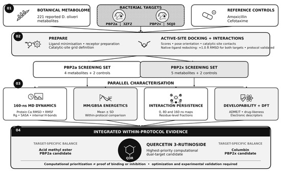
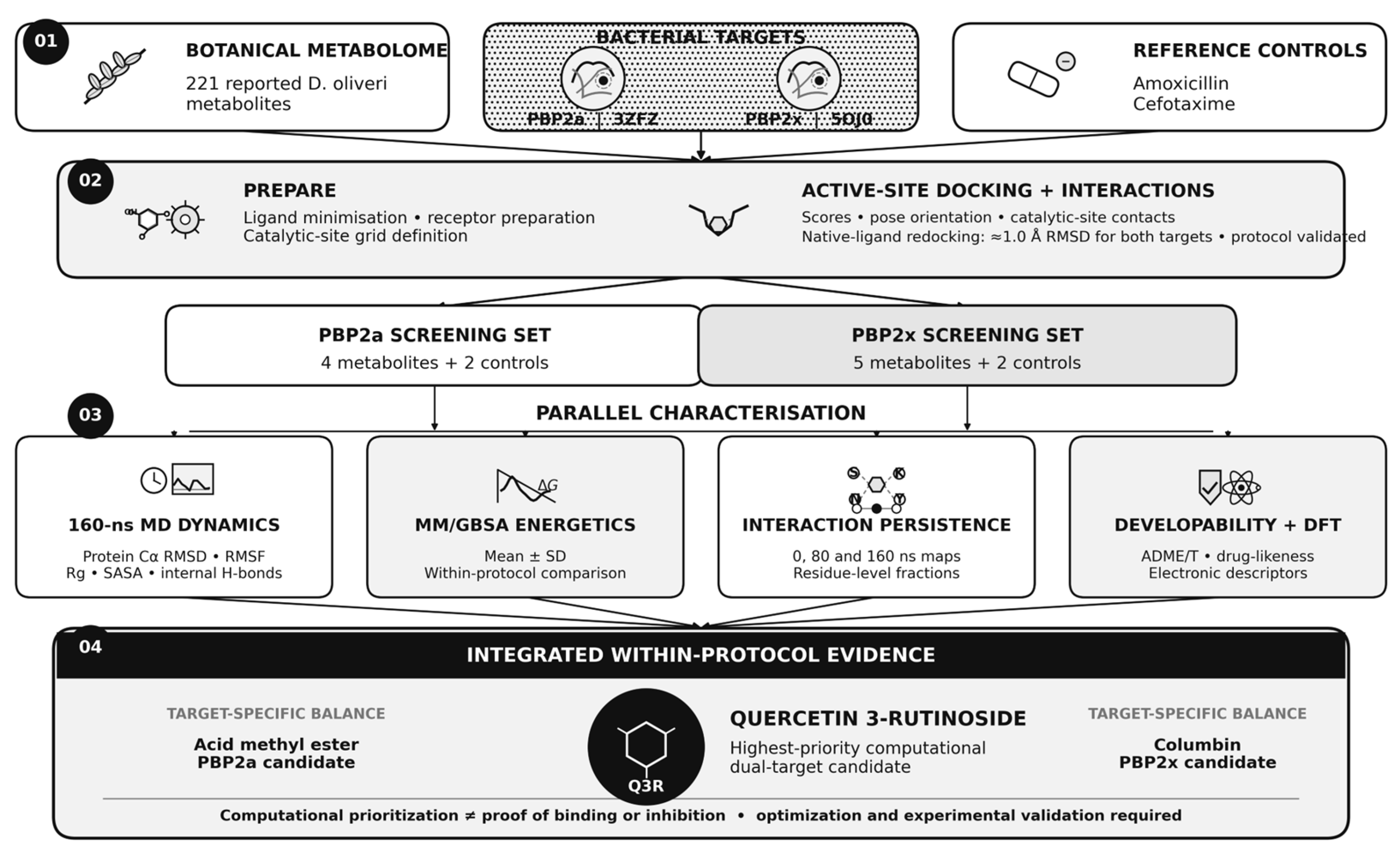
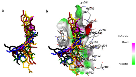
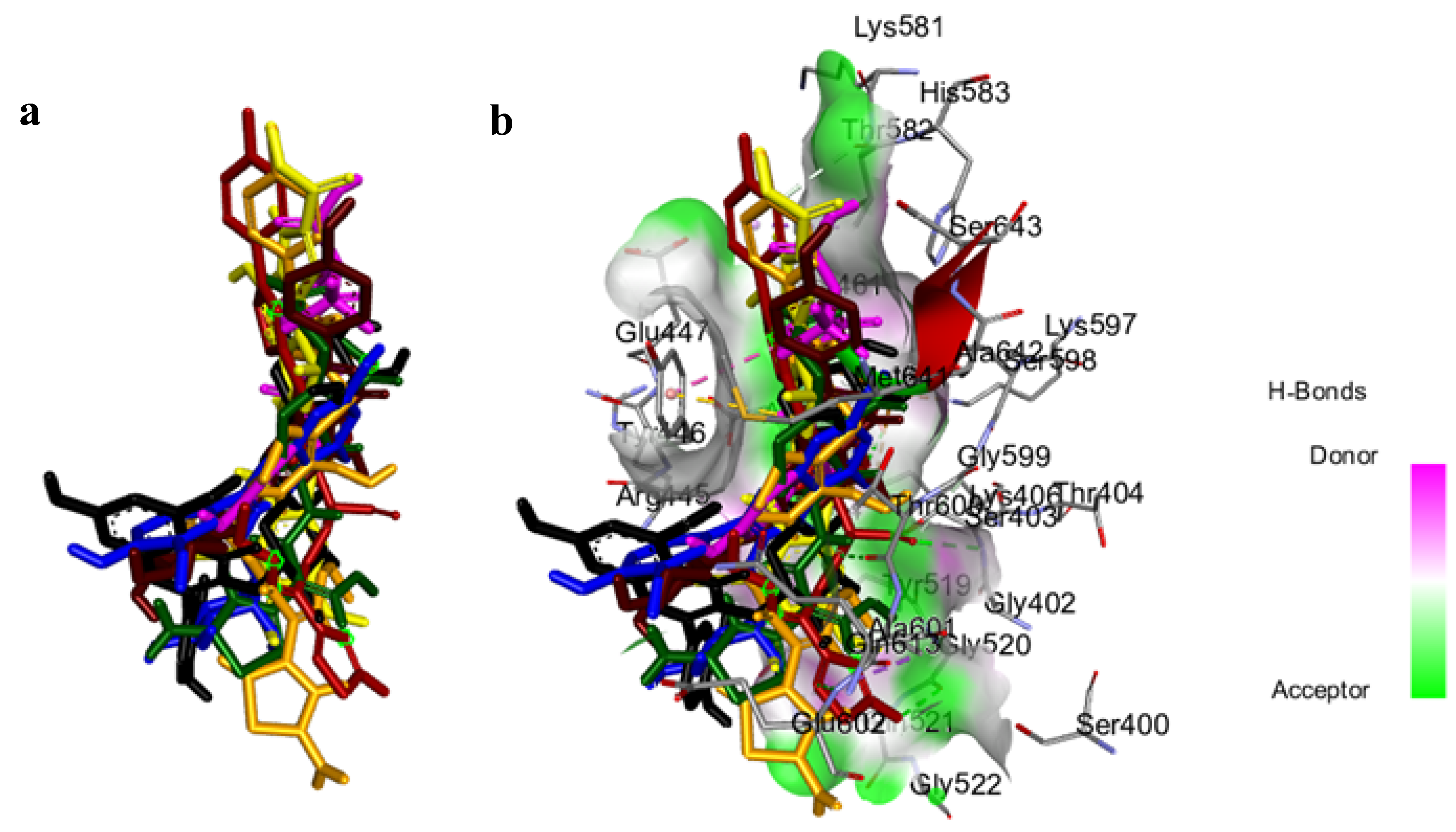
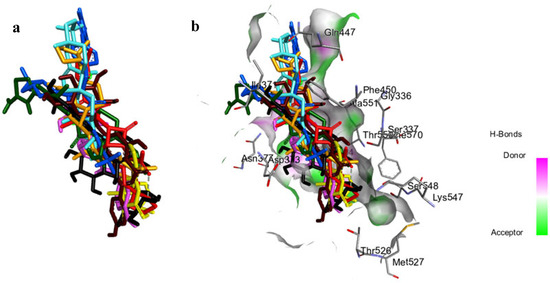
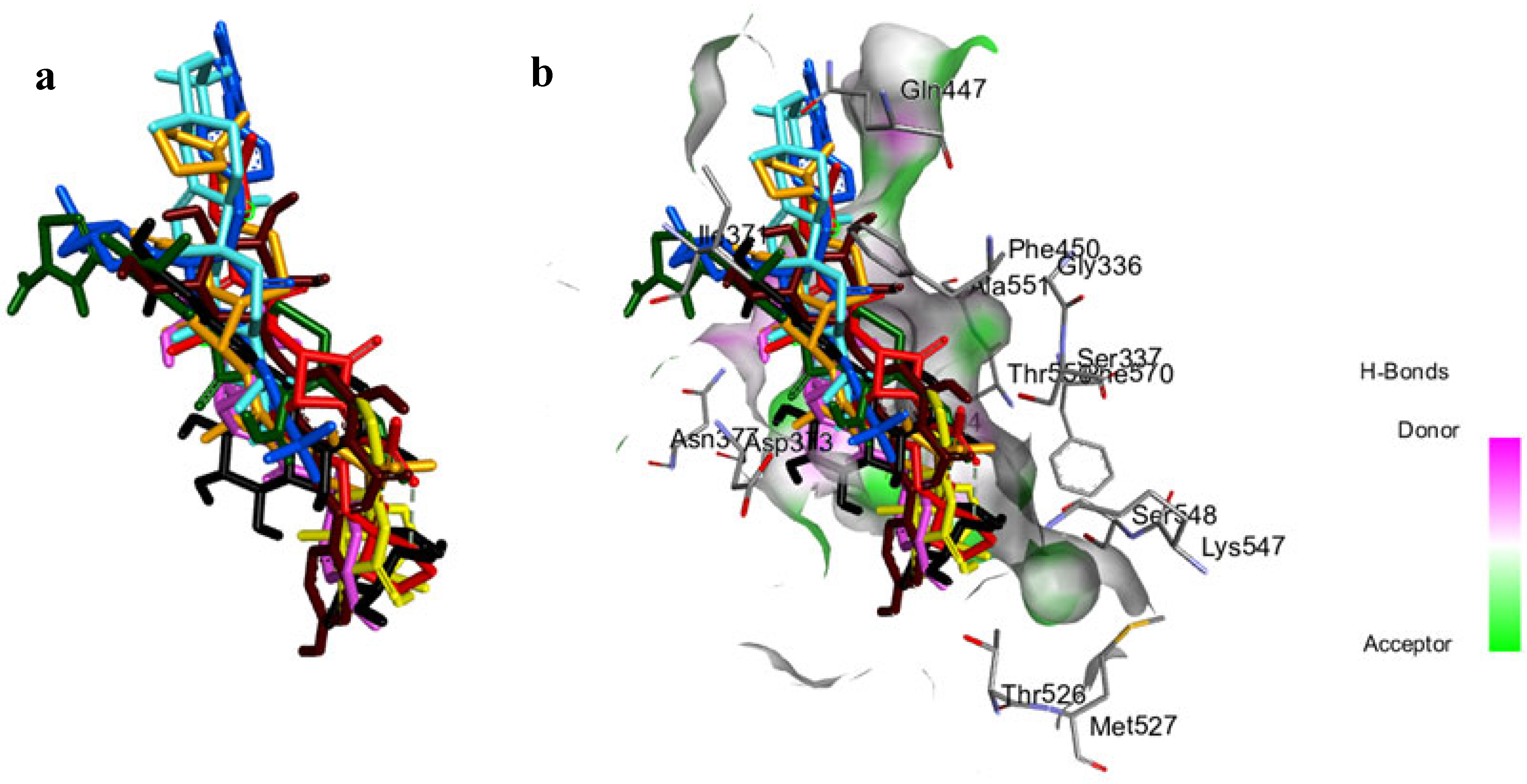
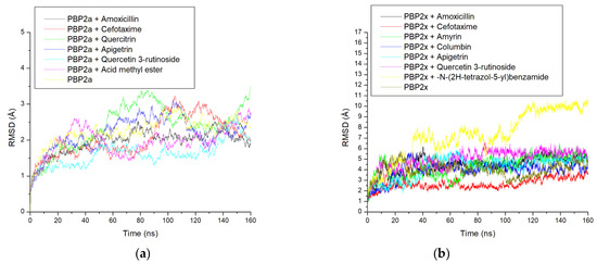
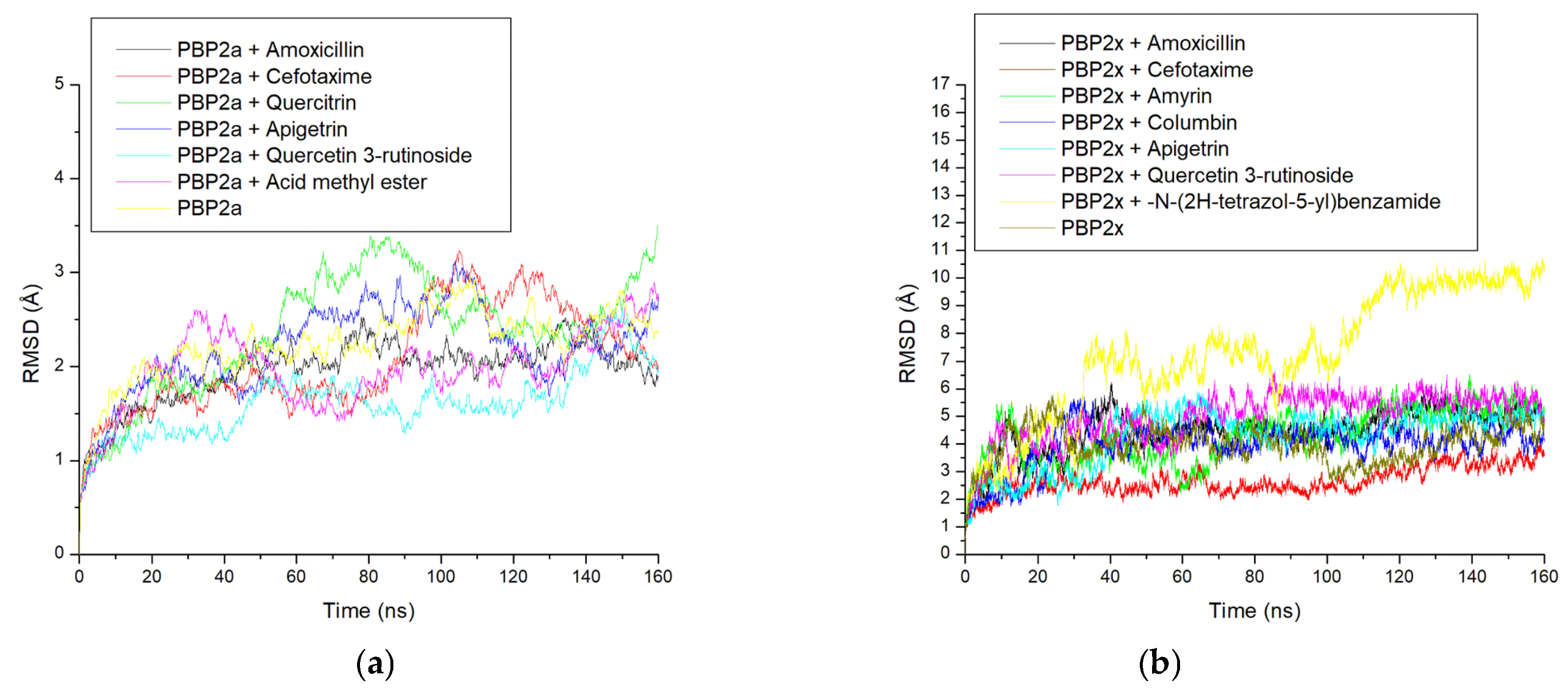
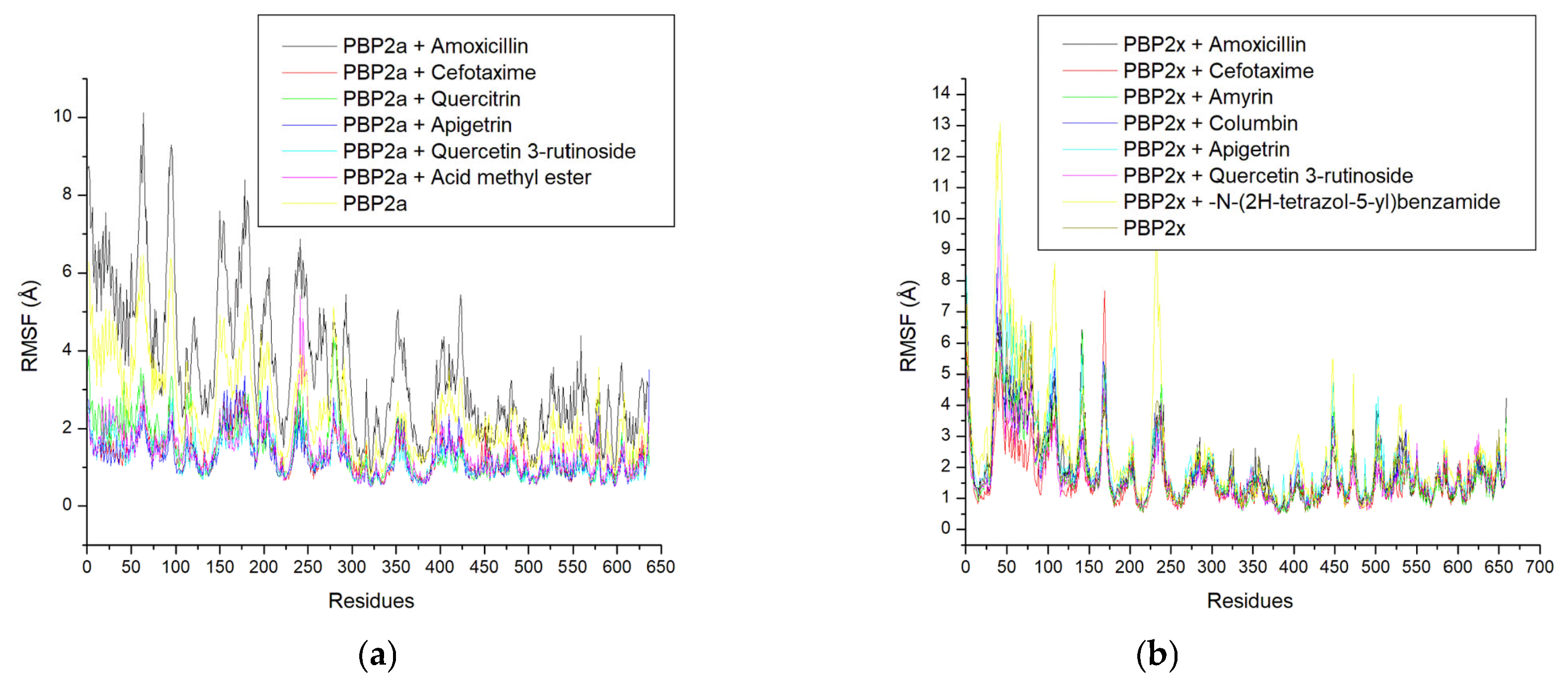
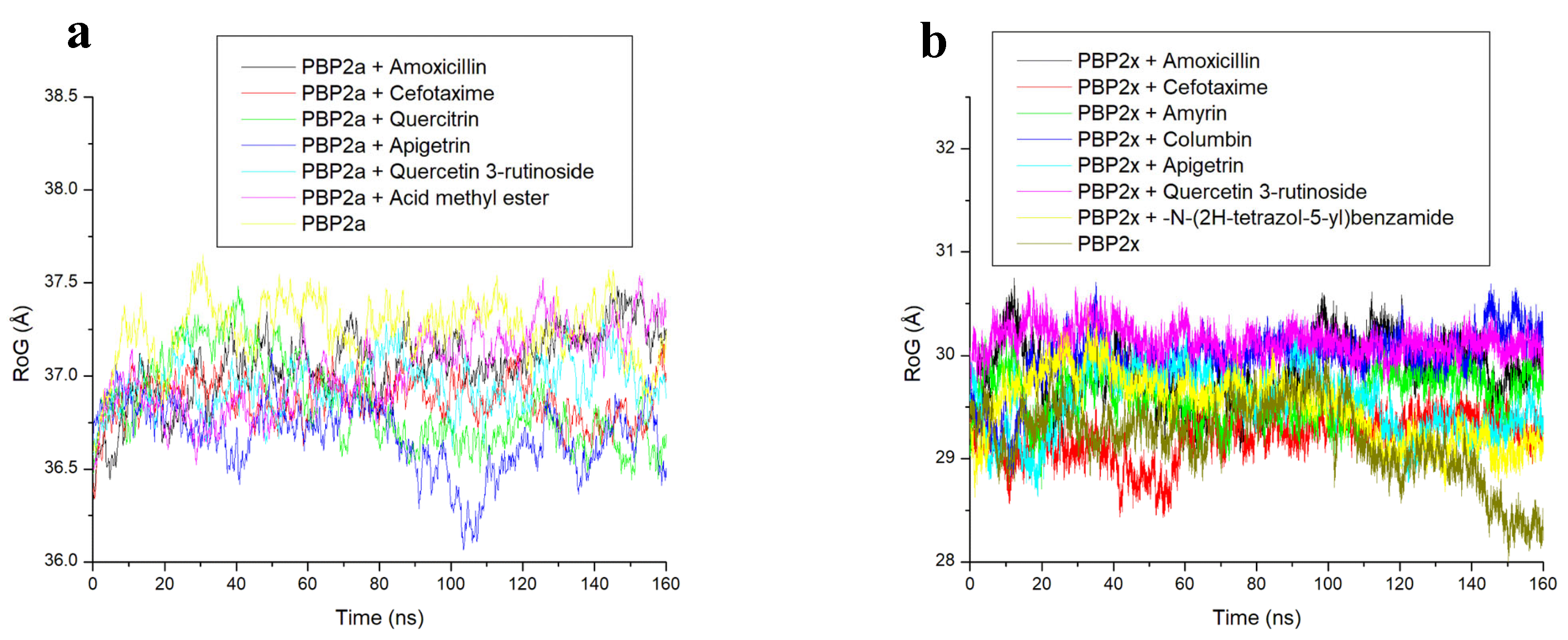
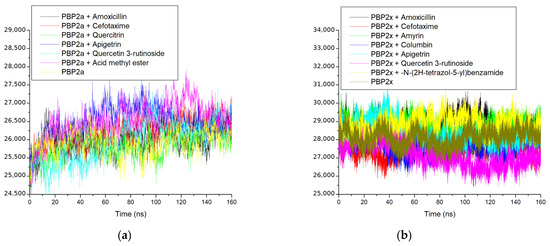
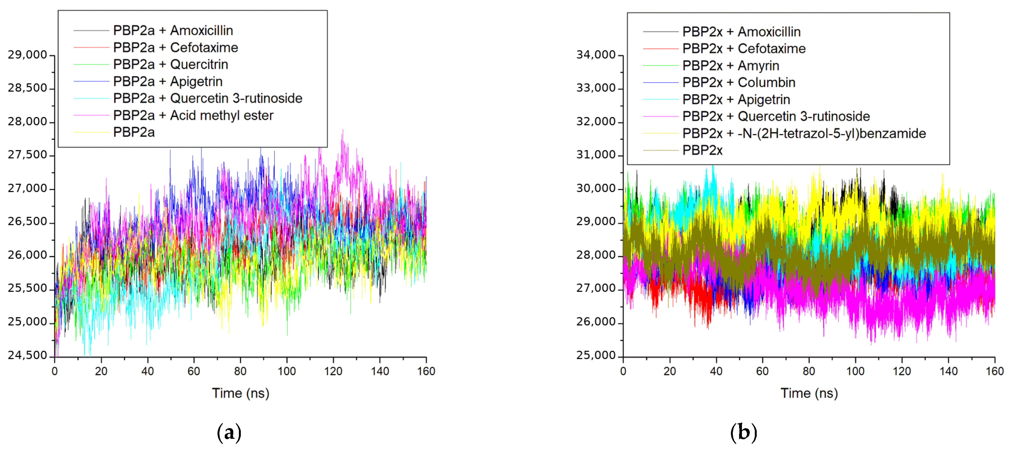
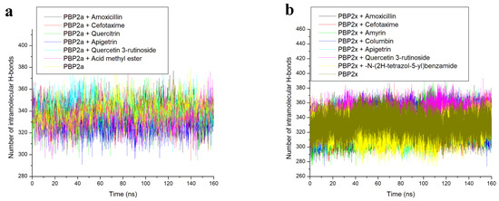
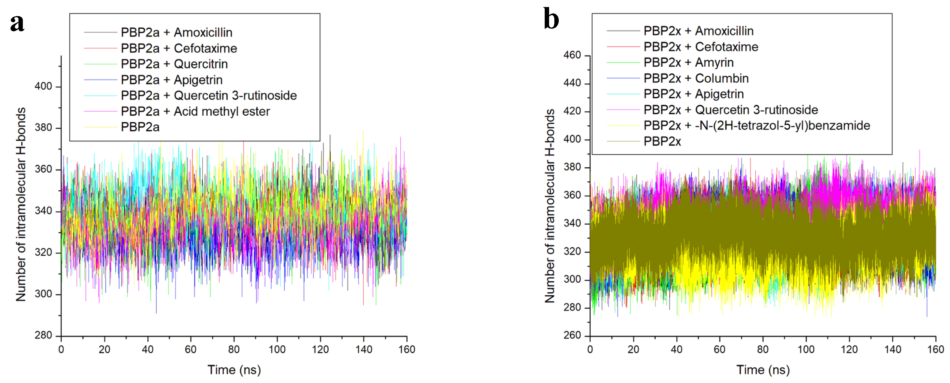
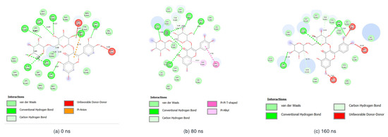
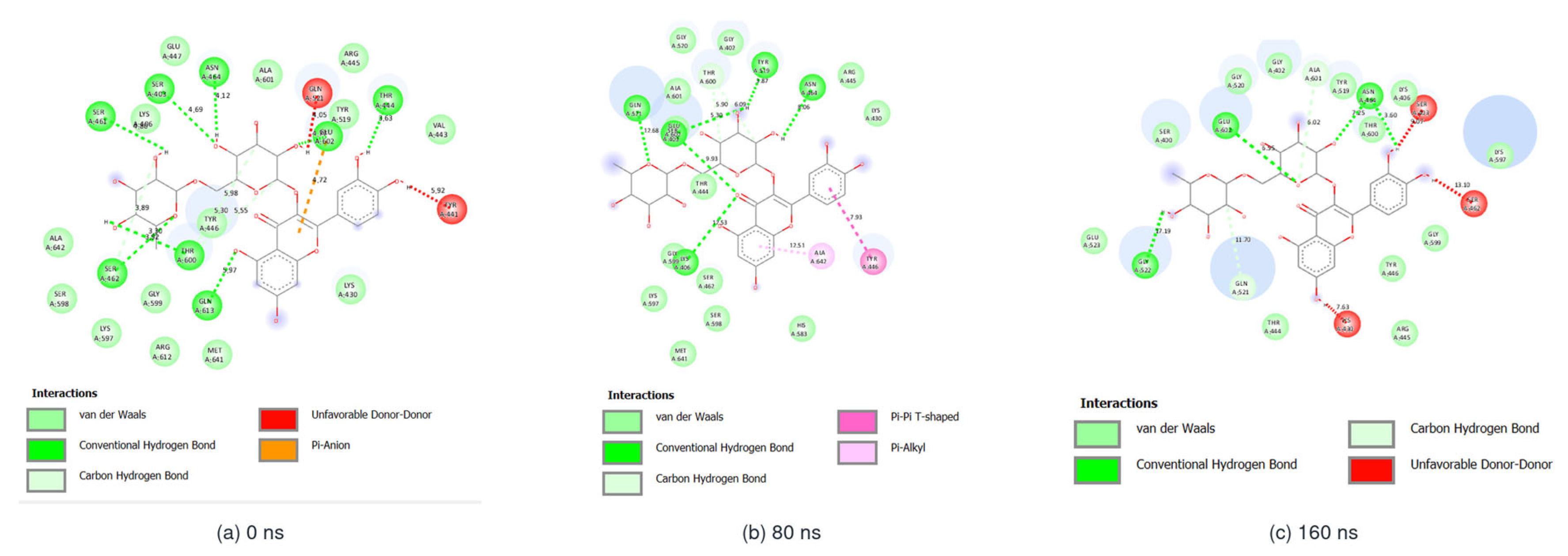
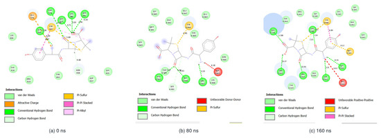
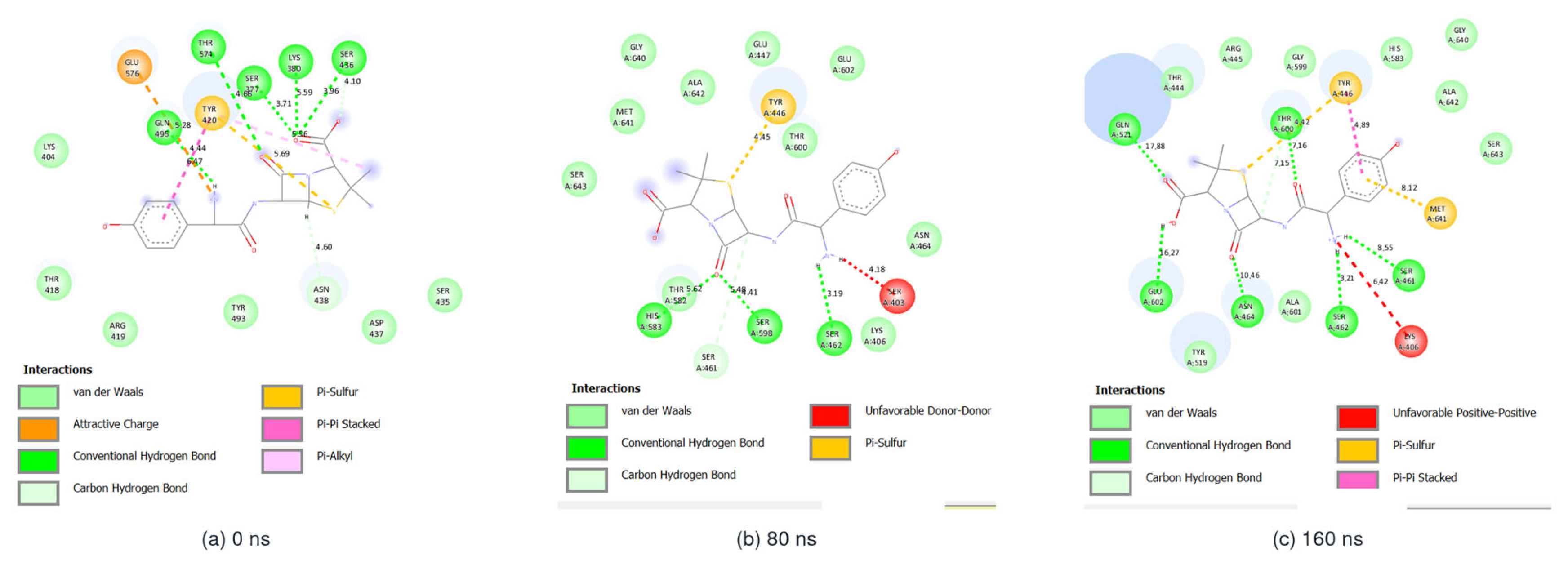
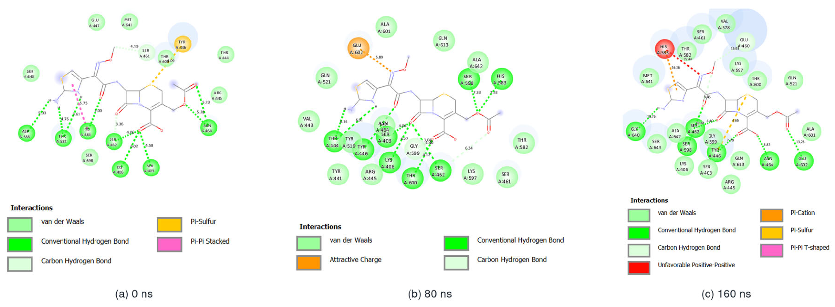
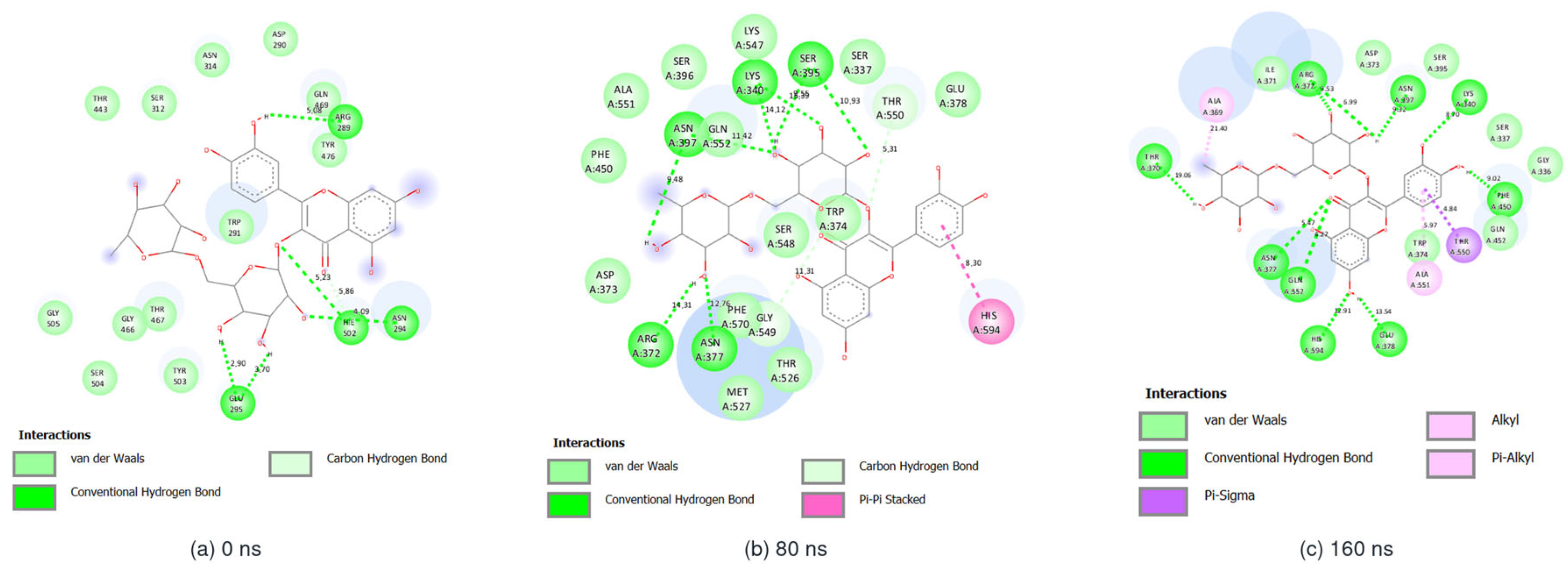
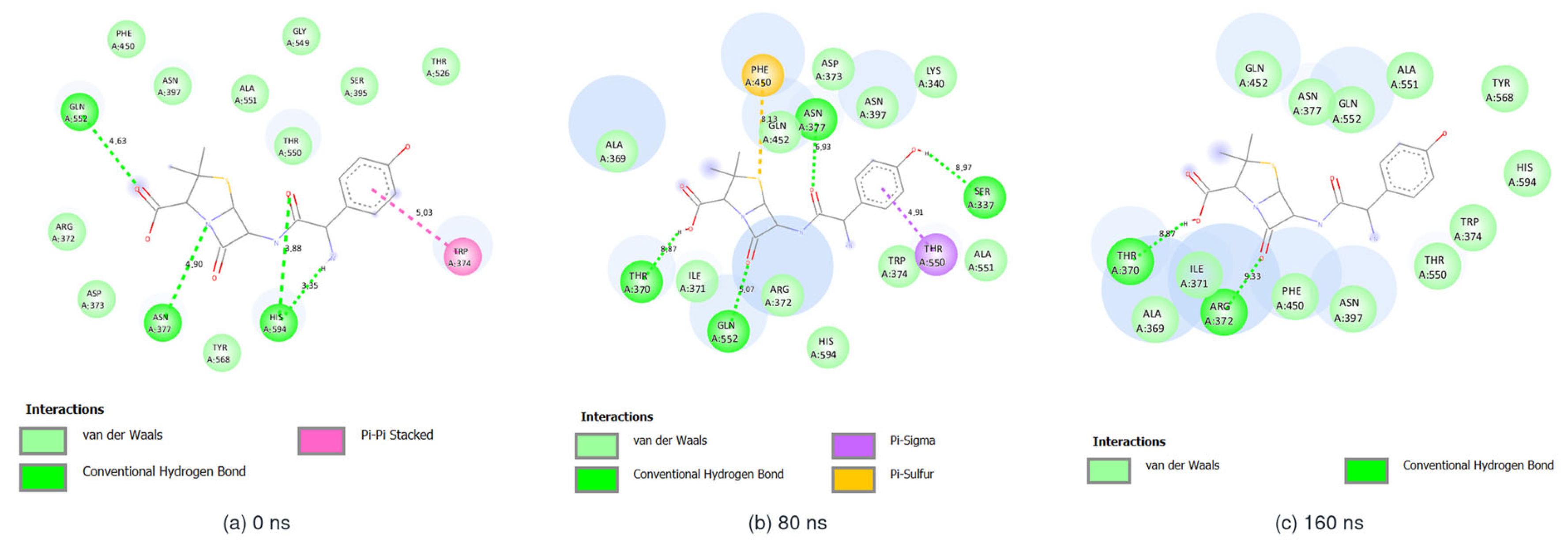
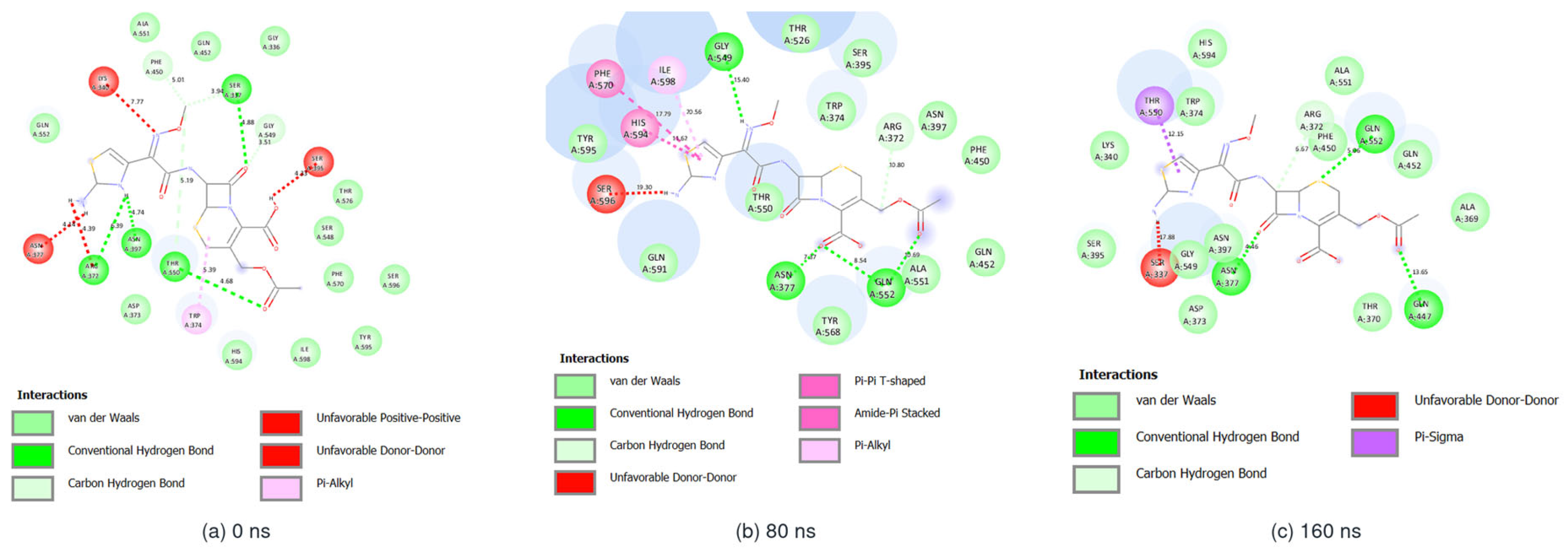
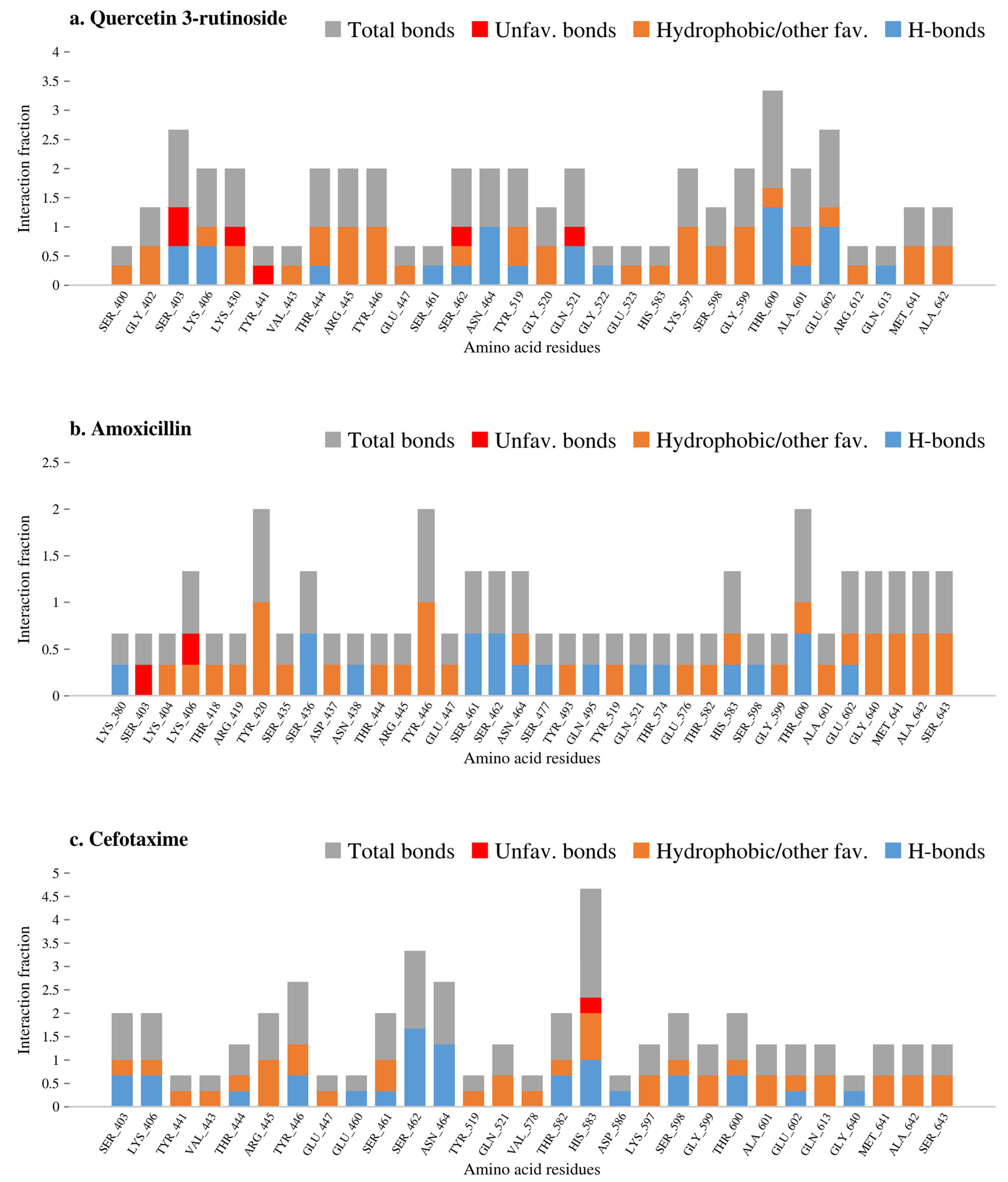
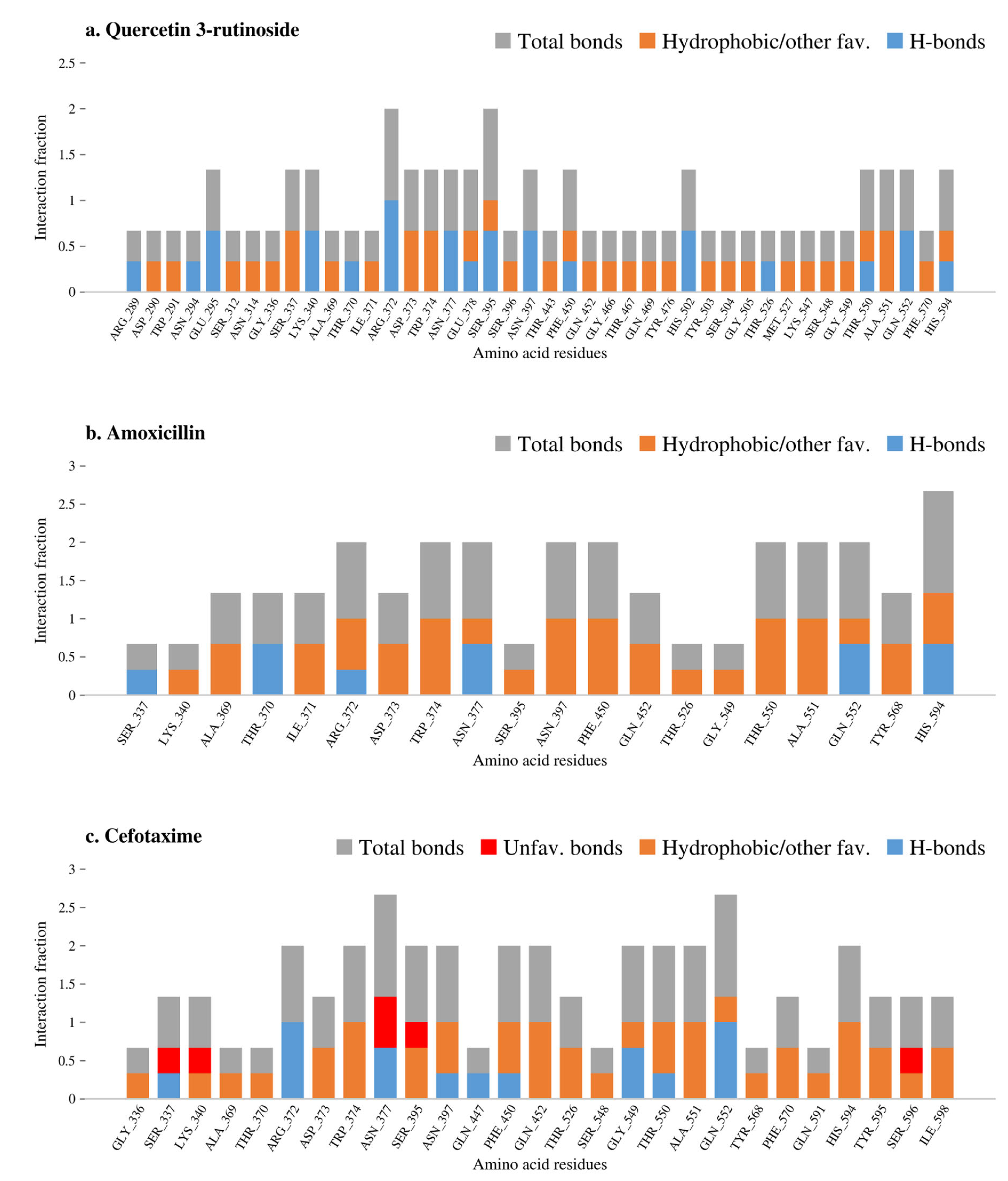
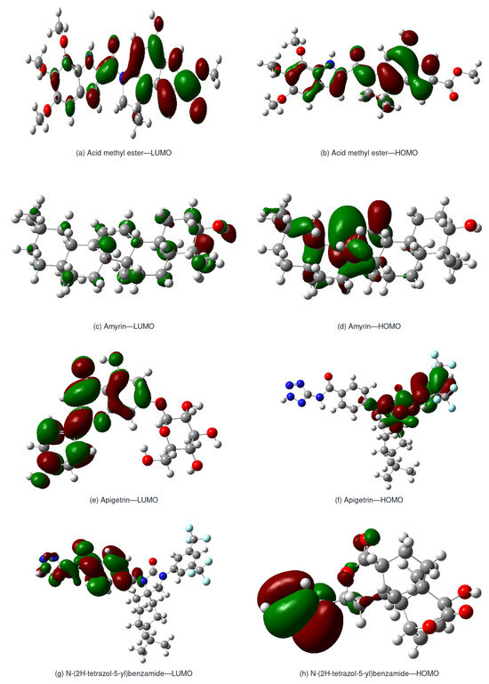
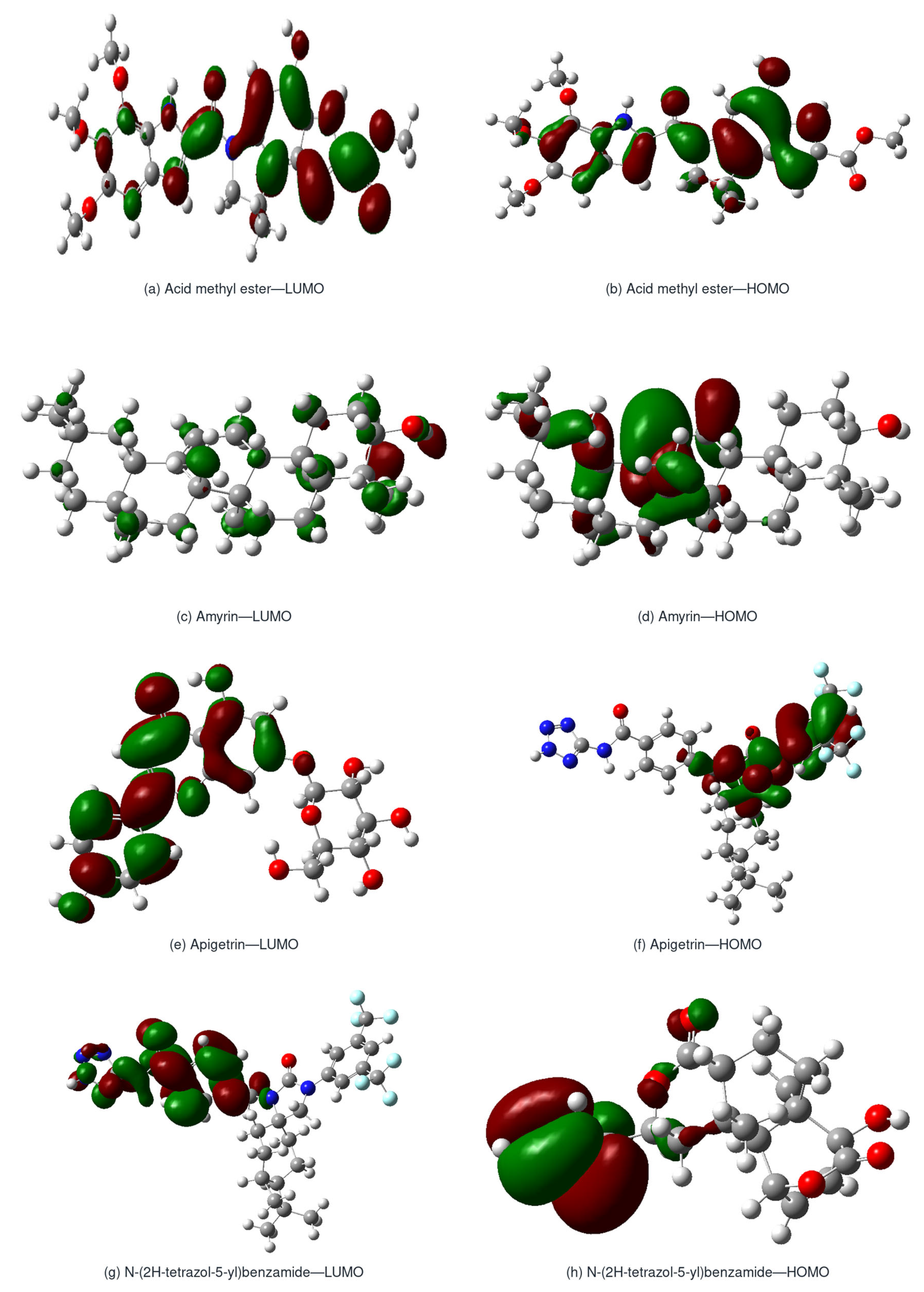
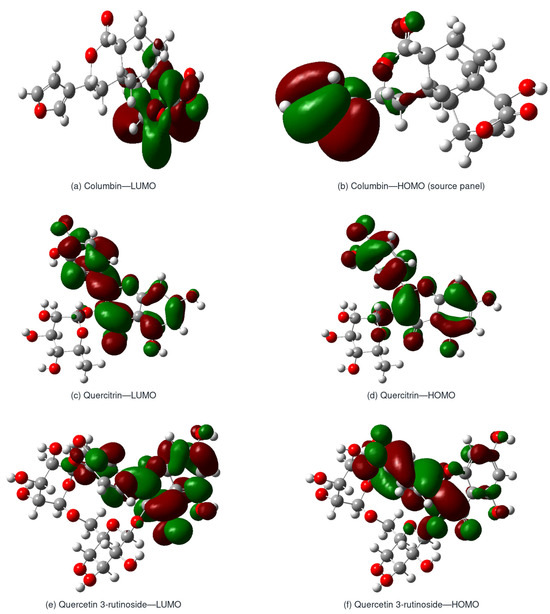
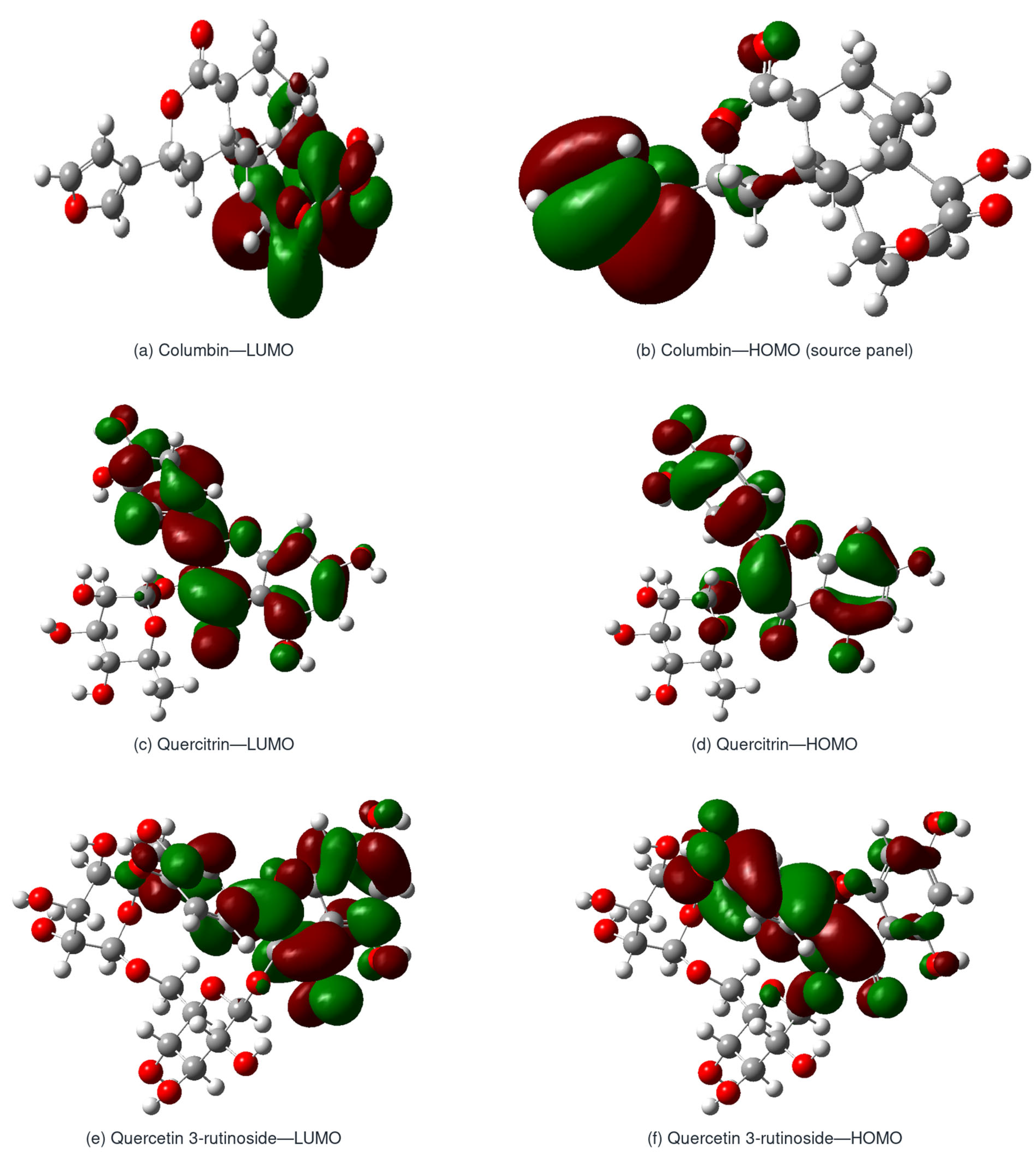
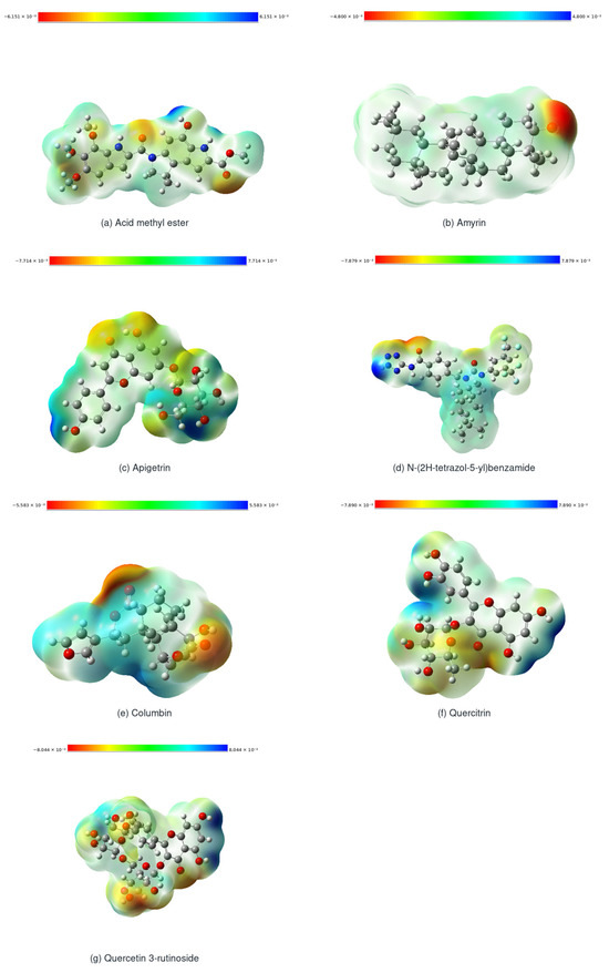
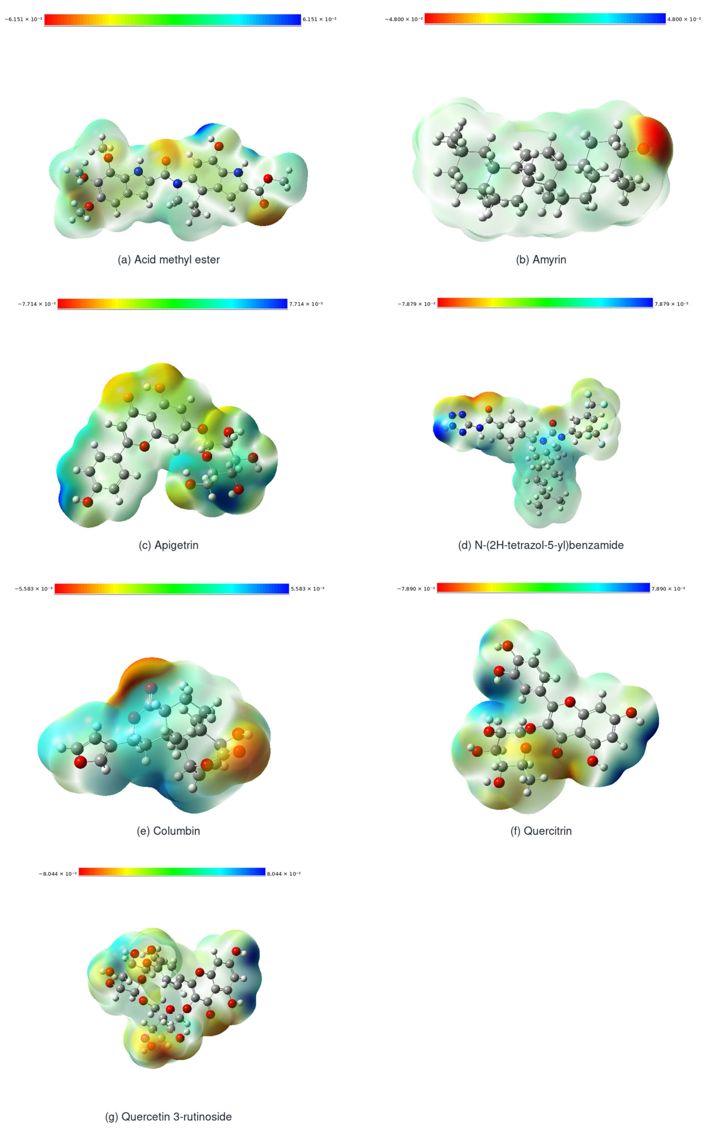

Disclaimer/Publisher’s Note: The statements, opinions and data contained in all publications are solely those of the individual author(s) and contributor(s) and not of MDPI and/or the editor(s). MDPI and/or the editor(s) disclaim responsibility for any injury to people or property resulting from any ideas, methods, instructions or products referred to in the content. |
© 2026 by the authors. Licensee MDPI, Basel, Switzerland. This article is an open access article distributed under the terms and conditions of the Creative Commons Attribution (CC BY) license.