


Abstract
Background/Objectives: This study investigated the effects of a single dose of psilocybin-containing Psilocybe cubensis extract on gut microbiota composition, microbial metabolism, and intestinal immune-related responses. Methods: A single dose of P. cubensis extract (35 mg/L) was subjected to colonic fermentation using the SHIME® model inoculated with fecal microbiota from individuals with severe anxiety. Psilocybin and psilocin concentrations, microbial metabolites, gut microbiota composition, intestinal barrier integrity, and cytokine responses were evaluated. Results: During colonic fermentation, the concentrations of both psilocybin and psilocin decreased significantly, suggesting their metabolism or degradation within the gut microbial environment. Treatment reduced ammonia production by 19–25% and decreased gamma-aminobutyric acid (GABA) levels. By contrast, the concentrations of the short-chain fatty acids acetic, propionic, and butyric increased by 2.26-, 2.69-, and 2.21-fold, respectively, after 20 h of fermentation. Microbial profiling revealed marked increases in the relative abundances of Bifidobacterium and Lactobacillus after 20 h and 15 days, together with a slight reduction in α-diversity. Fermentation supernatants also altered cytokine profiles and influenced intestinal barrier-related responses, although these effects reflected a complex immunomodulatory pattern rather than a uniform anti-inflammatory response. Conclusions: These findings indicate that psilocybin-containing P. cubensis extract can modulate gut microbiota composition and metabolic activity under in vitro colonic conditions. The observed changes in microbial metabolites, bacterial taxa, and host-related responses support the hypothesis that gut-microbiota-mediated mechanisms may contribute to the broader biological effects of psilocybin. However, these in vitro findings do not establish clinical benefits for intestinal or mental health, and further validation in animal models and well-controlled clinical studies is required.
1. Introduction
Clinical psychedelic studies remained limited until the early 1990s, when renewed interest emerged, driven by growing evidence on their therapeutic potential for the treatment of psychiatric disorders [1,2]. Psychedelics are found in living organisms such as plants and mushrooms and can also be synthesized (e.g., lysergic acid diethylamide—[LSD]). Psilocybin, one of the most extensively studied psychedelics, is a natural compound derived from the Psilocybe cubensis mushroom that has gained global attention due to its potential therapeutic application in the treatment of disorders such as depression and anxiety [3]. Recognized by the Food and Drug Administration (FDA) as a breakthrough therapy for selected psychiatric disorders, psilocybin is opening new avenues in mental health care, offering a promising alternative to conventional treatments.
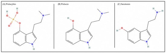
Psilocybin (O-phosphoryl-4-hydroxy-N,N-dimethyltryptamine—C12H17N2O4P, as shown in Figure 1A) [4] is an indole alkaloid compound derived from tryptophan and acts as an agonist of serotonin receptors, particularly the 5-hydroxytryptamine 2A (5HT2A) receptor [5]. The 5-HT2A receptor is a G protein-coupled receptor widely distributed throughout the central nervous system (CNS) and plays a key role in regulating physiological, psychological, and behavioral processes. Psilocybin is metabolized in the liver through enzymatic dephosphorylation, converting it into the active metabolite, psilocin (4-hydroxy-N,N-dimethyltryptamine—C12H16N2O, Figure 1B) [6], which exhibits a chemical structure like that of serotonin (5-hydroxytryptamine—C10H12N2O, Figure 1C) [7,8].
Figure 1.
Chemical structures of indoleamine compounds: (A) psilocybin [4], (B) psilocin [6], and (C) serotonin [8].
Following absorption, psilocin enters the systemic circulation and crosses the blood–brain barrier, where it exerts its psychoactive effects [9,10,11]. These effects typically begin 10–40 min after ingestion and may persist for 2–6 h, depending on factors such as the administered dose, body weight, age, individual metabolism, and tolerance [10,11,12]. Psilocybin and its metabolites are primarily excreted via the urine, bile, and feces, with elimination generally occurring within approximately 24 h after administration [13].
The mechanisms of action of psilocybin and psilocin remain incompletely understood. Emerging evidence suggests that psilocybin, traditionally investigated for its effects on the CNS, may also interact with peripheral systems, including the gut microbiome, thereby contributing to its broader therapeutic mechanisms [14]. The influence of psychedelics on the gut microbiota and its implications for mental health may advance our understanding of the microbiota–gut–brain axis (MGBA) as a critical regulatory pathway in mental health and disease.
Modulation of the gut microbiota, together with the serotonergic activity of psilocybin, may influence serotonin receptor signaling and affect mental health through immune and endocrine pathways within the gut–brain axis, potentially enhancing the therapeutic efficacy of psilocybin in depression and anxiety [14,15]. Globally, an estimated 280 million people are affected by major psychological disorders, many of whom fail to respond to conventional treatments [16], highlighting the need for innovative therapeutic approaches, including psychedelic-based interventions.
Recent advancements in microbiome research have established the MGBA as a sophisticated communication network linking the gastrointestinal (GI) tract and the CNS. This axis mediates bidirectional interactions through neural, endocrine, and immune pathways, demonstrating the potential of gut microbial communities to influence neuropsychiatric health and behavioral outcomes [17,18]. Serotonergic psychedelics, such as psilocybin, may alter microbial populations, potentially affecting gut–brain signaling and contributing to antidepressant efficacy through changes in microbial metabolites [17,19]. For instance, a significant correlation has been identified between specific bacterial groups in the gut microbiome and the density of 5-HT2A receptors. Members of the Ruminococcaceae family and the genus Candidatus exhibited a positive correlation, whereas members of the Lactobacillaceae family showed a negative correlation [18]. In addition, psilocybin induced notable changes in bacterial abundance at the phylum level. These effects were both dose- and time-dependent, with an increase in Verrucomicrobia and Actinobacteria and decreases in Proteobacteria [19].
Recent studies suggest that a single dose of psilocybin (25 mg) can induce rapid and sustained therapeutic benefits for psychological disorders, including treatment-resistant depression [20,21]. A double-blind trial involving 233 participants with treatment-resistant depression evaluated the effects of a single dose of psilocybin (25 mg, 10 mg, or 1 mg as a [control]). The study demonstrated that a 25 mg dose produced antidepressant effects, with measurable improvements observed within two days, and that a significant proportion of participants maintained these benefits for up to nine weeks [20]. However, a recent systematic review and meta-analysis indicated that the optimal psilocybin dose may range from 30 to 35 mg per 70 kg of body weight [22]. In addition to its central effects, evidence suggests that a single dose of psilocybin may transiently modulate the gut microbiota and increase serotonin (5-HT) production [23]. These changes may contribute to gut–brain communication and have been proposed as one of the mechanisms underlying the therapeutic effects of psilocybin. Furthermore, psilocybin-induced alterations in brain network connectivity may facilitate emotional processing and cognitive flexibility, potentially contributing to sustained clinical improvements [23].
Given the growing body of evidence supporting the gut–brain axis as a key regulator of behavior and mental health, investigating the interaction between psilocybin and the gut microbiome may uncover additional mechanisms by which psychedelic compounds exert their therapeutic effects. While clinical studies have demonstrated psilocybin’s efficacy in treating conditions such as treatment-resistant depression and anxiety [20,24], its influence on gut microbiota composition and metabolic activity remains poorly understood.
Therefore, this pioneering study aimed to address this knowledge gap by investigating the effects of a single dose of psilocybin-containing Psilocybe cubensis extract on the gut microbiota of individuals with severe anxiety using a dynamic colonic fermentation model.
2. Results and Discussion
2.1. Concentration of Psilocybin and Psilocin During Gastrointestinal Digestion and Colonic Fermentation
Table 1 presents the concentrations of psilocybin and psilocin throughout colonic fermentation. At the beginning of the experiment, approximately 1 g of P. cubensis mushroom extract was added to the system, resulting in an initial psilocybin concentration of 35 mg/L. The psilocybin concentration reached 35.35 ± 2.56 mg/L after 10 min of colonic fermentation (Table 1) but decreased significantly after 1 h (23.09 ± 0.79 mg/L) and remained at a similar level after 20 h (23.69 ± 1.15 mg/L). Psilocin levels, on the other hand, began decreasing after 20 h and continued to decrease until the end of the process.
Table 1.
Release of psilocybin and psilocin after gastrointestinal digestion and colonic fermentation of P. cubensis mushroom extract in the SHIME system.
The metabolism of psilocybin in the gastrointestinal tract involves its dephosphorylation under acidic gastric conditions and through the action of alkaline phosphatase, leading to the formation of psilocin, which is then absorbed and distributed throughout the body [3]. After absorption, psilocin is metabolized primarily in the liver by monoamine oxidase and aldehyde dehydrogenase through oxidative deamination and demethylation reactions, forming metabolites such as 4-hydroxy-indole-3-acetaldehyde to produce different metabolites, including 4-hydroxy-indole-3-acetic acid, and 4-hydroxy-indole-3-acetaldehyde. Therefore, psilocin is responsible for the therapeutic effect after administration of psilocybin [3].
Consequently, psilocin is considered the primary compound responsible for the therapeutic effects observed following psilocybin administration. However, it is important to note that the SHIME model does not include alkaline phosphatase in the digestion medium, which may lead to an underestimation of psilocybin conversion to psilocin. Therefore, we evaluated the direct effects of the P. cubensis mushroom extract on colonic fermentation.
2.2. Bacterial Metabolic Activity: Ammonia (NH4+), Gamma-Aminobutyric Acid (GABA) and Short-Chain Fatty Acid (SCFA) Production
Table 2 shows the ammonia concentrations after colonic fermentation. As shown in Table 2, ammonia levels decreased significantly throughout the SHIME® experiment. A significant reduction was first observed after 20 h of fermentation (−19%), followed by a further decrease after 3 days (−25%). Thereafter, ammonia levels remained relatively stable until the end of the fermentation process.
Table 2.
Ammonia concentration levels after colonic fermentation of P. cubensis mushroom extract.
Ammonia is a microbial metabolite generated by several gut microorganisms through the catabolism of proteins and amino acids. Ammonia production can also occur through the bacterial urease-mediated conversion of urea into carbamate and ammonia. In this context, Gram-negative facultative anaerobic bacteria represent major urease-producing microorganisms, including species belonging to the families Enterobacteriaceae and Veillonellaceae [25].
Ammonia has long been recognized as a metabolic waste product with well-established neurotoxic effects. Hyperammonemia may lead to degenerative outcomes, including mitochondrial dysfunction, impaired neuronal conduction, and alterations in cerebral glucose metabolism [26]. Therefore, the reduction in ammonia concentration observed in this study represents a promising outcome, suggesting that the P. cubensis mushroom extract containing psilocybin may contribute to the modulation of gut microbial activity and potentially reduce metabolic alterations associated with excessive ammonia accumulation.
Table 3 presents the production of GABA after colonic fermentation of the P. cubensis mushroom extract. The results demonstrate that fermentation of the extract by the gut microbiota significantly reduced GABA concentrations from 285.30 to 179.60 and 151.32 µmol/L after 9 and 15 days of fermentation, respectively. Several gut microbial species have been reported as GABA-producing microorganisms, including Bifidobacterium, Lactobacillus, and Bacteroides.
Table 3.
Production of gamma-aminobutyric acid (GABA) after colonic fermentation of P. cubensis mushroom extract.
The synthesis of GABA by gut microbiota requires the enzyme glutamic acid decarboxylase and glutamate as substrates, in addition to pyridoxal-5′-phosphate, as cofactors [27,28]. However, some bacteria have been reported as consumers of GABA, including Pseudomonas, Acinetobacter, and Brevibacterium, among others [29]. GABA is converted to succinic semialdehyde through transamination with α-ketoglutarate (as a co-substrate) by the action of GABA-transaminase. Subsequently, succinic semialdehyde is converted to succinate by succinic semialdehyde dehydrogenase [27]. In the present study, the reduction in GABA levels is likely associated with microbial degradation and utilization of GABA, including species traditionally recognized as GABA producers, such as Bifidobacterium and Lactobacillus. This response may represent an adaptive metabolic adjustment to the presence of the mushroom extract, suggesting that psilocybin may influence the functional activity of the gut microbiota in individuals with severe anxiety.
Therefore, the reduction in GABA observed herein may reflect a dynamic and context-dependent process, in which psilocybin influences both microbial composition and metabolite turnover. Further investigations are required to determine the precise contribution of these changes to the therapeutic effects associated with psilocybin.
Table 4 illustrates the concentrations of short-chain fatty acids (SCFAs) throughout the 15-day experimental period. All analyzed SCFAs increased significantly after 20 h of treatment with the P. cubensis mushroom extract, and these elevated levels were maintained throughout the 15-day fermentation period. Acetic acid increased by 2.26-fold, whereas propionic and butyric acids increased by 2.69- and 2.21-fold, respectively. Consequently, total SCFA production increased by 2.35-fold after 20 h of fermentation.
Table 4.
Concentration of short-chain fatty acids (SCFAs, mmol/L) after colonic fermentation of P. cubensis mushroom extract.
Short-chain fatty acids (SCFAs) are microbial metabolites generated mainly in the colon and cecum during the anaerobic fermentation of dietary substrates. Their biological effects are mediated, in part, through activation of G protein-coupled receptors, including GPR41, GPR43, and GPR109A, which are expressed in enteroendocrine and immune cells, adipocytes, and neurons. SCFAs can also suppress histone deacetylase activity, thereby influencing gene expression and several physiological processes. Through these mechanisms, they participate in the regulation of glucose utilization, energy homeostasis, lipid metabolism, and intestinal health. SCFAs also support epithelial barrier function by enhancing the expression of tight junction proteins, including occludin and zonula occludens-1 [30].
Decreased SCFA concentrations have been reported in experimental models of major depressive disorder. Conversely, supplementation with acetate, propionate, or butyrate has been shown to attenuate depressive-like behaviors in these models [31]. SCFAs may additionally influence emotional and behavioral responses by modulating the release and production of neuroactive compounds and gut-derived signaling molecules, including serotonin and GABA [32].
In particular, SCFAs can enhance tryptophan hydroxylase 1 (TPH1) expression and stimulate serotonin biosynthesis in intestinal enterochromaffin cells. These metabolites may also affect neural function through free fatty acid receptors, especially FFAR2 and FFAR3, corresponding to GPR43 and GPR41, respectively [32]. Furthermore, SCFAs reaching the brain may increase the expression of acetyl-CoA synthetase short-chain family member 2 (ACSS2), potentially reducing depressive-like behavior through modulation of the PPARγ–TPH2 signaling pathway [33].
Interestingly, in a phase 2 clinical trial involving participants with treatment-resistant depression, a single 25 mg dose of psilocybin significantly reduced depression scores over a three-week period [20]. In this context, the significant increase and sustained production of SCFAs observed in our study over the 15-day fermentation period may provide additional insights into potential mechanisms underlying psilocybin-associated therapeutic effects. The prolonged elevation in SCFAs could contribute to improved gut health, which, in turn, may influence gut–brain axis signaling and support the neuropsychiatric effects associated with psilocybin.
2.3. Richness and Diversity of the Gut Microbiota
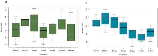
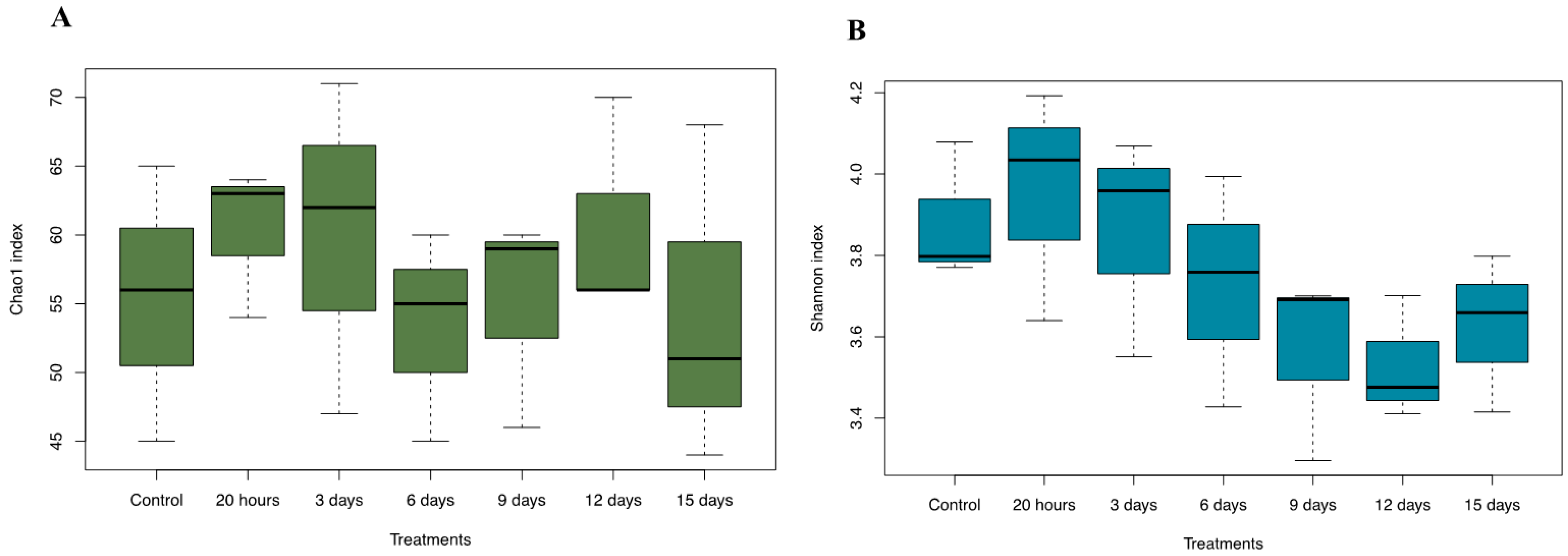
The alpha-diversity indices are presented in Figure 2. A tendency toward increased species richness (Chao1 index) was observed up to 3 days after treatment. However, this increase was followed by a gradual reduction in richness over time, although no significant differences were observed among the means according to the ANOVA test (Figure 1A).
Figure 2.
Alpha diversity of the ascending colon microbiota after SHIME fermentation of Psilocybe cubensis mushroom extract. (A) Chao1 index and (B) Shannon index at different treatment time points. Data represent the three parallel ascending colon reactors from a single SHIME experimental run (n = 1). No significant differences were observed among treatments according to one-way ANOVA (p > 0.05).
Regarding gut microbiota diversity, evaluated using the Shannon index, treatment with the P. cubensis mushroom extract showed a tendency to reduce bacterial diversity, although no statistically significant differences were detected according to the ANOVA test (Figure 2B). Higher alpha diversity is generally associated with a healthier and more resilient gut ecosystem [22]. However, direct evidence regarding the effects of psilocybin on gut microbiota diversity remains scarce. To date, only one published study has investigated the effects of oral psilocybin and related tryptamines on the gut microbiota of rats, reporting no significant alterations in alpha diversity following psilocybin administration [19].
The composition of the gut microbiota during colonic fermentation of the P. cubensis mushroom extract was investigated. Actinobacteriota and Firmicutes were the predominant phyla identified in the samples, with their relative abundances increasing throughout treatment. In contrast, Bacteroidota and Proteobacteria exhibited reduced relative abundances following treatment (Figure S1). The bacterial community was primarily composed of microorganisms belonging to the families Bifidobacteriaceae and Lactobacillaceae (Figure S2).
The relative abundance of Bifidobacteriaceae increased after 20 h of fermentation (from 39.73% to 43.88%) and again between 3 and 12 days of treatment (ranging from 43.93% to 48.70%), whereas Lactobacillaceae showed a significant increase after 15 days of colonic fermentation (Figure S2). Consistently, Bifidobacterium, the predominant genus detected in the samples, increased after 20 h and between 3 and 12 days of treatment, while Lactobacillus exhibited a marked increase after 15 days of colonic fermentation (Figure S3).
It is important to emphasize that comprehensive investigations evaluating the effects of serotonergic psychedelics on gut microbiota composition and ecological structure remain limited. Evidence from preclinical studies suggests that psilocybin may influence the composition and function of the intestinal microbiota. In one animal experiment, fecal analysis of psilocybin-treated rats revealed an increased relative abundance of Verrucomicrobia and Actinobacteria, accompanied by a reduction in Proteobacteria compared with untreated controls [19]. Another study showed that repeated psilocybin exposure enhanced social behavior and prolonged intestinal transit time in male wild-type mice. These effects were accompanied by changes in microbial composition, including lower abundances of Lactobacillus murinus, Lactobacillus animalis, and Alistipes dispar [34].
Recent findings have also identified Brunner’s glands in the duodenum as an important link between stress-responsive neural pathways and intestinal microbial homeostasis. Activation of the vagus nerve promoted the activity of these glands and favored the enrichment of intestinal Lactobacillus species. In contrast, selective removal of Brunner’s glands markedly decreased Lactobacillus abundance and increased vulnerability to infection. A polysynaptic neural pathway connecting the central nucleus of the amygdala to Brunner’s glands through parasympathetic vagal signaling was also characterized. Chronic stress reduced central amygdala activity and produced effects similar to those caused by gland ablation. Conversely, stimulation of either the central amygdala or parasympathetic vagal neurons activated Brunner’s glands and restored stress-associated disturbances in the gut microbiota and immune response [35].
Kelly and colleagues proposed that the therapeutic effects of psychedelics may involve interactions with the microbiota–gut–brain axis and suggested that interindividual differences in microbial activity could influence the clinical response to compounds such as psilocybin [18]. Supporting this hypothesis, psilocybin administration altered both behavior and intestinal microbial composition in C57BL/6J mice. Moreover, transferring intestinal contents from psilocybin-treated mice to untreated recipients induced behavioral changes resembling those observed in the donor animals. These findings suggest that the gut microbiota may contribute, at least in part, to the behavioral effects of psilocybin through microbiota–gut–brain signaling mechanisms [34].
2.4. Interplay Between Metabolite Production and Microbial Composition
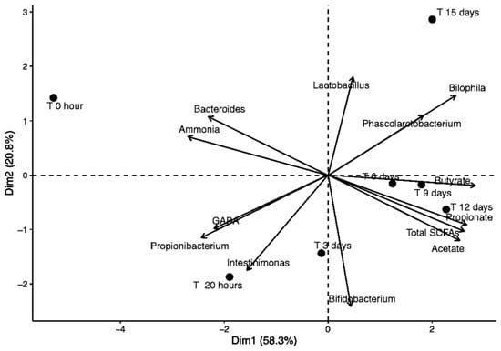
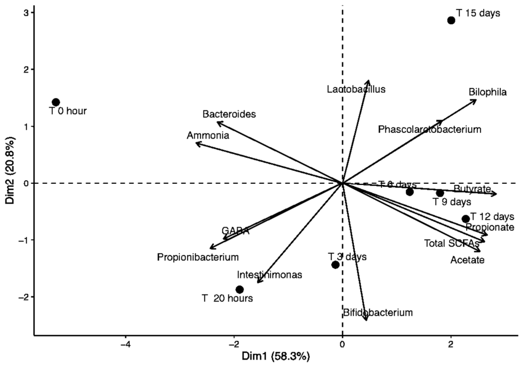
Figure 3 presents the principal component analysis (PCA) of metabolite production and microbial composition during the colonic fermentation of the P. cubensis mushroom extract. PCA revealed clear patterns in the temporal evolution of metabolites and microbial communities, indicating progressive changes in the colonic fermentation profile following the addition of the psilocybin-containing P. cubensis extract. The first principal component (PCA1) explained 58.33% of the data variability, whereas the second component (PCA2) accounted for 20.84%, together explaining 79.17% of the total variability. PCA1 demonstrated a marked transition in metabolic and microbial profiles throughout the experimental period, while the variability explained by PCA2 suggested additional sources of variation potentially associated with intermediate metabolites or less dominant microbial interactions.
Figure 3.
Principal component analysis of the metabolites and gut microbiota composition following colonic fermentation of the P. cubensis mushroom extract.
The analyzed metabolites showed strong correlations with PCA1. Regarding SCFAs, acetate (0.87), propionate (0.92), and butyrate (0.97) were strongly positively correlated with the first axis, indicating that the increase in these compounds over time represented a major contributor to the observed variation. The association between these metabolites and specific bacterial groups supports the involvement of carbohydrate-fermenting microorganisms, particularly Bifidobacterium, in SCFA production [30].
The distribution of bacterial communities along the principal components demonstrated distinct microbial patterns associated with different ecological niches. Bifidobacterium and Lactobacillus, which are commonly associated with carbohydrate fermentation and SCFA production, showed distinct positioning from Bilophila, a genus previously associated with inflammation, intestinal barrier dysfunction, and alterations in glucose metabolism [31].
The increase in the abundance of Phascolarctobacterium may be related to its capacity to utilize metabolic intermediates, such as lactate and succinate, for production of SCFAs [30]. Interestingly, Propionibacterium was associated with GABA production (Figure 3), suggesting that this microbial group may contribute to the regulation of GABA metabolism. Conversely, Bacteroides appeared to be associated with ammonia production in the fermented samples. Collectively, these findings suggest that the addition of P. cubensis mushroom extract influenced microbial composition and metabolic activity, favoring a microbial environment characterized by enhanced carbohydrate fermentation, as indicated by increased SCFA production and reduced abundance of Bacteroides and ammonia levels. These results support the hypothesis of reduced proteolytic activity within the fermentation system.
2.5. Immunological Response
In Figure S4, the cytotoxicity assessment is illustrated by flow cytometry using the FVS450 marker in Caco-2 cells. In this study, the colonic fermentation of the P. cubensis mushroom extract did not affect Caco-2 cell viability (Figure S4). Four experimental groups were analyzed: control (supernatant collected at time 0), 20 h (supernatant collected after 20 h of culture), 9 days (supernatant collected after 9 days of culture), and 15 days (supernatant collected after 15 days of culture). In this figure, data are expressed as mean ± standard deviation. However, colonic fermentation increased transepithelial/transendothelial electrical resistance (TEER) in Caco-2 cells, particularly after 15 days of treatment (Figure S5). TEER measurements were performed to evaluate intestinal epithelial barrier integrity. No significant differences were observed between treatments according to the Kruskal–Wallis test at the 5% significance level. Additionally, the colonic fermentation of the P. cubensis mushroom extract increased TNF-α levels and decreased IL-6 levels after 9 and 15 days of treatment, respectively (Figure S6). TEER is one of the parameters used to assess cellular barrier integrity, with higher electrical resistance across the cell monolayer indicating greater barrier integrity [32,33]. Clinical evidence indicates that patients with depressive or anxiety disorders may present gut microbial dysbiosis accompanied by increased circulating concentrations of lipopolysaccharide (LPS), zonulin, and fatty acid-binding protein 2 (FABP2). These findings are consistent with impaired intestinal barrier function and increased gut permeability [34]. Inflammatory signaling may further contribute to this barrier disruption, as elevated systemic levels of pro-inflammatory cytokines, particularly IL-6 and TNF-α, are frequently reported in stress-related and mood disorders [35]. In addition, extracts derived from psychedelic mushrooms have been shown to suppress the production of TNF-α, IL-1β, and IL-6 in human U937 macrophage cells [36]. Psilocybin may exert a complex immunomodulatory effect through its active metabolite psilocin, which interacts with the 5-HT2A receptor involved in immune regulation and cytokine signaling [37]. In addition, it has been shown that psilocybin, 4-AcO-DMT, and eugenol significantly reduce TNF-α and IFN-γ levels in human 3D intestinal tissue [38].
In the present study, colonic fermentation of the P. cubensis mushroom extract containing psilocybin was associated with maintenance of intestinal barrier integrity, although it did not induce a consistent anti-inflammatory profile. The observed increase in TEER after 15 days suggests improved epithelial barrier function, potentially mediated by alterations in microbial metabolites and cytokine signaling. Specifically, fermentation of the extract reduced IL-6 levels, a cytokine associated with chronic inflammation, whereas TNF-α levels increased, indicating a modulation rather than a generalized suppression of inflammatory signaling. This complex cytokine response may represent an adaptive immune modulation rather than a direct anti-inflammatory effect, highlighting the complexity of psilocybin-mediated interactions between microbial metabolites, epithelial cells, and immune responses. These findings are relevant because intestinal barrier integrity and immune regulation are important components in the pathophysiology of stress-related and mood disorders, which are frequently associated with impaired barrier function and chronic low-grade inflammation [39]. Therefore, the colonic fermentation of P. cubensis extract may represent a promising approach to improving gut barrier integrity, but further studies are needed to clarify the immunomodulatory effects of psilocybin.
Although the SHIME system represents a valuable approach for simulating colonic fermentation under controlled in vitro conditions, it still has limitations in understanding complex host–microbiota interactions, such as the absence of systemic immune responses, hormonal signaling, and the full diversity of host cell types. The Caco-2/THP-1 model, in turn, represents only a simplified interface between intestinal epithelial cells and immune cells. Therefore, the results obtained in this study, which utilized these systems, should be interpreted with caution. Further validation in animal models or clinical studies will be necessary to confirm the physiological relevance of the observed effects.
3. Materials and Methods
In this research, experiments were carried out using raw material of human feces from patients with severe anxiety.
3.1. Donor Selection
Fecal donors (n = 3; 1 woman, 2 men) aged 20–30 years with severe anxiety were recruited based on the Hamilton Anxiety Rating Scale. Donors with recent antibiotic use (within six months) or prebiotic, probiotic, medication, or supplement use (within three months) were excluded. Key donor characteristics are presented in Table S1. The fecal inoculum was prepared according to the methodology proposed by [40].
3.2. Dynamic Colonic Microbiome Model
The Simulator of the Human Intestinal Microbial Ecosystem (SHIME®) is a computer-controlled, dynamic in vitro system designed to reproduce the main physiological and microbial conditions of the human gastrointestinal tract. The conventional configuration consists of five sequentially connected vessels representing the stomach (R1), small intestine (R2), ascending colon (R3), transverse colon (R4), and descending colon (R5) [40]. Each compartment is independently regulated to simulate the corresponding gastrointestinal region, with controlled parameters such as pH, temperature, retention time, and working volume. A single SHIME experimental run was performed. Gastric (R1) and small intestinal (R2) digestion were each carried out in a single reactor. To enable parallel evaluation of the ascending colon, reactors R4 and R5 were reconfigured to operate under the same conditions as R3, resulting in three parallel ascending colon reactors. These reactors were maintained at a pH of 5.6–5.9 with a retention time of 20 h. Gastric conditions (R1) were maintained at a pH of 2.3–2.5 with a retention time of 2 h. Duodenal passage was simulated using 60 mL of artificial pancreatic juice (composition: 12.5 g/L NaHCO3, 3.6 g/L Oxgall, 0.9 g/L pancreatin; Sigma-Aldrich, St. Louis, MO, USA) with a retention time of 4 h. The system automatically adjusted reactor pH with sodium hydroxide or hydrochloric acid and maintained a temperature of 37 °C. Anaerobic conditions were maintained by flushing the reactors with nitrogen gas for 30 min each day. To reproduce the nutritional conditions of the proximal colon, each ascending colon vessel received 500 mL of sterile nutritional medium supplemented with 1.0 g/L arabinogalactan (Sigma), 2.0 g/L pectin (Sigma), 1.0 g/L xylan (Roth, Karlsruhe, Germany), 3.0 g/L starch (Êxodo, Hortolândia, Brazil), 0.4 g/L glucose (Synth, Diadema, Brazil), 3.0 g/L yeast extract (Neogen, Lansing, MI, USA), 1.0 g/L peptone (Kasvi, Italy), 4.0 g/L mucin (Sigma), and 0.5 g/L cysteine (Sigma). The medium was subsequently inoculated with 40 mL of fecal supernatant. The experiment lasted five weeks, divided into three phases: microbiota stabilization (Weeks 1–2): 300 mL of sterile feed medium was added daily; the control period (Week 3): 240 mL of feed medium plus 60 mL of pancreatic juice were added once daily; and psilocybin treatment (Week 4): a total of 240 mL of feed medium, 60 mL of pancreatic juice, and a single 1 g dose of Psilocybe cubensis mushroom extract, providing a psilocybin concentration of 35 mg/L, were added directly to the colonic vessels. Samples were collected from the control period, after 20 h of Psilocybe cubensis mushroom extract treatment, and after 3, 6, 9, 12 and 15 days of treatment, stored at −20 °C until analysis. During gastrointestinal digestion, samples were collected in triplicate from the stomach and small intestine reactors at each sampling time. During colonic fermentation, samples were collected from each of the three parallel ascending colon reactors.
3.3. Analysis of Psilocybin and Psilocin Metabolization in SHIME® by HPLC
To analyze psilocybin and psilocin metabolization via gut microbiota, samples were collected every 10 min during the first hour of colonic fermentation, beginning at time 0 when the extract entered the ascending colon. Subsequently, samples were collected after 20 h, and at 3, 6, 9, 12, and 15 days of colonic fermentation. Sample preparation was carried out by the successive collection of a psilocybin solution, which had a concentration of 35 mg/L at time zero, at different collection times. The collected samples were filtered using a PTFE 0.2 µm syringe filter and transferred to 2 mL vials, which were then taken for analysis. The chromatographic analysis was performed according to the previously published method [41]. A PerkinElmer LC-FLEXAR high-performance liquid chromatograph (HPLC) (PerkinElmer Inc., Waltham, MA, USA) coupled with a diode array detector (DAD) (PerkinElmer Inc., Waltham, MA, USA) was used. A Brownlee validated reversed-phase C18 chromatographic column measuring 150 mm × 4.6 mm ID × 5 μm with a pore size of 100 Å (PerkinElmer Inc., Waltham, MA, USA) was used. The column oven was maintained at a constant temperature of 30 °C. The volume of injection was 15 µL and the volumetric flow rate was 0.8 mL/min. The mobile phases consisted of ultrapure water acidified with 0.3% formic acid (solvent A) and acetonitrile containing 0.3% formic acid (solvent B) using the following elution gradient: (0.0 min, 5% solvent B), (2.4 min, 5% solvent B), (9.6 min, 30% solvent B), (10.1 min, 30% solvent B), (10.6 min, 98% solvent B), (17.6 min, 98% solvent B), (17.7 min, 5% solvent B). The wavelength used to monitor the compounds of interest was 266 nm. A calibration curve was constructed covering the concentration range between 5 and 100 mg/L.
3.4. Metabolic Activity Analysis
Metabolic activity was assessed in the simulated colonic environment to evaluate the functional responses of the gut microbiota to psilocybin exposure. Samples were collected after 20 h and on days 3, 6, 9, 12, and 15 of colonic fermentation. At each time point, three samples from each colonic vessel were analyzed (n = 9).
3.4.1. Determination of Ammonia (NH4+) and Short-Chain Fatty Acids (SCFAs)
Ammonia (NH4+) was quantified using an ammonia-selective ion meter (710A model, Orion, Thermo Scientific, Waltham, MA, USA) coupled to an ion-specific electrode (Model 95–12, Orion) according to [42].
SCFAs (acetic, propionic, and butyric acids) were analyzed using gas chromatography, as described in detail in the literature [43]. For this, colonic samples were centrifuged (14,000 rpm, 5 min), diluted 1:1 with MilliQ water, filtered (Millex®, Merck Millipore, Burlington, MA, USA, 0.45 μm), and injected into an Agilent HP-6890 gas chromatograph (Agilent, Santa Clara, CA, USA) with a DB-WAX capillary column under specific thermal conditions. Results were expressed as mmol/g.
3.4.2. Determination of Gamma-Aminobutyric Acid (GABA)
GABA levels were determined using an adapted GABase enzymatic assay based on a previously published protocol [44]. The reaction mixture was prepared directly in a 96-well microplate and contained 86 mmol Tris–HCl buffer at pH 9.0, 5 mmol α-ketoglutarate, 3.3 mmol 2-mercaptoethanol, 1.2 mmol NADP+, and 0.03 U GABase per well. Following equilibration of the reagents at 25 °C, either the GABA standard or the colonic fermentation supernatant was added to initiate the reaction. The increase in NADPH absorbance was monitored at 340 nm at 1 min intervals for 10 min using a Synergy HT microplate reader (BioTek, Winooski, VT, USA), with the temperature maintained at 25 °C. GABA concentrations were calculated using a standard curve prepared with 0.10, 0.25, 0.50, and 1 mmol GABA solutions and were reported as µmol/L.
3.5. Microbial Community Analysis via 16S rRNA Gene Sequencing
Bacterial DNA was extracted from fecal slurry samples using the ZymoBIOMICS DNA Miniprep Kit (Zymo Research, Irvine, CA, USA), following the manufacturer’s instructions. After extraction, DNA samples were stored at −20 °C until molecular analysis. Amplicon libraries targeting the V3–V4 region of the bacterial 16S rRNA gene, approximately 470 bp in length, were prepared using the primers 341F (5′-CCTAYGGGRBGCASCAG-3′) and 806R (5′-GGACTACNNGGGTATCTAAT-3′) [45]. The amplified products were sequenced on an Illumina platform using 250 bp paired-end reads, yielding approximately 50,000–100,000 raw reads per sample.
Sequence processing and microbial community analysis were performed using Quantitative Insights into Microbial Ecology 2 (QIIME 2), version 2022.2.0. During demultiplexing and quality filtering, reads with Phred scores below Q30, unsuitable sequence lengths, or chimeric sequences were removed. The resulting high-quality reads were used to identify amplicon sequence variants (ASVs). In parallel, operational taxonomic units (OTUs) were generated by clustering sequences at a 99% similarity threshold.
Taxonomic assignment of both ASVs and OTUs was performed using the SILVA 138 reference database. A classifier trained specifically on the V3–V4 region of the 16S rRNA gene was used to identify the microbial groups present in the samples [46]. Bacterial composition was subsequently evaluated at the phylum, family, and genus levels.
Rarefaction curves were generated in QIIME 2 to assess sequencing depth and sample coverage. Alpha diversity was evaluated using Chao1 and ACE indices to estimate microbial richness, whereas Shannon and Simpson indices were used to assess community diversity and evenness. Beta-diversity analyses were also conducted to compare the overall microbial community structure among samples.
3.6. Co-Culture of Caco-2 and THP1 Cells
Caco-2 cells (HTB-37) and THP1 cells (TIB-202) were co-cultured as described in the literature [43]. Caco-2 monolayers were seeded on 24-well semipermeable inserts and cultured until a TEER > 300 Ω·cm2 was achieved. THP1 cells were differentiated with PMA (50 ng/mL, Sigma) prior to co-culture. SHIME® colonic suspensions were added to the apical chamber, and cytokines (IL-6, IL-10, and TNF-α) were measured using ELISA after LPS stimulation (100 ng/mL).
3.7. Statistical Analysis
Statistical analysis was performed using RStudio software version 4.3.2 (RStudio Team, Boston, MA, USA). Prior to applying parametric tests, data were assessed for normality using the Shapiro–Wilk test and for homogeneity of variances using Bartlett’s test. One-way ANOVA followed by Tukey’s post hoc tests were used to assess psilocybin and psilocin contents, metabolic activity, cytotoxicity assessment, and microbial diversity at a 5% significance level (p > 0.05). Cytokine levels were tested using the Kruskal–Wallis test at a 5% significance level. Principal component analysis (PCA) was conducted to examine trends and correlations between the gut microbiota profile and metabolic activity.
4. Conclusions
This pioneering study demonstrates that in vitro colonic fermentation of a psilocybin-containing Psilocybe cubensis extract can alter gut microbial composition and metabolic activity in a microbiota model derived from individuals with severe anxiety. Progressive degradation of psilocybin and psilocin was observed throughout fermentation, together with sustained increases in short-chain fatty acid production and reductions in ammonia levels. These metabolic changes are generally associated with a more favorable colonic environment and may contribute to the maintenance of intestinal homeostasis.
Exposure to the mushroom extract also promoted changes in specific microbial taxa, including increases in potentially beneficial genera such as Bifidobacterium and Lactobacillus. In addition, alterations in cytokine production and transepithelial electrical resistance suggest that fermentation-derived metabolites may influence intestinal barrier-related and immunomodulatory responses. However, these findings should be interpreted cautiously, as the observed cytokine pattern was complex and the changes in TEER were not statistically significant.
Although no significant changes in α-diversity were detected, the metabolic activity indicates functional plasticity of the gut microbiota following exposure to the psilocybin-containing extract. Taken together, these results support the possibility that gut microbiota-mediated mechanisms may contribute to the broader biological effects associated with psilocybin. Nevertheless, the present in vitro findings do not establish clinical efficacy or demonstrate direct therapeutic effects on anxiety or other mental health conditions.
Further studies using animal models and well-controlled clinical trials are therefore required to clarify the interactions among psilocybin, the gut microbiota, intestinal physiology, and the central nervous system. Future investigations should incorporate appropriate control groups, validated measures of anxiety and other clinical outcomes, and longitudinal sampling to determine whether modulation of the gut microbial ecosystem contributes meaningfully to the potential therapeutic effects of psilocybin.
Supplementary Materials
The following supporting information can be downloaded at https://www.mdpi.com/article/10.3390/ddc5030046/s1. Figure S1: Gut microbiota composition during colonic fermentation of P. cubensis mushroom extract at phylum level. A: Actinobacteriota; B: Firmicutes; C: Bacteroidota; D: Proteobacteria; E: Desulfobacterota. Figure S2: Gut microbiota composition during colonic fermentation of P. cubensis mushroom extract at family level. A: Bifidobacteriaceae; B: Lactobacillaceae; C: Bacteroidaceae; D: Acidaminococcaceae; E: Propionibacteriaceae; F: Oscillospiraceae; G: Desulfovibrionaceae; H: Eggerthellaceae; I: Enterobacteriaceae. Figure S3: Gut microbiota composition during colonic fermentation of P. cubensis mushroom extract at genus level. A: Bifidobacterium; B: Lactobacillus; C: Bacteroides; D: Phascolarctobacterium; E: Propionibacterium; F: Intestinimonas; G: Bilophila. Figure S4: Cytotoxicity assessment by flow cytometry using the FVS450 marker in Caco-2 cells. Bars represent the percentage of viable cells after 72 h of exposure to the supernatant. Four experimental groups were analyzed: control (supernatant collected at time 0), 20 h (supernatant collected after 20 h of culture), 9 days (supernatant collected after 9 days of culture), and 15 days (supernatant collected after 15 days of culture). Data are expressed as mean ± standard deviation. Figure S5: Transepithelial resistance (TEER) assessment in Caco-2 cells 24 h after challenge with LPS (100 ng/mL). Values were normalized to the control group, set at 100%. Four experimental groups were analyzed: control (supernatant collected at time 0), 20 h (supernatant collected after 20 h of culture), 9 days (supernatant collected after 9 days of culture), and 15 days (supernatant collected after 15 days of culture). TEER measurements were performed to evaluate intestinal epithelial barrier integrity. Data are presented as mean ± standard deviation. No significant differences were observed between treatments according to the Kruskal–Wallis test at a 5% significance level. Figure S6: Quantification of pro-inflammatory and anti-inflammatory cytokines (IL-10, IL-6, and TNF-α) in the supernatant of THP-1 and Caco-2 co-cultures 24 h after challenge with LPS (100 ng/mL) using an ELISA assay. Values were normalized to the control group, corresponding to cells treated at time 0, set at 100%. Four experimental groups were analyzed: control (supernatant collected at time 0), 20 h (supernatant collected after 20 h of culture), 9 days (supernatant collected after 9 days of culture), and 15 days (supernatant collected after 15 days of culture). Cytokine concentrations were assessed to evaluate the inflammatory response of the cells. Data are expressed as mean ± standard deviation. No significant differences were observed between treatments according to the Kruskal–Wallis test at a 5% significance level; Table S1: Characteristics of fecal pool donors.
Author Contributions
Conceptualization, M.Z.d.S., M.K.S. and K.S.; methodology, M.Z.d.S., A.S., F.B., H.d.S.B., M.K.S., F.L.d.O., A.I.d.M., B.V.B.R., V.H.N., T.P.G., S.M.L.S. and M.V.L.F.; software, D.d.P.F., M.Z.d.S., A.S., F.B., H.d.S.B., M.K.S., F.L.d.O., A.I.d.M., B.V.B.R., V.H.N., T.P.G., S.M.L.S. and M.V.L.F.; formal analysis, M.Z.d.S., A.S., F.B., M.K.S., F.L.d.O., A.I.d.M., B.V.B.R., V.H.N., T.P.G., S.M.L.S. and M.V.L.F.; investigation, M.Z.d.S., A.S., F.B., H.d.S.B., M.K.S., F.L.d.O., A.I.d.M., B.V.B.R., V.H.N., T.P.G., S.M.L.S. and M.V.L.F.; resources, H.d.S.B. and K.S.; data curation, D.d.P.F., M.Z.d.S., A.S. and M.K.S.; writing—original draft preparation, M.K.S., D.d.P.F. and K.S.; writing—review and editing, M.K.S., M.Z.d.S., A.S., F.B., H.d.S.B., F.L.d.O., A.I.d.M., B.V.B.R., V.H.N., T.P.G., S.M.L.S., M.V.L.F., D.d.P.F. and K.S.; visualization, D.d.P.F. and H.d.S.B.; supervision, K.S.; project administration, K.S.; funding acquisition, H.d.S.B. and K.S. All authors have read and agreed to the published version of the manuscript.
Funding
This research was funded by the São Paulo Research Foundation (FAPESP), grant numbers 2024/08062-0 and 2022/08498-7.
Institutional Review Board Statement
The study was conducted in accordance with the Declaration of Helsinki and approved by the Ethics Committee of University of Araraquara (UNIARA) (CAAE: 75897323.6.0000.5383, approved on 11 March 2025) for studies involving humans.
Informed Consent Statement
Informed consent was obtained from all subjects involved in the study.
Data Availability Statement
Data presented in this study is contained within the article. Further inquiries can be directed to the corresponding author.
Conflicts of Interest
The authors declare no conflicts of interest.
Abbreviations
| ACSS2 | Acetyl-Coenzyme A Synthetase Short-Chain Family Member 2 |
| ANOVA | Analysis of Variance |
| ASV | Amplicon Sequence Variant |
| CNS | Central Nervous System |
| DAD | Diode Array Detector |
| ELISA | Enzyme-Linked Immunosorbent Assay |
| FABP2 | Fatty Acid-Binding Protein 2 |
| FDA | Food and Drug Administration |
| FFAR2 | Free Fatty Acid Receptor 2 |
| FFAR3 | Free Fatty Acid Receptor 3 |
| GABA | Gamma-Aminobutyric Acid |
| GI | Gastrointestinal |
| GPCR | G Protein-Coupled Receptor |
| GPR41 | G Protein-Coupled Receptor 41 |
| GPR43 | G Protein-Coupled Receptor 43 |
| GPR109a | G Protein-Coupled Receptor 109A |
| HPLC | High-Performance Liquid Chromatography |
| IFN-γ | Interferon Gamma |
| IL-1β | Interleukin 1 Beta |
| IL-6 | Interleukin 6 |
| IL-10 | Interleukin 10 |
| LPS | Lipopolysaccharide |
| LSD | Lysergic Acid Diethylamide |
| MGBA | Microbiota–Gut–Brain Axis |
| NADP+ | Nicotinamide Adenine Dinucleotide Phosphate |
| NADPH | Reduced Nicotinamide Adenine Dinucleotide Phosphate |
| NH4+ | Ammonium |
| OTU | Operational Taxonomic Unit |
| PCA | Principal Component Analysis |
| PMA | Phorbol 12-Myristate 13-Acetate |
| PTFE | Polytetrafluoroethylene |
| QIIME | Quantitative Insights Into Microbial Ecology |
| rRNA | Ribosomal Ribonucleic Acid |
| SCFAs | Short-Chain Fatty Acids |
| SHIME® | Simulator of the Human Intestinal Microbial Ecosystem |
| TEER | Transepithelial/Transendothelial Electrical Resistance |
| TNF-α | Tumor Necrosis Factor Alpha |
| TPH1 | Tryptophan Hydroxylase 1 |
| TPH2 | Tryptophan Hydroxylase 2 |
| 5-HT | 5-Hydroxytryptamine (Serotonin) |
| 5-HT2A | 5-Hydroxytryptamine Receptor 2A |
References
- Zhang, R.; Volkow, N.D. Brain Default-Mode Network Dysfunction in Addiction. Neuroimage 2019, 200, 313–331. [Google Scholar] [CrossRef] [Scilit]
- Argento, E.; Socias, M.E.; Hayashi, K.; Choi, J.; Mackay, L.; Christie, D.; Milloy, M.-J.; DeBeck, K. Psychedelic use is Associated With Reduced Daily Opioid Use Among People who use Illicit Drugs in a Canadian Ssetting. Int. J. Drug Policy 2022, 100, 103518. [Google Scholar] [CrossRef] [Scilit] [PubMed]
- Dinis-Oliveira, R.J. Metabolism of Psilocybin and Psilocin: Clinical and forensic toxicological relevance. Drug Metab. Rev. 2017, 49, 84–91. [Google Scholar] [CrossRef] [Scilit] [PubMed]
- National Center for Biotechnology Information. PubChem Compound Summary for CID 10624, Psilocybin. Available online: https://pubchem.ncbi.nlm.nih.gov/compound/Psilocybin (accessed on 15 July 2026).
- Vollenweider, F.X.; Vollenweider-Scherpenhuyzen, M.F.I.; Bäbler, A.; Vogel, H.; Hell, D. Psilocybin Induces Schizophrenia-like Psychosis in Humans via a Serotonin-2 Agonist Action. Neuroreport 1998, 9, 3897–3902. [Google Scholar] [CrossRef] [Scilit] [PubMed]
- National Center for Biotechnology Information. PubChem Compound Summary for CID 4980, Psilocin. 2026. Available online: https://pubchem.ncbi.nlm.nih.gov/compound/Psilocin (accessed on 15 July 2026).
- Brown, R.T.; Nicholas, C.R.; Cozzi, N.V.; Gassman, M.C.; Cooper, K.M.; Muller, D.; Thomas, C.D.; Hetzel, S.J.; Henriquez, K.M.; Ribaudo, A.S.; et al. Pharmacokinetics of Escalating Doses of Oral Psilocybin in Healthy Adults. Clin. Pharmacokinet. 2017, 56, 1543–1554. [Google Scholar] [CrossRef] [Scilit] [PubMed]
- National Center for Biotechnology Information. PubChem Compound Summary for CID 5202, Serotonin. Available online: https://pubchem.ncbi.nlm.nih.gov/compound/Serotonin (accessed on 15 July 2026).
- Bogadi, M.; Kaštelan, S. A Potential Effect of Psilocybin on Anxiety in Neurotic Personality Structures in Adolescents. Croat. Med. J. 2021, 62, 528–530. [Google Scholar] [CrossRef] [Scilit] [PubMed]
- Geiger, H.A.; Wurst, M.G.; Daniels, R.N. DARK Classics in Chemical Neuroscience: Psilocybin. ACS Chem. Neurosci. 2018, 9, 2438–2447. [Google Scholar] [CrossRef] [Scilit] [PubMed]
- Tylš, F.; Páleníček, T.; Horáček, J. Psilocybin—Summary of Knowledge and New Perspectives. Eur. Neuropsychopharmacol. 2014, 24, 342–356. [Google Scholar] [CrossRef] [Scilit] [PubMed]
- Passie, T.; Seifert, J.; Schneider, U.; Emrich, H.M. The Pharmacology of Psilocybin. Addctn. Biol. 2002, 7, 357–364. [Google Scholar] [CrossRef] [Scilit] [PubMed]
- Hasler, F.; Bourquin, D.; Brenneisen, R.; Vollenweider, F.X. Renal Excretion Profiles of Psilocin Following Oral Administration of Psilocybin: A Controlled Study in Man. J. Pharm. Biomed. Anal. 2002, 30, 331–339. [Google Scholar] [CrossRef] [Scilit] [PubMed]
- Caspani, G.; Ruffell, S.G.D.; Tsang, W.; Netzband, N.; Rohani-Shukla, C.; Swann, J.R.; Jefferies, W.A. Mind over Matter: The Microbial Mindscapes of Psychedelics and the Gut-Brain Axis. Pharmacol. Res. 2024, 207, 107338. [Google Scholar] [CrossRef] [Scilit] [PubMed]
- Wang, X.; Jun, F.; Lin, C.; Wang, X. Psychedelics and the Gut Microbiome: Unraveling the Interplay and Therapeutic Implications. ACS Chem. Neurosci. 2025, 16, 2747–2749. [Google Scholar] [CrossRef] [Scilit] [PubMed]
- WHO-World Health Organization. Mental Disorders. Available online: https://www.who.int/news-room/fact-sheets/detail/mental-disorders (accessed on 16 March 2026).
- Kargbo, R.B. Microbiome–Gut–Brain Axis Modulation: New Approaches in Treatment of Neuropsychological and Gastrointestinal Functional Disorders. ACS Med. Chem. Lett. 2023, 14, 692–695. [Google Scholar] [CrossRef] [Scilit] [PubMed]
- Kelly, J.R.; Clarke, G.; Harkin, A.; Corr, S.C.; Galvin, S.; Pradeep, V.; Cryan, J.F.; O’Keane, V.; Dinan, T.G. Seeking the Psilocybiome: Psychedelics Meet the Microbiota-Gut-Brain Axis. Int. J. Clin. Health Psychol. 2023, 23, 100349. [Google Scholar] [CrossRef] [Scilit] [PubMed]
- Xu, M.; Kiss, A.J.; Jones, J.A.; McMurray, M.S.; Shi, H. Effect of Oral Tryptamines on the Gut Microbiome of Rats—A Preliminary Study. PeerJ 2024, 12, e17517. [Google Scholar] [CrossRef] [Scilit] [PubMed]
- Goodwin, G.M.; Aaronson, S.T.; Alvarez, O.; Atli, M.; Bennett, J.C.; Croal, M.; DeBattista, C.; Dunlop, B.W.; Feifel, D.; Hellerstein, D.J.; et al. Single-Dose Psilocybin for a Treatment-Resistant Episode of Major Depression: Impact on Patient-Reported Depression Severity, Anxiety, Function, and Quality of Life. J. Affect. Disord. 2023, 327, 120–127. [Google Scholar] [CrossRef] [Scilit] [PubMed]
- Reed, F.; Foldi, C.J. Do the Therapeutic Effects of Psilocybin Involve Actions in the Gut? Trends Pharmacol. Sci. 2024, 45, 107–117. [Google Scholar] [CrossRef] [Scilit] [PubMed]
- Li, Z.; Zhou, J.; Liang, H.; Ye, L.; Lan, L.; Lu, F.; Wang, Q.; Lei, T.; Yang, X.; Cui, P.; et al. Differences in Alpha Diversity of Gut Microbiota in Neurological Diseases. Front. Neurosci. 2022, 16, 879318. [Google Scholar] [CrossRef] [Scilit] [PubMed]
- Császár-Nagy, N.; Bob, P.; Bókkon, I. A Multidisciplinary Hypothesis about Serotonergic Psychedelics. Is It Possible That a Portion of Brain Serotonin Comes From the Gut? J. Integr. Neurosci. 2022, 21, 148. [Google Scholar] [CrossRef] [Scilit] [PubMed]
- Ross, M.L.; Iyer, R.; Williams, M.L.; Boughey, M.; O’Callaghan, C.; Hiscock, R.; Dwyer, J. Psilocybin-Assisted Psychotherapy for Depression and Anxiety Associated with Life Threatening Illness: A Phase 2b Randomized Controlled Trial. Gen. Hosp. Psychiatry 2025, 96, 322–331. [Google Scholar] [CrossRef] [Scilit] [PubMed]
- Jakhar, D.; Sarin, S.K.; Kaur, S. Gut Microbiota and Dynamics of Ammonia Metabolism in Liver Disease. npj Gut Liver 2024, 1, 11. [Google Scholar] [CrossRef] [Scilit]
- Chen, Z.; Ruan, J.; Li, D.; Wang, M.; Han, Z.; Qiu, W.; Wu, G. The Role of Intestinal Bacteria and Gut–Brain Axis in Hepatic Encephalopathy. Front. Cell. Infect. Microbiol. 2021, 10, 595759. [Google Scholar] [CrossRef] [Scilit] [PubMed]
- Braga, J.D.; Thongngam, M.; Kumrungsee, T. Gamma-Aminobutyric Acid as a Potential Postbiotic Mediator in the Gut–Brain Axis. npj Sci. Food 2024, 8, 16. [Google Scholar] [CrossRef] [Scilit] [PubMed]
- Konstanti, P.; Ligthart, K.; Fryganas, C.; Constantinos, P.; Smidt, H.; de Vos, W.M.; Belzer, C. Physiology of γ-Aminobutyric Acid Production by Akkermansia muciniphila. Appl. Environ. Microbiol. 2024, 90, e01121-23. [Google Scholar] [CrossRef] [Scilit] [PubMed]
- Strandwitz, P.; Kim, K.H.; Terekhova, D.; Liu, J.K.; Sharma, A.; Levering, J.; McDonald, D.; Dietrich, D.; Ramadhar, T.R.; Lekbua, A.; et al. GABA-Modulating Bacteria of the Human Gut Microbiota. Nat. Microbiol. 2018, 4, 396–403. [Google Scholar] [CrossRef] [Scilit] [PubMed]
- Koh, A.; De Vadder, F.; Kovatcheva-Datchary, P.; Bäckhed, F. From Dietary Fiber to Host Physiology: Short-Chain Fatty Acids as Key Bacterial Metabolites. Cell 2016, 165, 1332–1345. [Google Scholar] [CrossRef] [Scilit] [PubMed]
- Natividad, J.M.; Lamas, B.; Pham, H.P.; Michel, M.-L.; Rainteau, D.; Bridonneau, C.; da Costa, G.; van Hylckama Vlieg, J.; Sovran, B.; Chamignon, C.; et al. Bilophila wadsworthia Aggravates High Fat Diet Induced Metabolic Dysfunctions in Mice. Nat. Commun. 2018, 9, 2802. [Google Scholar] [CrossRef] [Scilit] [PubMed]
- Malik, M.; Steele, S.A.; Mitra, D.; Long, C.J.; Hickman, J.J. Trans-Epithelial/Endothelial Electrical Resistance (TEER): Current State of Integrated TEER Measurements in Organ-on-a-Chip Devices. Curr. Opin. Biomed. Eng. 2025, 34, 100588. [Google Scholar] [CrossRef] [Scilit] [PubMed]
- Srinivasan, B.; Kolli, A.R.; Esch, M.B.; Abaci, H.E.; Shuler, M.L.; Hickman, J.J. TEER Measurement Techniques for in vitro Barrier Model Systems. SLAS Technol. 2015, 20, 107–126. [Google Scholar] [CrossRef] [Scilit] [PubMed]
- Stevens, B.R.; Goel, R.; Seungbum, K.; Richards, E.M.; Holbert, R.C.; Pepine, C.J.; Raizada, M.K. Increased Human Intestinal Barrier Permeability Plasma Biomarkers Zonulin and FABP2 Correlated with Plasma LPS and Altered Gut Microbiome in Anxiety or Depression. Gut 2018, 67, 1555–1557. [Google Scholar] [CrossRef] [Scilit] [PubMed]
- Doney, E.; Cadoret, A.; Dion-Albert, L.; Lebel, M.; Ménard, C. Inflammation-Driven Brain and Gut Barrier Dysfunction in Stress and Mood Disorders. Eur. J. Neurosci. 2021, 55, 2851–2873. [Google Scholar] [CrossRef] [Scilit] [PubMed]
- Nkadimeng, S.M.; Steinmann, C.M.; Eloff, J.N. Anti-Inflammatory Effects of Four Psilocybin-Containing Magic Mushroom Water Extracts in iitro on 15-Lipoxygenase Activity and on Lipopolysaccharide-Induced Cyclooxygenase-2 and Inflammatory Cytokines in Human U937 Macrophage Cells. J. Inflamm. Res. 2021, 14, 3729–3738. [Google Scholar] [CrossRef] [Scilit] [PubMed]
- Flanagan, T.W.; Nichols, C.D. Psychedelics as Anti-Inflammatory Agents. Int. Rev. Psychiatry 2018, 30, 363–375. [Google Scholar] [CrossRef] [Scilit] [PubMed]
- Robinson, G.I.; Li, D.; Wang, B.; Rahman, T.; Gerasymchuk, M.; Hudson, D.; Kovalchuk, O.; Kovalchuk, I. Psilocybin and Eugenol Reduce Inflammation in Human 3D EpiIntestinal Tissue. Life 2023, 13, 2345. [Google Scholar] [CrossRef] [Scilit] [PubMed]
- Madison, A.A.; Bailey, M.T. Stressed to the Core: Inflammation and Intestinal Permeability Link Stress-Related Gut Microbiota Shifts to Mental Health Outcomes. Biol. Psychiatry 2024, 95, 339–347. [Google Scholar] [CrossRef] [Scilit] [PubMed]
- Possemiers, S.; Marzorati, M.; Verstraete, W.; Van de Wiele, T. Bacteria and Chocolate: A Successful Combination for Probiotic Delivery. Int. J. Food Microbiol. 2010, 141, 97–103. [Google Scholar] [CrossRef] [Scilit] [PubMed]
- Pereira Galdino, T.; Oliveira, L.C.; Luz, M.A.; Costa Barbosa, R.; Torres, M.C.M.; Sivieri, K.; Fernandes, P.A.; Santos, M.L.; Lima, A.G.B.; Silva, S.M.L.; et al. Exploring the Frontiers of Psychedelics: A New Chromatographic Method for Detection and Quantification of Psilocybin and Psilocin in Psilocybe Cubensis Mushrooms. ACS Omega 2025, 10, 30695–30707. [Google Scholar] [CrossRef] [Scilit] [PubMed]
- Cárdenas-Castro, A.P.; Bianchi, F.; Tallarico-Adorno, M.A.; Montalvo-González, E.; Sáyago-Ayerdi, S.G.; Sivieri, K. In Vitro Colonic Fermentation of Mexican “Taco” from Corn-Tortilla and Black Beans in a Simulator of Human Microbial Ecosystem (SHIME®) System. Food Res. Int. 2019, 118, 81–88. [Google Scholar] [CrossRef] [Scilit] [PubMed]
- De Oliveira, F.L.; Salgaço, M.K.; de Oliveira, M.T.; Mesa, V.; Sartoratto, A.; Peregrino, A.M.; Ramos, W.S.; Sivieri, K. Exploring the Potential of Lactobacillus helveticus R0052 and Bifidobacterium longum R0175 as Promising Psychobiotics Using SHIME. Nutrients 2023, 15, 1521. [Google Scholar] [CrossRef] [Scilit] [PubMed]
- Cataldo, P.G.; Villena, J.; Elean, M.; Savoy de Giori, G.; Saavedra, L.; Hebert, E.M. Immunomodulatory Properties of a γ-Aminobutyric Acid-Enriched Strawberry Juice Produced by Levilactobacillus brevis CRL 2013. Front. Microbiol. 2020, 11, 610016. [Google Scholar] [CrossRef] [Scilit] [PubMed]
- Walters, W.; Hyde, E.R.; Berg-Lyons, D.; Ackermann, G.; Humphrey, G.; Parada, A.; Gilbert, J.A.; Jansson, J.K.; Caporaso, J.G.; Fuhrman, J.A.; et al. Improved Bacterial 16S RRNA Gene (V4 and V4-5) and Fungal Internal Transcribed Spacer Marker Gene Primers for Microbial Community Surveys. mSystems 2016, 1, e00009-15. [Google Scholar] [CrossRef] [Scilit] [PubMed]
- Quast, C.; Pruesse, E.; Yilmaz, P.; Gerken, J.; Schweer, T.; Yarza, P.; Peplies, J.; Glöckner, F.O. The SILVA Ribosomal RNA Gene Database Project: Improved Data Processing and Web-Based Tools. Nucleic Acids Res. 2013, 41, D590–D596. [Google Scholar] [CrossRef] [Scilit] [PubMed]

Disclaimer/Publisher’s Note: The statements, opinions and data contained in all publications are solely those of the individual author(s) and contributor(s) and not of MDPI and/or the editor(s). MDPI and/or the editor(s) disclaim responsibility for any injury to people or property resulting from any ideas, methods, instructions or products referred to in the content. |
© 2026 by the authors. Licensee MDPI, Basel, Switzerland. This article is an open access article distributed under the terms and conditions of the Creative Commons Attribution (CC BY) license.