


Abstract
Background/Objectives: Oral cancer remains a major global health challenge, with persistent limitations in treatment efficacy and significant therapy-related morbidity. Probiotics, owing to their immunomodulatory, anti-inflammatory, and microbiota-regulating properties, have emerged as potential therapeutic and adjuvant agents. This scoping review aimed to systematically map and critically appraise preclinical and clinical evidence regarding the therapeutic and supportive effects of probiotics in oral cancer. Methods: A comprehensive literature search was conducted across PubMed, Scopus, Web of Science, and Google Scholar without temporal restrictions, including studies published up to February 2026. Eligible studies comprised in vitro, in vivo, and clinical investigations evaluating the effects of live or non-viable probiotic interventions on oral cancer biology and related clinical outcomes. Results: Twenty-one studies were included: 13 in vitro, 3 in vivo, and 6 clinical studies. Preclinical evidence indicates that strains such as Lactiplantibacillus plantarum, Lactobacillus acidophilus, and Lacticaseibacillus paracasei exert selective antiproliferative effects (up to 85% inhibition) via apoptosis induction, modulation of PTEN/MAPK and NF-κB signaling, and reduction in pro-inflammatory mediators. In vivo models demonstrated tumor growth suppression and improved survival without significant toxicity. Clinically, probiotics reduced treatment-induced oral mucositis, improved salivary function, and enhanced microbiota stability and patient-reported outcomes. However, evidence on direct oncological endpoints remains limited. Conclusions: Probiotics demonstrate biologically plausible, strain-specific antitumor and supportive effects, with the strongest evidence supporting their role as adjunctive agents, particularly in managing treatment-related complications. Further well-designed in vivo and clinical studies are required to define optimal strains, dosing strategies, and integration with standard oncologic treatments.
1. Introduction
Oral cancer, encompassing malignancies of the lip and oral cavity, represents a major and growing global public health challenge [1]. According to the Global Cancer Observatory (GLOBOCAN), an estimated 389,498 new cases and 188,230 deaths were recorded worldwide in 2022 [2]. Earlier GLOBOCAN iterations estimated 377,713 new cases and 177,757 deaths in 2020 [3], compared with 354,900 cases in 2018 [4]. Although derived from successive GLOBOCAN updates, these estimates collectively suggest a sustained upward trend in the global burden of oral cancer. Globally, the disease ranks 16th in overall incidence [2], occupying the 12th position among men and the 18th among women, with an age-standardized incidence rate of approximately 4.0 per 100,000 population [5]. The disease disproportionately affects men and older individuals and exhibits marked socioeconomic disparities in incidence, stage at diagnosis, and mortality [6]. Despite advances in prevention and early detection, the burden of advanced-stage disease remains substantial in many regions.
Current management of oral cancer relies primarily on surgery, radiotherapy, chemotherapy, and, more recently, immunotherapy [7,8]. Although therapeutic innovations have improved locoregional control and survival in selected subgroups, overall outcomes for advanced-stage disease remain unsatisfactory [9]. Moreover, treatment-related morbidity is considerable and includes oral mucositis, xerostomia, dysphagia, opportunistic infections, and long-term impairment of speech and swallowing, all of which significantly compromise quality of life [10,11]. In addition, therapeutic resistance and locoregional recurrence continue to limit durable disease control [12]. These challenges underscore the need for complementary strategies that enhance antitumor efficacy while mitigating treatment-related toxicity.
In parallel, increasing attention has been directed toward the role of the oral microbiome in carcinogenesis. The concept of a tumor-associated microbiota has reshaped understanding of host–microbe interactions in cancer biology [13]. Microbial dysbiosis within the oral cavity may promote chronic inflammation, thereby facilitating malignant transformation [14]. Proposed mechanisms include enhanced oxidative stress, activation of pro-inflammatory signaling pathways such as NF-κB, dysregulation of immune surveillance, and increased production of cytokines including interleukin-6 and tumor necrosis factor-α [15]. These alterations contribute to a permissive tumor microenvironment characterized by sustained proliferation, angiogenesis, and immune evasion [16]. Consequently, modulation of the oral microbiota has emerged as a potential therapeutic avenue in oral oncology.
Within this context, probiotics, defined by the Food and Agriculture Organization/World Health Organization (FAO/WHO) as live microorganisms that confer a health benefit when administered in adequate amounts, have gained interest as microbiota-modulating agents [17]. Their proposed mechanisms include competitive exclusion of pathogenic species, reinforcement of epithelial barrier function, production of antimicrobial compounds and bioactive metabolites, and immunomodulatory effects at both local and systemic levels [18]. Beyond ecological rebalancing, preclinical studies suggest that specific probiotic strains may exert direct or indirect antitumor effects, including inhibition of cellular proliferation, induction of apoptosis, modulation of inflammatory mediators, and interference with oncogenic signaling pathways [19,20].
Emerging clinical evidence further indicates a potential adjuvant role for probiotics in the management of treatment-related complications, particularly radiation- or chemotherapy-induced oral mucositis, as well as in restoring microbial equilibrium following oncologic therapy [21]. However, the available data remain fragmented and heterogeneous, with variability in strains, doses, formulations, routes of administration, and outcome measures. No comprehensive mapping of the therapeutic and adjuvant effects of probiotics in oral cancer has yet clarified the scope, consistency, and limitations of the existing literature. Given this evolving but methodologically diverse body of evidence, a scoping review is warranted to systematically map current research, identify knowledge gaps, and delineate future investigative priorities. The present review, therefore, aims to comprehensively examine the therapeutic and adjuvant effects of probiotics in the management of oral cancer, encompassing in vitro, in vivo, and clinical studies.
- Research questions
This scoping review addressed the following research questions:
- What is the scope, nature, and strength of the available preclinical and clinical evidence regarding the therapeutic effects of probiotics in oral cancer?
- To what extent do probiotics exert clinically relevant adjuvant effects and biological activity, and what key gaps and limitations persist in the current evidence base?
2. Results
2.1. Study Selection and General Characteristics of Included Studies
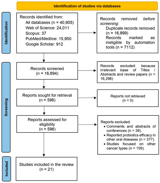
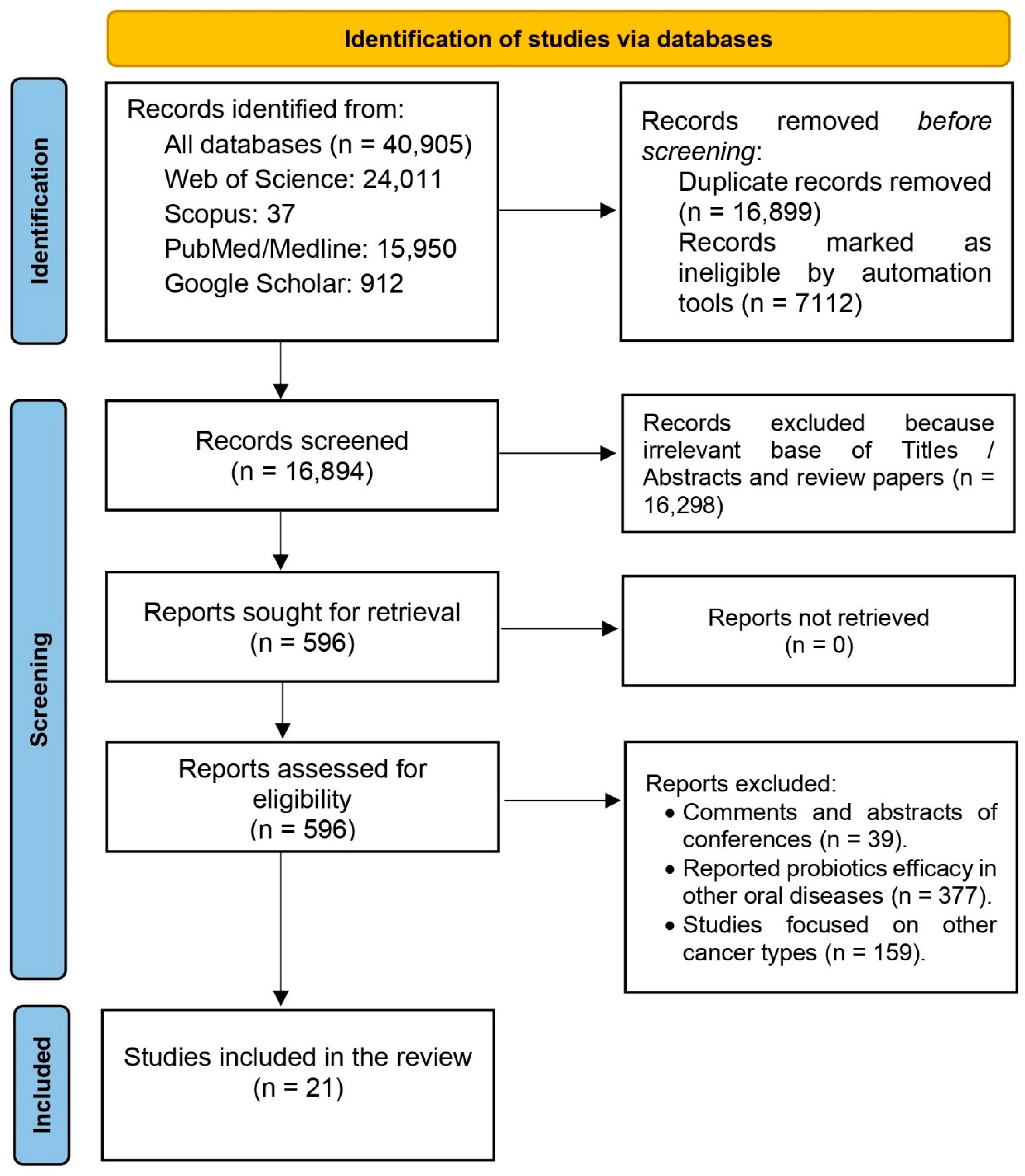
A total of 21 studies published between 2012 and 2026 were included in this scoping review, encompassing in vitro, in vivo, and clinical investigations (Figure 1). Preferred Reporting Items for Systematic reviews (PRISMA-ScR) Checklist can be found in the Supplementary Materials. Geographically, the majority of studies originated from Iran, India, China, Kuwait, Uganda, and Singapore. The distribution of study types comprised 13 in vitro studies, 3 in vivo preclinical studies, and 6 clinical trials. The total number of included studies was 21 rather than 22, as one study contributed to both in vitro and in vivo categories (Table 1). The evidence base is largely preclinical, with a substantial proportion of in vitro studies employing a range of OSCC cell lines to evaluate the antiproliferative and cytotoxic effects of probiotic strains. The most frequently utilized cell lines include HeLa-derived KB (ATCC CCL-17) [22,23,24,25,26], OSCC ATCC CRL-1628 [22,23,25], HNO97 [27,28], GMNL-346 [29], Cal27 [30], HN5 [31], HSC4 [32], Tca8113, SCC-9, and SAS [33]. These in vitro models have been used to assess a variety of probiotic interventions, including live bacterial cultures, cell-free supernatants (CFS), lysates, and heat-inactivated preparations, enabling evaluation of dose-dependent cytotoxicity and selectivity for cancer versus normal cells.
Figure 1.
PRISMA 2020 flow diagram for the selection of studies. The numbers (n) indicate the number of records identified at each stage of the screening process.
In vivo investigations utilized diverse animal models to replicate oral carcinogenesis and assess the therapeutic potential of probiotics. Experimental approaches included the 4-nitroquinoline 1-oxide (4NQO)-induced oral carcinogenesis model in rats [27,34], the 7,12-dimethylbenz[a]anthracene (DMBA) and liquid paraffin-induced buccal pouch model in golden Syrian hamsters [35], and xenograft models employing SAS cells in immunodeficient mice [29,36]. These models facilitated the evaluation of tumor incidence, tumor volume reduction, and the systemic safety profile of administered probiotic strains.
Clinical investigations, predominantly Phase Ib to III trials, focused on the adjuvant use of probiotics for the management of treatment-related oral complications in patients with head and neck cancers, including oral cancer. Primary endpoints included the prevention or mitigation of radiation-induced oral mucositis and secondary fungal infections [37,38,39,40,41]. Clinical studies included a total of 482 participants; notable heterogeneity was observed in study design, patient populations, and group allocation, with participants generally assigned to either a probiotic intervention or a control (placebo or standard care) group.
Across preclinical and clinical studies, a wide range of probiotic strains was investigated. Lactobacillus acidophilus [26,27] and Lactiplantibacillus plantarum strains (MCC 3016, Y33, Y8) [22,23,24,30] were among the most frequently studied. Other commonly examined strains included Limosilactobacillus fermentum KUMS-C9 and Lactiplantibacillus plantarum KUMS-C47 [25], Lacticaseibacillus paracasei GMNL-346 [29], Lactobacillus salivarius REN [34,42], Lactobacillus casei [28,42], Lactobacillus brevis CD2 [37,41], Lactococcus lactis [38], Lactobacillus rhamnosus GG [33], Bacillus clausii UBBC-07 [36], Acetobacter syzygii [26], Lactobacillus fermentum and Lactobacillus crispatus [31], marine-derived Lactobacillus species (TLMP1–TLMP5) [35], and the yeast Pichia fermentans YSH [32]. Multi-strain probiotic formulations were also assessed, including combinations of Lacticaseibacillus rhamnosus GG, L. casei, L. acidophilus, L. delbrueckii subsp. bulgaricus, Bifidobacterium infantis, B. brevis, and Streptococcus thermophilus [40], as well as alternative cocktails containing Bifidobacterium breve, B. longum, L. acidophilus, L. casei, L. bulgaricus, L. rhamnosus, and Streptococcus salivarius subsp. thermophilus [39].
The majority of probiotics investigated were Gram-positive microorganisms of human origin (oral or intestinal) or derived from traditional dairy products such as cheese and yogurt. Administration routes included suspensions, powders, lozenges, and mouthwashes, with dosing regimens ranging from 105 to 1010 CFU/mL or per day [33,38].
Table 1.
Summary of characteristics of included studies.
2.2. Preclinical Therapeutic Efficacy of Probiotics in the Management of Oral Cancer
- Lactobacillus acidophilus
Preclinical evidence suggests that L. acidophilus from traditional fermented dairy products exerts biologically relevant antitumor effects in OSCC models (Table 2). In vitro, strain PTCC 1643 reduced HeLa-derived KB cell proliferation by 48.5%, compared with 57.6% for cisplatin under similar conditions [26]. Strain ATCC 4356 also demonstrated significant cytotoxicity, decreasing HNO97 cell viability by 53% after 24 h [27]. Notably, enzymatic treatment of ATCC 4356-secreted metabolites enhanced cytotoxicity to 72.72% (pronase) and 51.5% (lipase) in HeLa-derived KB cells, whereas similarly treated cisplatin achieved only 42.4% inhibition [26], suggesting that proteinaceous or lipid-associated bacterial fractions may mediate antitumor activity. Mechanistically, apoptosis induction, potentially involving TRAIL-related pathways, has been proposed, although protein-level confirmation remains inconsistent [27]. Importantly, cytotoxic effects were markedly lower in normal KDR cells, indicating a degree of tumor selectivity [26]. Although limited to preclinical systems, these findings support further translational investigation of L. acidophilus as a potential adjunctive strategy in the management of oral cancer.
Table 2.
Summary of in vitro anti-oral cancer effects of probiotics.
- Lactiplantibacillus plantarum (formerly Lactobacillus plantarum)
Isolated from traditional fermented dairy products, particularly Greek cheeses and milk [22,43], Lactiplantibacillus plantarum is the most extensively investigated probiotic species in preclinical oral cancer research. In vitro, strain MCC 3016 reduced Cal27 cell viability by 48% at a multiplicity of infection (MOI) of 10 and by 60% at MOI 100 after 6 h of exposure [30]. The term Multiplicity of Infection (MOI) is the ratio of bacterial cells to target cancer cells in bacterium–host interaction assays, distinguishing it from its conventional virological usage. Notably, co-administration with 5-fluorouracil (5-FU) significantly enhanced chemosensitivity, lowering the 5-FU IC50 from 7.2 µM alone to 3.8–4.1 µM in the presence of either live bacteria or their cell-free supernatant [30]. Secreted metabolites from strains C14, M22, M42, and Y8 demonstrated IC50 values ranging from 3.44 to 6.4 µg/µL against HeLa-derived KB cells after 72 h [22], while strains C5, C28, M45, and Y33 exhibited IC50 values between 3.213 and 19.625 µg/µL at concentrations below 25 µg/mL [23].
Strain KUMS-C47 displayed one of the most pronounced selective effects, with an IC50 of 3.03 µg/µL at 72 h on HeLa-derived KB cells, compared with 2153.3 µg/µL on normal peripheral blood mononuclear cells (PBMCs), indicating superior tumor selectivity relative to paclitaxel and doxorubicin in normal cells [25]. Mechanistically, L. plantarum strains have been shown to induce apoptosis by increasing SMAC, caspase-3, and p53 expression; decreasing survivin levels; enhancing reactive oxygen species (ROS) production; and reducing the secretion of IL-6, IL-8, TNF-α, and VEGF. Additional modulation of PTEN activation and inhibition of the MAPK pathway have been reported [23,24,25,30]. Importantly, no significant cytotoxicity was observed in fibroblasts or HUVEC endothelial cells, reinforcing a favorable safety profile in vitro.
- Lacticaseibacillus paracasei (formerly Lactobacillus paracasei)
Originating from traditional Taiwanese and Iranian foods [44], Lacticaseibacillus paracasei GMNL-346 has demonstrated both in vitro and in vivo antitumor activity. Heat-inactivated GMNL-346 exhibited an IC50 of 7.12 log cells/mL in SAS and HSC3 cell lines after 72 h [29]. Treatment induced cell-cycle arrest in the G0/G1 phase in SAS cells and S phase in HSC3 cells, reduced DNA replication, upregulated RB and p16INK4a, downregulated CDK6, and suppressed tumor stemness markers including OCT4, SOX2, BMI1, EZH2, and c-Myc [29]. In an OSCC xenograft mouse model, oral administration of 1 × 10⁹ cells/day, five days per week for five weeks, increased survival to 40% compared with 0% in controls, reduced tumor weight by 85% in HSC3 and 26% in SAS tumors, and restored gut microbiota diversity with increased propionic and isobutyric acid levels [29]. No systemic toxicity was reported, supporting translational relevance.
- Limosilactobacillus fermentum (formerly Lactobacillus fermentum)
L. fermentum is a probiotic lactic acid bacterium naturally present in the human microbiota and commonly found in fermented foods such as dairy and plant products [45,46]. Data on L. fermentum strain 4–17 are heterogeneous. Strains GMNL-117 and GMNL-299 demonstrated IC50 values of 8.28 and 20.23 log cells/mL, respectively, for human OSCC cells [29]. Strain KUMS-C9 exhibited IC50 values of 7.53 µg/µL and 5.77 µg/µL at 48 and 72 h, respectively, in HeLa-derived KB cells, compared with 2453.6 µg/µL in normal PBMCs, indicating selective apoptosis associated with increased SMAC and decreased survivin expression [29]. Conversely, no significant anticancer activity was observed on HN5 cells, although acidic conditions reduced proliferation [31]. The residual viability values for Lactobacillus fermentum were 93%, 80%, 65%, 27%, 19%, and 11% at concentrations of 5, 10, 20, 30, 40, and 50 units, respectively, indicating a gradual, concentration-dependent reduction in tumor cell viability that remained broadly comparable to the control conditions (MRS and MP media) across lower and intermediate concentrations [31].
- Lactobacillus crispatus
L. crispatus is a Gram-positive, lactic acid-producing bacterium that naturally inhabits the female urogenital tract and vertebrate gastrointestinal tract, where it helps protect against bacterial vaginosis [47]. Unlike several other Lactobacillus species evaluated in oral squamous cell carcinoma models, L. crispatus strain SJ-3C-US has not demonstrated significant antiproliferative activity under the experimental conditions reported to date. In the HN5 cell line, exposure to this strain did not result in a measurable reduction in tumor cell viability [31]. The apparent decrease in cell viability observed with L. crispatus must be interpreted in the context of the experimental controls. Although residual viability declined from 89% to 8% across increasing concentrations, a comparable reduction was also observed in the control groups containing only the culture media (MRS and MP), which produced similar viability profiles [31]. This indicates that the observed cytotoxicity was largely attributable to the culture media conditions rather than to a strain-specific antiproliferative effect. Accordingly, compared with these controls, neither strain demonstrated a statistically significant antiproliferative activity in this experimental model, which supports the interpretation reported in the original study. The absence of detectable anticancer activity with L. crispatus underscores the need for systematic, strain-level functional evaluation rather than extrapolation based on taxonomic proximity.
- Lacticaseibacillus casei subsp. casei (formerly Lactobacillus casei subsp. casei)
L. casei is a lactic acid bacterium naturally present in the human and animal gastrointestinal tract, oral cavity, and vaginal mucosa, and is also commonly isolated from dairy products such as milk and cheese, fermented vegetables, and decomposing organic substrates [48]. In preclinical oral cancer models, strain-specific antiproliferative activity has been reported. At a density of 1 × 10⁸ CFU/mL, L. casei reduced proliferation of the HNO97 oral squamous cell carcinoma cell line by approximately 50% after 24 h of exposure, an effect associated with upregulation of TRAIL expression, suggesting activation of apoptosis-related signaling pathways [28]. However, this activity appears to be cell-line-dependent. No significant anticancer effect was observed in the TCA-8113 cell line, even at a higher concentration of 2 × 10⁹ CFU/mL [42]. This differential responsiveness underscores the importance of the tumor-specific biological context and highlights the variability of probiotic effects across distinct oral cancer cellular models.
- Lacticaseibacillus rhamnosus (formerly Lactobacillus rhamnosus)
L. rhamnosus is a lactic acid bacterium naturally present in the human intestinal and vaginal microbiota, first isolated in 1983, and also found in fermented dairy products such as yogurt and kefir, with notable acid tolerance [49]. The culture supernatant of L. rhamnosus GG at 108 CFU/mL (25% v/v) reduced SCC-9 cell viability to 49.0% and 41.6% after 24 and 48 h, respectively, compared with >94% in controls [33]. Combination with curcumin enhanced cytotoxicity but reduced selectivity, lowering normal cell viability to 84.12%. Mechanistically, apoptosis induction was linked to an increased Bax/Bcl-2 ratio, consistent with mitochondrial pathway activation [33].
- Ligilactobacillus salivarius (formerly Lactobacillus salivarius)
L. salivarius is a Gram-positive probiotic lactic acid bacterium naturally present in the human and animal microbiota (oral cavity, small intestine, and vagina), and has also been isolated from fermented foods and is valued for its antimicrobial properties [50]. At 3 × 10⁹ CFU/mL, L. salivarius REN inhibited TCA-8113 proliferation by 49% within 24 h, comparable to the COX-2 inhibitor NS-398 (50.94%), and was associated with downregulation of COX-2 expression [42]. In a 4-nitroquinoline-1-oxide (4NQO)-induced murine oral carcinogenesis model, oral administration of 5 × 10¹⁰ CFU/kg/day reduced tumor incidence from 65% to 0%, decreased oxidative DNA damage (8-OHdG), and lowered PCNA and COX-2 expression without detectable toxicity [34], providing one of the most compelling in vivo demonstrations of probiotic-mediated chemoprevention in oral cancer (Table 3).
Table 3.
Summary of in vivo anti-oral tumour effects of probiotics.
- Lactobacillus spp. and Bacteriocins
Experimental studies in a DMBA-induced oral carcinogenesis model in Syrian golden hamsters demonstrated that administration of Lactobacillus spp. or purified bacteriocins (2.5 mg/kg) markedly reduced tumor volumes from 34.94 mm3 to 1.04–7.08 mm3 and effectively inhibited progression to invasive squamous cell carcinoma [35]. Notably, purified bacteriocins did not induce mucositis, unlike whole probiotic cells, whereas 5-fluorouracil treatment resulted in severe alopecia [35]. Mechanistic analyses suggest that lactobacillary proteins interact with the α catalytic subunit of AMP-dependent protein kinase, providing a plausible pathway for the observed antiproliferative activity. Collectively, the preclinical evidence summarized in this review demonstrates that lactic acid bacteria exert robust antiproliferative and pro-apoptotic effects across diverse OSCC cell lines. These effects involve modulation of key signaling pathways, including TRAIL, p16INK4a/CDK6, PTEN/MAPK, NF-κB, and inflammatory mediators, while exhibiting selective cytotoxicity towards tumor cells (Table 4) [35]. In vivo data corroborate both preventive and therapeutic potential in chemically induced or xenograft models, with an overall favorable safety profile, supporting the translational promise of Lactobacillus species and their bacteriocins as adjunctive agents in oral oncology [35].
Table 4.
Summary of biological mechanisms of action identified with probiotics against tumour cells.
2.3. Clinical Effects of Probiotics as Adjuncts in Oral Cancer Management
2.3.1. Modulation of Antitumor Immune Response
Across the included studies, modulation of the antitumor immune response emerges as a central mechanistic pathway, predominantly documented in preclinical models. Administration of L. reuteri DSM 17938 and ATCC PTA 5289 in a murine model of 5-fluorouracil-induced mucositis significantly attenuated NF-κB p65 activation and reduced pro-inflammatory cytokine expression, including IL-1β and TNF-α. This was accompanied by decreased tissue infiltration of inflammatory cells and myeloperoxidase (MPO) activity [52]. Concomitantly, the cytoprotective Nrf2 axis was upregulated, evidenced by increased SOD-1, GPx-1, and HO-1 expression, with corroborative in vitro findings in TR146 oral epithelial cells demonstrating enhanced nuclear Nrf2 translocation and improved GSH/GSSG ratio [52]. Although classical adaptive antitumor immune markers, such as CD8+ T cells, NK cells, CD8+/Treg ratios, or tumor-infiltrating lymphocytes, were not systematically quantified in clinical trials, these preclinical results suggest a microenvironment less skewed toward chronic inflammation, potentially favoring effective antitumor immune activity [52].
2.3.2. Enhancement of Immunotherapy Response
No included study directly evaluated the impact of probiotics on objective response rate (ORR), progression-free survival (PFS), or overall survival (OS) in patients receiving immune checkpoint inhibitors, such as pembrolizumab or nivolumab. Nonetheless, observed modulation of the gut and oral microbiota, along with reduced systemic inflammation, provides a biologically plausible rationale for potential synergistic effects. This may involve attenuating immunosuppressive mediators and stabilizing the tumor microenvironment, thereby potentially enhancing responsiveness to immunotherapy [40,52].
2.3.3. Reduction in Treatment-Related Adverse Effects
The most robust clinical evidence pertains to mitigating anticancer treatment-related toxicities. Administration of B. clausii UBBC-07 significantly delayed mucositis onset (median 10 versus 8 days; p < 0.01), shortened its duration (12 versus 14 days; p < 0.05), and reduced severe cases (34.8% versus 69.6%; p < 0.05), with an overall clinical response rate of 65% compared to 30.4% in the placebo group [36]. Similarly, synbiotic mouth rinses reduced the grade 3 mucositis incidence to 11.59% versus 36.45% in controls (p < 0.001), with significant improvement in mean severity scores by the 30th radiotherapy session (1.75 versus 2.4; p < 0.001) and progressive benefit from the seventh session [39]. L. lactis AG013 decreased mean days with ulcerative mucositis by 35% and reduced unplanned consultations (36% versus 60%) without evidence of bacteremia or serious probiotic-related adverse events [38]. Conversely, a randomized trial of L. brevis CD2 showed no significant reduction in severe mucositis (40.6% versus 41.6%; p = 0.974), with tumor stage as the sole independent predictive factor (OR = 0.164; 95% CI, 0.040–0.673; p < 0.05) [37].
2.3.4. Modulation of Oral and Gut Microbiota
Probiotic administration has been shown to effectively modulate the oral and intestinal microbiota. Streptococcus salivarius demonstrated high viability (>1 × 10⁹ CFU), successful colonization with 17 ± 3% relative salivary abundance, and maintenance of alpha diversity (~182 ± 12 observed species; p > 0.05), without inducing dysbiosis. Network analysis revealed 8591 interactions, including 2141 negative interactions with periodontopathogens such as Campylobacter rectus and Treponema spp., indicating competitive ecological effects [53]. A multi-strain formulation including L. rhamnosus GG, L. casei, L. acidophilus, L. delbrueckii subsp. bulgaricus, B. infantis, B. brevis, and S. thermophilus significantly enhanced stimulated salivary flow (p = 0.0016), reduced total bacterial load (p = 0.0209), and decreased F. nucleatum prevalence (p = 0.0080 by qPCR; p = 0.0026 by culture) with strong methodological correlation (r = 0.923; p < 0.001) [40].
2.3.5. Impact on Tumor Growth in Preclinical Models
Preclinical models suggest that probiotics may indirectly inhibit tumor growth by reducing inflammation, activating antioxidant pathways, and maintaining epithelial integrity. Increased Ki-67 expression observed with L. reuteri in damaged oral epithelium reflects enhanced cell regeneration, while decreased oxidative stress markers, such as 8-OHdG and 4-HNE, confirm cytoprotective effects that limit chemotherapy-induced lesion progression [52].
2.3.6. Metabolic and Inflammatory Parameters
Although less frequently measured, systemic metabolic and inflammatory markers consistently indicate reduced inflammation and stabilized mucosal barriers. No systemic bacterial translocation or probiotic-related sepsis was reported, even in immunocompromised patients, supporting a favorable safety profile [38,40,52].
2.3.7. Patient-Reported Outcomes and Quality of Life
Improvements in quality of life, indirectly assessed through reductions in severe mucositis, fewer unplanned consultations, and enhanced salivary flow, suggest clinically meaningful functional benefits. However, standardized instruments such as the EORTC QLQ-C30 did not consistently reveal significant intergroup differences [37,39]. Overall, evidence converges to support a clinically relevant adjuvant effect of probiotics in oral cancer management, primarily through modulation of inflammation, stabilization of the microbiota, and mitigation of treatment-related toxicities, while maintaining a robust safety profile.
3. Discussion
This scoping review indicates that the field remains at an exploratory stage, predominantly supported by in vitro evidence, with comparatively limited in vivo validation and a small number of clinical trials. The strong concentration of studies in Asian countries likely reflects strong regional expertise in microbiome research and functional food sciences, alongside a strategic interest in cost-effective, biologically based adjuncts to conventional oncologic therapies [54]. However, this geographical clustering may introduce microbiological, dietary, and population-specific biases that restrict external validity, thereby underscoring the need for rigorously designed, multicenter trials across diverse populations to enhance global generalizability and support regulatory standardization of probiotics in oral cancer management.
A significant translational gap persists between experimental evidence and clinical applicability. Most studies rely on simplified in vitro OSCC cell line models assessed by metabolic assays, which, although suitable for preliminary strain screening, fail to reproduce the complexity of the tumor microenvironment, including immune, stromal, and microbiome interactions [55,56]. Most findings derive from controlled experimental systems that do not fully capture the biological complexity of human oral cancer, thereby limiting the extrapolation of observed effects to clinical settings. In vivo models based on chemical carcinogenesis (4-NQO, DMBA) or xenografts provide greater biological relevance but remain heterogeneous and only partially reflective of human disease [29,34,35]. While these models allow evaluation of tumor growth dynamics, inflammatory modulation, and systemic safety, they only partially reproduce the multifactorial etiology and immunological landscape of human oral cancer [57,58]. Moreover, the reported antitumor efficacy in animal models may reflect optimized experimental conditions that are not directly translatable to real-world clinical scenarios, and the observed effects are frequently strain-specific and context-dependent, further constraining generalizability. Importantly, the lack of large-scale randomized trials evaluating hard oncological endpoints, such as overall or progression-free survival, limits clinical interpretability, confining current evidence primarily to mechanistic and supportive effects rather than definitive antitumor efficacy [59]. Taken together, these findings should be interpreted as mechanistic and proof-of-concept evidence supporting a predominantly adjunctive role of probiotics, rather than as evidence of curative or standalone antitumor efficacy, underscoring the urgent need for robust, adequately powered clinical trials to validate these preliminary observations.
A central finding of this review is the pronounced inter-strain variability in antiproliferative activity, underscoring that probiotic effects in oral cancer cannot be generalized across species. It can also be explained by genetic and molecular heterogeneity within species [60]. Differences in accessory genomes, including strain-specific gene clusters encoding bacteriocins, exopolysaccharides, adhesion factors, and secondary metabolites, lead to distinct bioactive profiles [61]. Variability in secreted metabolites, cell wall structures, and surface proteins influences host receptor engagement and downstream signaling pathways, thereby modulating apoptosis, inflammation, and chemosensitivity [62]. Additionally, strain-specific transcriptional regulation and metabolic plasticity yield divergent functional outputs under similar conditions, supporting a strictly strain-dependent paradigm for probiotic-based strategies in oral cancer management [63]. These findings argue against a uniform “class effect” and strongly support a strain-specific paradigm.
When extrapolated to molar equivalents, the IC50 values reported for L. plantarum MCC3016, L. plantarum C47, L. fermentum C9, and L. paracasei GMNL-346 seldom fall within the ≤10 µg/µL range, a threshold considered indicative of very strong cytotoxic activity [64]. Accordingly, their intrinsic cytotoxic potency appears modest relative to conventional chemotherapeutic agents such as cisplatin, 5-fluorouracil, doxorubicin, or paclitaxel. Notably, exceptions have been reported: secreted metabolites of P. fermentans YSH demonstrated antiproliferative activity comparable to that of cisplatin in HSC4 cells [32], and L. salivarius showed effects on TCA-8113 cells comparable to those of NS-398 [42]. Beyond absolute potency, several probiotic strains display a comparatively favorable selectivity profile, characterized by higher inhibitory thresholds in normal fibroblasts or gingival epithelial cells than in malignant cell lines [25,26]. Although moderate in magnitude, this differential cytotoxicity suggests a pharmacologically relevant therapeutic window, particularly when combined with or used as an adjuvant in therapeutic strategies.
Preclinical investigations consistently demonstrate that live probiotic strains exert the most pronounced and selective antiproliferative effects against oral squamous cell carcinoma (OSCC) models. For instance, a live L. plantarum strain exhibited an IC50 of 3.03 µg/µL on KB cells, reflecting high tumor specificity [25]. Non-viable preparations, including heat-inactivated L. paracasei GMNL-346, produced an IC50 of 7.12 log cells/mL in SAS and HSC3 cells [29], whereas cell-free supernatants from L. acidophilus ATCC 4356, when enzymatically treated, increased cytotoxicity to 72.7% in KB cells, indicating that secreted metabolites contribute substantially to antiproliferative activity [27]. Collectively, these data underscore that while live organisms often achieve maximal tumor-selective effects, non-viable preparations retain mechanistic relevance and may serve as functionally active postbiotic interventions [65]. Nevertheless, direct quantitative comparisons are constrained by heterogeneous reporting units, underscoring a critical need for standardized expression of biological activity in future studies to enable rigorous cross-strain and preparation-type comparisons.
An additional methodological consideration concerns the historical use of the KB cell line (ATCC CCL-17) as a model of oral squamous cell carcinoma. It is now well established that KB cells are cross-contaminated with HeLa and are therefore classified as a HeLa derivative rather than a genuine oral cancer cell line, a limitation explicitly acknowledged by [66,67]. Consequently, studies reporting antiproliferative effects of probiotics using KB cells should be interpreted with caution, as the observed responses may reflect biological properties of cervical carcinoma rather than oral squamous cell carcinoma. While these experiments remain informative regarding general anticancer or cytotoxic potential, their disease specificity for oral cancer is limited, highlighting the importance of validating findings in well-authenticated OSCC cell lines.
However, such comparisons must be interpreted cautiously. Methodological heterogeneity, including non-standardized reporting units (CFU/mL versus µg/mL), divergent preparation types (live bacteria, cell-free supernatants, lysates), absence of formal synergy analyses, and variability in seeding densities, substantially limits cross-study comparability. These inconsistencies likely contribute to the wide dispersion of reported activities and complicate robust benchmarking against conventional chemotherapeutics. A further limitation arises from the heterogeneous reporting of probiotic activity across studies, including percentage inhibition, IC50 values expressed in µg/µL, and bacterial densities reported as log cells/mL, which are not directly convertible due to the biological nature of living microorganisms. Future research should adopt standardized reporting based on viable counts (CFU/mL or log CFU/mL) combined with clearly defined dose–response curves and IC50 calculations derived from these biological units, thereby enabling more consistent and biologically meaningful cross-study comparisons.
Regarding in vivo models, chemically induced carcinogenesis via 4-NQO or DMBA offers advantages in terms of reproducibility and stepwise dysplasia-to-carcinoma progression, partially mimicking carcinogen-driven human oral tumorigenesis [34,35]. Nevertheless, these models rely on acute, controlled exposures and do not capture the genetic heterogeneity, lifestyle-associated cofactors, or endogenous microbiome dynamics characteristic of human oral cancer. Xenograft models [29], while useful for tumor growth assessment, are conducted in immunodeficient animals and therefore preclude meaningful evaluation of immune-mediated mechanisms, which are central to the proposed probiotic mode of action [68,69,70]. The limited number of in vivo studies remains a critical barrier to translational extrapolation.
Despite experimental heterogeneity, a coherent mechanistic pattern emerges. Probiotics and their metabolites appear to activate mitochondrial apoptotic pathways (increased Bax/Bcl-2 ratio, caspase activation), modulate TRAIL signaling, restore p16INK4a expression, inhibit CDK6, activate PTEN, and suppress MAPK and NF-κB signaling [25,29,42]. While these pathways overlap with those targeted by conventional cytotoxic agents, the distinctive feature of probiotics lies in their multimodal and microenvironment-oriented activity [71]. Evidence of reduced oxidative stress [49], modulation of inflammation via NF-κB and COX-2 suppression, and alterations in oral microbial composition [40,53] suggest that probiotics may act upstream of direct DNA damage, reshaping the inflammatory and ecological context that sustains tumor progression [71]. This systems-level modulation supports the emerging concept of oncobiotics or microbiome-based interventions [72], wherein microbiome-directed strategies complement tumor-targeted therapies.
Clinically, the most consistent benefits of probiotic administration in oral cancer have been observed in the prevention and mitigation of radiotherapy- and chemotherapy-induced mucositis. Trials assessing single strains such as L. brevis CD2 [41] and B. clausii [36], as well as multistrain synbiotic formulations [39,40], have demonstrated reductions in mucositis severity and/or delayed onset, although these outcomes have not been uniformly confirmed in multicenter randomized settings [37]. Overall safety profiles appear favorable, with no significant signals of probiotic-related bacteremia; however, cautious risk–benefit evaluation remains necessary in severely immunocompromised patients. Collectively, the available clinical evidence more strongly supports positioning probiotics as adjuvants rather than primary anticancer therapies, a conclusion consistent with data reported in other oncologic contexts [73,74]. Their most plausible therapeutic contribution resides in modulating the tumor microenvironment, attenuating treatment-induced inflammation, and enhancing tolerability and supportive care outcomes within comprehensive multimodal cancer management strategies [75,76].
4. Limitations
Several limitations must be acknowledged when interpreting the current body of evidence. First, the translational maturity of the field remains limited, as rigorous evaluation of clinically meaningful oncologic endpoints has not yet been systematically undertaken, thereby constraining conclusions regarding direct antitumor efficacy in patients. Furthermore, the limited number of in vivo studies (3) represents a critical gap, restricting the ability to validate preclinical findings in complex biological systems and to accurately predict therapeutic efficacy and safety in clinical contexts. Second, substantial methodological heterogeneity across experimental designs, including variability in strain selection, preparation methods, dosing strategies, treatment duration, and outcome measures, impairs comparability and precludes quantitative synthesis or standardized dose–response assessment. The absence of harmonized IC50 reporting standards and formal pharmacodynamic or synergy analyses further complicates cross-study interpretation. Reproducibility and external validity also represent critical concerns. Most clinical investigations are single-center studies with relatively small sample sizes, which limit statistical power and increase susceptibility to contextual bias. Geographic clustering of research activity may additionally restrict generalizability across diverse populations, microbiome backgrounds, and dietary patterns. Mechanistic insights, although suggestive of coherent signaling modulation, are frequently derived from reductionist models and rarely supported by integrated multi-omics validation. Finally, the inclusion of heterogeneous head and neck cancer cohorts in several trials complicates extrapolation specifically to oral squamous cell carcinoma, thereby limiting disease-specific precision in clinical inference. Additionally, the substantial heterogeneity across clinical studies, including differences in design, overall sample size (482 participants), and interventions, limits direct comparability and generalization of the results.
5. Future Perspectives
Future research should prioritize rigorous multicenter randomized trials incorporating survival endpoints and validated mechanistic biomarkers to establish clinical relevance. Standardized GMP-grade, genomically characterized formulations are essential to ensure reproducibility and regulatory credibility. Strategic integration with immunotherapies, microbiome-based patient stratification, and the development of defined postbiotics should be pursued, supported by integrated multi-omics and tumor microenvironment analyses to clarify mechanisms and optimize the positioning of probiotics within multimodal oral cancer management.
6. Methodology
6.1. Study Design
This scoping review was conducted in accordance with the methodological framework proposed by Arksey and O’Malley and further refined by Westphaln et al. [77], and reported in compliance with the PRISMA-ScR guidelines [78]. The review protocol was registered in the Open Science Framework (OSF) as part of the associated project: https://osf.io/pb625 (accessed on 6 March 2026). The objective was to comprehensively map the available evidence regarding the therapeutic and adjuvant effects of probiotics in the management of oral cancer, including preclinical and clinical investigations. Given the heterogeneity of study designs and outcomes, a scoping methodology was selected to identify knowledge gaps, characterize mechanistic pathways, and synthesize translational evidence across experimental and clinical settings.
6.2. Eligibility Criteria
Studies were deemed eligible for inclusion if they investigated probiotic strains administered as single- or multi-strain formulations, encompassing live microorganisms, heat-inactivated preparations, bacterial lysates, metabolites, or cell-free supernatants. To ensure conceptual consistency with the objectives of this scoping review, only studies evaluating therapeutic, preventive, or adjuvant effects in the context of oral cancer, particularly OSCC, were considered. Eligible studies included experimental investigations conducted in vitro using established OSCC cell lines, in vivo studies employing animal models of oral carcinogenesis or xenograft systems, and interventional or observational clinical trials involving patients diagnosed with oral or head and neck cancers. Furthermore, studies were required to report measurable outcomes of biological, mechanistic, tumor-related, microbiological, or clinical relevance, thereby allowing comprehensive mapping of both preclinical and translational evidence.
No restrictions were applied regarding probiotic strain taxonomy, dosage, formulation, route of administration, or comparator groups to ensure comprehensive coverage and reflect the translational heterogeneity of probiotic interventions. No temporal limits were imposed during the initial search to capture the full historical scope of the field. However, eligibility was restricted to studies published in English and available up to 10 February 2026. The final selection was restricted to original, peer-reviewed research articles published in English. Reviews, editorials, conference abstracts without complete datasets, case reports, and non-peer-reviewed publications were excluded to preserve methodological rigor and analytical consistency consistent with standards for high-impact pharmacological research.
6.3. Information Sources and Search Strategy
A comprehensive literature search was conducted in major biomedical databases, including PubMed/MEDLINE, Scopus, Web of Science, and Google Scholar. The search strategy combined controlled vocabulary (MeSH terms for PubMed, Emtree for Scopus) and free-text keywords related to: “oral cancer” OR “oral squamous cell carcinoma” AND “probiotics” OR “Lactobacillus” OR “Synbiotics” AND “Anti-oral cancer” OR “adjuvant therapy” OR “antiproliferative effect”. The final search strategy was adapted to each database. An exhaustive search strategy was implemented in Google Scholar, ClinicalTrials.gov, and the reference lists of included studies, including original clinical studies involving heterogeneous oncological populations, to provide a broad contextual framework. Studies addressing head and neck cancers were systematically screened, and data specifically related to oral cancer were extracted and analyzed separately whenever possible to enable a focused and clinically relevant synthesis. In cases where trials involved mixed populations of head and neck cancers but explicitly stated that patients with oral cancer were included, without reporting oral cancer–specific outcomes, these studies were retained only when the effects of the intervention were reported consistently across the overall cohort, and no evidence suggested differential or contradictory responses in patients with oral cancer. This approach was adopted to preserve clinically relevant data on radiotherapy- and chemotherapy-associated mucositis, an adverse event common to head and neck malignancies, while acknowledging the current limitations in disease-specific reporting.
6.4. Study Selection
All identified records were imported into a reference management software, and duplicate entries were systematically removed prior to screening. Study selection was conducted independently by two reviewers (GTK and EJN) using a structured two-stage approach. In the first phase, titles and abstracts were screened to exclude studies that were clearly irrelevant to the scope and objectives of the review. In the second phase, the full texts of potentially eligible articles were retrieved and assessed in detail according to the predefined inclusion and exclusion criteria. Any discrepancies between reviewers were resolved through discussion and consensus; when agreement could not be reached, a third reviewer was consulted to ensure methodological rigor and selection transparency.
6.5. Data Charting Process
A standardized data extraction form was developed and pilot-tested to ensure consistency, clarity, and reproducibility in data charting. For each included study, detailed information was systematically collected, including the authors, year of publication, country of origin, and study design (in vitro, in vivo, or clinical trial). Specific characteristics of the probiotic intervention were documented, including the strain(s) investigated, formulation, and administered concentration or dose (expressed as CFU/mL, CFU/day, or equivalent units).
Information regarding the experimental or clinical cancer model was recorded, encompassing OSCC cell lines, animal models of oral carcinogenesis or xenograft systems, and defined patient populations. Comparator conditions or standard anticancer treatments were also extracted when applicable. Outcome measures were comprehensively documented and included primary and secondary endpoints, including cell viability, IC50 values, apoptosis-related markers, tumor volume, inflammatory mediators, mucositis grading, microbiological parameters, and survival outcomes. Safety data, including adverse events and tolerability profiles, were systematically recorded. Additionally, the biological origin of the probiotic strains (e.g., human-, dairy-, or environmental-derived) was documented to facilitate translational interpretation. Data extraction was independently performed by two reviewers, and cross-verification was conducted to ensure accuracy, completeness, and methodological reliability.
6.6. Data Synthesis and Mapping of Evidence
Given the substantial heterogeneity in study designs, experimental models, and reported endpoints, a descriptive analytical framework was adopted to systematically map and synthesize the available evidence. The included studies were stratified into three principal domains reflecting the translational continuum from bench to bedside.
The first domain comprised in vitro investigations, which predominantly explored antiproliferative activity, induction of apoptosis, modulation of intracellular signaling pathways, such as TRAIL-mediated cascades, PTEN/MAPK signaling, and NF-κB inhibition, and the selective cytotoxicity of probiotic-derived interventions toward malignant versus non-malignant cells. These studies were conducted using established OSCC cell lines, thereby providing mechanistic insights into direct tumor-cell interactions.
The second domain encompassed in vivo preclinical models, including 4-nitroquinoline-1-oxide (4NQO)-induced rat models of oral carcinogenesis, 7,12-dimethylbenz[a]anthracene (DMBA)-induced hamster buccal pouch models, and xenograft mouse systems. These studies evaluated outcomes such as tumor incidence, tumor volume reduction, modulation of oxidative stress biomarkers, histopathological alterations, and systemic safety parameters, thereby facilitating translational interpretation of probiotic-mediated effects in complex biological environments.
The third domain included clinical trials assessing the adjuvant role of probiotics in patients with oral and head and neck cancers, particularly in the management of treatment-related oral complications. Clinical endpoints primarily focused on radiation- or chemotherapy-induced mucositis severity and incidence of fungal or bacterial infections, salivary flow dynamics, patient-reported quality-of-life measures, and safety outcomes. Across all domains, probiotic interventions were further characterized and mapped according to taxonomic classification, formulation (single-strain versus multi-strain combinations), administered dose range (105–1010 CFU/mL or per day), route of administration (oral capsules, suspensions, lozenges, or mouthwash), and biological origin (human-derived, dairy-derived, or environmental), enabling a structured and pharmacologically relevant synthesis of the evidence landscape.
6.7. Risk of Bias Assessment
In line with scoping review methodology, a formal quantitative risk-of-bias assessment was not performed. However, methodological characteristics, including study design, sample size, comparator use, and reporting transparency, were descriptively examined to contextualize the strength and limitations of the available evidence.
7. Conclusions
In conclusion, current evidence does not support probiotics as primary cytotoxic agents in oral cancer, yet selected strains, particularly L. plantarum and L. paracasei GMNL-346, demonstrate reproducible in vitro efficacy, chemosensitizing potential, and preliminary in vivo antitumor effects. While their absolute cytotoxic potency is modest compared with conventional chemotherapeutics, several strains exhibit favorable selectivity for malignant cells over normal tissues. Clinically, probiotics consistently reduce treatment-related mucosal toxicity, highlighting their adjuvant value. Rather than direct cytotoxic agents, probiotics modulate the tumor microenvironment, immunity, and inflammatory signaling, aligning with the “oncobiotics” paradigm. Collectively, mechanistic plausibility, strain-specific activity, chemosensitization potential, and safety profiles support further investigation, with rigorous multicenter trials and standardized formulations required to establish their role as evidence-based adjuncts in integrative oral cancer therapy.
Supplementary Materials
The following are available online at https://www.mdpi.com/article/10.3390/ddc5020030/s1, Supplementary Material S1: Preferred Reporting Items for Systematic reviews and Meta-Analyses extension for Scoping Reviews (PRISMA-ScR) Checklist.
Author Contributions
All authors contributed substantially to the development of this study, including the conceptualization, methodology design, software utilization, validation, formal analysis, investigation, resource management, and data curation. G.T.K. prepared the initial draft of the manuscript. E.J.N. contributed to the critical revision and editing of the manuscript, data visualization, and overall supervision of the work. Project administration was coordinated by G.T.K. All authors have read and agreed to the published version of the manuscript.
Funding
This research was conducted without specific financial support from any public, commercial, or not-for-profit funding agencies.
Data Availability Statement
No new data were created or analyzed in this study. Data sharing is not applicable to this article.
Acknowledgments
The authors express their sincere appreciation to the university librarian for valuable assistance in developing the database search strategies used in this study. We also acknowledge Walter Sisulu University for providing internet access that facilitated the completion of this research.
Conflicts of Interest
The authors declare that there are no conflicts of interest related to this study.
References
- Ebrahimi, R.; Masouri, M.M.; Razeghian, I.; Azad, G.; Nejadghaderi, S.A. Lip, Oral Cavity, and Pharyngeal Cancers: Global Epidemiology, Risk Factors, and Prevention: A Narrative Review. Health Sci. Rep. 2025, 8, e71289. [Google Scholar] [CrossRef] [Scilit]
- Bray, F.; Laversanne, M.; Sung, H.; Ferlay, J.; Siegel, R.L.; Soerjomataram, I.; Jemal, A. Global cancer statistics 2022: GLOBOCAN estimates of incidence and mortality worldwide for 36 cancers in 185 countries. CA Cancer J. Clin. 2024, 74, 229–263. [Google Scholar] [CrossRef] [Scilit] [PubMed]
- Sung, H.; Ferlay, J.; Siegel, R.L.; Laversanne, M.; Soerjomataram, I.; Jemal, A.; Bray, F. Global Cancer Statistics 2020: GLOBOCAN Estimates of Incidence and Mortality Worldwide for 36 Cancers in 185 Countries. CA Cancer J. Clin. 2021, 71, 209–249. [Google Scholar] [CrossRef] [Scilit]
- Ferlay, J.; Colombet, M.; Soerjomataram, I.; Mathers, C.; Parkin, D.M.; Piñeros, M.; Znaor, A.; Bray, F. Estimating the global cancer incidence and mortality in 2018: GLOBOCAN sources and methods. Int. J. Cancer 2019, 144, 1941–1953. [Google Scholar] [CrossRef] [Scilit]
- WCRF. Mouth and Oral Cancer Statistics. Available online: https://www.wcrf.org/preventing-cancer/cancer-statistics/mouth-and-oral-cancer-statistics/ (accessed on 21 February 2026).
- Ghanem, A.S.; Tóth, Á.; Nagy, A.C. Socio-demographic disparities in global trends of lip and oral cavity neoplasms from 1990 to 2021. Sci. Rep. 2025, 15, 4230. [Google Scholar] [CrossRef] [Scilit]
- Melo-Alvim, C.; Neves, M.E.; Santos, J.L.; Abrunhosa-Branquinho, A.N.; Barroso, T.; Costa, L.; Ribeiro, L. Radiotherapy, Chemotherapy and Immunotherapy-Current Practice and Future Perspectives for Recurrent/Metastatic Oral Cavity Squamous Cell Carcinoma. Diagnostics 2022, 13, 99. [Google Scholar] [CrossRef] [Scilit]
- Gupta, S.; Singh, A.; Deorah, S.; Tomar, A. Immunotherapy in OSCC: Current trend and challenges. Crit. Rev. Oncol. Hematol. 2025, 209, 104672. [Google Scholar] [CrossRef] [Scilit] [PubMed]
- Chen, H.H.W.; Kuo, M.T. Improving radiotherapy in cancer treatment: Promises and challenges. Oncotarget 2017, 8, 62742–62758. [Google Scholar] [CrossRef] [Scilit] [PubMed]
- Hosseini, M.S.; Sanaie, S.; Mahmoodpoor, A.; Jabbari Beyrami, S.; Jabbari Beyrami, H.; Fattahi, S.; Jahanshahlou, F.; Zarei, M.; Rahimi Mamaghani, A.; Kuchaki Rafsanjani, M. Cancer treatment-related xerostomia: Basics, therapeutics, and future perspectives. Eur. J. Med. Res. 2024, 29, 571. [Google Scholar] [CrossRef] [Scilit]
- Shetty, S.S.; Maruthi, M.; Dhara, V.; de Arruda, J.A.A.; Abreu, L.G.; Mesquita, R.A.; Teixeira, A.L.; Silva, T.A.; Merchant, Y. Oral mucositis: Current knowledge and future directions. Dis. Mon. 2022, 68, 101300. [Google Scholar] [CrossRef] [Scilit]
- Wang, X.; Zhang, H.; Chen, X. Drug resistance and combating drug resistance in cancer. Cancer Drug Resist. 2019, 2, 141–160. [Google Scholar] [CrossRef] [Scilit]
- Yao, Y.; Zhu, Y.; Chen, K.; Chen, J.; Li, Y.; Li, D.; Wei, P. Microbiota in cancer: Current understandings and future perspectives. Signal Transduct. Target. Ther. 2026, 11, 39. [Google Scholar] [CrossRef] [Scilit]
- Mistry, L.N.; Mulla, S.A.; Patil, A.; Shah, S.; Pulaskar, S.P. A Narrative Review on Oral Microbiome Dysbiosis in Benign and Malignant Oral Lesions: Mechanistic Insights and Clinical Implications. Cureus 2026, 18, e102131. [Google Scholar] [CrossRef] [Scilit] [PubMed]
- Nishida, A.; Andoh, A. The Role of Inflammation in Cancer: Mechanisms of Tumor Initiation, Progression, and Metastasis. Cells 2025, 14, 488. [Google Scholar] [CrossRef] [Scilit]
- Wei, X.-Y.; Feng, H.-J.; Zhu, Y.-Y.; Guo, S.-J.; Wang, H.; Li, M.; Mei, Q. The immune microenvironment of pathogen-associated cancers and current clinical therapeutics. Mol. Cancer 2025, 24, 232. [Google Scholar] [CrossRef] [Scilit]
- Hill, C.; Guarner, F.; Reid, G.; Gibson, G.R.; Merenstein, D.J.; Pot, B.; Morelli, L.; Canani, R.B.; Flint, H.J.; Salminen, S.; et al. The International Scientific Association for Probiotics and Prebiotics consensus statement on the scope and appropriate use of the term probiotic. Nat. Rev. Gastroenterol. Hepatol. 2014, 11, 506–514. [Google Scholar] [CrossRef] [Scilit]
- Alam, S.T.; Rayhan, A.B.H.; Ahmed, M.U.; Khan, M.T.; Jame, J.F.; Islam, M.M.; Raj, M.A.R.; Uddin, M.A. Mechanisms of action and health benefits of probiotics: A comprehensive review. Explor. Drug Sci. 2025, 3, 1008129. [Google Scholar] [CrossRef] [Scilit]
- Park, S.H. Recent Research on the Role of Lactobacilli Probiotics in Cancer Management. Nutrients 2026, 18, 297. [Google Scholar] [CrossRef] [Scilit] [PubMed]
- Giorgi, C.; Lombardi, F.; Augello, F.R.; Alicka, Y.; Quintiliani, M.; Topi, S.; Cimini, A.; Castelli, V.; d’Angelo, M. Probiotics as Anti-Tumor Agents: Insights from Female Tumor Cell Culture Studies. Biomolecules 2025, 15, 657. [Google Scholar] [CrossRef] [Scilit]
- Minervini, G.; Franco, R.; Marrapodi, M.M.; Fiorillo, L.; Badnjević, A.; Cervino, G.; Cicciù, M. Probiotics in the Treatment of Radiotherapy-Induced Oral Mucositis: Systematic Review with Meta-Analysis. Pharmaceuticals 2023, 16, 654. [Google Scholar] [CrossRef] [Scilit]
- Haghshenas, B.; Nami, Y.; Kiani, A.; Moazami, N.; Tavallaei, O. Cytotoxic effect of potential probiotic Lactiplantibacillus plantarum KUMS-Y8 isolated from traditional dairy samples on the KB and OSCC human cancer cell lines. Heliyon 2023, 9, e20147. [Google Scholar] [CrossRef] [Scilit] [PubMed]
- Nami, Y.; Tavallaei, O.; Kiani, A.; Moazami, N.; Samari, M.; Derakhshankhah, H.; Jaymand, M.; Haghshenas, B. Anti-oral cancer properties of potential probiotic lactobacilli isolated from traditional milk, cheese, and yogurt. Sci. Rep. 2024, 14, 6398. [Google Scholar] [CrossRef] [Scilit] [PubMed]
- Asoudeh-Fard, A.; Barzegari, A.; Dehnad, A.; Bastani, S.; Golchin, A.; Omidi, Y. Lactobacillus plantarum induces apoptosis in oral cancer KB cells through upregulation of PTEN and downregulation of MAPK signalling pathways. Bioimpacts 2017, 7, 193–198. [Google Scholar] [CrossRef] [Scilit] [PubMed]
- Tavallaei, O.; Barghi, A.; Nami, Y.; Salehian, M.; Sabbaghhadi, A.; Haghshenas, B. Survivability, antimicrobial and anticancer effects, and cytotoxicity of Lactobacillus strains isolated from traditional Iranian cheese. Sci. Rep. 2025, 15, 25449. [Google Scholar] [CrossRef] [Scilit]
- Aghazadeh, Z.; Pouralibaba, F.; Yari Khosroushahi, A. The prophylactic effect of Acetobacter syzygii probiotic species against squamous cell carcinoma. J. Dent. Res. Dent. Clin. Dent. Prospect. 2017, 11, 208–214. [Google Scholar] [CrossRef] [Scilit]
- Al-Asfour, A.; Bhardwaj, R.G.; Karched, M. Growth Suppression of Oral Squamous Cell Carcinoma Cells by Lactobacillus Acidophilus. Int. Dent. J. 2024, 74, 1151–1160. [Google Scholar] [CrossRef] [Scilit]
- Rehaman, T.A.; Bhardwaj, R.G.; Sharma, N.; Ellepola, A.; Karched, M. Probiotic Lactobacillus casei Inhibits Oral Squamous Cell Carcinoma Growth and Induces Apoptosis In Vitro. Int. Dent. J. 2026, 76, 103960. [Google Scholar] [CrossRef] [Scilit]
- Chen, Y.-F.; Fan, C.-S.; Hung, Y.-C.; Chien, P.-J.; Tsai, W.-H.; Yeh, Y.-T.; Jhong, J.-H.; Hsieh, Y.-N.; Huang, Y.-H.; Jan, M.-S.; et al. Heat-killed Lacticaseibacillus paracasei GMNL-346 exerts anti-oral cancer effect by suppressing cancer stem cell activity and rebalancing gut microbiota dysbiosis. Int. J. Biol. Macromol. 2025, 334, 149033. [Google Scholar] [CrossRef] [Scilit]
- Fida, F.; Yuvarajan, S.; Ashwath, K.; Rekha, P.D. Lactiplantibacillus plantarum exerts anticancer effects and increase the chemosensitivity of 5-fluorouracil against oral cancer cells in vitro. Bioimpacts 2025, 15, 30427. [Google Scholar] [CrossRef] [Scilit]
- Mokhtari, S.; Atarbashi-Moghadam, S.; Motevaseli, E.; Ghafouri-Fard, S.; Hesampour, A. Lactobacillus fermentum and Lactobacillus crispatus Do Not Have Cytotoxic Effects on HN5 Oral Squamous Cell Carcinoma Cell Line. Int. J. Dent. 2021, 2021, 3034068. [Google Scholar] [CrossRef] [Scilit]
- Shamloo, H.B.; Shahabi, A.; Aghazadeh, Z.; Yari Khosroushahi, A. Pichia fermentans originates apoptosis in human oral squamous cell carcinoma by over-expressing BAX and CASP 9 genes. Cytotechnology 2020, 72, 445–454. [Google Scholar] [CrossRef] [Scilit] [PubMed]
- Udompatanakorn, C.; Ratthawongjirakul, P. A Combination of Curcumin and Lactobacillus rhamnosus GG Inhibits Viability and Induces Apoptosis in SCC-9 Human Oral Squamous Cell Carcinoma Cells. J. Evid. Based Integr. Med. 2024, 29, 2515690x241258369. [Google Scholar] [CrossRef] [Scilit]
- Zhang, M.; Wang, F.; Jiang, L.; Liu, R.; Zhang, L.; Lei, X.; Li, J.; Jiang, J.; Guo, H.; Fang, B.; et al. Lactobacillus salivarius REN inhibits rat oral cancer induced by 4-nitroquioline 1-oxide. Cancer Prev. Res. 2013, 6, 686–694. [Google Scholar] [CrossRef] [Scilit]
- Vidya, S.; Thiruneelakandan, G.; Krishnamoorthy, R.; Subbarayan, S.; Maran, B.A.V.; Alshuniaber, M.A.; Gatasheh, M.K.; Sunday, B.Y.; Ahmad, A. Exploring marine Lactobacillus and its protein for probiotic-based oral cancer therapy. Int. J. Biol. Macromol. 2024, 254, 127652. [Google Scholar] [CrossRef] [Scilit]
- Mirza, M.A.; Aruna, D.; Irukulla, M. Efficacy of Bacillus clausii UBBC—07 spores in the amelioration of oral mucositis in head and neck cancer patients undergoing radiation therapy. Cancer Treat. Res. Commun. 2022, 31, 100523. [Google Scholar] [CrossRef] [Scilit]
- De-Sanctis, V.; Belgioia, L.; Cante, D.; La-Porta, M.; Caspiani, O.; Guarnaccia, R.; Argenone, A.; Muto, P.; Musio, D.; De-Felice, F.; et al. Lactobacillus brevis CD2 for Prevention of Oral Mucositis in Patients With Head and Neck Tumors: A Multicentric Randomized Study. Anticancer. Res. 2019, 39, 1935–1942. [Google Scholar] [CrossRef] [Scilit] [PubMed]
- Limaye, S.A.; Haddad, R.I.; Cilli, F.; Sonis, S.T.; Colevas, A.D.; Brennan, M.T.; Hu, K.S.; Murphy, B.A. Phase 1b, multicenter, single blinded, placebo-controlled, sequential dose escalation study to assess the safety and tolerability of topically applied AG013 in subjects with locally advanced head and neck cancer receiving induction chemotherapy. Cancer 2013, 119, 4268–4276. [Google Scholar] [CrossRef] [Scilit] [PubMed]
- Manifar, S.; Koopaie, M.; Jahromi, Z.M.; Kolahdooz, S. Effect of synbiotic mouthwash on oral mucositis induced by radiotherapy in oral cancer patients: A double-blind randomized clinical trial. Support. Care Cancer 2022, 31, 31. [Google Scholar] [CrossRef] [Scilit] [PubMed]
- Pereira Riveros, T.; Jané Salas, E.; Lozano Borbalas, A.; Aguilera, F.R.; Vinuesa Aumedes, T. Effect of a Probiotic Combination on Clinical and Microbiological Oral Parameters in Head and Neck Cancer Patients: A Randomised Clinical Trial. Cancers 2025, 17, 2459. [Google Scholar] [CrossRef] [Scilit]
- Sharma, A.; Rath, G.K.; Chaudhary, S.P.; Thakar, A.; Mohanti, B.K.; Bahadur, S. Lactobacillus brevis CD2 lozenges reduce radiation- and chemotherapy-induced mucositis in patients with head and neck cancer: A randomized double-blind placebo-controlled study. Eur. J. Cancer 2012, 48, 875–881. [Google Scholar] [CrossRef] [Scilit]
- Wang, Y.; Zhang, M.; Fang, B.; Chen, L. Effects of Lactobacillus salivarius on oral cancer cell proliferation and apoptosis in vitro. Carpathian J. Food Sci. Technol. 2016, 8, 152–158. [Google Scholar]
- Garcia-Gonzalez, N.; Battista, N.; Prete, R.; Corsetti, A. Health-Promoting Role of Lactiplantibacillus plantarum Isolated from Fermented Foods. Microorganisms 2021, 9, 349. [Google Scholar] [CrossRef] [Scilit]
- Karimi Ardestani, S.; Tafvizi, F.; Tajabadi Ebrahimi, M. Heat-killed probiotic bacteria induce apoptosis of HT-29 human colon adenocarcinoma cell line via the regulation of Bax/Bcl2 and caspases pathway. Hum. Exp. Toxicol. 2019, 38, 1069–1081. [Google Scholar] [CrossRef] [Scilit]
- Melo, T.A.; Dos Santos, T.F.; Pereira, L.R.; Passos, H.M.; Rezende, R.P.; Romano, C.C. Functional Profile Evaluation of Lactobacillus fermentum TCUESC01: A New Potential Probiotic Strain Isolated during Cocoa Fermentation. BioMed Res. Int. 2017, 2017, 5165916. [Google Scholar] [CrossRef] [Scilit] [PubMed]
- Namshir, B.; Kim, G.-H.; Lkhagvasuren, N.; Jeong, S.-A.; Mijid, N.; Kim, W.-S. Fermentation and Functional Properties of Plant-Derived Limosilactobacillus fermentum for Dairy Applications. Fermentation 2025, 11, 286. [Google Scholar] [CrossRef] [Scilit]
- Pan, M.; Hidalgo-Cantabrana, C.; Barrangou, R. Host and body site-specific adaptation of Lactobacillus crispatus genomes. NAR Genom. Bioinform. 2020, 2, lqaa001. [Google Scholar] [CrossRef] [Scilit]
- Hill, D.; Sugrue, I.; Tobin, C.; Hill, C.; Stanton, C.; Ross, R.P. The Lactobacillus casei Group: History and Health Related Applications. Front. Microbiol. 2018, 9, 2107. [Google Scholar] [CrossRef] [Scilit]
- Segers, M.E.; Lebeer, S. Towards a better understanding of Lactobacillus rhamnosus GG--host interactions. Microb. Cell Fact. 2014, 13, S7. [Google Scholar] [CrossRef] [Scilit]
- Yang, Y.; Song, X.; Wang, G.; Xia, Y.; Xiong, Z.; Ai, L. Understanding Ligilactobacillus salivarius from Probiotic Properties to Omics Technology: A Review. Foods 2024, 13, 895. [Google Scholar] [CrossRef] [Scilit] [PubMed]
- Tomic, K.; Ventimiglia, E.; Robinson, D.; Häggström, C.; Lambe, M.; Stattin, P. Socioeconomic status and diagnosis, treatment, and mortality in men with prostate cancer. Nationwide population-based study. Int. J. Cancer 2018, 142, 2478–2484. [Google Scholar] [CrossRef] [Scilit]
- Gupta, N.; Ferreira, J.; Hong, C.H.L.; Tan, K.S. Lactobacillus reuteri DSM 17938 and ATCC PTA 5289 ameliorates chemotherapy-induced oral mucositis. Sci. Rep. 2020, 10, 16189. [Google Scholar] [CrossRef] [Scilit] [PubMed]
- Vesty, A.; Gear, K.; Boutell, S.; Taylor, M.W.; Douglas, R.G.; Biswas, K. Randomised, double-blind, placebo-controlled trial of oral probiotic Streptococcus salivarius M18 on head and neck cancer patients post-radiotherapy: A pilot study. Sci. Rep. 2020, 10, 13201. [Google Scholar] [CrossRef] [Scilit]
- Wong, E.Y.T.; Lee, J.W.J.; Lim, J.F.Y.; Toh, H.C. The East Asian gut microbiome and its role in oncology: A narrative review. Singap. Med. J. 2025, 66, 426–430. [Google Scholar] [CrossRef] [Scilit]
- Shah, S.; D’Souza, G.G.M. Modeling Tumor Microenvironment Complexity In Vitro: Spheroids as Physiologically Relevant Tumor Models and Strategies for Their Analysis. Cells 2025, 14, 732. [Google Scholar] [CrossRef] [Scilit] [PubMed]
- Moskal, K.; Khurana, N.; Siegert, L.; Lee, Y.S.; Clevers, H.; Elinav, E.; Puschhof, J. Modeling cancer–microbiome interactions in vitro: A guide to co-culture platforms. Int. J. Cancer 2025, 156, 2053–2067. [Google Scholar] [CrossRef] [Scilit] [PubMed]
- Van den Bosch, J.; Caz, N.; Martens, S.; Erens, C.; Rasking, L.; Gervois, P.; Nijsten, K.; Himmelreich, U.; Van Cauter, S.; Hillen, L.M.; et al. Optimizing Preclinical Models for Oral Cancer: The Influence of 4NQO Administration Routes on Tumor Development. Cancers 2025, 17, 2108. [Google Scholar] [CrossRef] [Scilit]
- Khayatan, D.; Hussain, A.; Tebyaniyan, H. Exploring animal models in oral cancer research and clinical intervention: A critical review. Vet. Med. Sci. 2023, 9, 1833–1847. [Google Scholar] [CrossRef] [Scilit]
- Delgado, A.; Guddati, A.K. Clinical endpoints in oncology—A primer. Am. J. Cancer Res. 2021, 11, 1121–1131. [Google Scholar]
- González-Sarrías, A.; Espín-Aguilar, J.C.; Romero-Reyes, S.; Puigcerver, J.; Alajarín, M.; Berná, J.; Selma, M.V.; Espín, J.C. Main Determinants Affecting the Antiproliferative Activity of Stilbenes and Their Gut Microbiota Metabolites in Colon Cancer Cells: A Structure–Activity Relationship Study. Int. J. Mol. Sci. 2022, 23, 15102. [Google Scholar] [CrossRef] [Scilit] [PubMed]
- Bhattacharya, A.; Joishy, T.K.; Khan, M.R. Exploration of the genomic potential of the probiotic bacterium Lactiplantibacillus plantarum JBC5 (LPJBC5). Discov. Bact. 2025, 2, 13. [Google Scholar] [CrossRef] [Scilit]
- Zhao, H.; Wu, L.; Yan, G.; Chen, Y.; Zhou, M.; Wu, Y.; Li, Y. Inflammation and tumor progression: Signaling pathways and targeted intervention. Signal Transduct. Target. Ther. 2021, 6, 263. [Google Scholar] [CrossRef] [Scilit] [PubMed]
- Kim, H.; Haque, M.A.; Razzak, M.A.; Jang, M.J.; Song, S.; Ku, S. Probiotic Development Strategy Centered on Stability and Regulatory Considerations. Compr. Rev. Food Sci. Food Saf. 2026, 25, e70320. [Google Scholar] [CrossRef] [Scilit] [PubMed]
- Ndebia, E.J.; Kamsu, G.T. Harnessing the Power of Natural Terpenoid Compounds Against Esophageal Squamous Cell Carcinoma: A Systematic Review. Future Pharmacol. 2025, 5, 21. [Google Scholar] [CrossRef] [Scilit]
- Balendra, V.; Rosenfeld, R.; Amoroso, C.; Castagnone, C.; Rossino, M.G.; Garrone, O.; Ghidini, M. Postbiotics as Adjuvant Therapy in Cancer Care. Nutrients 2024, 16, 2400. [Google Scholar] [CrossRef] [Scilit]
- JCRB Cell Bank (National Institutes of Biomedical Innovation, Health and Nutrition, Japan). Information [JCRB9027]: The Cell Line KB Has Been Known That This Cell Line Is a Subline of The HeLa; National Institutes of Biomedical Innovation, Health and Nutrition: Osaka, Japan, 1986. [Google Scholar]
- Jiang, L.; Zeng, X.; Wang, Z.; Chen, Q. Cell line cross-contamination: KB is not an oral squamous cell carcinoma cell line. Eur. J. Oral Sci. 2009, 117, 90–91. [Google Scholar] [CrossRef] [Scilit]
- Jung, J. Human tumor xenograft models for preclinical assessment of anticancer drug development. Toxicol. Res. 2014, 30, 1–5. [Google Scholar] [CrossRef] [Scilit] [PubMed]
- Li, Z.; Zheng, W.; Wang, H.; Cheng, Y.; Fang, Y.; Wu, F.; Sun, G.; Sun, G.; Lv, C.; Hui, B. Application of Animal Models in Cancer Research: Recent Progress and Future Prospects. Cancer Manag. Res. 2021, 13, 2455–2475. [Google Scholar] [CrossRef] [Scilit]
- Fujii, E.; Kato, A.; Suzuki, M. Patient-derived xenograft (PDX) models: Characteristics and points to consider for the process of establishment. J. Toxicol. Pathol. 2020, 33, 153–160. [Google Scholar] [CrossRef] [Scilit]
- Ahmad, M.F.; Ahmad, F.A.; Alsayegh, A.A.; Zeyaullah, M.; Babalghith, A.O.; Faidah, H.; Ahmed, F.; Khanam, A.; Mozaffar, B.; Kambal, N.; et al. Probiotics and Cancer: Mechanistic Insights and Organ-Specific Impact. Biomolecules 2025, 15, 879. [Google Scholar] [CrossRef] [Scilit]
- Turner, D. Oncobiotic Cancer Drug Could Be a Step Forward for Science; Drug Discovery World (DDW): Tunbridge Wells, UK, 2023. [Google Scholar]
- Shi, K.; Zhang, Q.; Zhang, Y.; Bi, Y.; Zeng, X.; Wang, X. Association between probiotic therapy and the risk of hepatocellular carcinoma in patients with hepatitis B-related cirrhosis. Front. Cell. Infect. Microbiol. 2023, 12, 1104399. [Google Scholar] [CrossRef] [Scilit] [PubMed]
- Zhang, F.; Lo, E.K.K.; Chen, C.; Lee, J.C.-Y.; Felicianna; Ismaiah, M.J.; Leung, H.K.M.; Tsang, D.H.L.; El-Nezami, H. Probiotics Mixture, Prohep: A Potential Adjuvant for Low-Dose Sorafenib in Metabolic Dysfunction–Associated Steatotic Liver Disease–Associated Hepatocellular Carcinoma Suppression Through Modulating Gut Microbiota. Probiotics Antimicrob. Proteins 2025, 18, 956–972. [Google Scholar] [CrossRef] [Scilit] [PubMed]
- Kaur, K. Role of Probiotics in Enhancing Immune Function and Improving the Effectiveness of Treatments for Pancreatic Cancer. Microorganisms 2025, 13, 2687. [Google Scholar] [CrossRef] [Scilit] [PubMed]
- Moreira, M.M.; Carriço, M.; Capelas, M.L.; Pimenta, N.; Santos, T.; Ganhão-Arranhado, S.; Mäkitie, A.; Ravasco, P. The impact of pre-, pro- and synbiotics supplementation in colorectal cancer treatment: A systematic review. Front. Oncol. 2024, 14, 1395966. [Google Scholar] [CrossRef] [Scilit] [PubMed]
- Westphaln, K.K.; Regoeczi, W.; Masotya, M.; Vazquez-Westphaln, B.; Lounsbury, K.; McDavid, L.; Lee, H.; Johnson, J.; Ronis, S.D. From Arksey and O’Malley and Beyond: Customizations to enhance a team-based, mixed approach to scoping review methodology. MethodsX 2021, 8, 101375. [Google Scholar] [CrossRef] [Scilit]
- Tricco, A.C.; Lillie, E.; Zarin, W.; O’Brien, K.K.; Colquhoun, H.; Levac, D.; Moher, D.; Peters, M.D.J.; Horsley, T.; Weeks, L.; et al. PRISMA Extension for Scoping Reviews (PRISMA-ScR): Checklist and Explanation. Ann. Intern. Med. 2018, 169, 467–473. [Google Scholar] [CrossRef] [Scilit]

Disclaimer/Publisher’s Note: The statements, opinions and data contained in all publications are solely those of the individual author(s) and contributor(s) and not of MDPI and/or the editor(s). MDPI and/or the editor(s) disclaim responsibility for any injury to people or property resulting from any ideas, methods, instructions or products referred to in the content. |
© 2026 by the authors. Licensee MDPI, Basel, Switzerland. This article is an open access article distributed under the terms and conditions of the Creative Commons Attribution (CC BY) license.