


Abstract
Glutamate receptors constitute one of the most structurally diverse and functionally versatile families of signaling proteins in the animal kingdom, mediating the primary excitatory neurotransmission in the vertebrate central nervous system while also fulfilling ancestral roles in metabolic coordination, osmotic regulation, and intercellular communication in organisms lacking centralized nervous systems. This review integrates perspectives from structural biology, evolutionary neuroscience, and systems physiology to provide an updated synthesis of glutamate receptor biology across the tree of life. We examine the molecular architecture and functional properties of ionotropic glutamate receptors AMPA (GluA), NMDA (GluN), and kainate (GluK) and metabotropic glutamate receptors (mGlu1–8), highlighting how subunit composition, RNA editing, alternative splicing, post-translational modifications, and auxiliary protein interactions expand receptor functional diversity beyond what primary gene expression alone can account for. Comparative analyses across vertebrate and non-vertebrate model systems, including Drosophila melanogaster, Caenorhabditis elegans, echinoderms, and cnidarians, reveal deeply conserved ligand-binding and gating mechanisms alongside lineage-specific adaptations that reflect distinct ecological and circuit-level demands. At the systems level, glutamate receptors govern information encoding, oscillatory synchronization, memory consolidation, and homeostatic network stability through coordinated ionotropic and metabotropic signaling. Their dysregulation underlies a broad spectrum of neurological and psychiatric disorders, including excitotoxicity, Alzheimer’s disease, schizophrenia, epilepsy, and major depressive disorder. Recent advances in cryo-electron microscopy, allosteric pharmacology, and gene-editing technologies offer new avenues for subtype-selective therapeutic intervention. Together, these findings underscore glutamate receptors as dynamic molecular platforms that couple synaptic biophysics to emergent neural computation and represent critical targets for next-generation neurotherapeutics.
1. Introduction
Glutamate (Glu) is the predominant excitatory neurotransmitter in the vertebrate central nervous system, mediating most of the excitatory synaptic transmission and shaping complex patterns of neural computation. Through its receptors (GluRs), glutamate regulates neuronal communication, synaptic plasticity, circuit development, and higher-order processes such as learning and memory [1,2]. Beyond vertebrates, glutamatergic signaling has been identified in diverse eukaryotic lineages, including basal metazoans, protists, and plants, indicating that the molecular foundations of glutamate-mediated signaling emerged long before the evolution of centralized nervous systems [3,4,5]. GluRs are among the most extensively characterized signaling proteins in biology, yet they remain among the most therapeutically intractable. Decades of high-resolution structural work, pharmacological dissection, and genetic analysis have produced an extraordinarily detailed picture of how these receptors are built, gated, and regulated.
The biological actions of glutamate are mediated by two major receptor families: ionotropic glutamate receptors (iGluRs), which function as ligand-gated ion channels, and metabotropic glutamate receptors (mGluRs), which belong to the class C G protein-coupled receptor family. Together, these receptor systems constitute one of the most versatile signaling networks in biology, integrating rapid electrical communication with slower modulatory signaling pathways. Their discovery and characterization have profoundly advanced our understanding of synaptic transmission, neural plasticity [6], sensory processing [7], and cognitive function [8]. Over the past several decades, structural biology, electrophysiology, molecular genetics, and comparative genomics have revealed a remarkable degree of conservation and diversification within glutamate receptor families. Although the core mechanisms governing ligand recognition, receptor activation, and signal transduction are broadly conserved, receptor function has been extensively modified through gene duplication, alternative splicing, RNA editing, post-translational regulation, and interactions with auxiliary proteins [9,10]. Yet, despite decades of research, the evolutionary origins, molecular plasticity, and physiological breadth of GluRs continue to expand.
The family of GluRs exemplifies the interplay between molecular plasticity and neurophysiological diversity. Structural studies have uncovered an extraordinary range of conformational states that mediate receptor activation [11,12], desensitization [11,13], and allosteric modulation [14,15]. At the same time, research in both classical and emerging model organisms has demonstrated that glutamatergic signaling systems undergo significant diversification, accommodating unique ecological pressures, developmental programs, and architectural features [16,17]. This duality, conservation and diversification, has inspired a reevaluation of GluRs not only as mediators of excitatory transmission but also as dynamic molecular platforms that integrate metabolic, developmental, and behavioral signals.
As new high-resolution approaches illuminate synaptic nanoarchitecture and receptor kinetics, and as comparative work in marine invertebrates, early-diverging metazoans, and non-bilaterian lineages uncovers ancestral signaling motifs, the field is poised for a synthesis of evolutionary and mechanistic perspectives. This review synthesizes current knowledge of glutamate receptor biology from molecular, evolutionary, physiological, pharmacological, and pathological perspectives. We first examine receptor classification and structural organization, followed by an analysis of molecular plasticity and evolutionary diversification across taxa. We then discuss the roles of glutamate receptors in neural function, pharmacological modulation, and disease, emphasizing how conserved molecular frameworks have been adapted to support increasingly complex nervous systems. By integrating evidence from both vertebrate and non-vertebrate models, this review highlights glutamate receptors as evolutionarily conserved yet highly adaptable regulators of cellular communication and neural function.
2. Classification of Glutamate Receptors
Glutamate receptors (GluRs) comprise a diverse family of membrane proteins that mediate the cellular effects of L-Glu, the principal excitatory neurotransmitter in the metazoan nervous system. Although both receptor families (iGluRs and mGluRs) respond to the same endogenous ligand, they differ fundamentally in their structural organization, activation mechanisms, signaling kinetics, and physiological functions (Figure 1). Ionotropic receptors mediate rapid excitatory neurotransmission by directly controlling ion flux across the plasma membrane, whereas metabotropic receptors regulate intracellular signaling pathways through G protein-dependent mechanisms. Together, these receptor systems provide complementary modes of glutamatergic signaling, enabling both fast synaptic communication and longer-lasting modulation of neuronal activity. Table 1 presents a structured summary that synthesizes the structural, functional, and signaling properties of GluR subclasses in a format aligned with receptor pharmacology and molecular neurobiology standards.
Figure 1.
Structural organization and classification of glutamate receptors. Schematic representation of ionotropic (iGluRs) and metabotropic (mGluRs) glutamate receptors. iGluRs are tetrameric ligand-gated ion channels composed of four subunits forming a central ion-conducting pore, each containing an amino-terminal domain (ATD), a ligand-binding domain (LBD; S1/S2), transmembrane segments (M1–M4) including the M2 pore loop, and an intracellular C-terminal domain (CTD). AMPA, NMDA, and kainate receptors differ in subunit composition, ion permeability, and gating properties, as well as in their interactions with auxiliary proteins. mGluRs are class C G protein-coupled receptors that function as obligate dimers, with each protomer comprising a Venus flytrap domain (VFT), a cysteine-rich domain (CRD), and a seven-transmembrane (7TM) domain. Group I mGluRs primarily couple to Gq/11 proteins, whereas Groups II and III couple to Gi/o proteins. Na+-sodium ions, Ca2+-calcium ions, K+-potassium ions, and Mg2+-magnesium ions.
Table 1.
Classification of glutamate receptor subclasses, principal auxiliary proteins, splice variants, and key structural–functional properties.
2.1. Ionotropic Glutamate Receptors (iGluRs)
Ionotropic Glutamate Receptors (iGluR, present nomenclature; GRI, past nomenclature) are ligand-gated ion channels that mediate rapid synaptic transmission on a millisecond timescale. Upon Glu binding, these receptors undergo conformational changes that open a transmembrane pore permeable to cations, leading to membrane depolarization and excitatory postsynaptic currents. Structurally, iGluRs are tetrameric assemblies composed of homologous subunits, each containing (1) an extracellular amino-terminal domain (ATD), (2) a bilobed ligand-binding domain (LBD), (3) three transmembrane helices (M1, M3, M4), (4) a re-entrant pore loop (M2), and (5) an intracellular C-terminal domain (CTD) involved in trafficking and signaling [12,30]. Their heterotetrameric composition, modular domain organization, ligand-binding domain, transmembrane channel, and tightly regulated gating kinetics enable precise and fast temporal control of synaptic excitation. Comparative genomics reveals that additional iGluR-like proteins and orphan Glu-binding receptors exist across multiple non-vertebrate lineages, suggesting that the canonical vertebrate classification represents only a subset of broader molecular diversity [4]. Based on sequence homology, pharmacological profiles, and electrophysiological properties, iGluRs are subdivided into three principal subfamilies (Figure 1) [31,32]:
- i.
- AMPA Receptors (AMPARs) mediate the majority of fast excitatory neurotransmission in the central nervous system. These receptors are assembled from combinations of GluA1–GluA4 subunits and exhibit rapid activation and desensitization kinetics. AMPARs are primarily permeable to Na+ and K+; however, calcium permeability is determined by RNA editing at the Q/R site of the GluA2 subunit. AMPAR trafficking and synaptic insertion are tightly regulated processes underlying synaptic plasticity mechanisms such as long-term potentiation (LTP) and long-term depression (LTD) [33,34].
- ii.
- NMDA Receptors (NMDARs) are distinguished by their voltage-dependent Mg2+ block, high calcium permeability, and slower gating kinetics. Functional NMDARs are obligate heterotetramers typically composed of two GluN1 subunits and two GluN2 (A–D) and/or GluN3 (A–B) subunits. Activation requires simultaneous Glu binding (to GluN2) and glycine or D-serine binding (to GluN1), conferring coincidence detection properties critical for Hebbian plasticity. Due to their calcium permeability, NMDARs serve as key regulators of activity-dependent gene expression, synaptic remodeling, and neurodevelopmental processes. Dysregulation is implicated in excitotoxicity and multiple neuropathologies [21,35].
- iii.
- Kainate Receptors (KARs) are composed of GluK1–GluK5 subunits and exhibit intermediate kinetic properties compared to AMPARs and NMDARs. While capable of mediating postsynaptic excitatory currents, KARs also function presynaptically to modulate neurotransmitter release. Their dual ionotropic and modulatory roles contribute to fine-tuning synaptic transmission and network excitability [36,37].
2.2. Metabotropic Glutamate Receptors (mGluRs)
mGluRs belong to class C G protein-coupled receptors (GPCRs). Unlike iGluRs, mGluRs do not form ion channels but instead initiate intracellular signaling cascades through heterotrimeric G proteins. Functional receptors exist as obligate dimers and possess (1) a large extracellular “Venus flytrap” domain responsible for Glu binding, (2) a cysteine-rich domain linking to the ligand-binding domain, and (3) a seven-transmembrane helical region that mediates signal transduction. Eight mGluRs subtypes (mGlu1–mGlu8, present nomenclature; GRM1–GRM8, past nomenclature) have been identified and are categorized into three groups (I–III) based on sequence similarity, second-messenger coupling, synaptic localization, and pharmacological properties (Figure 1) [29,38].
- i.
- Group I Receptors (mGlu1 and mGlu5) couple primarily to Gq/11 proteins, activating phospholipase C (PLC), leading to inositol trisphosphate (IP3) production, diacylglycerol (DAG) formation, and intracellular calcium mobilization. These receptors are typically located postsynaptically and enhance neuronal excitability [24,26].
- ii.
- Group II Receptors (mGlu2 and mGlu3) couple to Gi/o proteins and inhibit adenylyl cyclase, reducing intracellular cAMP levels. These receptors frequently function as presynaptic autoreceptors or heteroreceptors, suppressing neurotransmitter release and regulating synaptic gain [24,39].
- iii.
- Group III Receptors (mGlu4, mGlu6, mGlu7, and mGlu8) also couple predominantly to Gi/o proteins and generally exert inhibitory effects on synaptic transmission. Although they share common signaling mechanisms with Group II receptors, they differ in expression patterns, ligand sensitivity, and physiological specialization. For example, mGluR6 plays an essential role in signal transduction within retinal ON-bipolar cells [29,40].
2.3. Functional Integration of iGluRs and mGluRs
Although classified into distinct receptor families, iGluRs and mGluRs operate cooperatively within glutamatergic circuits. iGluRs provide rapid membrane depolarization and mediate fast excitatory signaling, whereas mGluRs regulate neuronal excitability, neurotransmitter release, intracellular signaling, and long-term synaptic adaptations. Their coordinated activity contributes to synaptic plasticity, circuit development, sensory processing, and behavioral regulation.
This classification reflects not only structural differences but also complementary signaling strategies that have evolved to support increasingly complex nervous systems. Comparative studies across vertebrates and invertebrates indicate that both receptor families emerged early during metazoan evolution and retain highly conserved structural features despite extensive lineage-specific diversification [41,42,43,44]. Continued molecular, pharmacological, and comparative analyses are revealing additional layers of receptor complexity, including alternative splicing, RNA editing, auxiliary proteins, and noncanonical signaling pathways that further expand the functional repertoire of glutamatergic signaling systems [45,46].
Key differences between GluRs are summarized in four points: (1) iGluRs are tetrameric ion channels with intrinsic pore-forming domains, whereas mGluRs are dimeric GPCRs containing a large extracellular Venus flytrap ligand-binding domain; (2) iGluRs mediate rapid excitatory postsynaptic currents (milliseconds), while mGluRs regulate slower neuromodulatory processes (hundreds of milliseconds to seconds); (3) cooperative signaling between NMDAR and mGlu1 is a key determinant of long-term synaptic plasticity; and (4) alternative splicing, RNA editing (e.g., GluA2 Q/R site), and auxiliary proteins (e.g., TARPs for AMPARs) significantly expand receptor functional heterogeneity.
3. Molecular and Structural Plasticity
Efficient synaptic transmission depends not only on the release of neurotransmitters but also on the ability of synapses to dynamically modify their strength and efficacy in response to neuronal activity [17,47]. This phenomenon, known as synaptic plasticity, underlies fundamental processes such as learning, memory formation, and adaptive behavioral responses. As the principal mediators of excitatory neurotransmission in the central nervous system, GluRs play a central role in these activity-dependent changes by continuously adjusting synaptic signaling in response to physiological demands.
Glutamate receptors exhibit remarkable molecular and structural plasticity, enabling excitatory neurotransmission to adapt dynamically across developmental stages, physiological conditions, and evolutionary contexts. This versatility arises from regulatory mechanisms operating at multiple levels, including transcriptional and post-transcriptional control, post-translational modification, subunit assembly, receptor trafficking, interactions with auxiliary proteins, and nanoscale synaptic organization [48]. Collectively, these processes modulate receptor abundance, localization, biophysical properties, and signaling capacity, allowing glutamatergic transmission to respond rapidly to neuronal activity and environmental stimuli.
Rather than functioning as static membrane proteins, GluRs exist within highly dynamic molecular networks that continuously remodel receptor composition and function. Consequently, receptor properties are determined not only by their core subunit architecture but also by a complex interplay of regulatory mechanisms that collectively shape synaptic efficacy and neuronal communication.
3.1. Transcriptional and Post-Transcriptional Regulation
3.1.1. Activity-Dependent Gene Expression
Neuronal activity regulates GluR expression through calcium-dependent signaling pathways initiated primarily by NMDAR activation. Downstream effectors, including calcium/calmodulin-dependent protein kinases (CaMKs), mitogen-activated protein kinases (MAPKs), and cAMP response element-binding protein (CREB), coordinate transcriptional programs that modify receptor abundance and subunit composition. Developmental transitions frequently involve changes in receptor expression, such as the well-characterized switch in NMDAR subunit composition that alters channel kinetics, calcium permeability, and synaptic integration [49]. Similar regulatory mechanisms have been identified in several non-vertebrate systems, suggesting that activity-dependent transcriptional control represents an evolutionarily conserved strategy for adapting glutamatergic signaling to developmental and environmental demands [50,51].
3.1.2. RNA Editing
RNA editing substantially expands receptor diversity by modifying intracellular interaction domains, regulatory motifs, and trafficking signals. For example, GluN1 splice variants alter C-terminal domains involved in protein interactions and membrane targeting, whereas AMPAR flip/flop isoforms influence receptor desensitization and recovery kinetics [52,53]. Likewise, alternative splice variants of mGlu1 and mGlu5 regulate scaffold interactions and downstream signaling. RNA editing provides an additional level of post-transcriptional regulation. Editing at the Q/R site of the GluA2 subunit, and in selected kainate receptor subunits, controls calcium permeability, channel conductance, and susceptibility to excitotoxicity. This single nucleotide modification exemplifies how subtle molecular changes can profoundly influence receptor physiology and neuronal function [54].
3.2. Post-Translational Modifications
Post-translational modifications (PTMs) provide rapid and reversible mechanisms for regulating receptor function independently of gene expression. These modifications influence receptor trafficking, membrane stability, signaling efficiency, and synaptic localization.
3.2.1. Phosphorylation
Phosphorylation is among the best-characterized mechanisms regulating GluR activity. Protein kinases including PKA, PKC, and CaMKII phosphorylate intracellular receptor domains, thereby modifying channel conductance, membrane insertion, and receptor stability. In AMPARs, phosphorylation promotes receptor recruitment during long-term potentiation, whereas NMDAR phosphorylation influences channel gating and subcellular localization [52,55]. For mGluRs, phosphorylation regulates receptor desensitization, internalization, and signaling duration through interactions with G protein-coupled receptor kinases (GRKs) and β-arrestins [13].
3.2.2. Ubiquitination
Ubiquitination targets GluRs for endocytosis and degradation through proteasomal and lysosomal pathways, providing a mechanism for activity-dependent receptor turnover and synaptic scaling. By regulating receptor abundance at the synapse, ubiquitination contributes to long-term synaptic remodeling and maintenance of neuronal homeostasis [56].
3.2.3. Palmitoylation
Reversible palmitoylation regulates receptor clustering, membrane retention, and interactions with postsynaptic scaffold proteins. By controlling receptor mobility and the assembly of signaling complexes within the postsynaptic density, this lipid modification provides an additional mechanism for fine-tuning excitatory synaptic transmission [57].
3.3. Auxiliary Proteins and Allosteric Modulation
The functional diversity of GluRs is further expanded through interactions with auxiliary proteins that regulate receptor trafficking, gating, pharmacological properties, and synaptic targeting. In AMPARs, transmembrane AMPA receptor regulatory proteins (TARPs), cornichon homologs (CNIHs), and related auxiliary proteins influence channel kinetics and receptor localization, whereas Neto proteins perform analogous functions for kainate receptors [58,59]. Developmental or activity-dependent changes in auxiliary protein expression therefore provide an efficient mechanism for modifying receptor function without altering core receptor subunits [60].
3.4. Receptor Trafficking and Synaptic Organization
The regulated movement of receptors between intracellular compartments and the plasma membrane is a central component of synaptic plasticity.
3.4.1. Exocytosis and Endocytosis
Long-term potentiation is characterized by activity-dependent insertion of AMPARs into the postsynaptic membrane, whereas long-term depression involves receptor internalization through clathrin-mediated endocytosis. Although NMDAR trafficking is more tightly regulated, developmental redistribution between synaptic and extrasynaptic compartments also contributes to the balance between adaptive plasticity and stress-associated signaling pathways [61].
3.4.2. Synaptic Nanodomains
Super-resolution microscopy has revealed that GluRs are organized into discrete nanodomains rather than being uniformly distributed across the postsynaptic membrane. Precise alignment of these receptor clusters with presynaptic release sites optimizes neurotransmission, whereas activity-dependent reorganization of nanodomains provides an additional mechanism for regulating synaptic efficacy [62].
3.5. Structural Remodeling of Synapses
Molecular plasticity is tightly coupled to structural remodeling of dendritic spines and synaptic architecture. Spine enlargement during long-term potentiation is accompanied by AMPAR accumulation and actin cytoskeleton reorganization, whereas spine shrinkage during long-term depression is associated with receptor internalization. NMDAR-mediated calcium influx activates signaling pathways that coordinate cytoskeletal remodeling, while mGluRs contribute through second-messenger signaling mechanisms [63].
3.6. Homeostatic Plasticity
In addition to Hebbian forms of plasticity, GluRs participate in homeostatic mechanisms that stabilize neuronal network activity. Chronic reductions in neuronal activity typically increase AMPAR surface expression, whereas prolonged hyperactivity promotes receptor downregulation. These compensatory adjustments preserve excitation–inhibition balance while maintaining relative differences in synaptic strength, thereby ensuring stable information processing under changing physiological conditions [64]. The mechanisms described above operate as components of an integrated regulatory network rather than as independent processes. Transcriptional regulation influences receptor assembly, post-translational modifications control trafficking and stability, auxiliary proteins fine-tune receptor function, and dynamic redistribution at synapses reshapes neuronal communication [65]. Together, these coordinated mechanisms provide the molecular foundation for the remarkable adaptability of glutamatergic signaling, enabling excitatory synapses to continuously adjust their strength, organization, and functional properties in response to developmental, physiological, and environmental demands.
4. Evolutionary Diversity of Glutamate Receptors
Glutamate receptors (GluRs) represent one of the most ancient and evolutionarily conserved signaling systems in biology. Comparative genomic, structural, and functional studies indicate that the molecular foundations of glutamatergic signaling predate the emergence of centralized nervous systems, reflecting an ancestral role in cellular communication that was later adapted for synaptic neurotransmission. The widespread occurrence of glutamate receptor homologs across metazoans and several non-metazoan lineages highlights the deep evolutionary origin of glutamate-mediated signaling and underscores its remarkable versatility as a molecular communication system. Although the overall architecture of GluRs has remained highly conserved throughout evolution, successive rounds of gene duplication, sequence divergence, domain specialization, and regulatory innovation have generated extensive receptor diversity. These evolutionary processes expanded the functional repertoire of glutamatergic signaling, enabling increasingly sophisticated regulation of neuronal communication as nervous systems became more complex [4].
4.1. Early Origins of Glutamatergic Signaling
Glutamate functions as a signaling molecule in a broad range of organisms, including plants, fungi, protists, and animals [66,67]. Homologs of glutamate receptor-like proteins have been identified in several non-animal lineages, suggesting that glutamate-responsive signaling systems evolved before the emergence of multicellular animals. In these organisms, glutamate primarily regulates environmental sensing, ion homeostasis, and intercellular communication rather than synaptic transmission. The evolution of multicellularity provided new opportunities for the functional diversification of glutamatergic signaling. Rather than evolving entirely new receptor systems, early metazoans adapted pre-existing glutamate-responsive proteins for neuronal communication. Comparative analyses indicate that the core structural elements responsible for ligand recognition, receptor activation, and signal transduction were established early in animal evolution and have remained remarkably conserved despite extensive lineage-specific diversification [68]. This evolutionary continuity illustrates how ancient molecular frameworks were repeatedly modified to support increasingly specialized neural functions.
Although both AMPARs and NMDARs undergo dynamic trafficking, the underlying mechanisms and functional outcomes differ according to the biological context. During synaptic plasticity, AMPAR trafficking is characterized by the rapid insertion and removal of receptors at the postsynaptic membrane, providing the principal mechanism for the expression of long-term potentiation (LTP) and long-term depression (LTD) through changes in synaptic strength [69]. NMDARs also undergo activity-dependent trafficking during synaptic plasticity, although to a lesser extent, with receptor mobility and synaptic retention primarily regulating calcium signaling and the induction of plasticity rather than directly altering synaptic efficacy [70]. In contrast, developmental regulation is dominated by gradual changes in receptor composition. AMPARs undergo developmental shifts in subunit expression that modify calcium permeability and channel kinetics, whereas NMDARs exhibit a well-characterized transition from predominantly GluN2B-containing receptors in immature synapses to increased incorporation of GluN2A subunits during maturation [49,71]. Thus, whereas synaptic plasticity relies primarily on rapid receptor trafficking, neuronal development is characterized by progressive remodeling of receptor subunit composition, together enabling glutamatergic synapses to adapt across both short and long temporal scales.
4.2. Evolution of Ionotropic Glutamate Receptors
Ionotropic glutamate receptors most likely evolved from ancestral ligand-gated ion channels that progressively specialized during early metazoan evolution [72]. Despite substantial sequence divergence among receptor subfamilies, their modular organization, including the amino-terminal domain, ligand-binding domain, transmembrane pore, and intracellular regulatory regions, has been largely preserved. Gene duplication was a major driver of iGluR diversification. Expansion of ancestral receptor genes generated the AMPA, NMDA, and kainate receptor subfamilies, each acquiring distinct electrophysiological and pharmacological properties [73]. Subsequent duplication and divergence produced the multiple receptor subunits observed in extant vertebrates and many invertebrate taxa. The emergence of NMDARs represented a particularly important evolutionary innovation because voltage-dependent Mg2+ block and high calcium permeability provided a molecular mechanism for coincidence detection, activity-dependent plasticity, and increasingly complex neural computation [74]. Diversification of AMPAR and KAR subunits further expanded the range of channel kinetics, ion selectivity, and regulatory interactions available to evolving nervous systems.
Comparative genomic analyses reveal substantial variation in receptor repertoires among invertebrates. While some lineages have retained ancestral receptor complements, others exhibit lineage-specific expansions or gene losses. These contrasting evolutionary trajectories demonstrate how conserved receptor architectures have been selectively modified to meet the ecological and physiological demands of different animal groups.
4.3. Evolution of Metabotropic Glutamate Receptors
Phylogenetic evidence indicates that mGluRs also originated early during metazoan evolution and subsequently diversified through repeated gene duplication and functional specialization [43,75]. The three majors vertebrate mGluR groups likely arose through ancient duplication events followed by divergence in intracellular signaling properties, expression patterns, and physiological functions. Although the overall domain architecture of mGluRs has remained highly conserved, intracellular regions responsible for G protein coupling, receptor trafficking, and scaffold interactions have undergone substantial diversification [73,76]. These modifications contributed to the evolution of increasingly sophisticated neuromodulatory networks that complement the rapid signaling mediated by ionotropic receptors.
4.4. Gene Duplication and Functional Diversification
Genome duplication has been one of the principal mechanisms driving glutamate receptor evolution, particularly within vertebrates [77,78]. The two rounds of whole-genome duplication that occurred early during vertebrate evolution generated numerous receptor paralogs, many of which subsequently underwent subfunctionalization or neofunctionalization. As a result, individual receptor isoforms acquired specialized expression patterns, regulatory mechanisms, and physiological roles. Additional lineage-specific duplication events further expanded receptor diversity. Teleost fishes, for example, experienced an additional whole-genome duplication that produced numerous glutamate receptor paralogs, providing valuable opportunities to investigate the evolutionary consequences of gene duplication for receptor function and regulation [79,80].
Compared with vertebrates, many invertebrates retain more streamlined receptor repertoires while preserving the core molecular principles of glutamatergic signaling. Consequently, vertebrate complexity appears to result primarily from expansion and specialization of existing receptor families rather than from the evolution of fundamentally new receptor classes. Across both vertebrate and invertebrate taxa, GluRs regulate synaptic transmission, neuronal development, circuit formation, experience-dependent plasticity, and behavioral adaptation. While vertebrate systems provide exceptional mechanistic resolution, non-vertebrate organisms offer critical evolutionary context, together providing complementary perspectives on glutamatergic signaling (Table 2).
Table 2.
Comparative Overview of Glutamate Receptors in Vertebrate and Non-Vertebrate Models. CNS—Central Nervous System; NMJ—neuromuscular junction.
4.5. Evolutionary Conservation and Functional Innovation
A defining feature of glutamate receptor evolution is the coexistence of exceptional structural conservation and extensive functional diversification [89]. The core receptor architecture has been maintained for hundreds of millions of years, reflecting strong evolutionary constraints on ligand recognition and signal transduction [90]. In contrast, regulatory domains, expression patterns, auxiliary protein interactions, and intracellular signaling networks have remained considerably more flexible [91]. Rather than generating entirely new receptor classes, evolution has primarily expanded glutamatergic signaling through modifications of conserved molecular frameworks. Mechanisms such as alternative splicing, RNA editing, post-translational regulation, and interactions with auxiliary proteins have substantially increased receptor diversity while preserving fundamental structural organization. This strategy has enabled nervous systems to acquire remarkable physiological complexity without compromising essential receptor functions.
Comparative studies indicate that the structural framework of GluRs was already established before the divergence of bilaterian animals [75]. Subsequent vertebrate evolution increased receptor diversity through genome duplication, splice variation, expanded intracellular regulatory domains, and increasingly sophisticated protein interaction networks. In contrast, many invertebrates preserve ancestral receptor architectures while adapting glutamatergic signaling to distinct nervous system organizations, including diffuse nerve nets and compact ganglionic circuits. Deuterostome invertebrates, particularly echinoderms, occupy a uniquely informative phylogenetic position because they bridge protostome models and vertebrates, providing valuable insight into the evolutionary transition toward more complex nervous systems (Table 3).
Table 3.
Comparative Molecular Domain Analysis of Glutamate receptors between vertebrates and invertebrates.
Although remarkable progress has been achieved through comparative genomics, structural biology, and single-cell transcriptomics, several fundamental questions remain unresolved. The evolutionary relationships among ancestral receptor forms, the origins of key regulatory mechanisms, and the selective pressures responsible for receptor diversification continue to be active areas of investigation. Expanding genomic and functional studies in underrepresented taxa, particularly marine invertebrates and early-diverging metazoans, will be essential for reconstructing the evolutionary history of glutamatergic signaling. Integrating these comparative data with structural, physiological, and pharmacological studies will further clarify how conserved receptor frameworks were modified to generate the extraordinary diversity of nervous systems observed throughout the animal kingdom.
5. Experimental Models for the Study of Glutamate Receptors
Experimental model organisms have been indispensable for elucidating the molecular, cellular, physiological, and evolutionary properties of GluRs. No single model recapitulates every aspect of glutamatergic signaling; rather, each provides unique experimental advantages that have collectively shaped current understanding of receptor structure, function, regulation, and disease. Comparative studies across vertebrate and non-vertebrate species have revealed both the remarkable evolutionary conservation of glutamatergic signaling and the lineage-specific adaptations that support diverse nervous system architectures. Together, these complementary models have transformed GluRs from molecules studied primarily in mammalian neurobiology into broadly conserved regulators of cellular communication across the animal kingdom.
5.1. Non-Vertebrate Models
Non-vertebrate organisms have been instrumental in revealing the evolutionary origins and functional diversity of glutamatergic signaling. Their relatively simple nervous systems, powerful genetic and developmental tools, and broad phylogenetic representation provide opportunities to investigate receptor function from perspectives that cannot be readily addressed in vertebrate models. Rather than representing primitive versions of vertebrate systems, these organisms have uncovered fundamental principles governing receptor organization, synaptic function, neural development, and behavioral regulation.
5.1.1. Early-Diverging Metazoans
Cnidarians and other early-diverging metazoans provide valuable insight into the ancestral functions of glutamate receptors. Despite possessing diffuse nerve nets rather than centralized brains, these organisms express both ionotropic and metabotropic glutamate receptor homologs. Functional studies indicate that glutamatergic signaling participates in sensory processing, behavioral responses, and neural coordination, demonstrating that sophisticated receptor systems evolved before the emergence of complex nervous systems [67,75]. These findings support the hypothesis that glutamate initially functioned as a general signaling molecule that was subsequently co-opted for specialized neuronal communication during animal evolution.
5.1.2. Protostome Models
Protostome invertebrates have provided some of the most influential experimental systems for dissecting glutamate receptor biology. In Drosophila melanogaster, powerful genetic approaches have elucidated mechanisms of receptor trafficking, synapse assembly, activity-dependent plasticity, and intracellular signaling [83,84]. Similarly, the fully mapped nervous system of Caenorhabditis elegans has enabled precise analyses of glutamatergic circuits involved in sensory integration, locomotion, and behavior [85,98]. Together, these models have demonstrated that many molecular mechanisms governing receptor localization, synaptic organization, and plasticity are deeply conserved across Bilateria while illustrating how conserved signaling pathways can support diverse neural architectures.
5.1.3. Mollusks as Models
Mollusks have played a foundational role in the study of the cellular mechanisms underlying learning and memory. Large, individually identifiable neurons in Aplysia californica provided unprecedented experimental access to the molecular basis of synaptic plasticity, leading to many of the concepts that now underpin modern neuroscience [75]. Studies in this model established fundamental relationships between glutamate receptor activation, calcium signaling, gene expression, and long-term changes in synaptic strength. Cephalopods represent a striking example of convergent neural evolution. Octopuses, cuttlefish, and squids possess highly centralized nervous systems capable of sophisticated learning, problem-solving, and behavioral flexibility. Although glutamatergic signaling in cephalopods remains less extensively characterized than in vertebrates, available evidence indicates that GluRs play important roles in sensory processing, motor coordination, and higher-order cognitive functions [99,100]. Consequently, cephalopods provide a unique opportunity to investigate how similar behavioral complexity can arise through independently evolved nervous systems.
5.1.4. Echinoderms as Emerging Models
Among marine invertebrates, echinoderms occupy a particularly informative evolutionary position because they are deuterostomes and therefore share a more recent common ancestor with vertebrates than most invertebrate model organisms [87,101]. This phylogenetic relationship, combined with their distinctive nervous system organization and remarkable regenerative capacity, makes echinoderms especially attractive for comparative studies of glutamate receptor biology. Genomic and transcriptomic analyses have identified extensive repertoires of both ionotropic and metabotropic glutamate receptors in multiple echinoderm species. Many of these receptors exhibit structural features that closely resemble their vertebrate counterparts, indicating strong conservation of the molecular framework underlying glutamatergic signaling. Expression studies further demonstrate widespread distribution of GluRs throughout the nervous system and sensory structures, suggesting broad roles in neural communication and environmental responsiveness. Perhaps the greatest promise of echinoderm models lies in their extraordinary capacity for neural regeneration. Species capable of regenerating substantial portions of the nervous system provide exceptional opportunities to investigate how glutamatergic signaling contributes to neuronal repair, circuit reconstruction, and functional recovery [87,88]. These processes remain difficult to study in traditional vertebrate models and may reveal conserved regenerative mechanisms with important implications for translational neuroscience. Moreover, the accessibility of echinoderm larvae facilitates developmental analyses of receptor expression, neurogenesis, and circuit assembly, making these organisms increasingly valuable for integrating evolutionary, developmental, and regenerative perspectives on glutamatergic signaling.
Collectively, non-vertebrate models have demonstrated that the core molecular mechanisms governing glutamate receptor activation, synaptic localization, and activity-dependent plasticity were established early during animal evolution. At the same time, they reveal how conserved receptor frameworks have been repeatedly adapted to support distinct nervous system organizations, developmental programs, ecological niches, and behavioral repertoires.
5.2. Vertebrate Models
Vertebrate models have provided the mechanistic foundation for contemporary understanding of glutamate receptor biology. Their greater nervous system complexity, combined with sophisticated genetic, electrophysiological, and imaging methodologies, has enabled detailed investigation of receptor function from the molecular to the behavioral level [32,102,103]. These models have also been indispensable for translating basic discoveries into clinically relevant insights.
5.2.1. Mammalian Models
Mammalian systems, particularly rodents, have served as the primary experimental platforms for investigating glutamate receptor biology. Research in mice and rats has provided fundamental insights into receptor pharmacology, synaptic transmission, neural circuit function, and activity-dependent plasticity [104,105]. The development of transgenic and gene-targeting technologies has further enabled precise manipulation of receptor subunits and associated signaling pathways, facilitating direct analyses of receptor function in vivo [106]. Genetically modified mouse models have been instrumental in defining the physiological roles of specific glutamate receptor subtypes. Targeted deletion or modification of individual receptor genes has revealed their contributions to learning, memory, sensory processing, motor control, and emotional regulation. These studies have also established important links between receptor dysfunction and numerous neurological and psychiatric disorders. The availability of well-characterized behavioral paradigms, advanced imaging approaches, and electrophysiological techniques has made mammalian systems indispensable for connecting molecular mechanisms to circuit-level function and behavior. Consequently, mammalian models remain the principal framework for translational studies aimed at developing therapeutic strategies targeting glutamatergic signaling pathways.
5.2.2. Zebrafish Model
The zebrafish (Danio rerio) model provides advantages for in vivo imaging, developmental studies, and genetic manipulation. GluR subunits in zebrafish exhibit strong homology to mammalian orthologs, with conserved pharmacological properties [80,107]. Transparent embryos permit direct visualization of glutamatergic circuit development and activity-dependent synaptogenesis, facilitating investigation of developmental plasticity and neurotoxicity. Zebrafish are also increasingly used in pharmacological and disease-modeling studies. Their suitability for high-throughput screening has accelerated efforts to identify compounds that modulate glutamatergic signaling and to investigate the molecular basis of neurological disorders associated with receptor dysregulation.
5.2.3. Amphibian and Avian Models
Amphibians and birds have provided complementary insights into glutamate receptor biology, particularly in developmental neurobiology and sensory system organization.
Studies in amphibian embryos have contributed to our understanding of activity-dependent neural development and synaptic maturation [108]. Their accessibility during embryogenesis has allowed detailed investigations of receptor expression patterns and the role of glutamatergic signaling in circuit formation. Avian models have been especially informative for examining sensory processing, learning, and neural plasticity [109]. Research in birds has demonstrated how glutamatergic signaling contributes to complex behaviors, including vocal learning and auditory processing. These systems provide valuable opportunities to investigate the relationships among receptor function, neural circuit organization, and adaptive behavior.
Taken together, vertebrate models provide unparalleled mechanistic resolution, whereas non-vertebrate organisms supply the evolutionary and comparative framework necessary to understand how glutamate receptor systems originated and diversified. The integration of discoveries across these complementary experimental models has transformed our understanding of glutamatergic signaling from a predominantly vertebrate-centered perspective into a comprehensive evolutionary framework spanning the entire animal kingdom. This comparative approach not only strengthens our understanding of receptor biology but also facilitates the identification of conserved molecular mechanisms that may ultimately guide the development of novel therapeutic strategies targeting glutamatergic dysfunction.
6. Neurophysiological Diversity and System-Level Functions
GluRs enable a wide array of neural actions by modulating membrane excitability, synaptic transmission, circuit dynamics, and behavior. Receptor subunit composition and localization determine synaptic integration, excitation–inhibition balance, and plasticity [110] demonstrating functional diversity at multiple biological scales.
At the cellular level, iGluR- and mGluR-dependent mechanisms receptor subunit composition dictates channel kinetics, calcium permeability, and coupling to intracellular signaling cascades [111]. These mechanisms underpin long-term potentiation, long-term depression, and homeostatic plasticity across synapses. Differentiated receptor distributions across circuits enable specialized sensory, motor, and cognitive computations. At the circuit level, differential expression of iGluRs and mGluRs shapes sensory processing, motor coordination, and associative learning [112,113]. In sensory systems, GluRs contribute to temporal precision, gain control, and stimulus discrimination [114]. In motor circuits, they regulate pattern generation and neuromuscular integration [115]. Deep brain structures, including the hippocampus, amygdala, and cerebellum, rely on finely tuned glutamatergic signaling to implement memory encoding, emotional regulation, and predictive processing [116]. Neurophysiological diversity of GluRs arises from combinatorial subunit assembly, RNA editing, alternative splicing, post-translational modification, subcellular targeting, and interaction with auxiliary proteins. These mechanisms generate a continuum of receptor phenotypes whose biophysical and signaling properties are finely tuned to specific cellular and circuit contexts. This neurophysiological diversity reflects the capacity of the Glu signaling system to adapt across developmental stages, behavioral states, and environmental contexts. Some of them are explained in the following next lines.
6.1. Subunit Composition and Heteromerization
iGluRs assemble as tetramers composed of homologous but distinct subunits [117]. AMPAR (GluA1–4), NMDAR (GluN1 combined with GluN2A–D and/or GluN3A–B), and KAR (GluK1–5) exhibit distinct gating kinetics, ion selectivity, and pharmacological profiles depending on subunit stoichiometry [118]. For example, the incorporation of GluN2B versus GluN2A subunits into NMDARs alters channel open probability, deactivation kinetics, and calcium influx duration, thereby modifying the temporal integration window for synaptic coincidence detection [119,120]. Similarly, AMPAR calcium permeability is critically determined by RNA editing at the Q/R site of the GluA2 subunit, influencing both synaptic strength and vulnerability to excitotoxic stress [121]. This combinatorial assembly expands the functional repertoire of GluRs without requiring an extensive increase in gene number.
6.2. Alternative Splicing
Alternative splicing generates receptor isoforms with distinct intracellular C-terminal domains that interact differentially with scaffolding proteins, kinases, and cytoskeletal elements. In AMPARs, flip/flop splice variants alter desensitization kinetics and recovery rates, affecting synaptic transmission fidelity during high-frequency activity [18,122]. RNA editing, particularly at the GluA2 Q/R site, modifies ion permeability and channel conductance. Developmental regulation of editing levels ensures appropriate control of calcium signaling during circuit maturation.
6.3. Auxiliary Subunits and Protein–Protein Interactions
Receptor-associated proteins substantially modulate receptor trafficking, gating, and synaptic stabilization. AMPARs interact with TARPs, cornichon homologs, and scaffolding proteins such as PSD-95, which anchor receptors to postsynaptic densities and influence channel kinetics [65]. NMDAR associate with intracellular complexes linking them to signaling cascades, including CaMKII and Src-family kinases [52]. These interactions create microdomains in which calcium influx is tightly coupled to downstream biochemical pathways. Thus, receptor function cannot be fully understood in isolation; it is embedded within dynamic multiprotein assemblies that define synaptic microarchitecture.
6.4. Subcellular Localization and Compartmentalization
GluR diversity is further refined by spatial distribution within neurons. Synaptic versus extrasynaptic localization profoundly influences signaling outcomes. Synaptic NMDAR activation is generally associated with plasticity-promoting pathways, whereas extrasynaptic activation can engage pro-apoptotic signaling cascades [123]. Dendritic compartmentalization also determines how receptors shape local integration. Group I mGluRs localized perisynaptically regulate dendritic calcium release and local protein synthesis, modulating spine morphology and long-term structural plasticity [124]. Presynaptically, KAR and mGluRs regulate neurotransmitter release probability, shaping short-term plasticity and synaptic reliability.
6.5. Developmental Regulation
During neural development, GluR expression profiles shift in a temporally regulated manner. For example, early predominance of GluN2B-containing NMDAR supports prolonged calcium signaling and enhanced plasticity, facilitating synaptic refinement [125]. Later developmental transitions toward GluN2A-dominant receptors narrow the integration window, stabilizing mature circuit function. In many species, including invertebrate systems, receptor expression patterns are reorganized during metamorphosis and maturation, aligning excitatory signaling properties with changes in behavioral repertoire and ecological demands.
6.6. Circuit-Level Functions
While molecular diversity defines receptor properties at the synaptic level, system-level functions emerge from coordinated receptor activity across distributed neural networks. GluRs support information processing, plasticity, oscillatory synchronization, behavioral adaptation, and homeostatic regulation. At the systems level, GluRs govern how neural circuits encode and transform information: AMPARs provide rapid depolarization with millisecond precision, enabling temporal coding and spike timing-dependent plasticity (STDP); NMDARs integrate synaptic inputs over longer timescales due to slower kinetics and voltage dependence, functioning as nonlinear amplifiers of coincident activity; KAR modulate excitatory and inhibitory transmission, influencing oscillatory synchronization; and metabotropic receptors adjust excitability and synaptic gain over longer timescales, integrating neuromodulatory context. In structures such as the hippocampus, these receptor classes cooperate to generate theta and gamma oscillations that coordinate memory encoding [126]. Within the cerebral cortex, receptor heterogeneity shapes hierarchical processing and recurrent network dynamics [127].
6.7. Sensory Processing
GluRs mediate primary afferent transmission in sensory pathways and determine dynamic range, adaptation, and contrast sensitivity. In the visual system, photoreceptor release of Glu activates iGluRs and mGluRs in bipolar and ganglion cells, creating parallel ON and OFF pathways [128]. In auditory circuits, rapid AMPAR-mediated kinetics enable precise temporal resolution necessary for sound localization [129]. System-level performance, such as frequency discrimination or contrast detection, thus depends on receptor subtype distribution and kinetics within specialized circuits. Motor systems rely on glutamatergic excitation to activate spinal and supraspinal networks. NMDAR-dependent plasticity contributes to motor learning, while mGluR signaling modulates excitability thresholds during skill acquisition [130]. Within cerebellar circuits, Group I mGluRs are essential for long-term depression at parallel fiber-Purkinje cell synapses, supporting adaptive timing and coordination. System-level motor refinement therefore reflects coordinated ionotropic and metabotropic receptor signaling.
6.8. Learning, Memory, and Cognition
Perhaps the most extensively studied functions of GluRs involve their roles in learning and memory. The cellular basis of memory formation is rooted in GluR-dependent synaptic plasticity [131]. AMPAR insertion strengthens synapses during long-term potentiation, whereas NMDAR activation initiates intracellular cascades that stabilize structural modifications [52,132]. Distributed memory networks require coordinated receptor activity across multiple brain regions, including the hippocampus and prefrontal cortical areas. mGluRs further regulate local protein synthesis and synaptic scaling, ensuring long-term consolidation and circuit stability [133]. Cognitive flexibility depends on dynamic modulation of excitatory strength, allowing circuits to adapt to changing environmental contingencies.
6.9. Network Stability and Excitation–Inhibition Balance
Neural systems must maintain excitation–inhibition balance to prevent runaway activity or functional collapse. GluRs contribute to homeostatic plasticity mechanisms, including synaptic scaling and regulation of intrinsic excitability [64]. Group II and III mGluRs provide negative feedback control over neurotransmitter release, buffering excessive excitation. At the network level, this modulation stabilizes oscillatory dynamics and prevents epileptiform synchronization. Conversely, dysregulation of glutamatergic signaling contributes to pathologies such as epilepsy, schizophrenia, and neurodegenerative disorders. Excessive calcium influx through NMDAR can trigger excitotoxic cascades, whereas receptor hypofunction may impair cognitive integration [134]. So, system-level functions of GluRs reflect multiscale integration: molecular scale–subunit composition and channel kinetics; cellular scale–synaptic integration and plasticity; circuit scale–oscillatory coordination and signal propagation; behavioral scale–learning, perception, and adaptive responses. This hierarchical organization ensures that molecular diversity translates into functional versatility. GluRs therefore serve as central nodes linking synaptic biophysics to emergent neural computation.
The neurophysiological functions of GluRs emerge from interactions occurring across multiple levels of biological organization. At the molecular level, receptor subunit composition and biophysical properties determine signaling characteristics. At the cellular level, receptor activation regulates excitability and intracellular signaling. At the circuit level, glutamatergic transmission shapes connectivity, oscillatory dynamics, and information processing. Finally, at the behavioral level, these mechanisms support perception, learning, memory, and adaptive responses. This hierarchical organization illustrates how a conserved family of signaling proteins can generate extraordinary functional diversity. By linking synaptic biophysics to neural computation and behavior, GluRs serve as central integrators of nervous system function. Their physiological importance also explains why alterations in glutamatergic signaling are associated with numerous neurological and psychiatric disorders, which are examined in the following sections.
7. Pharmacology and Modulation of GluRs
The pharmacological landscape of GluRs is exceptionally rich, encompassing endogenous ligands, competitive and non-competitive antagonists, positive and negative allosteric modulators, and receptor-specific toxins. Their pharmacological complexity arises from the diversity of receptor subtypes, ligand-binding properties, ion permeability profiles, and regulatory mechanisms. Understanding the pharmacology of GluRs has been central to elucidating the mechanisms of excitatory neurotransmission and synaptic modulation. The pharmacological tools have also facilitated the exploration of glutamatergic signaling across diverse taxa, revealing both conserved and lineage-specific regulatory features. Pharmacological regulation of GluRs involves not only direct ligand binding but also indirect modulation through auxiliary proteins, post-translational modifications, and intracellular signaling pathways (Figure 2).
Figure 2.
Pharmacological modulation of glutamate receptors. Schematic representation of a glutamatergic synapse highlighting key mechanisms of pharmacological modulation of glutamate receptors (GluRs). Glutamate released from the presynaptic terminal binds to postsynaptic ionotropic receptors (AMPA, NMDA, and kainate receptors) and metabotropic glutamate receptors (mGluRs). Orthosteric agonists include glutamate and subtype-selective ligands (AMPA, NMDA, and kainate), whereas competitive antagonists (e.g., CNQX, NBQX, and APV) and non-competitive blockers inhibit receptor activation or ion flux. Allosteric modulators bind to regulatory sites distinct from the ligand-binding domain, including positive allosteric modulators (PAMs) of AMPA receptors and both positive and negative modulators (PAMs/NAMs) of mGluRs. Endogenous modulators further regulate receptor function, including voltage-dependent Mg2+ block of NMDA receptors, as well as modulation by Zn2+, polyamines, and co-agonists such as glycine and D-serine. These mechanisms collectively regulate synaptic transmission and plasticity mediated by glutamate receptors.
7.1. Pharmacology of Ionotropic Glutamate Receptors
For iGluRs, selective agonists, such as AMPA and NMDA [135,136], have enabled precise characterization of channel function, while antagonists such as NBQX, AP5, and perampanel are critical tools for dissecting synaptic mechanisms [137]. Allosteric modulators offer a means to fine-tune receptor gating without directly competing with Glu, expanding therapeutic possibilities. mGluR pharmacology is equally diverse. Orthosteric ligands targeting the Venus flytrap domain can selectively engage specific receptor groups, while allosteric modulators acting on transmembrane regions achieve receptor subtype specificity [138]. These agents have facilitated detailed mapping of mGluR signaling and enabled preclinical exploration of treatments for neurological and psychiatric disorders. Beyond classical pharmacology, receptor function is modulated by auxiliary proteins, RNA editing, synaptic scaffolding complexes, redox conditions, and lipid signaling pathways. Such multilayered modulation provides a sophisticated regulatory architecture that can be leveraged for therapeutic intervention while minimizing off-target effects.
7.1.1. AMPA Receptors
Pharmacologically, AMPARs are activated by Glu and the selective agonist AMPA. Competitive antagonists such as CNQX and NBQX inhibit receptor activation by occupying the Glu-binding site, whereas noncompetitive antagonists modulate channel gating through distinct binding domains [137]. Positive allosteric modulators, including compounds from the ampakine family, enhance receptor activity by slowing desensitization and prolonging channel open time. These agents have attracted attention for their potential cognitive-enhancing and neuroprotective properties. A key determinant of AMPAR pharmacology is RNA editing at the Q/R site of the GluA2 subunit, which regulates calcium permeability and sensitivity to intracellular polyamines [139]. Receptors lacking edited GluA2 exhibit increased calcium permeability and distinct pharmacological properties, contributing to synaptic plasticity mechanisms as well as excitotoxic vulnerability under pathological conditions.
7.1.2. NMDA Receptors
NMDAR pharmacological activation requires the simultaneous binding of Glu to GluN2 subunits and glycine or D-serine to GluN1 subunits [140]. This dual-ligand requirement allows tight regulation of receptor activity. In addition, channel opening is controlled by a voltage-dependent block by extracellular Mg2+ ions, which is relieved upon membrane depolarization. Numerous pharmacological agents target NMDARs at different sites. Competitive antagonists such as APV block Glu binding, whereas glycine-site antagonists inhibit co-agonist interaction [141]. Noncompetitive channel blockers, including MK-801 and ketamine, bind within the channel pore and prevent ion flux [142]. These pharmacological properties have made NMDARs key targets in research on synaptic plasticity, learning, and memory, as well as in clinical contexts involving neurodegeneration, chronic pain, and psychiatric disorders.
7.1.3. Kainate Receptors
KAR pharmacology is defined by sensitivity to kainic acid and related agonists. Subunit composition strongly influences ligand affinity and channel kinetics. Certain antagonists originally developed for AMPARs also affect KARs, though newer compounds with higher selectivity have improved experimental resolution. In addition to their role as ion channels, KARs can also activate metabotropic-like signaling pathways through interactions with G proteins and intracellular cascades [31]. This dual functionality highlights the complexity of glutamatergic pharmacology and underscores the diverse regulatory mechanisms that operate within excitatory synapses.
7.2. Pharmacology of Metabotropic Glutamate Receptors
Metabotropic group I receptors (mGlu1 and mGlu5) are typically located postsynaptically and couple to Gq/11 proteins, activating phospholipase C and promoting intracellular calcium release. Selective agonists such as DHPG and antagonists including MPEP and CPCCOEt have been widely used to investigate their functions in synaptic plasticity and excitability [143]. Group II receptors (mGlu2 and mGlu3) and Group III receptors (mGlu4, mGlu6, mGlu7, and mGlu8) generally couple to Gi/o proteins, leading to inhibition of adenylate cyclase and reduced cAMP levels [144]. These receptors are frequently located presynaptically, where they regulate neurotransmitter release through negative feedback mechanisms. Selective pharmacological agents targeting these receptors have provided insights into their roles in synaptic modulation and neuroprotection. A major advance in mGluR pharmacology has been the development of positive and negative allosteric modulators (PAMs and NAMs), which bind to receptor sites distinct from the orthosteric Glu-binding pocket [145]. These modulators offer enhanced subtype selectivity and have become promising therapeutic candidates for disorders involving glutamatergic dysregulation.
7.3. Endogenous and Activity-Dependent Modulation
Beyond direct ligand interactions, GluR function is dynamically modulated by a range of endogenous mechanisms. These include interactions with auxiliary proteins, post-translational modifications, and changes in receptor trafficking and localization. Auxiliary subunits such as TARPs significantly influence AMPAR gating kinetics, pharmacological sensitivity, and synaptic targeting [65]. Similarly, NMDARs interact with scaffolding proteins within the postsynaptic density, including PSD-95 and related MAGUK family members, which regulate receptor stability and signaling efficiency [146]. Post-translational modifications such as phosphorylation, ubiquitination, and palmitoylation also play key roles in modulating receptor activity. For instance, phosphorylation of AMPAR subunits by protein kinases can alter channel conductance and promote receptor insertion into the synaptic membrane, processes that are central to long-term potentiation and synaptic plasticity [131]. In addition, extracellular factors, including zinc ions, polyamines, protons, and neurosteroids, can modulate GluR activity by interacting with specific binding sites or altering channel gating dynamics. These modulatory influences allow glutamatergic signaling to respond to metabolic state, neuronal activity, and environmental conditions.
7.4. Therapeutic Perspectives
Because GluRs regulate fundamental aspects of neural communication, their dysregulation has been implicated in numerous neurological and psychiatric conditions, including epilepsy, stroke, neurodegenerative diseases, schizophrenia, and chronic pain. Excessive activation of iGluRs, particularly NMDARs, can lead to excitotoxicity through sustained calcium influx and downstream activation of cell death pathways [131]. Pharmacological agents that reduce glutamatergic overactivation therefore represent potential neuroprotective strategies. At the same time, insufficient glutamatergic signaling may contribute to cognitive deficits and neuropsychiatric disorders, motivating the development of modulators that enhance receptor function in a controlled manner [147,148]. Recent therapeutic approaches increasingly focus on allosteric modulators and subtype-selective compounds that fine-tune receptor activity without completely blocking physiological signaling [149]. Such strategies aim to maintain the essential roles of GluRs in normal synaptic function while minimizing adverse effects associated with broad inhibition.
Advances in structural biology, cryo-electron microscopy, and computational modeling have dramatically expanded our understanding of GluR pharmacology. High-resolution receptor structures now reveal the molecular architecture of ligand-binding domains, channel pores, and allosteric regulatory sites, providing valuable frameworks for rational drug design. At the same time, comparative studies across vertebrate and invertebrate systems, including marine organisms frequently used in neurobiological research, are uncovering evolutionary variations in glutamatergic signaling. These findings highlight how receptor pharmacology has been shaped by ecological pressures and neural system specialization. Continued integration of structural, pharmacological, and evolutionary approaches will deepen our understanding of GluR modulation and facilitate the development of next-generation therapeutics targeting glutamatergic signaling pathways.
8. Pathophysiological Implications of GluRs Dysfunction
Under physiological conditions, glutamatergic signaling is tightly regulated through a balance between neurotransmitter release, receptor activation, and Glu clearance by transporters. Disruption of this balance can lead to abnormal receptor activation and downstream cellular stress, contributing to a wide spectrum of neurological, neurodevelopmental, and neuropsychiatric disorders. Pathophysiological alterations in GluR signaling may arise from several mechanisms, including excessive extracellular Glu accumulation, altered receptor expression, mutations affecting receptor structure or function, and dysregulation of intracellular signaling pathways (Figure 3). These disturbances can lead to changes in neuronal excitability, synaptic plasticity, and network stability. Mutations affecting mGluR signaling, scaffold protein interactions, or synaptic localization disrupt network homeostasis and impair cognitive flexibility. Additionally, imbalances in glial Glu transport contribute to pathological receptor activation, oxidative stress, and neuroinflammation [150,151].
Figure 3.
Pathophysiological mechanisms of glutamate receptor dysregulation. Schematic representation of the excitotoxic cascade induced by excessive glutamatergic signaling. Elevated extracellular glutamate leads to overactivation of ionotropic glutamate receptors (AMPA and NMDA receptors), resulting in excessive calcium influx. Increased intracellular calcium triggers mitochondrial dysfunction, oxidative stress, and protease activation. Mitochondrial calcium overload promotes ATP depletion, while reactive oxygen species induce lipid peroxidation and cellular damage. Activation of calcium-dependent proteases (e.g., calpains) contributes to cytoskeletal disruption. These processes converge to induce neuronal injury and neurodegeneration. Representative associated disorders include stroke, Alzheimer’s disease, amyotrophic lateral sclerosis (ALS), and epilepsy. The red dots represent glutamate molecules.
Understanding the pathophysiological roles of GluRs has therefore become a major focus of modern neuroscience, providing insights into disease mechanisms and identifying potential therapeutic targets. Next paragraphs and Table 4, examine how alterations in GluR function contribute to major neurological and psychiatric conditions, highlighting the cellular mechanisms underlying excitotoxicity, synaptic dysfunction, and neurodegeneration. Figure 3 also represents a compilation of some of the most known neurological neuropsychiatric disorders and their disease-specific alterations in GluRs.
8.1. Excitotoxicity and Neuronal Injury
One of the most extensively studied pathological consequences of GluR dysregulation is excitotoxicity. These include activation of calcium-dependent proteases, phospholipases, and endonucleases, as well as the generation of reactive oxygen species and mitochondrial dysfunction [152]. Collectively, these events promote structural damage, metabolic failure, and ultimately neuronal death. Excitotoxic mechanisms are especially prominent in acute neurological insults such as ischemic stroke and traumatic brain injury. During ischemia, reduced energy availability compromises Glu uptake by astrocytic transporters, leading to elevated extracellular Glu levels and widespread receptor activation [153]. Similar processes may occur during prolonged seizures or hypoxic conditions, where excessive neuronal firing increases Glu release. Although excitotoxicity has traditionally been associated with acute injury, growing evidence suggests that chronic low-level excitotoxic signaling may also contribute to progressive neuronal damage in neurodegenerative disorders.
8.2. Neurodegenerative Disorders
Dysregulation of glutamatergic signaling has been implicated in several neurodegenerative diseases, including Alzheimer’s disease, Parkinson’s disease, Huntington’s disease, and amyotrophic lateral sclerosis.
8.2.1. Alzheimer’s Disease
In Alzheimer’s disease, abnormal glutamate signaling has been linked to synaptic dysfunction, cognitive decline, and progressive neuronal loss. Soluble amyloid-β oligomers alter glutamate receptor trafficking and signaling, disrupting synaptic plasticity and promoting pathological calcium influx. Chronic activation of extrasynaptic NMDARs has been proposed as a key mechanism contributing to neuronal dysfunction and degeneration. These observations provided the rationale for the development of NMDAR-targeted therapies such as memantine, which reduces pathological receptor activation while preserving normal synaptic transmission [154].
8.2.2. Huntington’s Disease
Huntington’s disease is characterized by selective degeneration of striatal neurons, which are particularly sensitive to excitotoxic damage mediated by NMDAR activation. Mutant huntingtin protein has been proposed to alter receptor trafficking and enhance NMDAR activity, thereby increasing neuronal susceptibility to Glu-induced toxicity [155].
8.2.3. Amyotrophic Lateral Sclerosis
Amyotrophic lateral sclerosis represents one of the strongest examples of glutamate-associated neurotoxicity. Impaired Glu uptake by astrocytes has been associated with elevated extracellular Glu concentrations and excitotoxic injury to motor neurons [156].
8.2.4. Parkinson’s Disease
Although Parkinson’s disease is primarily associated with degeneration of dopaminergic neurons, glutamatergic dysfunction contributes substantially to disease progression and motor symptoms. Altered glutamate transmission within basal ganglia circuits affects motor control and may participate in the development of treatment-related complications such as dyskinesias [157].
8.3. Psychiatric Disorders
In addition of neurodegeneration, abnormal GluR signaling is also implicated in several psychiatric conditions. In particular, growing evidence suggests that alterations in glutamatergic transmission, particularly in mGluR dysfunction, contribute to the pathophysiology of disorders such as schizophrenia, autism spectrum disorders, major depressive disorder and anxiety disorders and other mood disorders [158].
8.3.1. Schizophrenia
Among psychiatric disorders, schizophrenia has perhaps the strongest association with glutamatergic dysfunction. The Glu hypothesis of schizophrenia proposes that hypofunction of NMDARs on inhibitory interneurons leads to reduced GABAergic inhibition and increased cortical network excitability [159]. This imbalance may contribute to the cognitive deficits and perceptual disturbances characteristic of the disorder. Support for this hypothesis derives from the observation that NMDAR antagonists such as ketamine and phencyclidine can induce schizophrenia-like symptoms in healthy individuals [134].
8.3.2. Mood and Anxiety Disorders
In major depressive disorder, dysregulation of glutamatergic synaptic plasticity has been linked to impaired neural circuit function in mood-related brain regions like affective disorders [160]. Recent clinical findings demonstrating rapid antidepressant effects of NMDAR antagonists have renewed interest in GluR modulation as a therapeutic strategy [161]. Anxiety-related disorders have also been associated with alterations in GluR expression and signaling within limbic circuits [162]. mGluRs play important roles in regulating synaptic transmission and emotional processing, making them promising targets for pharmacological intervention.
8.4. Epilepsy and Network Hyperexcitability
Epileptic disorders provide another example of how GluR dysregulation can disrupt neural network stability. Seizures arise from abnormal synchronization of neuronal populations, often driven by excessive excitatory signaling combined with insufficient inhibitory control. Alterations in the expression or function of AMPAR and NMDAR can enhance excitatory synaptic transmission and lower the threshold for seizure generation [163]. In some forms of epilepsy, mutations affecting GluR subunits or associated proteins have been identified, highlighting the genetic contribution to receptor-mediated hyperexcitability [164]. In addition, altered AMPAR trafficking and subunit composition have been linked to chronic pain [165]. mGluR also play complex roles in seizure regulation. Depending on the subtype and neural context, activation of these receptors can either enhance or suppress neuronal excitability [166]. Consequently, pharmacological modulation of specific mGluR subtypes is being explored as a potential strategy for controlling epileptic activity.
8.5. Neurodevelopmental Disorders
GluRs are essential for normal brain development, influencing processes such as synaptogenesis, neuronal migration, and activity-dependent circuit refinement. Disruptions in receptor signaling during critical developmental periods can therefore have long-lasting effects on neural connectivity and cognitive function. Alterations in GluR expressions or function have been implicated in neurodevelopmental disorders [167].
8.5.1. Autism Spectrum Disorders
Autism spectrum disorders have been associated with alterations in glutamatergic signaling, including changes in receptor expression, synaptic plasticity, and excitation–inhibition balance. Both ionotropic and metabotropic receptor pathways have been implicated, although the specific mechanisms likely vary among individuals and genetic backgrounds.
8.5.2. Intellectual Disability and Developmental Syndromes
Mutations affecting glutamate receptor genes or associated signaling proteins can impair synaptic maturation and cognitive development. Numerous receptor-associated variants have been linked to intellectual disability, developmental delay, and deficits in adaptive behavior.
8.5.3. Fragile X Syndrome
Fragile X syndrome has provided important insights into the relationship between mGluR signaling and neurodevelopmental pathology. Dysregulated Group I mGluR activity contributes to abnormal protein synthesis and synaptic function, leading to the development of the influential mGluR theory of Fragile X pathogenesis [168].
8.6. Acute Neurological Injury
Glutamate receptor-mediated excitotoxicity is a major contributor to neuronal damage following acute injury.
8.6.1. Ischemic Stroke
During cerebral ischemia, energy failure impairs ion homeostasis and promotes excessive glutamate release. The resulting overstimulation of glutamate receptors leads to calcium overload, oxidative stress, and neuronal death [169].
8.6.2. Traumatic Brain Injury
Traumatic brain injury similarly triggers abnormal glutamate accumulation and receptor activation. In addition to immediate cellular damage, these processes may contribute to long-term cognitive deficits and increased susceptibility to neurodegenerative disease [152].
Table 4.
Glutamate receptor alterations across neurological and psychiatric disorders.
9. Conclusions and Future Directions
The evidence reviewed here establishes GluRs not merely as mediators of fast excitatory transmission but as evolutionarily ancient, structurally modular, and functionally adaptive molecular systems whose complexity scales with the demands of the nervous systems they inhabit. Several overarching conclusions emerge from this synthesis. The conservation of core structural domains, the bilobed ligand-binding domain of ionotropic receptors and the Venus flytrap module of metabotropic receptors across organisms as phylogenetically distant as cnidarians, echinoderms, and mammals argues that the fundamental architecture of Glu signaling was established prior to bilaterian divergence. What evolution has varied is not the core machinery, but the regulatory periphery: C-terminal domains, auxiliary subunit networks, splice variants, and RNA editing sites. This distinction carries direct implications for drug discovery, suggesting that therapeutic windows may reside in lineage-specific regulatory layers rather than in the conserved ligand-binding cores that have historically been targeted.
The molecular plasticity mechanisms reviewed here transcriptional regulation, RNA editing, post-translational modification, auxiliary protein association, and nanoscale synaptic redistribution, do not operate independently but function as an integrated hierarchical network. Disruption at any one level propagates across others, which likely explains why single-target pharmacological strategies have so frequently produced insufficient efficacy or unacceptable side effects. The clinical failures of broad NMDA antagonism in stroke and the partial success of ketamine in depression both reflect this principle: the therapeutic challenge is not simply to modulate a receptor, but to intervene in a dynamic multiprotein system embedded in a specific circuit context.
Looking forward, the integration of cryo-electron microscopy with in vivo functional imaging will be essential for bridging static structural snapshots with the dynamic conformational landscapes governing receptor behavior at active synapses. Expansion of comparative genomic datasets to systematically underrepresented phyla will clarify when key regulatory innovations first appeared and what functional pressures drove their emergence. In the therapeutic domain, subtype-selective allosteric modulators, biased ligands that preferentially engage plasticity-promoting over excitotoxic signaling pathways, and cell-type-targeted delivery strategies represent the most promising avenues for translating mechanistic insight into clinical benefit.
What this review ultimately makes clear is that GluR biology cannot be fully understood within any single disciplinary frame. A unified perspective one that holds evolutionary conservation, molecular plasticity, and clinical pathology in simultaneous view is not merely intellectually satisfying. It is the necessary foundation for the next generation of discoveries in glutamatergic neuroscience.
Funding
This research was funded by Secihti, grant number CF-2023-I-935 to A.O.
Institutional Review Board Statement
Not applicable.
Informed Consent Statement
Not applicable.
Data Availability Statement
No new data were created or analyzed in this study. Data sharing is not applicable to this article.
Acknowledgments
T.N.O.-B. acknowledges C.G.C.H. for creating the figures using BioRender.com.
Conflicts of Interest
The authors declare no conflicts of interest.
References
- Zhang, D.; Hua, Z.; Li, Z. The Role of Glutamate and Glutamine Metabolism and Related Transporters in Nerve Cells. CNS Neurosci. Ther. 2024, 30, e14617. [Google Scholar] [CrossRef] [Scilit]
- Teleanu, R.I.; Niculescu, A.G.; Roza, E.; Vladâcenco, O.; Grumezescu, A.M.; Teleanu, D.M. Neurotransmitters—Key Factors in Neurological and Neurodegenerative Disorders of the Central Nervous System. Int. J. Mol. Sci. 2022, 23, 5954. [Google Scholar] [CrossRef] [Scilit]
- Campagnaro, G.D.; Kaysel Cruz, A. Genetic and Functional Dissection of the Glutamate-Proline Pathway Reveals a Shortcut for Glutamate Catabolism in Leishmania. FEBS J. 2025, 292, 2645–2670. [Google Scholar] [CrossRef] [Scilit]
- Ramos-Vicente, D.; Grant, S.G.; Bayés, À. Metazoan Evolution and Diversity of Glutamate Receptors and Their Auxiliary Subunits. Neuropharmacology 2021, 195, 108640. [Google Scholar] [CrossRef] [Scilit]
- Simon, A.A.; Navarro-Retamal, C.; Feijó, J.A. Merging Signaling with Structure: Functions and Mechanisms of Plant Glutamate Receptor Ion Channels. Annu. Rev. Plant Biol. 2023, 74, 415–452. [Google Scholar] [CrossRef] [Scilit]
- Fernández de Sevilla, D.; Nuñez, A.; Araque, A.; Buño, W. Metabotropic Regulation of Synaptic Plasticity. Neuroscience 2021, 456, 1–3. [Google Scholar] [CrossRef] [Scilit]
- Kellner, V.; Kersbergen, C.J.; Li, S.; Babola, T.A.; Saher, G.; Bergles, D.E. Dual Metabotropic Glutamate Receptor Signaling Enables Coordination of Astrocyte and Neuron Activity in Developing Sensory Domains. Neuron 2021, 109, 2545–2555.e7. [Google Scholar] [CrossRef] [Scilit]
- Kos, J.A.; Langiu, M.; Hellyer, S.D.; Gregory, K.J. Pharmacology, Signaling and Therapeutic Potential of Metabotropic Glutamate Receptor 5 Negative Allosteric Modulators. ACS Pharmacol. Transl. Sci. 2024, 7, 3671–3690. [Google Scholar] [CrossRef] [Scilit]
- Arendt, D. Elementary Nervous Systems. Philos. Trans. R. Soc. B Biol. Sci. 2021, 376, 20200347. [Google Scholar] [CrossRef] [Scilit]
- Martí-Solans, J.; Børve, A.; Bump, P.; Hejnol, A.; Lynagh, T. Peripheral and Central Employment of Acid-Sensing Ion Channels during Early Bilaterian Evolution. eLife 2023, 12, e81613. [Google Scholar] [CrossRef] [Scilit]
- Strauss, A.; Gonzalez-Hernandez, A.J.; Lee, J.; Abreu, N.; Selvakumar, P.; Salas-Estrada, L.; Kristt, M.; Marx, D.C.; Gilliland, K.; Melancon, B.J.; et al. Structural Basis of Allosteric Modulation of Metabotropic Glutamate Receptor Activation and Desensitization. bioRxiv 2023. [Google Scholar] [CrossRef] [Scilit]
- Yelshanskaya, M.V.; Sobolevsky, A.I. Structural Insights into Function of Ionotropic Glutamate Receptors. Biochem. (Mosc.) Suppl. Ser. A Membr. Cell Biol. 2022, 16, 190–206. [Google Scholar] [CrossRef] [Scilit]
- Abreu, N.; Acosta-Ruiz, A.; Xiang, G.; Levitz, J. Mechanisms of Differential Desensitization of Metabotropic Glutamate Receptors. Cell Rep. 2021, 35, 109050. [Google Scholar] [CrossRef] [Scilit]
- Geoffroy, C.; Paoletti, P.; Mony, L. Positive Allosteric Modulation of NMDA Receptors: Mechanisms, Physiological Impact and Therapeutic Potential. J. Physiol. 2022, 600, 233–259. [Google Scholar] [CrossRef] [Scilit]
- Orgován, Z.; Ferenczy, G.G.; Keserű, G.M. Allosteric Molecular Switches in Metabotropic Glutamate Receptors. ChemMedChem 2021, 16, 81–93. [Google Scholar] [CrossRef] [Scilit]
- Klauer, M.J.; Willette, B.K.A.; Tsvetanova, N.G. Functional Diversification of Cell Signaling by GPCR Localization. J. Biol. Chem. 2024, 300, 105668. [Google Scholar] [CrossRef] [Scilit]
- Roy, A.; Kumari, S.; Singh, A.; Gupta, R.K.; Kumar, A. The Nervous System Decoded: Structural Dynamics, Functional Integration, and Emerging Neuroscientific Frontiers. Int. J. Sci. Res. Technol. 2025, 2, 28–52. [Google Scholar] [CrossRef]
- Perozzo, A.M.; Brown, P.M.G.E.; Bowie, D. Alternative Splicing of the Flip/Flop Cassette and TARP Auxiliary Subunits Engage in a Privileged Relationship That Fine-Tunes AMPA Receptor Gating. J. Neurosci. 2023, 43, 2837–2849. [Google Scholar] [CrossRef] [Scilit]
- Couch, T.; McCullock, T.W.; MacLean, D.M. The Impact of Subunit Type, Alternative Splicing, and Auxiliary Proteins on AMPA Receptor Trafficking. J. Biol. Chem. 2025, 301, 108569. [Google Scholar] [CrossRef] [Scilit]
- Lutzu, S.; Castillo, P.E. Modulation of NMDA Receptors by G-Protein-Coupled Receptors: Role in Synaptic Transmission, Plasticity and Beyond. Neuroscience 2020, 456, 27–42. [Google Scholar] [CrossRef] [Scilit]
- Dupuis, J.P.; Nicole, O.; Groc, L. NMDA Receptor Functions in Health and Disease: Old Actor, New Dimensions. Neuron 2023, 111, 2312–2328. [Google Scholar] [CrossRef] [Scilit]
- Selvakumar, P.; Lee, J.; Khanra, N.; He, C.; Munguba, H.; Kiese, L.; Broichhagen, J.; Reiner, A.; Levitz, J.; Meyerson, J.R. Structural and Compositional Diversity in the Kainate Receptor Family. Cell Rep. 2021, 37, 109891. [Google Scholar] [CrossRef] [Scilit]
- Gangwar, S.P.; Yelshanskaya, M.V.; Yen, L.Y.; Newton, T.P.; Sobolevsky, A.I. Structure and Gating of Kainate Receptors. Front. Pharmacol. 2025, 16, 1662316. [Google Scholar] [CrossRef] [Scilit]
- Mao, L.-M.; Bodepudi, A.; Chu, X.-P.; Wang, J.Q. Group I Metabotropic Glutamate Receptors and Interacting Partners: An Update. Int. J. Mol. Sci. 2022, 23, 840. [Google Scholar] [CrossRef] [Scilit]
- Martínez-Gallego, I.; Rodríguez-Moreno, A.; Andrade-Talavera, Y. Role of Group I Metabotropic Glutamate Receptors in Spike Timing-Dependent Plasticity. Int. J. Mol. Sci. 2022, 23, 7807. [Google Scholar] [CrossRef] [Scilit]
- Kukaj, T.; Sattler, C.; Zimmer, T.; Schmauder, R.; Benndorf, K. Kinetic Fingerprinting of Metabotropic Glutamate Receptors. Commun. Biol. 2023, 6, 104. [Google Scholar] [CrossRef] [Scilit]
- Zhu, X.; Luo, M.; An, K.; Shi, D.; Hou, T.; Warshel, A.; Bai, C. Exploring the Activation Mechanism of Metabotropic Glutamate Receptor 2. Proc. Natl. Acad. Sci. USA 2024, 121, e2401079121. [Google Scholar] [CrossRef] [Scilit]
- Mao, L.M.; Mathur, N.; Mahmood, T.; Rajan, S.; Chu, X.P.; Wang, J.Q. Phosphorylation and Regulation of Group II Metabotropic Glutamate Receptors (MGlu2/3) in Neurons. Front. Cell Dev. Biol. 2022, 10, 1022544. [Google Scholar] [CrossRef] [Scilit]
- McCullock, T.W.; Cardani, L.P.; Kammermeier, P.J. Signaling Specificity and Kinetics of the Human Metabotropic Glutamate Receptors. Mol. Pharmacol. 2024, 105, 104–115. [Google Scholar] [CrossRef] [Scilit]
- Hansen, K.B.; Wollmuth, L.P.; Bowie, D.; Furukawa, H.; Menniti, F.S.; Sobolevsky, A.I.; Swanson, G.T.; Swanger, S.A.; Greger, I.H.; Nakagawa, T.; et al. Structure, Function, and Pharmacology of Glutamate Receptor Ion Channels. Pharmacol. Rev. 2021, 73, 1469–1658. [Google Scholar] [CrossRef] [Scilit]
- Magdaleno Roman, J.Y.; Chapa González, C. Glutamate and Excitotoxicity in Central Nervous System Disorders: Ionotropic Glutamate Receptors as a Target for Neuroprotection. Neuroprotection 2024, 2, 137–150. [Google Scholar] [CrossRef] [Scilit]
- Brunetti, V.; Soda, T.; Berra-Romani, R.; De Sarro, G.; Guerra, G.; Scarpellino, G.; Moccia, F. Two Signaling Modes Are Better than One: Flux-Independent Signaling by Ionotropic Glutamate Receptors Is Coming of Age. Biomedicines 2024, 12, 880. [Google Scholar] [CrossRef] [Scilit]
- Zhang, D.; Ivica, J.; Krieger, J.M.; Ho, H.; Yamashita, K.; Stockwell, I.; Baradaran, R.; Cais, O.; Greger, I.H. Structural Mobility Tunes Signalling of the GluA1 AMPA Glutamate Receptor. Nature 2023, 621, 877–882. [Google Scholar] [CrossRef] [Scilit]
- Kamalova, A.; Nakagawa, T. AMPA Receptor Structure and Auxiliary Subunits. J. Physiol. 2021, 599, 453–469. [Google Scholar] [CrossRef] [Scilit]
- Stroebel, D.; Paoletti, P. Architecture and Function of NMDA Receptors: An Evolutionary Perspective. J. Physiol. 2021, 599, 2615–2638. [Google Scholar] [CrossRef] [Scilit]
- Mayer, M.L. Structural Biology of Kainate Receptors. Neuropharmacology 2021, 190, 108511. [Google Scholar] [CrossRef] [Scilit]
- Gangwar, S.P.; Yelshanskaya, M.V.; Nadezhdin, K.D.; Yen, L.Y.; Newton, T.P.; Aktolun, M.; Kurnikova, M.G.; Sobolevsky, A.I. Kainate Receptor Channel Opening and Gating Mechanism. Nature 2024, 630, 762–768. [Google Scholar] [CrossRef] [Scilit]
- Zhuang, R.; Yan, Z.; Gao, Y.; Nurmamat, A.; Zhang, S.; Xiu, M.; Zhou, Y.; Pang, Y.; Li, D.; Zhao, L.; et al. Evolutionary and Functional Analysis of Metabotropic Glutamate Receptors in Lampreys. Fish Physiol. Biochem. 2024, 50, 1861–1877. [Google Scholar] [CrossRef] [Scilit]
- Qunies, A.M.; Emmitte, K.A. Negative Allosteric Modulators of Group II Metabotropic Glutamate Receptors: A Patent Review (2015–Present). Expert Opin. Ther. Pat. 2021, 31, 687–708. [Google Scholar] [CrossRef] [Scilit]
- Habrian, C.; Latorraca, N.; Fu, Z.; Isacoff, E.Y. Homo- and Hetero-Dimeric Subunit Interactions Set Affinity and Efficacy in Metabotropic Glutamate Receptors. Nat. Commun. 2023, 14, 8288. [Google Scholar] [CrossRef] [Scilit]
- Shmukler, Y.B.; Nikishin, D.A. Non-Neuronal Transmitter Systems in Bacteria, Non-Nervous Eukaryotes, and Invertebrate Embryos. Biomolecules 2022, 12, 271. [Google Scholar] [CrossRef] [Scilit]
- Zayed, O.; Hewedy, O.A.; Abdelmoteleb, A.; Ali, M.; Youssef, M.S.; Roumia, A.F.; Seymour, D.; Yuan, Z.C. Nitrogen Journey in Plants: From Uptake to Metabolism, Stress Response, and Microbe Interaction. Biomolecules 2023, 13, 1443. [Google Scholar] [CrossRef] [Scilit]
- Dyakonova, V.E. Origin and Evolution of the Nervous System: New Data from Comparative Whole Genome Studies of Multicellular Animals. Russ. J. Dev. Biol. 2022, 53, 55–64. [Google Scholar] [CrossRef] [Scilit]
- Sachkova, M.Y. Evolutionary Origin of the Nervous System from Ctenophora Prospective. Evol. Dev. 2024, 26, e12472. [Google Scholar] [CrossRef] [Scilit]
- Laboute, T.; Zucca, S.; Holcomb, M.; Patil, D.N.; Garza, C.; Wheatley, B.A.; Roy, R.N.; Forli, S.; Martemyanov, K.A. Orphan Receptor GPR158 Serves as a Metabotropic Glycine Receptor: MGlyR. Science 2023, 379, 1352–1358. [Google Scholar] [CrossRef] [Scilit]
- Burada, A.P.; Vinnakota, R.; Lambolez, B.; Tricoire, L.; Kumar, J. Structural Biology of Ionotropic Glutamate Delta Receptors and Their Crosstalk with Metabotropic Glutamate Receptors. Neuropharmacology 2021, 196, 108683. [Google Scholar] [CrossRef] [Scilit]
- Chaudhary, U. Neuromodulation in Neural Circuits. In Expanding Senses Using Neurotechnology; Springer: Cham, Switzerland, 2025; pp. 225–245. [Google Scholar] [CrossRef] [Scilit]
- Burada, A.P.; Vinnakota, R.; Kumar, J. Cryo-EM Structures of the Ionotropic Glutamate Receptor GluD1 Reveal a Non-Swapped Architecture. Nat. Struct. Mol. Biol. 2020, 27, 84–91. [Google Scholar] [CrossRef] [Scilit]
- Bowie, D.; Wang, X.T.; Miguez-Cabello, F.; Perozzo, A.M. The AMPA Receptor Life Cycle: Assembly, Regulation and Synaptic Diversity. Nat. Rev. Neurosci. 2026, 27, 551–570. [Google Scholar] [CrossRef] [Scilit]
- Gaidin, S.G.; Kosenkov, A.M. Calcium-Permeable AMPA Receptors: An Underestimated Pharmacological Target for the Therapy of Brain Pathologies. Neural Regen. Res. 2023, 18, 2669–2670. [Google Scholar] [CrossRef] [Scilit]
- Ma, H.; Khaled, H.G.; Wang, X.; Mandelberg, N.J.; Cohen, S.M.; He, X.; Tsien, R.W. Excitation–Transcription Coupling, Neuronal Gene Expression and Synaptic Plasticity. Nat. Rev. Neurosci. 2023, 24, 672–692. [Google Scholar] [CrossRef] [Scilit]
- Tahiri, E.; Corti, E.; Duarte, C.B. Regulation of Synaptic NMDA Receptor Activity by Post-Translational Modifications. Neurochem. Res. 2025, 50, 110. [Google Scholar] [CrossRef] [Scilit]
- Braunstein, H.; Ventura, A.C.; Colman-Lerner, A. Modeling the Use of Transient Ligand Binding Information by AMPA Receptors. npj Syst. Biol. Appl. 2025, 11, 68. [Google Scholar] [CrossRef] [Scilit]
- Zhai, J.; Koh, J.H.; Soong, T.W. RNA Editing of Ion Channels and Receptors in Physiology and Neurological Disorders. Oxf. Open Neurosci. 2022, 1, kvac010. [Google Scholar] [CrossRef] [Scilit]
- Choquet, D.; Opazo, P.; Zhang, H. AMPA Receptor Diffusional Trapping Machinery as an Early Therapeutic Target in Neurodegenerative and Neuropsychiatric Disorders. Transl. Neurodegener. 2025, 14, 8. [Google Scholar] [CrossRef] [Scilit]
- Maio, B.; Aria, F.; Louros, S.R.; Osterweil, E.K. Mechanisms of Proteostasis in Neuronal Development and Plasticity. Curr. Opin. Neurobiol. 2026, 96, 103143. [Google Scholar] [CrossRef] [Scilit]
- Jansen, M.; Beaumelle, B. How Palmitoylation Affects Trafficking and Signaling of Membrane Receptors. Biol. Cell 2022, 114, 61–72. [Google Scholar] [CrossRef] [Scilit]
- Saunders, N.; Niu, L. A Comparative Study of NETO1 and NETO2 on Channel-Opening Kinetics of GluK2 Kainate Receptors. J. Biol. Chem. 2025, 301, 110888. [Google Scholar] [CrossRef] [Scilit]
- Hale, W.D.; Romero, A.M.; Koylass, N.; Warrick, C.R.; Qiu, Z.; Huganir, R.L.; Twomey, E.C. Structure of Transmembrane AMPA Receptor Regulatory Protein Subunit Γ2. Nat. Commun. 2025, 16, 671. [Google Scholar] [CrossRef] [Scilit]
- Yen, L.Y.; Newton, T.P.; Yelshanskaya, M.V.; Aktolun, M.; Gangwar, S.P.; Clausen, R.P.; Kurnikova, M.G.; Sobolevsky, A.I. Auxiliary Subunits Reshape Structural Asymmetry and Functional Plasticity in Heterotetrameric GluA1/A2 AMPA Receptor Core. Nat. Commun. 2026, 17, 4191. [Google Scholar] [CrossRef] [Scilit]
- Mochida, S. Mechanisms of Synaptic Vesicle Exo- and Endocytosis. Biomedicines 2022, 10, 1593. [Google Scholar] [CrossRef] [Scilit]
- Damenti, M.; Levy, A.D.; Blanpied, T.A. Scales of Postsynaptic Nanostructure: Molecules, Nanoclusters, and Nanodomains. Adv. Neurobiol. 2026, 48, 191–215. [Google Scholar] [CrossRef] [Scilit]
- Wichmann, C.; Kuner, T. Heterogeneity of Glutamatergic Synapses: Cellular Mechanisms and Network Consequences. Physiol. Rev. 2021, 102, 269–318. [Google Scholar] [CrossRef] [Scilit]
- Lu, H.; Diaz-Pier, S.; Lenz, M.; Vlachos, A. The Interplay between Homeostatic Synaptic Scaling and Homeostatic Structural Plasticity Maintains the Robust Firing Rate of Neural Networks. eLife 2025, 12, RP88376. [Google Scholar] [CrossRef]
- Qneibi, M.; Bdir, S.; Bdair, M.; Aldwaik, S.A.; Heeh, M.; Sandouka, D.; Idais, T. Exploring the Role of AMPA Receptor Auxiliary Proteins in Synaptic Functions and Diseases. FEBS J. 2025, 292, 2433–2478. [Google Scholar] [CrossRef] [Scilit]
- Zhang, X.; Zhang, L.; Si, Y.; Wen, X.; Wang, L.; Song, L. Unveiling the Functional Diversity of Ionotropic Glutamate Receptors in the Pacific Oyster (Crassostrea gigas) by Systematic Studies. Front. Physiol. 2023, 14, 1280553. [Google Scholar] [CrossRef] [Scilit]
- Hauser, F.; Koch, T.L.; Grimmelikhuijzen, C.J.P. Review: The Evolution of Peptidergic Signaling in Cnidaria and Placozoa, Including a Comparison with Bilateria. Front. Endocrinol. 2022, 13, 973862. [Google Scholar] [CrossRef] [Scilit]
- Valencia-Montoya, W.A.; Pierce, N.E.; Bellono, N.W. Evolution of Sensory Receptors. Annu. Rev. Cell Dev. Biol. 2024, 40, 353–379. [Google Scholar] [CrossRef] [Scilit]
- Keifer, J. Regulation of AMPAR Trafficking in Synaptic Plasticity by BDNF and the Impact of Neurodegenerative Disease. J. Neurosci. Res. 2022, 100, 979–991. [Google Scholar] [CrossRef] [Scilit]
- Italia, M.; Ferrari, E.; Diluca, M.; Gardoni, F. NMDA and AMPA Receptors at Synapses: Novel Targets for Tau and α-Synuclein Proteinopathies. Biomedicines 2022, 10, 1550. [Google Scholar] [CrossRef] [Scilit]
- Beaurain, M.; Salabert, A.S.; Payoux, P.; Gras, E.; Talmont, F. NMDA Receptors: Distribution, Role, and Insights into Neuropsychiatric Disorders. Pharmaceuticals 2024, 17, 1265. [Google Scholar] [CrossRef] [Scilit]
- Wang, G.; Sun, Y.Q.; Wang, F.; Wang, Z.Y.; Sun, N.Y.; Wei, M.J.; Shen, Y.T.; Li, Y.J.; Sun, Q.Q.; Fujaya, Y.; et al. Genome-Wide Characterization of Four Gastropod Species Ionotropic Receptors Reveals Diet-Linked Evolutionary Patterns of Functional Divergence. Animals 2026, 16, 172. [Google Scholar] [CrossRef] [Scilit]
- Estrela, K.A.R.; Senninger, L.; Arndt, J.; Kabas, M.; Schmid, F.; Dillmann, L.; Auer, S.; Stepfer, T.; Flor, P.J.; Uschold-Schmidt, N. Blocking Metabotropic Glutamate Receptor Subtype 7 via the Venus Flytrap Domain Promotes a Chronic Stress-Resilient Phenotype in Mice. Cells 2022, 11, 1817. [Google Scholar] [CrossRef] [Scilit]
- Bender, P.A.; Chakraborty, S.; Durham, R.J.; Berka, V.; Carrillo, E.; Jayaraman, V. Bi-Directional Allosteric Pathway in NMDA Receptor Activation and Modulation. Nat. Commun. 2024, 15, 8841. [Google Scholar] [CrossRef] [Scilit]
- Nikitin, M.A.; Borman, S.I. Glutamate and GABA Receptors of Nerveless Animals (Placozoa): Preadaptation to Neurotransmission. Biol. Bull. Rev. 2024, 14, 1–13. [Google Scholar] [CrossRef] [Scilit]
- Levy, I.; Arvidson, R. Cephalic Ganglia Transcriptomics of the American Cockroach Periplaneta americana (Blattodea: Blattidae). J. Insect Sci. 2024, 24, 12. [Google Scholar] [CrossRef] [Scilit]
- Zhu, Y.; Zhang, S.; Wei, J.; Dolgetta-Garcia, D.; Jindrich, K.; Liu, H.; Shi, C.; Pan, R.; Chen, Y.; Xu, Y.; et al. Whole-Genome Duplication Shaped Cell-Type Evolution in the Vertebrate Brain. Nature 2026, 656, 659–669. [Google Scholar] [CrossRef] [Scilit]
- Itoigawa, A.; Nakagita, T.; Toda, Y. The Remarkable Diversity of Vertebrate Bitter Taste Receptors: Recent Advances in Genomic and Functional Studies. Int. J. Mol. Sci. 2024, 25, 12654. [Google Scholar] [CrossRef] [Scilit]
- Zhang, G.; Lübke, L.; Chen, F.; Beil, T.; Takamiya, M.; Diotel, N.; Strähle, U.; Rastegar, S. Neuron-Radial Glial Cell Communication via BMP/ID1 Signaling Is Key to Long-Term Maintenance of the Regenerative Capacity of the Adult Zebrafish Telencephalon. Cells 2021, 10, 2794. [Google Scholar] [CrossRef] [Scilit]
- Martins, M.L.; Pinheiro, E.F.; Saito, G.A.; De Lima, C.A.C.; Leão, L.K.R.; Batista, E.d.J.O.; Passos, A.d.C.F.; Gouveia, A.; Oliveira, K.R.H.M.; Herculano, A.M. Distinct Acute Stressors Produce Different Intensity of Anxiety-like Behavior and Differential Glutamate Release in Zebrafish Brain. Front. Behav. Neurosci. 2024, 18, 1464992. [Google Scholar] [CrossRef] [Scilit]
- Elhussiny, M.E.A.; Carini, G.; Mingardi, J.; Tornese, P.; Sala, N.; Bono, F.; Fiorentini, C.; La Via, L.; Popoli, M.; Musazzi, L.; et al. Modulation by Chronic Stress and Ketamine of Ionotropic AMPA/NMDA and Metabotropic Glutamate Receptors in the Rat Hippocampus. Prog. Neuropsychopharmacol. Biol. Psychiatry 2021, 104, 110033. [Google Scholar] [CrossRef] [Scilit]
- Li, S.; Raychaudhuri, S.; Lee, S.A.; Brockmann, M.M.; Wang, J.; Kusick, G.; Prater, C.; Syed, S.; Falahati, H.; Ramos, R.; et al. Asynchronous Release Sites Align with NMDA Receptors in Mouse Hippocampal Synapses. Nat. Commun. 2021, 12, 677. [Google Scholar] [CrossRef] [Scilit]
- Soares, C.C.; Rizzo, A.; Maresma, M.F.; Meier, P. Autocrine Glutamate Signaling Drives Cell Competition in Drosophila. Dev. Cell 2024, 59, 2974–2989.e5. [Google Scholar] [CrossRef] [Scilit]
- He, K.; Dickman, D. Building and Modifying Diverse Synaptic Properties: Insights from Drosophila. Curr. Opin. Neurobiol. 2025, 92, 102995. [Google Scholar] [CrossRef] [Scilit]
- Lamassiaude, N.; Courtot, E.; Corset, A.; Charvet, C.L.; Neveu, C. Pharmacological Characterization of Novel Heteromeric GluCl Subtypes from Caenorhabditis elegans and Parasitic Nematodes. Br. J. Pharmacol. 2022, 179, 1264–1279. [Google Scholar] [CrossRef] [Scilit]
- Sato, H.; Kunitomo, H.; Fei, X.; Hashimoto, K.; Iino, Y. Glutamate Signaling from a Single Sensory Neuron Mediates Experience-Dependent Bidirectional Behavior in Caenorhabditis elegans. Cell Rep. 2021, 35, 109177. [Google Scholar] [CrossRef] [Scilit]
- Sania, R.E.; Cardoso, J.C.R.; Louro, B.; Marquet, N.; Canário, A.V.M. A New Subfamily of Ionotropic Glutamate Receptors Unique to the Echinoderms with Putative Sensory Role. Mol. Ecol. 2021, 30, 6642–6658. [Google Scholar] [CrossRef] [Scilit]
- Shmukler, Y.B.; Alyoshina, N.M.; Nikishina, Y.O.; Nikishin, D.A. Expression Dynamics of Neurotransmitter System Genes in Early Sea Urchin Embryos: Insights from a Four-Species Comparative Transcriptome Analysis. Biology 2025, 14, 1262. [Google Scholar] [CrossRef] [Scilit]
- Karagöl, A.; Karagöl, T. A Conserved Motif as an Evolutionary Kernel for β-Sheet Oligomerization Revealed by Divergence Analysis of Helix-to-Sheet Transitions in EAA1 Isoforms. bioRxiv 2025. [Google Scholar] [CrossRef] [Scilit]
- Ceballos, C.; Matos, C.A.; Wang, L.; Merighi, A.; Sbriz, M.; Lossi, L. Chemical Multiplexing in the Nervous System: Molecular Architecture, Functional Stratification, and Pathophysiological Plasticity of Neuropeptide-Classical Neurotransmitter Cotransmission. Front. Mol. Neurosci. 2026, 19, 1839947. [Google Scholar] [CrossRef] [Scilit]
- Srivastava, J.; Yadav, J.; Dubey, N. Glutamate Receptors as Regulators of Synaptic Plasticity in Neuropsychiatric Disorders: Pathological Insight and Translational Perspectives. Cogn. Neurodyn. 2026, 20, 82. [Google Scholar] [CrossRef] [Scilit]
- Riaz, B.; Zhang, Y.; Riaz, A.; Jiang, W.; Sadia, H.; Qin, Y.; Chen, G.; Chen, Z.H.; Deng, F.; Zeng, F. Structural and Functional Diversity of Glutamate Receptors-Like Channels in Plants. Physiol. Plant. 2025, 177, e70313. [Google Scholar] [CrossRef] [Scilit]
- Masternak, M.; Koch, A.; Laulumaa, S.; Tapken, D.; Hollmann, M.; Jørgensen, F.S.; Kastrup, J.S. Differences between the GluD1 and GluD2 Receptors Revealed by GluD1 X-Ray Crystallography, Binding Studies and Molecular Dynamics. FEBS J. 2023, 290, 3781–3801. [Google Scholar] [CrossRef] [Scilit]
- Chou, T.H.; Epstein, M.; Fritzemeier, R.G.; Akins, N.S.; Paladugu, S.; Ullman, E.Z.; Liotta, D.C.; Traynelis, S.F.; Furukawa, H. Molecular Mechanism of Ligand Gating and Opening of NMDA Receptor. Nature 2024, 632, 209–217. [Google Scholar] [CrossRef] [Scilit]
- Krishna Kumar, K.; Wang, H.; Habrian, C.; Latorraca, N.R.; Xu, J.; O’Brien, E.S.; Zhang, C.; Montabana, E.; Koehl, A.; Marqusee, S.; et al. Stepwise Activation of a Metabotropic Glutamate Receptor. Nature 2024, 629, 951–956, Correction in Nature 2024, 629, E13. https://doi.org/10.1038/s41586-024-07470-5. [Google Scholar] [CrossRef] [Scilit]
- Liu, L.; Lin, L.; Shen, C.; Rondard, P.; Pin, J.P.; Xu, C.; Liu, J. Asymmetric Activation of Dimeric GABAB and Metabotropic Glutamate Receptors. Am. J. Physiol. Cell Physiol. 2023, 325, C79–C89. [Google Scholar] [CrossRef] [Scilit]
- Seven, A.B.; Barros-Álvarez, X.; de Lapeyrière, M.; Papasergi-Scott, M.M.; Robertson, M.J.; Zhang, C.; Nwokonko, R.M.; Gao, Y.; Meyerowitz, J.G.; Rocher, J.P.; et al. G-Protein Activation by a Metabotropic Glutamate Receptor. Nature 2021, 595, 450–454. [Google Scholar] [CrossRef] [Scilit]
- Li, H.; Wang, R.; Pan, J.; Chen, J.; Hao, X. Bioinformatics Analysis of the Glutamate-Gated Chloride Channel Family in Bursaphelenchus xylophilus. Int. J. Mol. Sci. 2025, 26, 3477. [Google Scholar] [CrossRef] [Scilit]
- Thomas, J.T.; Huerlimann, R.; Schunter, C.; Watson, S.A.; Munday, P.L.; Ravasi, T. Transcriptomic Responses in the Nervous System and Correlated Behavioural Changes of a Cephalopod Exposed to Ocean Acidification. BMC Genom. 2024, 25, 635. [Google Scholar] [CrossRef] [Scilit]
- Kang, G.; Allard, C.; Valencia-Montoya, W.; Joo Kim, J.; van Giesen, L.; Kilian, P.; Bai, X.; Bellono, N.; Hibbs, R.E. Structural and Evolutionary Studies on Cephalopod Chemotactile Receptors. Biophys. J. 2023, 122, 393a–394a. [Google Scholar] [CrossRef] [Scilit]
- Zhang, X.; Ren, W.; Yang, C.; Wang, L.; Song, L. The Glutamate-GABA Balance Axis in Immune and Stress Regulation of Aquaculture Invertebrates. Aquaculture 2026, 613, 743377. [Google Scholar] [CrossRef] [Scilit]
- Heir, R.; Abbasi, Z.; Komal, P.; Altimimi, H.F.; Franquin, M.; Moschou, D.; Chambon, J.; Stellwagen, D. Astrocytes Are the Source of TNF Mediating Homeostatic Synaptic Plasticity. J. Neurosci. 2024, 44, e2278222024. [Google Scholar] [CrossRef] [Scilit]
- Xu, X.; Mei, B.; Yang, Y.; Li, J.; Weng, J.; Yang, Y.; Zhu, Q.; Zhang, H.; Liu, X. Astrocytes Lingering at a Crossroads: Neuroprotection and Neurodegeneration in Neurocognitive Dysfunction. Int. J. Biol. Sci. 2025, 21, 3122. [Google Scholar] [CrossRef] [Scilit]
- Kovalenko, A.A.; Zakharova, M.V.; Schwarz, A.P.; Dyomina, A.V.; Zubareva, O.E.; Zaitsev, A.V. Changes in Metabotropic Glutamate Receptor Gene Expression in Rat Brain in a Lithium–Pilocarpine Model of Temporal Lobe Epilepsy. Int. J. Mol. Sci. 2022, 23, 2752. [Google Scholar] [CrossRef] [Scilit]
- Kovalenko, I.L.; Galyamina, A.G.; Smagin, D.A.; Kudryavtseva, N.N. Effects of Chronic Social Stress on the Expression of Neurotransmitter System–Associated Genes in the Hypothalamus of Male Mice. Russ. J. Genet. 2024, 60, 737–746. [Google Scholar] [CrossRef] [Scilit]
- Meeker, R.B.; Greenwood, R.S.; Hayward, J.N. Glutamate Receptors in the Rat Hypothalamus and Pituitary. Endocrinology 1994, 134, 621–629. [Google Scholar] [CrossRef] [Scilit]
- Dogra, D.; Phan, V.A.; Zhang, S.; Gavrilovici, C.; DiMarzo, N.; Narang, A.; Ibhazehiebo, K.; Kurrasch, D.M. Modulation of NMDA Receptor Signaling and Zinc Chelation Prevent Seizure-like Events in a Zebrafish Model of SLC13A5 Epilepsy. PLoS Biol. 2025, 23, e3002499. [Google Scholar] [CrossRef] [Scilit]
- Funkhouser, H.A.; Holloway, A.; Slack, K.L.; Hopkins, W.A. Depleted Dissolved Oxygen Concentrations Impair the Embryonic and Early Larval Development of Eastern Hellbenders (Cryptobranchus alleganiensis). J. Exp. Zool. Part A Ecol. Integr. Physiol. 2026, 345, 112–126. [Google Scholar] [CrossRef] [Scilit]
- Herold, C.; Ockermann, P.N.; Amunts, K. Behavioral Training Related Neurotransmitter Receptor Expression Dynamics in the Nidopallium Caudolaterale and the Hippocampal Formation of Pigeons. Front. Physiol. 2022, 13, 883029. [Google Scholar] [CrossRef] [Scilit]
- Stockwell, I.; Watson, J.F.; Greger, I.H. Tuning Synaptic Strength by Regulation of AMPA Glutamate Receptor Localization. BioEssays 2024, 46, e2400006. [Google Scholar] [CrossRef] [Scilit]
- D’Andrea, T.; Fucile, S. Reduction of the Ca2+ Permeability of Ligand-Gated Ion Channels as a Strategy against Excitotoxicity. Front. Cell. Neurosci. 2025, 19, 1617006. [Google Scholar] [CrossRef] [Scilit]
- Bell, T.K.; Craven, A.R.; Hugdahl, K.; Noeske, R.; Harris, A.D. Functional Changes in GABA and Glutamate during Motor Learning. eNeuro 2023, 10, ENEURO.0356-20.2023. [Google Scholar] [CrossRef] [Scilit]
- Granato, J.M.; Silver, N.; Hobbins, A.; Randolph, J.; Stout, A.; Gelbard, H.A.; Gunnersen, J.M.; Mackenzie, S.J.; White, P.M.; Hammond, J.W. SEZ6L2 Loss Disrupts Motor Coordination, Cognitive Function, and Synaptic Connectivity. bioRxiv 2026. [Google Scholar] [CrossRef] [Scilit]
- Noorman, S.; Fahrenfort, J.J.; Heilbron, M.; Sergent, C.; Zantvoord, J.B.; van Gaal, S.; Stein, T. Effects of Expectation, Attention, and NMDA Receptor Blockade on Feedforward and Feedback Processing. J. Neurosci. 2026, 46, e0674252026. [Google Scholar] [CrossRef] [Scilit]
- Petrov, K.; Lenina, O.; Leroy, J.; Bernard, V.; Germain, T.; Truong, C.; Nurullin, L.; Sibgatullina, G.; Ohno, K.; Samigullin, D.; et al. An A7 Nicotinic and GABAB Receptor-Mediated Pathway Controls Acetylcholine Release in the Tripartite Neuromuscular Junction. J. Physiol. 2025, 603, 507–527. [Google Scholar] [CrossRef] [Scilit]
- Mohanta, S.; Afrasiabi, M.; Casey, C.P.; Tanabe, S.; Redinbaugh, M.J.; Kambi, N.A.; Phillips, J.M.; Polyakov, D.; Filbey, W.; Austerweil, J.L.; et al. Predictive Feedback, Early Sensory Representations, and Fast Responses to Predicted Stimuli Depend on NMDA Receptors. J. Neurosci. 2021, 41, 10130–10147. [Google Scholar] [CrossRef] [Scilit]
- Zhang, M.; Feng, J.; Xie, C.; Song, N.; Jin, C.; Wang, J.; Zhao, Q.; Zhang, L.; Wang, B.; Sun, Y.; et al. Assembly and Architecture of Endogenous NMDA Receptors in Adult Cerebral Cortex and Hippocampus. Cell 2025, 188, 1198–1207.e13. [Google Scholar] [CrossRef] [Scilit]
- Schackert, F.K.; Biedermann, J.; Abdolvand, S.; Minniberger, S.; Song, C.; Plested, A.J.R.; Carloni, P.; Sun, H. Mechanism of Calcium Permeation in a Glutamate Receptor Ion Channel. J. Chem. Inf. Model. 2023, 63, 1293–1300. [Google Scholar] [CrossRef] [Scilit]
- Korinek, M.; Serra, M.C.; Rahman, F.E.S.A.; Dobrovolski, M.; Kuchtiak, V.; Abramova, V.; Fili, K.; Tomovic, E.; Krausova, B.H.; Krusek, J.; et al. Disease-Associated Variants in GRIN1, GRIN2A and GRIN2B Genes: Insights into NMDA Receptor Structure, Function, and Pathophysiology. Physiol. Res. 2024, 73, S413–S434. [Google Scholar] [CrossRef] [Scilit]
- Han, W.; Yuan, H.; Allen, J.P.; Kim, S.; Shaulsky, G.H.; Perszyk, R.E.; Traynelis, S.F.; Myers, S.J. Opportunities for Precision Treatment of GRIN2A and GRIN2B Gain-of-Function Variants in Triheteromeric N-Methyl-D-Aspartate Receptors. J. Pharmacol. Exp. Ther. 2022, 381, 54–66. [Google Scholar] [CrossRef] [Scilit]
- Zinchenko, V.P.; Dolgacheva, L.P.; Tuleukhanov, S.T. Calcium-Permeable AMPA and Kainate Receptors of GABAergic Neurons. Biophys. Rev. 2024, 16, 165–171. [Google Scholar] [CrossRef] [Scilit]
- Voicu, V.; Toader, C.; Șerban, M.; Covache-Busuioc, R.A.; Ciurea, A.V. Systemic Neurodegeneration and Brain Aging: Multi-Omics Disintegration, Proteostatic Collapse, and Network Failure Across the CNS. Biomedicines 2025, 13, 2025. [Google Scholar] [CrossRef] [Scilit]
- Frycz, B.A.; Dutsch-Wicherek, M.; Płaza, O.; Szulc, A.; Bijata, M.; Wlodarczyk, J.; Dzwonek, J. The Synaptic Triad in Depression: How Stress-Related Pathways Converge on BDNF, NMDA Receptor, and MMP-9. Pharmacol. Rep. 2026, 78, 351–373. [Google Scholar] [CrossRef] [Scilit]
- Perera-Murcia, G.R.; Prior-González, M.; Gutiérrez-Vera, B.; López-Huerta, V.G.; Gómez-Martínez, C.; Contreras-López, R.; Hernández, L.T.; Ramiro-Cortés, Y. Altered Structural Plasticity Mediated by MGlu and NMDA Receptors and Impaired Cognition in a Genetic ASD Model (Shank3+/− Mice). J. Neurosci. 2026, 46, e0791252025. [Google Scholar] [CrossRef] [Scilit]
- Yang, X.; Gong, R.; Qin, L.; Bao, Y.; Fu, Y.; Gao, S.; Yang, H.; Ni, J.; Yuan, T.F.; Lu, W. Trafficking of NMDA Receptors Is Essential for Hippocampal Synaptic Plasticity and Memory Consolidation. Cell Rep. 2022, 40, 111217. [Google Scholar] [CrossRef] [Scilit]
- Mohammadi, H.; Zargaran, S.J.; Khajehpour, H.; Adibi, I.; Rahimiforoushani, A.; Karimi, S.; Serej, N.D.; Alam, N.R. Human Short-Term Memory Learning Based on Dynamic Glutamate Levels and Oscillatory Activities: Concurrent Metabolic and Electrophysiological Studies Using Event-Related Functional-MRS and EEG Modalities. Cogn. Process. 2025, 27, 365–384. [Google Scholar] [CrossRef] [Scilit]
- Lenninger, M.; Herman, P.; Skoglund, M.; Kumar, A. NMDAR-Mediated Shift of Neuronal Gain across the Cortical Hierarchy. bioRxiv 2026. [Google Scholar] [CrossRef] [Scilit]
- Ichinose, T.; Habib, S. On and off Signaling Pathways in the Retina and the Visual System. Front. Ophthalmol. 2022, 2, 989002. [Google Scholar] [CrossRef] [Scilit]
- Bender, P.T.R.; McCollum, M.; Boyd-Pratt, H.; Mendelson, B.Z.; Anderson, C.T. Synaptic Zinc Potentiates AMPA Receptor Function in Mouse Auditory Cortex. Cell Rep. 2023, 42, 112932. [Google Scholar] [CrossRef] [Scilit]
- Wierońska, J.M.; Cieślik, P.; Burnat, G.; Kalinowski, L. Activation of Metabotropic Glutamate Receptor (MGlu2) and Muscarinic Receptors (M1, M4, and M5), Alone or in Combination, and Its Impact on the Acquisition and Retention of Learning in the Morris Water Maze, NMDA Expression and CGMP Synthesis. Biomolecules 2023, 13, 1064. [Google Scholar] [CrossRef] [Scilit]
- de León-López, C.A.M.; Carretero-Rey, M.; Khan, Z.U. AMPA Receptors in Synaptic Plasticity, Memory Function, and Brain Diseases. Cell. Mol. Neurobiol. 2025, 45, 14. [Google Scholar] [CrossRef] [Scilit]
- Rodríguez-Moreno, A.; Paulsen, O. Long-Term Potentiation in the Brain: A Synaptic Memory Mechanism. Physiol. Rev. 2026, 106, 2473–2520. [Google Scholar] [CrossRef] [Scilit]
- Samadi, M.; Hales, C.A.; Lustberg, D.J.; Farris, S.; Ross, M.R.; Zhao, M.; Hepler, J.R.; Harbin, N.H.; Robinson, E.S.J.; Banks, P.J.; et al. Mechanisms of MGluR-Dependent Plasticity in Hippocampal Area CA2. Hippocampus 2023, 33, 730–744. [Google Scholar] [CrossRef] [Scilit]
- Zhang, T.; Liu, C.; Zhong, N.; Wang, Y.; Huang, Y.; Zhang, X. Advances in the Treatment of Cognitive Impairment in Schizophrenia: Targeting NMDA Receptor Pathways. Int. J. Mol. Sci. 2024, 25, 10668. [Google Scholar] [CrossRef] [Scilit]
- Xu, Y.Y.; Du, J.J.; Du, Z.A.; Liu, D.L.; Chen, K.D.; Che, Y.C.; Wu, F.M.; Yu, G.Y.; Zhang, W.B.; Zheng, X.F.; et al. Enriched Environment Prevented Glutamate Receptor Loss in the Prefrontal Cortex to Alleviate CUMS-Induced Depression-like Behaviors. J. Affect. Disord. 2026, 395, 120634. [Google Scholar] [CrossRef] [Scilit]
- Nikolaev, M.; Tikhonov, D. Light-Sensitive Open Channel Block of Ionotropic Glutamate Receptors by Quaternary Ammonium Azobenzene Derivatives. Int. J. Mol. Sci. 2023, 24, 13773. [Google Scholar] [CrossRef] [Scilit]
- Wang, G.H.; Chuang, A.Y.; Lai, Y.C.; Chen, H.I.; Hsueh, S.W.; Yang, Y.C. Pre-Synaptic and Post-Synaptic A-Type K+ Channels Regulate Glutamatergic Transmission and Switching of the Network into Epileptiform Oscillations. Br. J. Pharmacol. 2022, 179, 3754–3777. [Google Scholar] [CrossRef] [Scilit]
- Acher, F.C.; Cabayé, A.; Eshak, F.; Goupil-Lamy, A.; Pin, J.P. Metabotropic Glutamate Receptor Orthosteric Ligands and Their Binding Sites. Neuropharmacology 2022, 204, 108886. [Google Scholar] [CrossRef] [Scilit]
- Peterson, L.; Coca, R.; Parikh, S.; McCarthy, K.; Man, H.Y. ADAR2-Mediated Q/R Editing of GluA2 in Homeostatic Synaptic Plasticity. Sci. Signal. 2025, 18, eadr1442. [Google Scholar] [CrossRef] [Scilit]
- Folorunso, O.O.; Brown, S.E.; Baruah, J.; Harvey, T.L.; Jami, S.A.; Radzishevsky, I.; Wolosker, H.; McNally, J.M.; Gray, J.A.; Vasudevan, A.; et al. D-Serine Availability Modulates Prefrontal Cortex Inhibitory Interneuron Development and Circuit Maturation. Sci. Rep. 2023, 13, 9595. [Google Scholar] [CrossRef] [Scilit]
- Netolicky, J.; Zahumenska, P.; Misiachna, A.; Kolcheva, M.; Rehakova, K.; Hemelikova, K.; Kortus, S.; Langore, E.; Doderovic, J.; Ladislav, M.; et al. Distinct Regulation of Early Trafficking of the NMDA Receptors by the Ligand-Binding Domains of the GluN1 and GluN2A Subunits. J. Neurosci. 2025, 45, e0226242025. [Google Scholar] [CrossRef] [Scilit]
- Gattuso, J.J.; Wilson, C.; Hannan, A.J.; Renoir, T. Acute Administration of the NMDA Receptor Antagonists Ketamine and MK-801 Reveals Dysregulation of Glutamatergic Signalling and Sensorimotor Gating in the Sapap3 Knockout Mouse Model of Compulsive-like Behaviour. Neuropharmacology 2023, 239, 109689. [Google Scholar] [CrossRef] [Scilit]
- Landucci, E.; Berlinguer-Palmini, R.; Baccini, G.; Boscia, F.; Gerace, E.; Mannaioni, G.; Pellegrini-Giampietro, D.E. The Neuroprotective Effects of MGlu1 Receptor Antagonists Are Mediated by an Enhancement of GABAergic Synaptic Transmission via a Presynaptic CB1 Receptor Mechanism. Cells 2022, 11, 3015. [Google Scholar] [CrossRef] [Scilit]
- Xu, C.; Zhou, Y.; Liu, Y.; Lin, L.; Liu, P.; Wang, X.; Xu, Z.; Pin, J.P.; Rondard, P.; Liu, J. Specific Pharmacological and Gi/o Protein Responses of Some Native GPCRs in Neurons. Nat. Commun. 2024, 15, 1990. [Google Scholar] [CrossRef] [Scilit]
- Poulie, C.B.M.; Larsen, Y.; Leteneur, C.; Barthet, G.; Bjørn-Yoshimoto, W.E.; Malhaire, F.; Nielsen, B.; Pin, J.P.; Mulle, C.; Pickering, D.S.; et al. (S)-2-Mercaptohistidine: A First Selective Orthosteric GluK3 Antagonist. ACS Chem. Neurosci. 2022, 13, 1580–1587. [Google Scholar] [CrossRef] [Scilit]
- Ma, L.; Sun, D.; Wen, S.; Yuan, J.; Li, J.; Tan, X.; Cao, S. PSD-95 Protein: A Promising Therapeutic Target in Chronic Pain. Mol. Neurobiol. 2024, 62, 3361–3375. [Google Scholar] [CrossRef] [Scilit]
- Bacchi, A.D. Beyond the Neuron: The Integrated Role of Glia in Psychiatric Disorders. Neuroglia 2025, 6, 15. [Google Scholar] [CrossRef] [Scilit]
- Miguel-Hidalgo, J.J. Neuroprotective Astroglial Response to Neural Damage and Its Relevance to Affective Disorders. Explor. Neuroprot. Ther. 2023, 3, 328–345. [Google Scholar] [CrossRef] [Scilit]
- Li, P.; Wang, J.; Wang, M.; Chen, X.; Zhu, H.; Dong, M. Development of GluN2A NMDA Receptor Positive Allosteric Modulators: Recent Advances and Perspectives. Bioorg. Med. Chem. 2025, 124, 118194. [Google Scholar] [CrossRef] [Scilit]
- Zhang, Y.; Chu, J.-M.-T.; Wong, G.-T.-C. Cerebral Glutamate Regulation and Receptor Changes in Perioperative Neuroinflammation and Cognitive Dysfunction. Biomolecules 2022, 12, 597. [Google Scholar] [CrossRef] [Scilit]
- Passlick, S.; Rose, C.R.; Petzold, G.C.; Henneberger, C. Disruption of Glutamate Transport and Homeostasis by Acute Metabolic Stress. Front. Cell. Neurosci. 2021, 15, 637784. [Google Scholar] [CrossRef] [Scilit]
- Hou, J.; Klippel, K.; Bose, P. Glutamate Excitotoxicity: A Key Secondary Injury Mechanism of Traumatic Brain Injury and Spinal Cord Injury. Front. Biosci.-Landmark 2025, 30, 38904. [Google Scholar] [CrossRef] [Scilit]
- Kosenkov, A.M.; Maiorov, S.A.; Gaidin, S.G. Astrocytic NMDA Receptors. Biochemistry 2024, 89, 1045–1060. [Google Scholar] [CrossRef] [Scilit]
- Revett, T.J.; Baker, G.B.; Jhamandas, J.; Kar, S. Glutamate System, Amyloid β Peptides and Tau Protein: Functional Interrelationships and Relevance to Alzheimer Disease Pathology. J. Psychiatry Neurosci. 2025, 38, 6–23. [Google Scholar] [CrossRef] [Scilit]
- Yu, S.P.; Choi, E.; Jiang, M.Q.; Wei, L. Acute and Chronic Excitotoxicity in Ischemic Stroke and Late-Onset Alzheimer’s Disease. Neural Regen. Res. 2025, 20, 1981–1988. [Google Scholar] [CrossRef] [Scilit]
- Arnold, F.J.; Putka, A.F.; Raychaudhuri, U.; Hsu, S.; Bedlack, R.S.; Bennett, C.L.; La Spada, A.R. Revisiting Glutamate Excitotoxicity in Amyotrophic Lateral Sclerosis and Age-Related Neurodegeneration. Int. J. Mol. 2024, 25, 5587. [Google Scholar] [CrossRef] [Scilit]
- Alborghetti, M.; Ceccherelli, A.; Caridi, M.; Nicoletti, F.; Battaglia, G.; Bruno, V. Targeting Metabotropic Glutamate Receptors for Symptomatic and Disease-Modifying Treatment in Parkinson’s Disease. npj Park. Dis. 2025, 11, 290. [Google Scholar] [CrossRef] [Scilit]
- Ioannou, D.; Kavvadas, D.; Papamitsou, T. Schizophrenia Genetic Profiling and Association to Depressive and Immune Disorders and Environmental Factors. Arch. Hell. Med./Arheia Ellenikes Iatr. 2026, 43, 10. [Google Scholar]
- Nakahara, T.; Tsugawa, S.; Noda, Y.; Ueno, F.; Honda, S.; Kinjo, M.; Segawa, H.; Hondo, N.; Mori, Y.; Watanabe, H.; et al. Glutamatergic and GABAergic Metabolite Levels in Schizophrenia-Spectrum Disorders: A Meta-Analysis of 1H-Magnetic Resonance Spectroscopy Studies. Mol. Psychiatry 2021, 27, 744–757. [Google Scholar] [CrossRef] [Scilit]
- Grassi, G.; Scillitani, E.; Cecchelli, C. New Horizons for Obsessive-Compulsive Disorder Drug Discovery: Is Targeting Glutamate Receptors the Answer? Expert Opin. Drug Discov. 2024, 19, 1235–1245. [Google Scholar] [CrossRef] [Scilit]
- Egunlusi, A.O.; Joubert, J. NMDA Receptor Antagonists: Emerging Insights into Molecular Mechanisms and Clinical Applications in Neurological Disorders. Pharmaceuticals 2024, 17, 639. [Google Scholar] [CrossRef] [Scilit]
- Swanson, C.J.; Bures, M.; Johnson, M.P.; Linden, A.M.; Monn, J.A.; Schoepp, D.D. Metabotropic Glutamate Receptors as Novel Targets for Anxiety and Stress Disorders. Nat. Rev. Drug Discov. 2005, 4, 131–144. [Google Scholar] [CrossRef] [Scilit]
- Gao, Y.; Liu, N.; Chen, J.; Zheng, P.; Niu, J.; Tang, S.; Peng, X.; Wu, J.; Yu, J.; Ma, L. Neuropharmacological Insight into Preventive Intervention in Posttraumatic Epilepsy Based on Regulating Glutamate Homeostasis. CNS Neurosci. Ther. 2023, 29, 2430–2444. [Google Scholar] [CrossRef] [Scilit]
- Vieira, M.M.; Peng, S.; Won, S.; Hong, E.; Inati, S.K.; Thurm, A.; Thiam, A.H.; Kim, S.; Myers, S.J.; Badger, J.D.; et al. A Frameshift Variant of GluN2A Identified in an Epilepsy Patient Results in NMDA Receptor Mistargeting. J. Neurosci. 2024, 44, e0557232023. [Google Scholar] [CrossRef] [Scilit]
- Huang, Y.; Chen, S.R.; Pan, H.L. A2δ-1-Linked NMDA and AMPA Receptors in Neuropathic Pain and Gabapentinoid Action. J. Neurochem. 2025, 169, e70064. [Google Scholar] [CrossRef] [Scilit]
- Iacobas, D.A.; Veliskova, J.; Chachua, T.; Chern, C.R.; Vieira, K.; Iacobas, S.; Velíšek, L. Neurotransmission Sex Dichotomy in the Rat Hypothalamic Paraventricular Nucleus in Healthy and Infantile Spasm Model. Curr. Issues Mol. Biol. 2025, 47, 380. [Google Scholar] [CrossRef] [Scilit]
- Oya, M.; Matsuoka, K.; Kubota, M.; Fujino, J.; Tei, S.; Takahata, K.; Tagai, K.; Yamamoto, Y.; Shimada, H.; Seki, C.; et al. Increased Glutamate and Glutamine Levels and Their Relationship to Astrocytes and Dopaminergic Transmissions in the Brains of Adults with Autism. Sci. Rep. 2023, 13, 11655. [Google Scholar] [CrossRef] [Scilit]
- Protic, D.; Breeze, E.; Mendoza, G.; Zafarullah, M.; Abbeduto, L.; Hagerman, R.; Coffey, C.; Cudkowicz, M.; Durbin-Johnson, B.; Ashwood, P.; et al. Negative Effect of Treatment with MGluR5 Negative Allosteric Modulator AFQ056 on Blood Biomarkers in Young Individuals with Fragile X Syndrome. SAGE Open Med. 2024, 12, 20503121241282401. [Google Scholar] [CrossRef] [Scilit]
- Khan, S.; Khan, M.M.; Badruddeen; Ahmad, U.; Akhtar, W.; Islam, A. Exploring NMDAR Pathways in Ischemic Stroke: Implications for Neurotoxic and Neuroprotective Mechanisms and Therapeutic Strategies. Naunyn-Schmiedeberg’s Arch. Pharmacol. 2025, 398, 15173–15208. [Google Scholar] [CrossRef] [Scilit]
- Wang, Y.; Wang, J.; Chen, X.; Lin, Z.; You, Z.; He, K.; Guo, T.; Zhao, J.; Huang, Q.; Ni, R.; et al. Tau Pathology Is Associated with Postsynaptic Metabotropic Glutamate Receptor 5 (MGluR5) in Early Alzheimer’s Disease in a Sex-Specific Manner. Alzheimer’s Dement. 2025, 21, e70004. [Google Scholar] [CrossRef] [Scilit]
- Almohmadi, N.H.; Al-kuraishy, H.M.; Al-Gareeb, A.I.; Albuhadily, A.K.; Abdelaziz, A.M.; Jabir, M.S.; Alexiou, A.; Papadakis, M.; Batiha, G.E.S. Glutamatergic Dysfunction in Neurodegenerative Diseases Focusing on Parkinson’s Disease: Role of Glutamate Modulators. Brain Res. Bull. 2025, 225, 111349. [Google Scholar] [CrossRef] [Scilit]
- Kuwar, O.K.; Singh, S. Baicalein-Mediated Regulation of Nrf2/ARE, NFĸB, and MAPK Signaling in Huntington’s Disease: A Promising Strategy against Neuroinflammation and Neurodegeneration. Inflammopharmacology 2025, 33, 3789–3803. [Google Scholar] [CrossRef] [Scilit]
- Li, S.H.; Colson, T.L.L.; Abd-Elrahman, K.S.; Ferguson, S.S.G. Metabotropic Glutamate Receptor 5 Antagonism Reduces Pathology and Differentially Improves Symptoms in Male and Female Heterozygous ZQ175 Huntington’s Mice. Front. Mol. Neurosci. 2022, 15, 801757. [Google Scholar] [CrossRef] [Scilit]
- Shibata, N.; Kataoka, I.; Okamura, Y.; Murakami, K.; Kato, Y.; Yamamoto, T.; Masui, K. Implications for Soluble Iron Accumulation, Oxidative Stress, and Glial Glutamate Release in Motor Neuron Death Associated with Sporadic Amyotrophic Lateral Sclerosis. Neuropathology 2025, 45, 177–201. [Google Scholar] [CrossRef] [Scilit]
- Silva-Hucha, S.; Hernández, R.G.; Baena-López, D.; Fernández de Sevilla, M.E.; Paradas, C.; Morcuende, S. Excitotoxicity in Amyotrophic Lateral Sclerosis: A Key Pathogenic Mechanism. Brain Commun. 2026, 8, fcag098. [Google Scholar] [CrossRef] [Scilit]
- Zeng, Y.; Qu, Z.Y.; Zhu, Q.W.; Wang, H.Y.; Zhou, Y.; Gao, Z.B. New Advances in Small Molecule Drugs Targeting NMDA Receptors. Acta Pharmacol. Sin. 2025, 47, 3–21. [Google Scholar] [CrossRef] [Scilit]
- Holmes, S.E.; Asch, R.H.; Davis, M.T.; DellaGioia, N.; Pashankar, N.; Gallezot, J.D.; Nabulsi, N.; Matuskey, D.; Sanacora, G.; Carson, R.E.; et al. Differences in Quantification of the Metabotropic Glutamate Receptor 5 Across Bipolar Disorder and Major Depressive Disorder. Biol. Psychiatry 2023, 93, 1099–1107. [Google Scholar] [CrossRef] [Scilit]
- Jiang, Y.; Tao, M.; Zhang, C.; Wu, J.; Xie, J.; He, X.; Fan, N.; Qu, W.; Wang, C.; Zhang, J.; et al. Glutamatergic Cannabinoid Receptor 1 Confers Neuroprotection against Ischemic Stroke by Suppressing Excitotoxicity and Anoxic Depolarization. Neuropharmacology 2026, 291, 110926. [Google Scholar] [CrossRef] [Scilit]
- Zhou, S.; Lou, Y.Y.; Ying, X.Z. Glutamate Exacerbates Traumatic Brain Injury-Induced Acute Lung Injury Through NMDAR/ROS/Ca2+ Signaling Pathway in Pulmonary Endothelial Cells. Kaohsiung J. Med. Sci. 2025, 41, e70087. [Google Scholar] [CrossRef] [Scilit]
- Meringolo, M.; Montanari, M.; D’Antoni, S.; Martella, G.; El Atiallah, I.; Ponterio, G.; Tassone, A.; Reverte, I.; Caprioli, D.; Strimpakos, G.; et al. Impairment of Group I Metabotropic Glutamate Receptors in the Dorsal Striatum of the R451C-Neuroligin 3 Mouse Model of Autism Spectrum Disorder. J. Neurochem. 2025, 169, e70253. [Google Scholar] [CrossRef] [Scilit]
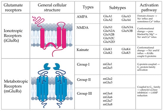
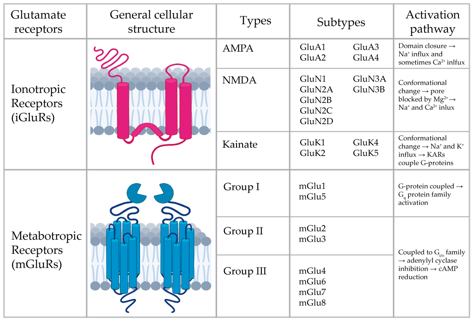
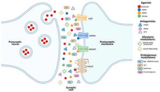
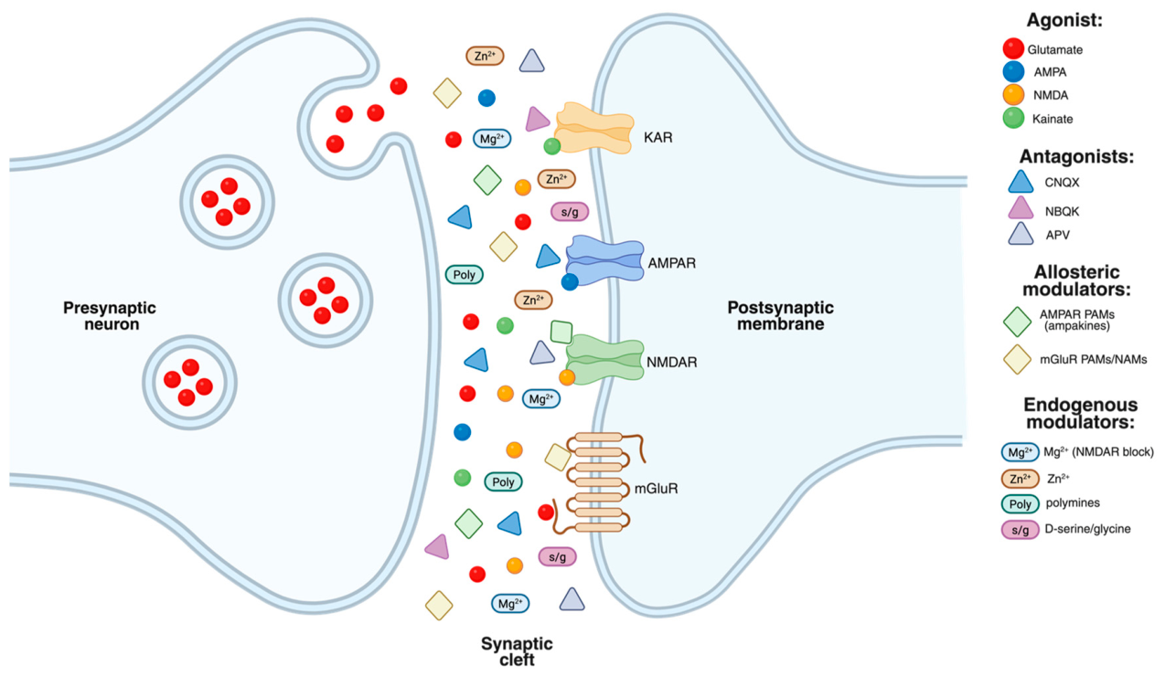
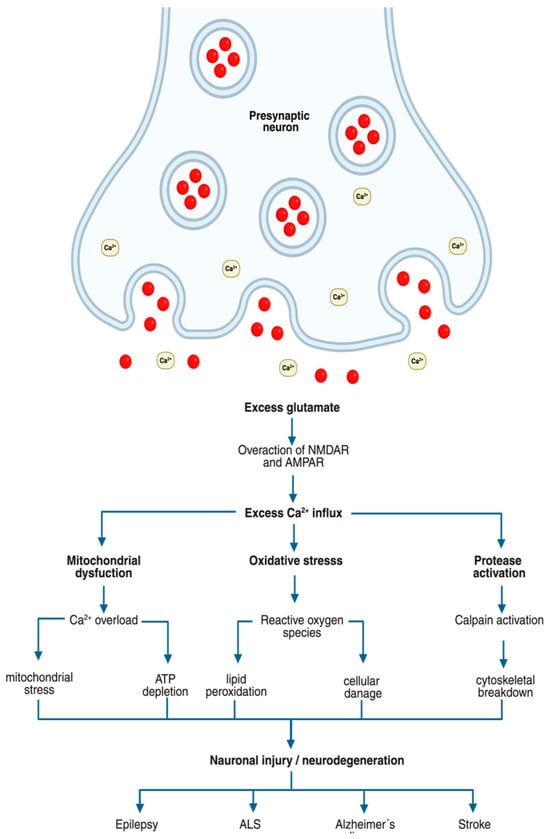
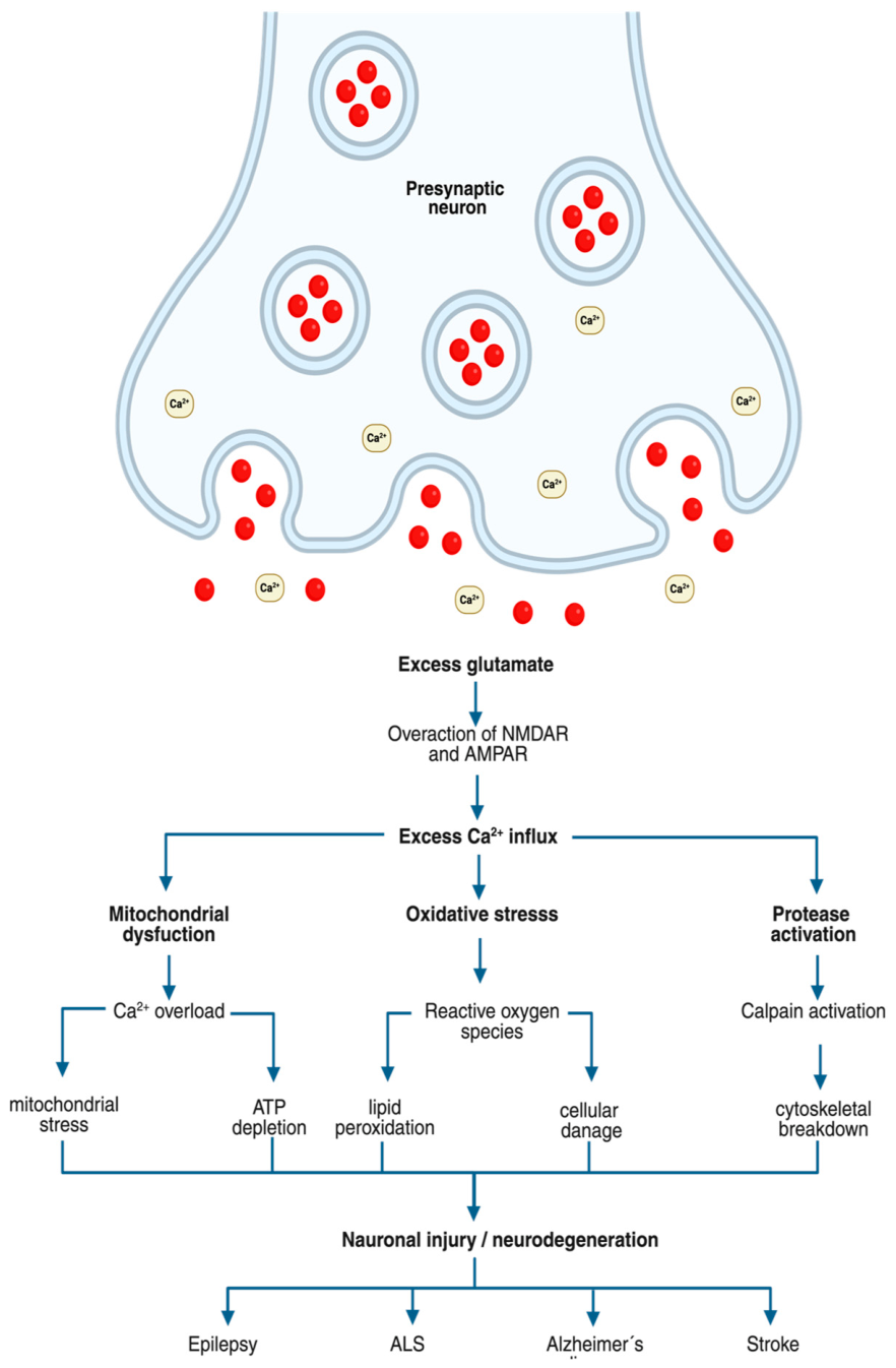

Disclaimer/Publisher’s Note: The statements, opinions and data contained in all publications are solely those of the individual author(s) and contributor(s) and not of MDPI and/or the editor(s). MDPI and/or the editor(s) disclaim responsibility for any injury to people or property resulting from any ideas, methods, instructions or products referred to in the content. |
© 2026 by the authors. Licensee MDPI, Basel, Switzerland. This article is an open access article distributed under the terms and conditions of the Creative Commons Attribution (CC BY) license.