Article

Robust Intrinsic Dorsoventral Organization of Hippocampal Sharp Wave–Ripples Persists During Cannabinoid Modulation


by
Athina Miliou
1,
Panagiota Giannakopoulou
1,
Agathi Erda
1,
Ioanna-Alexia Tsiokou
1,
Eleni-Despoina Mavriki
1,
Giota Tsotsokou
1,
Ioanna-Maria Sotiropoulou
1 and
Costas Papatheodoropoulos
1,2,*
1
Laboratory of Physiology, Department of Medicine, University of Patras, 26504 Rio, Greece
2
Laboratory of Experimental Animals, School of Health Sciences, University of Patras, 26504 Rio, Greece
*
Author to whom correspondence should be addressed.
Receptors 2026, 5(3), 30; https://doi.org/10.3390/receptors5030030
Submission received: 10 April 2026 / Revised: 21 July 2026 / Accepted: 14 September 2026 / Published: 17 September 2026

Abstract

Background/Objectives: The hippocampus exhibits pronounced functional and physiological heterogeneity along its dorsoventral axis. Sharp wave–ripple (SWR) complexes are highly organized hippocampal network events that depend on coordinated excitation and inhibition and contribute to memory processing. This study investigated whether modulation of the cannabinoid system differentially affects SWRs and associated neuronal activity in the dorsal and ventral hippocampus. Methods: Extracellular field potentials and multiunit activity (MUA) were recorded from the CA1 stratum pyramidale of acute dorsal and ventral rat hippocampal slices. Spontaneous SWRs were analyzed under control conditions and following application of the cannabinoid type 1 (CB1) receptor agonists ACEA and WIN55,212-2, cannabidiol (CBD), and the G-protein-gated inwardly rectifying potassium channel blocker tertiapin-Q. CB1 receptor expression was assessed in dorsal and ventral CA3 using Western blot analysis. Results: Baseline recordings revealed pronounced dorsoventral differences in SWR dynamics. Ventral slices exhibited shorter inter-event intervals, larger sharp-wave amplitudes, higher ripple frequency and power, and greater SWR-associated neuronal activity than dorsal slices. Under the present experimental and analytical conditions, ACEA, WIN55,212-2, and CBD did not produce statistically significant changes in any of the examined SWR or MUA parameters. Tertiapin-Q selectively increased ripple power but did not reveal cannabinoid sensitivity when followed by WIN55,212-2 application. CB1 receptor expression was comparable between dorsal and ventral CA3. Conclusions: Despite marked intrinsic dorsoventral differences in SWR dynamics, acute cannabinoid-system modulation produced no statistically detectable changes in most SWR and MUA parameters examined. These findings indicate that the intrinsic dorsoventral organization of hippocampal SWR activity is robust and remains preserved during acute cannabinoid-system modulation under the present experimental conditions.

1. Introduction

Sharp wave–ripples (SWRs) are transient hippocampal population events characterized by a large sharp wave in the local field potential (LFP) accompanied by fast ripple oscillations (140–250 Hz), and represent one of the most synchronous patterns in the mammalian brain [1]. They occur predominantly during quiet wakefulness and non-REM sleep and are tightly linked to the reactivation of hippocampal neuronal ensembles, thereby supporting memory consolidation, memory-guided behavior, and hippocampal–cortical communication [1,2,3]. The generation of SWRs depends on precisely coordinated interactions between excitatory inputs, primarily arising from CA3, and local inhibitory interneuron networks in CA1. Consequently, even subtle shifts in excitation/inhibition (E/I) balance can profoundly alter SWR occurrence and structure, making SWRs a sensitive readout of hippocampal network dynamics and neuromodulatory influences.
The endocannabinoid system constitutes a major neuromodulatory system regulating synaptic transmission, circuit dynamics, network oscillations, and diverse brain functions, including cognition, learning, memory, and emotional processing [4,5,6]. Endocannabinoids act primarily through cannabinoid type 1 (CB1) receptors, which are abundantly expressed in the hippocampus, predominantly on presynaptic terminals of GABAergic interneurons, particularly cholecystokinin-positive basket cells, and, to a lesser extent, on glutamatergic terminals [4,7,8]. CB1 receptors are primarily coupled to Gi/o proteins and modulate several intracellular signaling pathways, including the inhibition of adenylyl cyclase, suppression of voltage-gated Ca2+ channels, and activation of G-protein-gated inwardly rectifying K+ (GIRK) channels. Through these mechanisms, CB1 receptor activation reduces neurotransmitter release and dynamically regulates the E/I balance of hippocampal circuits [9]. Classical forms of endocannabinoid signaling, such as depolarization-induced suppression of inhibition or excitation, exemplify this activity-dependent retrograde control of synaptic transmission [10].
Although the hippocampus represents a major target of endocannabinoid modulation, CB1 receptors are widely distributed throughout the CNS, including the cerebral cortex and cerebellum, where they contribute to the regulation of cognitive, sensorimotor, and motor functions [11]. Consequently, synthetic cannabinoids, which often exhibit high affinity and efficacy at CB1 receptors, exert region-specific effects across multiple brain circuits. For example, repeated exposure to AKB48 has been associated with CB1 receptor downregulation and persistent molecular alterations in the prefrontal cortex and cerebellum, whereas JWH-018 induces CB1 receptor-dependent behavioral and neuroplastic changes involving cortical, hippocampal, striatal, and cerebellar regions [11,12,13]. These findings underscore the broad CNS actions of cannabinoids and provide a wider context for investigating cannabinoid modulation of hippocampal network activity.
Pharmacological activation of CB1 receptors has been reported to alter hippocampal network oscillations associated with cognitive function. In vivo studies have shown that cannabinoids such as Δ9-tetrahydrocannabinol (THC) and CP55,940 reduce theta and gamma oscillations and impair hippocampus-dependent behavior [14]. In vitro studies further indicate that CB1 receptor activation can modulate intrinsic hippocampal rhythms, including gamma oscillations and SWRs, presumably through presynaptic inhibition of excitatory transmission and altered interneuron–pyramidal coordination [15,16,17]. However, the magnitude and consistency of these effects vary across experimental conditions, and their dependence on circuit context and analytical approach remains incompletely understood. These inconsistencies may reflect differences in experimental preparation, recording conditions, pharmacological protocols, analytical approaches, and the hippocampal regions examined.
In contrast to CB1 receptor agonists, cannabidiol (CBD) is a non-intoxicating phytocannabinoid with a more complex and indirect mode of action. CBD exhibits low affinity for the orthosteric CB1 receptor site but can act as a negative allosteric modulator, reducing the efficacy and potency of CB1 receptor agonists [18,19,20]. In addition, CBD interacts with multiple molecular targets, including the 5-HT1A, TRPV1, and GPR55 receptors, thereby exerting context-dependent effects on neuronal excitability [20,21]. Despite the rapidly expanding therapeutic use of CBD and growing interest in its neuromodulatory actions, comparatively little is known about how CBD influences hippocampal network oscillations. In particular, it remains unclear whether CBD directly modulates SWRs or alters their properties indirectly through interactions with CB1 receptor-dependent mechanisms.
An additional and critical dimension in understanding cannabinoid effects on hippocampal function is the pronounced functional and anatomical heterogeneity along the dorsoventral (septotemporal) axis of the hippocampus. Although functional specialization along the hippocampal longitudinal axis has traditionally been described as a dorsal–ventral dichotomy, accumulating evidence indicates that this organization is better viewed as a continuum comprising functional gradients and partially overlapping domains [22,23,24,25,26]. These functional specializations are accompanied by differences in connectivity patterns, intrinsic neuronal excitability, synaptic organization, and excitation/inhibition (E/I) balance along the longitudinal hippocampal axis [25,27,28]. Such regional specialization gives rise to distinct network dynamics, including differences in oscillatory activity and short-term neuronal responses. Importantly, SWRs themselves exhibit dorsoventral heterogeneity. Both in vivo and in vitro studies have reported differences in SWR incidence, waveform characteristics, and susceptibility to perturbations along the hippocampal axis [29,30,31]. These observations suggest that neuromodulatory systems, including the endocannabinoid system, may exert region-specific effects on SWR-generating circuits. Nevertheless, most previous studies examining cannabinoid effects on hippocampal oscillations have focused on a single hippocampal segment and have not systematically addressed dorsoventral differences.
Taken together, these considerations raise key unresolved questions: whether cannabinoid signaling differentially modulates SWR activity along the dorsoventral hippocampal axis, and whether different classes of cannabinoids, such as CB1 receptor agonists and CBD, exert distinct effects on SWR dynamics depending on regional circuit properties. Addressing these questions is essential for linking cellular and network-level cannabinoid actions to their differential impact on cognitive and emotional processes.
In the present study, we investigated the effects of cannabinoid signaling on SWR activity along the dorsoventral axis of the hippocampus. Using extracellular recordings from the CA1 region in dorsal and ventral hippocampal slices, we examined how CB1 receptor agonists and CBD influence SWR occurrence, waveform properties, and associated neuronal activity. Our aim was to determine whether cannabinoid modulation of hippocampal network dynamics differs between the dorsal and ventral hippocampus and how intrinsic circuit organization shapes these effects on SWR activity.

2. Materials and Methods

2.1. Experimental Animals and Hippocampal Slice Preparation

Male Wistar rats were obtained from the Animal Facility of the Medical School of the University of Patras (license No. EL-13-BIOexp-04) and housed under controlled environmental conditions (21 ± 1 °C; 12 h light/dark cycle) with ad libitum access to food and water. All procedures were conducted in accordance with European Directive 2010/63/EU and were approved by the Protocol Evaluation Committee of the Department of Medicine, University of Patras, and the Directorate of Veterinary Services of the Achaia Prefecture of the Western Greece Region (approval no. 5661/37, 18 January 2021). A total of 156 hippocampi from 78 rats were used. The required number of animals was estimated using G*Power 3.1 software. Animals were deeply anesthetized with diethyl ether (ChemLab NV, Zedelgem, Belgium) and decapitated using a guillotine. The brain was rapidly removed from the cranium and immersed in ice-cold (2–4 °C) artificial cerebrospinal fluid (ACSF) containing (in mM): 124 NaCl, 4 KCl, 2 CaCl2, 2 MgSO4, 26 NaHCO3, 1.25 NaH2PO4, and 10 glucose. The ACSF was continuously equilibrated with 95% O2 and 5% CO2 and maintained at pH 7.4. Following removal of the brain, the hippocampi were carefully dissected free from the surrounding tissue and positioned under direct visual control on the stage of a McIlwain tissue chopper. The dorsal (septal) and ventral (temporal) poles were identified from the anatomical orientation of the intact hippocampus. Transverse slices, 550 μm thick, were prepared perpendicular to the longitudinal hippocampal axis from the dorsal and ventral segments. Specifically, tissue located 0.5–3.5 mm from the dorsal and ventral ends of the hippocampus was used. Immediately after preparation, slices were transferred to a custom-made Plexiglas interface-type recording chamber and continuously perfused with oxygenated ACSF at a flow rate of approximately 1.5 mL/min. The recording temperature was maintained at 30 ± 0.5 °C, and the chamber atmosphere was humidified with a mixture of 95% O2 and 5% CO2. Recordings were initiated after a recovery period of at least 90 min.

2.2. Electrophysiology and Data Analysis

Spontaneous field potentials were recorded from the CA1 region of hippocampal slices, with electrodes positioned in the stratum pyramidale to monitor population activity. CA3 was examined only in the Western blot experiments assessing CB1 receptor expression, as described below. Recordings were obtained using carbon fiber electrodes under visual guidance and amplified, band-pass filtered (0.5 Hz–2 kHz), and digitized at 10 kHz for offline analysis. Spontaneous activity consisted of sharp wave–ripple complexes (SWRs) and multiunit activity (MUA). SWRs were identified in the CA1 pyramidal layer as large-amplitude events with superimposed high-frequency oscillations. Only slices displaying stable activity for at least 20 min were selected for further analysis. Events occurred either in isolation or in clusters, defined as sequences of SWRs separated by short inter-event intervals (intra-cluster interval, approximately 100 ms), as identified from the IEI distributions (Figure 1A). For SWR analysis, signals were downsampled to 1 kHz and low-pass filtered at 35 Hz to isolate the sharp-wave component (Figure 1B). Events were detected using threshold-based detection followed by visual verification. The following parameters were quantified: (i) SWR amplitude; (ii) inter-event interval (IEI); (iii) probability of cluster occurrence; (iv) ripple peak frequency; and (v) ripple power. All events meeting the predefined detection criteria were included in the SWR amplitude and IEI analyses, including closely spaced SWRs occurring within clusters. Ripple frequency and power were quantified following spectral analysis of local field potentials performed using fast Fourier transform (FFT) (Figure 1C). Power spectra were computed from continuous recordings using a Hanning window with a duration of 1.638 s, yielding a frequency resolution of approximately 0.61 Hz. Ripple peak frequency was defined as the frequency corresponding to the maximum power within the band between 75 and 250 Hz. Ripple power was quantified as the peak value of the power spectrum within the ripple range.
MUA was extracted from band-pass filtered signals (400 Hz–1.5 kHz) and detected using threshold-based spike identification with visual confirmation. MUA occurring between SWRs was defined as MUA-Base, while MUA occurring during SWRs was defined as MUA-SWR. MUA-Base was quantified as firing rate (spikes/s) during inter-SWR periods, whereas MUA-SWR was quantified as the peak firing rate derived from peri-event time histograms aligned to SWR peaks (Figure 1C). The temporal relationship between unit firing and SWR peak was quantified as MUA-Delay. For each slice, SWR and MUA parameters were quantified during control conditions, after drug application, and after washing out the drug, using stable recording periods (5 last minutes of each period). Drug effects were expressed as within-slice changes relative to the control.
The following drugs were used in this study: the potent and highly selective agonist of CB1 receptors N-(2-Chloroethyl)-5Z,8Z,11Z,14Z-eicosatetraenamide (ACEA), the potent aminoalkylindole cannabinoid receptor agonist [(3R)-2,3-dihydro-5-methyl-3-(4-morpholinylmethyl)pyrrolo-[1,2,3-de]-1,4-benzoxazin-6-yl]-1-naphthalenyl-methanone, monomethane sulfonate ((+)-WIN 55,212-2 (mesylate)), and the natural cannabinoid cannabidiol (2-[(1R,6R)-3-Methyl-6-(1-methylethenyl)-2-cyclohexen-1-yl]-5-pentyl-1,3-benzenediol, CBD), and Tertiapin Q (trifluoroacetate salt, TPN-Q). ACEA and CBD were purchased from Tocris Cookson Ltd., Bristol, UK; WIN 55,212-2 and Tertiapin-Q were purchased from Cayman Chemical Company, Michigan, USA. Also, CBD was kindly provided by Dr. Maria Chalampalaki (Faculty of Pharmacy, National and Kapodistrian University of Athens, Athens, Greece).
ACEA was applied at 5 nM, WIN55,212-2 at 10 μM, CBD at 50 μM, and TPN-Q at 50 nM. The drug concentrations used were selected based on previous electrophysiological studies demonstrating the biological activity of these compounds in hippocampal and other neural preparations [30,32,33]. All drugs were prepared as concentrated stock solutions and stored at −20 °C. ACEA was dissolved in ethanol, TPN-Q was dissolved in water, CBD was dissolved in DMSO, and WIN55,212-2 was dissolved in DMF. All drugs were diluted in ACSF immediately before use to obtain the required final concentrations. Following dilution in ACSF, the final concentrations of ethanol, DMF, and DMSO in the recording solution were 0.005%, 0.05%, and 0.02% (v/v), respectively. DMSO was additionally examined as a separate vehicle control at the concentration used in the CBD experiments.
Following acquisition of a stable baseline recording, drugs were bath-applied through continuously perfusing ACSF; no compound was applied directly onto the slice. Drug-containing ACSF was delivered to the interface recording chamber at the same flow rate as control ACSF (~1.5 mL/min). Each compound was applied for 30 min, and data obtained during the final 5 min of application were used for analysis. Drug application was followed, where applicable, by washout with drug-free ACSF. For the combined pharmacological protocol, TPN-Q was applied before WIN55,212-2, and WIN55,212-2 was subsequently applied in the continued presence of TPN-Q.

2.3. Immunoblotting

Because hippocampal SWRs are considered to arise primarily within recurrent CA3 circuitry before propagating to CA1, CB1 receptor expression was assessed in dorsal and ventral CA3. This analysis complemented our previous characterization of CB1 receptor expression in dorsal and ventral CA1 [32]. To assess CB1 receptor expression along the dorsoventral axis, the CA3 region was isolated from dorsal and ventral hippocampal slices and homogenized in 1% SDS containing protease inhibitors. Protein concentration was determined spectrophotometrically. Equal amounts of protein (25 μg per lane) were separated by SDS-PAGE and transferred onto PVDF membranes. Membranes were blocked with 5% non-fat milk in PBST and incubated overnight at 4 °C with primary antibodies against CB1 (rabbit monoclonal; 1:1000; Abcam, Cambridge, UK; Cat. No. ab259323; RRID: AB_3676209) and β-actin (mouse monoclonal; 1:10,000; Thermo Fisher Scientific, Waltham, MA, USA; Cat. No. MA5-15739; RRID: AB_10979409). After washing, membranes were incubated with appropriate HRP-conjugated secondary antibodies. Protein bands were visualized using enhanced chemiluminescence and imaged with a ChemiDoc MP system (Bio-Rad, Hercules, CA, USA). Band intensities were quantified using ImageLab 6.1 software, and CB1 expression was normalized to β-actin. Data are expressed as the ratio of CB1 to β-actin optical density for each sample.

2.4. Statistics

The experimental unit in this study was the hippocampal slice. Each slice was exposed to a single pharmacological condition, and comparisons were performed between control and drug application within the same slice. Data were analyzed using linear mixed-effects models (LMMs) to account for repeated measurements within slices. For each dependent variable (SWR and MUA parameters), fixed effects included Drug condition (control vs. drug), Hippocampal region (dorsal vs. ventral), and their interaction. A random intercept was included for slice to account for within-slice dependencies. The primary effect of interest was the interaction between Drug and Region (dorsal–ventral), indicating differential drug effects along the dorsoventral axis. When appropriate, post hoc comparisons were performed using estimated marginal means with correction for multiple comparisons. Because relatively few slices were obtained from each animal, animal identity was not included as a random effect, in line with standard practice in slice electrophysiology studies. Model assumptions, including normality of residuals and homogeneity of variance, were evaluated before interpretation of the results. For descriptive purposes, within-slice changes (Δ or percentage change from control) were also calculated. Plots display the median, interquartile range (25th–75th percentiles), mean, 5th and 95th percentiles, and outliers. Comparisons between the dorsal and ventral hippocampus across the measured variables were conducted using an independent-samples t-test. Statistical significance was set at p < 0.05.

3. Results

3.1. Baseline Characteristics of Sharp Wave–Ripples Along the Dorsoventral Hippocampal Axis

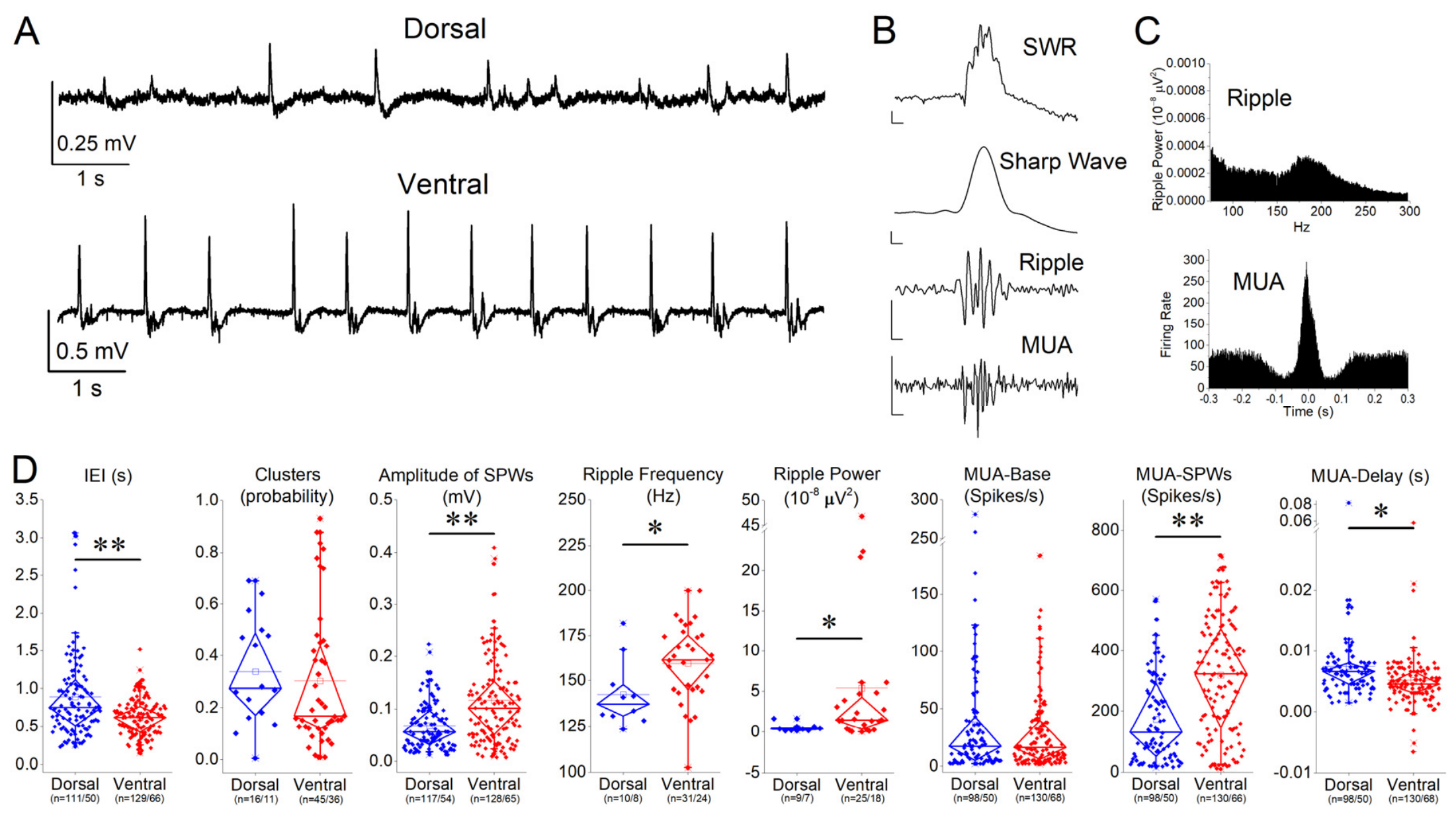
Spontaneous network activity in the CA1 region consisted of recurrent SWRs recorded from the stratum pyramidale and multiunit activity (MUA) occurring either during SWR events or independently (Figure 1A,B). Under control conditions, SWR complexes were recorded in both dorsal and ventral hippocampal slices included in the study (Figure 1A). Only slices exhibiting stable spontaneous SWR activity during the baseline recording period were included in the subsequent pharmacological experiments. Quantitative analysis, however, revealed pronounced differences in several SWR properties along the dorsoventral axis. The inter-event interval (IEI) was significantly shorter in the ventral compared with the dorsal hippocampus (independent samples t-test, p < 0.001), indicating a higher rate of spontaneous SWR generation in the ventral hippocampus (Figure 1D). In contrast, SWR cluster probability did not differ significantly between the dorsal and ventral hippocampal slices (p = 0.987), suggesting a similar temporal grouping of events despite differences in overall occurrence rate. Analysis of SWR waveform properties showed that sharp wave amplitude was significantly greater in the ventral slices (p < 0.001). In addition, both ripple frequency and ripple power were higher in the ventral compared to dorsal hippocampus (p = 0.045 and p = 0.034, respectively), indicating differences in the fast oscillatory component of SWRs along the dorsoventral axis.
Baseline neuronal firing was assessed using MUA (Figure 1B,D). MUA recorded outside SWRs (MUA-Base) did not differ between the dorsal and ventral hippocampus (p = 0.971), indicating comparable baseline excitability. In contrast, MUA associated with SWRs (MUA-SWR) was significantly higher in the ventral slices (p < 0.001), reflecting enhanced neuronal recruitment during SWR events. Furthermore, the timing of neuronal firing relative to the SWR peak (MUA-Delay) differed significantly between regions (p = 0.038), indicating altered temporal coordination of unit activity within SWRs along the dorsoventral axis.
Taken together, these results demonstrate that SWRs in the ventral hippocampus occur at a higher rate, exhibit larger amplitudes and stronger ripple components, and recruit more robust and temporally distinct neuronal firing compared to the dorsal hippocampus, while baseline firing rates remain comparable between regions.

3.2. Effects of ACEA on SWRs and Neuronal Activity

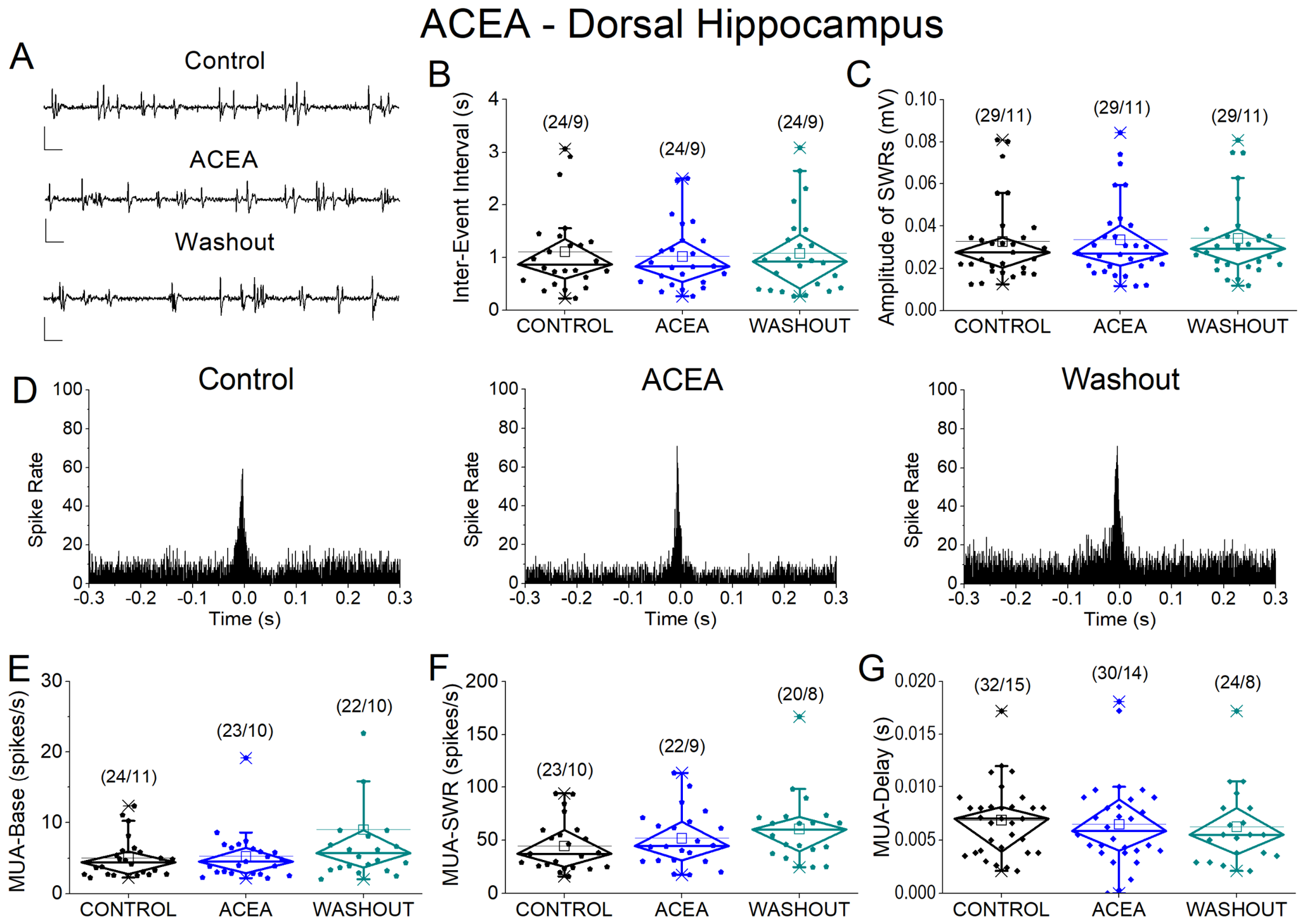
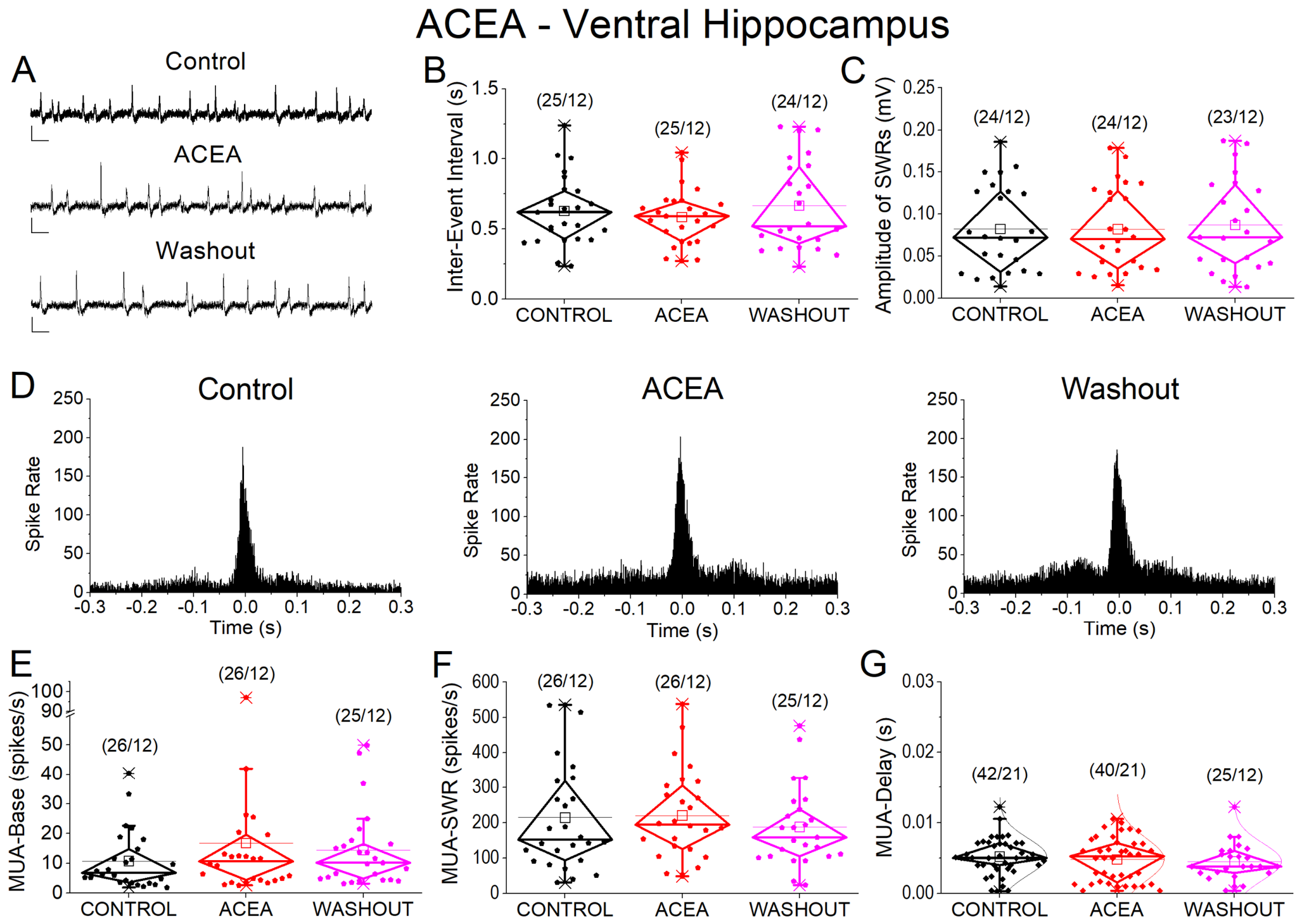
To investigate the effects of CB1 receptor activation on hippocampal network oscillations, slices were exposed to the selective CB1 receptor agonist ACEA (5 nM). Activation of CB1 receptors with ACEA did not significantly affect any of the examined properties of spontaneous SWRs or associated neuronal activity. LMM analysis revealed no effect of condition on inter-event interval (IEI), cluster probability, SWR amplitude, ripple frequency, or ripple power (all p > 0.5) either in the dorsal (Figure 2A–C) or ventral hippocampus (Figure 3A–C). Similarly, ACEA did not alter baseline multiunit activity (MUA-Base), SWR-associated firing rate (MUA-SWR), or the temporal delay of neuronal firing relative to SWR peaks (MUA-Delay) (all p > 0.2) either in the dorsal (Figure 2E–G) or ventral hippocampus (Figure 3E–G). No significant interactions between condition and region were observed for any parameter (all p > 0.7), indicating that CB1 receptor activation did not differentially affect the dorsal and ventral hippocampal slices. In contrast, significant main effects of hippocampal segment were consistently observed for several variables, including IEI, SWR amplitude, and MUA measures (all p < 0.05), confirming robust dorsoventral differences in hippocampal network activity.

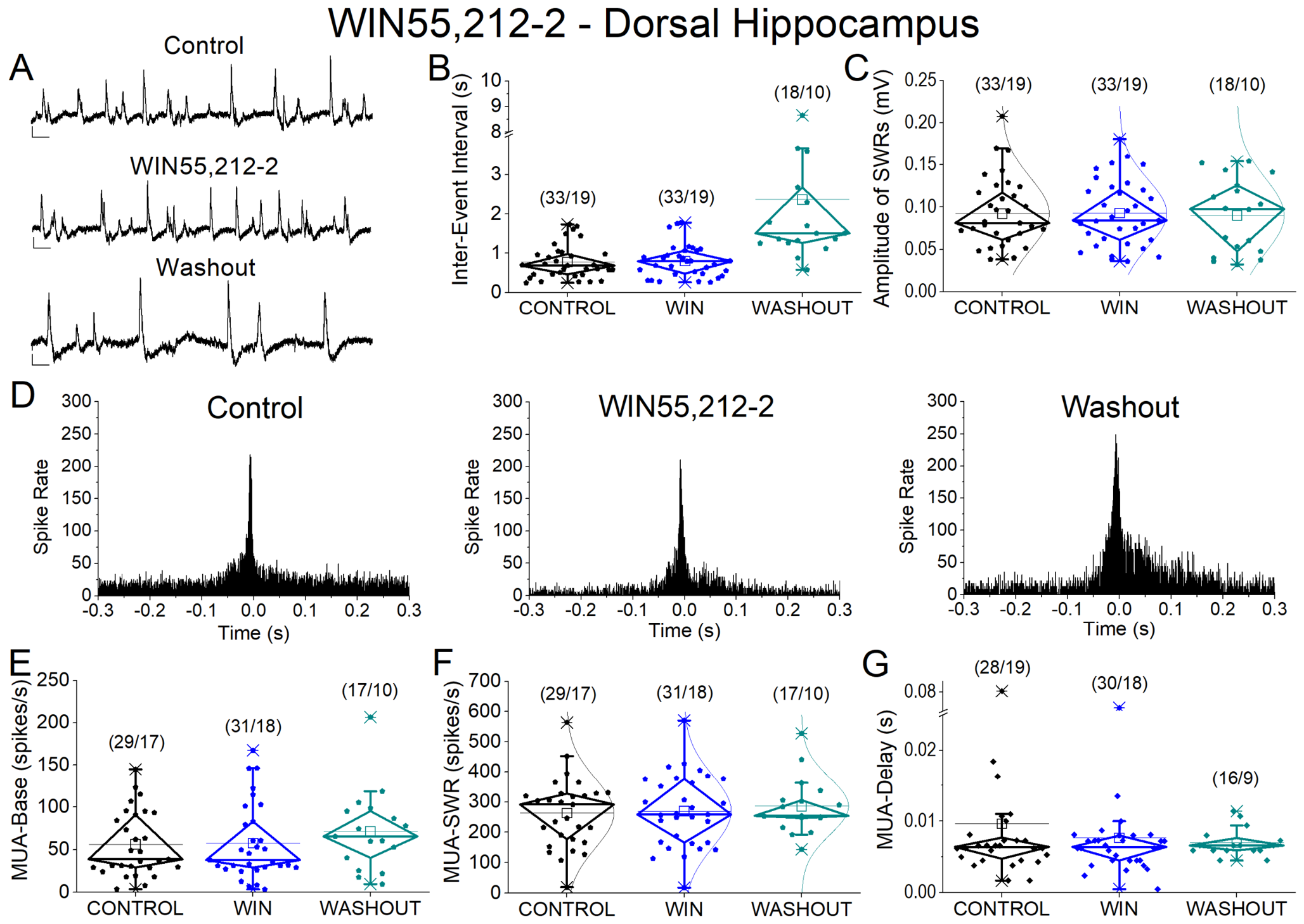
3.3. Effects of WIN 55,212-2 on SWRs and Neuronal Activity

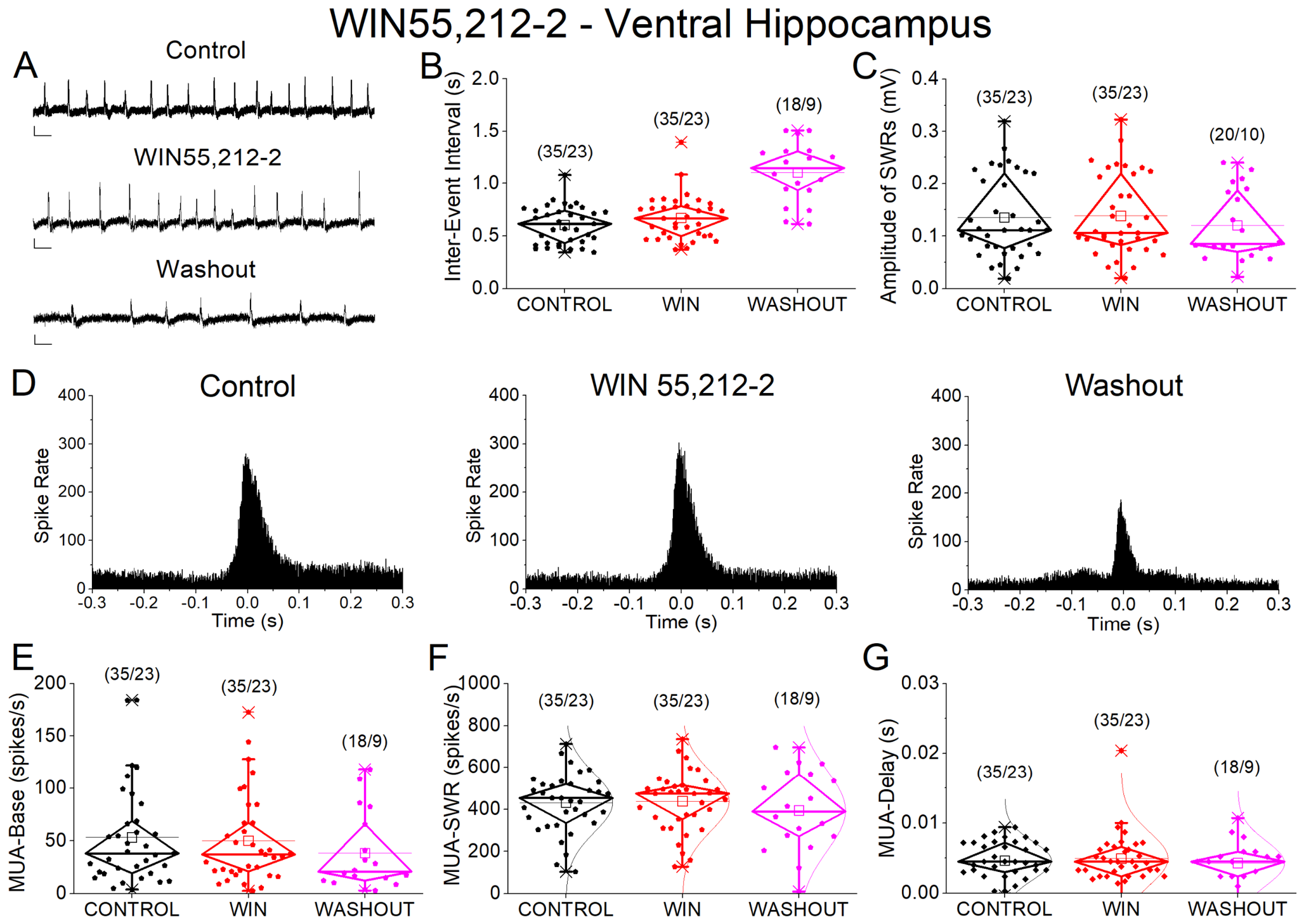
Application of WIN 55,212-2 (10 μM) did not significantly alter the SWR properties or neuronal firing either in the dorsal (Figure 4A–G) or ventral hippocampus (Figure 5A–G). No significant effects of condition were observed on IEI, cluster probability, sharp wave amplitude, MUA-Base, MUA-SWR, or MUA-Delay (all p > 0.5), and no condition × region interactions were detected (all p > 0.4). Significant main effects of hippocampal segment persisted for several parameters, including IEI, amplitude, and MUA-SWR (all p < 0.05), indicating that dorsoventral differences in network dynamics remained unaffected by WIN. The number of observations differed among some outcome variables because a clearly identifiable ripple component was not present in every slice under all experimental conditions, particularly in the dorsal hippocampal recordings. Therefore, ripple frequency and ripple power were analyzed only when the ripple component could be reliably quantified. Missing values were not imputed, and the linear mixed-effects mode was fitted using the available valid observations for each outcome. The number of slices and animals included in each analysis are indicated in the corresponding figures.

3.4. Effects of Vehicle (DMSO) on SWRs and Neuronal Activity

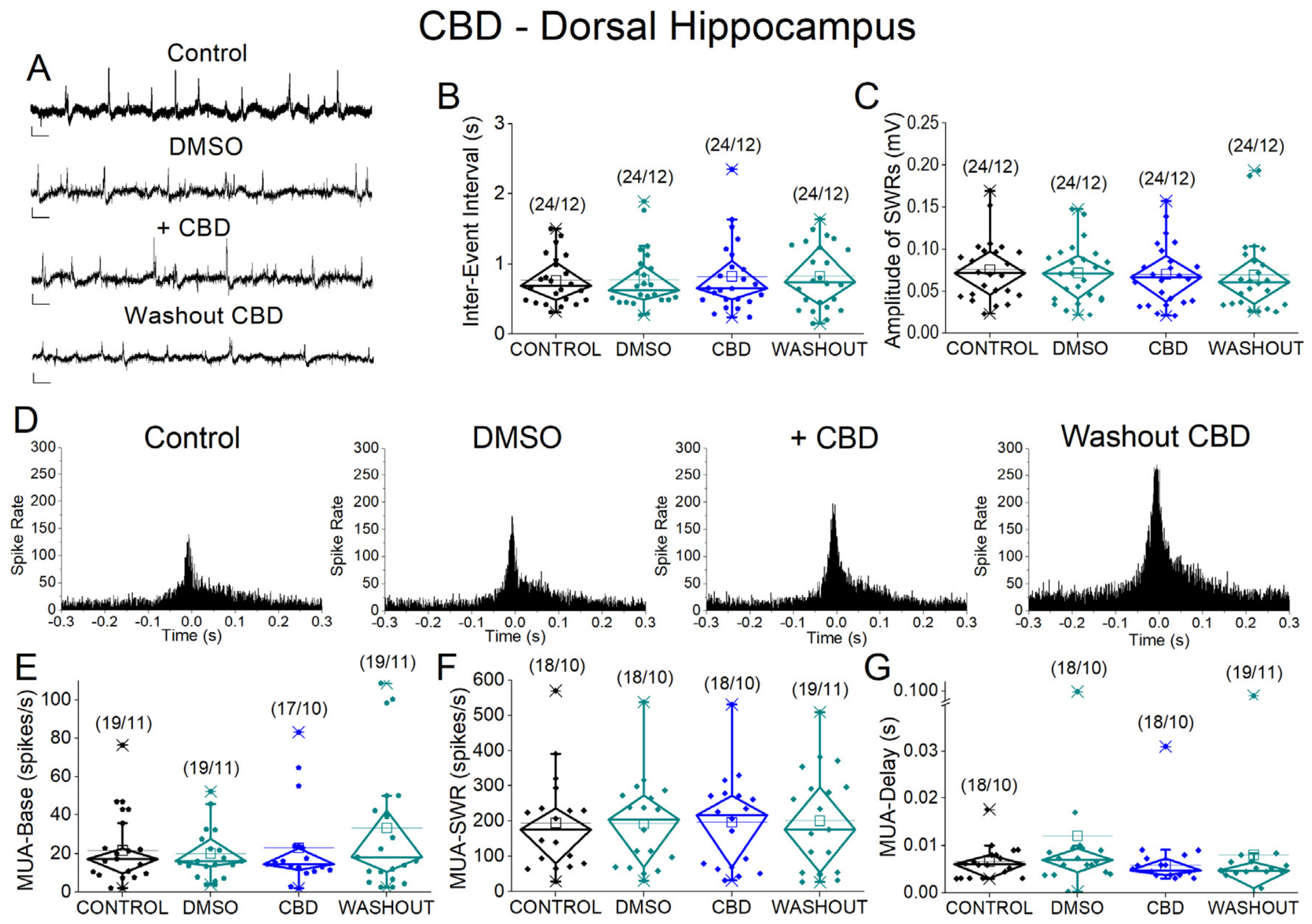
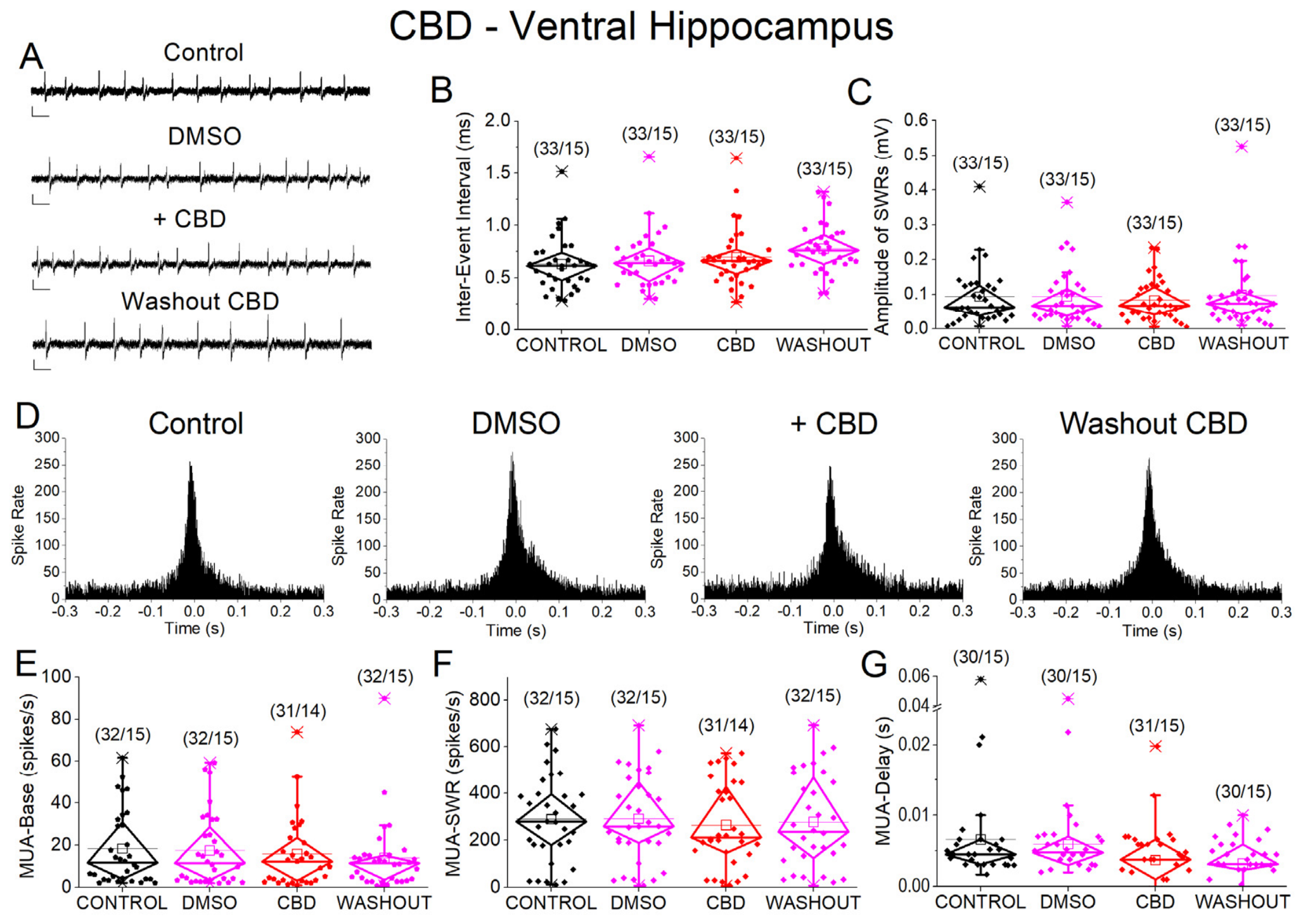
Because CBD was dissolved in DMSO, we first assessed whether the vehicle itself affected spontaneous network activity. Application of DMSO (0.02%) did not significantly alter any of the examined SWR or MUA parameters in either segment of the hippocampus (dorsal hippocampus, Figure 6A–G; ventral hippocampus, Figure 7A–G). More specifically, no significant main effects of condition were observed for IEI, cluster probability, amplitude, ripple frequency, ripple power, MUA-Base, MUA-SWR, or MUA-Delay (all p > 0.25), and no condition × region interactions were detected. In contrast, significant dorsoventral differences persisted for several parameters, including IEI, cluster probability, ripple power, and MUA-SWR (all p < 0.05), indicating that vehicle application does not interfere with the intrinsic organization of hippocampal network activity.

3.5. Effects of Cannabidiol (CBD) on Hippocampal Sharp Wave–Ripples

We next examined whether CBD influences hippocampal network oscillations associated with SWRs. Compared with the vehicle (DMSO), CBD (50 μM) did not significantly affect SWR dynamics or neuronal activity. Specifically, LMM analysis revealed no significant effect of condition on IEI, cluster probability, SWR amplitude, ripple frequency, ripple power, MUA-Base, MUA-SWR, or MUA-Delay (all p > 0.08) in either the dorsal (Figure 6A–G) or ventral hippocampus (Figure 7A–G). No significant condition × region interactions were observed for any parameter, indicating that CBD did not differentially influence dorsal and ventral hippocampal activity. These findings indicate that CBD produced no statistically significant effects on spontaneous SWRs or associated neuronal firing under the present experimental conditions.

3.6. Effects of TPN-Q on SWR and Neuronal Activity

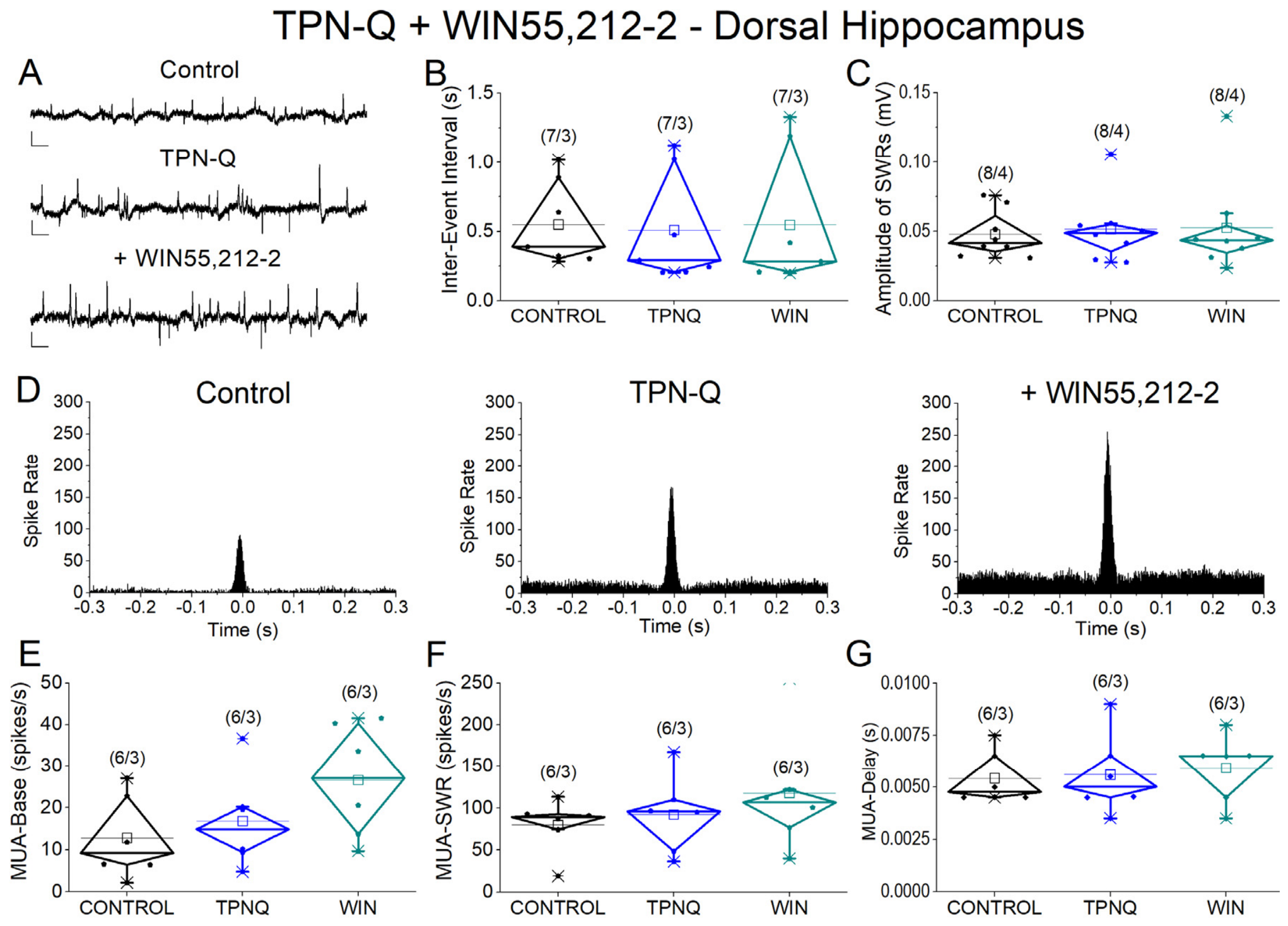
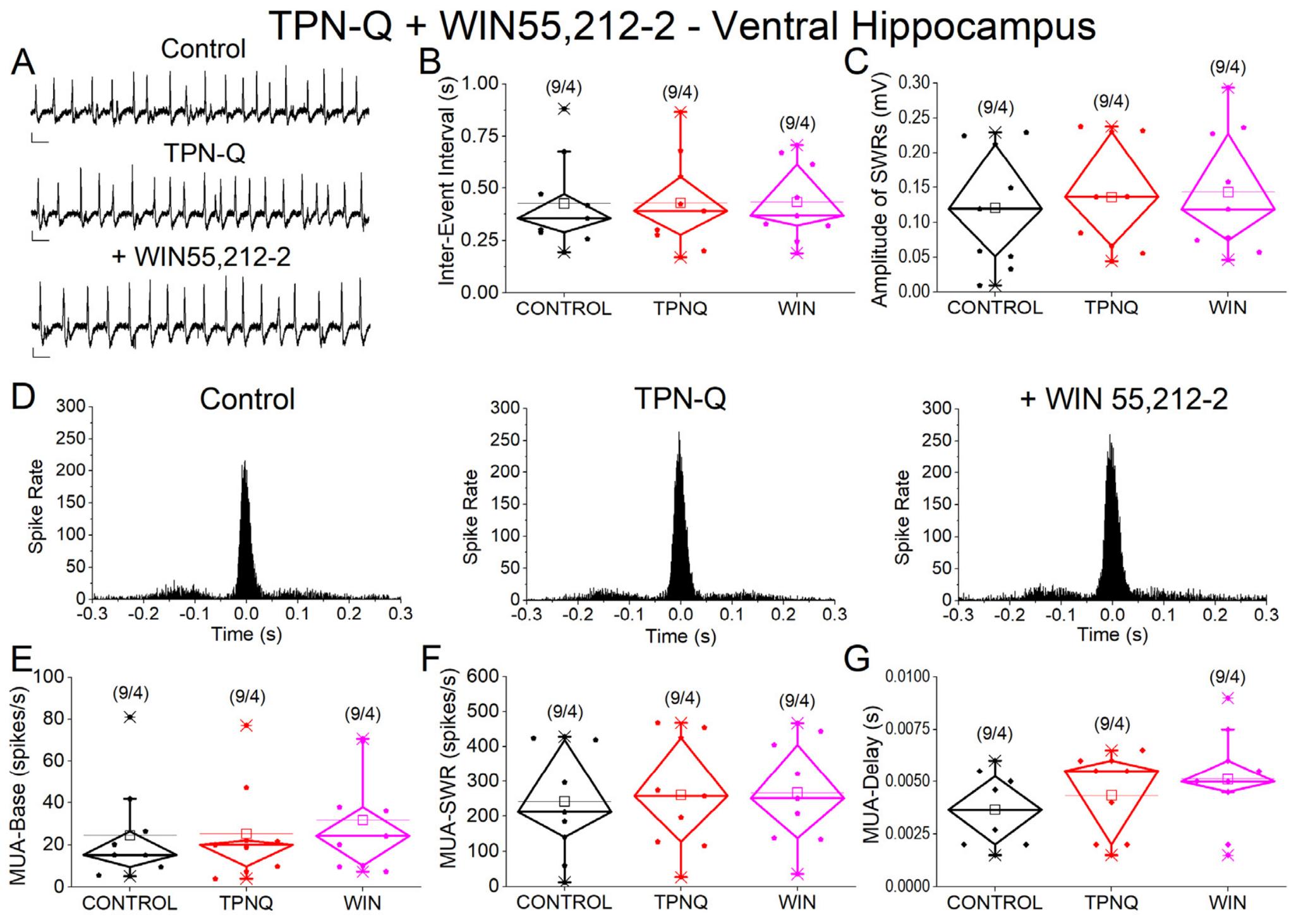
To assess the contribution of GIRK-dependent conductances to hippocampal network dynamics, TPN-Q was applied prior to cannabinoid receptor activation. TPN-Q (50 nM) did not significantly affect IEI, cluster probability, SWR amplitude, ripple frequency, MUA-Base, MUA-SWR, or MUA-Delay (all p > 0.5) in either the dorsal (Figure 8A–G) or ventral hippocampus (Figure 9A–G), and no condition × region interactions were observed. In contrast, TPN-Q significantly increased ripple power (p = 0.003), indicating a selective effect on the high-frequency oscillatory component of SWRs. These results indicate that GIRK channel blockade selectively increased ripple power without significantly affecting SWR occurrence or neuronal recruitment.

3.7. Effects of WIN on SWR and Neuronal Activity in the Presence of TPN-Q

To determine whether GIRK channel blockade modifies the effects of cannabinoid receptor activation, WIN 55,212-2 was applied in the presence of TPN-Q. Under these conditions, WIN did not significantly affect any of the examined properties of spontaneous SWRs or associated neuronal activity. Specifically, LMM analysis revealed no significant effect of condition on inter-event interval (IEI), cluster probability, SWR amplitude, ripple frequency, or ripple power (all p > 0.5) in either the dorsal (Figure 8A–G) or the ventral hippocampus (Figure 9A–G). Similarly, WIN did not alter the baseline multiunit activity (MUA-Base), SWR-associated firing rate (MUA-SWR), or the temporal delay of neuronal firing relative to SWR peaks (MUA-Delay) (all p > 0.4) in either segment of the hippocampus. No significant interactions between condition and region were observed for any parameter, indicating that no evidence was found that GIRK channel blockade altered the effects of WIN in either the dorsal or ventral hippocampus. In contrast, significant main effects of hippocampal segment persisted for several parameters, including SWR amplitude, ripple power, and MUA-SWR (all p < 0.05), confirming robust dorsoventral differences in hippocampal network activity that were not influenced by the combined application of TPN-Q and WIN.

3.8. CB1 Receptor Expression Is Comparable Between Dorsal and Ventral Hippocampus

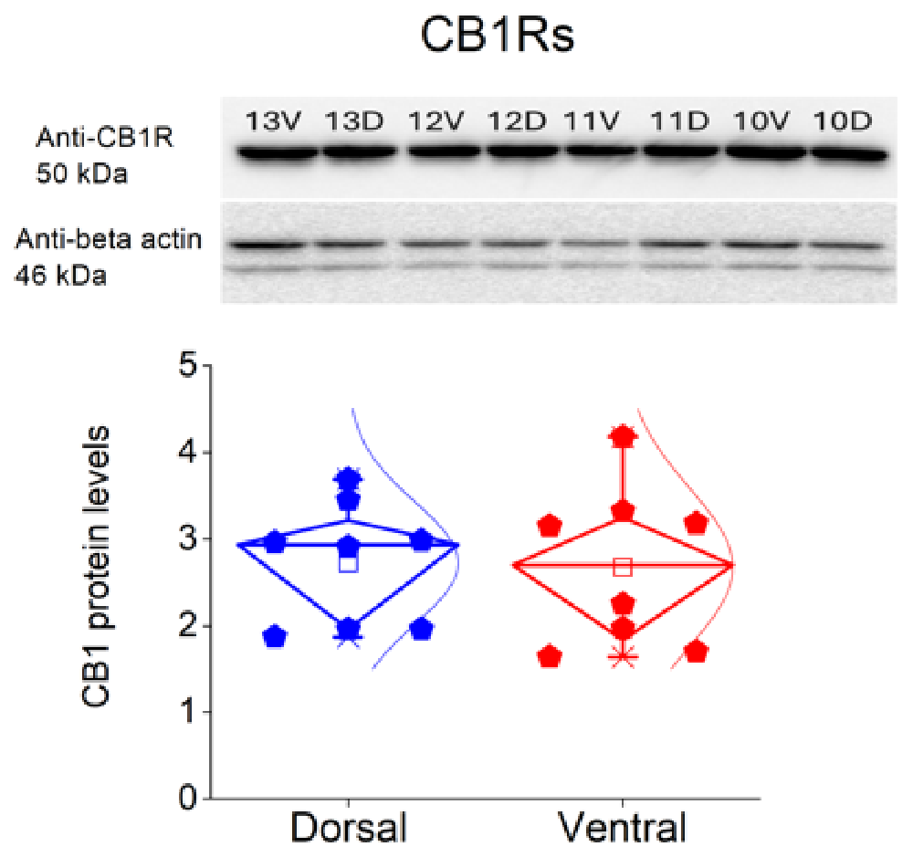
To determine whether differences in CB1 receptor expression might contribute to the observed dorsoventral differences in SWR activity, we compared CB1 receptor levels in dorsal and ventral CA3 using Western blot analysis. Quantitative analysis revealed no significant difference in CB1 receptor expression between the dorsal and ventral hippocampal samples (paired t-test, t = −0.31, p = 0.772; Figure 10). These findings indicate that the functional differences in SWR dynamics along the dorsoventral axis are unlikely to arise from differences in the overall abundance of CB1 receptors. Instead, they may reflect region-specific variations in receptor localization, circuit organization, or downstream signaling mechanisms.

4. Discussion

The present study demonstrates pronounced dorsoventral differences in spontaneous SWR activity in hippocampal slices. These findings are broadly consistent with previous in vitro studies reporting enhanced SWR activity and neuronal recruitment in the ventral hippocampus [29,30,31]. However, in contrast to in vivo studies reporting larger sharp-wave amplitudes in the dorsal hippocampus [34,35], the present data showed greater amplitudes in the ventral slices. This discrepancy may reflect methodological differences, as in vivo recordings preserve extrahippocampal inputs and longitudinal hippocampal connectivity, whereas transverse slice preparations isolate local circuitry [36,37]. Accordingly, slice recordings primarily reflect intrinsic dorsoventral differences in local network dynamics, whereas in vivo measurements capture the integrated activity of the longitudinal hippocampal system [34,38].
Despite these robust baseline dorsoventral differences, no statistically significant effects of cannabinoid-related compounds were detected in most of the SWR and MUA parameters examined under the present experimental and analytical conditions in either hippocampal segment. Specifically, neither selective CB1 receptor activation (ACEA), non-selective cannabinoid receptor activation (WIN55,212-2), CBD, nor GIRK channel blockade (TPN-Q) significantly altered the intrinsic dorsoventral organization of SWR activity. Together, these findings indicate that dorsoventral differences in SWR activity were maintained across all pharmacological conditions examined, whereas acute cannabinoid modulation produced no statistically significant effects on the measured SWR and MUA parameters under the present experimental and analytical conditions.
Previous studies have reported that cannabinoid receptor activation suppresses hippocampal oscillations and disrupts SWRs, reducing their incidence, ripple power, and associated neuronal recruitment [14,16,17,39]. In contrast, the present study did not detect significant effects of CB1 receptor activation or CBD on the measured SWR and MUA variables. This apparent discrepancy may reflect, at least in part, differences in pharmacological specificity and analytical approaches.
From a pharmacological perspective, studies employing endogenous cannabinoids such as anandamide may involve the activation of multiple targets beyond CB1 receptors, including TRPV1 channels [17], resulting in more complex and variable effects on hippocampal network activity. In contrast, the present study primarily examined selective CB1 receptor activation (ACEA), non-selective cannabinoid receptor activation (WIN55,212-2), and CBD, which may engage distinct signaling pathways. Methodological differences may also contribute to the discrepant findings. In several previous reports, the observed cannabinoid-induced changes were relatively modest in magnitude, and their detection may therefore depend on the statistical approach employed. Paired analyses emphasize within-slice changes, whereas linear mixed-effects models explicitly account for both within- and between-slice variability, potentially influencing the detection of subtle effects.
Taken together, previously reported alterations in SWR dynamics may arise from a combination of pharmacological differences, CB1-dependent and non-canonical signaling mechanisms, and differences in analytical methodology. Overall, under the present experimental and analytical conditions, no statistically significant effects of acute cannabinoid modulation were detected on the measured SWR and MUA parameters.
A central finding of this study is the persistence of strong dorsoventral differences in SWR dynamics across all experimental conditions. The ventral hippocampus exhibited higher SWR rates, larger amplitudes, and stronger neuronal recruitment, consistent with previous in vitro work [29,30,31]. These differences likely reflect intrinsic variations in circuit organization, synaptic dynamics, and excitation–inhibition balance along the hippocampal axis. Importantly, these regional characteristics were preserved following cannabinoid receptor activation and GIRK channel blockade under the present experimental conditions, indicating that acute pharmacological manipulation of these pathways did not measurably alter dorsoventral differences in SWR dynamics. Together, these findings suggest that the intrinsic dorsoventral organization of SWR dynamics is maintained despite acute cannabinoid-related pharmacological manipulation.
Blockade of GIRK channels with TPN-Q produced only limited effects on the SWR dynamics, with the exception of a selective modulation of ripple power. This finding suggests that GIRK-dependent conductances may contribute to the fine-tuning of high-frequency oscillatory synchronization, without playing a major role in SWR generation or neuronal recruitment. Notably, in a previous study [30], GIRK blockade increased the SWR rate in the dorsal hippocampus and enhanced the probability of clustered events, indicating a role in the temporal organization of network activity. The absence of such effects in the present dataset may reflect differences in analytical approach and experimental design. Interestingly, GIRK blockade did not reveal any latent sensitivity of SWRs to cannabinoid receptor activation, as combined application of TPN-Q and WIN also failed to alter the SWR or MUA parameters. These findings do not support the hypothesis that GIRK channel activity masks acute cannabinoid effects on SWR dynamics under the present experimental conditions.
Comparable CB1 receptor expression in dorsal and ventral CA3 suggests that differences in receptor abundance within the principal SWR-generating region are unlikely to account for the observed dorsoventral differences in SWR dynamics. Rather, they may be related to regional differences in circuit architecture, synaptic organization, or downstream signaling pathways. Although receptor expression was quantified in CA3, cannabinoid modulation of SWR-associated activity is likely to depend on coordinated signaling across the CA3–CA1 network [1]. Notably, a similar dissociation between receptor expression and functional output has been reported in CA1 hippocampal circuits, where CB1 receptor levels were comparable between dorsal and ventral regions despite pronounced differences in network excitability and short-term neuronal dynamics [32], supporting the idea that dorsoventral functional specialization of cannabinoid signaling may depend more on circuit-level and intracellular mechanisms than on receptor abundance alone. However, similar receptor expression does not exclude regional differences in receptor localization, coupling efficiency, downstream signaling, or cell-type-specific expression. For example, CB1 receptors are expressed predominantly by cholecystokinin (CCK)-positive GABAergic interneurons, whereas parvalbumin (PV)-positive basket cells, which are thought to play a central role in ripple synchronization, express little or no CB1 receptor [6,40,41,42]. Consequently, acute cannabinoid receptor activation may preferentially modulate CCK-dependent inhibitory transmission while largely preserving the PV-mediated inhibitory network underlying ripple generation. Although the present study did not directly examine defined interneuron populations, this cell-type-specific organization may partly account for the limited cannabinoid effects observed on the SWR and MUA parameters examined.
Taken together, these findings suggest that CB1 receptor expression alone is not sufficient to predict the functional modulation of network activity, particularly in the context of intrinsically generated oscillations such as SWRs. SWRs are widely implicated in memory consolidation, replay, and hippocampal–cortical communication [1]. Disruption of SWRs by cannabinoids has been proposed as a mechanism underlying cannabinoid-induced memory impairment [2,3,14,16]. The present findings suggest that such effects may depend critically on network state and circuit context, and may involve mechanisms beyond the direct modulation of intrinsic SWR-generating circuits. Instead, cannabinoid-induced alterations in memory may involve the modulation of input pathways, network coordination, or large-scale brain dynamics, rather than local SWR generation per se. This distinction is important for understanding how cannabinoids influence hippocampal function in vivo and for interpreting their cognitive and behavioral effects.
Accordingly, the present findings should not be interpreted as evidence that intrinsic SWR-generating circuits are generally insensitive or resistant to cannabinoid modulation. Rather, under the present experimental and analytical conditions, no statistically significant effects of cannabinoid-related compounds were detected in the SWR and MUA parameters examined, with the exception of the increase in ripple power following TPN-Q application. Effects on other aspects of hippocampal network function, including synaptic currents, cell-type-specific activity, or the temporal and neuronal organization of SWR-associated ensembles, were not directly assessed and therefore cannot be excluded.
Several limitations should be considered when interpreting the present findings. The experiments were performed in acute hippocampal slices, a preparation that lacks long-range connections, interactions with other brain regions, and the broader neuromodulatory influences present in vivo. Consequently, the present findings should not be extrapolated directly to hippocampal network activity or hippocampus-dependent behavior in the intact brain. In addition, only acute pharmacological manipulations were examined, and therefore the receptor, synaptic, and network adaptations associated with chronic cannabinoid exposure were not addressed. Finally, all experiments were conducted in male rats; thus, the extent to which the present findings generalize to females remains unknown. Although our group has recently investigated sex-related differences in hippocampal physiology in dedicated studies [31], the influence of sex on the cannabinoid modulation of SWRs was beyond the scope of the present work. Future studies should determine whether cannabinoid modulation of SWRs differs between sexes, across hippocampal subfields, and during development or aging. Combining in vivo recordings with behavioral approaches will also be important for establishing the functional significance of these network dynamics.

5. Conclusions

These findings suggest that acute cannabinoid modulation does not measurably alter the intrinsic dorsoventral organization of spontaneous SWR activity under the present experimental conditions, and that cannabinoid effects on hippocampal function may involve mechanisms beyond local SWR generation.

Author Contributions

Conceptualization, C.P.; investigation, A.M., P.G., A.E., I.-A.T., E.-D.M., G.T. and I.-M.S.; formal analysis, A.M., P.G., A.E., I.-A.T., E.-D.M., G.T. and I.-M.S., C.P.; resources, C.P.; writing—original draft preparation, C.P.; writing—review and editing, A.M. and C.P.; supervision, C.P.; project administration, C.P.; funding acquisition, C.P. All authors have read and agreed to the published version of the manuscript.

Funding

The research project was supported by the Empeirikeion Foundation (#10). A.M. is a recipient of a postgraduate fellowship from the Hellenic Foundation for Research and Innovation (HFRI) (grant number: 19416). G.T. was financially supported by the “Polembros Shipping Limited”, as a recipient of a PhD fellowship.

Institutional Review Board Statement

The animal study protocol was approved by the Research Ethics Committee of the University of Patras and the Directorate of Veterinary Services of the Achaia Prefecture of Western Greece Region (reg. number: 5661/37, 18 January 2021). The treatment of animals and all experimental procedures used in this study were conducted in accordance with the European Communities Council Directive Guidelines for the Care and Use of Laboratory Animals (2010/63/EU—European Commission).

Informed Consent Statement

Not applicable.

Data Availability Statement

The minimal dataset supporting the conclusions of this study, including all variables used for statistical analysis, is available from the corresponding author upon reasonable request.

Acknowledgments

The authors would like to thank Leonidas Leontiadis and George Trompoukis for their valuable technical assistance in this study. The authors also gratefully acknowledge Maria Chalampalaki (Faculty of Pharmacy, National and Kapodistrian University of Athens) for the generous provision of cannabidiol, which was essential for the completion of this work.

Conflicts of Interest

The authors declare no conflicts of interest.

Abbreviations

The following abbreviations are used in this manuscript:
ACEAN-(2-Chloroethyl)-5Z,8Z,11Z,14Z-eicosatetraenamide
ACSFArtificial cerebrospinal fluid
CBDCannabidiol
CB1Cannabinoid receptor type 1
E/IExcitation/Inhibition balance
FFTFast Fourier transform
GIRKG-protein-gated inwardly rectifying potassium channels
IEIInter-event interval
LFPLocal field potential
LMMLinear mixed-effects model
MUAMultiunit activity
MUA-BaseBaseline multiunit activity
MUA-DelayDelay of multiunit activity relative to SWR peak
MUA-SWRSWR-associated multiunit activity
SWRSharp wave–ripple
TPN-QTertiapin-Q
WIN 55,212-2[(3R)-2,3-dihydro-5-methyl-3-(4-morpholinylmethyl)pyrrolo-[1,2,3-de]-1,4-benzoxazin-6-yl]-1-naphthalenylmethanone

References

  1. Buzsaki, G. Hippocampal sharp wave-ripple: A cognitive biomarker for episodic memory and planning. Hippocampus 2015, 25, 1073–1188. [Google Scholar] [CrossRef] [Scilit]
  2. Hall, A.F.; Wang, D.V. The two tales of hippocampal sharp-wave ripple content: The rigid and the plastic. Prog. Neurobiol. 2023, 221, 102396. [Google Scholar] [CrossRef] [Scilit]
  3. Xie, B.; Zhen, Z.; Guo, O.; Li, H.; Guo, M.; Zhen, J. Progress on the hippocampal circuits and functions based on sharp wave ripples. Brain Res. Bull. 2023, 200, 110695. [Google Scholar] [CrossRef] [Scilit]
  4. Lu, H.C.; Mackie, K. Review of the Endocannabinoid System. Biol. Psychiatry Cogn. Neurosci. Neuroimaging 2021, 6, 607–615. [Google Scholar] [CrossRef] [Scilit]
  5. Figueiredo, A.; Cheer, J.F. Endocannabinoid regulation of hippocampus-dependent memory. Exp. Neurol. 2023, 364, 114384. [Google Scholar] [CrossRef] [Scilit]
  6. Robledo-Menendez, A.; Vella, M.; Grandes, P.; Soria-Gomez, E. Cannabinoid control of hippocampal functions: The where matters. FEBS J. 2022, 289, 2162–2175. [Google Scholar] [CrossRef] [Scilit]
  7. Katona, I.; Freund, T.F. Multiple functions of endocannabinoid signaling in the brain. Annu. Rev. Neurosci. 2012, 35, 529–558. [Google Scholar] [CrossRef] [Scilit]
  8. Kawamura, Y.; Fukaya, M.; Maejima, T.; Yoshida, T.; Miura, E.; Watanabe, M.; Ohno-Shosaku, T.; Kano, M. The CB1 cannabinoid receptor is the major cannabinoid receptor at excitatory presynaptic sites in the hippocampus and cerebellum. J. Neurosci. Off. J. Soc. Neurosci. 2006, 26, 2991–3001. [Google Scholar] [CrossRef] [Scilit]
  9. Kano, M.; Ohno-Shosaku, T.; Hashimotodani, Y.; Uchigashima, M.; Watanabe, M. Endocannabinoid-mediated control of synaptic transmission. Physiol. Rev. 2009, 89, 309–380. [Google Scholar] [CrossRef] [Scilit]
  10. Wilson, R.I.; Nicoll, R.A. Endogenous cannabinoids mediate retrograde signalling at hippocampal synapses. Nature 2001, 410, 588–592, Correction in Nature 2001, 411, 974. [Google Scholar] [CrossRef] [Scilit]
  11. Corli, G.; Roda, E.; Tirri, M.; Bilel, S.; De Luca, F.; Strano-Rossi, S.; Gaudio, R.M.; De-Giorgio, F.; Fattore, L.; Locatelli, C.A.; et al. Sex-specific behavioural, metabolic, and immunohistochemical changes after repeated administration of the synthetic cannabinoid AKB48 in mice. Br. J. Pharmacol. 2024, 181, 1361–1382. [Google Scholar] [CrossRef] [Scilit]
  12. De Luca, F.; Corli, G.; Bassi, M.; Bilel, S.; Merli, D.; Lonati, D.; Schicchi, A.; Rossi, P.; Bottone, M.G.; Locatelli, C.A.; et al. The Synthetic Cannabinoid AKB-48 Induces Cell Death in Murine Cerebellum Through Different Signaling Pathways. Int. J. Mol. Sci. 2026, 27, 3867. [Google Scholar] [CrossRef] [Scilit]
  13. Corli, G.; De Luca, F.; Bilel, S.; Bassi, M.; Roda, E.; Rossi, P.; Fattore, L.; Locatelli, C.A.; Marti, M. Repeated treatment with JWH-018 progressively increases motor activity and aggressiveness in male mice: Involvement of CB(1) cannabinoid and D(1)/D(2) dopaminergic receptors. Eur. J. Pharmacol. 2025, 998, 177633. [Google Scholar] [CrossRef] [Scilit]
  14. Robbe, D.; Montgomery, S.M.; Thome, A.; Rueda-Orozco, P.E.; McNaughton, B.L.; Buzsaki, G. Cannabinoids reveal importance of spike timing coordination in hippocampal function. Nat. Neurosci. 2006, 9, 1526–1533. [Google Scholar] [CrossRef] [Scilit]
  15. Holderith, N.; Németh, B.; Papp, O.I.; Veres, J.M.; Nagy, G.A.; Hájos, N. Cannabinoids attenuate hippocampal γ oscillations by suppressing excitatory synaptic input onto CA3 pyramidal neurons and fast spiking basket cells. J. Physiol. 2011, 589, 4921–4934. [Google Scholar] [CrossRef] [Scilit]
  16. Maier, N.; Morris, G.; Schuchmann, S.; Korotkova, T.; Ponomarenko, A.; Bohm, C.; Wozny, C.; Schmitz, D. Cannabinoids disrupt hippocampal sharp wave-ripples via inhibition of glutamate release. Hippocampus 2012, 22, 1350–1362. [Google Scholar] [CrossRef] [Scilit]
  17. Sun, Y.; Norimoto, H.; Pu, X.P.; Matsuki, N.; Ikegaya, Y. Cannabinoid receptor activation disrupts the internal structure of hippocampal sharp wave-ripple complexes. J. Pharmacol. Sci. 2012, 118, 288–294. [Google Scholar] [CrossRef] [Scilit]
  18. Laprairie, R.B.; Bagher, A.M.; Kelly, M.E.; Denovan-Wright, E.M. Cannabidiol is a negative allosteric modulator of the cannabinoid CB1 receptor. Br. J. Pharmacol. 2015, 172, 4790–4805. [Google Scholar] [CrossRef] [Scilit]
  19. Priestley, R.; Glass, M.; Kendall, D. Functional Selectivity at Cannabinoid Receptors. Adv. Pharmacol. 2017, 80, 207–221. [Google Scholar] [CrossRef] [Scilit]
  20. Schouten, M.; Dalle, S.; Mantini, D.; Koppo, K. Cannabidiol and brain function: Current knowledge and future perspectives. Front. Pharmacol. 2023, 14, 1328885. [Google Scholar] [CrossRef] [Scilit]
  21. Peng, J.; Fan, M.; An, C.; Ni, F.; Huang, W.; Luo, J. A narrative review of molecular mechanism and therapeutic effect of cannabidiol (CBD). Basic Clin. Pharmacol. Toxicol. 2022, 130, 439–456. [Google Scholar] [CrossRef] [Scilit]
  22. Fanselow, M.S.; Dong, H.W. Are the dorsal and ventral hippocampus functionally distinct structures? Neuron 2010, 65, 7–19. [Google Scholar] [CrossRef] [Scilit]
  23. Bannerman, D.M.; Sprengel, R.; Sanderson, D.J.; McHugh, S.B.; Rawlins, J.N.; Monyer, H.; Seeburg, P.H. Hippocampal synaptic plasticity, spatial memory and anxiety. Nat. Rev. Neurosci. 2014, 15, 181–192. [Google Scholar] [CrossRef] [Scilit]
  24. Strange, B.A.; Witter, M.P.; Lein, E.S.; Moser, E.I. Functional organization of the hippocampal longitudinal axis. Nat. Rev. Neurosci. 2014, 15, 655–669. [Google Scholar] [CrossRef] [Scilit]
  25. Turner, V.S.; O’Sullivan, R.O.; Kheirbek, M.A. Linking external stimuli with internal drives: A role for the ventral hippocampus. Curr. Opin. Neurobiol. 2022, 76, 102590. [Google Scholar] [CrossRef] [Scilit]
  26. Genon, S.; Bernhardt, B.C.; La Joie, R.; Amunts, K.; Eickhoff, S.B. The many dimensions of human hippocampal organization and (dys)function. Trends Neurosci. 2021, 44, 977–989. [Google Scholar] [CrossRef] [Scilit]
  27. Papatheodoropoulos, C. Electrophysiological evidence for long-axis intrinsic diversification of the hippocampus. Front. Biosci. 2018, 23, 109–145. [Google Scholar] [CrossRef] [Scilit]
  28. Topolnik, L.; Tamboli, S. The role of inhibitory circuits in hippocampal memory processing. Nat. Rev. Neurosci. 2022, 23, 476–492. [Google Scholar] [CrossRef] [Scilit]
  29. Kouvaros, S.; Papatheodoropoulos, C. Prominent differences in sharp waves, ripples and complex spike bursts between the dorsal and the ventral rat hippocampus. Neuroscience 2017, 352, 131–143. [Google Scholar] [CrossRef] [Scilit]
  30. Trompoukis, G.; Rigas, P.; Leontiadis, L.J.; Papatheodoropoulos, C. I(h), GIRK, and KCNQ/Kv7 channels differently modulate sharp wave—Ripples in the dorsal and ventral hippocampus. Mol. Cell. Neurosci. 2020, 107, 103531. [Google Scholar] [CrossRef] [Scilit]
  31. Miliou, A.; Tsotsokou, G.; Tsouka, M.; Papatheodoropoulos, C. Sex- and Region-Dependent Differences in Sharp Wave-Ripples Along the Long Axis of the Hippocampus. Cells 2026, 15, 1109. [Google Scholar] [CrossRef] [Scilit]
  32. Tsotsokou, G.; Sotiropoulou, I.M.; Stampolitis, K.; Oikonomou, G.D.; Avdi, A.P.; Papatheodoropoulos, C. Cannabinoid Modulation of Excitability and Short-Term Neuronal Dynamics in the Dorsal and Ventral Hippocampus. Biology 2025, 14, 642. [Google Scholar] [CrossRef] [Scilit]
  33. Ahrens, J.; Demir, R.; Leuwer, M.; de la Roche, J.; Krampfl, K.; Foadi, N.; Karst, M.; Haeseler, G. The nonpsychotropic cannabinoid cannabidiol modulates and directly activates alpha-1 and alpha-1-Beta glycine receptor function. Pharmacology 2009, 83, 217–222. [Google Scholar] [CrossRef] [Scilit]
  34. Patel, J.; Schomburg, E.W.; Berényi, A.; Fujisawa, S.; Buzsáki, G. Local generation and propagation of ripples along the septotemporal axis of the hippocampus. J. Neurosci. Off. J. Soc. Neurosci. 2013, 33, 17029–17041. [Google Scholar] [CrossRef] [Scilit]
  35. Sosa, M.; Joo, H.R.; Frank, L.M. Dorsal and Ventral Hippocampal Sharp-Wave Ripples Activate Distinct Nucleus Accumbens Networks. Neuron 2019, 105, 725–741. [Google Scholar] [CrossRef] [Scilit]
  36. Swanson, L.W.; Wyss, J.M.; Cowan, W.M. An autoradiographic study of the organization of intrahippocampal association pathways in the rat. J. Comp. Neurol. 1978, 181, 681–715. [Google Scholar] [CrossRef] [Scilit]
  37. Ishizuka, N.; Weber, J.; Amaral, D.G. Organization of intrahippocampal projections originating from CA3 pyramidal cells in the rat. J. Comp. Neurol. 1990, 295, 580–623. [Google Scholar] [CrossRef] [Scilit]
  38. Sullivan, D.; Csicsvari, J.; Mizuseki, K.; Montgomery, S.; Diba, K.; Buzsáki, G. Relationships between hippocampal sharp waves, ripples, and fast gamma oscillation: Influence of dentate and entorhinal cortical activity. J. Neurosci. Off. J. Soc. Neurosci. 2011, 31, 8605–8616. [Google Scholar] [CrossRef] [Scilit]
  39. Kim, J.; Huang, H.; Gilbert, E.T.; Arndt, K.C.; English, D.F.; Jia, X. T-DOpE probes reveal sensitivity of hippocampal oscillations to cannabinoids in behaving mice. Nat. Commun. 2024, 15, 1686. [Google Scholar] [CrossRef] [Scilit]
  40. Katona, I.; Sperlágh, B.; Sík, A.; Käfalvi, A.; Vizi, E.S.; Mackie, K.; Freund, T.F. Presynaptically located CB1 cannabinoid receptors regulate GABA release from axon terminals of specific hippocampal interneurons. J. Neurosci. Off. J. Soc. Neurosci. 1999, 19, 4544–4558. [Google Scholar] [CrossRef] [Scilit]
  41. Marsicano, G.; Lutz, B. Expression of the cannabinoid receptor CB1 in distinct neuronal subpopulations in the adult mouse forebrain. Eur. J. Neurosci. 1999, 11, 4213–4225. [Google Scholar] [CrossRef] [Scilit]
  42. Klausberger, T.; Somogyi, P. Neuronal diversity and temporal dynamics: The unity of hippocampal circuit operations. Science 2008, 321, 53–57. [Google Scholar] [CrossRef] [Scilit]
Figure 1. Baseline properties of SWRs and associated neuronal activity in dorsal and ventral hippocampus. (A) Representative extracellular field recordings from the CA1 stratum pyramidale illustrating spontaneous SWRs in dorsal (top) and ventral (bottom) hippocampal slices under control conditions. (B) Example of an individual SWR event. Top trace: raw signal (SWR). Middle traces: low-pass filtered signal (35 Hz) showing the sharp wave component and band-pass filtered signal (90–250 Hz) showing ripple oscillations. Bottom trace: band-pass filtered signal (0.4–1.5 kHz) revealing multiunit activity (MUA). Calibration bars: 50 μV, 10 ms. (C) Representative spectral and temporal characteristics of SWRs. Top: ripple power spectrum. Bottom: peri-event time histogram of MUA aligned to the SWR peak. (D) Quantification of SWR and MUA parameters in dorsal (blue) and ventral (red) hippocampus, including inter-event interval (IEI), probability of clustered events, sharp wave amplitude, ripple frequency, ripple power, baseline firing rate (MUA-Base), SWR-associated firing rate (MUA-SWR), and MUA delay relative to SWR peak (MUA-Delay). Numbers in parentheses in this and the following figures indicate the number of slices/rats. Significant differences between dorsal and ventral hippocampus are indicated (independent t-test, * p < 0.05, ** p < 0.01).
Receptors 05 00030 g001
Figure 2. Effects of the CB1 receptor agonist ACEA (5 nM) on SWRs and MUA in the dorsal hippocampus. (A) Representative extracellular recordings from the CA1 stratum pyramidale under control conditions (top), during ACEA application (middle), and after washout (bottom). Calibration bars: 50 μV, 0.5 s. (B,C) Quantification of SWR properties across conditions. (B) Inter-event interval (IEI). (C) Sharp wave amplitude. No significant effects of ACEA were observed. (D) Peri-event time histograms of MUA aligned to SWR peak under control (left), ACEA (middle), and washout (right) conditions. (EG) Quantification of neuronal activity parameters. (E) Baseline firing rate (MUA-Base). (F) SWR-associated firing rate (MUA-SWR). (G) MUA delay relative to SWR peak (MUA-Delay). Individual data points in diamond plots represent individual slices, while squares indicate mean values. No significant effects of ACEA were detected for any parameter. Statistical analysis was performed using LMM (Control vs. ACEA).
Receptors 05 00030 g002
Figure 3. Effects of the CB1 receptor agonist ACEA (5 nM) on SWRs and MUA in the ventral hippocampus. (A) Representative extracellular recordings from the CA1 stratum pyramidale under control conditions (top), during ACEA application (middle), and after washout (bottom). Calibration bars: 50 μV, 0.5 s. (B,C) Quantification of SWR properties across conditions. (B) Inter-event interval (IEI). (C) Sharp wave amplitude. No significant effects of ACEA were observed. (D) Peri-event time histograms of multiunit activity (MUA) aligned to SWR peak under control (left), ACEA (middle), and washout (right) conditions. (EG) Quantification of MUA. (E) Baseline firing rate (MUA-Base). (F) SWR-associated firing rate (MUA-SWR). (G) MUA delay relative to SWR peak (MUA-Delay). Individual data points in diamond plots represent individual slices, while squares indicate mean values. No significant effects of ACEA were detected for any parameter. Statistical analysis was performed using LMM (Control vs. ACEA). Washout data, which were available for only a subset of slices, are shown for completeness but were not included in the statistical comparisons.
Receptors 05 00030 g003
Figure 4. Effects of the cannabinoid receptor agonist WIN55,212-2 (10 μM) on SWRs and MUA in the dorsal hippocampus. (A) Representative extracellular recordings from the CA1 stratum pyramidale under control conditions (top), during WIN55,212-2 application (middle), and after washout (bottom). Calibration bars: 50 μV, 0.5 s. (B,C) Quantification of SWR properties across conditions. (B) Inter-event interval (IEI). (C) Sharp wave amplitude. No significant effects of WIN55,212-2 were observed. (D) Peri-event time histograms of multiunit activity (MUA) aligned to SWR peak under control (left), WIN55,212-2 (middle), and washout (right) conditions. (EG) Quantification of neuronal activity parameters. (E) Baseline firing rate (MUA-Base). (F) SWR-associated firing rate (MUA-SWR). (G) MUA delay relative to SWR peak (MUA-Delay). Individual data points in diamond plots represent individual slices, while squares indicate mean values. No significant effects of WIN55,212-2 were detected for any parameter. Statistical analysis was performed using LMM (Control vs. WIN55,212-2). Washout data, which were available for only a subset of slices, are shown for completeness but were not included in the statistical comparisons.
Receptors 05 00030 g004
Figure 5. Effects of the cannabinoid receptor agonist WIN55,212-2 (10 μM) on SWRs and MUA in the ventral hippocampus. (A) Representative extracellular recordings from the CA1 stratum pyramidale under control conditions (top), during WIN55,212-2 application (middle), and after washout (bottom). Calibration bars: 50 μV, 0.5 s. (B,C) Quantification of SWR properties across conditions. (B) Inter-event interval (IEI). (C) Sharp wave amplitude. No significant effects of WIN55,212-2 were observed. (D) Peri-event time histograms of multiunit activity (MUA) aligned to SWR peak under control (left), WIN55,212-2 (middle), and washout (right) conditions. (EG) Quantification of MUA parameters. (E) Baseline firing rate (MUA-Base). (F) SWR-associated firing rate (MUA-SWR). (G) MUA delay relative to SWR peak (MUA-Delay). Individual data points in diamond plots represent individual slices, while squares indicate mean values. No significant effects of WIN55,212-2 were detected for any parameter. Statistical analysis was performed using LMM (Control vs. WIN55,212-2). Washout data, which were available for only a subset of slices, were not included in the statistical comparisons.
Receptors 05 00030 g005
Figure 6. Effects of vehicle (DMSO, 0.02% v/v) and cannabidiol (CBD, 50 μM) on SWRs and MUA in the dorsal hippocampus. (A) Representative extracellular recordings from the CA1 stratum pyramidale under control conditions (top), during application of DMSO (second trace), CBD (third trace), and after washout (bottom). Calibration bars: 50 μV, 0.5 s. (B,C) Quantification of SWR properties across conditions. (B) Inter-event interval (IEI). (C) Sharp wave amplitude. Neither DMSO nor CBD produced significant effects. (D) Peri-event time histograms of multiunit activity (MUA) aligned to SWR peak under control, DMSO, CBD, and washout conditions. (EG) Quantification of neuronal activity parameters. (E) Baseline firing rate (MUA-Base). (F) SWR-associated firing rate (MUA-SWR). (G) MUA delay relative to SWR peak (MUA-Delay). Individual data points in diamond plots represent individual slices, while squares indicate mean values. No significant effects of DMSO or CBD were detected for any parameter. Statistical analysis was performed using LMM (Control vs. DMSO and Control vs. CBD); washout data, which were available for only a subset of slices, were not included in the statistical comparisons.
Receptors 05 00030 g006
Figure 7. Effects of vehicle (DMSO, 0.02% v/v) and cannabidiol (CBD, 50 μM) on SWRs and associated neuronal activity in the ventral hippocampus. (A) Representative extracellular recordings from the CA1 stratum pyramidale under control conditions (top), during application of DMSO (second trace), CBD (third trace), and after washout (bottom). Calibration bars: 50 μV, 0.5 s. (B,C) Quantification of SWR properties across conditions. (B) Inter-event interval (IEI). (C) Sharp wave amplitude. Neither DMSO nor CBD produced significant effects. (D) Peri-event time histograms of multiunit activity (MUA) aligned to SWR peak under control, DMSO, CBD, and washout conditions. (EG) Quantification of neuronal activity parameters. (E) Baseline firing rate (MUA-Base). (F) SWR-associated firing rate (MUA-SWR). (G) MUA delay relative to SWR peak (MUA-Delay). Individual data points in diamond plots represent individual slices, while squares indicate mean values. No significant effects of DMSO or CBD were detected for any parameter. Statistical analysis was performed using LMM (Control vs. DMSO and Control vs. CBD). Washout data, which were available for only a subset of slices, were not included in the statistical comparisons.
Receptors 05 00030 g007
Figure 8. Effects of GIRK channel blockade with TPN-Q (50 nM) and subsequent application of WIN55,212-2 (10 μM) on SWRs and associated neuronal activity in the dorsal hippocampus. (A) Representative extracellular recordings from the CA1 stratum pyramidale under control conditions (top), during TPN-Q application (middle), and after subsequent application of WIN55,212-2 (bottom). Calibration bars: 50 μV, 0.5 s. (B,C) Quantification of SWR properties across conditions. (B) Inter-event interval (IEI). (C) Sharp wave amplitude. TPN-Q produced no significant effects on SWR properties, and subsequent application of WIN55,212-2 did not alter these parameters. (D) Peri-event time histograms of MUA aligned to SWR peak under control (left), TPN-Q (middle), and WIN55,212-2 (right) conditions. (EG) Quantification of neuronal activity parameters. (E) Baseline firing rate (MUA-Base). (F) SWR-associated firing rate (MUA-SWR). (G) MUA delay relative to SWR peak (MUA-Delay). Individual data points in diamond plots represent individual slices, while squares indicate mean values. TPN-Q produced only minor, non-significant changes, and no additional effects were observed following WIN55,212-2 application. Statistical analysis was performed using LMM (Control vs. TPN-Q and TPN-Q vs. WIN55,212-2).
Receptors 05 00030 g008
Figure 9. Effects of GIRK channel blockade with TPN-Q (50 nM) and subsequent application of WIN55,212-2 (10 μM) on SWRs and MUA in the ventral hippocampus. (A) Representative extracellular recordings from the CA1 stratum pyramidale under control conditions (top), during TPN-Q application (middle), and after subsequent application of WIN55,212-2 (bottom). Calibration bars: 50 μV, 0.5 s. (B,C) Quantification of SWR properties across conditions. (B) Inter-event interval (IEI). (C) Sharp wave amplitude. TPN-Q produced no significant effects on SWR properties, and subsequent application of WIN55,212-2 did not alter these parameters. (D) Peri-event time histograms of MUA aligned to SWR peak under control (left), TPN-Q (middle), and WIN55,212-2 (right) conditions. (EG) Quantification of MUA parameters. (E) Baseline firing rate (MUA-Base). (F) SWR-associated firing rate (MUA-SWR). (G) MUA delay relative to SWR peak (MUA-Delay). Individual data points in diamond plots represent individual slices, while squares indicate mean values. TPN-Q produced only limited, non-significant effects, and no additional effects were observed following WIN55,212-2 application. Statistical analysis was performed using LMM (Control vs. TPN-Q and TPN-Q vs. WIN55,212-2).
Receptors 05 00030 g009
Figure 10. Comparable CB1 receptor expression in the dorsal and ventral CA3 regions of the hippocampus. Representative Western blot showing CB1 receptor immunoreactivity (~50 kDa) in the dorsal (D) and ventral (V) hippocampal samples, with β-actin (~46 kDa) used as the loading control (top). Lane labels indicate individual samples from the dorsal and ventral hippocampus. Quantification of CB1 protein levels (normalized to β-actin) revealed no significant difference between the dorsal and ventral hippocampus. Data are presented as individual samples from corresponding rats and squares indicate mean values, with distributions illustrated using box plots.
Receptors 05 00030 g010
Disclaimer/Publisher’s Note: The statements, opinions and data contained in all publications are solely those of the individual author(s) and contributor(s) and not of MDPI and/or the editor(s). MDPI and/or the editor(s) disclaim responsibility for any injury to people or property resulting from any ideas, methods, instructions or products referred to in the content.