


Abstract
Drawing-centred and drawing-associated visual-representation strategies are used in anatomy and histology education, but the literature spans learner-generated drawing, guided drawing, screencasts, multisensory approaches, and model-based visual abstraction. This systematic evidence map examined their use and conducted an exploratory quantitative synthesis of objective learning outcomes among health science students. The review was prospectively registered in PROSPERO (CRD420261360533) and reported according to PRISMA 2020. Searches finalized in April 2026 covered PubMed, Scopus, Web of Science Core Collection, CINAHL, and MEDLINE via Ovid, with backward and forward citation searching. After deduplication and supplementary searching, 1129 records were screened, 181 full-text reports were assessed, and 100 unique studies were included in the evidence map. Fifty-one studies were preliminary quantitative candidates. Six independent contrasts met the numerical meta-analysis criteria; however, only four were sufficiently aligned with drawing-centred learning to form the primary quantitative subset. Random-effects meta-analysis using Hedges g, REML estimation, and Hartung–Knapp confidence intervals yielded a positive but statistically uncertain primary estimate (g = 0.18, 95% CI −0.27 to 0.64; I2 = 50.1%; τ2 = 0.037; prediction interval −0.85 to 1.22). The broader six-contrast model was also positive but uncertain (g = 0.29, 95% CI −0.08 to 0.67). Certainty of evidence was very low. These analyses are exploratory rather than confirmatory because few conceptually comparable studies were available.
1. Introduction
Teaching anatomy and histology in health science programmes requires learners to process visually complex information, but the two domains do not impose identical cognitive demands. Anatomy often requires the construction of three-dimensional mental representations from two-dimensional images, specimens, and schematic resources, whereas histology requires recognition of microscopic patterns, interpretation of tissue organization across sections and preparations, and connection of these observations to functional concepts [1,2,3,4]. These differences are important when evaluating drawing because a strategy that supports spatial transformation in anatomy may not operate in exactly the same way as a strategy that supports pattern discrimination in histology.
Student-generated drawing has attracted attention because it can function as a generative learning activity rather than merely as an illustrative product. By producing drawings, sketches, diagrams, annotations, or schematic representations, learners may need to select relevant features, organize spatial or morphological relationships, integrate visual and verbal information, and externalise their current understanding [5,6,7,8,9,10]. In this sense, drawing may support learning by making structure and organization visible to the learner and to the instructor.
However, the educational literature uses drawing-related terminology unevenly. Some studies compare diagram-based and microscopic-image representations [11], while others examine learner-generated drawing during a course, guided drawing tutorials, instructor-generated drawing, drawing screencasts, tablet-based drawing, diagram completion, haptico-visual drawing, body painting [12], modelling, or active visual abstraction through models. Broader scholarship has also discussed drawing as a pathway to deeper understanding and retention [13]. These approaches may share a representational component, but they should not be treated as cognitively identical. Therefore, a clear conceptual boundary is needed between drawing-centred interventions and broader drawing-associated active visual-representation strategies.
Systematic reviews and meta-analyses in anatomy education have mainly focused on related but distinct resources, including three-dimensional visualization technologies, anatomy laboratory pedagogies, physical models, plastinated specimens, and augmented reality [1,2,14,15,16]. These reviews are valuable, but they do not directly clarify how drawing-centred strategies, or adjacent active visual-representation strategies identified through drawing-anchored searches, are represented in anatomy and histology education.
This review had two objectives: (1) to map drawing-centred and drawing-associated visual-representation strategies used in anatomy and/or histology education among health science students; and (2) to synthesize objective learning and performance outcomes when studies were conceptually comparable and numerically complete. The primary quantitative synthesis was limited to four drawing-centred contrasts. A secondary exploratory model added two conceptually more distant, model-based contrasts to examine how broadening the intervention definition affected the pooled estimate. Here, exploratory indicates that the quantitative analyses were non-confirmatory because of the small number of studies, conceptual heterogeneity, and very low certainty of evidence.
2. Materials and Methods
2.1. Protocol and Registration
This systematic evidence map with exploratory meta-analysis was designed in accordance with the Preferred Reporting Items for Systematic Reviews and Meta-Analyses (PRISMA) 2020 framework and planned with reference to Cochrane methodological guidance [17,18]. The review protocol was prepared prospectively and registered in PROSPERO before formal study identification, screening, data extraction, risk-of-bias assessment, and synthesis (registration number CRD420261360533). The registration record is available at https://www.crd.york.ac.uk/PROSPERO/view/CRD420261360533 (accessed on 20 July 2026). Protocol-level amendments that affected the source list, eligibility operationalisation, or synthesis strategy are reported below and in the Supplementary Materials.
The registered protocol focused on student-generated drawing. During eligibility calibration and before final synthesis, the intervention domain was operationalized to retain drawing-associated strategies when active visual construction was a central instructional component. To manage the resulting conceptual heterogeneity, these studies were included in the evidence map, whereas the primary quantitative synthesis was limited to drawing-centred interventions; model-based contrasts were examined only in a secondary exploratory model. This amendment is documented in the Supplementary Materials.
2.2. Search Strategy and Information Sources
A comprehensive search strategy was developed to identify comparative and descriptive studies evaluating drawing-centred and drawing-associated visual-representation interventions in anatomy and/or histology learning among health science students. Search terms were anchored in drawing, sketching, diagramming, annotation, and student-generated visual representation and combined with anatomy/histology and health-science student terms. In brief, the strategy combined three concept blocks: morphology-domain terms, drawing/diagramming/annotation terms, and health-science student or health-professions education terms. Electronic searches were finalized in April 2026 in PubMed, Scopus, Web of Science Core Collection, CINAHL, and MEDLINE via Ovid. No operational date restriction was documented in the search record, and no language filter was applied at the database-search stage. One retrieved Chinese-language report on anatomy and drawing in obstetrics and gynecology could not be assessed reliably because a usable translation was unavailable [19] (PMID: 21609574).
Embase had initially been considered during protocol planning but was removed from the final source list before formal screening because institutional access was not available. This amendment was made before study selection, data extraction, risk-of-bias assessment, and synthesis, and was documented as part of the review workflow.
Backward citation searching and forward citation searching were undertaken as supplementary identification methods. Retrieved supplementary records were matched against the Zotero-deduplicated master library to determine whether they represented genuinely additional records. The finalized electronic search strategies, database-specific strings, and operational limits are provided in Supplementary Table S2; full-text exclusion reasons are reported in Supplementary Table S4.
2.3. Eligibility Criteria
Eligible studies included undergraduate or entry-level health science students enrolled in formal higher education programmes, including medicine, dentistry, nursing, physiotherapy/kinesiology, and related health professions, who were studying anatomy, histology, human morphology, or equivalent curricular modules. Studies with mixed populations were included only when data for the target health science student population were reported separately or could be extracted separately. Studies in postgraduate trainees or residents were retained only for the broader evidence map or secondary exploratory interpretation when the educational domain was directly relevant, and such differences were considered as indirectness rather than as evidence of identical instructional effects.
The intervention domain comprised educational strategies in which drawing or active visual representation was central to anatomy and/or histology learning. For synthesis, interventions were classified into four operational strata.
2.3.1. Learner-Generated Drawing
Learner-generated drawing required students to produce drawings, sketches, diagrams, annotations, schematic reproductions, diagram completions, draw-and-explain products, or digital drawings. These interventions represented the closest operational match to generative drawing and were eligible for the drawing-centred primary synthesis when the quantitative criteria in Section 2.6 were met.
2.3.2. Drawing-Supported Instruction
Drawing-supported instruction used instructor-generated drawing, drawing tutorials, drawing screencasts, or staged visual construction. These interventions were retained within the drawing-centred domain but were distinguished from learner-generated drawing because the learner’s degree of representational agency differed.
2.3.3. Drawing-Associated Active Visual Representation
Drawing-associated interventions combined visual representation with another sensory, material, or modelling mechanism, including haptico-visual drawing, body painting, modelling, artistic three-dimensional abstraction, and multimodal representational activities. They were retained in the evidence map and, when quantitatively eligible, in the secondary exploratory model; they were not interpreted as equivalent to learner-generated drawing.
2.3.4. Adjacent Visual-Learning Strategies
Adjacent visual-learning strategies included three-dimensional or model-based resources without a clear learner-generated or drawing-supported mechanism. They were not eligible for the drawing-centred primary synthesis. They were retained descriptively only when active visual construction or manipulation was a central instructional component explicitly linked to morphology learning; entry into the secondary model additionally required all quantitative criteria specified in Section 2.6.
Eligible comparators included teaching approaches that did not require student-generated drawing or equivalent active visual representation, such as usual teaching, lecture-based instruction, practical sessions without drawing, atlas- or image-based study, microscopy without drawing, written note-taking, or digital resources without drawing. Active-vs-active visual comparisons were retained only when relevant to the evidence map or exploratory sensitivity analyses. Such contrasts were interpreted as relative comparisons between active visual strategies rather than as estimates of the absolute effect of drawing against usual teaching.
Both randomized and non-randomized comparative study designs were eligible, including randomized controlled trials, cluster-randomized trials, quasi-experimental studies with concurrent comparators, controlled before-after studies, crossover trials, and cohort-type comparative educational studies. Single-group pre-post studies without a comparator, descriptive studies, purely qualitative studies, editorials, letters, narrative reviews, protocols, conference abstracts without sufficient data, and studies that did not permit extraction of comparative outcome data were excluded from quantitative synthesis. The population, intervention, comparator, outcomes, and eligible study designs are summarized in Table 1.
Table 1.
PICO framework.
2.4. Study Selection and Data Extraction
All records identified through the electronic database searches were imported into Zotero (version 9.0.6) for reference management and deduplication. The deduplicated library was then supplemented with unique records identified through backward and forward citation searching. Title/abstract screening and full-text eligibility assessment were completed in duplicate by two reviewers (A.B.-M. and J.J.V.-F.) using predefined eligibility criteria. Inter-reviewer agreement for screening was substantial (Cohen’s kappa = 0.80). Discrepant records were rechecked against the prespecified eligibility framework before final inclusion coding. Reasons for exclusion at the full-text stage were recorded using a structured coding framework.
Data extraction and study coding were performed by three reviewers (V.P.-L., M.M.-Sch., and D.S.-Ch.) using standardized forms. Extracted information included study design, participant characteristics, discipline, educational context, intervention characteristics, comparator characteristics, outcome definitions, measurement timing, and numerical data required for synthesis. Coding disagreements, ambiguous extraction decisions, or uncertain quantitative-feasibility decisions were resolved by consensus after rechecking the full text and the prespecified eligibility framework. For quantitative feasibility assessment, each candidate study was classified according to the availability of sample size, mean, standard deviation or convertible dispersion data, outcome comparability, timing, conceptual proximity to drawing-centred learning, and risk of double-counting.
2.5. Risk of Bias and Certainty of Evidence
Risk of bias was assessed according to study design by three reviewers (V.P.-L., M.M.-Sch., and D.S.-Ch.) using a standardized judgement framework. Randomized studies were evaluated using the Cochrane Risk of Bias 2 framework [20], whereas non-randomized and quasi-randomized comparative studies were interpreted using ROBINS-I principles where applicable [21]. Formal domain-level assessment focused on the six strict meta-ready contrasts because these studies informed the quantitative models. The broader 100-study evidence map was not treated as a causal effectiveness synthesis and was summarized using an abbreviated design-level appraisal based on study design, comparator structure, outcome type, quantitative role, and proximity to drawing-centred mechanisms. Study-level considerations informing the six quantitative risk-of-bias judgments are reported in Section 3.7. Certainty of evidence was evaluated using GRADE [22].
2.6. Statistical Analysis
Quantitative synthesis was undertaken only when studies were sufficiently comparable in educational domain, intervention concept, outcome type, and assessment timing and when the required arm-level numerical data were available. Eligibility for the primary synthesis required: (1) a learner-generated or drawing-supported intervention as defined in Section 2.3; (2) a concurrent comparator; (3) an objective post-intervention learning or performance outcome; (4) compatible assessment timing; (5) extractable arm-level sample size, mean, and standard deviation; and (6) an independent contrast without participant double-counting. The primary model therefore included four drawing-centred contrasts. The secondary exploratory model applied the same numerical criteria but additionally allowed two conceptually more distant model-based contrasts. The secondary model was used to examine the influence of broadening the representational mechanism and was not interpreted as an estimate of learner-generated drawing alone.
Continuous outcomes were numerical learning or performance measures summarized by a mean and standard deviation, such as examination scores, knowledge-test scores, or performance scores. Because eligible studies used different measurement scales, effects were expressed as standardized mean differences and calculated as Hedges g, including the small-sample correction. For each eligible contrast, the intervention and comparator groups were extracted using the reported sample size, mean, and standard deviation. Effect directions were harmonized so that positive values indicated better objective learning or performance in the drawing-centred or drawing-associated intervention group.
Statistical analyses were performed in RStudio version 2026.04.0+526 using R version 4.5.2 and the metafor package version 5.0-1. Random-effects models were fitted using restricted maximum likelihood estimation. Given the small number of studies and anticipated between-study variability in educational interventions, populations, and outcome measures, Hartung–Knapp adjustment was used for the confidence interval of the pooled effect estimate. Statistical heterogeneity was assessed using tau2, the Q statistic, and I2. Prediction intervals were calculated to describe the expected range of true effects in a future comparable study. All analyses were based on one eligible contrast per study whenever possible to avoid double-counting participants. Where conceptually related intervention arms were combined, this decision was documented and evaluated in sensitivity analysis. The arm-level dataset, methodological decisions, arm-combination decisions, sensitivity definitions, and reproducible R/metafor code are provided in Supplementary Table S5. For Styn et al. [7], the tablet-based and paper-based drawing arms were combined to avoid double-counting the shared writing-summary comparator.
The pooled estimates were interpreted according to magnitude, direction, confidence interval, heterogeneity, prediction interval, and conceptual proximity to drawing-centred learning, rather than solely on statistical significance. Because the evidence base was pedagogically heterogeneous, the quantitative synthesis was treated as exploratory and embedded within a broader systematic evidence map.
2.7. Sensitivity Analyses: Methods
Sensitivity analyses were conducted to examine whether the quantitative findings were robust to key methodological and conceptual decisions. These analyses were not intended to establish confirmatory subgroup effects, given the small number of studies, but rather to evaluate the influence of potentially debatable contrasts on the direction and magnitude of the summary estimate.
First, within the drawing-centred subset, we examined the effect of excluding the combined-arm study and the effect of excluding the active-comparator contrast. Second, for the broader six-contrast model, we repeated the analysis in three scenarios: excluding Joseph et al. [23], excluding Koh et al. [24], and excluding both studies, because these contrasts involve a working model and an artistic/color-coded 3D model rather than learner-generated drawing. Additional exploratory checks excluded active technology/model comparator contrasts and combined-arm transformations.
Meta-regression was not performed because the number of studies within the analytically coherent quantitative domain was insufficient to support reliable covariate modelling. Similarly, formal subgroup analyses were not treated as confirmatory. Differences by intervention family, comparator type, discipline, and timing of assessment were addressed descriptively within the evidence map and narrative synthesis.
3. Results
3.1. Study Selection
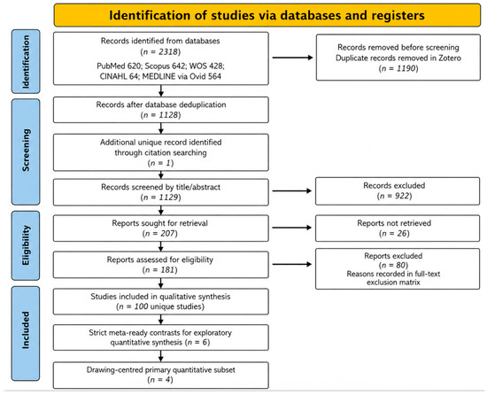
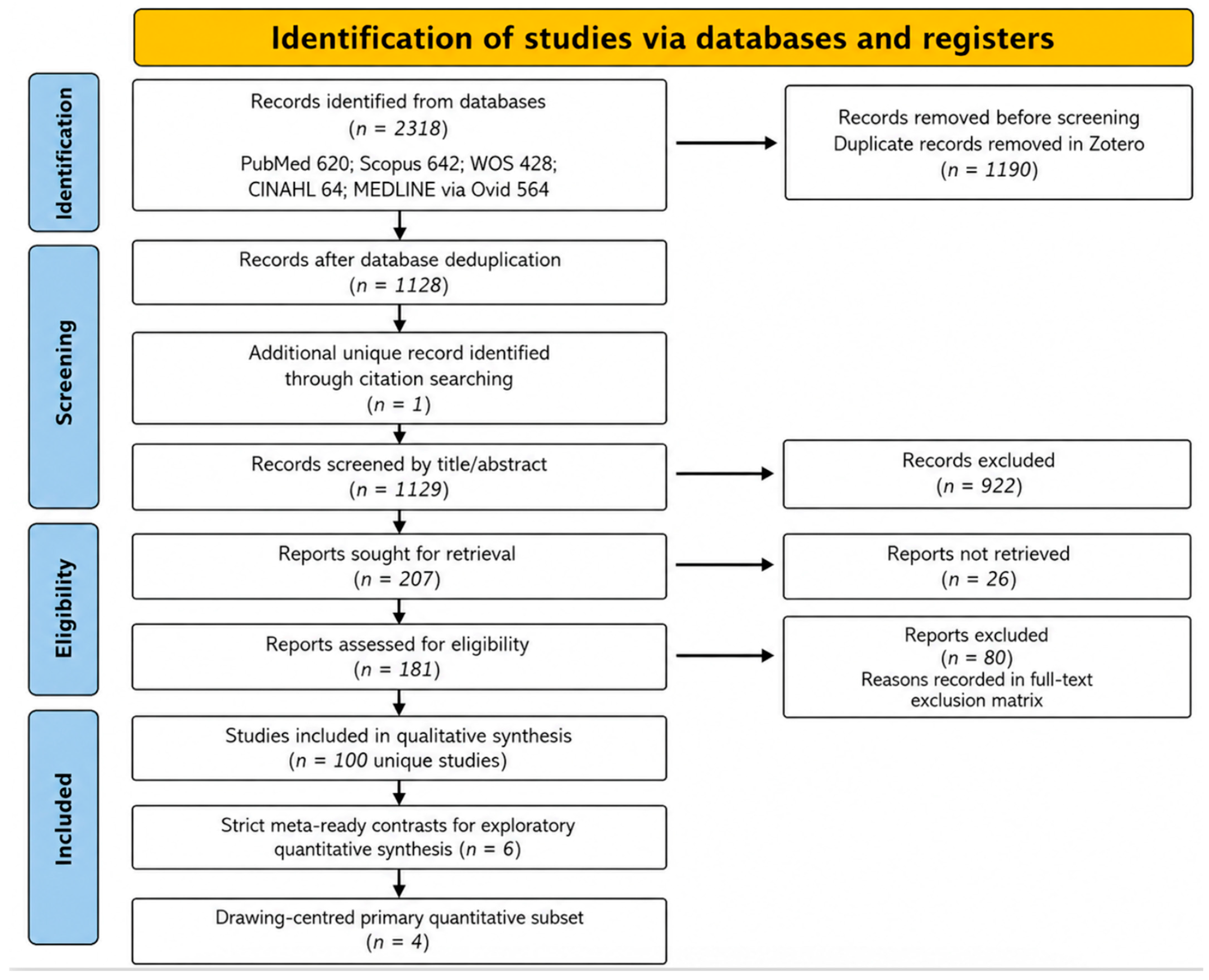
The finalized electronic database searches identified 2318 records: PubMed (n = 620), Scopus (n = 642), Web of Science Core Collection (n = 428), CINAHL (n = 64), and MEDLINE via Ovid (n = 564). Deduplication in Zotero removed 1190 duplicate records, leaving 1128 unique database-derived records.
Backward citation searching contributed one additional unique record after matching against the deduplicated library, whereas primary forward citation searching did not contribute additional unique records. The final title/abstract screening corpus therefore comprised 1129 records. Of the 207 reports sought for retrieval, 26 could not be obtained and 181 were assessed at full-text level. After full-text assessment, 80 reports were excluded. The remaining 101 included reports were consolidated into 100 unique studies for the qualitative evidence map. Of these, 51 were assessed as preliminary quantitative candidates. Six independent contrasts met strict meta-ready criteria for exploratory quantitative synthesis, and four of these were classified as the drawing-centred primary quantitative subset. The complete study-selection counts are summarized in Table 2 and illustrated in Figure 1.
Table 2.
PRISMA 2020 flow counts [17].
Figure 1.
PRISMA 2020 flow diagram showing identification, screening, retrieval, eligibility, qualitative inclusion of 100 unique studies, restriction to six strict meta-ready contrasts for exploratory quantitative synthesis, and the drawing-centred primary quantitative subset of four contrasts [17].
3.2. Characteristics of Included Studies
The qualitative evidence map included 100 unique studies addressing drawing-based, drawing-supported, drawing-associated active visual-representation, and adjacent representational strategies in anatomy, histology, human morphology, physiology-integrated morphology, and related health sciences education contexts. The included evidence base was dominated by gross anatomy, surface anatomy, applied anatomy, neuroanatomy, brain imaging, and histology/microscopic morphology, but also included cardiovascular, physiology-integrated, and broader visual-learning contexts (Table 3; Supplementary Table S1).
Table 3.
Descriptive map of the 100 unique studies included in the qualitative synthesis, grouped by educational domain and intervention family.
Across the included evidence base, interventions encompassed learner-generated drawing, guided sketching, diagrammatic learning, draw-and-explain activities, digital or tablet-based drawing, drawing tutorials or screencasts, body painting, haptico-visual drawing, modelling, 3D/model-based visual abstraction, and other drawing-associated active visual-representation approaches. These intervention families were retained in the evidence map but were not treated as cognitively equivalent for quantitative interpretation.
When grouped at the domain level, included studies most commonly addressed gross or applied anatomy (n = 65), histology/cytology/microscopic morphology (n = 15), neuroanatomy or brain-imaging contexts (n = 9), integrated morphology-physiology or cardiovascular learning (n = 7), and other health sciences or visual-learning contexts (n = 4).
The study-level characteristics of the 100 unique studies included in the evidence map were extracted from the final full-text eligibility matrix. Because the complete table is extensive, a descriptive map is presented in Table 3, and the full study-level table is provided as Supplementary Table S1, with citations for each included study.
An abbreviated design-level appraisal showed that the broader evidence base was methodologically heterogeneous. Based on the study-design labels in Supplementary Table S1, the 100 studies comprised randomized or quasi-randomized comparative studies (n = 21), non-randomized comparative/cohort studies (n = 23), pre-post or within-participant evaluations (n = 7), descriptive educational evaluations (n = 13), qualitative studies (n = 13), survey/perception or usability evaluations (n = 11), and other mixed educational designs (n = 12). This appraisal was not a substitute for formal risk-of-bias assessment and was used only to constrain the narrative synthesis to descriptive claims.
3.3. Outcome-Oriented Narrative Synthesis
3.3.1. Objective Academic Performance and Conceptual Integration
Studies of objective academic performance and conceptual integration were considered according to the representational mechanism. Learner-generated drawing studies examined whether producing drawings, diagrams, or annotations supported knowledge acquisition, structure recognition, examination scores, or applied interpretation. Drawing-supported studies evaluated guided tutorials, instructor-generated drawing, or drawing screencasts. Drawing-associated studies examined multisensory or model-based activities and were interpreted separately because they did not require the same degree of learner-generated representation. Many studies could not be pooled because they used pre-post designs, crossover formats without extractable paired data, historical cohorts, missing dispersion values, incompatible outcome constructs, or insufficient numerical reporting.
3.3.2. Retention, Cognitive Load, and Visuospatial or Representational Outcomes
Evidence for retention, cognitive load, and visuospatial or representational outcomes was more fragmented. Anatomy and neuroanatomy studies tended to emphasize spatial organization, three-dimensional reasoning, structure–function relationships, mental rotation, or image interpretation. Histology and cytology studies more often emphasized microscopic pattern recognition, diagram production, tissue identification, and retention of visual features. These differences support separate interpretation rather than a single assumed mechanism across anatomy and histology.
3.3.3. Learner Perceptions, Engagement, and Implementation Outcomes
Learner perceptions, engagement, satisfaction, self-efficacy, and implementation-related outcomes were frequently positive, but they were usually reported in descriptive, survey-based, or mixed-methods designs. These outcomes inform acceptability and pedagogical feasibility, but they were not treated as evidence of effectiveness unless accompanied by objective comparative learning outcomes. Haptico-visual drawing, body painting, modelling, and three-dimensional visual abstraction were interpreted as drawing-associated or adjacent representational approaches rather than as learner-generated drawing.
3.4. Quantitative Feasibility and Evidence Mapping
Among the 100 unique studies included in the evidence map, 51 were identified as preliminary quantitative candidates because they appeared to report comparative educational outcomes potentially relevant to synthesis. A strict numerical extraction audit showed that only a minority provided the full arm-level information required for conservative quantitative synthesis. Specifically, 10 rows had exact arm-level sample size, mean, and standard deviation data; 11 were classified as partial or conditional because they required additional assumptions, conversions, paired/crossover handling, graph digitization, or missing variance information; and 30 were not suitable for pooling because numerical data, outcome compatibility, or design requirements were insufficient.
Within the 51 quantitative candidates, the most frequent intervention families included drawing or diagrammatic learning, digital/live drawing or drawing video, student-generated drawing, digital drawing or sketching, 3D/model-based visualization, modelling or 3D visualization with a drawing component, and multisensory drawing/body-painting/modeling approaches. This distribution confirmed that the quantitative evidence base was broader than pure freehand drawing alone and supported the decision to distinguish a drawing-centred primary quantitative subset from a broader secondary exploratory model.
The primary quantitative synthesis was restricted to four drawing-centred contrasts with objective learning/performance outcomes, compatible timing, and complete arm-level data. Two additional strict meta-ready contrasts were retained only in the broader secondary exploratory model because they reflected model-based active visual abstraction rather than drawing-centred learning.
3.5. Quantitative Synthesis of Objective Learning/Performance Outcomes
The primary quantitative synthesis included four drawing-centred contrasts: a drawing tutorial, a drawing screencast, tablet/paper drawing, and teaching through drawing. Two additional strict meta-ready contrasts, Joseph et al. [23] and Koh et al. [24], were retained in the broader secondary exploratory analysis because they involved an artistic/color-coded 3D model and a working model rather than learner-generated or drawing-supported learning. The characteristics and quantitative roles of the six strict meta-ready contrasts are summarized in Table 4.
Table 4.
Characteristics of the six strict meta-ready contrasts and their quantitative role.
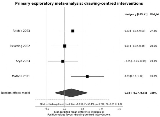
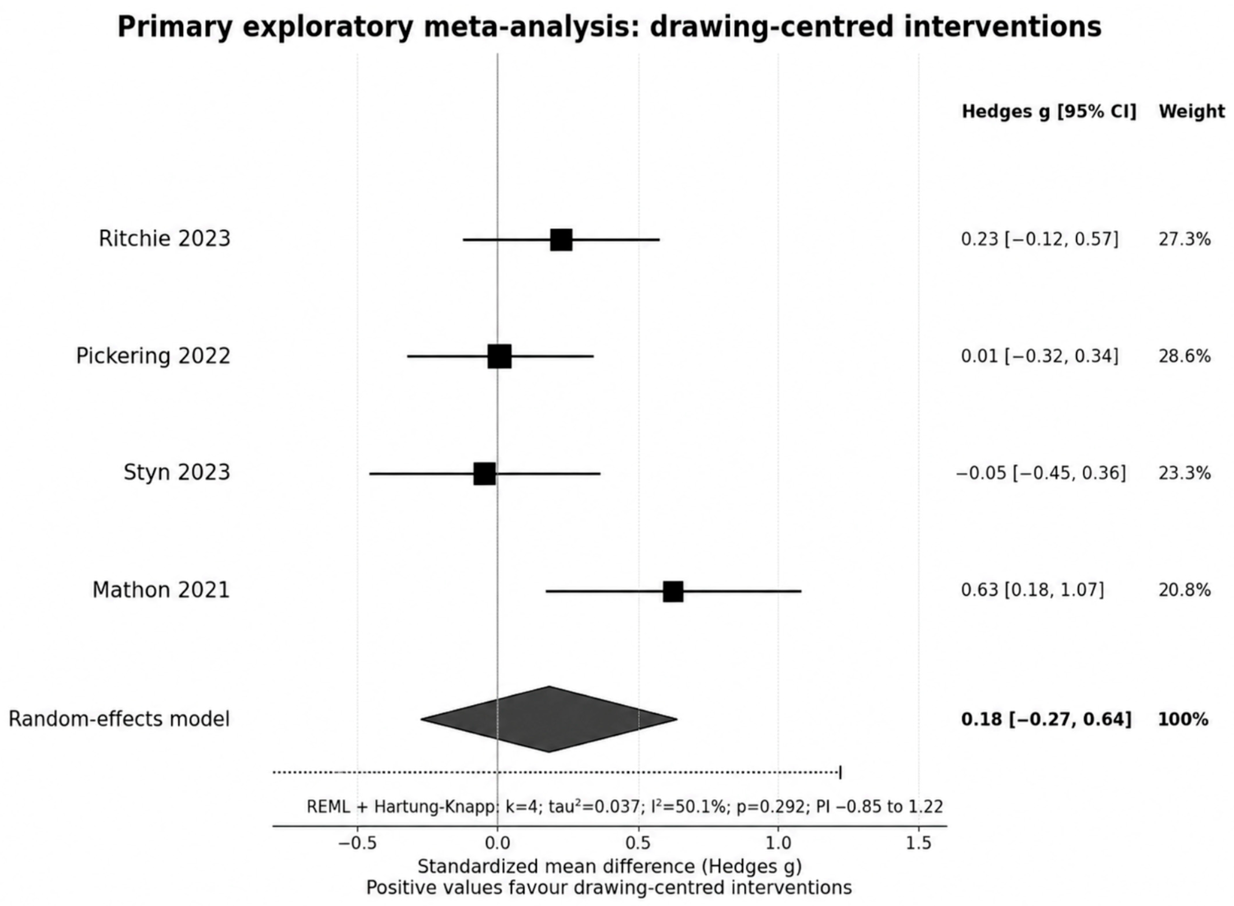
The drawing-centred primary random-effects estimate was directionally positive but statistically uncertain for objective learning/performance outcomes. The summary effect was Hedges g = 0.18, with a Hartung–Knapp 95% confidence interval from −0.27 to 0.64. Between-study heterogeneity was moderate (I2 = 50.1%; τ2 = 0.037), and the prediction interval ranged from −0.85 to 1.22. Therefore, although the point estimate favored drawing-centred interventions, both the confidence interval and prediction interval indicated substantial uncertainty.
Accordingly, the primary quantitative findings were interpreted as suggestive rather than confirmatory. The broader six-contrast secondary model, which also included the artistic/color-coded 3D model and working-model studies, remained positive but uncertain (Hedges g = 0.29, 95% CI −0.08 to 0.67; I2 = 53.7%; τ2 = 0.058; prediction interval −0.49 to 1.08). This broader estimate was not interpreted as the effect of learner-generated drawing alone. The study-level effects and pooled estimate for the drawing-centred primary model are shown in Figure 2.
Figure 2.
Primary exploratory random-effects meta-analysis of drawing-centred interventions for objective learning/performance outcomes. Positive Hedges g values favour drawing-centred interventions. The contributing studies were Ritchie et al. [26], Pickering et al. [27], Styn et al. [7], and Mathon et al. [28]. REML, restricted maximum likelihood; HK, Hartung–Knapp; PI, prediction interval.
3.6. Sensitivity Analyses: Results
Sensitivity analyses evaluated whether the primary estimate and the broader exploratory model were driven by specific conceptual or statistical decisions. Within the drawing-centred primary subset, excluding the combined-arm study yielded Hedges g = 0.26 (95% CI −0.49 to 1.01), and excluding the active-comparator contrast yielded Hedges g = 0.26 (95% CI −0.55 to 1.07). Both analyses remained directionally positive but very imprecise. The results of the primary, secondary, and sensitivity models are summarized in Table 5.
Table 5.
Quantitative synthesis and exploratory sensitivity analyses.
Across primary and secondary sensitivity analyses, the pooled estimates remained positive in direction, but all Hartung–Knapp confidence intervals crossed the null. The study-exclusion analyses of Joseph et al. [23] and Koh et al. [24] reduced the conceptual breadth of the analysis but did not convert the evidence into a statistically certain effect. These findings support the interpretation that the available quantitative evidence is directionally suggestive but very uncertain. Exploratory sensitivity plots are provided in Supplementary Figure S1.
3.7. Risk of Bias, Reporting Bias, and Certainty of Evidence
3.7.1. Study-Level Risk of Bias
Study-level risk of bias was evaluated for all six strict meta-ready contrasts because these studies informed either the drawing-centred primary analysis or the broader secondary exploratory model. Randomized trials were appraised using RoB 2 [20], whereas non-randomized and quasi-randomized comparisons were interpreted using ROBINS-I principles [21]. Judgments considered allocation or confounding, deviations from intended interventions, missing outcome data, outcome measurement, selective reporting, and the overall risk of bias.
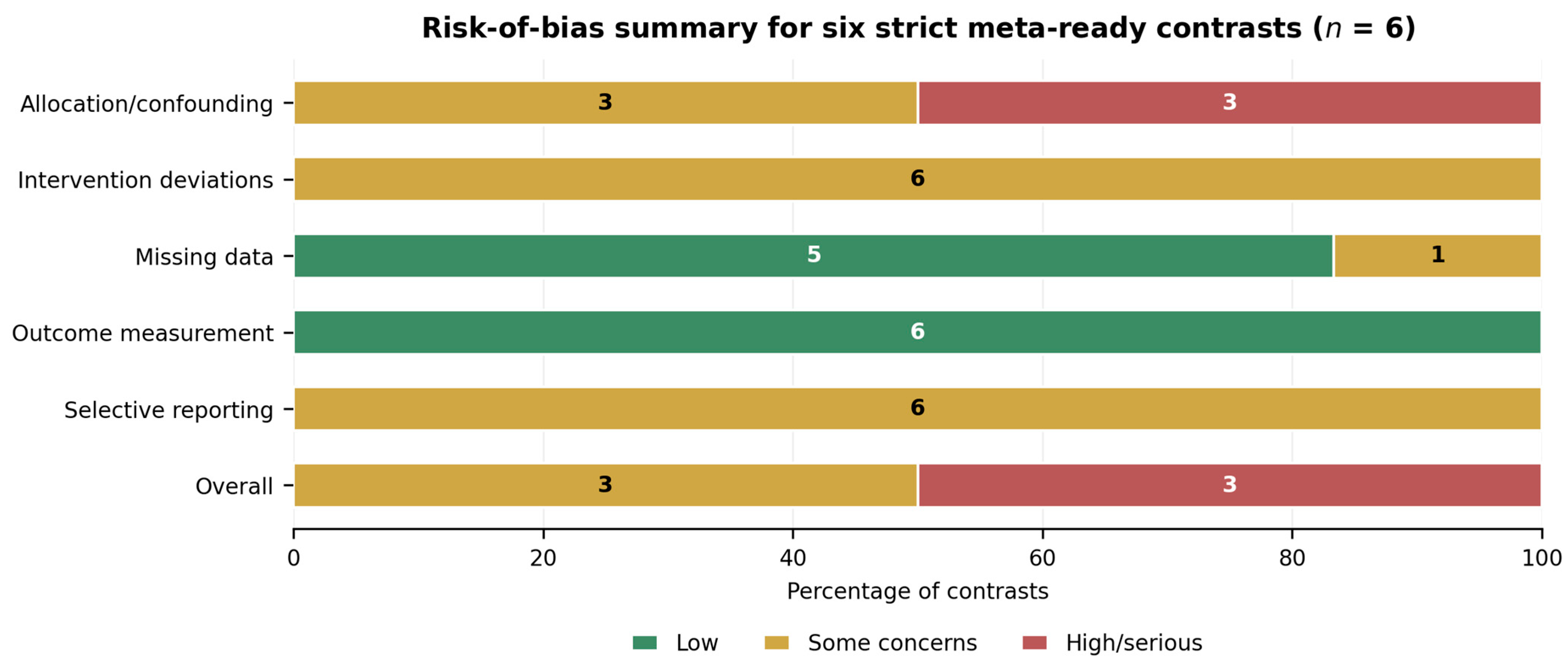
Overall, three contrasts had high risk of bias or serious study limitations, and three had some concerns. At the study level, Ritchie et al. [26] and Joseph et al. [23] were limited by non-randomized comparisons and the resulting potential for selection and confounding; Pickering et al. [27] used quasi-random allocation and an active comparator, which also limited causal interpretation. Koh et al. [24], Styn et al. [7], and Mathon et al. [28] used randomized designs, but incomplete methodological reporting prevented low-risk judgments. Table 6 reports the principal design and reporting considerations for each contrast, while the distribution of domain-level and overall risk-of-bias judgments across the six contrasts is summarized in Figure 3. Complete domain-level forms are provided in the risk-of-bias supporting material.
Table 6.
Study-level design and reporting considerations informing the risk-of-bias judgments.
Figure 3.
Risk-of-bias summary plot for the six strict meta-ready contrasts. Low risk = green, some concerns = amber, and high risk/serious limitations = red. Study-level considerations are reported in Table 6 [7,23,24,26,27,28].
3.7.2. Risk of Bias Due to Missing Results
Formal small-study effect testing and funnel-plot asymmetry assessment were not performed because fewer than ten studies were included in the quantitative synthesis [18]. Reporting bias was therefore considered qualitatively. The evidence base showed a substantial reduction from 100 unique studies included in the evidence map to 51 preliminary quantitative candidates, six strict meta-ready contrasts, and four drawing-centred primary contrasts. This reduction indicates that incomplete numerical reporting, incompatible outcome constructs, heterogeneous timing, and conceptual heterogeneity were major constraints on quantitative synthesis.
3.7.3. Certainty of Evidence
Using the GRADE framework, the certainty of evidence for the drawing-centred primary objective learning/performance outcome was judged to be very low [22]. The body of evidence was downgraded for risk-of-bias concerns, inconsistency, imprecision, and residual indirectness related to intervention delivery and comparator differences. The broader six-contrast secondary exploratory model was also judged as very low certainty, with additional indirectness because not all contributing contrasts represented drawing-centred learning; some estimated relative effects between active visual strategies or model-based representational activities. The corresponding GRADE judgments for the primary and secondary models are summarized in Table 7.
Table 7.
GRADE summary of findings for the drawing-centred primary subset and broader secondary model.
GRADE rationale: the drawing-centred primary subset was downgraded for risk-of-bias concerns, inconsistency, residual indirectness, and imprecision. The broader secondary exploratory model was additionally downgraded for serious indirectness because it included active visual-abstraction and working-model contrasts that should not be interpreted as pure learner-generated drawing. Publication bias was not formally assessed because fewer than ten studies were included.
4. Discussion
4.1. Principal Findings
This systematic evidence map identified a broad and heterogeneous literature on drawing-based, drawing-supported, and drawing-associated active visual-representation strategies in anatomy, histology, morphology, and related health sciences education. Although 100 unique studies were included in the map, only six independent contrasts met strict meta-ready criteria and only four were sufficiently close to drawing-centred learning to form the primary quantitative subset. This distinction is central to the interpretation: the field is pedagogically active, but the evidence that can support quantitative inference about drawing-centred learning remains sparse.
The drawing-centred primary meta-analysis suggested a small positive but statistically uncertain effect on objective learning/performance outcomes (Hedges g = 0.18). The confidence interval crossed the null, and the prediction interval was wide, indicating that future comparable studies may plausibly show no effect, negative effects, or larger positive effects. The broader six-contrast secondary model was also positive but uncertain (Hedges g = 0.29), and sensitivity analyses excluding Joseph et al. [23], Koh et al. [24], or both did not materially change the core conclusion: available quantitative evidence is suggestive, not confirmatory [7,23,24,26,27,28].
4.2. Interpretation in Relation to Anatomy and Health Sciences Education
The findings are consistent with the theoretical rationale that drawing can operate as a generative learning activity: learners may need to select relevant information, organize structural relationships, transform visual material, and externalise understanding [5,6,7,8,9,10]. However, the analysis emphasizes that not all drawing-associated interventions invoke the same cognitive mechanism. Learner-generated drawing, guided drawing, drawing screencasts, haptico-visual drawing, body painting, and model-based visual abstraction may all involve representation, but they differ in learner agency, sensory input, scaffolding, motor demands, and comparator contamination.
The distinction between anatomy and histology is also important. In anatomy and neuroanatomy, drawing may be most relevant when learners must organize spatial relationships, transform two-dimensional resources into three-dimensional representations, or reason through topographic structure. In histology, drawing may support attention to microscopic patterns, tissue organization, and discriminative visual features. The present review therefore does not claim that drawing has the same function across both domains. Rather, it maps how drawing and adjacent representational strategies have been used across morphological education and identifies where rigorous comparative evidence is currently available.
The relationship between drawing and haptics deserves particular attention. Reid et al. [29] and related haptico-visual approaches suggest that drawing may serve as a bridge between tactile exploration, visual observation, and spatial understanding. Such studies are highly relevant conceptually, but they should be interpreted as multisensory representational strategies rather than as conventional learner-generated drawing. Accordingly, the synthesis treats haptico-visual drawing as part of the evidence map and discussion, while avoiding overgeneralization from these designs to the pooled drawing-centred effect.
Compared with neighbouring anatomy-education reviews on 3D visualization, laboratory pedagogies, physical models, plastinated specimens, and augmented reality [1,2,14,15,16], the present review addresses a narrower representational question but reaches a similar methodological conclusion: active visual resources may be useful under some instructional conditions, yet effects depend on implementation, scaffolding, outcome alignment, comparator choice, and study design. Generalization to other medical subject areas should be made cautiously because comparable drawing-centred effectiveness syntheses remain limited.
4.3. Implications for Curriculum and Instructional Design
For curriculum design, the findings support cautious, outcome-aligned use of drawing rather than indiscriminate addition of drawing activities. Drawing is more defensible when it is integrated with a specific morphological reasoning task, scaffolded through modelling or feedback, and linked explicitly to assessment expectations. In anatomy, this may involve drawing to clarify spatial relations, layered organization, pathways, or clinically relevant topography. In histology, it may involve drawing or annotating microscopic features to strengthen pattern recognition and conceptual integration.
Comparator choice is critical. A drawing screencast compared with mixed reality estimates the relative effect of two active visual approaches, not the absolute effect of drawing compared with usual teaching [27]. Similarly, artistic/color-coded 3D models and working models may reflect active visual abstraction or model-based learning rather than drawing-centred learning [23,24]. Educators and researchers should therefore describe the representational mechanism precisely and avoid labelling all visual or model-based activities as drawing interventions.
4.4. Strengths and Limitations
A major strength of this review is the transparent PRISMA 2020 workflow, including explicit database counts, deduplication, supplementary citation searching, full-text eligibility coding, and a conservative decision not to force all quantitative candidates into a single effectiveness estimate [17]. The analysis further distinguishes by separating the 100-study evidence map, the four-contrast drawing-centred primary model, and the broader six-contrast secondary exploratory model. The quantitative analysis used Hedges g, random-effects modelling, REML estimation, Hartung–Knapp adjustment, and prediction intervals, which are appropriate for a small and heterogeneous educational evidence base [18].
Several limitations should be acknowledged. First, the drawing-centred primary meta-analysis included only four contrasts, which limits precision and precludes confirmatory subgroup analysis or meta-regression. Second, risk-of-bias concerns remained important across the quantitative subset. Third, even the drawing-centred subset included variation in learner-generated drawing, guided drawing, drawing screencasts, comparator type, and timing. Fourth, anatomy and histology were mapped together because both are morphology-intensive health-science education domains, but they differ in cognitive demands and should not be assumed to share identical mechanisms. Fifth, the search was anchored in drawing, sketching, diagramming, and annotation terms and did not include dedicated education databases such as ERIC or PsycINFO; therefore, the evidence map should not be interpreted as a comprehensive review of all active visual, 3D, augmented-reality, mixed-reality, or model-based strategies independent of drawing. Sixth, 26 reports sought for retrieval could not be obtained. Separately, one retrieved Chinese-language report on anatomy and drawing in obstetrics and gynecology could not be assessed reliably because a usable translation was unavailable [19] (PMID: 21609574). Seventh, many studies in the broader evidence base did not provide sufficient numerical data for pooling. Finally, the certainty of evidence was judged very low.
4.5. Implications for Future Research
Future studies should use rigorous comparative designs, clearer allocation procedures, prespecified outcomes, and transparent reporting of sample sizes, means, standard deviations, assessment timing, and attrition. Researchers should distinguish between learner-generated drawing, instructor-generated drawing, guided drawing, drawing screencasts, diagram completion, haptico-visual drawing, body painting, model-based visualisation, and other active visual-representation approaches. This distinction is essential because these interventions may not share the same cognitive mechanism, instructional dose, or comparator contamination risk.
Future trials should prioritize objective learning outcomes, delayed retention, conceptual transfer, spatial or representational reasoning, and cognitive load, while avoiding exclusive reliance on satisfaction or perceived usefulness. Where possible, studies should report enough data to support future meta-analysis and should avoid multiple uncorrected outcomes without a clearly prespecified primary outcome. The field would benefit from a core outcome set for anatomy and histology education studies involving drawing-centred and active visual-representation interventions.
5. Conclusions
Drawing-based and drawing-associated active visual-representation strategies are widely used across anatomy, histology, and related health sciences education. The evidence map shows a diverse field, but the quantitative evidence for drawing-centred interventions remains sparse. The primary drawing-centred exploratory meta-analysis suggested a positive but statistically uncertain effect on objective learning/performance outcomes, and the broader six-contrast model remained similarly uncertain. Given the small number of strict meta-ready studies, moderate heterogeneity, risk-of-bias concerns, indirectness, imprecision, and very low certainty of evidence, the findings should be interpreted as directionally suggestive rather than confirmatory. The review supports continued pedagogical use and rigorous evaluation of drawing-centred strategies, but it does not establish drawing as an isolated, universally effective intervention.
Supplementary Materials
The following supporting information can be downloaded at: https://www.mdpi.com/article/10.3390/ime5030067/s1. Supplementary Table S1, study-level characteristics of the evidence map; Supplementary Table S2, full electronic search strategies; Supplementary Table S3, PRISMA 2020 checklist; Supplementary Table S4, reports excluded after full-text assessment with structured exclusion reasons; Supplementary Figure S1, exploratory sensitivity analyses for the quantitative synthesis; Supplementary Table S5, arm-level dataset and statistical code for the primary drawing-centred analysis, broader secondary exploratory model, study-exclusion sensitivity analyses, sensitivity-analysis definitions, and arm-combination decisions; and risk-of-bias/GRADE supporting material. Appendix S1. Reports not retrieved.
Author Contributions
Conceptualization, V.P.-L., D.S.-C., M.M.-S. and C.B.-E.; Methodology, A.B.-M., G.O. and J.J.C.-S.; Software, M.O.-D. and A.B.-M.; Validation, A.B.-M., J.J.V.-F., G.O. and J.J.C.-S.; Formal Analysis, A.B.-M.; Investigation, V.P.-L., D.S.-C., M.M.-S., R.V.-S., M.R.-L., D.L. and A.B.-M.; Data Curation, V.P.-L., M.M.-S., D.S.-C., R.V.-S., M.R.-L., D.L., J.J.C.-S. and L.P.-A.; Screening, A.B.-M. and J.J.V.-F.; Risk-of-Bias Assessment, V.P.-L., M.M.-S. and D.S.-C.; Writing—Original Draft Preparation, A.B.-M. and J.J.V.-F.; Writing—Review and Editing, L.P.-A., M.P.M., R.V.-S., M.R.-L., D.L., G.O., G.C.-S., M.O.-D., J.J.C.-S. and J.S.-G.; Visualization, M.O.-D. and J.F.-P.; Supervision, A.B.-M., J.J.C.-S. and J.S.-G.; Project Administration, V.P.-L., D.S.-C., M.M.-S. and A.B.-M. All authors have read and agreed to the published version of the manuscript.
Funding
This research received no external funding.
Institutional Review Board Statement
Not applicable. This systematic evidence map and exploratory quantitative synthesis used previously published literature and did not involve new human participants, human tissue, identifiable personal data, or experimental interventions.
Informed Consent Statement
Not applicable.
Data Availability Statement
All data supporting the primary drawing-centred model, broader secondary exploratory model, and sensitivity analyses are available in Supplementary Table S5, including arm-level sample sizes, means, standard deviations, Hedges g values, variances, standard errors, confidence intervals, random-effects weights, arm-combination decisions, sensitivity-analysis definitions, study-exclusion models, and reproducible R/metafor code. Study-level characteristics of the evidence map are provided in Supplementary Table S1; the full electronic search strategies are provided in Supplementary Table S2; the PRISMA 2020 checklist is provided in Supplementary Table S3; and full-text exclusion reasons are provided in Supplementary Table S4.
Conflicts of Interest
The authors declare no conflicts of interest.
References
- Yammine, K.; Violato, C. A meta-analysis of the educational effectiveness of three-dimensional visualization technologies in teaching anatomy. Anat. Sci. Educ. 2015, 8, 525–534. [Google Scholar] [CrossRef] [Scilit] [PubMed]
- Wilson, A.B.; Miller, C.H.; Klein, B.A.; Taylor, M.A.; Goodwin, M.; Boyle, E.K.; Brown, K.; Hoppe, C.; Lazarus, M. A meta-analysis of anatomy laboratory pedagogies. Clin. Anat. 2018, 31, 19–27. [Google Scholar] [CrossRef] [Scilit] [PubMed]
- Na, Y.; Clary, D.W.; Rose-Reneau, Z.B.; Segars, L.; Hanson, A.; Brauer, P.; Wright, B.W.; Keim, S.A. Spatial Visualization of Human Anatomy through Art Using Technical Drawing Exercises. Anat. Sci. Educ. 2022, 15, 587–598. [Google Scholar] [CrossRef] [Scilit] [PubMed]
- Cahayani, W.A.; Rahayu, I.D.; Riawan, W. Enhancing histology education through a multimodal approach: Integrating digital annotation, manual drawing, and histological batik design. Anat. Sci. Educ. 2026, 19, 525–534. [Google Scholar] [CrossRef] [Scilit] [PubMed]
- Noorafshan, A.; Hoseini, L.; Amini, M.; Dehghani, M.R.; Kojuri, J.; Bazrafkan, L. Simultaneous anatomical sketching as learning by doing method of teaching human anatomy. J. Educ. Health Promot. 2014, 3, 50. [Google Scholar] [CrossRef] [Scilit] [PubMed]
- Leconte, L.; Jamet, E. Does learning by drawing from a multimedia document improve knowledge and self-assessment in anatomical education? Adv. Health Sci. Educ. 2026. advance online publication. [Google Scholar] [CrossRef] [Scilit] [PubMed]
- Styn, A.; Scheiter, K.; Fischer, M.R.; Shiozawa, T.; Behrmann, F.; Steffan, A.; Kugelmann, D.; Berndt, M. Effects of tablet-based drawing and paper-based methods on medical students’ learning of gross anatomy. Anat. Sci. Educ. 2023, 16, 266–279. [Google Scholar] [CrossRef] [Scilit] [PubMed]
- Beach, I.R.; D’Agostino, E.N.; Thakrar, R.; Lunardini, D.J. Learning by drawing and modeling: Teaching modalities for spinal anatomy in medical students. Anat. Sci. Educ. 2023, 16, 1041–1045. [Google Scholar] [CrossRef] [Scilit] [PubMed]
- Shapiro, L.; Bell, K.; Dhas, K.; Branson, T.; Louw, G.; Keenan, I.D. Focused Multisensory Anatomy Observation and Drawing for Enhancing Social Learning and Three-Dimensional Spatial Understanding. Anat. Sci. Educ. 2020, 13, 488–503. [Google Scholar] [CrossRef] [Scilit] [PubMed]
- Schwamborn, A.; Mayer, R.E.; Thillmann, H.; Leopold, C.; Leutner, D. Drawing as a generative activity and drawing as a prognostic activity. J. Educ. Psychol. 2010, 102, 872–879. [Google Scholar] [CrossRef] [Scilit]
- Sreevidyalatha, G.M.; Shashikala, P. Fostering proficiency in cell identification: A comparative analysis of diagrams and microscopic images for optimal cytopathology learning in competency-based medical education curriculum. J. Med. Sci. Health 2024, 10, 47–51. [Google Scholar] [CrossRef] [Scilit]
- Jariyapong, P.; Punsawad, C.; Bunratsami, S.; Kongthong, P. Body painting to promote self-active learning of hand anatomy for preclinical medical students. Med. Educ. Online 2016, 21, 30833. [Google Scholar] [CrossRef] [Scilit] [PubMed]
- Weiss, V.; Kinley, V.; Khalaf, N.; Sillevis, R. Enhancing medical education through drawing: A pathway to deeper understanding and retention. Med. Res. Arch. 2025, 13, 1–7. [Google Scholar] [CrossRef] [Scilit]
- Yammine, K.; Violato, C. The effectiveness of physical models in teaching anatomy: A meta-analysis of comparative studies. Adv. Health Sci. Educ. Theory Pract. 2016, 21, 883–895. [Google Scholar] [CrossRef] [Scilit] [PubMed]
- Goh, J.S.K.; Chandrasekaran, R.; Sirasanagandla, S.R.; Acharyya, S.; Mogali, S.R. Efficacy of plastinated specimens in anatomy education: A systematic review and meta-analysis. Anat. Sci. Educ. 2024, 17, 712–721. [Google Scholar] [CrossRef] [Scilit] [PubMed]
- Bolek, K.A.; de Jong, G.; Henssen, D. The effectiveness of the use of augmented reality in anatomy education: A systematic review and meta-analysis. Sci. Rep. 2021, 11, 15292. [Google Scholar] [CrossRef] [Scilit]
- Page, M.J.; McKenzie, J.E.; Bossuyt, P.M.; Boutron, I.; Hoffmann, T.C.; Mulrow, C.D.; Shamseer, L.; Tetzlaff, J.M.; Akl, E.A.; Brennan, S.E.; et al. The PRISMA 2020 statement: An updated guideline for reporting systematic reviews. BMJ 2021, 372, n71. [Google Scholar] [CrossRef] [Scilit] [PubMed]
- Higgins, J.P.T.; Thomas, J.; Chandler, J.; Cumpston, M.; Li, T.; Page, M.J.; Welch, V.A. (Eds.) Cochrane Handbook for Systematic Reviews of Interventions, 2nd ed.; John Wiley & Sons: Chichester, UK, 2019. [Google Scholar]
- Lang, J.H. Anatomy and drawing of obstetrics and gynecology. Zhonghua Fu Chan Ke Za Zhi 2011, 46, 241–243. (In Chinese) [Google Scholar] [PubMed]
- Sterne, J.A.; Savović, J.; Page, M.J.; Elbers, R.G.; Blencowe, N.S.; Boutron, I.; Cates, C.J.; Cheng, H.Y.; Corbett, M.S.; Eldridge, S.M.; et al. RoB 2: A revised tool for assessing risk of bias in randomised trials. BMJ 2019, 366, l4898. [Google Scholar] [CrossRef] [Scilit] [PubMed]
- Sterne, J.A.; Hernán, M.A.; Reeves, B.C.; Savović, J.; Berkman, N.D.; Viswanathan, M.; Henry, D.; Altman, D.G.; Ansari, M.T.; Boutron, I.; et al. ROBINS-I: A tool for assessing risk of bias in non-randomised studies of interventions. BMJ 2016, 355, i4919. [Google Scholar] [CrossRef] [Scilit] [PubMed]
- Balshem, H.; Helfand, M.; Schünemann, H.J.; Oxman, A.D.; Kunz, R.; Brozek, J.; Vist, G.E.; Falck-Ytter, Y.; Meerpohl, J.; Norris, S.; et al. GRADE guidelines: 3. Rating the quality of evidence. J. Clin. Epidemiol. 2011, 64, 401–406. [Google Scholar] [CrossRef] [Scilit] [PubMed]
- Joseph, B.; Rebello, G.; Mayya, S. The use of a working model for teaching functional and applied surgical anatomy of the subtalar joint. Foot Ankle Int. 2006, 27, 286–292. [Google Scholar] [CrossRef] [Scilit] [PubMed]
- Koh, M.Y.; Tan, G.J.S.; Mogali, S.R. Spatial ability and 3D model colour-coding affect anatomy performance: A cross-sectional and randomized trial. Sci. Rep. 2023, 13, 7879. [Google Scholar] [CrossRef] [Scilit]
- Rafi, A.; Rauf, A.; Anwar, M.I. Significance of actually drawing microscopic images and its impact on students’ understanding of histology. J. Dow Univ. Health Sci. Karachi 2017, 11, 77–81. [Google Scholar] [CrossRef] [Scilit]
- Ritchie, H.E.; Oakes, D.; Drury, H.; Ollerenshaw, S.; Hegedus, E. Can drawing instruction help students with low visuospatial ability in learning anatomy? Anat. Sci. Educ. 2023, 16, 252–265. [Google Scholar] [CrossRef] [Scilit] [PubMed]
- Pickering, J.D.; Panagiotis, A.; Ntakakis, G.; Athanassiou, A.; Babatsikos, E.; Bamidis, P.D. Assessing the difference in learning gain between a mixed reality application and drawing screencasts in neuroanatomy. Anat. Sci. Educ. 2022, 15, 628–635. [Google Scholar] [CrossRef] [Scilit] [PubMed]
- Mathon, B.; Chougar, L.; Carpentier, A.; Amelot, A. Teaching brain imaging through a drawing method may improve learning in medical students. Eur. Radiol. 2021, 31, 4195–4202. [Google Scholar] [CrossRef] [Scilit] [PubMed]
- Reid, S.; Shapiro, L.; Louw, G. How Haptics and Drawing Enhance the Learning of Anatomy. Anat. Sci. Educ. 2019, 12, 164–172. [Google Scholar] [CrossRef] [Scilit] [PubMed]
- Alsaid, B.; Bertrand, M. Students’ memorization of anatomy, influence of drawing. Morphologie 2016, 100, 2–6. [Google Scholar] [CrossRef] [Scilit] [PubMed]
- Shapiro, L.; Hobbs, E.; Keenan, I.D. Transforming musculoskeletal anatomy learning with haptic surface painting. Anat. Sci. Educ. 2023, 16, 677–693. [Google Scholar] [CrossRef] [Scilit] [PubMed]
- Horne, C.A.; Hepworth, D.; Saunders, E.; Keenan, I.D. Everyone can draw: An inclusive and transformative activity for conceptualization of topographic anatomy. Anat. Sci. Educ. 2024, 17, 1080–1096. [Google Scholar] [CrossRef] [Scilit]
- Singal, A.; Chaudhary, P.; Aggarwal, N.; Patra, A. Students’ Perception of Newly Experimented “Rangoli Art” in Anatomy Education: A Pilot Study. Med. Sci. Educ. 2023, 33, 887–892. [Google Scholar] [CrossRef] [Scilit] [PubMed]
- Crossing, E.; Smyth, L.; Valter, K.; Webb, A. Visualizing the Human Body Using an Artistic Approach. Adv. Exp. Med. Biol. 2023, 1392, 129–148. [Google Scholar] [CrossRef] [Scilit] [PubMed]
- Langlois, J.; Hamstra, S.J.; Dagenais, Y.; Lemieux, R.; Lecourtois, M.; Yetisir, E.; Bellemare, C.; Bergeron, G.; Wells, G.A. Objects drawn from haptic perception and vision-based spatial abilities. Anat. Sci. Educ. 2024, 17, 433–443. [Google Scholar] [CrossRef] [Scilit] [PubMed]
- Lau, I.; Sun, Z. The role of 3D printed heart models in immediate and long-term knowledge acquisition in medical education. Rev. Cardiovasc. Med. 2022, 23, 22. [Google Scholar] [CrossRef] [Scilit] [PubMed]
- Phillips, P.S. Running a life drawing class for pre-clinical medical students. Med. Educ. 2000, 34, 1020–1025. [Google Scholar] [CrossRef] [Scilit]
- Sharma, B.; Nasir, N.; Alhazmi, A.; Bridget, B.; Ali, M. Visualizing Anatomy: Comparing the Pedagogical Impact of Chalkboard Teaching and 3D Models. Clin. Anat. 2025. advance online publication. [Google Scholar] [CrossRef] [Scilit] [PubMed]
- Motzko, M.; Dennis, J.F. Usage of Student-Created Anatomical Diagrams Shared on Social Media. Med. Sci. Educ. 2023, 33, 191–204. [Google Scholar] [CrossRef] [Scilit] [PubMed]
- Pathak, D.; Kapoor, K.; Choudhary, R.K.; Singh, O.; Uppal, V.; Bansal, N. Sketch-based animation teaching in veterinary anatomy via virtual mode during COVID-19 versus physical mode: A comparative analysis. J. Vis. Commun. Med. 2025, 48, 69–75. [Google Scholar] [CrossRef] [Scilit] [PubMed]
- Backhouse, M.; Fitzpatrick, M.; Hutchinson, J.; Thandi, C.S.; Keenan, I.D. Improvements in anatomy knowledge when utilizing a novel cyclical “Observe-Reflect-Draw-Edit-Repeat” learning process. Anat. Sci. Educ. 2017, 10, 7–22. [Google Scholar] [CrossRef] [Scilit] [PubMed]
- Chaudhuri, J.D. Changes in the learning styles and approaches of students following incorporation of drawing during cadaveric dissection. Clin. Anat. 2021, 34, 437–450. [Google Scholar] [CrossRef] [Scilit] [PubMed]
- Moore, C.M.; Lowe, C.; Lawrence, J.; Borchers, P. Developing observational skills and knowledge of anatomical relationships in an art and anatomy workshop using plastinated specimens. Anat. Sci. Educ. 2011, 4, 294–301. [Google Scholar] [CrossRef] [Scilit] [PubMed]
- Paymard, M.; Naderian, H.; Hassani Bafrani, H.; Azami Tameh, A.; Mirsafi Niasar, M.; Rahimi, H.; Hosseini, H.S.; Rafat, A. An evaluation of the use of 3D-printed anatomical models in anatomy education. Surg. Radiol. Anat. 2025, 47, 199. [Google Scholar] [CrossRef] [Scilit] [PubMed]
- Yuan, Q.; Chen, X.; Zhai, J.; Chen, Y.; Liu, Q.; Tan, Z.; Chen, G.; Zhuang, K.; Zhang, J.; Xu, X.; et al. Application of 3D modeling and fusion technology of medical image data in image teaching. BMC Med. Educ. 2021, 21, 194. [Google Scholar] [CrossRef] [Scilit] [PubMed]
- Liou, K.T.; George, P.; Baruch, J.M.; Luks, F.I. Clinical sketches: Teaching medical illustration to medical students. Med. Educ. 2014, 48, 525. [Google Scholar] [CrossRef] [Scilit] [PubMed]
- Naug, H.L.; Colson, N.J.; Donner, D.G. Promoting metacognition in first year anatomy laboratories using plasticine modeling and drawing activities: A pilot study of the “blank page” technique. Anat. Sci. Educ. 2011, 4, 231–234. [Google Scholar] [CrossRef] [Scilit] [PubMed]
- Sharma, B.; Alhazmi, A.; Nasir, N.; Bridget, B. Innovative Learning in Anatomy Education: Assessing the Impact of Low-Cost 3D Deep Learning Anatomical Models in Museum-Based Instruction. J. Surg. Educ. 2026, 83, 103748. [Google Scholar] [CrossRef] [Scilit]
- Narayanan, S.; Adikesavan, P.N.; Ananthy, V. Teaching Pterygopalatine Fossa Anatomy Using 3D Images and Physical Model: A Novel Teaching Strategy. Med. Sci. Educ. 2024, 34, 901–908. [Google Scholar] [CrossRef] [Scilit] [PubMed]
- Chen, S.; Pan, Z.; Wu, Y.; Gu, Z.; Li, M.; Liang, Z.; Zhu, H.; Yao, Y.; Shui, W.; Shen, Z.; et al. The role of three-dimensional printed models of skull in anatomy education: A randomized controlled trail. Sci. Rep. 2017, 7, 575. [Google Scholar] [CrossRef] [Scilit] [PubMed]
- Laakkonen, J. Drawing in Veterinary Anatomy Education: What Do Students Use It For? Anat. Sci. Educ. 2021, 14, 799–807. [Google Scholar] [CrossRef] [Scilit] [PubMed]
- Pickering, J.D. Anatomy drawing screencasts: Enabling flexible learning for medical students. Anat. Sci. Educ. 2015, 8, 249–257. [Google Scholar] [CrossRef] [Scilit] [PubMed]
- Ng, C.L.; Liu, X.; Chee, S.C.J.; Ngo, R.Y.S. An Innovative 3-dimensional Model of the Epitympanum for Teaching of Middle Ear Anatomy. Otolaryngol. Head. Neck Surg. 2015, 153, 832–837. [Google Scholar] [CrossRef] [Scilit] [PubMed]
- Elgendy, H.; Cui, X.; Watkins, T.; McQuistan, M. Teaching Dental Drawings for Freshman Dental Students and Its Correlation with Manual Dexterity. Int. J. Dent. 2023, 2023, 5685003. [Google Scholar] [CrossRef] [Scilit] [PubMed]
- Pérez-Higueras, J.J.; Oteo, B.; Gancedo-Caravia, L.; Arias, A. Influence of the incorporation of small-group practical seminars to a traditional lecture for the understanding of root canal morphology classification systems. Eur. J. Dent. Educ. 2024, 28, 371–376. [Google Scholar] [CrossRef] [Scilit] [PubMed]
- Agarwal, A.; Souza, A.D.; Jyostna, B.; Shetty, A.; Bhat, N. Introducing 3D printed models of fractures in osteology learning improves clinical reasoning skills among first-year medical students: A pilot study. BMC Med. Educ. 2025, 25, 190. [Google Scholar] [CrossRef] [Scilit] [PubMed]
- Joewono, M.; Karmaya, I.N.M.; Wirata, G.; Yuliana Widianti, I.G.A.; Wardana, I.N.G. Drawing method can improve musculoskeletal anatomy comprehension in medical faculty student. Anat. Cell Biol. 2018, 51, 14–18. [Google Scholar] [CrossRef] [Scilit] [PubMed]
- Greene, S.J. The use and effectiveness of interactive progressive drawing in anatomy education. Anat. Sci. Educ. 2018, 11, 445–460. [Google Scholar] [CrossRef] [Scilit] [PubMed]
- Lim, M.W.; Burt, G.; Rutter, S.V. Use of three-dimensional animation for regional anaesthesia teaching: Application to interscalene brachial plexus blockade. Br. J. Anaesth. 2005, 94, 372–377. [Google Scholar] [CrossRef] [Scilit] [PubMed]
- Beck, S.; Whalley, G.; Coffey, S.; Hawley, A.; Anakin, M. Cardiac Ultrasound Training for Medical Students Utilizing Drawing and Ward-Based Instruction. MedEdPORTAL 2025, 21, 11485. [Google Scholar] [CrossRef] [Scilit] [PubMed]
- Loke, Y.H.; Harahsheh, A.S.; Krieger, A.; Olivieri, L.J. Usage of 3D models of tetralogy of Fallot for medical education: Impact on learning congenital heart disease. BMC Med. Educ. 2017, 17, 54. [Google Scholar] [CrossRef] [Scilit] [PubMed]
- Zhou, F.; Yang, Y.; Liu, J.; Zhu, X. AI + Drawing Enhances the Efficiency of Human Anatomy Education. Clin. Anat. 2026. advance online publication. [Google Scholar] [CrossRef] [Scilit] [PubMed]
- Pickering, J.D. Measuring learning gain: Comparing anatomy drawing screencasts and paper-based resources. Anat. Sci. Educ. 2017, 10, 307–316. [Google Scholar] [CrossRef] [Scilit] [PubMed]
- Fyllos, A.; Zibis, A.; Dailiana, Z.H. Description and Validation of an Innovative and Effective Hand-Shaped Suture-Training Model for Medical Students. J. Hand Microsurg. 2021, 13, 185–188. [Google Scholar] [CrossRef] [Scilit] [PubMed]
- Pang, M.; Tong, X.; Zhao, X.; Wan, J.; Yin, H.; Yu, N.; Ma, C.; Zhang, W.; Xu, Y.; Ren, Y.; et al. Virtual sketching-based dental anatomy module improves learners’ abilities to use computer-aided design to create dental restorations and prostheses. Anat. Sci. Educ. 2024, 17, 1251–1264. [Google Scholar] [CrossRef] [Scilit] [PubMed]
- Kim, G.; Bhasin, S.; Zheng, L.; Ma, S.; Abedrabbo, N.; Mook, J.; Yi, V.N.; Li, L.W.; Burner, D.; Owens, K.E.; et al. Anatomy Drawing: Dissecting the Impact of Art in Medicine. Med. Sci. Educ. 2025, 35, 991–1000. [Google Scholar] [CrossRef] [Scilit] [PubMed]
- Latre-Navarro, L.; Quintas-Hijós, A.; Sáez-Bondía, M.J. The combined effects of an anatomy program integrating drawing and gamification on basic psychological needs satisfaction among sport sciences students: Results of a natural experiment. Anat. Sci. Educ. 2024, 17, 366–378. [Google Scholar] [CrossRef] [Scilit] [PubMed]
- Van Vlasselaer, N.; Van den Broeck, J.; Serrien, B.; Cattrysse, E. Creating and hosting 3D photogrammetry models for anatomical education: An accessible and student-centered approach. Anat. Sci. Educ. 2025, 18, 657–669. [Google Scholar] [CrossRef] [Scilit] [PubMed]
- Kotzé, S.H.; Mole, C.G.; Greyling, L.M. The translucent cadaver: An evaluation of the use of full body digital X-ray images and drawings in surface anatomy education. Anat. Sci. Educ. 2012, 5, 287–294. [Google Scholar] [CrossRef] [Scilit] [PubMed]
- Scogin, W.M.; Sanford, D.; Greenway, M.B.; Ledbetter, M.; Washmuth, N.B. The influence of body painting on L4 spinous process palpation accuracy in novice palpators. J. Man. Manip. Ther. 2024, 32, 429–434. [Google Scholar] [CrossRef] [Scilit] [PubMed]
- Bertrand, M.M.; Besseyrias, A.; Gouzi, F.; Tricot, A. Showing the drawing hand of the teacher in an anatomy video lecture-Effect on the student’s learning, motivation, and cognitive load. Anat. Sci. Educ. 2026, 19, 430–439. [Google Scholar] [CrossRef] [Scilit] [PubMed]
- Li, W.; Ding, Z.; Feng, S.; Xu, W.; Qi, H.; Zhu, Q.; Xiao, B.; Zhu, S.; Sun, M.; Yuan, F. Evaluating a problem-based learning model integrated with 3D anatomy software and software-assisted annotation in undergraduate spinal surgery education: A randomized controlled trial. BMC Med. Educ. 2026, 26, 301. [Google Scholar] [CrossRef] [Scilit] [PubMed]
- Latre-Navarro, L.; Quintas-Hijós, A. Neurocognitive outcomes of reinforced representational modelling in anatomical learning: The role of executive functions. Ann. Anat. 2026, 264, 152783. [Google Scholar] [CrossRef] [Scilit] [PubMed]
- Veerabhadrappa, S.K.; Vadivel, J.K.; Yadav, S.; Jain, V.K.; Marya, A.; Selvaraj, S. Effectiveness of ChatGPT-assisted and drawing-based learning in enhancing understanding of intraoral radiographic anatomy among dental students: A comparative study. BMC Oral Health 2026, 26, 1048. [Google Scholar] [CrossRef] [Scilit] [PubMed]
- Shin, D.S.; Kim, D.H.; Park, J.S.; Jang, H.G.; Chung, M.S. Evaluation of anatomy comic strips for further production and applications. Anat. Cell Biol. 2013, 46, 210–216. [Google Scholar] [CrossRef] [Scilit] [PubMed]
- Troy, J.R.; Bergh, M.S. Development and Efficacy of a Canine Pelvic Limb Model Used to Teach the Cranial Drawer and Tibial Compression Tests in the Stifle Joint. J. Vet. Med. Educ. 2015, 42, 127–132. [Google Scholar] [CrossRef] [Scilit] [PubMed]
- Naug, H.L.; Colson, N.J.; Donner, D. Experiential Learning, Spatial Visualization and Metacognition: An Exercise with the “Blank Page” Technique for Learning Anatomy. Health Prof. Educ. 2016, 2, 51–57. [Google Scholar] [CrossRef] [Scilit]
- Rodrigues, M.; Afonso, J.D.; Lúcio, I.; Proença, L.; Mendes, J.J.; Rodrigues, P.; Raidou, R.G.; Lopes, D.S. Mind Mapping Anatomical Illustrations: Pilot Evaluation of Paper- and Slide-Based Educational Tools. In EG VCBM 2025—Eurographics Workshop on Visual Computing for Biology and Medicine, Short Paper Proceedings; Fellner, D., Ed.; Eurographics Association: Eindhoven, The Netherlands, 2025. [Google Scholar] [CrossRef]
- Nuuyoma, V.; Josef, E. The Use of Drawing as Learning Activity in a Human Anatomy Course: Implications for Learning Scientific Foundation of Nursing. Afr. J. Nurs. Midwifery 2021, 23, 1–18. [Google Scholar] [CrossRef] [Scilit] [PubMed]
- Shin, D.S.; Seo, E.; Kim, K.Y.; Chung, B.S. Effects of a Free Electronic Book on Systemic Anatomy with Schematics on Student Learning. Int. J. Morphol. 2023, 41, 1909–1914. [Google Scholar] [CrossRef] [Scilit]
- Latre-Navarro, L.; Quintas-Hijós, A. Digital Gamification and Visual Modeling for Learning Regulation in Biomedical Education. Educ. Sci. 2025, 15, 1542. [Google Scholar] [CrossRef] [Scilit]
- Narayanan, S.; Shankar, N. Comparison of illustrative handouts and self-drawing on learning outcomes from anatomy lectures. Natl. J. Clin. Anat. 2021, 10, 209–213. [Google Scholar] [CrossRef] [Scilit] [PubMed]
- Lyon, P.; Letschka, P.; Ainsworth, T.; Haq, I. An exploratory study of the potential learning benefits for medical students in collaborative drawing: Creativity, reflection and ‘critical looking’. BMC Med. Educ. 2013, 13, 86. [Google Scholar] [CrossRef] [Scilit] [PubMed]
- Martínez, E.; Villacañas, M.; Moreno, M.; Flores, D. Drawing as a learning tool for anatomy: Design and implementation of a method. Span. J. Med. Educ. 2025, 6, 1–12. [Google Scholar] [CrossRef] [Scilit]
- Kermanian, F.; Zamani, S.; Mahakizadeh, S. Art-based assignment in head and neck anatomical course, a dynamic experience. J. Educ. Health Promot. 2023, 12, 328. [Google Scholar] [CrossRef] [Scilit] [PubMed]
- Sreenivasan, S.; Nakhate, M.S. Evaluation of a novel method for teaching drawing of histology diagrams to first year MBBS students. Natl. Med. J. India 2024, 37, 209–214. [Google Scholar] [CrossRef] [Scilit] [PubMed]
- Kotzé, S.H.; Mole, C.G. Making large class basic histology lectures more interactive: The use of draw-along mapping techniques and associated educational activities. Anat. Sci. Educ. 2015, 8, 463–470. [Google Scholar] [CrossRef] [Scilit] [PubMed]
- Adkins, E.N.; Barrett, E.; D’Amato, J.; Maxwell, R.A.; Kindig, M. Learning Pelvic Anatomy and Pathology Through Drawing: An Interactive Session in the Obstetrics and Gynecology Clerkship. MedEdPORTAL 2023, 19, 11363. [Google Scholar] [CrossRef] [Scilit] [PubMed]
- Rafi, A.; Anwar, M.I.; Manzoor, S.; Anwar, S. Drawing is an important tool to learn context-based histology in an integrated undergraduate medical curriculum. J. Taibah Univ. Med. Sci. 2023, 18, 886–893. [Google Scholar] [CrossRef] [Scilit] [PubMed]
- Robinson, P.G.; Newman, D.; Reitz, C.L.; Vaynberg, L.Z.; Bahga, D.K.; Levitt, M.H. A large drawing of a nephron for teaching medical students renal physiology, histology, and pharmacology. Adv. Physiol. Educ. 2018, 42, 192–199. [Google Scholar] [CrossRef] [Scilit] [PubMed]
- Cracolici, V.; Judd, R.; Golden, D.; Cipriani, N.A. Art as a Learning Tool: Medical Student Perspectives on Implementing Visual Art into Histology Education. Cureus 2019, 11, e5207. [Google Scholar] [CrossRef] [Scilit] [PubMed]
- Balemans, M.C.M.; Kooloos, J.G.M.; Donders, A.R.T.; Van der Zee, C.E.E.M. Actual drawing of histological images improves knowledge retention. Anat. Sci. Educ. 2016, 9, 60–70. [Google Scholar] [CrossRef] [Scilit] [PubMed]
- Basset, C.A.; Spatola, G.F.; Uzzo, M.L.; Jurjus, A.R.; Leone, A. Evaluating the effectiveness of drawing as a pedagogical tool in teaching histology and human anatomy to dental and allied health sciences: A three-year observational study. Ital. J. Anat. Embryol. 2024, 128, 93–99. [Google Scholar] [CrossRef] [Scilit]
- Wiedemeier, A.M.D.; Tolson, K.M. Drawn to Biology: Student Attitudes and Perceptions about Visual Art Integration in a Biology Laboratory. Am. Biol. Teach. 2025, 87, 26–31. [Google Scholar] [CrossRef] [Scilit]
- Alonso-González, M.; Hernández-Barrenechea, A.; Torres-Hernández, A.; González-La Nuez, O. Assessment of the use of drawing microscopic images in the teaching of biomedical sciences. Rev. Med. Electron. 2022, 44, 941–951. [Google Scholar]
- Sreenivasan, S. Students’ Perception of Interactive Progressive Drawing Using a Graphic Tablet in Histology Education: A Mixed-Method Evaluation Study. Natl. J. Clin. Anat. 2023, 12, 210–215. [Google Scholar] [CrossRef] [Scilit] [PubMed]
- Cogdell, B.; Torsney, B.; Stewart, K.; Smith, R.A. Technological and traditional drawing approaches encourage active engagement in histology classes for science undergraduates. Biosci. Educ. 2012, 19, 1–15. [Google Scholar] [CrossRef] [Scilit]
- Slominski, T.N.; Momsen, J.L.; Montplaisir, L.M. Drawing on student knowledge of neuroanatomy and neurophysiology. Adv. Physiol. Educ. 2017, 41, 212–221. [Google Scholar] [CrossRef] [Scilit] [PubMed]
- Courneya, C.A. Heartfelt images: Learning cardiac science artistically. Med. Humanit. 2018, 44, 20–27. [Google Scholar] [CrossRef] [Scilit] [PubMed]
- Gonzalez, A.A.; Pacheco, G.; Pino, S.; Merino, C. Testing an augmented reality-based learning sequence for renal physiology with biomedical students. Adv. Physiol. Educ. 2025, 49, 1034–1044. [Google Scholar] [CrossRef] [Scilit] [PubMed]
- Olson, H.L.; Turin, D.R.; Petzold, A.M. Lighting up the NMJ: Developing an LED-based model of the neuromuscular junction for the undergraduate classroom. Adv. Physiol. Educ. 2020, 44, 482–487. [Google Scholar] [CrossRef] [Scilit] [PubMed]
- Murphy, M.P.; Hung, W. Using systems modeling to facilitate undergraduate physiology student learning and retention of difficult concepts. Adv. Physiol. Educ. 2024, 48, 867–872. [Google Scholar] [CrossRef] [Scilit] [PubMed]
- Braithwaite, S.S.; Thrasher, A.B. Drawing for Retention in Emergency Medical Services Education Part 1: Technique and Process. Pedagog. Health Promot. 2025, 12, 108–117. [Google Scholar] [CrossRef] [Scilit]
- Braithwaite, S.S.; Thrasher, A.B. Drawing for Retention in Emergency Medical Services Education Part 2: Impact on Student Learning. Pedagog. Health Promot. 2025, 12, 118–127. [Google Scholar] [CrossRef] [Scilit]
- Alsaid, B. Slide shows vs graphic tablet live drawing for anatomy teaching. Morphologie 2016, 100, 210–215. [Google Scholar] [CrossRef] [Scilit] [PubMed]
- Akter, M.; Sharmin, K.M.; Amin, N.F. Medical Undergraduates’ Written Assessment Profile of Illustration-based Neuroanatomy. Mymensingh Med. J. 2020, 29, 926–938. [Google Scholar] [PubMed]
- Gheysens, K.; Lebeau, R.; Glendinning, D. Teaching Spinal Cord Neuroanatomy through Drawing: An Interactive, Step-Wise Module. MedEdPORTAL 2017, 13, 10592. [Google Scholar] [CrossRef] [Scilit] [PubMed]
- Ranaweera, S.P.N.; Montplaisir, L.M. Students’ illustrations of the human nervous system as a formative assessment tool. Anat. Sci. Educ. 2010, 3, 227–233. [Google Scholar] [CrossRef] [Scilit] [PubMed]
- Yohannan, D.G.; Oommen, A.M.; Kumar, A.S.; Devanand, S.; Ut, M.R.; Sajan, N.; Thomas, N.E.; Anzer, N.; Raju, N.K.; Thomas, B.; et al. “Visualization matters”—Stereoscopic visualization of 3D graphic neuroanatomic models through AnaVu enhances basic recall and radiologic anatomy learning when compared with monoscopy. BMC Med. Educ. 2024, 24, 932. [Google Scholar] [CrossRef] [Scilit] [PubMed]
- Rao, B.R.C.; Asha, B.; Uma, S.V. Effectiveness of Incorporating Animated Drawings in PowerPoint Presentations for Teaching Neuroanatomy to First-phase MBBS Students. Natl. J. Clin. Anat. 2025, 14, 137–141. [Google Scholar] [CrossRef] [Scilit] [PubMed]
- Craik, K.; Collings, A.J. A preliminary study into the impact of using three-dimensional models in forensic anthropology learning and teaching. Sci. Justice 2022, 62, 814–821. [Google Scholar] [CrossRef] [Scilit] [PubMed]
- Lyon, P.; Letschka, P.; Ainsworth, T.; Haq, I. Drawing Pedagogies in Higher Education: The Learning Impact of a Collaborative Cross-disciplinary Drawing Course. Int. J. Art. Des. Educ. 2018, 37, 221–232. [Google Scholar] [CrossRef] [Scilit]
- Moretti, V.; Scavarda, A.; Green, M. Learning by drawing: Understanding the potential of comics-based courses in medical education through a qualitative study. BMC Med. Educ. 2025, 25, 563. [Google Scholar] [CrossRef] [Scilit] [PubMed]
- Fougner, M.; Kordahl, H.L. Nude drawing—A relevant tool in Health Professions Education? Developing skills in observation method for reflective physiotherapy practice. Arts Health 2012, 4, 16–25. [Google Scholar] [CrossRef] [Scilit]

Disclaimer/Publisher’s Note: The statements, opinions and data contained in all publications are solely those of the individual author(s) and contributor(s) and not of MDPI and/or the editor(s). MDPI and/or the editor(s) disclaim responsibility for any injury to people or property resulting from any ideas, methods, instructions or products referred to in the content. |
© 2026 by the authors. Published by MDPI on behalf of the Academic Society for International Medical Education. Licensee MDPI, Basel, Switzerland. This article is an open access article distributed under the terms and conditions of the Creative Commons Attribution (CC BY) license.