


Abstract
Peripheral exudative hemorrhagic chorioretinopathy (PEHCR) is an uncommon but likely underrecognized degenerative retinal disorder characterized by peripheral subretinal and sub-retinal pigment epithelium hemorrhagic and exudative lesions, predominantly affecting elderly individuals. The condition frequently mimics choroidal melanoma and other peripheral retinal diseases, often resulting in diagnostic uncertainty and occasionally unnecessary invasive procedures. Recent advances in multimodal imaging have substantially improved the recognition and characterization of PEHCR, suggesting possible associations with polypoidal choroidal vasculopathy and the pachychoroid disease spectrum. A narrative literature review was performed using PubMed/MEDLINE, Scopus, Embase, and Web of Science. Original studies, retrospective case series, multimodal imaging reports, and relevant review articles published in English were included according to their relevance to PEHCR epidemiology, imaging, pathogenesis, differential diagnosis, and treatment. Particular attention was devoted to recent developments in ultra-widefield imaging, optical coherence tomography, indocyanine green angiography, and optical coherence tomography angiography. PEHCR typically presents with peripheral subretinal hemorrhage, exudation, pigment epithelial detachments, and occasionally vitreous hemorrhage. Multimodal imaging, particularly ultra-widefield imaging and ICGA, has substantially improved the diagnosis of PEHCR and supports its relationship with the pachychoroid-PCV spectrum. Although many patients can be managed conservatively, individualized treatment based on lesion activity remains essential, while prospective studies are needed to establish evidence-based management strategies.
1. Introduction
Peripheral exudative hemorrhagic chorioretinopathy (PEHCR) is a rare and incompletely understood retinal disorder characterized by hemorrhagic and exudative changes involving the peripheral retina, retinal pigment epithelium (RPE), and choroid [1,2,3,4,5]. Although initially considered an unusual peripheral variant of age-related macular degeneration (AMD), PEHCR is currently regarded as a distinct clinical entity whose pathogenesis likely involves complex degenerative and vascular mechanisms [2,6,7,8]. The disease predominantly affects elderly individuals and frequently presents with peripheral subretinal hemorrhage, sub-RPE hemorrhage, lipid exudation, pigment epithelial detachments (PEDs), fibrosis, and vitreous hemorrhage [1,3,5].
Since its original description by Reese and colleagues in the 1960s, PEHCR has remained diagnostically challenging because its clinical appearance may closely resemble that of choroidal melanoma, choroidal metastasis, retinal vasoproliferative tumors, or peripheral polypoidal choroidal vasculopathy (PCV) [5,9]. Indeed, several historical series reported cases initially misdiagnosed as choroidal melanoma, occasionally resulting in unnecessary enucleation [5,10]. The elevated hemorrhagic appearance of peripheral lesions, especially when associated with fibrosis and pigmentary changes, explains why PEHCR continues to represent one of the most important pseudomelanomatous conditions in ocular oncology [10].
In recent years, major advances in multimodal retinal imaging have substantially improved the understanding of PEHCR. Ultra-widefield imaging, enhanced depth imaging optical coherence tomography (EDI-OCT), indocyanine green angiography (ICGA), and OCT angiography (OCTA) have revealed vascular abnormalities closely resembling those observed in PCV and pachychoroid spectrum disorders [6,7,8,11,12]. Several authors proposed that PEHCR may represent a peripheral manifestation of pachychoroid disease characterized by abnormal choroidal vasculature, aneurysmal dilations, and chronic choroidal hyperpermeability [6,7,12]. This theory is further supported by the response of some lesions to anti-vascular endothelial growth factor (anti-VEGF) therapy and photodynamic therapy (PDT) [13,14,15].
Despite increasing recognition, the exact natural history and optimal management strategies of PEHCR remain incompletely defined. The rarity of the disease and the absence of large prospective studies have limited the development of evidence-based guidelines. Consequently, diagnosis and treatment are still largely based on small retrospective case series and expert opinion.
The aim of this narrative review is to summarize current evidence regarding epidemiology, pathogenesis, multimodal imaging, differential diagnosis, and management of PEHCR, while highlighting unresolved controversies and future directions in this evolving field.
2. Literature Search Strategy
A narrative literature review was conducted using PubMed/MEDLINE, Scopus, Embase, and Web of Science databases. English-language original studies, retrospective case series, multimodal imaging studies, and relevant review articles published up to January 2026 were selected based on their relevance to the epidemiology, pathophysiology, multimodal imaging, differential diagnosis, natural history, and the management of peripheral exudative hemorrhagic chorioretinopathy (PEHCR). Search terms included combinations of Medical Subject Headings (MeSH) and free-text keywords such as “Peripheral Exudative Hemorrhagic Chorioretinopathy”, “PEHCR”, “peripheral retinal hemorrhage”, “peripheral exudative retinal degeneration”, “pachychoroid”, “polypoidal choroidal vasculopathy”, “ultra-widefield imaging”, “optical coherence tomography”, “OCT angiography”, “retinal pigment epithelium detachment”, and “choroidal melanoma differential diagnosis”. The initial database search identified 243 records. After duplicate removal, 182 records underwent title and abstract screening. Seventy-two full-text articles were assessed for eligibility, and 48 publications were ultimately included in this narrative review.
Priority was given to original clinical studies, retrospective case series, multimodal imaging reports, histopathologic investigations, and review articles published in peer-reviewed journals. References from selected articles were manually screened to identify additional relevant publications. Given the rarity of PEHCR, case reports and observational studies were also included when considered clinically relevant.
Particular attention was directed toward studies focusing on multimodal imaging findings, pathophysiologic hypotheses, pachychoroid-related features, and treatment outcomes. Only articles published in English were included.
3. Epidemiology and Risk Factors
PEHCR predominantly affects elderly Caucasian individuals, with most series reporting a mean age at diagnosis ranging from 70 to 85 years [1,5,16,17,18]. A female predominance has been observed in several cohorts, although this finding has not been universally confirmed [5,16]. Bilateral involvement has been reported in approximately 9% to 43% of cases, suggesting that PEHCR may reflect a diffuse choroidal or retinal degenerative process rather than a localized abnormality [5,16,18]. Interestingly, the female predominance observed in most PEHCR series resembles the epidemiologic pattern described in neovascular age-related macular degeneration. Although the underlying mechanisms remain uncertain, shared age-related vascular alterations, hormonal influences after menopause, and common choroidal degenerative pathways may partially explain this association. The frequent coexistence of AMD and PEHCR further supports the hypothesis that these disorders may share common pathogenic mechanisms.
The lesions are most frequently located temporally between the equator and ora serrata, although circumferential and multifocal involvement may occur [1]. Systemic hypertension, cardiovascular disease, and anticoagulant or antiplatelet therapy are commonly reported among affected patients and may contribute to the hemorrhagic manifestations of the disease [17,19]. The frequent coexistence of AMD, peripheral drusen, and PCV-like lesions further supports a common degenerative and vascular background [6,20].
Several investigators emphasized the importance of peripheral retinal examination in elderly patients because PEHCR lesions are often asymptomatic and may remain undetected without meticulous fundus evaluation [1,5]. Ultra-widefield imaging has significantly improved the recognition of peripheral retinal abnormalities and likely contributed to the increasing number of diagnosed cases in recent years [11]. The main epidemiologic and clinical characteristics reported in the literature are summarized in Table 1.
Table 1.
Epidemiologic and Clinical Characteristics of PEHCR.
4. Pathophysiology
The pathogenesis of PEHCR remains incompletely understood and is likely multifactorial, involving complex interactions among degenerative, vascular, hemodynamic, and retinal pigment epithelium (RPE)-related mechanisms. Historically, PEHCR was considered a peripheral manifestation of age-related macular degeneration (AMD) because affected patients frequently exhibited advanced age, drusen, RPE abnormalities, and degenerative retinal changes similar to those observed in AMD [3,20]. According to the traditional degenerative hypothesis, progressive alterations in Bruch’s membrane, chronic dysfunction of the RPE, and age-related choroidal vascular fragility may predispose one to recurrent subretinal and sub-RPE hemorrhage, exudation, and fibrovascular degeneration within the peripheral retina [20]. Degeneration of Bruch’s membrane may additionally impair metabolic exchange between the choroid and outer retina, thereby promoting ischemia, oxidative stress, and chronic inflammatory changes that contribute to vascular instability and hemorrhagic complications.
Several authors have proposed that the peripheral retina and choroid may be particularly susceptible to degenerative changes because of age-related reductions in choroidal perfusion and structural support [1,3], although several studies have also suggested that the peripheral retina may retain relative structural resilience during physiological aging compared with the macular region [21]. The peripheral localization of lesions could therefore reflect regional differences in choroidal vascular architecture, hydrostatic pressure, or mechanical susceptibility of the peripheral RPE-Bruch’s membrane complex. Repeated episodes of hemorrhage and exudation may subsequently induce chronic inflammation, fibrosis, pigmentary remodeling, and progressive retinal degeneration, explaining the mixed hemorrhagic and fibrotic appearance frequently observed in longstanding lesions [16].
More recent multimodal imaging studies, however, have increasingly challenged the concept of PEHCR as a purely degenerative disorder and instead support the hypothesis of a primary choroidal vascular abnormality [6,7,8]. In particular, indocyanine green angiography (ICGA) frequently demonstrates aneurysmal dilations, polypoidal lesions, and branching vascular networks remarkably similar to those described in polypoidal choroidal vasculopathy (PCV) [6,7]. These findings strongly suggest that abnormal choroidal vasculature may play a central pathogenic role in the development of PEHCR. The observation of recurrent hemorrhagic pigment epithelial detachments (PEDs), vascular hyperpermeability, and exudative activity further reinforces the possibility that PEHCR represents a peripheral manifestation of a broader pachychoroid-PCV spectrum.
Enhanced depth imaging optical coherence tomography (EDI-OCT) studies have additionally revealed increased choroidal thickness, dilated outer choroidal vessels (pachyvessels), and the attenuation of the choriocapillaris in selected patients with PEHCR [8,12]. These findings closely resemble those observed in pachychoroid spectrum diseases and support the pachychoroid hypothesis as a major pathogenic model. According to this theory, chronic choroidal congestion and hyperpermeability may generate increased hydrostatic pressure within the choroid, leading to progressive RPE dysfunction, ischemic stress, and recurrent episodes of subretinal or sub-RPE hemorrhage [12]. Persistent choroidal vascular overload may also promote aneurysmal vascular remodeling and the formation of polypoidal lesions, similar to those encountered in PCV.
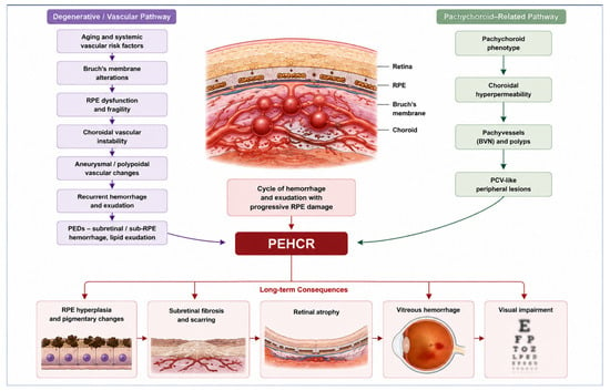
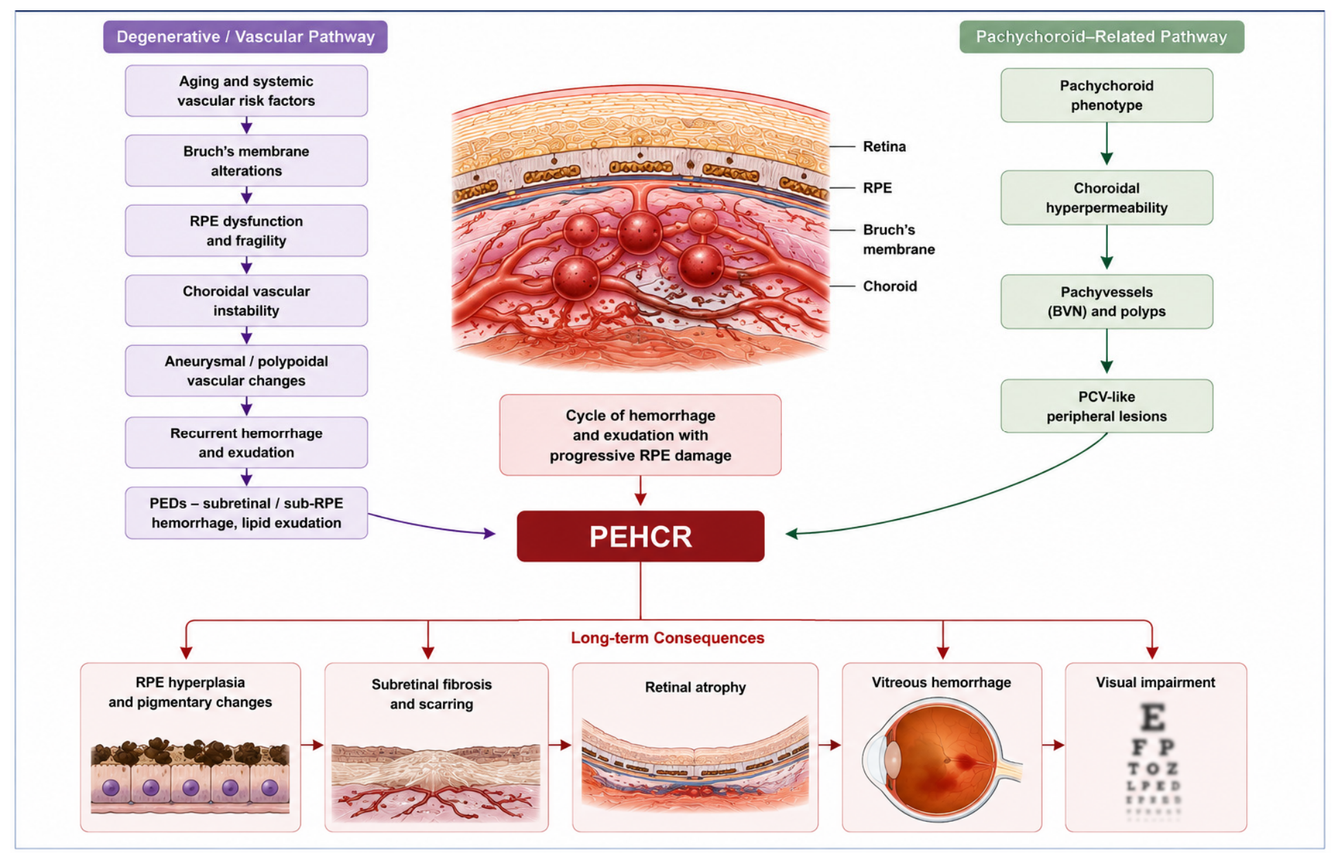
A schematic overview of the hypothesized pathogenic pathways underlying PEHCR is shown in Figure 1. Current evidence suggests that degenerative and vascular mechanisms are not mutually exclusive but may instead coexist and interact throughout disease progression. Age-related degeneration of Bruch’s membrane and RPE dysfunction may create a permissive environment for abnormal choroidal vascular remodeling, whereas chronic vascular congestion and hyperpermeability may further aggravate retinal degeneration and hemorrhagic instability.
Figure 1.
Proposed Pathophysiological Mechanisms of PEHCR. Systemic vascular factors likely contribute substantially to disease development and progression. Hypertension, arteriosclerotic cardiovascular disease, and anticoagulant or antiplatelet therapy are frequently reported in patients with PEHCR and may increase the propensity for recurrent hemorrhage [17,19]. Nevertheless, these therapies are generally considered contributing rather than causative factors because many patients exhibit imaging evidence of underlying vascular abnormalities independent of systemic treatment. Altered vascular autoregulation, endothelial dysfunction, and impaired choroidal perfusion associated with systemic vascular disease may further exacerbate hemorrhagic events and chronic exudation.
The therapeutic response observed following intravitreal anti-vascular endothelial growth factor (anti-VEGF) therapy and photodynamic therapy (PDT) also provides indirect evidence supporting a vascular pathogenesis closely related to PCV and pachychoroid disease [13,14,15]. Anti-VEGF therapy may reduce vascular permeability, subretinal fluid accumulation, and hemorrhagic activity, while PDT may induce regression of abnormal aneurysmal vascular complexes and decrease choroidal hyperpermeability. These treatment responses strongly suggest that active vascular remodeling and choroidal dysfunction play central roles in disease activity rather than representing merely secondary degenerative phenomena.
Despite substantial advances in multimodal imaging, several important questions regarding the pathogenesis of PEHCR remain unresolved. It is still unclear whether PEHCR should be considered a distinct clinical entity, an advanced peripheral variant of AMD, or part of a broader pachychoroid-PCV disease spectrum. Future histopathologic investigations, genetic studies, and longitudinal multimodal imaging analyses will likely be essential to clarify the precise mechanisms underlying this disorder and to better define its nosologic classification within retinal and choroidal vascular diseases. Future multimodal imaging studies should also investigate quantitative choroidal biomarkers, including choroidal volume and the choroidal vascularity index (CVI). These parameters have emerged as valuable indicators of choroidal structural remodeling in several pachychoroid-related disorders and may help clarify whether PEHCR represents a peripheral manifestation within the pachychoroid spectrum.
Age-related degeneration of Bruch’s membrane and RPE, associated with choroidal vascular fragility and pachychoroid-related abnormalities, may induce aneurysmal vascular dilations, chronic exudation, recurrent hemorrhage, and progressive fibrosis. Chronic vascular congestion and choroidal hyperpermeability may contribute to the development of hemorrhagic PEDs and peripheral retinal degeneration.
5. Clinical Presentation
The clinical manifestations of PEHCR are remarkably heterogeneous and largely depend on lesion location, activity, chronicity, anatomical extension, and the development of secondary complications [1,5]. In many patients, the disease remains asymptomatic for prolonged periods and is discovered incidentally during routine dilated fundus examination or ultra-widefield retinal imaging performed for unrelated indications [1]. This asymptomatic presentation is particularly common in lesions confined to the extreme temporal periphery without posterior extension. Conversely, symptomatic patients may present with blurred vision, floaters, photopsias, metamorphopsia, peripheral visual field defects, or sudden visual loss secondary to vitreous hemorrhage or macular involvement [5]. Symptoms are often intermittent and may correlate with recurrent hemorrhagic episodes or fluctuations in exudative activity.
Funduscopically, PEHCR typically appears as elevated hemorrhagic or hemorrhagic–exudative lesions involving the peripheral subretinal and/or sub-retinal pigment epithelium (sub-RPE) space [1,5]. The lesions frequently exhibit complex morphological characteristics, including variable pigmentation, lipid exudation, subretinal fibrosis, pigment epithelial detachments (PEDs), and localized areas of retinal pigment epithelium hyperplasia. Fresh lesions often demonstrate dark red or orange subretinal hemorrhage associated with serosanguineous PEDs, whereas chronic lesions may evolve into densely fibrotic and pigmented scars with extensive RPE remodeling [16]. In some eyes, the coexistence of old and new hemorrhagic components suggests recurrent cycles of vascular instability and repeated bleeding.
The temporal peripheral retina between the equator and ora serrata represents the most frequently affected location, although circumferential or multifocal involvement has also been described [1]. Bilateral lesions may occur in a substantial proportion of patients, further supporting the concept of a diffuse degenerative or vascular disorder rather than an isolated focal abnormality [5,16]. Lesion size and extent are highly variable, ranging from small localized hemorrhagic PEDs to large peripheral hemorrhagic masses associated with extensive exudation and fibrosis.
As the disease progresses, hemorrhage and exudation may extend posteriorly toward the posterior pole and macula, potentially resulting in significant visual deterioration [17]. Macular involvement may occur secondary to subretinal fluid accumulation, lipid exudation, vitreous hemorrhage, or extension of hemorrhagic detachment. Vitreous hemorrhage represents one of the most clinically significant complications of PEHCR and may occasionally constitute the initial presenting manifestation [17]. In severe cases, dense vitreous hemorrhage may obscure fundus visualization and create substantial diagnostic uncertainty, sometimes requiring ultrasonography or pars plana vitrectomy for adequate assessment and management.
Additional clinical findings reported in PEHCR include peripheral drusen, RPE atrophy, chronic subretinal fluid, peripheral disciform scarring, and areas of chorioretinal degeneration [6,20]. Some patients may also exhibit concurrent features of age-related macular degeneration (AMD), pachychoroid disease, or polypoidal choroidal vasculopathy (PCV), supporting the hypothesis of shared pathogenic mechanisms [6,7,8]. Hemorrhagic PEDs and recurrent exudative episodes further reinforce the possible vascular relationship between PEHCR and peripheral PCV.
Because of their elevated configuration, pigmentation, and hemorrhagic appearance, PEHCR lesions frequently mimic choroidal melanoma and other peripheral choroidal tumors. This pseudomelanomatous presentation remains one of the most clinically important aspects of the disease [5,10]. Careful peripheral retinal examination combined with multimodal imaging—including ultra-widefield imaging, OCT, fluorescein angiography, ICGA, OCTA, and B-scan ultrasonography—is therefore essential to establish the correct diagnosis and avoid unnecessary invasive procedures or overtreatment. Recognition of characteristic imaging findings, stability over time, and the absence of typical ultrasonographic melanoma features are fundamental for differentiating PEHCR from malignant lesions [10,22]. The principal clinical manifestations associated with PEHCR are summarized in Table 2.
Table 2.
Clinical Features of PEHCR.
6. Multimodal Imaging
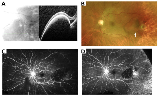
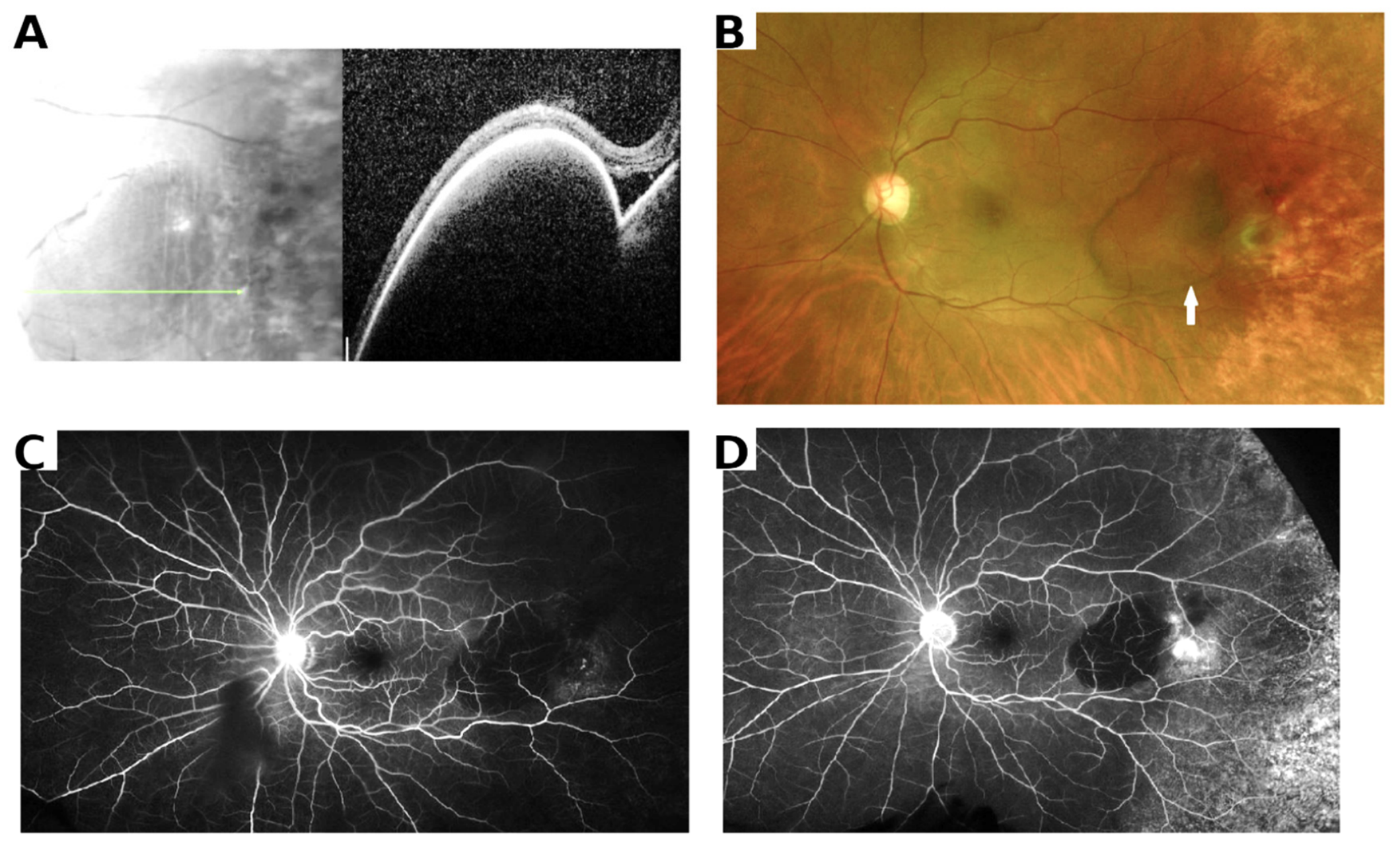
Multimodal imaging represents the cornerstone of PEHCR diagnosis and management [6,7,8,9,10,11,12]. Ultra-widefield imaging has dramatically improved the evaluation of peripheral retinal lesions and currently represents one of the most important diagnostic tools [11]. Representative multimodal imaging findings of PEHCR are illustrated in Figure 2. Ultra-widefield color fundus photography allows documentation of hemorrhage, exudation, fibrosis, and lesion extension, whereas fundus autofluorescence demonstrates mixed hypoautofluorescent and hyperautofluorescent alterations corresponding to RPE degeneration and chronic hemorrhagic changes [11].
Figure 2.
Representative multimodal imaging of peripheral exudative hemorrhagic chorioretinopathy (PEHCR). (A) Spectral-domain optical coherence tomography (SD-OCT) demonstrating a large hemorrhagic pigment epithelial detachment (PED) with shallow overlying subretinal fluid. (B) Ultra-widefield color fundus photograph showing a temporal hemorrhagic PED associated with adjacent fibrovascular changes and peripheral subretinal hemorrhage (arrow). (C) Early phase fluorescein angiography demonstrating hypofluorescence due to blockage from the hemorrhagic PED. (D) Late-phase fluorescein angiography showing persistent blocked fluorescence with mild peripheral staining and no evidence of intrinsic tumor circulation. Together, these multimodal imaging findings are characteristic of PEHCR and highlight the value of multimodal retinal imaging in distinguishing PEHCR from choroidal melanoma. Adapted from Elwood et al., Medicina (2023), distributed under the Creative Commons Attribution 4.0 International (CC BY 4.0) License [8].
Fluorescein angiography may reveal blocked fluorescence caused by hemorrhage and areas of late leakage associated with active exudative components [6]. Nevertheless, ICGA is considered particularly valuable because it frequently demonstrates aneurysmal dilations, polypoidal lesions, and branching vascular networks closely resembling those observed in PCV [6,7]. These findings strongly support the theory that PEHCR may represent a peripheral form of PCV or pachychoroid disease.
OCT typically demonstrates hemorrhagic PEDs, subretinal fluid, hyperreflective subretinal material, and outer retinal disruption [8]. In chronic disease, retinal thinning and fibrotic degeneration may become evident. EDI-OCT studies revealed increased choroidal thickness and dilated pachyvessels in selected patients, further supporting a pachychoroid-related pathogenesis [8,12].
OCTA may additionally reveal abnormal vascular flow patterns and branching vascular networks without dye injection [23]. However, peripheral OCTA imaging remains technically challenging and its precise role in PEHCR continues to evolve. Ultrasonography remains fundamental in the differential diagnosis with choroidal melanoma because PEHCR lesions generally demonstrate high internal reflectivity without acoustic hollowness [5]. The characteristic findings associated with each imaging modality are summarized in Table 3.
Table 3.
Multimodal Imaging Findings in PEHCR.
Because PEHCR lesions are typically located between the equator and the ora serrata, conventional 30–55° fundus imaging frequently fails to document the entire lesion. Ultra-widefield imaging enables comprehensive visualization of lesion extension, multifocal involvement, peripheral hemorrhage, and exudation within a single acquisition. Furthermore, UWF imaging facilitates longitudinal follow-up by allowing accurate comparison of lesion morphology over time and improves communication with ocular oncologists when differentiating PEHCR from peripheral choroidal tumors.
7. Differential Diagnosis
The differential diagnosis of PEHCR includes a broad spectrum of benign and malignant retinal and choroidal disorders, many of which may present with peripheral hemorrhagic and exudative lesions that closely resemble the characteristic appearance of PEHCR. Among these entities, choroidal melanoma remains the most critical diagnosis to exclude because PEHCR may clinically simulate both pigmented and amelanotic melanoma, particularly when lesions are elevated, associated with subretinal hemorrhage, fibrosis, or pigment epithelial detachment [5,10]. Historically, numerous cases of PEHCR were initially misdiagnosed as melanoma, occasionally resulting in unnecessary invasive procedures, including enucleation [1,5]. Careful longitudinal observation and multimodal imaging are therefore essential in distinguishing these conditions. In contrast to PEHCR, choroidal melanoma typically demonstrates progressive enlargement over time, low-to-medium internal reflectivity on B-scan ultrasonography, acoustic hollowness, intrinsic vascular pulsations, and the classic mushroom- or collar-button configuration resulting from rupture through Bruch’s membrane [22]. Additional features favoring melanoma include the presence of orange pigment, subretinal fluid extending posteriorly, and documented tumor growth during follow-up.
Choroidal metastases may also present as elevated hemorrhagic peripheral lesions and occasionally pose substantial diagnostic difficulty, especially in elderly patients [24]. However, metastatic lesions generally exhibit creamy-yellow coloration, multifocality, diffuse choroidal infiltration, and relatively rapid progression. Unlike PEHCR, they are frequently associated with a known systemic malignancy, particularly breast or lung carcinoma, and often display placoid morphology with extensive subretinal fluid on imaging [24]. Ultrasonography typically reveals lower lesion thickness and less pronounced dome-shaped elevation compared with melanoma. Clinical history and systemic evaluation therefore play a pivotal role in the differential diagnosis.
Retinal vasoproliferative tumors (VPTs) may similarly produce exudation, intraretinal or subretinal hemorrhage, and secondary retinal detachment, thereby mimicking PEHCR in selected cases [25]. Nevertheless, VPTs usually occur in younger patients and are commonly associated with retinal gliosis, telangiectatic vessels, intraretinal exudation, and surrounding pigmentary changes. These lesions are primarily retinal rather than choroidal in origin and frequently arise in association with inflammatory, vascular, or degenerative retinal disorders [25]. Fluorescein angiography often demonstrates prominent vascular leakage and feeding retinal vessels, features that are less characteristic of PEHCR.
The relationship between PEHCR and peripheral polypoidal choroidal vasculopathy (PCV) remains particularly intriguing and continues to stimulate considerable debate. Several investigators proposed that both disorders may represent different manifestations of the same underlying pachychoroid-driven vascular abnormality [6,7]. ICGA findings demonstrating polypoidal aneurysmal dilations and branching vascular networks strongly support this hypothesis and suggest that PEHCR may represent a peripheral variant within the PCV spectrum [6,7,12]. Moreover, both conditions share common features including hemorrhagic PEDs, recurrent subretinal hemorrhage, exudation, pachyvessels, and responsiveness to anti-VEGF therapy and PDT [13,14,15]. Nevertheless, peripheral PCV tends to demonstrate more clearly identifiable aneurysmal lesions and vascular networks on ICGA, whereas PEHCR may present with more extensive hemorrhagic and fibrotic degeneration.
Retinal arterial macroaneurysms should additionally be considered in the differential diagnosis because they may cause multilayered retinal hemorrhage, exudation, and vitreous hemorrhage [19]. However, macroaneurysms are generally distinguishable by their direct association with retinal arterial branches and by their characteristic angiographic appearance showing focal arterial dilation and leakage. They are frequently associated with systemic hypertension and arteriosclerotic vascular disease and are usually located along the temporal retinal arcades rather than in the extreme peripheral fundus.
Other less frequent entities entering the differential diagnosis include peripheral retinal angiomatous lesions, organized subretinal hemorrhage, peripheral fibrovascular scars, and inflammatory or infectious chorioretinal disorders. In some cases, chronic PEHCR lesions with marked fibrosis and pigmentation may simulate organized hematoma or even peripheral retinal detachment, further complicating diagnosis. Consequently, accurate identification of PEHCR requires integration of detailed clinical examination, multimodal imaging findings, ultrasonographic characteristics, systemic history, and careful longitudinal follow-up.
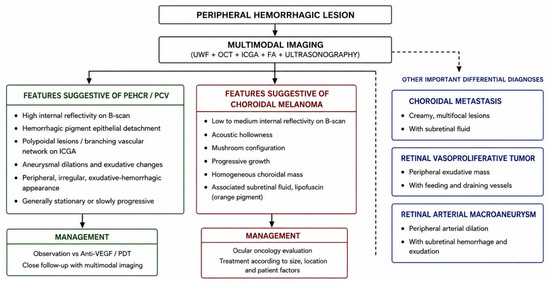
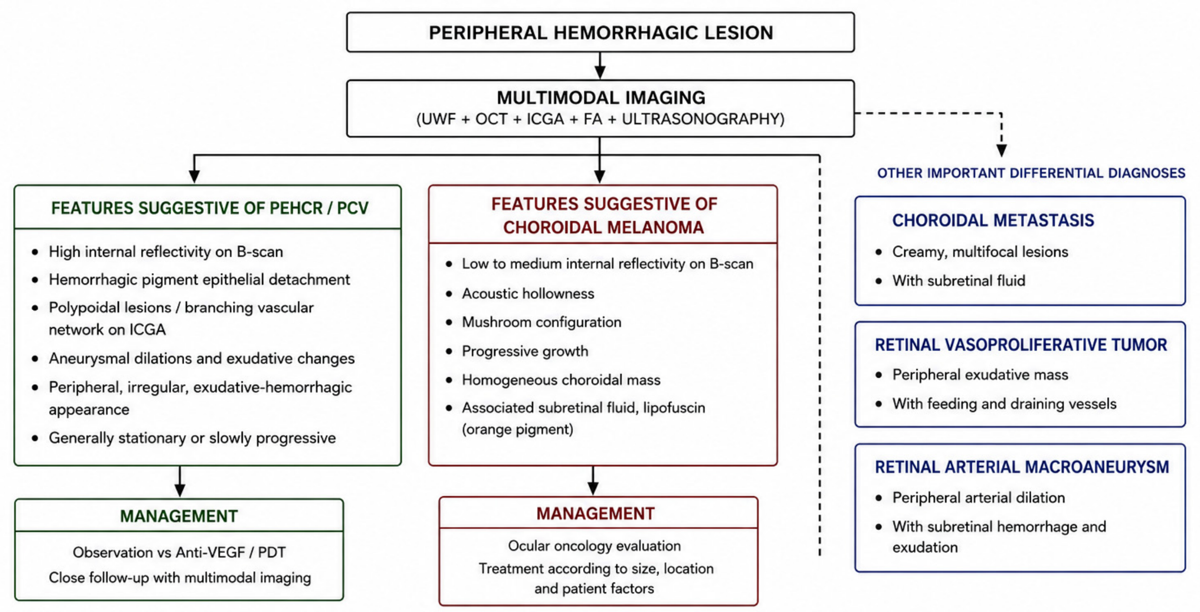
Awareness of PEHCR among retinal specialists and ocular oncologists remains crucial because misdiagnosis may lead to unnecessary biopsies, overtreatment, or major surgical procedures. The increasing use of ultra-widefield imaging, OCT, ICGA, and OCTA has substantially improved diagnostic accuracy and has enhanced the recognition of the vascular and pachychoroid-related features underlying this condition. The principal differential diagnostic features distinguishing PEHCR from other peripheral hemorrhagic lesions are summarized in Table 4 and schematically illustrated in Figure 3.
Table 4.
Differential Diagnosis of PEHCR.
Figure 3.
Diagnostic Algorithm for Peripheral Hemorrhagic Lesions.
Peripheral exudative hemorrhagic chorioretinopathy (PEHCR) should be differentiated from several benign and malignant ocular disorders, particularly choroidal melanoma. Multimodal imaging, including ultra-widefield imaging, optical coherence tomography (OCT), indocyanine green angiography (ICGA), fluorescein angiography (FA), and B-scan ultrasonography, plays a crucial role in distinguishing these entities. Lesions demonstrating high internal reflectivity, hemorrhagic pigment epithelial detachments, peripheral aneurysmal dilations, and branching vascular networks on ICGA are more suggestive of PEHCR or peripheral polypoidal choroidal vasculopathy. Conversely, low-to-medium internal reflectivity, acoustic hollowness, progressive growth, and mushroom-shaped configuration are characteristic findings of choroidal melanoma. Additional differential diagnoses include choroidal metastasis, retinal vasoproliferative tumor, retinal arterial macroaneurysm, and peripheral retinal angiomatous lesions. Accurate diagnosis is essential to avoid unnecessary invasive procedures and inappropriate treatment.
8. Natural History and Management
The natural history of PEHCR is highly variable and remains incompletely characterized because of the rarity of the disease, the heterogeneous clinical presentation, and the absence of large prospective longitudinal studies [1,3]. Clinical evolution ranges from long-term stability without significant progression to recurrent hemorrhagic episodes associated with progressive fibrosis, exudation, and vitreoretinal complications [17]. In many patients, lesions remain confined to the peripheral retina and may remain asymptomatic for years, particularly when the macula is spared. Conversely, some eyes demonstrate recurrent cycles of hemorrhage and exudation that may progressively extend posteriorly toward the posterior pole, eventually compromising visual function.
Visual prognosis is generally favorable when lesions remain peripheral and inactive; however, extensive exudation, recurrent subretinal hemorrhage, vitreous hemorrhage, or macular involvement may result in substantial and sometimes irreversible visual impairment [1,17]. Chronic disease may lead to subretinal fibrosis, retinal pigment epithelium (RPE) atrophy, outer retinal degeneration, and disciform scarring, all of which contribute to permanent visual dysfunction. In some patients, recurrent hemorrhagic activity may produce cumulative retinal damage despite apparent anatomical stabilization of the lesion.
Several retrospective studies have suggested that a considerable proportion of PEHCR lesions follow a relatively indolent course and may undergo spontaneous stabilization or partial regression over time [1,11]. Nevertheless, lesion behavior is unpredictable, and recurrent hemorrhage may occur even after prolonged periods of quiescence. Consequently, careful long-term follow-up remains essential, particularly in patients demonstrating active exudation, hemorrhagic pigment epithelial detachments (PEDs), or posterior extension toward the macula.
Observation remains the preferred management strategy for asymptomatic and stable lesions without evidence of active exudation or progressive hemorrhage [1]. Conservative management includes periodic clinical examination and multimodal imaging follow-up using ultra-widefield imaging, OCT, fluorescein angiography, ICGA, and occasionally ultrasonography to evaluate lesion stability, recurrent bleeding, and macular involvement. Monitoring is especially important because PEHCR may clinically mimic malignant lesions such as choroidal melanoma, and documented lesion stability over time may significantly assist in confirming the diagnosis.
Over the past decade, intravitreal anti-vascular endothelial growth factor (anti-VEGF) therapy has emerged as one of the most important treatment options for active exudative PEHCR [13,14,15]. The rationale for anti-VEGF therapy derives from the increasing recognition of PCV-like vascular abnormalities and pachychoroid-related mechanisms underlying the disease. Bevacizumab, ranibizumab, aflibercept, and more recently faricimab have been used with variable success to reduce subretinal fluid, exudation, hemorrhagic activity, and vascular leakage [13,14]. Several reports documented anatomical improvement following anti-VEGF injections, including regression of hemorrhagic PEDs, reduction in exudation, and stabilization of visual acuity. However, treatment response appears heterogeneous, and some lesions demonstrate persistent or recurrent activity despite repeated injections. The optimal treatment regimen, injection interval, and the duration of therapy remain undefined because of the lack of prospective controlled studies.
Faricimab has recently attracted interest because of its dual inhibition of VEGF-A and angiopoietin-2 pathways, potentially offering improved vascular stabilization and reduced choroidal hyperpermeability. Although clinical experience in PEHCR remains limited, preliminary observations suggest that faricimab may represent a promising therapeutic option in refractory or recurrent exudative disease [13,14,15]. Future studies will be required to determine whether dual-pathway inhibition offers advantages over conventional anti-VEGF monotherapy in this setting.
Photodynamic therapy (PDT) may be particularly beneficial in lesions demonstrating PCV-like vascular abnormalities on ICGA, including aneurysmal dilations and branching vascular networks [15]. PDT may induce selective thrombosis and regression of abnormal choroidal vascular complexes while reducing choroidal hyperpermeability and chronic exudation. Combination therapy using anti-VEGF agents together with PDT has also been proposed in selected refractory cases, mirroring treatment strategies employed in PCV. Nevertheless, evidence supporting PDT in PEHCR remains limited to small retrospective series and isolated case reports.
Peripheral laser photocoagulation has historically been employed in localized lesions characterized by exudation or peripheral vascular abnormalities [26]. However, this modality is currently less favored because of inconsistent efficacy, limited penetration in hemorrhagic lesions, and the potential risk of retinal scarring, visual field defects, and induction of further fibrosis. Cryotherapy has also been described historically but is rarely utilized in contemporary practice because of the availability of less destructive therapeutic alternatives.
Pars plana vitrectomy may become necessary in cases complicated by dense non-clearing vitreous hemorrhage, tractional changes, or persistent diagnostic uncertainty [17]. Surgical intervention generally provides satisfactory anatomical outcomes and may restore media clarity, allowing accurate retinal assessment and preventing secondary complications related to chronic vitreous hemorrhage. However, postoperative visual recovery may be limited by underlying retinal degeneration, chronic subretinal fibrosis, or associated macular pathology. In some cases, surgery may additionally facilitate exclusion of intraocular malignancy when clinical diagnosis remains uncertain.
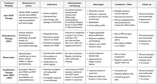
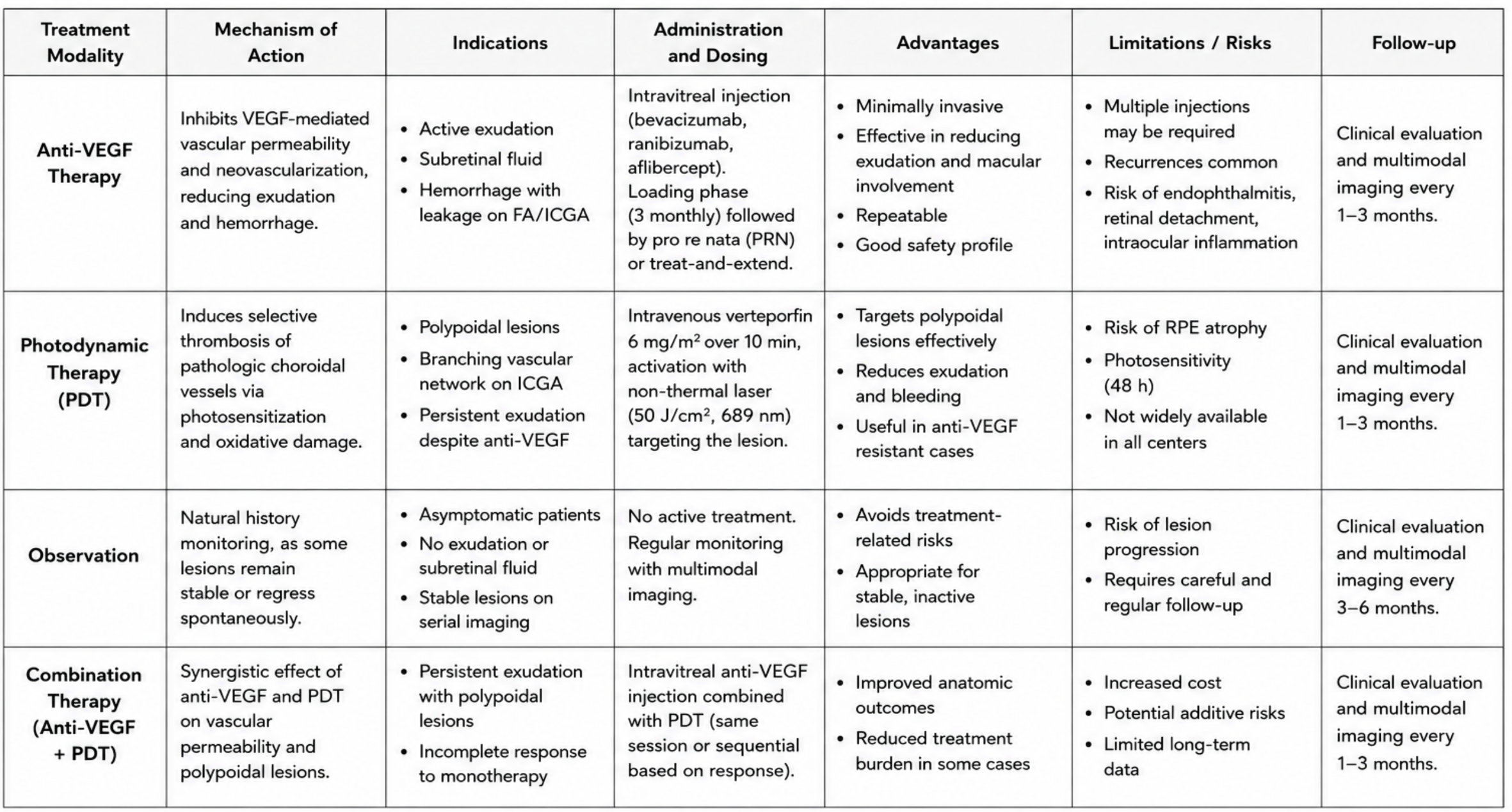
Despite substantial advances in multimodal imaging and therapeutic options, no standardized evidence-based management guidelines currently exist for PEHCR. Treatment decisions are therefore largely individualized and based on lesion activity, symptom severity, imaging findings, posterior extension, and the presence of complications. Future multicenter prospective studies integrating multimodal imaging, functional outcomes, and long-term follow-up will likely be essential to define optimal therapeutic strategies, identify prognostic factors, and better characterize the natural history of this complex disorder. Current therapeutic approaches and their principal indications are summarized in Table 5, while a proposed management algorithm is illustrated in Figure 4.
Table 5.
Therapeutic Strategies in PEHCR.
Figure 4.
Proposed Therapeutic Algorithm for PEHCR.
Summary of the principal management approaches currently adopted for PEHCR according to lesion activity, multimodal imaging findings, and clinical presentation. Observation remains appropriate for asymptomatic and stable peripheral lesions without evidence of active exudation or subretinal fluid. Intravitreal anti-vascular endothelial growth factor (anti-VEGF) therapy represents the most frequently employed treatment for active exudative disease, hemorrhagic pigment epithelial detachments, and lesions associated with subretinal fluid or leakage on fluorescein angiography and indocyanine green angiography. Photodynamic therapy (PDT) may be particularly useful in cases demonstrating polypoidal or branching vascular network abnormalities suggestive of peripheral polypoidal choroidal vasculopathy. Combination therapy with anti-VEGF agents and PDT may provide synergistic effects in refractory or recurrent lesions. Careful follow-up with multimodal imaging, including ultra-widefield imaging, OCT, fluorescein angiography, and ICGA, is essential for monitoring lesion progression, therapeutic response, and recurrence.
9. Conclusions
Peripheral exudative hemorrhagic chorioretinopathy is an uncommon but clinically important retinal disorder that continues to represent a major diagnostic challenge because of its remarkable resemblance to choroidal melanoma and other malignant lesions. Recent advances in multimodal imaging substantially improved the understanding of the disease and increasingly support a relationship between PEHCR, PCV, and pachychoroid spectrum disorders.
Although many lesions remain stable and may be managed conservatively, active exudative disease and severe hemorrhagic complications may require anti-VEGF therapy, laser photocoagulation, photodynamic therapy, or vitreoretinal surgery according to lesion characteristics and disease activity. Improved awareness among retinal specialists and ocular oncologists is essential to avoid unnecessary invasive procedures and overtreatment.
Future prospective studies integrating multimodal imaging, histopathologic investigations, and long-term clinical follow-up will likely be essential to clarify the exact pathogenesis of PEHCR and establish evidence-based management guidelines.
Author Contributions
Conceptualization, F.M.; methodology, F.M.; software, F.M. and L.D.L.; validation, A.M. and S.L.; formal analysis, F.M.; investigation, L.D.L.; resources, F.M.; data curation, E.M.V. and L.D.L.; writing—original draft preparation, F.M.; writing—review and editing, L.D.L., F.M., S.L. and A.B.; visualization, F.M., S.L. and A.B.; supervision, A.M., S.L. and E.M.V. All authors have read and agreed to the published version of the manuscript.
Funding
This research received no external funding.
Institutional Review Board Statement
This study is a literature review and did not involve the generation or analysis of new human data, samples, or clinical material. Therefore, ethics committee approval was not required.
Informed Consent Statement
Not applicable. This study did not involve human participants.
Data Availability Statement
No new data were created or analyzed in this study. Data sharing is not applicable to this article.
Conflicts of Interest
The authors declare no conflicts of interest.
Abbreviations
The following abbreviations are used in this manuscript:
| AMD | Age-Related Macular Degeneration |
| BVN | Branching Vascular Network |
| EDI-OCT | Enhanced Depth Imaging Optical Coherence Tomography |
| FA | Fluorescein Angiography |
| FAF | Fundus Autofluorescence |
| ICGA | Indocyanine Green Angiography |
| OCT | Optical Coherence Tomography |
| OCTA | Optical Coherence Tomography Angiography |
| PCV | Polypoidal Choroidal Vasculopathy |
| PDT | Photodynamic Therapy |
| PED | Pigment Epithelial Detachment |
| PEHCR | Peripheral Exudative Hemorrhagic Chorioretinopathy |
| PRN | Pro Re Nata |
| RPE | Retinal Pigment Epithelium |
| SRF | Subretinal Fluid |
| UWF | Ultra-Widefield |
| VEGF | Vascular Endothelial Growth Factor |
References
- Shields, C.L.; Salazar, P.F.; Mashayekhi, A.; Shields, J.A. Peripheral exudative hemorrhagic chorioretinopathy simulating choroidal melanoma in 173 eyes. Ophthalmology 2009, 116, 529–535. [Google Scholar] [CrossRef] [Scilit] [PubMed]
- Mantel, I.; Uffer, S.; Zografos, L. Peripheral exudative hemorrhagic chorioretinopathy: A clinical, angiographic, and histologic study. Am. J. Ophthalmol. 2009, 148, 932–938.e1. [Google Scholar] [CrossRef] [Scilit] [PubMed]
- Mirshahi, A.; Höhn, F.; Baatz, H.; Müller, M.; Hattenbach, L.O. exudative haemorrhagic chorioretinopathy: Clinical and angiographic findings. Klin. Monatsblatter Fur Augenheilkd. 2009, 226, 659–663. [Google Scholar] [CrossRef] [Scilit] [PubMed]
- Annesley, W.H., Jr. Peripheral exudative hemorrhagic chorioretinopathy. Trans. Am. Ophthalmol. Soc. 1980, 78, 321–364. [Google Scholar] [PubMed] [PubMed Central]
- Shields, J.A.; Augsburger, J.J.; Brown, G.C.; Stephens, R.F. The differential diagnosis of posterior uveal melanoma. Ophthalmology 1980, 87, 518–522. [Google Scholar] [CrossRef] [Scilit] [PubMed]
- Goldman, D.R.; Freund, K.B.; McCannel, C.A.; Sarraf, D. Peripheral polypoidal choroidal vasculopathy as a cause of peripheral exudative hemorrhagic chorioretinopathy: A report of 10 eyes. Retina 2013, 33, 48–55. [Google Scholar] [CrossRef] [Scilit] [PubMed]
- Gedik, S.; Akova, Y.; Yilmaz, G.; Bozbeyoğlu, S. Indocyanine green and fundus fluorescein angiographic findings in patients with active ocular Behcet’s disease. Ocul. Immunol. Inflamm. 2005, 13, 51–58. [Google Scholar] [CrossRef] [Scilit] [PubMed]
- Elwood, K.F.; Richards, P.J.; Schildroth, K.R.; Mititelu, M. Peripheral Exudative Hemorrhagic Chorioretinopathy (PEHCR): Diagnostic and Therapeutic Challenges. Medicina 2023, 59, 1507. [Google Scholar] [CrossRef] [Scilit] [PubMed] [PubMed Central]
- Yannuzzi, L.A.; Sorenson, J.; Spaide, R.F.; Lipson, B. Idiopathic polypoidal choroidal vasculopathy (IPCV). Retina 1990, 10, 1–8. [Google Scholar] [CrossRef] [Scilit] [PubMed]
- Badawi, A.H.; Semidey, V.A.; Magliyah, M.; Al-Dhibi, H. Updated Systematic Review and Clinical Spectrum of Peripheral Exudative Hemorrhagic Chorioretinopathy. Middle East Afr. J. Ophthalmol. 2020, 27, 4–9. [Google Scholar] [CrossRef] [Scilit] [PubMed] [PubMed Central]
- Safir, M.; Zloto, O.; Fabian, I.D.; Moroz, I.; Gaton, D.D.; Vishnevskia-Dai, V. Peripheral Exudative Hemorrhagic Chorioretinopathy with and without treatment-Clinical and multimodal imaging characteristics and prognosis. PLoS ONE 2022, 17, e0275163. [Google Scholar] [CrossRef] [Scilit] [PubMed] [PubMed Central]
- Shroff, D.; Sharma, M.; Chhablani, J.; Gupta, P.; Gupta, C.; Shroff, C. Peripheral Exudative Hemorrhagic Chorioretinopathy-A New Addition to the spectrum of pachychoroid disease? Retina 2021, 41, 1518–1525. [Google Scholar] [CrossRef] [Scilit] [PubMed]
- Seibel, I.; Hager, A.; Duncker, T.; Riechardt, A.I.; Nürnberg, D.; Klein, J.P.; Rehak, M.; Joussen, A.M. Anti-VEGF therapy in symptomatic peripheral exudative hemorrhagic chorioretinopathy (PEHCR) involving the macula. Graefe’s Arch. Clin. Exp. Ophthalmol. 2016, 254, 653–659. [Google Scholar] [CrossRef] [Scilit] [PubMed]
- Shao, J.; Choudhary, M.M.; Schachat, A.P. Neovascular Age-Related Macular Degeneration. Dev. Ophthalmol. 2016, 55, 125–136. [Google Scholar] [CrossRef] [Scilit] [PubMed]
- Schmidt-Erfurth, U.; Hasan, T. Mechanisms of action of photodynamic therapy with verteporfin for the treatment of age-related macular degeneration. Surv. Ophthalmol. 2000, 45, 195–214. [Google Scholar] [CrossRef] [Scilit] [PubMed]
- Shields, C.L.; Manalac, J.; Das, C.; Ferguson, K.; Shields, J.A. Choroidal melanoma: Clinical features, classification, and top 10 pseudomelanomas. Curr. Opin. Ophthalmol. 2014, 25, 177–185. [Google Scholar] [CrossRef] [Scilit] [PubMed]
- Mazal, Z. Peripheral exudative hemorrhagic chorioretinopathy. Cesk Slov. Oftalmol. 2019, 75, 80–84. [Google Scholar] [CrossRef] [Scilit] [PubMed]
- Castro-Navarro, V.; Behar-Cohen, F.; Chang, W.; Joussen, A.M.; Lai, T.Y.Y.; Navarro, R.; Pearce, I.; Yanagi, Y.; Okada, A.A. Pachychoroid: Current concepts on clinical features and pathogenesis. Graefe’s Arch. Clin. Exp. Ophthalmol. 2021, 259, 1385–1400. [Google Scholar] [CrossRef] [Scilit] [PubMed] [PubMed Central]
- Kuhli-Hattenbach, C.; Fischer, I.B.; Schalnus, R.; Hattenbach, L.O. Subretinal hemorrhages associated with age-related macular degeneration in patients receiving anticoagulation or antiplatelet therapy. Am. J. Ophthalmol. 2010, 149, 316–321.e1. [Google Scholar] [CrossRef] [Scilit] [PubMed]
- Fleckenstein, M.; Schmitz-Valckenberg, S.; Chakravarthy, U. Age-Related Macular Degeneration: A Review. JAMA 2024, 331, 147–157. [Google Scholar] [CrossRef] [Scilit] [PubMed] [PubMed Central]
- Kiciński, K.; Gawęcki, M. Choroidal and Retinal Thicknesses in Healthy Eyes Measured with Ultra-Wide-Field Optical Coherence Tomography. Diagnostics 2024, 14, 1114. [Google Scholar] [CrossRef] [Scilit] [PubMed] [PubMed Central]
- Singh, A.D.; Turell, M.E.; Topham, A.K. Uveal melanoma: Trends in incidence, treatment, and survival. Ophthalmology 2011, 118, 1881–1885. [Google Scholar] [CrossRef] [Scilit] [PubMed]
- Spaide, R.F.; Klancnik, J.M., Jr.; Cooney, M.J. Retinal vascular layers imaged by fluorescein angiography and optical coherence tomography angiography. JAMA Ophthalmol. 2015, 133, 45–50. [Google Scholar] [CrossRef] [Scilit] [PubMed]
- Shields, C.L.; Welch, R.J.; Malik, K.; Acaba-Berrocal, L.A.; Selzer, E.B.; Newman, J.H.; Mayro, E.L.; Constantinescu, A.B.; Spencer, M.A.; McGarrey, M.P.; et al. Uveal Metastasis: Clinical Features and Survival Outcome of 2214 Tumors in 1111 Patients Based on Primary Tumor Origin. Middle East Afr. J. Ophthalmol. 2018, 25, 81–90. [Google Scholar] [CrossRef] [Scilit] [PubMed] [PubMed Central]
- Shields, C.L.; Kaliki, S.; Al-Dahmash, S.; Rojanaporn, D.; Shukla, S.Y.; Reilly, B.; Shields, J.A. Retinal vasoproliferative tumors: Comparative clinical features of primary vs secondary tumors in 334 cases. JAMA Ophthalmol. 2013, 131, 328–334. [Google Scholar] [CrossRef] [Scilit] [PubMed]
- Singer, M.A.; Tan, C.S.; Surapaneni, K.R.; Sadda, S.R. Targeted photocoagulation of peripheral ischemia to treat rebound edema. Clin. Ophthalmol. 2015, 9, 337–341. [Google Scholar] [CrossRef] [Scilit] [PubMed] [PubMed Central][Green Version]

Disclaimer/Publisher’s Note: The statements, opinions and data contained in all publications are solely those of the individual author(s) and contributor(s) and not of MDPI and/or the editor(s). MDPI and/or the editor(s) disclaim responsibility for any injury to people or property resulting from any ideas, methods, instructions or products referred to in the content. |
© 2026 by the authors. Licensee MDPI, Basel, Switzerland. This article is an open access article distributed under the terms and conditions of the Creative Commons Attribution (CC BY) license.