


Abstract
Background/Aims: This study set out to compare patient experiences and attitudes toward virtual reality (VR) versus in-office ophthalmic functional testing, as well as their eye exam visit preferences. Methods: A total of 64 patients undergoing a comparative clinical trial including VR and in-office versions of 24-2 visual field perimetry, Pelli–Robson contrast sensitivity, ETDRS visual acuity, Ishihara plates, and Amsler Grid tests were given a survey that included Likert-scaled questions regarding overall experience, speed, and comfort of broad VR testing versus in-office tests, as well as binary modality preference, like or dislike of various exam modality aspects, ease of understanding each test, distance traveled to their appointment, visit frequency, and remote care preferences. Results: In terms of overall experience, 59 (93.7%) participants rated VR as “Liked” or “Very Much Liked” compared to 38 (60.3%) for in-office tests (χ2 p < 0.0001). Regarding comfort, 59 (93.7%) and 38 (60.3%) participants at least “Liked” VR and in-office tests, respectively (χ2 p < 0.0001). For speed, 60 (95.2%) and 36 (57.1%) participants at least “Liked” VR and in-office tests, respectively (χ2 p < 0.0001). Accordingly, 11 (17.5%) patients liked all aspects of in-office tests, while 54 (85.7%) patients said the same for VR (χ2 p < 0.0001). Overall, 33 (52.4%) patients disliked something about in-office tests, while 20 (31.7%) said the same for VR (χ2 p < 0.0001). There were no differences in ability to understand tests between the two modalities. We noted a variable distribution of distance traveled and visits per year in our population, yet there was also a strong preference for implementing more remote eye testing options. Conclusions: Development and implementation of virtual reality analogs to in-office, functional ophthalmic exams can improve patient experience and facilitate remote care delivery.
1. Introduction
High patient volume in ophthalmic workflows remains a widespread challenge globally, often leading to clinic backlogs and worsened patient experiences [1,2,3]. This problem is compounded by a growing supply–demand mismatch in eye care. In the United States, the Health Resources and Services Administration’s Health Workforce Simulation Model projects a 30% national shortage of full-time-equivalent ophthalmologists by 2035, with workforce adequacy in non-metropolitan regions falling as low as 29%—placing ophthalmology among the 2 specialties with the lowest projected workforce adequacy of 38 surveyed [4]. These supply constraints disproportionately burden patients with chronic eye disease, despite consistent evidence that local availability of vision specialists improves outcomes in diseases such as diabetic retinopathy and age-related macular degeneration [5]. Geographic analyses have repeatedly shown that roughly one quarter of U.S. counties have no practicing eye care providers, and that subspecialty access is even more constrained. For example, 85.6% of U.S. counties lack a practicing cornea specialist [6,7]. While primary care providers are more readily accessible, they currently lack the tools necessary to screen for and accurately quantify vision deficits, particularly for early-stage ocular diseases [8].
To address limited access to vision specialists and increase access to vision care, ophthalmology has increasingly adopted telemedicine, especially following the COVID-19 pandemic [9,10,11]. Asynchronous, store-and-forward tele-ophthalmology programs integrated into primary care clinics and federally qualified health centers have demonstrated feasibility, high acceptability, and the ability to detect previously undiagnosed ocular and systemic disease in a substantial proportion of patients [12,13]. However, many of these programs remain anchored to retinal imaging, and structured functional testing (visual fields, contrast sensitivity, color vision, metamorphopsia, and acuity) is rarely delivered outside the specialty clinic. Concurrently, ophthalmic screening is becoming more popular—particularly through emerging technologies such as virtual reality (VR), which enables faster testing and disease detection while offering an additional decision point regarding the need for more comprehensive examination based on screening results.
VR-based perimetry, in particular, has progressed rapidly toward clinical maturity. A recent systematic review by Hekmatjah and colleagues identified at least 14 prospective comparative studies of VR perimetry versus standard automated perimetry in glaucoma, with most reporting moderate-to-strong correlation in global indices but persistent heterogeneity in stimulus algorithms, headset hardware, and reliability metrics [14]. A complementary systematic review by Selvan and colleagues, evaluating VR headsets for perimetry across mixed populations, reached similar conclusions and emphasized that patient-facing usability data remain comparatively under-reported [15]. Multicenter validation studies—including the NOVA trial of the Radius VR perimeter and a prospective comparison of the VisuALL platform with the Humphrey Field Analyzer—have shown concordance in glaucoma staging sufficient to support adjunctive clinical use, while also highlighting that test–retest variability, fixation monitoring, and patient comfort remain meaningful determinants of test reliability [16,17].
Beyond perimetry, head-mounted VR platforms have also been validated for specialized indications such as superior visual field testing prior to functional eyelid surgery, with sensitivity and specificity comparable to standard automated perimetry [18,19]. At-home and remote-administered VR visual field testing has likewise been shown to be feasible, with reasonable retention and compliance over periods of up to two years in motivated glaucoma cohorts [20,21,22].
Despite this expanding validation literature, patient-centered evaluation of VR vision testing remains limited. Existing studies focus on a single test, most often perimetry, within a single diagnostic group, primarily glaucoma [23,24]. Patient comfort, perceived speed, and willingness to use a given modality are critical determinants of adherence, particularly for chronic conditions such as glaucoma. In the present study, our group surveyed patients who had just completed one or more vision test, including VR and/or in-office versions of Ishihara plates for color deficit testing, Amsler Grid for metamorphopsia, Pelli–Robson for contrast sensitivity, 24-2 perimetry for visual fields, and ETDRS for visual acuity, as part of a randomized study [25]. The primary focus was on patient perceptions of overall experience, speed, and comfort between VR and in-office tests. Secondary measures included patient preferences, aspects they liked or disliked about each modality, ease of understanding test instructions, and modality preference. Lastly, the survey assessed the frequency of ophthalmology visits, distance traveled to the practice, and preference for increased remote testing capabilities. This study is unique among the existing patient experience literature in three respects: (1) the survey was administered after a multi-test panel rather than a single test, more accurately representing the realities of test-taking, including patient fatigue and cumulative comfort; (2) the cohort included a balanced mix of healthy and diseased eyes spanning anterior segment, retinal, optic nerve, and oculomotor pathology; and (3) the cohort was demographically diverse, with substantial representation of African American and Hispanic patients, groups often under-represented in VR validation studies.
2. Materials and Methods
2.1. Overall Study Design
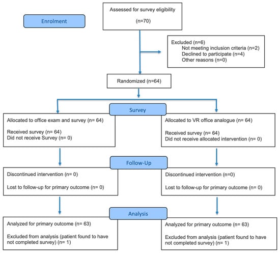
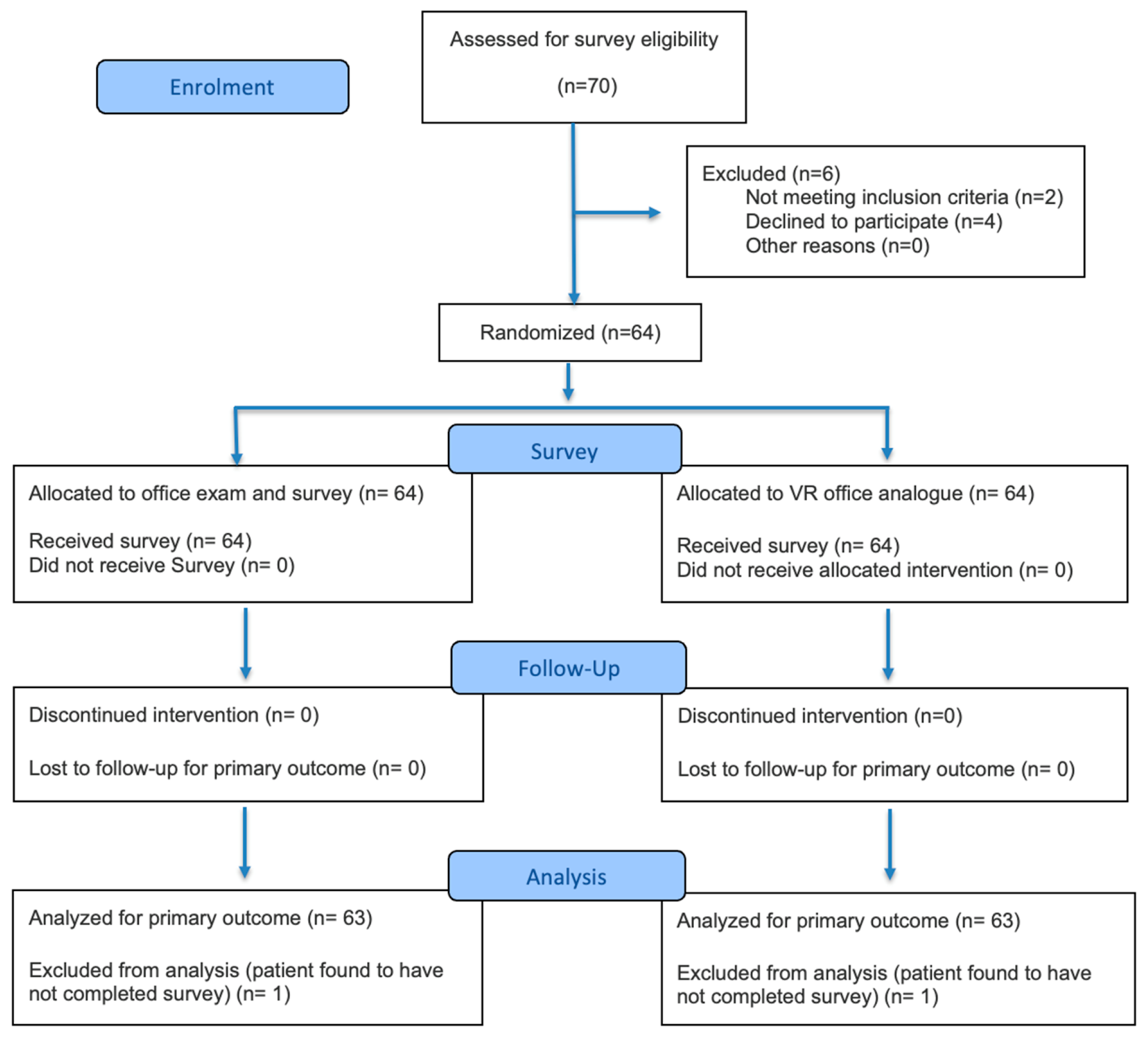
This was a single-center randomized survey study that followed the recommendations of the Consolidated Standards of Reporting Trials (CONSORT), as demonstrated in Figure 1 [26]. This study was approved by the Institutional Review Board (IRB) of the Icahn School of Medicine at Mount Sinai (IRB #20-00358, approval date: 20 January 2020) and registered under NCT04980430 (registered 18 July 2021). Patient recruitment took place from September 2022 until September 2023 and was conducted at the Mount Sinai 102nd Street Ophthalmology Clinic on a voluntary basis, with randomization of either the in-office test followed by the VR analog, or vice versa. A notable limitation of this study is that not all patients completed every examination described above. While this may have affected responses due to varying difficulties of the exams, the goal of this survey was to provide a preliminary subjective comparison of the two testing modalities overall, rather than between specific tests. Patients were asked to complete a survey of their perceptions of both modalities for the tests they completed (Table 1). Any patient-specific data was de-identified prior to data collection, and a study number was assigned to each participant. This study adhered to the ethical principles outlined in the Declaration of Helsinki.
Figure 1.
Participant flow diagram of the progress through the phases of the survey study.
Table 1.
Survey participant demographic data.
VR testing was performed using HTC Vive Pro Eye (HTC Corporation, Taoyuan, Taiwan) or Oculus Go (Oculus VR, Menlo Park, CA, USA) headsets, each delivering an identical panel of tests powered by SteamVR software. Ishihara tile, Pelli–Robson, and ETDRS VR analogs were appropriately scaled, and responses were recorded by selecting from several choices. The Amsler VR analog consisted of free-hand drawing with a VR controller against a virtual grid. The 24-2 VR analog was a suprathreshold version used to evaluate the viability of accelerated, single contrast-level testing (25 decibels) against standard in-office perimetry from sensitivity and patient preference perspectives. In-office assessment included Humphrey Visual Field Analysis (HVFA) 24-2 perimetry (SITA Fast or Full Threshold) (Carl Zeiss Meditec, Inc., Dublin, CA, USA), Precision Vision’s ETDRS (Precision Vision, Woodstock, IL, USA) and Pelli–Robson charts, and Amsler grid printouts. The HVFA was administered by a certified technician as part of the patient’s visit at the ophthalmology clinic, which precluded administration of a suprathreshold version more closely matching the accelerated 24-2 VR test used. All other examinations were delivered by trained research assistants. The survey was administered on a tablet, and the patient was given time and private space to independently rate their responses.
2.2. Statistical Analysis
All statistical analyses were conducted in GraphPad Prism version 7. Chi-squared tests (χ2) were used for multi-option response comparisons between the two testing modalities. All graphs were made with GraphPad Prism version 7.
3. Results
A total of sixty-four participants successfully completed at least one of the five tests in both VR and in-office formats and completed the survey, as summarized in Table 1. The majority of participants were under 60 years old (n = 41; 64.1%), female (n = 41; 64.1%), and identified as African American (n = 23; 35.9%), Caucasian (n = 22; 34.3%), or Hispanic (n = 11; 17.2%). The study cohort included patients with either healthy eyes (n = 21; 32.8%) or diseased eyes (n = 43; 67.2%), with the latter group consisting of anterior chamber, retinal, optic nerve, and oculomotor diseases.
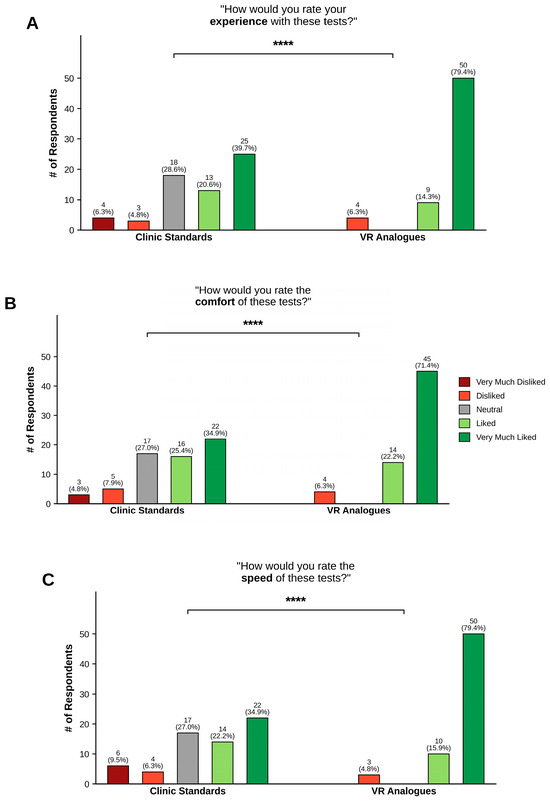
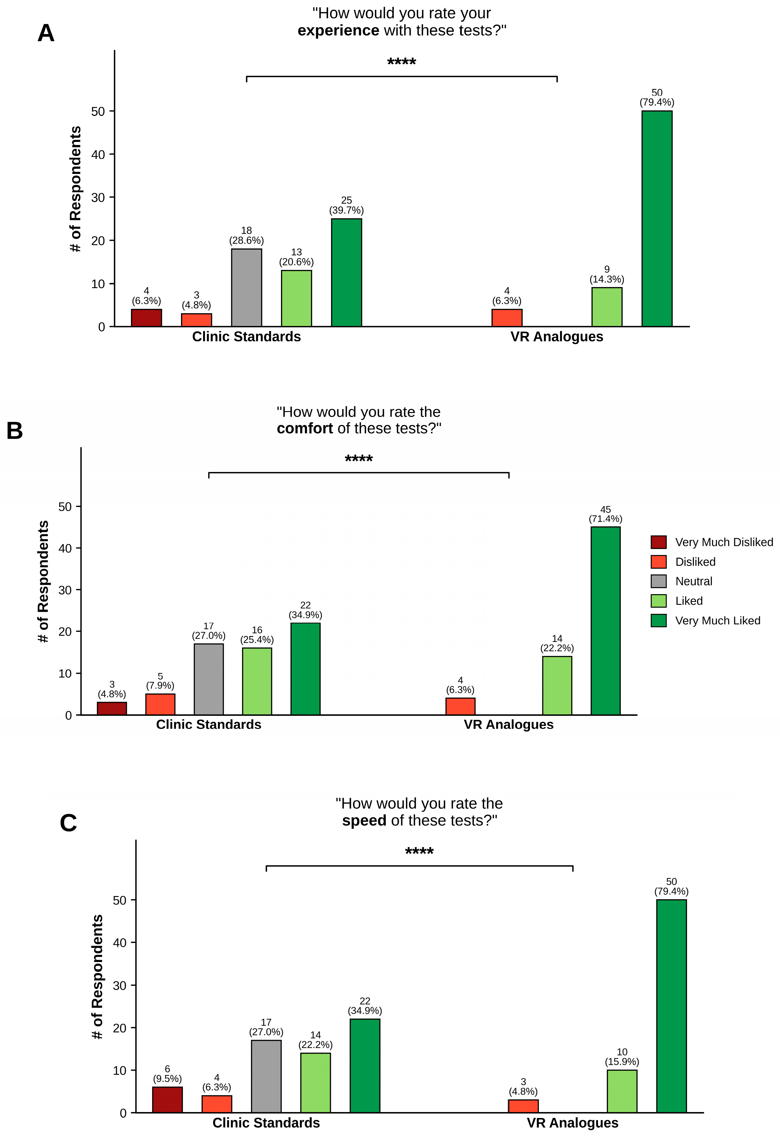
Figure 2 depicts the response distributions for the three Likert-scaled questions on the survey. For the first question, “How would you rate your experience with these tests?” (Figure 2A), the distribution for the VR analog was significantly more positive than for the clinical standard, with 59 participants (93.7%) expressing a favorable opinion of VR compared to 38 (60.3%) for in-office tests (p < 0.0001; χ2 test). Regarding comfort (Figure 2B), 59 participants (93.7%) at least “Liked” VR compared to 38 (60.3%) for in-office tests (p < 0.0001; χ2 test). For the last question, “How would you rate the speed of these tests?” (Figure 2C), 60 participants (95.2%) at least “Liked” VR compared to 36 (57.1%) for in-office tests (p < 0.0001; χ2 test). The observed differences in speed preferences might be affected by the delivery of VR suprathreshold perimetry, which was notably quicker than the HVFA 24-2 SITA Fast and Full Threshold algorithms. This consideration is relevant for the deployment of these tests in non-vision-specialist locations and for patient adherence to regular testing for specific disease states.
Figure 2.
Likert-scaled questions comparing patient preferences for standard in-clinic versus VR exams. Patients were more likely to prefer the VR analog battery than clinic standards in terms of overall experience (A), comfort (B), and speed (C). **** p < 0.0001 (χ2 test).
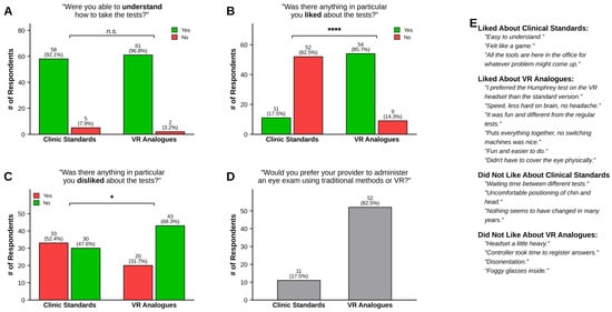
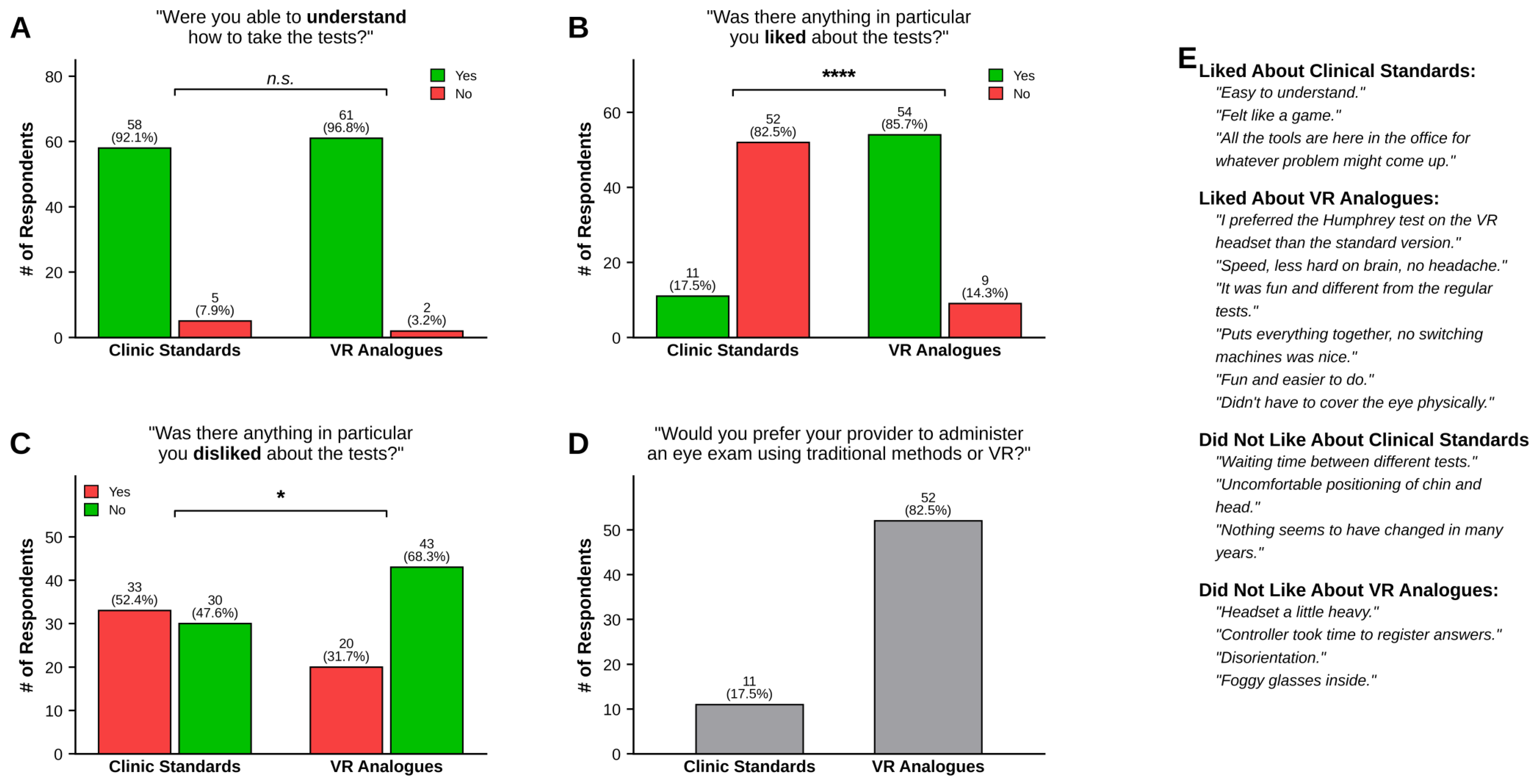
To control for technician clarity and the simplicity of VR testing, this study evaluated whether patients experienced greater difficulty understanding either of the testing modalities. As shown in Figure 3A, there was no significant difference in the proportion of patients who understood both modalities, with 58 (92.1%) and 61 (96.8%) of participants understanding the clinical standards and VR tests, respectively (p = 0.2433; χ2 test). Despite equal clarity in understanding, participants were significantly more likely to like an aspect of VR testing (54, 85.7%) than in-office tests (11, 17.5%) (Figure 3B; p < 0.0001; χ2 test). Although understanding of the tests was comparable, participants were significantly less likely to dislike an aspect of VR testing (20, 31.7%) than in-office tests (33, 52.3%) (Figure 3C; p < 0.0001; χ2 test). When presented with the option of standard in-office testing or VR, a strong majority of the patients chose VR (Figure 3D). Figure 3E depicts representative patient quotes for the “Likes” and “Dislikes” of each modality of testing.
Figure 3.
Binary questions on understanding tests and attitudes towards aspects of the different testing modalities. (A) There was no difference in ability to understand clinic standards versus VR analogs. (B) Patients liked significantly more aspects of the VR tests than the clinic standards. (C) Patients disliked significantly fewer aspects of the VR tests than the clinic standards. (D) Patients preferred taking VR tests over clinic standards. (E) Representative quotes about factors that were liked or disliked in each testing modality. * p < 0.05; **** p < 0.0001 (χ2 test).
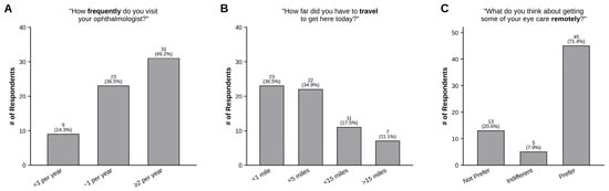
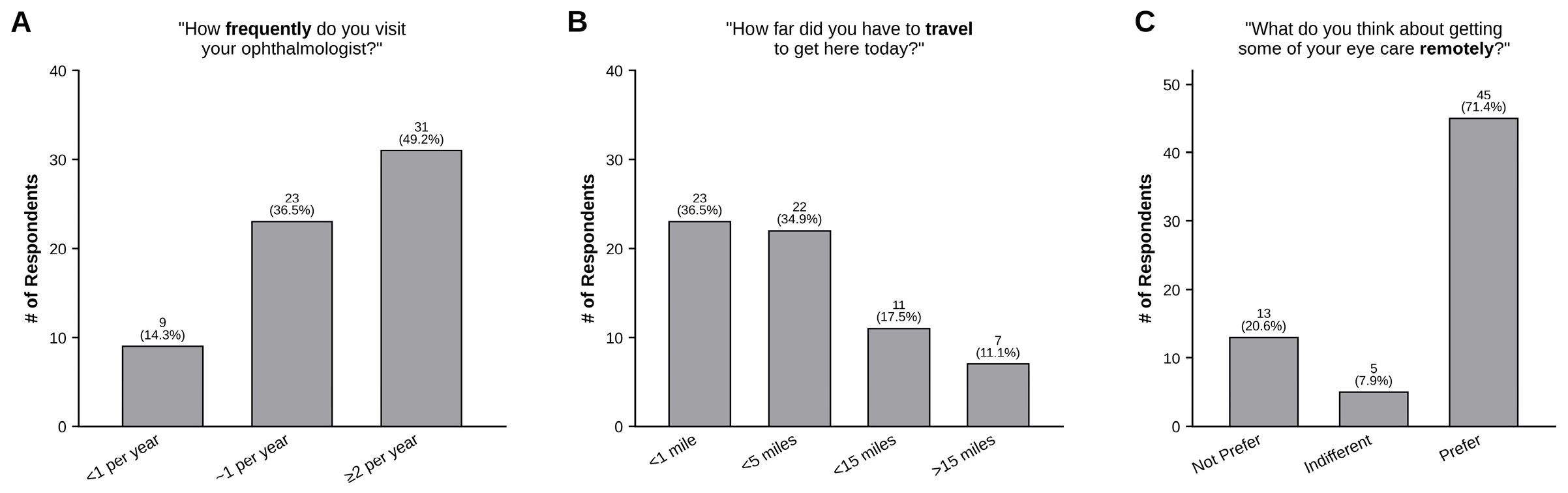
Given the diversity of our study population in terms of health status, we assessed factors such as annual ophthalmology visits, distance traveled to appointments, and attitudes towards receiving eye care outside of traditional vision specialist facilities, including pharmacies, urgent care centers, or at home. Most patients reported seeing their ophthalmologist at least two times per year (31, 49.2%), with additional distributions of less than one (9, 14.3%), approximately one (23, 36.5%), visit per year (Figure 4A). As illustrated in Figure 4B, the majority of patients resided near the appointment site, with the distribution being as follows: <1 mile (23, 36.5%), <5 miles (22, 34.9%), <15 miles (11, 17.5%), and >15 miles (7, 11.1%). Regarding the potential of vision testing outside of a dedicated clinic, a majority of patients preferred this notion (45, 71.4%), compared to those who did not like the idea (13, 20.6%) or were indifferent (5, 7.9%) (Figure 4C).
Figure 4.
Questions on visit frequency, travel distance, and non-specialist vision testing preferences with accompanying quote and cross analysis. (A) Most patients in this population saw their ophthalmologist approximately twice per year, with a broad distribution of overall visits. (B) Most patients in this study lived within 5 miles of the clinic, though several lived much farther away. (C) Many participants would prefer to receive some form of vision testing outside the ophthalmology clinic.
This study also investigates whether distance traveled to appointments was associated with perception of overall experience with standard clinical tests. Although the average ratings were similar across distances traveled, an analysis of response distributions suggested that patients who travel different distances report distinct perceptions of in-office experiences (p < 0.0001; overall χ2 test). This held true when performing a χ2 test against individual distance categories: <1 mile vs. <5 miles (p = 0.0013), <1 mile vs. <15 miles (p < 0.0001), <1 mile vs. >15 miles (p < 0.0001), <5 miles vs. <15 miles (p < 0.0001), <5 miles vs. >15 miles (p = 0.0138), and <15 miles vs. >15 miles (p < 0.0001). These findings suggest that more attention should be given to alternative administration strategies for eye testing, particularly for those who travel greater distances.
4. Discussion
This study quantified participant opinions on VR testing within ophthalmic workflows compared to standard in-office functional vision tests for metamorphopsia, color deficit, contrast sensitivity, visual fields, and visual acuity. Among patients with diverse backgrounds, visit frequencies, and distances traveled to eye appointments, patients preferred VR in terms of overall experience, comfort, and speed. Participants identified more favorable and fewer unfavorable aspects of VR testing and indicated a preference toward VR over standard in-office tests when given a choice. Additionally, many patients also liked the notion of moving some aspects of vision testing outside of ophthalmic workflows.
These findings are concordant with and expand upon the emerging literature on patient experience with VR-based vision testing. Hu and colleagues, in a home-monitoring feasibility study combining VR visual field testing with rebound tonometry, reported that 100% of participants rated the VR perimetry testing as easy to use, and 94.4% rated it as useful, with patients describing a sense of empowerment from generating their own visual function data between clinic visits [20]. Lallouette and Gillmann, in a study using a VR headset with real-time eye tracking, similarly found high comfort, acceptance, and tolerability across both healthy and glaucomatous subjects [22]. McLaughlin and colleagues additionally demonstrated that self-administered, remotely supervised VR visual field testing yields acceptable inter-test and test–retest reliability with high usability ratings [21]. The present study is distinguished by its multi-test panel, which more accurately reflects the cumulative cognitive and physical load of a real clinic visit, and by its inclusion of a broader patient population in terms of their diversity and varying ocular diseases. The consistency of reported patient preferences towards VR testing across these independent studies suggests that the preference for VR is not limited to a single test, headset, or disease state.
Results from this study bring up two concepts for consideration—(1) VR has the potential to accelerate ophthalmic (and optometric) workflows and improve patient experience, possibly improving relatively low patient adherence rates to ophthalmic appointments [27,28], and (2) VR may have the potential to increase quantitative vision testing capabilities in third-party locations, such as pharmacy clinics, primary care workflows, and at-home monitoring, which a majority of participants in this study were in favor of. This second consideration is particularly salient in light of the projected shortage of ophthalmologists [4,6], as well as growing evidence that primary-care-based tele-ophthalmology can detect ocular disease and prompt meaningful changes in ocular and systemic disease management [12,13]. VR delivery of vision screening may therefore provide a viable functional-testing complement to retinal-imaging-based tele-ophthalmology, bridging the existing gap that prevents primary care providers and tertiary centers from providing an additional touch point for early eye disease detection.
Existing virtual reality technologies have thus far largely focused on creating direct analogs of in-clinic tests, such as full-threshold perimetry, which is beneficial for consideration (1) above. However, the time these tests take can be limiting to settings described in consideration (2). VR allows for test alterations that accelerate testing time, such as suprathreshold testing and altered stimulus presentation strategies depending on the condition of interest and participants’ cooperation [19,29]. These alterations can be helpful to enable VR implementation in third-party locations and can also further gamify tests to make the eye exam experience more interactive for patients.
There are several factors to consider in VR implementation, as observed in the qualitative responses from participants in this study. Lightweight headsets that are commercially available should be considered to minimize excessive neck strain, particularly for older individuals who might require more frequent testing and could find certain head positions difficult to maintain for optimal target fixation. Recent advances in head-mounted display hardware, including pancake lenses, high-resolution micro-OLED panels, and integrated eye tracking, have meaningfully reduced device weight and improved fixation monitoring, addressing several of the comfort and reliability concerns identified in earlier VR perimetry literature [30]. Vertigo and nausea have been reported in a small percentage of individuals during VR use, necessitating close monitoring by providers. Due to high rates of surgical facemask utilization in clinics, excessive fogging in VR headsets with glasses users is expected—the provider might suggest removing the mask during examination. The results of this study indicate that the shortcomings of VR use from the patient perspective are outweighed by the benefits relative to in-office tests. The technology thus holds great promise for utility in the field of vision health.
This study is limited by its analysis of patient responses from different testing experiences and the investigation of tests that are not identical across VR and in-office settings; however, it does highlight the importance of performing further standardized trials to assess patient perspectives across these modalities. Several specific limitations merit emphasis. First, the suprathreshold 24-2 VR perimetry algorithm used in this protocol is intrinsically faster than the in-office SITA Fast and Full Threshold algorithms, which may partly explain the magnitude of the speed-preference effect, and a head-to-head comparison against a threshold VR algorithm is necessary to decouple modality from algorithm. Second, the sample of 64 participants from a single urban clinic limits generalizability, particularly to rural populations who, in fact, stand to benefit most from remote screening. Third, technology literacy, prior VR exposure, and age-related comfort with head-mounted devices were not formally measured, despite their possible impact on responses. Future studies should stratify responses by demographic, medical, and social factors, such as age, disease segment, and technology literacy, and incorporate parallel objective performance data—for example, test–retest reliability, agreement with in-clinic gold standards, etc.—to allow for co-analysis of patient experience and diagnostic accuracy endpoints.
5. Conclusions
Given the growing adoption of innovative technologies such as virtual reality (VR) for ophthalmic vision screening, this study evaluated VR’s integration into the ophthalmic workflow and its patient usability. Survey data from a diverse cohort of patients with various ocular pathologies show that patients consider VR to be a more efficient and well-tolerated testing modality compared to commonly used in-office tests. Participants found the VR experience user-friendly and comfortable, and their positive feedback highlights an interest in technologies for more flexible and accessible eye care. While VR testing is not intended to replace comprehensive in-office ophthalmic testing, these findings—taken together with recent independent reports of the feasibility, validity, and acceptability of VR perimetry—support its potential to expand screening options, especially in settings without access to specialty care. Continued validation and integration of these novel devices has the potential to enhance remote visual testing by bringing it into everyday clinical practice and contributing to expanding the outreach of preventive eye care.
Author Contributions
Conceptualization, M.L., R.A.S. and A.J.W.; methodology, M.L. and R.A.S.; software, A.J.W. and V.N.N.; validation, M.L., R.A.S., A.J.W., C.C. and O.O.O.; formal analysis, A.J.W. and R.A.S.; investigation, C.C. and O.O.O.; resources, M.L., R.A.S., C.C., O.O.O. and V.N.N.; data curation, A.J.W., C.C. and O.O.O.; writing—original draft preparation, M.L. and R.A.S.; writing—review and editing, M.L., R.A.S., A.J.W., C.C., O.O.O. and V.N.N.; visualization, M.L., R.A.S. and A.J.W.; supervision, H.R. and J.G.C.; project administration, H.R. and J.G.C.; funding acquisition, M.L., R.A.S. and J.G.C. All authors have read and agreed to the published version of the manuscript.
Funding
This work was funded by the 4D Technology Development Program grant at the Icahn School of Medicine at Mount Sinai, awarded to M.L., R.A.S., A.J.W., and J.C.
Institutional Review Board Statement
The study was conducted in accordance with the Declaration of Helsinki and approved by the Institutional Review Board (IRB) of the Icahn School of Medicine at Mount Sinai (IRB #20-00358, approval date: 20 January 2020). The IRB determined that the study carries no greater than minimal risk.
Informed Consent Statement
Informed consent was obtained from all subjects involved in the study.
Data Availability Statement
The original contributions presented in this study are included in the article. Further inquiries can be directed to the corresponding author.
Acknowledgments
During the preparation of this manuscript/study, the author(s) used GraphPad Prism version 7 for the purpose of data analysis. The authors have reviewed and edited the output and take full responsibility for the content of this publication.
Conflicts of Interest
Margarita Labkovich, Randal A. Serafini, and Andrew J. Warburton are inventors on patent applications for VR test designs and methods of administration. The principal investigators (Harsha Reddy and James G. Chelnis) are not inventors on patents relevant to this study, and do not belong to a commercial entity that could commercialize the technologies tested in this study.
References
- Munavalli, J.R.; Rao, S.V.; Srinivasan, A.; Srinivas, A.; van Merode, F. The optimization in workflow management: Ophthalmology. J. Health Manag. 2016, 18, 21–30. [Google Scholar] [CrossRef] [Scilit]
- Ciulla, T.A.; Tatikonda, M.V.; Elmaraghi, Y.A.; Hussain, R.M.; Hill, A.L.; Clary, J.M.; Hattab, E. Lean six sigma techniques to improve ophthalmology clinic efficiency. Retina 2018, 38, 1688–1698. [Google Scholar] [CrossRef] [Scilit] [PubMed]
- McMullen, M.; Netland, P.A. Wait time as a driver of overall patient satisfaction in an ophthalmology clinic. Clin. Ophthalmol. 2013, 7, 1655–1660. [Google Scholar] [CrossRef] [Scilit] [PubMed]
- Berkowitz, S.T.; Finn, A.P.; Parikh, R.; Kuriyan, A.E.; Patel, S. Ophthalmology workforce projections in the United States, 2020 to 2035. Ophthalmology 2024, 131, 133–139. [Google Scholar] [CrossRef] [Scilit] [PubMed]
- Gibson, D.M. Eye care availability and access among individuals with diabetes, diabetic retinopathy, or age-related macular degeneration. JAMA Ophthalmol. 2014, 132, 471–477. [Google Scholar] [CrossRef] [Scilit] [PubMed]
- Gibson, D.M. The geographic distribution of eye care providers in the United States: Implications for a national strategy to improve vision health. Prev. Med. 2015, 73, 30–36. [Google Scholar] [CrossRef] [Scilit] [PubMed]
- Patel, R.H.; Mothy, D.; Choudhry, H.S.; Grinberg, A.; Bhasin, J.; Dastjerdi, M.H. Cornea eye care coverage based on geographic distribution and population demographics in the United States. AJO Int. 2025, 2, 100139. [Google Scholar] [CrossRef] [Scilit]
- Labkovich, M.; Paul, M.; Kim, E.; Serafini, R.A.; Lakhtakia, S.; Valliani, A.A.; Warburton, A.J.; Patel, A.; Zhou, D.; Sklar, B.; et al. Portable hardware and software technologies for addressing ophthalmic health disparities: A systematic review. Digit Health 2022, 8, 20552076221090042. [Google Scholar] [CrossRef] [Scilit] [PubMed]
- Areaux, R.G.; de Alba Campomanes, A.G.; Indaram, M.; Shah, A.S. Your eye doctor will virtually see you now: Synchronous patient-to-provider virtual visits in pediatric tele-ophthalmology. J. Am. Assoc. Pediatr. Ophthalmol. Strabismus 2020, 24, 197–203. [Google Scholar] [CrossRef] [Scilit] [PubMed]
- Kapoor, S.; Eldib, A.; Hiasat, J.; Scanga, H.; Tomasello, J.; Alabek, M.; Ament, K.; Arner, D.; Benson, A.; Berret, K.; et al. Developing a pediatric ophthalmology telemedicine program in the COVID-19 crisis. J. Am. Assoc. Pediatr. Ophthalmol. Strabismus 2020, 24, 204–208. [Google Scholar] [CrossRef] [Scilit] [PubMed]
- Patel, S.; Hamdan, S.; Donahue, S. Optimising telemedicine in ophthalmology during the COVID-19 pandemic. J. Telemed. Telecare 2022, 28, 498–501. [Google Scholar] [CrossRef] [Scilit] [PubMed]
- Pinhas, A.; Pinhas, B.; Dmitruk, E.; Pinhas, S. Eye and systemic disease management changes after teleophthalmology screening in primary care: Retrospective cross-sectional pilot study of 200 consecutive patients. JMIR Form. Res. 2025, 9, e81918. [Google Scholar] [CrossRef] [Scilit] [PubMed]
- Swain, T.A.; McGwin, G.; Girkin, C.A.; Owsley, C. Telemedicine screening for eye disorders in federally qualified health centers: Relationship to vision-targeted health-related quality of life. Telemed. J. e-Health 2024, 30, e2080–e2086. [Google Scholar] [CrossRef] [Scilit] [PubMed]
- Hekmatjah, N.; Chibututu, C.; Han, Y.; Keenan, J.D.; Oatts, J.T. Virtual reality perimetry compared to standard automated perimetry in adults with glaucoma: A systematic review. PLoS ONE 2025, 20, e0318074. [Google Scholar] [CrossRef] [Scilit] [PubMed]
- Selvan, K.; Mina, M.; Abdelmeguid, H.; Gulsha, M.; Vincent, A.; Sarhan, A. Virtual reality headsets for perimetry testing: A systematic review. Eye 2024, 38, 1041–1064. [Google Scholar] [CrossRef] [Scilit] [PubMed]
- Bradley, C.; Ahmed, I.I.K.; Samuelson, T.W.; Chaglasian, M.; Barnebey, H.; Radcliffe, N.; Bacharach, J. Validation of a wearable virtual reality perimeter for glaucoma staging, the NOVA trial: Novel Virtual Reality Field Assessment. Transl. Vis. Sci. Technol. 2024, 13, 10. [Google Scholar] [CrossRef] [Scilit] [PubMed]
- Griffin, J.M.; Slagle, G.T.; Vu, T.A.; Eis, A.; Sponsel, W.E. Prospective comparison of VisuALL virtual reality perimetry and Humphrey automated perimetry in glaucoma. J. Curr. Glaucoma Pract. 2024, 18, 4–9. [Google Scholar] [CrossRef] [Scilit] [PubMed]
- Patel, A.J.; Lee, W.W.; Ziff, M.; Munshi, H.M.; Chang, T.C.; Grajewski, A.L.; Wester, S.T.; Tse, D.T.; Tse, B.C. Superior visual field testing using virtual reality with and without eye tracking for functional upper eyelid surgery evaluation: A pilot study. Ophthalmic Plast. Reconstr. Surg. 2023, 39, 381–385. [Google Scholar] [CrossRef] [Scilit] [PubMed]
- Labkovich, M.; Warburton, A.J.; Ying, S.; Valliani, A.A.; Kissel, N.; Serafini, R.A.; Mathew, R.; Paul, M.; Hovstadius, S.M.; Navarro, V.N.; et al. Virtual reality hemifield measurements for corrective surgery eligibility in ptosis patients: A pilot clinical trial. Transl. Vis. Sci. Technol. 2022, 11, 35. [Google Scholar] [CrossRef] [Scilit] [PubMed]
- Hu, G.Y.; Prasad, J.; Chen, D.K.; Alcantara-Castillo, J.C.; Patel, V.N.; Al-Aswad, L.A. Home monitoring of glaucoma using a home tonometer and a novel virtual reality visual field device: Acceptability and feasibility. Ophthalmol. Glaucoma 2023, 6, 121–128. [Google Scholar] [CrossRef] [Scilit] [PubMed]
- McLaughlin, D.E.; Savatovsky, E.J.; O’Brien, R.C.; Vanner, E.A.; Munshi, H.K.; Pham, A.H.; Grajewski, A.L. Reliability of visual field testing in a telehealth setting using a head-mounted device: A pilot study. J. Glaucoma 2024, 33, 15–23. [Google Scholar] [CrossRef] [Scilit] [PubMed]
- Lallouette, A.; Gillmann, K. Patient comfort, acceptance, and tolerability of virtual reality (VR) headsets with real-time eye tracking for remote visual field testing. J. Clin. Med. 2025, 14, 3219. [Google Scholar] [CrossRef] [Scilit] [PubMed]
- Shi, R.B.; Li-Han, L.Y.; Kherani, I.N.; Trope, G.E.; Buys, Y.M.; Wong, W.; Eizenman, M. Virtual reality portable perimetry and home monitoring of glaucoma: Retention and compliance over a 2-year period. Ophthalmol. Sci. 2025, 5, 100639. [Google Scholar] [CrossRef] [Scilit] [PubMed]
- Bibak-Bejandi, Z.; Razavi, A.; Niktinat, H.; Yucel, Z.J.; Sebhat, A.M.; Bibak-Bejandi, R.; Arabpour, Z.; Khandaker, A.N.; Sanchez, J.; Nusair, O.; et al. Virtual reality and augmented reality in ophthalmology: A recent update. Digit Health 2025, 11, 20552076251387047. [Google Scholar] [CrossRef] [Scilit] [PubMed]
- Cheng, C.P.; Serafini, R.A.; Labkovich, M.; Warburton, A.J.; Navarro, V.; Shaik, N.; Reddy, H.; Chelnis, J.G. Clinical report: Virtual reality enables comparable contrast sensitivity measurements to in-office testing (pilot study). Optom. Vis. Sci. 2024, 101, 124–128. [Google Scholar] [CrossRef] [Scilit] [PubMed]
- Hopewell, S.; Chan, A.W.; Collins, G.S.; Hróbjartsson, A.; Moher, D.; Schulz, K.F.; Tunn, R.; Aggarwal, R.; Berkwits, M.; Berlin, J.A.; et al. CONSORT 2025 Statement: Updated guideline for reporting randomised trials. BMJ 2025, 388, e081123. [Google Scholar] [CrossRef] [Scilit] [PubMed]
- McMullen, M.J.; Netland, P.A. Lead time for appointment and the no-show rate in an ophthalmology clinic. Clin. Ophthalmol. 2015, 9, 513–516. [Google Scholar] [CrossRef] [Scilit] [PubMed]
- Chiam, M.; Kunselman, A.R.; Chen, M.C. Characteristics associated with new patient appointment no-shows at an academic ophthalmology department in the United States. Am. J. Ophthalmol. 2021, 229, 210–219. [Google Scholar] [CrossRef] [Scilit] [PubMed]
- Razeghinejad, R.; Gonzalez-Garcia, A.; Myers, J.S.; Katz, L.J. Preliminary report on a novel virtual reality perimeter compared with standard automated perimetry. J. Glaucoma 2021, 30, 17–23. [Google Scholar] [CrossRef] [Scilit] [PubMed]
- Lim, D.Z.; Hosamani, S.; Aung, T.; Lim, C.H.L. Understanding virtual/augmented reality devices and their application in ophthalmology. J. Retin. 2024, 9, 95–104. [Google Scholar] [CrossRef] [Scilit]

Disclaimer/Publisher’s Note: The statements, opinions and data contained in all publications are solely those of the individual author(s) and contributor(s) and not of MDPI and/or the editor(s). MDPI and/or the editor(s) disclaim responsibility for any injury to people or property resulting from any ideas, methods, instructions or products referred to in the content. |
© 2026 by the authors. Licensee MDPI, Basel, Switzerland. This article is an open access article distributed under the terms and conditions of the Creative Commons Attribution (CC BY) license.