


Abstract
Background: Lateral eye movements are part of natural ocular motility, and their effect on tear film behavior is largely unknown because they are usually not evaluated during tear film examinations, where patients are required to look straight ahead. This study aimed to determine what happens to the tear film during lateral eye movements in the absence of blinking. Methods: Tear film dynamics during a sequence of open-eye lateral gaze maneuvers were recorded using an infrared camera system (TearView). Results: The study included 15 healthy participants (5 female; median age 26) who had no ocular surface-related complaints. It was observed that lateral eye movements exposed new ocular surface areas not previously covered by the tear film. The tear film margins reacted to lateral eye movements by spreading towards the newly uncovered ocular surface areas, thereby recoating them. This response was asymmetrical, with a tear reservoir in the lacus lacrimalis of the medial canthus providing additional tear film spread only during ocular abduction. Conclusions: These findings expand our understanding of tear film physiology beyond static gaze. They might also bear implications for dry eye disease and oculoplastic surgical techniques affecting the medial canthus.
1. Introduction
Knowledge about tear film behavior has come from studying the stationary eye in forward gaze with blinking under instruction [1]. Little is known about tear film dynamics during lateral eye movements [2]. Lateral eye movements expose an area of ocular surface previously covered by the eyelid with minimal change to palpebral fissure height. They occur frequently in everyday life. Reading provides an example of lateral saccades of moderate rates and size [3]. Many, but not all, saccades are followed by a blink [4,5,6,7]. For example, smooth pursuit lateral movements, such as during finger-following, are usually not accompanied by blinks [5]. The extreme case of lateral movement is maximal eye adduction to abduction, a change of gaze of almost 90 degrees [8]. While not strictly physiological, these large movements are most likely to reveal what happens to the newly exposed ocular surface in terms of the tear film.
Yokoi et al. investigated the effects of lateral eye movements on the tear film using fluorescein and interferometry. These techniques were limited to the precorneal tear film [9,10] and could not reveal what occurs with the tear film over the bulbar conjunctiva. With new technology, detecting infrared emittance [11,12,13,14], it is now possible to investigate the tear film in these areas. This technology has been used to investigate the dynamics of the tear film in response to vertical eye movements and revealed that the fully formed tear film expanded to cover the newly exposed ocular surface [13]. The purpose of this research was to determine if a similar phenomenon occurs with lateral eye movements.
2. Materials and Methods
2.1. Recruitment and Experimental Conditions
The Institutional Review Board/local ethics committee of Saarland University, Homburg, Germany, approved this study (Ha 83/20; approval date: 8 June 2020). The research was conducted in compliance with the Declaration of Helsinki, and informed consent was obtained from all participants.
2.2. Experimental Procedures
Participants were seated at a slit lamp (SL800, Zeiss Meditec AG, Jena, Germany) with the infrared camera (TearView, Beyond700 Ltd., Sydney, Australia) attached to a proprietary arm inserted into the slit lamp’s accessory turret hole. With the participant placed in the headrest, the infrared camera was focused on the eye using the slit lamp mechanism. For consistency and convenience, only the right eyes were imaged. Long-wavelength infrared radiation, naturally emitted from the ocular surface, was captured at a resolution of 640 × 480 pixels, with a noise equivalent temperature resolution of 30 mK, and a frame rate of 25 frames per second in real time. The camera is designed so that the field of view is filled by the ocular surface and adnexa. This enables visualization of the tear film over the entire palpebral fissure, from the nasal canthus to the temporal canthus, and from the lower to the upper lid margin [12].
2.3. Ocular Movement Paradigms
A baseline recording of tear film formation following a spontaneous blink was made with participants in primary gaze. Movement paradigms were based on previously published work where fluorescein and interferometry were used to study tear film dynamics [2]. The participants were instructed to keep their eyes open and without blinking, and move their eyes rapidly towards an acoustic signal on one or the other side of the head.
2.3.1. Rapid Excursion Movement
Participants rapidly (as quickly as possible) moved their eyes from maximal abduction to adduction repeatedly.
2.3.2. Adduction (Nasal) First Paradigm
Participants performed an extreme nasal saccade (adduction) following an acoustic signal. After waiting ~5 s, performing an extreme temporal saccade (abduction).
2.3.3. Abduction (Temporal) First Paradigm
This was identical to the adduction paradigm, but starting with an extreme temporal saccade and moving the eye nasally on instruction.
2.3.4. Smooth Pursuit Paradigm (Finger Following)
Participants were asked to track the examiner’s finger as it moved from temporal to nasal and vice versa.
2.3.5. Lid Wipe Experiment
Based on the results from the other experimental paradigms, the possibility of the lacus lacrimalis being a tear reservoir was investigated. In four participants, the caruncle region was wiped with a Blephaclean sterile lid wipe (Thea Pharma, Clermont-Ferrand, France), and then the abduction first paradigm was performed.
Note on Terminology
We define the fully formed tear film as the complete tear film consisting of a mucoaqueous subphase and a lipid layer [15]. We use this term because it is known that a complete tear film, all components working together, resists evaporation, and this cannot be attributed to the lipid layer alone [16,17]. Since evaporative cooling (dark patches) is not seen with TearView following the spread of a normal tear film (the images are uniform in appearance), this is interpreted as representing a fully formed tear film. When evaporative cooling is observed, the infrared images do not indicate the component or components of the tear film responsible, just that the tear film is dysfunctional [12]. The tear film margin as seen with TearView demarcates the border between the complete(d) tear film and the incomplete one. While the ocular surface beyond the margin might be moist and covered by a “subaqueous layer” [2], it is not covered by a complete tear film. Tear film spread, flow, or expansion means an increase in the area of the complete tear film.
Note on the Figures
Images and videos were not cropped or manipulated post-recording. In some of the images (particularly in the videos), the infrared reflection of the camera lens as well as the examiner’s hand can be seen. These reflections are artifacts and bear no significance. In the top right corner, there is an eye (and a nose) symbol indicating the position of the cornea (blue). The numbers in the box at the bottom right indicate experimental time (in seconds). The blue bar indicates that a saccade/eye movement has been performed.
3. Results
Participants were 10 males, 5 females (mean age 32 ± 15 years, median age 26, range 21–70) with no ocular surface-related complaints (OSDI < 13) or contact lens wear.
Slit lamp examination confirmed no abnormalities of the lids and anterior segment, and participants did not use any topical medication.
In forward gaze following a blink, all participants showed a normal tear film characterized by a dark front swiftly moving to and disappearing at the upper eyelid margin. Thereafter, with the eye open, the tear film was even and unchanging with no discernible difference between the cornea and conjunctiva. The participants in this study were the same participants from the previously published study [13].
3.1. Rapid Excursion Movement Recordings
In all participants, repeated rapid adduction and abduction movements showed that the central tear film, largely identical to the precorneal tear film, moved with the eye (Supplemental Video S1). This means that the ocular surface did not move underneath the tear film. There was no lag between the eye movements and tear film movement, meaning there were no movements due to inertia. Other evidence that the tear film was moving with the eye came from occasional examples when an inclusion/defect in the tear film could be seen. An example is shown in a later figure (Figure 3, bottom row).
3.2. Adduction (Nasal) First Paradigm Recordings
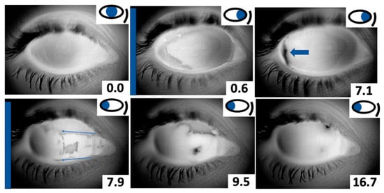
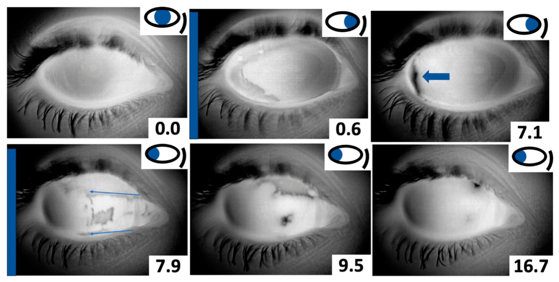
The initial adduction saccade exposed a crescent of ocular surface with the tear film margins initially retaining the shape of the temporal canthal angle (Figure 1; 0.6 s). The existing preocular tear film expanded slowly to cover the exposed ocular surface. The tear film did not expand out from the tear menisci at the lid margins. Typically, this expansion was incomplete, leaving a small crescent adjacent to the canthus (Supplemental Video S2; Figure 1; 7.2 s). A maximal abduction saccade after the adduction revealed different features: The tear film did not entirely separate from the medial canthus. Instead, a blotchy tear film was immediately present, appearing to have stretched out from the lacus lacrimalis region (Figure 1; 7.9 s). This new tear film only covered a region extending from the upper and lower anatomical limits of the lacus lacrimalis, leaving uncovered surfaces above and below (Figure 1; 7.9 s, blue arrows). These uncovered regions were eventually covered by the expansion of the fully formed tear film, and from the tear film originating from the lacus lacrimalis (Supplemental Video S2, Figure 1; 16.7 s), i.e., tear flow to cover the newly exposed ocular surface was not from the lid margins towards the exposed areas. These findings were consistent across all participants, meaning that the movement direction of the tear film in response to eye movement was the same for all of them. There was never a case where the tear film did not expand, nor where it expanded in the opposite direction.
Figure 1.
Adduction (nasal) first paradigm. An initial adduction (0.6 s) revealed a temporal crescent of exposed ocular surface, which was slowly filled in with tear film from nasal to temporal, leaving a small, uncovered portion next to the lateral canthus (7.1 s; arrow). The abduction saccade (7.9 s) revealed new tear film forming immediately, bounded by the extent of the lacus lacrimalis (arrows). With time, complete coverage of the ocular surface occurred (9.5 s).
3.3. Abduction (Temporal) First Paradigm Recordings
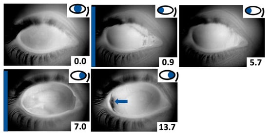
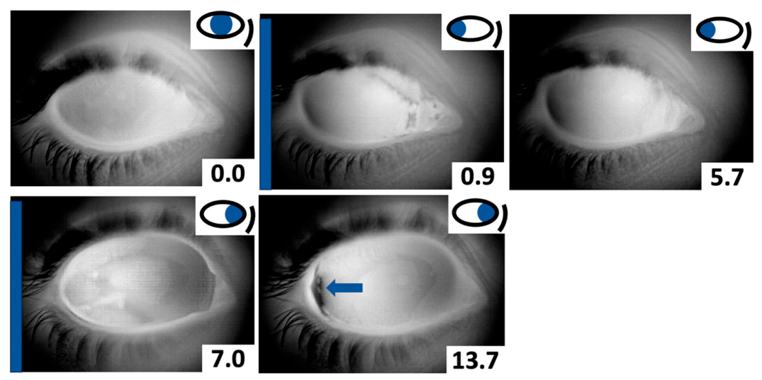
Changes to the tear film and its subsequent expansion were almost identical to those described for the adduction first paradigm (Figure 2). The main difference was that it took a shorter time for the tear film to fill in the exposed eye surface in the medial canthus upon the initial abduction (Supplemental Video S3). This is ascribed to the initial ocular movement being less extreme (45 degrees), compared to the above paradigm where the ocular abduction movement was about 90 degrees (abduction paradigm; compare Figure 1; 7.9 s onwards with Figure 2; 0–5.7 s). This was consistent across participants, i.e., tear film expansion was in the same direction for all of them.
Figure 2.
Abduction (temporal) first paradigm. An initial abduction (0.9 s) showed tear film being “drawn out” of the lacus lacrimalis. The tear film margin above the lacus lacrimalis retained the outline of the upper portion of the medial canthus. Within about 5 s, complete coverage of the ocular surface was achieved (5.7 s). The subsequent adduction saccade (7.0 s) revealed a large area of uncovered ocular surface. The tear film failed to completely cover this area, leaving a small crescent of exposure, developing an intense evaporation signal (13.7 s; arrow).
3.4. Smooth Pursuit Paradigm/Finger Following Recordings
Finger following is the classic test to examine smooth pursuit eye movements [18].
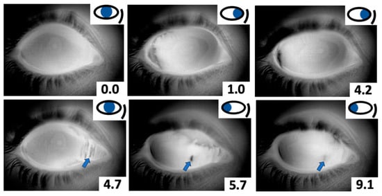
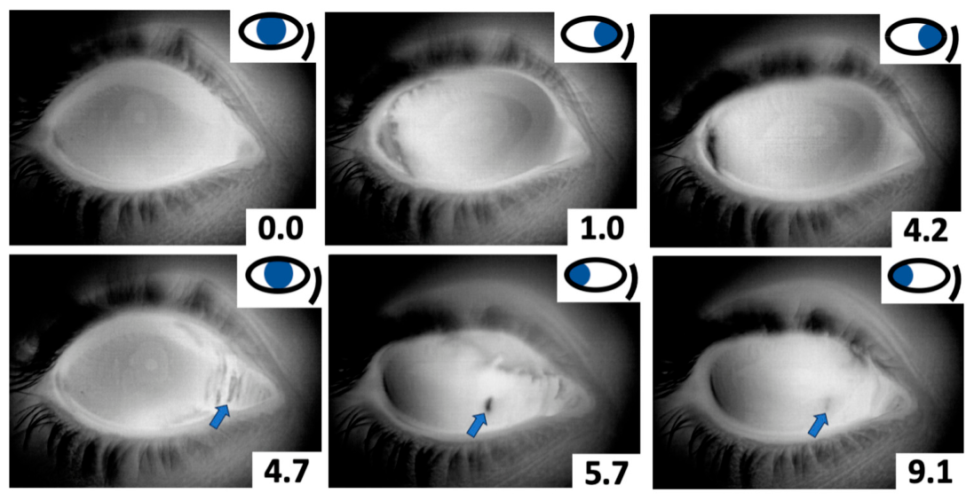
Eye movements tracking the examiner’s finger were slower than the extreme saccade movements. Consequently, the tear film started to fill in the newly exposed ocular surface even during the eye movement (Supplemental Video S4). This meant that there was barely any uncovered area left as the eye stopped moving, both during abduction and adduction. During slow abduction, a series of dark vertical bands appeared near the medial canthus, which then rapidly disappeared as the eye continued moving (Figure 3; 4.7 s). This might be explained by the tear film initially being embedded within the conjunctival folds and stretched out like unfolding pleats with lateral movement. We did not observe any obvious differences regarding these phenomena across participants.
Figure 3.
Finger following. Following the finger of the examiner into adduction had the tear film “filling in the gaps”, even as the eye was being rotated (1.0 s), leaving only a small defect in the very periphery (4.2 s). A subsequent slow abduction revealed vertical dark bands of exposed ocular surface (4.7 s, arrow), which quickly disappeared through tear film expansion (5.7 s). An incidental defect in the tear film indicated the amount of eye rotation (9.1 s, arrow).
3.5. Lid Wipe Experiment Recordings
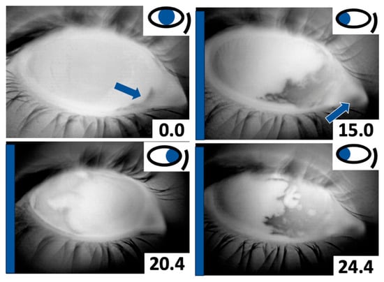
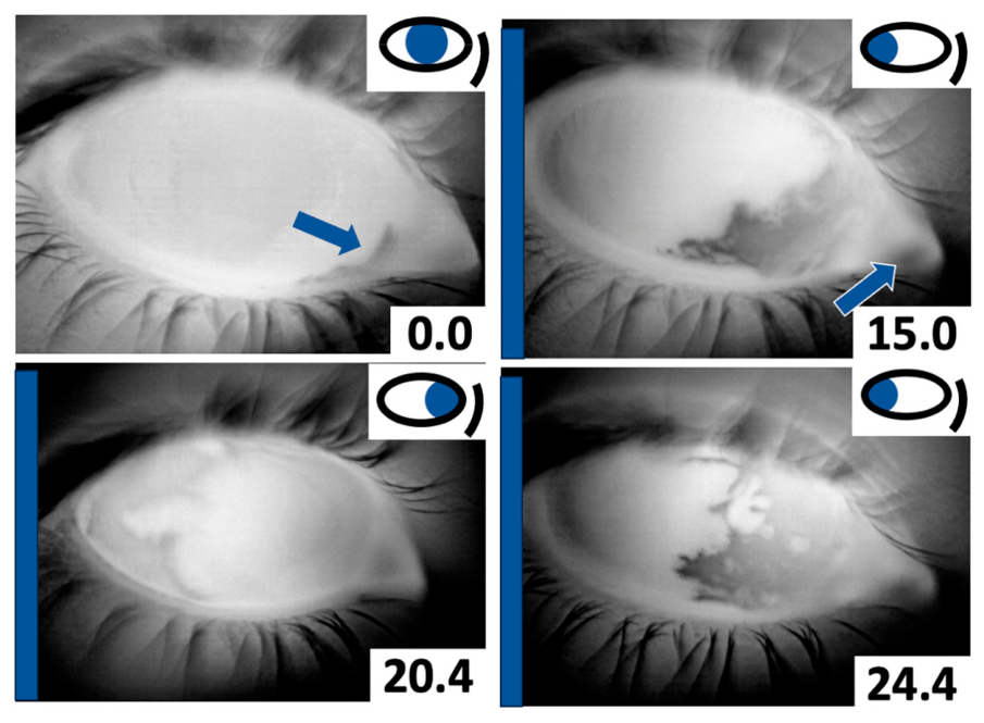
A lid wipe was used to gently wipe the caruncular region (lacus lacrimalis) prior to saccadic movements. In all four participants, abduction revealed that the tear film separated from the lid margin and retained the outline of the nasal canthus, similar to that observed at the temporal canthus following adduction (Supplemental Video S5). This was different from normal conditions in that there was no spread from the caruncle region, but instead, there was a slight flow from the existing tear film towards the caruncle (Figure 4).
Figure 4.
Caruncle wipe experiment. After gently wiping the caruncular region with a Blephaclean wipe, only a minor defect in the tear film could be seen in forward gaze (0.0 s, blue arrow). Abduction of the eyeball revealed that no new tear film is transferred onto the nasal bulbar conjunctiva. Moreover, the emittance of the caruncle (dark appearance) revealed increased evaporation (15.0 s, arrow). Eye adduction showed an absence of a reservoir on the temporal side, with an initial retention of the outline of the canthal angle (20.4 s). A second abduction maneuver showed a couple of tear film specks (24.4 s).
4. Discussion
Our experiments parallel the eye movement/tear film experiments of Yokoi et al. [2]. Instead of interferometry and fluorescein tear film staining, infrared imaging was used to passively capture tear film dynamics over the whole ocular surface without disturbing the tear film. Adding additional volume as needed for fluoresceine staining was also unnecessary with this technique [19], neither was bright light shone into the eye; a likely cause of reflex tearing in both fluoresceine staining and interferometry.
The main findings were that the newly exposed ocular surface was covered by the spreading of fully formed tear film, and in the case of the medial canthus, additional tear film from the lacus lacrimalis. This matches our previous findings where tear film movements following vertical eye movements were studied [13].
4.1. The “Fluid Shell”
A detailed comparison with the work of Yokoi et al. [2] is presented in Appendix A. To briefly summarize, infrared imaging in the present study demonstrated that the tear film can be regarded as a fluid shell that moves synchronously with the eye (Rapid excursion movement, see Supplemental Video S1), which is generally in accordance with the findings of Yokoi et al. The fully formed tear film moves as a single unit with eye movement, and there is no lag upon eye movement initiation. This indicates that there is no momentum-driven movement of the tear film.
4.2. The Reservoir Function of the Lacus Lacrimalis
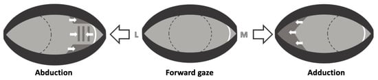
The lacus lacrimalis is generally overlooked in terms of tear film function and dry eye assessment. Anatomically, it comprises the semilunar fold, the lacrimal caruncle, and the adjacent lid margins of the medial canthus and is filled with tear fluid [20]. It has been regarded as merely a pool of used tears ready for drainage through the puncta [21,22,23]. Our data suggest that the lacus lacrimalis also provides a reservoir of tears for use during abduction to coat the newly exposed ocular surface (Figure 1; 7.9 s; Figure 5). This is likely to be assisted by unfolding of the conjunctival folds during globe rotation and movement of the canthus itself [24,25,26,27]. This makes the tear film response to lateral movements asymmetrical, with almost instant coverage of the newly exposed bulbar conjunctiva with tear film only occurring during abduction of the globe (Figure 5). Also worth noting is that despite there being no meibomian glands at the margins of the medial canthus [28], evaporative cooling in the infrared images was never observed in the lacus lacrimalis in forward gaze or subsequent to spread following lateral gaze. This indicates that it is protected by a complete tear film, including a mucoaqueous and lipid layer. The lipid layer must arise from neighboring meibomian glands from either eyelid, from sebaceous glands present in the caruncle [29,30], or both. Secreted proteins, which form an integral part of the mucoaqueous layer, might be secreted directly by the caruncular tissue or by adjacent conjunctiva [31,32].
Figure 5.
Schematic of the asymmetrical tear film response to lateral eye movements coming from forward gaze as seen in the infrared image. The approximate position of the cornea is depicted with a dotted circle. The approximate position of the plica semilunaris, the lateral border of the lacus lacrimalis, is depicted with a white arc. (Forward gaze) shows the post-blink fully formed tear film (light grey). Upon (Adduction), the tear film moves together with the eye, leaving an uncovered darker crescent of ocular surface around the lateral canthus. The fully formed tear film expands laterally to cover this area (white arrows). Upon (Abduction), tear film is immediately present across the newly exposed bulbar conjunctiva, often with vertical defects (vertical dark grey columns). The tear film then spreads in both directions at the same time, originating from the fully formed tear film and the lacus lacrimalis (white arrows). L: lateral; M: medial.
The concept that the lacus lacrimalis has a tear reservoir function was supported by the lid wipe experiment, where the results can be explained by the reservoir being removed, and hence, the tear film spread upon lateral eye movement was solely from the fully formed tear film. This closely resembled what was seen in the lateral canthus following adduction (compare to Figure 5).
The purpose of the observed asymmetry in tear film response, spreading from the medial canthus (lacus lacrimalis) during abduction but not adduction, remains speculative. One plausible explanation is the protection of the medial canthal region, including the lacrimal puncta and drainage system, from desiccation by rapid tear film recoating. The lacus lacrimalis primarily collects excess tears for drainage via the puncta; swift coverage during abduction could offer additional protection for this critical drainage pathway. Tear film spread originating in the medial canthus might also be a remnant of the action of the nictitating membrane (third eyelid), which exists in almost all mammals to some extent except for the higher primates, and which sweeps the eye from medial to lateral during a blink [33]. In contrast, the lateral canthus lacks drainage structures and benefits from proximity to the lacrimal gland, providing sufficient aqueous tears without the need for additional spreading [30].
There are clinical implications if the lacus lacrimalis has a tear reservoir. Oculoplastic surgery of the medial canthal region could disrupt the structure of the lacus lacrimalis, with the consequence of compromised tear film formation upon lateral eye movements [34]. Diseases of the medial canthus could similarly compromise the reservoir function. For instance, pterygia can lead to a loss of vertical length of plica semilunaris [35] and lead to a unique tear film break-up pattern [36]. Interestingly, lacrimal caruncular epitheliopathy has been associated with meibomian gland dysfunction [37]. In this case, an incomplete tear film might be present in the lacus lacrimalis due to lack of meibum and this could lead to the caruncular epitheliopathy.
4.3. Comparison to Predictions
Prior to our studies, it has not been possible to directly observe the dynamic behavior of the tear film over the peripheral bulbar conjunctiva in response to lateral eye movement. Nevertheless, predictions have been made. Yokoi et al. [2] used lateral eye movement paradigms similar to ours and made predictions based on the fact that during abduction, the area of exposed nasal bulbar conjunctiva increases and the area of exposed temporal conjunctiva decreases, and vice versa during adduction. On the decreasing side, they speculated that the lipid layer would either be compressed or redistributed into the marginal lipid reservoir. On the increasing side, they supposed that the tear film might be stretched out or that the ocular surface might just be covered with a “subaqueous phase” drawn forth from behind the eyelids. They also predicted that for the flow of the lipid (layer) to occur, the lipid layer would need to be continuous with the lipid in the meibomian reservoirs from which lipids would be drawn. In addition, they expected the central region of the tear film, being a fluid shell, to remain stable.
In agreement with Yokoi et al., using infrared imaging, we found that the central fluid shell remained intact during lateral eye movements. However, we observed that the fully formed tear film separated from the lid margins during adduction and then expanded laterally to cover the newly exposed ocular surface (Figure 5), rather than stretching or flowing from the lid margins as predicted by Yokoi et al. This tear film expansion towards the lid margin has also been observed following vertical eye movements and so appears to be a consistent feature of tear film physiology [13]. A new finding in this study was the observation of tear film flow originating at the lacus lacrimalis. While not strictly part of the meibomian reservoirs of the lid margins, it does seem to be continuous with the fully formed tear film during abduction (Figure 5) and thus in agreement with Yokoi et al.’s predictions.
The mechanism behind the expansion of the tear film in response to eye movements in the absence of blinking is unknown. One possibility is that the expansion is due to shear forces that are exerted on the tear film fluid shell by the eyelids during eye movement. This idea differs from Yokoi et al., who concluded that shearing during horizontal saccades would be “insufficient to disturb the tear film structure greatly” [2]. However, Tiffany found that the shear rates between blinking and saccades are almost identical [38], so that the resulting shear stress should be similar as well [39]. Another possibility states that the tear film is able to adjust itself to cover a palpebral fissure of differing size without the need for shear forces. This possibility has been explored in detail in a previous publication by the authors [13].
4.4. Limitations
The study was limited to 15 participants and thus had a small sample size. However, observed phenomena were consistent across all participants, and the number of participants is comparable to previous studies on tear film dynamics [40,41].
No quantitative analysis and statistics were performed. This is consistent with an observational study wherein phenomena are reported for the first time, with the phenomena consistent for all the participants. This is similar to studies of Yokoi et al. [2], in which they reported the dark arc as a new phenomenon in lateral eye movement studies and did not quantify it. Moreover, there is no consensus on what, where, and how to measure tear film dynamics such as lipid layer spreading speed, coverage area, and time to stabilization of the tear film [42]. For clinical studies, it could be useful to quantify tear film dynamics following eye movement, where there would be at least two distinct populations to compare. A study comparing healthy participants and dry eye disease patients could measure tear film spreading velocity, stabilization time, and surface coverage with a view to determining if examining tear film behavior during lateral eye movement could provide an additional diagnostic test for dry eye disease.
5. Conclusions
The tear film forms a gel-like fluid shell over the exposed ocular surface and moves with the ocular surface during large lateral movements through which previously retropalpebral ocular surface lacking a tear film becomes exposed. The fully formed tear film responds to this by spreading outwards from its margins to cover the newly exposed ocular surface area. On the nasal side, this spread is supported by a tear reservoir made up of the lacus lacrimalis. Overall, this blink-independent tear film spread appears to be sufficient to cover newly exposed ocular surface area during physiological eye movements, e.g., during finger following and small saccades. These sorts of eye movements are common during everyday activities such as reading or looking outside a car window while it is moving [43,44].
Investigating how the tear film copes with eye movements broadens our understanding of tear film physiology. It might also bear implications for oculoplastic surgical techniques affecting the medial canthus. Further investigation of tear film dynamics during eye movements and their role in ocular surface health and disease is needed.
Supplementary Materials
The following supporting information can be downloaded at: https://www.mdpi.com/article/10.3390/jcto4030018/s1, Video S1: Rapid Excursion Movement; Video S2: Adduction (Nasal) First Paradigm; Video S3: Abduction (Temporal) First Paradigm; Video S4: Smooth Pursuit Paradigm/Finger Following; Video S5: Lid Wipe Experiment.
Author Contributions
Conceptualization, T.A.; Methodology, T.A. and F.N.F.; Investigation, T.A., F.N.F. and B.S.; Resources, F.N.F. and B.S.; Writing—original draft, T.A. and T.J.M.; Writing—review & editing, T.A., F.N.F., T.L.B., F.M.d.R., S.O.J. and B.S.; Visualization, T.A.; Supervision, S.O.J., T.J.M. and B.S.; Project administration, F.N.F. All authors have read and agreed to the published version of the manuscript.
Funding
This research received no external funding.
Institutional Review Board Statement
The Institutional Review Board/Local ethics committee of Saarland University, Homburg, Germany, approved this study (Ha 83/20, approval date: 8 June 2020). The research was conducted in compliance with the Declaration of Helsinki.
Informed Consent Statement
Informed consent was obtained from all subjects involved in the study.
Data Availability Statement
The data presented in this study are available on request from the corresponding author. The data are not publicly available due to patient privacy and ethical restrictions.
Conflicts of Interest
Thomas James Millar is employed by Beyond 700 Pty Ltd., the manufacturer of the TearView device used in this study. Timon Ax has received travel funding from Beyond 700 Pty Ltd. The TearView device used in this study was provided by Beyond 700 Pty Ltd. Beyond 700 Pty Ltd. had no role in the study design, data collection, data analysis, decision to publish, or preparation of the manuscript. All other authors declare no conflict of interest.
Abbreviations
The following abbreviations are used in this manuscript:
| OSDI | Ocular Surface Disease Index |
| mK | Millikelvin |
Appendix A. Comparison with Fluorescein Staining and Interferometry
Using fluorescein staining, Yokoi et al. described dark arcs in the pre-corneal tear film that formed at the end of a horizontal return saccade [2]. This was attributed to the lid margins at the canthus partially covering the cornea, causing meniscus-induced thinning of the pre-corneal tear film. The presence of dark arcs implied gel-like properties of the marginal tear film and, with it, the presence of a tear film fluid shell that moves with the eye. The images of the dark arcs from lateral movement appear to be similar to the tear film margins observed with TearView (compare [2]; Figure 7f and our Figure 1; 0.6 s). However, they likely do not correspond to the same process. Once formed, the dark arcs in the fluorescein images were not filled in by the surrounding fluorescein-stained tear film and remained stable until they were painted over by another eye rotation. With infrared imaging, the examiners observed arcuate margins but no arcs (which would be delineated by a margin on each side of it) on eye rotation (Figure 1; 0.6 s). These margins, while very briefly retaining the shape of the canthal angle, were dynamic and expanded almost immediately from the post-blink fully formed tear film covering newly exposed ocular surface area (Figure 1; 7.1 s). This likely means that the dark arcs are not identical to what can be observed in the infrared image.
References
- McMonnies, C.W. The clinical and experimental significance of blinking behavior. J. Optom. 2020, 13, 74–80. [Google Scholar] [CrossRef] [Scilit] [PubMed]
- Yokoi, N.; Bron, A.J.; Georgiev, G.A. The precorneal tear film as a fluid shell: The effect of blinking and saccades on tear film distribution and dynamics. Ocul. Surf. 2014, 12, 252–266. [Google Scholar] [CrossRef] [Scilit] [PubMed]
- Land, M.F.; Tatler, B.W. The human eye movement repertoire. In Looking and Acting Vision and Eye Movements in Natural Behaviour; Oxford University Press: Oxford, UK, 2009; pp. 13–25. [Google Scholar]
- Rottach, K.G.; Das, V.E.; Wohlgemuth, W.; Zivotofsky, A.Z.; Leigh, R.J.; Buonocore, A.; Chen, C.-Y.; Tian, X.; Idrees, S.; Münch, T.A.; et al. Properties of horizontal saccades accompanied by blinks. J. Neurophysiol. 1998, 79, 2895–2902. [Google Scholar] [CrossRef] [Scilit] [PubMed]
- Evinger, C.; Shaw, M.D.; Peck, C.K.; Manning, K.A.; Baker, R. Blinking and associated eye movements in humans, guinea pigs, and rabbits. J. Neurophysiol. 1984, 52, 323–339. [Google Scholar] [CrossRef] [Scilit] [PubMed]
- Tada, H.; Iwasaki, S. Spontaneous eyeblinks elicited by vertical eye movements. Percept. Mot. Ski. 1985, 60, 191–200. [Google Scholar] [CrossRef] [Scilit]
- Tada, H. Spontaneous blinking during a visual tracking performance. Fukushima J. Med. Sci. 1978, 25, 91–100. [Google Scholar] [PubMed]
- Kim, J.H.; Lim, H.W. Range of Eye Movement in a Normal Population and Its Relationship to Age. J. Korean Ophthalmol. Soc. 2017, 58, 698–705. [Google Scholar] [CrossRef] [Scilit]
- Yokoi, N.; Yamada, H.; Mizukusa, Y.; Bron, A.J.; Tiffany, J.M.; Kato, T.; Kinoshita, S. Rheology of tear film lipid layer spread in normal and aqueous tear-deficient dry eyes. Investig. Ophthalmol. Vis. Sci. 2008, 49, 5319–5324. [Google Scholar]
- Yokoi, N.; Georgiev, G.A. Tear Film-Oriented Diagnosis and Tear Film-Oriented Therapy for Dry Eye Based on Tear Film Dynamics. Investig. Ophthalmol. Vis. Sci. 2018, 59, DES13–DES22. [Google Scholar]
- Kokkinakis, J.B.; Schuett, B.S.; Millar, T.J. Effects on the Human Tear Film.of Applying Skin Lipids to the Ocular Surface. Cornea 2023, 42, 1562–1571. [Google Scholar] [PubMed]
- Coroneo, M.T.M.; Kokkinakis, J.B.; Lee, B.W.B.; Chen, H.B.; Tat, L.B. High-Resolution Ocular Surface Imaging: Real-Time Visualization of Tear Film Dysfunction. Cornea 2024, 43, 1072–1079. [Google Scholar] [PubMed]
- Ax, T.; de Ribot, F.M.; Fries, F.N.; Bothe, T.L.; Jensen, S.O.; Millar, T.J.; Seitz, B. Lifting the Lid on Tear Film Dynamics: Tear Film Movement Upon Vertical Gaze Change. Curr. Eye Res. 2026. Epub ahead of print. [Google Scholar] [CrossRef] [Scilit]
- Millar, T.J.; Vittitow, J.; Cavet, M.; Ahmed, S.; Borchman, D. In Vitro and In Vivo Visualization of Perfluorohexyloctane, an Eye Drop for Dry Eye Disease, Using Infrared Emissivity. Cornea 2025, 44, 350–359. [Google Scholar] [CrossRef] [Scilit] [PubMed]
- Yokoi, N.; Georgiev, G.A. Tear-film-oriented diagnosis for dry eye. Jpn. J. Ophthalmol. 2019, 63, 127–136. [Google Scholar] [PubMed]
- Borchman, D. Does the Tear Film Lipid Layer Inhibit THE Rate of Evaporation of Tears? EC Ophthalmol. 2015, 3, 251–253. [Google Scholar]
- Millar, T.J.; Schuett, B.S. The real reason for having a meibomian lipid layer covering the outer surface of the tear film—A review. Exp. Eye Res. 2015, 137, 125–138. [Google Scholar] [PubMed]
- Rodin, E.A. Impaired Ocular Pursuit Movements. Arch. Neurol. 1964, 10, 327–330. [Google Scholar] [CrossRef] [Scilit] [PubMed]
- Mooi, J.K.; Wang, M.T.; Lim, J.; Müller, A.; Craig, J.P. Minimising instilled volume reduces the impact of fluorescein on clinical measurements of tear film stability. Cont. Lens Anterior Eye 2017, 40, 170–174. [Google Scholar] [CrossRef] [Scilit] [PubMed]
- Kontis, T.C.; Papel, I.D.; Larrabee, W.F. Surgical anatomy of the eyelids. Facial Plast. Surg. 1994, 10, 1–5. [Google Scholar] [CrossRef] [Scilit] [PubMed]
- Contino, A. Über die Entwicklung der Karunkel und der Plica semilunaris beim Menschen. Albrecht Graefes Arch. Ophthalmol. 1909, 71, 1–51. [Google Scholar] [CrossRef] [Scilit]
- Ali, M.J.; Zetzsche, M.; Scholz, M.; Hahn, D.; Gaffling, S.; Heichel, J.; Hammer, C.M.; Bräuer, L.; Paulsen, F. New insights into the lacrimal pum. Ocul. Surf. 2020, 18, 689–698. [Google Scholar] [PubMed]
- Kakizaki, H.; Takahashi, Y.; Nakano, T.; Ikeda, H.; Selva, D.; Leibovitch, I. The anatomical relationships between the lower eyelid retractors and the lacrimal caruncle: A microscopic study. Am. J. Ophthalmol. 2010, 150, 905–908. [Google Scholar] [CrossRef] [Scilit] [PubMed]
- Maurice, D.M. Mixing of the tear film under the eyelids. Adv. Exp. Med. Biol. 1994, 350, 263–266. [Google Scholar] [CrossRef] [Scilit] [PubMed]
- Murube, J.; ChenZhuo, L.; Murube, E.; Rivas, L. The lacunar sulci: A new test to measure the shrinkage of ocular surface, and its relation with the number of goblet cells. In Lacrimal Gland, Tear Film, and Dry Eye Syndromes 3; Sullivan, D.A., Stern, M.E., Tsubota, K., Dartt, D.A., Sullivan, R.M., Bromberg, B.B., Eds.; Advances in Experimental Medicine and Biology; Springer: Boston, MA, USA, 2002; Volume 506, pp. 363–369. [Google Scholar] [CrossRef] [Scilit] [PubMed]
- Lopez Garcia, J.S.; Garcia Lozano, I.; Martinez Garchitorena, J. A study of lacunar folds in relation to the etiology and severity of dry eye. Arch. Soc. Esp. Oftalmol. 2004, 79, 169–175. [Google Scholar] [PubMed]
- Cook, T.; Goldberg, R.A.; Douglas, R.; Eshaghian, B.; Shorr, N.; Fagien, S. The horizontal dynamic of the medial and lateral canthus. Ophthalmic Plast. Reconstr. Surg. 2003, 19, 297–304. [Google Scholar] [CrossRef] [Scilit] [PubMed]
- Knop, E.; Knop, N.; Millar, T.; Obata, H.; Sullivan, D.A. The international workshop on meibomian gland dysfunction: Report of the subcommittee on anatomy, physiology, and pathophysiology of the meibomian gland. Investig. Ophthalmol. Vis. Sci. 2011, 52, 1938–1978. [Google Scholar] [CrossRef] [Scilit]
- Kathuria, S.S.; Howarth, D.; Hurwitz, J.J.; Oestreicher, J. An anatomic and histologic study of the caruncle. Ophthalmic Plast. Reconstr. Surg. 1999, 15, 407–411. [Google Scholar] [CrossRef] [Scilit] [PubMed]
- Wolff, E. The muco-cutaneous junction of the lid margin and the distribution of the tear fluid. Trans. Ophthalmol. Soc. UK 1946, 66, 291–308. [Google Scholar]
- Ou, S.; Jeyalatha, M.V.; Mao, Y.; Wang, J.; Chen, C.; Zhang, M.; Liu, X.; Liang, M.; Lin, S.; Wu, Y.; et al. The Role of Ectodysplasin A on the Ocular Surface Homeostasis. Int. J. Mol. Sci. 2022, 23, 15700. [Google Scholar] [CrossRef] [Scilit] [PubMed]
- Bello, M.; Morales-Farfán, A.; Martínez-Colín, E.J.; Lezama, I.; Lamas, M. Optimizing Tear Collection in Mice for mRNA and Protein Analysis. J. Vis. Exp. 2024, 209, e66955. [Google Scholar]
- Prince, J.H. Anatomy and Histology of the Eye and Orbit in Domestic Animals; C.C. Thomas: Springfield, IL, USA, 1960. [Google Scholar]
- Shorr, N.; Baylis, H.I.; Goldberg, R.A.; Perry, J.D. Transcaruncular approach to the medial orbit and orbital apex. Ophthalmology 2000, 107, 1459–1463. [Google Scholar] [CrossRef] [Scilit] [PubMed]
- Kim, M.; Chun, Y.S.; Kim, K.W. Evaluation of Primary Pterygia on Basis of the Loss of Vertical Length of Plica Semilunaris. Transl. Vis. Sci. Technol. 2021, 10, 18. [Google Scholar] [CrossRef] [Scilit] [PubMed]
- Chang, J.; Lin, X.; Kang, Z.; Xu, R.; Xue, C. The unique properties of tear film breakup process in patients with nasal unilateral pterygium. Optom. Vis. Sci. 2024, 101, 62–70. [Google Scholar] [CrossRef] [Scilit] [PubMed]
- Rojas, K.A.; Greiner, J.V.; Oliver, P.; Glonek, T. Correlation of Lacrimal Caruncle Epitheliopathy with Symptom Severity and Tearfilm Breakup Time in Dry Eye Disease Secondary to Meibomian Gland Dysfunction. Investig. Ophthalmol. Vis. Sci. 2024, 65, 6592. [Google Scholar]
- Tiffany, J.M. The viscosity of human tears. Int. Ophthalmol. 1991, 15, 371–376. [Google Scholar] [CrossRef] [Scilit] [PubMed]
- Mutharasan, R.; Srinivas, S. Topographic Distribution of Blink Induced Shear Stress (BLISS) on The Corneal Surface. Investig. Ophthalmol. Vis. Sci. 2002, 43, 974. [Google Scholar]
- Goto, E.; Tseng, S.C.G. Differentiation of Lipid Tear Deficiency Dry Eye by Kinetic Analysis of Tear Interference Images. Arch. Ophthalmol. 2003, 121, 173–180. [Google Scholar] [CrossRef] [Scilit] [PubMed]
- King-Smith, P.E.; Fink, B.A.; Nichols, J.J.; Nichols, K.K.; Braun, R.J.; McFadden, G.B. The contribution of lipid layer movement to tear film thinning and breakup. Investig. Ophthalmol. Vis. Sci. 2009, 50, 2747–2756. [Google Scholar] [CrossRef] [Scilit]
- Ax, T.; Jensen, S.O.; Millar, T.J. How best to measure tear film formation: An opinion. Front. Physiol. 2026, 17, 1833744. [Google Scholar] [CrossRef] [Scilit] [PubMed]
- Hyönä, J.; Kaakinen, J.K. Eye Movements During Reading. In Eye Movement Research: An Introduction to Its Scientific Foundations and Applications; Klein, C., Ettinger, U., Eds.; Springer International Publishing: Cham, Switzerland, 2019; pp. 239–274. [Google Scholar]
- Lappi, O.; Pekkanen, J.; Rinkkala, P.; Tuhkanen, S.; Tuononen, A.; Virtanen, J.-P. Humans use Optokinetic Eye Movements to Track Waypoints for Steering. Sci. Rep. 2020, 10, 4175. [Google Scholar] [CrossRef] [Scilit] [PubMed]
Disclaimer/Publisher’s Note: The statements, opinions and data contained in all publications are solely those of the individual author(s) and contributor(s) and not of MDPI and/or the editor(s). MDPI and/or the editor(s) disclaim responsibility for any injury to people or property resulting from any ideas, methods, instructions or products referred to in the content. |
© 2026 by the authors. Licensee MDPI, Basel, Switzerland. This article is an open access article distributed under the terms and conditions of the Creative Commons Attribution (CC BY) license.