


Abstract
Background: Faricimab is one of the most potent anti-vascular endothelial growth factors used in the management of diabetic macular edema (DME). However, real-world benefits regarding its timing and efficacy are still being explored. Methods: This retrospective non-randomized pilot study aimed to evaluate the efficacy of intravitreal faricimab in the treatment of DME. Eyes initially treatment-naïve for DME with a follow-up of 1 year were grouped as: group 1, where faricimab was introduced within the first six months after the start of treatment; group 2, where it was initiated six or more months after treatment with other drugs. Study parameters included changes in best corrected visual acuity (BCVA) and optical coherence tomography based structural parameters within the 6 × 6 mm optical coherence tomography (OCT) scan regions. Results: Forty-two eyes from 26 patients were analyzed. No statistically significant differences were observed between the groups in cluster-weighted proportions of intra- or sub-retinal fluid, retinal thickness or volume parameters, although group 1 showed modest numerical benefits. SRF showed a trend towards qualitative reduction in group 1, although IRF showed persistence in both groups. Adjusted linear mixed-effects modeling demonstrated no significant impact of early faricimab initiation on functional and anatomical outcomes, which appeared to be influenced by the baseline BCVA, glycemic control, and the number of injections, nullifying the benefits. Conclusions: Faricimab demonstrated modest anatomical improvements with earlier treatment in eyes initially treatment-naïve for DME. Further prospective studies are indicated to assess the treatment strategy and the timing of introduction with faricimab in such eyes.
1. Introduction
Recent evidence appears to indicate a role for both vascular endothelial growth factors (VEGF)-A and Angiopoetin-2 (Ang-2) in the pathophysiology of diabetic macular edema (DME) [1]. Historically proven treatment strategies, such as focal laser photocoagulation and steroids, have been surpassed by newer intravitreal pharmacotherapeutic agents, including anti-VEGF drugs, as the standard of care for the management of DME [2,3,4,5]. However, pitfalls such as the need for frequent and persistent therapy contribute to significant burden for patients, as well as the fact that approximately one-fifth of patients are refractory to anti-VEGF monotherapy have sparked interest in identifying novel therapeutic agents to treat DME [6,7,8,9,10].
Faricimab (Vabysmo, Genentech, San Francisco, CA, USA) is a promising new drug approved in January 2022 for the treatment of DME [11]. It is the first antibody-specific injectable drug that independently neutralizes both VEGF-A and Ang-2, thereby allowing the vessel-stabilizing effects of the Tie2 signaling pathway, thereby stabilizing the retinal blood vessels, while at the same time acting synergistically with VEGF-A inhibition, resulting in reduced leakage and inflammation [1,5]. Clinical evidence for the efficacy of faricimab is supported by one- and two-year data from the Phase III YOSEMITE and RHINE trials in DME, which demonstrated that faricimab yielded non-inferior vision gains compared with aflibercept, with superior drying efficacy, most clearly evident in the matched phase of the study when subjects in both the faricimab and aflibercept arms were treated with monthly therapy [7,8]. While patients in the faricimab arm of the trial achieved similar visual results and better drying with fewer injections compared to aflibercept, it is unclear because of the study design whether aflibercept-treated patients would have achieved similar visual outcomes if dosed using a similar strategy as the faricimab arm. Regardless, the apparent better drying efficacy has offered hope that there may be a prospect for more durable and less frequent therapy for patients [10].
Despite these promising findings, real-world outcomes for anti-VEGF therapies have frequently deviated from those observed in clinical trials, highlighting the need for real-world evidence [12]. These discrepancies can be attributed to the challenges of frequent injections, the burden of regular monitoring, economic factors, and variable adherence to long-term therapeutic regimens. Furthermore, restrictions imposed by the payers may impose restrictions on using higher efficacy agents earlier, and given the chronicity of the disease, the demonstration of anatomic/visual benefits is imperceptible. There have been relatively few real-world studies that have examined the outcomes of intravitreal faricimab (IVF) in patients with DME, especially those with previous anti-VEGF treatment. Most of these studies focus on visual acuity, central retinal thickness (CRT), central macular thickness, and retinal volume (CRV) [13,14,15]. More importantly, there is a paucity of literature investigating the efficacy of faricimab depending on when it is introduced during the treatment course for a patient with DME. This is particularly important, since many payers often insist on step therapy with other drugs, mandating that treatment of naïve patients start with a less expensive agent. This can have a significant bearing on the final anatomical and functional outcomes if the disease activity is not curtailed effectively at the outset.
This study aimed to investigate the efficacy of intravitreal faricimab based on the early versus delayed initiation in eyes which were initially treatment-naïve for DME, by evaluating visual outcomes as well as a panel of OCT-based anatomic parameters including retinal thickness (RT) and volume (RV), and presence of subretinal fluid (SRF) and intraretinal fluid (IRF). The primary objective was to gain insight into the structural and functional outcomes in eyes initially treatment-naïve for DME subsequently treated with faricimab with or without prior anti-VEGF therapy.
2. Materials and Methods
2.1. Study Design
This was a retrospective non-randomized observational analysis of eyes which were initially treatment-naïve for DME secondary to type 2 diabetes mellitus (T2DM). This study was approved by the University of Louisville School of Medicine institutional review board (IRB number: 24.0273, approval date: 16 April 2024). All eyes received treatment with intravitreal injections of faricimab at some point during their treatment course, between January 2022 and November 2023, and were followed for a duration of one year. Patients who were treated with intravitreal steroid injections, had recent cataract surgery within the last three months prior to starting treatment or underwent surgery during the treatment period, had advanced glaucoma or were on concomitant treatment for glaucoma, had a history of concomitant laser photocoagulation, had a history of or were on treatment for uveitis, or had inadequate follow-up of at least 1 year were also excluded from this analysis. All patients had an IOP ranging from 12 mm Hg to 18 mm Hg using applanation technique. All data was entered into a de-identified fashion. The study was compliant with the Health Insurance Portability and Accountability Act (HIPAA) and adhered to the tenets set forth in the Declaration of Helsinki. Informed consent from all patients was obtained at the time of imaging.
2.2. Patient Population
The study cohort comprised initially treatment-naïve eyes with DME secondary to type 2 DM treated with an anti-vascular endothelial growth factor (VEGF) agent (bevacizumab, ranibizumab, aflibercept, or faricimab) for the study eye based on the treating physician’s preference and practice pattern during the follow-up period. The eyes were divided into two groups: group 1 included all eyes in which faricimab was initiated at the start or within six months of initiation of treatment. Group 2 included all eyes that initiated on faricimab more than six months after the start of treatment for DME with other anti-VEGF agents. The treatment regimen for all subjects consisted of initial loading doses of at least three intravitreal faricimab injections administered Q4W (whether given from the outset or following initiation), followed by either pro re nata or a treat-and-extend regimen, as per the treating physician’s discretion.
2.3. Study Parameters
All OCT data was acquired using a Heidelberg Spectralis OCT (Heidelberg Engineering, Heidelberg, Germany) with a 20 × 20 degree scan pattern centered on the fovea with acquisition settings of (512 A-scans X 96 B-scans, with an average of 16 automatic real-time tracking per scan). Scans with poor centration or poor image quality precluding accurate quantitative assessment were excluded. Quantitative OCT parameters including the following: The central subfield thickness (CST, µm) and volume (CSV, mm3) were measured in the 1 mm region centered at the fovea; The central retinal thickness (CRT, µm) was measured at the foveal center point; The central retinal volume (CRV, mm3) was measured within the ETDRS (Early Treatment of Diabetic Retinopathy Study) grid. All these parameters were recorded from the automated values provided by the instrument software after carefully checking for any segmentation errors. Any segmentation errors were corrected using the segmentation propagation tool. In addition, the presence or absence of subretinal (SRF) and intraretinal fluid (IRF) was noted based on review of all B-scans of the 20 × 20 degree-volume OCT. IRF was identified by the presence of any hypo-reflective spaces within the neurosensory retina, and SRF was identified as a hypo-reflective space between the ellipsoid zone (EZ) band and the retinal pigment epithelium (RPE) band on OCT. The changes in IRF/SRF were noted in all the B-scans within the volumetric map. The graders were masked for the study groups. Graders TG, BS, LH, and SW graded the OCT scans, and final adjudication was made by the senior grader AV. Being qualitative, no intergrader agreement was performed for IRF/SRF estimations.
The best corrected visual acuity (BCVA-ETDRS, converted to LogMAR notation) was also collected from every subject at every visit. Demographic information, including age, sex, and relevant medical history (such as duration of diabetes and presence of comorbid conditions), was also recorded when available (Table 1). Clinical variables such as the types and numbers of prior anti-VEGF treatments received were also documented, and any patient with prior treatment for DME was excluded from the analysis.
Table 1.
Demographic distribution of patients in the study cohort.
2.4. Statistical Analysis
The demographic and clinical characteristics of the participants were systematically summarized, and comparisons between patients who initiated faricimab treatment within six months (group 1) and those who started after six months (group 2) were conducted using the relevant statistical method as described below. Categorical variables were analyzed using Fisher’s Exact Test, and continuous variables were compared using the Wilcoxon Rank Sum test.
All analyses were performed in R version 4.2.2 (R Core Team, Vienna, Austria). The ‘htestClust’ package in R was utilized to estimate cluster-weighted proportions and their corresponding 95% confidence intervals for various clinical characteristics. This approach was chosen to account for potential clustering within the data, ensuring that the estimates accurately reflect the underlying population characteristics while properly addressing any intra-cluster correlation. Non-clustered data was represented as a median and interquartile range. Linear mixed-effects models were used to correct for inter-eye correlations.
Longitudinal continuous variables were modeled using linear mixed-effects models with random intercepts for patient ID and eye. These models incorporated random effects for patient ID and the eye, accommodating the repeated measures within individual patients and controlling for potential intra-patient correlations. For binary variables (SRF), a mixed-effects logistic regression model of the form [SRF~Timing + (1|ID/Eye)] was fitted using a logit link function. To further evaluate potential confounding, additional exploratory mixed-effects analyses were performed, adjusting for baseline BCVA, HbA1c, total number of intravitreal injections, and number of faricimab injections. Additionally, logistic regression analysis was utilized to explore the association between the presence of subretinal fluid (SRF) and the timing of faricimab treatment initiation, providing insight into whether early versus late treatment significantly impacted the presence or absence of SRF outcomes.
3. Results
Forty-two eyes from 26 patients, including 11 males (44%), were followed for a period of 1 year and were retrospectively analyzed in this exploratory study. Both eyes were included in 16 patients (32 eyes). Table 1 summarizes the demographic details of the study cohort in both groups. The groups were well matched for age (p = 0.606), with the median age (interquartile range [IQR]) of the study population being 68.0 [63.0–76.0] years. No significant differences were observed in other demographic baseline characteristics. The median number of total intravitreal injections was comparable between the groups (overall median [IQR] = 8.0 [5.0, 12.0]; p = 0.143). Similarly, the median number of faricimab injections did not differ significantly between groups (overall median [IQR] = 2.5 [1.0, 4.0]; p = 0.907). No adverse safety outcomes, ocular or systemic, were noted during this time period.
3.1. Comparison of Parameters Between the Study Groups
Table 2 compares the key parameters at baseline between the study groups. Some amount of IRF was noted to be persistently present in all eyes during the follow-up period. A higher proportion of eyes showed SRF in group 2 (16.4%, 95% CI 0.0–33.0%) compared to group 1 (7.1%, 95% CI 0.0–41.4%), although this difference was not statistically significant (p = 0.922). Similarly, other study parameters, including the BCVA, were comparable at baseline between both the groups.
Table 2.
Comparison of the baseline study parameters between the study groups.
3.2. Comparison of the Change in the Study Parameters at One Year
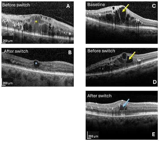
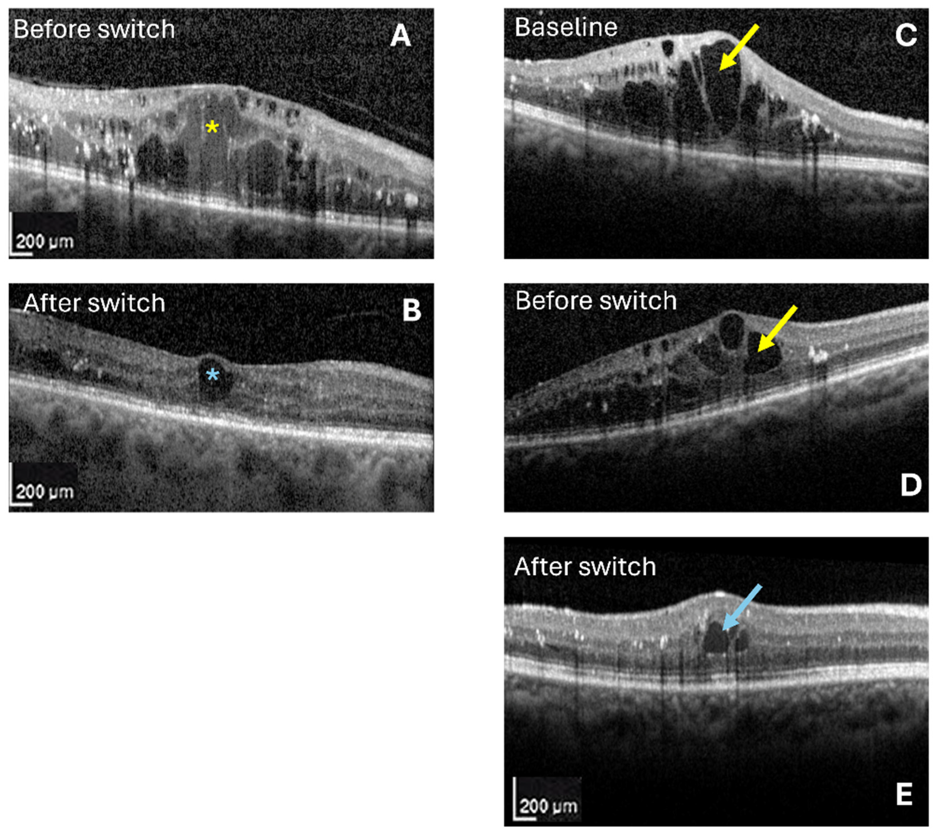
Table 3 illustrates the change in study parameters at the end of the 1-year follow-up period. Median LogMAR BCVA showed a marginal reduction in group 1 (0.09; interquartile range, IQR 0.0–0.24) compared to group 2 (0.0, IQR −0.13–0.08). IRF was noted in all eyes during the follow-up period, so the net change (ΔIRF) was zero. SRF reduction was reported in 2 out of 8 or 25% of eyes from group 1, while group 2 saw SRF reduction in 5 out of 26 eyes or 19%. Given a small percentage of eyes showing changes in SRF, the median (ΔSRF) was reported as zero. CST, CRT, and CRV showed a modest reduction in group 1 compared to group 2, which did not reach statistical significance. Interestingly, the median thickness values slightly increased in group 2 during the 1-year follow-up period (ΔCST = 18, IQR −46 to 95; ΔCRT = 3.5, IQR −45 to 40). The median CSV, however, showed a minimal, but numerically greater reduction in group 2 (−0.03, IQR −0.11 to 0.0) as compared to group 1 (−0.01, IQR −0.03 to 0.02), which did not reach statistical significance (p = 0.355). Figure 1 illustrates the exemplary images from both the groups, with OCT scans before and after faricimab initiation.
Table 3.
Comparison of the change in study parameters from baseline to 1 year.
Figure 1.
Optical coherence tomography (OCT) scans centered at the fovea in both groups, at baseline (A,C,D) and at the last follow-up (B,E), one month after the last faricimab injection during the study period. (A,B) were acquired from group 1, with (A) (at presentation) showing extensive intraretinal fluid (IRF, yellow asterisk), CST: 562 µm, CRV: 0.48 mm3; (B) (after initiating faricimab) showing a resolution of fluid, with a persistent intraretinal fluid pocket over the fovea (blue asterisk); CST: 189 µm, CRV: 0.26 mm3. (C–E) were acquired from group 2, with (C) showing IRF (yellow arrow), being treated with other anti-VEGF agents (bevacizumab in this example); CST: 587 µm, CRV: 0.47 mm3. (D) was acquired at the baseline just before faricimab initiation (IRF denoted by the yellow arrow); CST: 395 µm, CRV: 0.39 mm3; and (E) depicting changes at the last follow-up, illustrating the reduction in IRF (blue arrow) at the last follow-up visit; CST: 314 µm, CRV: 0.31 mm3.
3.3. Timing of Faricimab Initiation and Subgroup Analysis
Table 4 summarizes the results of linear mixed-effects models and logistic regression analyses. No statistically significant differences were observed in anatomic outcomes based on the timing of faricimab initiation (<6 months vs. >6 months). CST (β = 11.36, 95% confidence intervals, CI −50.87 to 73.59, p = 0.721) and CRT (β = 9.06, CI −24.89 to 43.03, p = 0.601) showed moderate numerical differences but without statistical significance, whereas CSV and CRV remained practically constant. A linear mixed-effects model was used to assess the association between faricimab initiation timing and different continuous variables (CST, CRT, CSV, CRV, LogMAR BCVA) in all the study eyes. After adjusting for baseline BCVA, HbA1C, total number of injections/faricimab injections, the timing of faricimab initiation was not independently associated with the change in visual acuity, CRT, or CST. This confirmed that there was no statistically significant effect of early faricimab initiation on this study cohort.
Table 4.
Adjusted group differences in clinical outcomes for faricimab treatment timing and sub-group analysis for patients initiating faricimab after 6 months.
4. Discussion
This retrospective chart analysis evaluated the efficacy of intravitreal faricimab, focusing on early versus a delayed initiation in eyes initially treatment-naïve for DME. Earlier faricimab initiation demonstrating a modest effect on changes in OCT-based structural metrics in this study underscores the need for individualized treatment strategies. Delayed initiation (group 2), while effective in maintaining visual acuity, resulted in a modest increase in thickness values as compared to group 1. These results highlight the possible functional efficacy of faricimab even with a delayed initiation in such eyes. While this increase in thickness values appears unusual, this figure was a change from the baseline to 1 year, with the last follow-up appointment being about 4 weeks after the last injection. This may have had an impact on the change analysis, with a mild, apparently nonsignificant increase in the values, which is generally noted a few weeks after an anti-VEGF injection. This may be important as the delayed initiation of faricimab may have been less effective in maintaining the macular thickness over time, where early initiation appeared to be effective in maintaining the thickness. Although these results may be clinically important, they did not reach statistical significance in our study. A slight decrease in visual acuity in group 1 (LogMAR 0.09, equivalent to approximately 4–5 letters) is difficult to explain given the retrospective nature of the study, although the chronicity (duration) of DME and the effects of higher glycemic load in this group (median HbA1c 8.1%, compared to 7.5% in group 2), with thinner baseline subfield thickness and volume parameters, may have an important role to play. Although there were no statistically significant differences in the demographic data, there were numerical differences in sex and ethnic variability that may be potential confounders which should be considered when assessing our results.
Furthermore, the adjusted linear mixed-effects model (Table 4) did not reveal any association of the timing of faricimab initiation on the functional or anatomical outcomes in this study cohort. Although the average change in BCVA (Table 3) in both the groups (an average decline of approximately one line Snellen’s equivalent in group 1 when compared to group 2) did not show statistical significance, the range of values suggested that there was a marginal improvement (albeit by a few letters) in some eyes even in group 2. The lack of such association on BCVA in group 1 versus group 2 may be due to a limited sample size, and the heterogeneity of the disease impacted by chronicity, systemic factors and the limited follow-up period.
Although the other study parameters showed no statistically significant difference between the groups, the numerical differences were all in the direction of or in favor of earlier initiation of faricimab. More importantly, given the chronic nature of this disease, maintained visual acuity suggests functional success even with a delayed initiation. Although no quantitative assessment of SRF/IRF was done in this study, graders noted a trend towards better SRF reduction in group 1. These findings, however, cannot be concluded definitely due to the qualitative nature of analysis. Previous reports have suggested that anatomical improvements do not always correlate with immediate functional gains in patients with DME [13]. Whether these anatomic benefits will translate to superior visual outcomes in the long-term, however, remains to be determined.
We speculate that the dual action of faricimab targeting both VEGF and Angiopoietin-2 (Ang-2) pathways may contribute to the observed anatomic benefit, though the possibility of an impact of a greater molar equivalent of anti-VEGF effect cannot be excluded [15,16]. This finding additionally aligns with those from the YOSEMITE and RHINE trials, which demonstrated the superiority of faricimab in improving anatomical outcomes compared to aflibercept [7,8,10].
Although more patients could achieve a complete resolution of SRF after initiating faricimab in both groups, all subjects demonstrated at least some persistent IRF in all groups. It should be noted that given the reduction in the OCT parameters (even in cases without SRF), we suspect that there was a reduction in the amount of IRF following faricimab initiation. This was also the impression of the OCT graders based on a qualitative inspection of the OCT scans; however, we are unable to conclude this definitively as a quantitative fluid compartment analysis was not performed in this study. This may be a target for future trials as automated fluid segmentation tools become more widely available. Across all groups, at least some IRF persisted, despite reductions in other structural metrics. While qualitative observations suggested a potential decrease in IRF volume, this study did not include quantitative fluid compartment analysis. The persistence of IRF may reflect a refractory response to therapy, degenerative changes, or residual non-active fluid, consistent with previous studies on chronic DME [17]. The results also emphasize the importance of early intervention in mitigating the chronic effects of DME. However, they highlight the challenges of achieving complete fluid resolution, particularly in real-world settings where adherence to consistent treatment schedules may be challenging. Further research is needed to determine these findings’ long-term clinical implications and refine the treatment protocols.
The results of this study are consistent with previous studies showing that certain patients with DME, particularly those with long-standing disease, may have a more refractory response to treatment [18,19,20]. While this may certainly highlight that mechanisms aside from VEGF and Ang-2 may be relevant to DME pathophysiology and may need to be targeted in some patients, it is also possible that residual IRF in some patients may be degenerative and not a sign of persistent disease activity [21,22,23].
The lack of statistically significant changes in BCVA between the early and late treatment groups emphasizes the complex relationship between retinal morphology and visual function in eyes with DME. Existing evidence highlights that visual gains may not always correlate with the structural improvement in fluid levels [6,23]. DRCR.net protocol V also highlighted that some eyes with good vision could maintain this vision over an extended period of time despite the presence of persistent center-involved DME [24]. On the other hand, we have decades of data that show chronic diabetic macular edema can lead to irreversible vision loss. Unfortunately, it is still unclear exactly when the tipping point occurs (based on severity or duration of edema), and when vision loss cannot be recovered. This information will be crucial in defining the optimal intervention points for therapy or a change in therapy. In the absence of better data, it may be prudent to maintain the retina as dry as early as possible.
This study also underscores the potential real-world challenges associated with anti-VEGF therapies, including faricimab [12,15,20,25,26]. While clinical trials demonstrate the drug’s efficacy with relatively consistent and frequent injections, real-world outcomes often differ from idealistic trajectories due to patient adherence issues, economic burdens, and other logistical difficulties of frequent monitoring. Furthermore, the proven efficacy in prolonging the treatment interval and improving the anatomical outcomes with possible maintenance of improvements in visual acuity is well reflected in this study. Of note, there was no significant difference between the number of overall intravitreal injections or the faricimab injections in both groups. This suggests that patients with a larger time gap when initiating more potent agents like faricimab may require more intensive treatment over time to maintain visual acuity. These findings further highlight the need for additional real-world studies to validate the optimal timing and frequency of faricimab administration in clinical practice.
The strengths of the study were the uniformity of demographic data, as well as the baseline study characteristics. Our study is not without limitations, which should be considered when analyzing our findings. First, the retrospective design inherently limits the ability to establish causality. Furthermore, in some cases, it may have introduced elements of bias, as the patients were treated as needed (PRN) and with a “treat and extend” regimen, depending upon the treating physician. There is also likely selection bias as to why one agent was chosen versus another for initiation of therapy as well as the timing of initiation. Additionally, the relatively small sample size and the exclusion of patients with inadequate follow-up reduce the generalizability of our findings. The lack of statistically significant differences may also be ascribed to a small sample size. That is why this study was designed as a pilot study, as the real-world difficulties can be improved efficiently to design a randomized trial in the future. The study was underpowered to detect modest between-group differences, supporting the interpretation that non-significant findings may reflect limited sample size rather than the absence of effect. Among patients with documented faricimab timing data, the median time from the first faricimab injection to the final assessment was 30.5 days (IQR, 0.0–70.8 days). However, complete treatment-timing information, including injection intervals and final injection-to-OCT intervals, was not available for all patients in this retrospective dataset. Consequently, these metrics could only be evaluated in a subset of patients and should be interpreted with caution. The lack of long-term follow-up also hinders our ability to evaluate the longevity of the anatomical and functional improvements observed, and whether the apparent anatomic benefits would have translated to better visual outcomes in the long term. Furthermore, only three eyes within group 1 had intravitreal faricimab as the first-line treatment, and thus we could not perform further sub-analysis within group 1 with regard to starting with faricimab vs. another agent. Although the unadjusted analysis suggested a potential association between earlier faricimab initiation and visual acuity outcomes, this association was no longer observed after adjustment for baseline visual acuity, HbA1c, and treatment exposure variables. These findings suggest that baseline disease characteristics and treatment-related factors may have contributed to the differences observed in the unadjusted analyses. Given the retrospective design and limited sample size, the present findings should be considered exploratory and hypothesis-generating rather than evidence of a causal effect of treatment timing. Lastly, while we accounted for various clinical characteristics and used cluster-weighted analyses to reduce intra-cluster correlations, other unmeasured confounding factors may have influenced our results. Last but not least, the quantification of IRF volume may have added further insight into the possible differences between groups, and therefore, this may qualify as an important biomarker which needs to be investigated in future studies.
5. Conclusions
In this small retrospective pilot cohort, earlier faricimab introduction showed numerical but statistically non-significant anatomical changes. After adjustment for baseline visual acuity, glycemic control, and treatment exposure, timing of faricimab initiation was not independently associated with meaningful functional or anatomical improvement. These findings should be interpreted as hypothesis-generating and require validation in larger prospective studies with standardized treatment protocols. The goal is to define an optimal integration of faricimab in the management of DME.
Author Contributions
Conceptualization, T.G., B.S., L.H., S.W., M.W.D., W.W., C.B., M.K.G., S.R.S. and A.V.; Methodology, B.S., S.R.S. and A.V.; Validation, A.V.; Formal analysis, T.G., L.H., S.W., M.W.D., S.R.S. and A.V.; Investigation, T.G., B.S., L.H., S.W., M.W.D., W.W., C.B., M.K.G., S.R.S. and A.V.; Resources, T.G., B.S., S.W., W.W., C.B., M.K.G., S.R.S. and A.V.; Data curation, T.G., B.S., L.H., S.W., M.W.D., M.K.G., S.R.S. and A.V.; Writing—original draft, T.G., B.S., L.H., S.W., M.W.D., W.W., C.B., M.K.G., S.R.S. and A.V.; Writing—review and editing, T.G., B.S., L.H., S.W., M.W.D., W.W., C.B., M.K.G., S.R.S. and A.V.; Visualization, T.G., B.S., L.H., S.W., M.W.D., W.W., C.B., M.K.G., S.R.S. and A.V.; Supervision, T.G., M.W.D., C.B., S.R.S. and A.V.; Project administration, S.R.S. and A.V. All authors have read and agreed to the published version of the manuscript.
Funding
This research received no external funding.
Institutional Review Board Statement
The study was conducted in accordance with the Declaration of Helsinki and approved by the Institutional Review Board of University of Louisville (IRB number: 24.0273; approval date: 16 April 2024).
Informed Consent Statement
Informed consent was obtained from all subjects involved in the study at the time of imaging.
Data Availability Statement
The data presented in this study are available on request from the corresponding author. The data are not publicly available due to privacy restrictions.
Conflicts of Interest
The authors declare no conflicts of interest.
Abbreviations
The following abbreviations have been appropriately defined in the manuscript.
| BCVA | Best corrected visual acuity |
| CI | Confidence intervals |
| CRT | Central retinal thickness |
| CRV | Central retinal volume |
| CST | Central subfield thickness |
| CVS | Central retinal volume |
| DME | Diabetic macular edema |
| ETRDS | Early treatment Diabetic Retinopathy Study |
| EZ | Ellipsoid zone |
| HbA1C | Glycosylated hemoglobin |
| IQR | Interquartile range |
| IRF | Intraretinal fluid |
| IVF | Intravitreal faricimab |
| OCT | Optical coherence tomography |
| PRN | As needed (pro re nata) |
| SRF | Subretinal fluid |
| VEGF | Vascular endothelial growth factor |
References
- Kusuhara, S.; Fukushima, Y.; Ogura, S.; Inoue, N.; Uemura, A. Pathophysiology of Diabetic Retinopathy: The Old and the New. Diabetes Metab. J. 2018, 42, 364–376. [Google Scholar] [CrossRef] [Scilit] [PubMed]
- Takamura, Y.; Ohkoshi, K.; Murata, T. New Strategies for Treatment of Diabetic Macular Edema. J. Ophthalmol. 2018, 2018, 4292154. [Google Scholar] [CrossRef] [Scilit] [PubMed]
- Haritoglou, C.; Maier, M.; Neubauer, A.S.; Augustin, A.J. Current concepts of pharmacotherapy of diabetic macular edema. Expert. Opin. Pharmacother. 2020, 21, 467–475. [Google Scholar] [CrossRef] [Scilit] [PubMed]
- Ciulla, T.A.; Bracha, P.; Pollack, J.; Williams, D.F. Real-world Outcomes of Anti-Vascular Endothelial Growth Factor Therapy in Diabetic Macular Edema in the United States. Ophthalmol. Retin. 2018, 2, 1179–1187. [Google Scholar] [CrossRef] [Scilit]
- Massin, P.; Creuzot-Garcher, C.; Kodjikian, L.; Girmens, J.F.; Delcourt, C.; Fajnkuchen, F.; Glacet-Bernard, A.; Guillausseau, P.J.; Guthux, F.; Blin, P.; et al. Real-World Outcomes after 36-Month Treatment with Ranibizumab 0.5 mg in Patients with Visual Impairment due to Diabetic Macular Edema (BOREAL-DME). Ophthalmic Res. 2021, 64, 577–586. [Google Scholar] [PubMed]
- Bressler, N.M.; Beaulieu, W.T.; Glassman, A.R.; Blinder, K.J.; Bressler, S.B.; Jampol, L.M.; Melia, M.; Wells, J.A., 3rd; Diabetic Retinopathy Clinical Research Network. Persistent Macular Thickening Following Intravitreous Aflibercept, Bevacizumab, or Ranibizumab for Central-Involved Diabetic Macular Edema with Vision Impairment: A Secondary Analysis of a Randomized Clinical Trial. JAMA Ophthalmol. 2018, 136, 257–269. [Google Scholar] [PubMed]
- Ishida, S.; Chen, S.J.; Murata, T.; Ogura, Y.; Ruamviboonsuk, P.; Sakamoto, T.; Fujita, T.; Kawano, M.; Ohsawa, S.; Abreu, F.; et al. Efficacy, Durability, and Safety of Faricimab in Patients from Asian Countries with Diabetic Macular Edema: 1-Year Subgroup Analysis of the Phase III YOSEMITE and RHINE Trials. Asia Pac. J. Ophthalmol. 2023, 12, 451–459. [Google Scholar] [CrossRef] [Scilit]
- Wong, T.Y.; Haskova, Z.; Asik, K.; Baumal, C.R.; Csaky, K.G.; Eter, N.; Ives, J.A.; Jaffe, G.J.; Korobelnik, J.F.; Lin, H.; et al. Faricimab Treat-and-Extend for Diabetic Macular Edema: Two-Year Results from the Randomized Phase 3 YOSEMITE and RHINE Trials. Ophthalmology 2024, 131, 708–723. [Google Scholar] [PubMed]
- Sahni, J.; Patel, S.S.; Dugel, P.U.; Khanani, A.M.; Jhaveri, C.D.; Wykoff, C.C.; Hershberger, V.S.; Pauly-Evers, M.; Sadikhov, S.; Szczesny, P.; et al. Simultaneous Inhibition of Angiopoietin-2 and Vascular Endothelial Growth Factor-A with Faricimab in Diabetic Macular Edema: BOULEVARD Phase 2 Randomized Trial. Ophthalmology 2019, 126, 1155–1170. [Google Scholar] [PubMed]
- Wykoff, C.C.; Abreu, F.; Adamis, A.P.; Basu, K.; Eichenbaum, D.A.; Haskova, Z.; Lin, H.; Loewenstein, A.; Mohan, S.; Pearce, I.A.; et al. Efficacy, durability, and safety of intravitreal faricimab with extended dosing up to every 16 weeks in patients with diabetic macular oedema (YOSEMITE and RHINE): Two randomised, double-masked, phase 3 trials. Lancet 2022, 399, 741–755. [Google Scholar] [CrossRef] [Scilit] [PubMed]
- Laura Joszt, M. FDA Approves Faricimab to Treat Wet AMD and DME; AJMC: Cranbury, NJ, USA, 2022; Available online: https://www.ajmc.com/view/fda-approves-fariximab-to-treat-wet-amd-and-dme (accessed on 31 January 2022).
- Quah, N.Q.X.; Javed, K.M.A.A.; Arbi, L.; Hanumunthadu, D. Real-World Outcomes of Faricimab Treatment for Neovascular Age-Related Macular Degeneration and Diabetic Macular Edema. Clin. Ophthalmol. 2024, 27, 1479–1490. [Google Scholar] [CrossRef] [Scilit]
- Wolfrum, P.; Böhm, E.W.; Lorenz, K.; Stoffelns, B.; Pfeiffer, N.; Korb, C.A. Short-Term Clinical Outcomes of Patients with Diabetic Macular Edema Following a Therapy Switch to Faricimab. J. Clin. Med. 2024, 13, 4508. [Google Scholar] [CrossRef] [Scilit] [PubMed]
- Penha, F.M.; Masud, M.; Khanani, Z.A.; Thomas, M.; Fong, R.D.; Smith, K.; Chand, A.; Khan, M.; Gahn, G.; Melo, G.B.; et al. Review of real-world evidence of dual inhibition of VEGF-A and ANG-2 with faricimab in NAMD and DME. Int. J. Retin. Vitr. 2024, 10, 5. [Google Scholar] [CrossRef] [Scilit]
- Durrani, A.F.; Momenaei, B.; Wakabayashi, T.; Vemula, S.; Pandit, S.A.; Hsu, J.; Ho, A.C.; Spirn, M.J.; Klufas, M.A.; Garg, S.J.; et al. Conversion to faricimab after prior anti-vascular endothelial growth factor therapy for persistent diabetic macular oedema. Br. J. Ophthalmol. 2024, 108, 1257–1262. [Google Scholar] [CrossRef] [Scilit] [PubMed]
- Downey, L.; Sivaprasad, S.; Chhabra, R.; Bailey, C.; Chakrabarti, S.; Elsherbiny, S.; Patel, J.; Silvestri, G.; Watson, S.L.; Williams, G.; et al. Dual pathway inhibition with faricimab for previously treated neovascular age-related macular degeneration and diabetic macular oedema: Guidance from a UK panel of retina specialists. Eye 2024, 38, 3077–3086. [Google Scholar] [CrossRef] [Scilit] [PubMed]
- Park, J.; Felfeli, T.; Kherani, I.Z.; Altomare, F.; Chow, D.R.; Wong, D.T. Prevalence and clinical implications of subretinal fluid in retinal diseases: A real-world cohort study. BMJ Open Ophthalmol. 2023, 8, e001214. [Google Scholar] [CrossRef] [Scilit] [PubMed]
- Hsieh, T.C.; Deng, G.H.; Chang, Y.C.; Chang, F.L.; He, M.S. A real-world study for timely assessing the diabetic macular edema refractory to intravitreal anti-VEGF treatment. Front. Endocrinol. 2013, 14, 1108097. [Google Scholar]
- Virgili, G.; Parravano, M.; Evans, J.R.; Gordon, I.; Lucenteforte, E. Anti-vascular endothelial growth factor for diabetic macular oedema: A network meta-analysis. Cochrane Database Syst. Rev. 2023, 10, CD007419. [Google Scholar] [CrossRef] [Scilit]
- Shah, S.U.; Maturi, R.K. Therapeutic Options in Refractory Diabetic Macular Oedema. Drugs 2017, 77, 481–492. [Google Scholar] [CrossRef] [Scilit] [PubMed]
- Midena, E.; Torresin, T.; Schiavon, S.; Danieli, L.; Polo, C.; Pilotto, E.; Midena, G.; Frizziero, L. The Disorganization of Retinal Inner Layers Is Correlated to Müller Cells Impairment in Diabetic Macular Edema: An Imaging and Omics Study. Int. J. Mol. Sci. 2023, 24, 9607. [Google Scholar] [CrossRef] [Scilit] [PubMed]
- Browning, D.J.; Stewart, M.W.; Lee, C. Diabetic macular edema: Evidence-based management. Indian J. Ophthalmol. 2018, 66, 1736–1750. [Google Scholar] [CrossRef] [Scilit] [PubMed]
- Patil, N.S.; Mihalache, A.; Dhoot, A.S.; Popovic, M.M.; Muni, R.H.; Kertes, P.J. The Impact of Residual Retinal Fluid Following Intravitreal Anti-Vascular Endothelial Growth Factor Therapy for Diabetic Macular Edema and Macular Edema Secondary to Retinal Vein Occlusion: A Systematic Review. Ophthalmic Surg. Lasers Imaging Retin. 2023, 54, 50–58. [Google Scholar] [CrossRef] [Scilit]
- Baker, C.W.; Glassman, A.R.; Beaulieu, W.T.; Antoszyk, A.N.; Browning, D.J.; Chalam, K.V.; Grover, S.; Jampol, L.M.; Jhveri, C.D.; Melia, M.; et al. Effect of initial management with aflibercept vs laser photocoagulation vs observation on vision loss among patients with diabetic macular edema involving the center of the macula and good visual acuity: A randomized clinical trial. JAMA 2019, 321, 1880–1894. [Google Scholar] [PubMed]
- Borchert, G.A.; Kiire, C.A.; Stone, N.M.; Akil, H.; Gkika, T.; Fischer, M.D.; Xue, K.; Cehajic-Kapetanovic, J.; MacLaren, R.E.; Charbel Issa, P.; et al. Real-world six-month outcomes in patients switched to faricimab following partial response to anti-VEGF therapy for neovascular age-related macular degeneration and diabetic macular oedema. Eye 2024, 38, 3569–3577. [Google Scholar] [PubMed]
- Nasimi, S.; Nasimi, N.; Grauslund, J.; Vergmann, A.S.; Subhi, Y. Real-World Efficacy of Intravitreal Faricimab for Diabetic Macular Edema: A Systematic Review. J. Pers. Med. 2024, 14, 913. [Google Scholar] [CrossRef] [Scilit] [PubMed]

Disclaimer/Publisher’s Note: The statements, opinions and data contained in all publications are solely those of the individual author(s) and contributor(s) and not of MDPI and/or the editor(s). MDPI and/or the editor(s) disclaim responsibility for any injury to people or property resulting from any ideas, methods, instructions or products referred to in the content. |
© 2026 by the authors. Licensee MDPI, Basel, Switzerland. This article is an open access article distributed under the terms and conditions of the Creative Commons Attribution (CC BY) license.