


Abstract
Backgrond/Objectives: Human anatomy education has traditionally relied on cadaveric dissection as the reference standard for acquiring anatomical knowledge. However, decreasing availability of donated bodies, curricular changes, and rapid technological advances have encouraged the progressive integration of complementary educational resources, including historical anatomical collections, physical models, plastinated specimens, and digital technologies. At the same time, anatomical museums have evolved from repositories of scientific heritage into dynamic educational environments that promote public engagement, accessibility, and lifelong learning. Methods: This article presents the educational experience of the Human Anatomy and Histology Institute of the University of Palermo, where historical anatomical collections, cadaveric dissection, plastination, three-dimensional (3D) digitization, virtual anatomy, and interactive educational technologies have been integrated within a single multimodal teaching framework. Results: The strengths and limitations of each educational resource are discussed in relation to contemporary anatomy education and museum communication. Our experience suggests that digital technologies achieve their greatest educational value when they complement rather than replace traditional anatomy teaching. Conclusions: Integrating historical collections with cadaveric study, physical models, and immersive digital tools offers a comprehensive educational approach capable of enhancing anatomical understanding, improving accessibility, and promoting more engaging learning experiences for students and the wider public.
1. Introduction
Human anatomy is a fundamental component of undergraduate medical education, with cadaveric dissection traditionally regarded as the gold standard for acquiring anatomical knowledge [1]. Nevertheless, anatomical education has long faced important challenges related to cadaver procurement, preservation techniques, donor shortages, increasing operational costs, and continuous curricular reforms, prompting many medical schools to explore complementary educational resources [2,3].
Among the earliest alternatives to cadaveric specimens, anatomical wax models emerged during the late seventeenth century and progressively became an integral component of anatomical teaching. Anatomical ceroplastics is a specialized anatomical modeling technique that employs wax to reproduce human structures with high morphological fidelity for scientific and educational use. Owing to its remarkable ability to reproduce fine morphological details together with the natural appearance of tissues, blood vessels, and internal organs, wax represented an ideal material for the creation of highly realistic and durable anatomical models [2]. Originating in Italy, the tradition of anatomical ceroplastics subsequently spread throughout Europe, reflecting the close collaboration between anatomists and artists in producing models that combined scientific accuracy with exceptional artistic quality.
During the twentieth century, wax models were progressively complemented and, in many institutions, replaced by durable three-dimensional (3D) anatomical models better suited for routine educational activities. More recently, the digital revolution has profoundly transformed anatomy education through the introduction of technologies such as three-dimensional digitization, virtual reality (VR), and augmented reality (AR), providing increasingly interactive, immersive, and student-centred learning experiences [4]. Rather than replacing traditional educational resources, these technologies offer new opportunities to integrate cadaveric dissection, anatomical collections, physical models, and digital visualization into complementary learning pathways.
Beyond their educational role in medical training, historical anatomical collections have progressively evolved into museum environments with broader scientific, cultural, and social missions. According to the current International Council of Museums (ICOM) definition, museums should be accessible and inclusive institutions that promote participation, diversity, and lifelong learning [5]. Within this framework, accessibility extends beyond the removal of physical barriers to include cognitive, sensory, and experiential dimensions of the visitor experience. Contemporary museology increasingly emphasizes the design of educational environments capable of accommodating diverse audiences with different sensory and cognitive profiles [6,7]. Recent studies have further highlighted the value of combining structured educational pathways with digital technologies to improve cognitive and sensory accessibility, particularly for neurodivergent visitors [8].
Against this background, the experience of the Human Anatomy and Histology Institute of the University of Palermo provides an example of how historical anatomical heritage can be integrated with cadaveric dissection, physical models, plastination, three-dimensional digitization, and virtual anatomy technologies within a unified educational framework. The present article describes this integrated approach, discussing the strengths and limitations of each educational resource and illustrating how their complementary use may enhance anatomy teaching, museum communication, and public engagement.
2. Human Anatomical Heritage in Palermo
The University of Palermo preserves one of the most important historical anatomical collections in southern Italy, representing more than five centuries of anatomical teaching, scientific investigation, and medical education. Although the present Institute of Human Anatomy was formally established in 1932, the anatomical heritage currently preserved within its collections reflects a much longer academic tradition, documenting the evolution of anatomical knowledge, teaching methodologies, and specimen preparation techniques from the Renaissance to the present day [9].
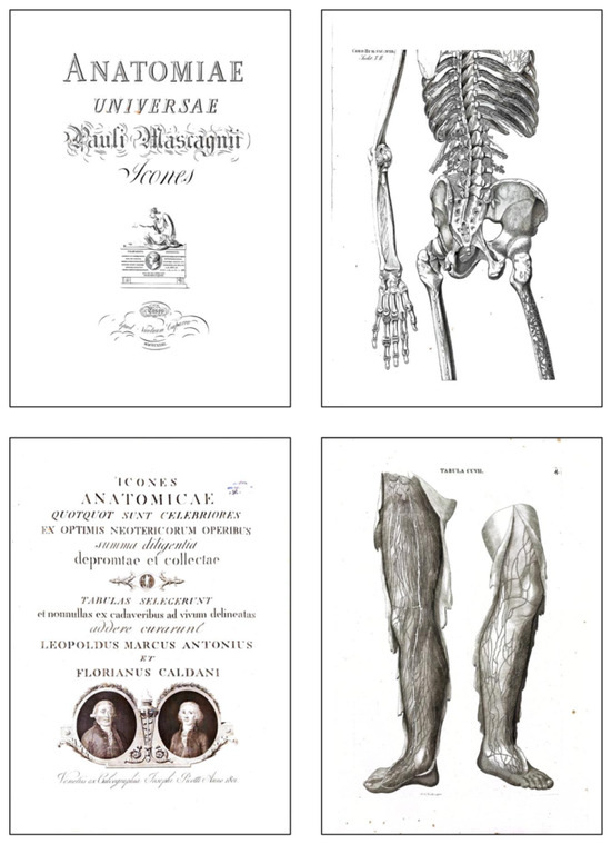
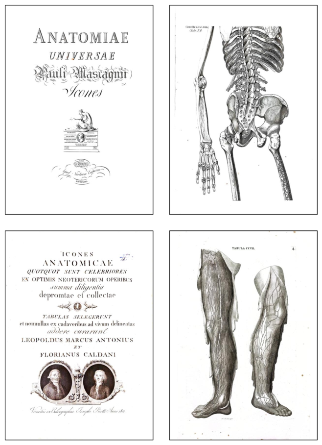
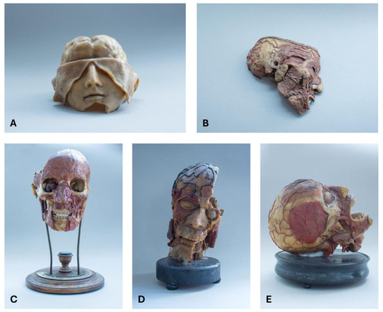
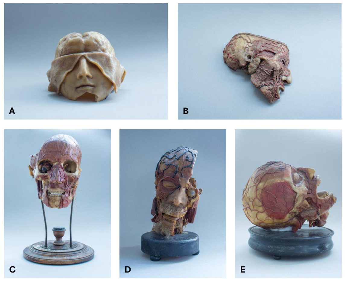
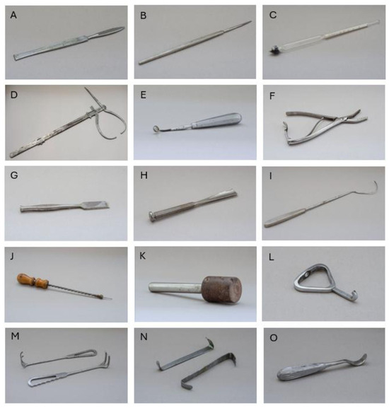
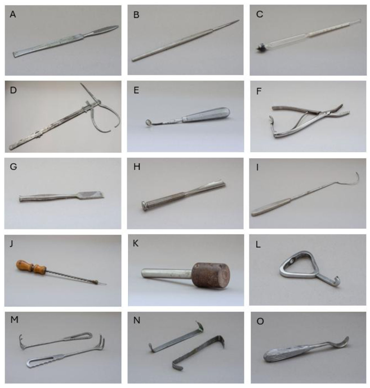
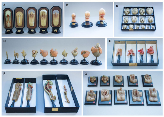
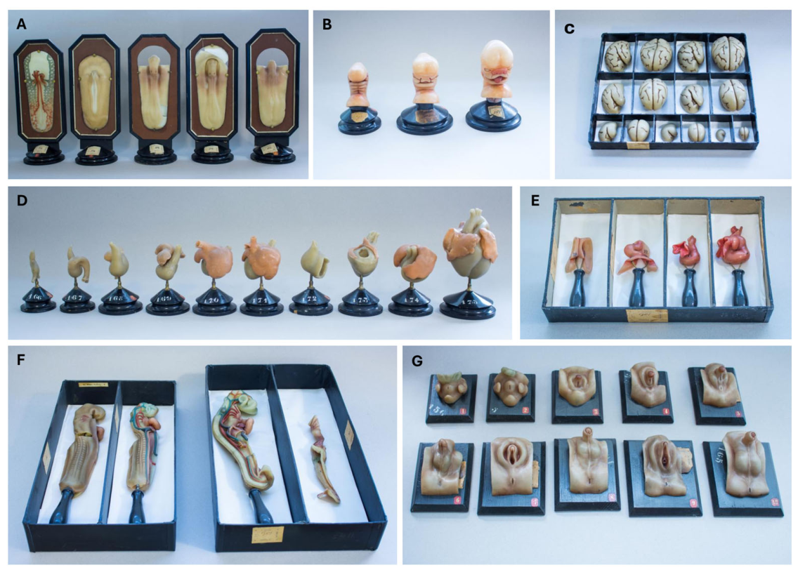
Today, this heritage forms the core of the University Human Anatomy Collection, where historical books, anatomical preparations, scientific instruments, and teaching models are displayed within an educational museum environment. Rather than functioning solely as a repository of historical artefacts, the collection aims to illustrate the evolution of anatomical education and to promote dialogue between historical heritage and contemporary biomedical sciences. It includes rare anatomical atlases (Figure 1), anatomical wax models attributed to the Sicilian school of anatomical ceroplastics (Figure 2), historical instruments formerly employed for anatomical dissection, specimen preservation, and the preparation and monitoring of preservation solutions (Figure 3), as well as embryological teaching models that remain valuable educational resources today (Figure 4).
Figure 1.
Representative images from two historical books preserved in the Anatomical Collection of the University of Palermo. (Above): Anatomiae Universae by Paolo Mascagni (1755–1815). First published in 1823, this work remained the standard textbook for the education of medical and surgical students for many decades. (Left): the book cover. (Right): one of the anatomical plates. (Below): Icones anatomicae: quotquot sunt celebriores ex optimis neotericorum operibus summa diligentia depromptae et collectae by Leopoldo Marco Antonio Caldani (1725–1813). One of the most monumental and comprehensive collections of anatomical illustrations ever produced in the history of medicine, this work combined extraordinary scientific value for its time with outstanding artistic quality, representing a synthesis of the finest anatomical knowledge of the period. (Left): the book cover. (Right): one of the anatomical plates.
Figure 2.
Anatomical wax models of the skull. (A): Late eighteenth-century skull with the calvaria removed. (B): Late nineteenth-century skull showing the vascular and nervous systems. (C): Late nineteenth-century wax anatomical head attributed to the Sicilian ceroplast Graffeo. (D): Early nineteenth-century wax anatomical head attributed to the Sicilian ceroplast Graffeo. (E): Early nineteenth-century skull containing preserved organic material.
Figure 3.
Instruments for anatomical dissection, early twentieth century. (A,B): Scalpels. (C): Gay-Lussac alcoholometer. (D): Anatomical caliper. (E): Volkmann curette. (F): Kocher clamp. (G,H): Surgical chisels. (I): Suture-passing needle. (J): Surgical perforator. (K): Surgical mallet. (L): Retractor hook. (M): Volkmann retractor. (N): Farabeuf retractor. (O): Spatula retractor.
Figure 4.
Wax models illustrating selected stages of human embryonic and fetal development, including the embryonic disc (A), the facial prominences (B), the developing brain (C), the developing heart (D), the primitive heart tube (E), the embryo as a whole (F), and the developing external male genitalia (G).
The collection is regularly opened to the public during World Anatomy Day (15 October) and is also accessible throughout the year through guided tours organized in collaboration with cultural initiatives such as Le Vie dei Tesori and educational activities dedicated to primary and secondary schools. These initiatives continue the educational mission of the Institute by promoting scientific dissemination, stimulating curiosity among younger generations, and encouraging public engagement with the history of medicine and human anatomy. These public engagement activities are supported by the Institute’s academic staff as well as by members of the Institute in various positions (undergraduate students, doctoral candidates, and postdoctoral researchers), who actively participate in the planning and realization of guided tours. Their involvement serves a dual purpose: to ensure high-quality scientific communication for visitors while also providing valuable training in public engagement and “third mission” activities, which are considered an integral part of academic and professional development within the Institute. The activities are organized using existing institutional resources and include, for example, for children, coloring pictures of anatomical structures based on the models they have observed, creating simple anatomical models using plasticine, and placing organs in their correct anatomical positions on a template of the human body. These practical activities reinforce the anatomical concepts introduced during the guided tour.
From a museological perspective, historical anatomical collections represent not only scientific resources but also powerful educational tools capable of creating narrative, emotional, and multisensory experiences. Their artistic, historical, and scientific value allows visitors to establish meaningful connections with anatomical heritage while fostering a deeper appreciation of the evolution of medical knowledge [6,10].
Among the most valuable specimens preserved in the collection are the anatomical wax models representing the head and other body regions (Figure 2), traditionally attributed to the Sicilian school of anatomical ceroplastics. This artistic and scientific tradition originated with the pioneering work of the Sicilian ceroplast Gaetano Giulio Zumbo (ca. 1656–1701), whose anatomical models profoundly influenced the development of wax modelling in Europe [11]. Combining remarkable anatomical accuracy with exceptional artistic craftsmanship, Zumbo’s work established a tradition that was subsequently continued by later Sicilian ceroplasts, including Graffeo, to whom several models preserved in Palermo are traditionally attributed. The flourishing of anatomical wax modelling during the eighteenth century, particularly following the establishment of the Florentine ceroplastics workshop, contributed to the dissemination of these teaching models throughout Italy and beyond, where they became indispensable educational resources for the study of human anatomy [11].
The collection also houses a remarkable series of embryological teaching models produced in Freiburg by Adolf Ziegler and Friedrich Ziegler under the scientific supervision of Wilhelm His Sr. (Figure 4). These wax models illustrate successive stages of embryonic and fetal development with exceptional morphological accuracy, allowing students and visitors to appreciate the complex processes underlying human morphogenesis. By transforming dynamic developmental events into tangible three-dimensional representations, these models continue to provide an effective educational resource for understanding early human development. The same anatomical resources are used for both healthcare students and the general public, with the approach adapted to the target audience. For students, the focus is on anatomical detail and clinical correlations, whereas for the public, the emphasis is on stimulating curiosity and promoting interest in human anatomy and biomedical sciences. These differentiated educational pathways are currently being refined and will be further structured and evaluated in future studies.
3. From Historical Collections to Modern Anatomy Education
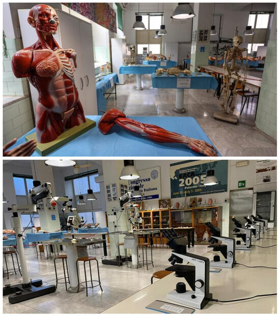
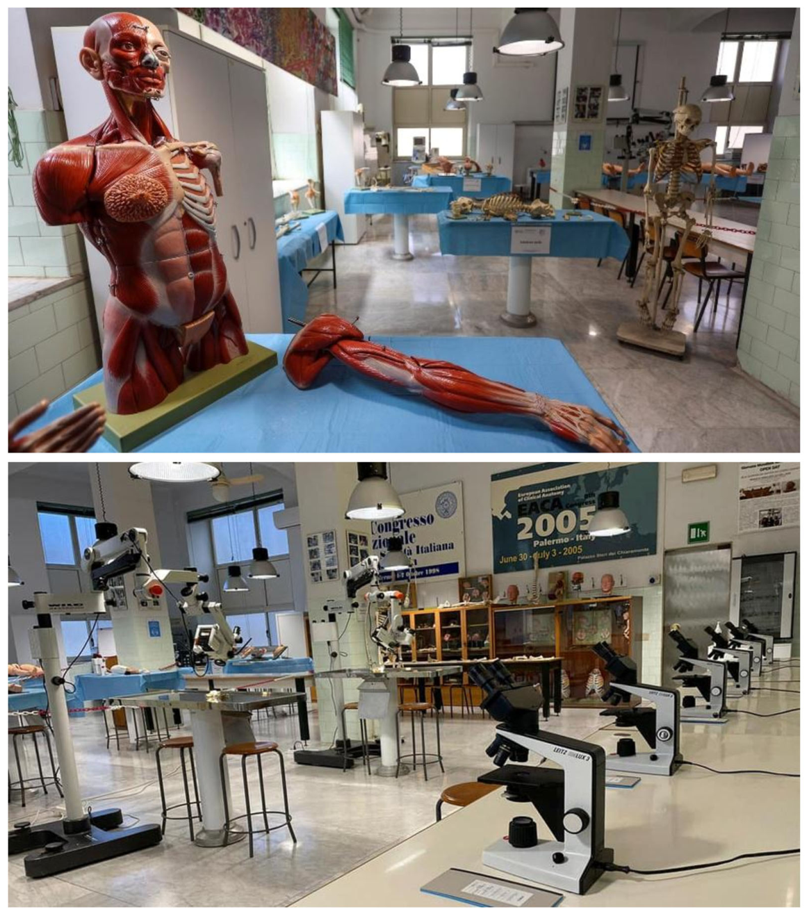
The educational pathway continues beyond the historical collection into the contemporary teaching facilities of the Institute, where traditional anatomical resources are integrated with modern educational technologies. Visitors are introduced to the gross anatomy room, where three-dimensional anatomical models, human skeletal specimens, and histological preparations are routinely used for undergraduate teaching and practical laboratory sessions (Figure 5). Here, students can directly compare anatomical models with real human skeletal specimens while simultaneously exploring tissue organization through microscopic observation. This integrated learning environment enables the progressive transition from macroscopic to microscopic anatomy, reinforcing the relationship between structure and function across different levels of biological organization. The skeletal material held at our Institute dates mainly from the late 19th and early 20th centuries. At that time, anatomical collections in Italy were commonly assembled from bodies that had not been claimed after death, often including homeless people who had died without relatives or heirs. Unfortunately, after the Second World War, the original paper documents attesting the origin of these specimens were lost or destroyed. Consequently, it is no longer possible to reconstruct the identity or individual history of most of the remains. Historical collections require careful assessment of provenance and ethical context, according to the International Federation of Associations of Anatomists (IFAA) guidelines published in 2012 [12]. Whilst contemporary donations are governed by current legislation and informed consent procedures [13], many historical artefacts were acquired during periods characterized by different ethical standards and limited documentation. Where information on provenance is incomplete, these collections must be presented transparently, acknowledging their historical context and the difficulties involved in reconstructing their origins. Both educational and museum activities should raise awareness and emphasize the importance of a respectful and informed approach to human remains, which are classified as a special category of “culturally sensitive materials” under the ICOM Code of Ethics for Museums [13,14].
Figure 5.
Representative views of the gross anatomy room at our Institute, where three-dimensional anatomical models, human skeletal specimens, and histological slides are exhibited to the public during World Anatomy Day. (Above): Three-dimensional anatomical models and human skeletal specimens. (Below): Histological microscopes and histological slides available for student observation.
The educational experience is further strengthened by the recently established plastination laboratory, where selected anatomical specimens obtained from cadaveric dissection are permanently preserved using modern plastination techniques. Besides preventing tissue degradation and ensuring long-term conservation, plastination allows unique anatomical specimens to remain available for teaching, scientific dissemination, and museum exhibition. Together with the historical collections, plastinated specimens provide a direct link between classical cadaveric dissection and contemporary anatomy education.
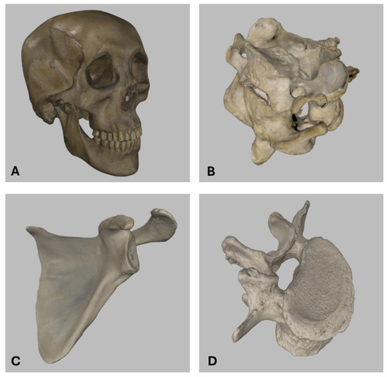
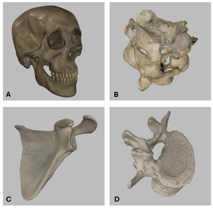
In parallel, the Institute has progressively introduced three-dimensional (3D) digitization technologies into its educational activities. Human skeletal specimens and cadaveric preparations are digitally acquired using 3D scanning systems and subsequently displayed on large interactive touchscreens, allowing students to freely rotate, enlarge, and examine anatomical structures while interacting with instructors during practical lessons (Figure 6). This approach considerably enhances spatial understanding of complex anatomical relationships while preserving the educational value of the original specimens. The 3D models of the specimens are acquired through photogrammetry using a structured-light scanner, allowing accurate digital reconstruction of surface morphology. The models are shared with students through the Tinalp platform, which enables visualization and interactive exploration of the digital specimens. The use of donated specimens for digital imaging was covered by the informed consent provided during the donation process, in which the procedures for image acquisition, storage, duration of retention, and intended use were explicitly specified [12,15]. In accordance with recommendations for good practice in human tissue image acquisition, the use of images of donated specimens was restricted to clearly defined education and research purposes. The images were stored on secure, password-protected devices accessible only to designated faculty, staff, and authorized students. They were not shared on social media or other publicly accessible, non-password-protected internet platforms, thereby preventing the misuse or inappropriate dissemination of images of donated bodies for non-academic purposes [12].
Figure 6.
Examples of human skeletal specimens digitized by three-dimensional (3D) scanning and made available to students through large interactive display screens, allowing them to rotate, enlarge, and examine the models while actively interacting with instructors during the study of osteology. (A): Complete skull. (B): Atlas and axis in articulation. (C): Scapula. (D): Lumbar vertebra.
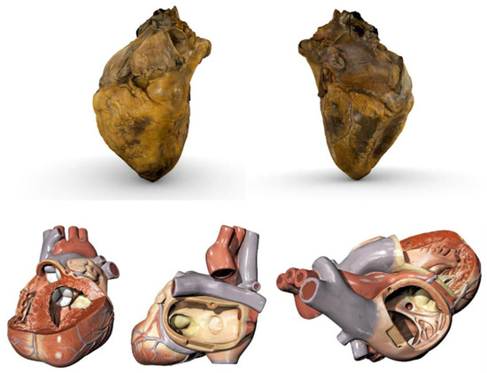
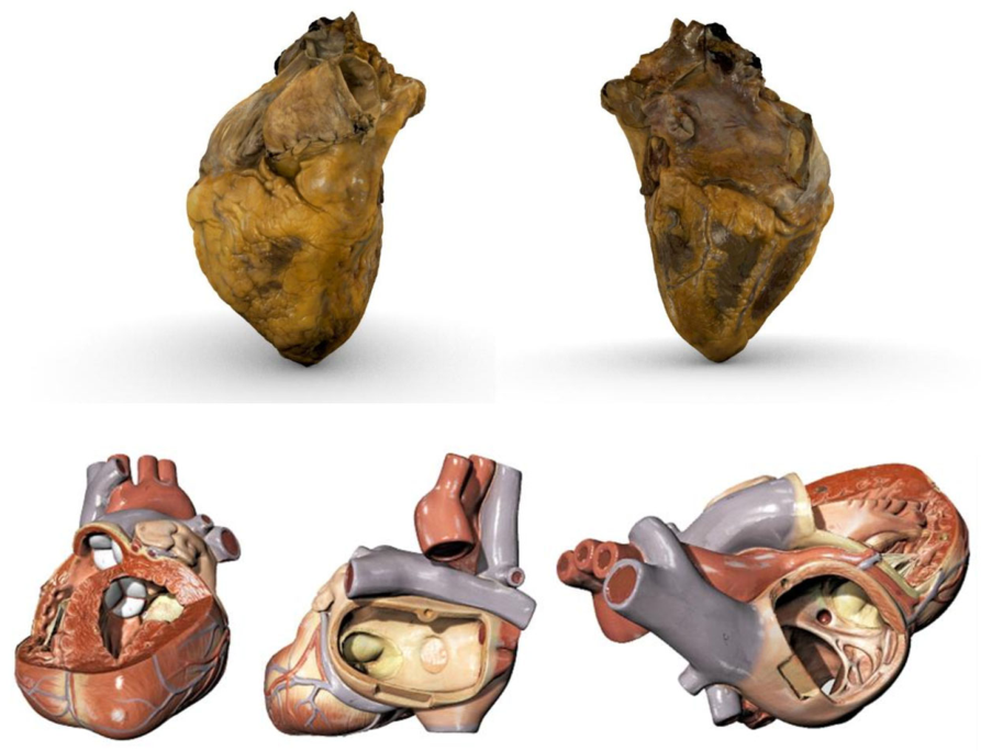
The integration of cadaveric anatomy with digital technologies is further exemplified by three-dimensional reconstructions of real human organs and anatomical teaching models (Figure 7). Cadaveric specimens acquired in the dissection room can be digitally reconstructed and explored from multiple perspectives, whereas anatomical models may be virtually opened to reveal internal structures that are not visible during external observation alone. The accompanying Supplementary Videos provide students with an interactive learning resource that complements direct observation and facilitates a deeper understanding of complex three-dimensional anatomy.
Figure 7.
Integration of cadaveric anatomy, three-dimensional (3D) digitization, and anatomical models in anatomy teaching. (Above): A human heart obtained during cadaveric dissection and digitized by 3D scanning, showing the anterior surface (left) and the posterior and inferior surfaces (right). An additional video of the 3D model is provided as Supplementary Video S1, enabling interactive visualization of the specimen and enhancing the understanding of its three-dimensional morphology. (Below): Three frames extracted from a video generated by 3D scanning of a plastic anatomical heart model. The complete video, provided as Supplementary Video S2, allows students to visualize internal anatomical structures that are concealed during external inspection and become visible only when the model is opened. The integration of cadaveric dissection, where students can examine the real human heart, with the study of anatomical models in the gross anatomy room enhances the understanding of the three-dimensional morphology of the heart and, by extension, of other anatomical structures.
Taken together, these educational resources illustrate the philosophy currently adopted at the University of Palermo, where historical anatomical heritage, cadaveric dissection, plastination, physical anatomical models, and digital technologies are not regarded as alternative approaches but as complementary components of a single integrated educational framework. Rather than replacing traditional anatomy teaching, technological innovation expands its educational potential, creating a multimodal learning environment capable of addressing the needs of university students, healthcare professionals, and the general public alike.
4. Strengths and Limitations of Traditional Anatomical Resources
A comprehensive understanding of human anatomy requires the ability to visualize three-dimensional relationships among anatomical structures while simultaneously developing spatial reasoning and visuospatial skills [16]. Among all available educational resources, cadaveric dissection continues to represent the reference standard for anatomy teaching, allowing students to appreciate the texture, consistency, and natural anatomical variability of human tissues while promoting long-lasting knowledge retention [17]. Beyond its scientific value, cadaveric dissection also plays a fundamental role in fostering empathy, professionalism, and ethical awareness, encouraging students to regard body donors as their first “silent mentors” [17].
Despite these educational advantages, the availability of cadavers has progressively declined in many countries because of religious and cultural traditions, ethical concerns, economic constraints, concerns regarding disease transmission, and increasingly complex legislative frameworks [18]. Italy has experienced a similar situation. Although Law No. 10/2020 was specifically introduced to promote body donation for education and research, delays in its implementation together with persistent administrative and legal barriers have limited its practical impact, contributing to the continuing shortage of donated bodies available for medical education [18,19,20].
For these reasons, anatomy educators are increasingly relying on complementary educational resources, including high-fidelity anatomical models, plastinated specimens, prosections, and digital technologies [21]. Recent qualitative studies have shown that students perceive these resources as fulfilling different educational needs rather than representing interchangeable alternatives. Three-dimensional printed models are considered particularly effective for understanding general anatomical organization and organ morphology, whereas plastinated specimens are preferred for studying complex anatomical regions and appreciating the realistic appearance of human tissues [17]. Conversely, some students may initially experience cognitive overload when learning exclusively through cadaveric specimens because of their intrinsic anatomical complexity [22]. Similarly, Davis and colleagues reported that anatomical models are generally perceived positively when used alongside cadaveric material, although individual teaching tools may differ in their educational effectiveness according to their intended learning objectives [21]. These observations suggest that no single educational resource is sufficient to address all learning needs and reinforce the value of integrating multiple complementary approaches within anatomy education.
A similar principle applies to historical anatomical collections and medical museums. While these institutions preserve an invaluable scientific and cultural heritage and remain powerful educational resources, traditional museum exhibitions are often perceived as static and insufficiently interactive, particularly by younger generations accustomed to digital learning environments. Passive observation alone may limit visitors’ engagement with complex anatomical concepts and reduce opportunities for meaningful educational interaction. Consequently, anatomical museums are increasingly evolving from repositories of historical artefacts into dynamic educational environments capable of combining historical collections with innovative communication strategies and interactive learning experiences [8].
Contemporary museum studies recognize accessibility as a multidimensional concept encompassing physical, cognitive, social, and sensory dimensions [6,23]. Although many museums have successfully improved architectural accessibility, cognitive and sensory accessibility remain comparatively underdeveloped. Environmental factors such as excessive noise, visual complexity, lighting conditions, or overcrowding may negatively affect comfort and learning, particularly among visitors with autism spectrum disorder and other neurodevelopmental conditions [24,25]. In response, museums are increasingly adopting principles derived from Universal Design for Learning (UDL) and Design for All, promoting multimodal communication, adaptable educational pathways, and flexible learning experiences capable of accommodating diverse audiences [7,26,27].
Within this evolving educational framework, digitization has emerged as a valuable strategy for both preserving and disseminating anatomical heritage. Advances in photogrammetry and three-dimensional acquisition technologies now allow museum specimens to be accurately digitized and incorporated into virtual museum environments, considerably expanding public access to collections that often remain physically inaccessible [28]. Rather than replacing traditional anatomical resources, these digital technologies provide new opportunities for integrating historical collections, cadaveric anatomy, and interactive educational experiences, supporting the multimodal teaching philosophy described in the present study.
5. Digital Technologies as Complementary Educational Tools
Medical education is undergoing a profound transformation driven by the progressive integration of digital technologies into traditional teaching methodologies. Alongside cadaveric dissection, anatomical models, and museum collections, novel educational resources such as virtual patients, three-dimensional (3D) anatomical atlases, VR, AR, and simulation-based learning platforms are increasingly being incorporated into anatomy curricula [29,30]. One of the principal motivations behind this evolution has been the need to bridge the gap between classical anatomy teaching and the imaging modalities routinely employed in contemporary clinical practice, including computed tomography (CT), radiography, ultrasonography, and magnetic resonance imaging (MRI) [31].
The development of increasingly accurate three-dimensional digital representations of the human body has considerably expanded the possibilities for anatomy education [4]. Among the most influential initiatives, the Visible Human Project established a digital reference model of the entire human body by integrating CT, MRI, and cryosection datasets, thereby enabling virtual dissection and interactive exploration of anatomical structures from multiple perspectives [4]. Students can progressively visualize different anatomical systems by adding or removing individual tissue layers, an approach that facilitates spatial understanding and reinforces the three-dimensional organization of the human body [32].
VR and AR have attracted increasing attention in recent years, particularly following the COVID-19 pandemic, which accelerated the adoption of digital educational technologies [33]. Several studies have investigated the effectiveness of VR and AR in anatomy education. Recent meta-analyses demonstrated that VR is associated with significantly greater gains in anatomical knowledge compared with conventional teaching methods, including textbooks, lectures, two-dimensional (2D) images, audio-based instruction, and 3D models. However, current evidence suggests that VR should be regarded as a complementary rather than a replacement educational tool [34,35,36]. The effectiveness of VR in anatomy education is influenced by several factors, including the level of immersion, quality of the simulated models, duration of training, prior familiarity with VR technology, baseline anatomical knowledge, educational topic, interactivity, usability, cognitive load, and VR-related side effects, which may vary according to individual characteristics, such as gender and alertness [35,37].
These technological advances have also created new opportunities for anatomical museums and historical collections. Interactive 3D models, virtual anatomy platforms, AR, and immersive digital environments enable museums to move beyond passive exhibition strategies, transforming visitors into active participants in the learning process. Digital technologies may enhance sensory and cognitive accessibility through personalized educational pathways, adjustable visual complexity, multimodal information delivery, audio guidance, and self-paced exploration [28]. Such approaches have the potential to improve the inclusiveness of museum experiences across heterogeneous audiences with different educational backgrounds, ages, and abilities. Nevertheless, immersive technologies should be implemented thoughtfully, since excessive sensory stimulation may negatively affect some visitors, particularly individuals with neurodevelopmental conditions, emphasizing the importance of person-centred educational design [38,39,40].
Interactive visualization tools further contribute to contextualizing historical anatomical specimens by linking them with contemporary biomedical knowledge and clinical imaging. Rather than replacing traditional collections, digital technologies expand their educational potential while preserving their historical and scientific value. This educational philosophy has progressively guided the development of anatomy teaching at the Human Anatomy and Histology Institute of the University of Palermo. In recent years, AR and VR systems have been incorporated into undergraduate anatomy courses, complementing cadaveric dissection, historical collections, plastinated specimens, and physical anatomical models. Preliminary student feedback has suggested high levels of interest and satisfaction, particularly regarding the improved understanding of three-dimensional anatomical relationships, spatial orientation, and active participation during practical teaching sessions (Figure 6). However, these observations are based on small-scale questionnaires without a formal study design or statistical analysis and cannot be considered conclusive evidence. A structured evaluation of student feedback using an appropriate methodology is planned for the coming academic years. Our experience suggests that digital technologies achieve their greatest educational value when they complement rather than replace traditional anatomy teaching, contributing to an integrated learning environment in which historical collections, cadaveric dissection, plastination, physical models, and virtual anatomy mutually reinforce one another.
6. Conclusions
The experience of the Human Anatomy and Histology Institute of the University of Palermo demonstrates that historical anatomical heritage and modern digital technologies should not be regarded as alternative educational resources, but rather as complementary components of an integrated anatomy teaching strategy. Historical collections preserve the scientific, cultural, and artistic legacy of anatomy while continuing to stimulate curiosity, critical thinking, and public engagement. At the same time, digital technologies—including three-dimensional digitization, virtual reality, augmented reality, and interactive educational platforms—offer new opportunities to enhance anatomical understanding through immersive, flexible, and student-centred learning experiences.
Within anatomical museums, the integration of historical collections with digital resources extends well beyond technological innovation. It contributes to transforming traditional exhibitions into dynamic educational environments that promote accessibility, inclusiveness, and lifelong learning while preserving the historical integrity of anatomical heritage. Future developments should therefore combine technological advances with educational strategies specifically designed to accommodate the cognitive, sensory, and experiential diversity of museum visitors, in accordance with contemporary principles of inclusive museum practice [5,6].
Our experience further suggests that the future of anatomy education will increasingly rely on multimodal learning environments in which historical collections, cadaveric dissection, plastinated specimens, physical anatomical models, and digital technologies are integrated within a single educational framework. Each of these resources possesses distinctive educational strengths that compensate for the limitations of the others, allowing students to develop a more comprehensive understanding of human anatomy while simultaneously fostering ethical awareness, spatial reasoning, and clinical thinking.
Rather than replacing traditional anatomy teaching, digital technologies achieve their greatest educational value when they complement and reinforce established educational resources. In this perspective, the dialogue between anatomical heritage and technological innovation represents not only an opportunity to modernize anatomy education but also an effective strategy for preserving the educational relevance of historical collections and ensuring that they remain active components of contemporary medical education, scientific communication, and public engagement.
Supplementary Materials
The following supporting information can be downloaded at: https://www.mdpi.com/article/10.3390/anatomia5030021/s1, Video S1: Three-dimensional (3D) scanned model of a cadaveric human heart, showing its external morphology; Video S2: Three-dimensional (3D) scanned model of a plastic anatomical heart model, showing internal cardiac structures.
Author Contributions
Conceptualization, G.D., M.D.M., C.C.B., F.M. (Francesca Monachino), F.C.; methodology, G.D., M.D.M., G.V., F.M. (Francesca Monachino); writing—original draft preparation G.D., M.D.M., G.V., F.M. (Francesca Monachino), M.F., D.S.; writing—review and editing, F.D.P., F.P., F.R., F.B., C.C.B., F.M. (Filippo Macaluso), F.C.; visualization M.F., D.S., F.D.P., F.P., F.R., F.B.; supervision, F.D.P., F.P., F.R., F.B.; funding acquisition, C.C.B., F.M. (Filippo Macaluso), F.C. All authors have read and agreed to the published version of the manuscript.
Funding
This research was funded by Connecting Europe Facility—Digital (CEF Digital), Call CEF-DIG-2021-5GSMARTCOM PRJ-1494 (title of project: “5 G 4 A Smart Sicilian Academic Campus—5G4ASSAC”).
Institutional Review Board Statement
Not applicable.
Informed Consent Statement
Not applicable.
Data Availability Statement
The original contributions presented in this study are included in the article/Supplementary Materials. Further inquiries can be directed to the corresponding author.
Conflicts of Interest
The authors declare no conflicts of interest.
Abbreviations
The following abbreviations are used in this manuscript:
| 3D | Three-dimensional |
| VR | Virtual Reality |
| AR | Augmented Reality |
| ICOM | International Council of Museums |
| CT | Computed Tomography |
| MRI | Magnetic Resonance Imaging |
References
- Korf, H.W.; Wicht, H.; Snipes, R.L.; Timmermans, J.P.; Paulsen, F.; Rune, G.; Baumgart-Vogt, E. The dissection course—Necessary and indispensable for teaching anatomy to medical students. Ann. Anat. 2008, 190, 16–22. [Google Scholar] [CrossRef] [Scilit] [PubMed]
- Ballestriero, R. Anatomical models and wax Venuses: Art masterpieces or scientific craft works? J. Anat. 2010, 216, 223–234. [Google Scholar] [CrossRef] [Scilit] [PubMed]
- Drake, R.L.; McBride, J.M.; Lachman, N.; Pawlina, W. Medical education in the anatomical sciences: The winds of change continue to blow. Anat. Sci. Educ. 2009, 2, 253–259. [Google Scholar] [CrossRef] [Scilit] [PubMed]
- Bisht, B.; Hope, A.; Paul, M.K. From papyrus leaves to bioprinting and virtual reality: History and innovation in anatomy. Anat. Cell Biol. 2019, 52, 226–235. [Google Scholar] [CrossRef] [Scilit] [PubMed]
- ICOM. Museum Definition. International Council of Museums. 2022. Available online: https://icom.museum/en/resources/standards-guidelines/museum-definition/ (accessed on 26 May 2026).
- Falk, J.H.; Dierking, L.D. The Museum Experience Revisited, 1st ed.; Routledge: London, UK, 2013. [Google Scholar] [CrossRef] [Scilit]
- Stephanidis, C. The Universal Access Handbook, 1st ed.; CRC Press: Boca Raton, FL, USA, 2009. [Google Scholar]
- Hutson, J.; Hutson, P. Inclusive Smart Museums: Engaging Neurodiverse Audiences and Enhancing Cultural Heritage; Springer Nature: Berlin/Heidelberg, Germany, 2024. [Google Scholar]
- University of Palermo. Collezione di Anatomia Umana. UNIPA Heritage, University of Palermo, Palermo, Italy, n.d. Available online: https://musei.unipa.it/it/collezioni/collezione-di-anatomia-umana (accessed on 25 June 2026).
- Hooper-Greenhill, E. Museums and Education: Purpose, Pedagogy, Performance, 1st ed.; Routledge: London, UK, 2007. [Google Scholar] [CrossRef] [Scilit]
- Chen, J.C.; Amar, A.P.; Levy, M.L.; Apuzzo, M.L. The development of anatomic art and sciences: The ceroplastica anatomic models of La Specola. Neurosurgery 1999, 45, 883–892. [Google Scholar] [CrossRef] [Scilit] [PubMed]
- Cornwall, J.; Hildebrandt, S.; Champney, T.H.; Billings, B.; Schmitt, B.; Winkelmann, A. IFAA recommendations for the ethical use of anatomical images. Anat. Sci. Educ. 2024, 17, 7–10. [Google Scholar] [CrossRef] [Scilit] [PubMed]
- Cornwall, J.; Callahan, D.; Wee, R. Ethical issues surrounding the use of images from donated cadavers in the anatomical sciences. Clin. Anat. 2016, 29, 30–36. [Google Scholar] [CrossRef] [Scilit] [PubMed]
- Licata, M.; Bonsignore, A.; Boano, R.; Monza, F.; Fulcheri, E.; Ciliberti, R. Study, conservation and exhibition of human remains: The need of a bioethical perspective. Acta Biomed. 2020, 91, e2020110. [Google Scholar] [CrossRef] [Scilit] [PubMed]
- Cornwall, J.; Champney, T.H.; de la Cova, C.; Hall, D.; Hildebrandt, S.; Mussell, J.C.; Winkelmann, A.; Deleon, V.B. American Association for Anatomy recommendations for the management of legacy anatomical collections. Anat. Rec. 2024, 307, 2787–2815. [Google Scholar] [CrossRef] [Scilit] [PubMed]
- Langlois, J.; Bellemare, C.; Toulouse, J.; Wells, G.A. Spatial abilities and anatomy knowledge assessment: A systematic review. Anat. Sci. Educ. 2017, 10, 235–241. [Google Scholar] [CrossRef] [Scilit] [PubMed]
- Radzi, S.; Chandrasekaran, R.; Peh, Z.K.; Rajalingam, P.; Yeong, W.Y.; Mogali, S.R. Students’ learning experiences of three-dimensional printed models and plastinated specimens: A qualitative analysis. BMC Med. Educ. 2022, 22, 695. [Google Scholar] [CrossRef] [Scilit] [PubMed]
- De Caro, R.; Boscolo-Berto, R.; Artico, M.; Bertelli, E.; Cannas, M.; Cappello, F.; Carpino, G.; Castorina, S.; Cataldi, A.; Cavaletti, G.A.; et al. The Italian law on body donation: A position paper of the Italian College of Anatomists. Ann. Anat. 2021, 238, 151761. [Google Scholar] [CrossRef] [Scilit]
- Bucalo, M.; Craxì, L.; Giaimo, G.; Rappa, F. La normativa sulla destinazione del corpo allo studio e alla ricerca: Una riflessione multidisciplinare sulle questioni ancora aperte. Responsab. Medica 2023, 4, 399–412. [Google Scholar]
- Giaimo, G. Il lascito del proprio corpo a fini didattici e di ricerca. Il nobile (ma vano) intento della legge 10/2020. Biolaw J. 2021, 2, 171–190. [Google Scholar] [CrossRef]
- Davis, C.R.; Bates, A.S.; Ellis, H.; Roberts, A.M. Human anatomy: Let the students tell us how to teach. Anat. Sci. Educ. 2014, 7, 262–272. [Google Scholar] [CrossRef] [Scilit] [PubMed]
- Skulmowski, A. Are realistic details important for learning with visualizations or can depth cues provide sufficient guidance? Cogn. Process. 2024, 25, 351–361. [Google Scholar] [CrossRef] [Scilit] [PubMed]
- World Health Organization. International Classification of Functioning, Disability and Health (ICF), 1st ed.; World Health Organization: Geneva, Switzerland, 2001. [Google Scholar]
- MacLennan, K.; Woolley, C.; Andsensory, E.; Heasman, B.; Starns, J.; George, B.; Manning, C. “It Is a Big Spider Web of Things”: Sensory Experiences of Autistic Adults in Public Spaces. Autism Adulthood 2023, 5, 411–422. [Google Scholar] [CrossRef] [Scilit] [PubMed]
- Silverman, F.; Tyszka, A.C. Supporting Participation for Children With Sensory Processing Needs and Their Families: Community-Based Action Research. Am. J. Occup. Ther. 2017, 71, 483–492. [Google Scholar] [CrossRef] [Scilit] [PubMed]
- Rose, D.H. Universal Design for Learning. J. Spec. Educ. Technol. 2001, 16, 66–67. [Google Scholar] [CrossRef] [Scilit]
- Kasemsarn, K.; Sawadsri, A.; Harrison, D.; Nickpour, F. Museums for Older Adults and Mobility-Impaired People: Applying Inclusive Design Principles and Digital Storytelling Guidelines—A Review. Heritage 2024, 7, 1893–1916. [Google Scholar] [CrossRef] [Scilit]
- Jędrzejewski, Z.; Loranger, B.; Clancy, J.A. Virtual Anatomy Museum: Facilitating Public Engagement Through an Interactive Application. In Biomedical Visualisation; Springer: Cham, Switzerland, 2020; Volume 7, pp. 1–18. [Google Scholar] [CrossRef] [Scilit] [PubMed]
- Behmadi, S.; Asadi, F.; Okhovati, M.; Ershad Sarabi, R. Virtual reality-based medical education versus lecture-based method in teaching START triage lessons in emergency medical students: Virtual reality in medical education. J. Adv. Med. Educ. Prof. 2022, 10, 48–53. [Google Scholar] [CrossRef] [Scilit] [PubMed]
- Peng, M.J.; Chen, H.Y.; Chen, P.; Tan, Z.; Hu, Y.; To, M.K.; He, E. Virtual reality-based surgical planning simulator for tumorous resection in FreeForm Modeling: An illustrative case of clinical teaching. Quant. Imaging Med. Surg. 2024, 14, 2060–2068. [Google Scholar] [CrossRef] [Scilit] [PubMed]
- Fasel, J.H.; Aguiar, D.; Kiss-Bodolay, D.; Montet, X.; Kalangos, A.; Stimec, B.V.; Ratib, O. Adapting anatomy teaching to surgical trends: A combination of classical dissection, medical imaging, and 3D-printing technologies. Surg. Radiol. Anat. 2016, 38, 361–367. [Google Scholar] [CrossRef] [Scilit] [PubMed]
- McMenamin, P.G.; Quayle, M.R.; McHenry, C.R.; Adams, J.W. The production of anatomical teaching resources using three-dimensional (3D) printing technology. Anat. Sci. Educ. 2014, 7, 479–486. [Google Scholar] [CrossRef] [Scilit] [PubMed]
- Lottering, T.; Billings, B.; Brits, D.; Hutchinson, E.; Kramer, B. The ethical use of digital technology in teaching anatomy: A southern African perspective. Ann. Anat. 2022, 244, 151990. [Google Scholar] [CrossRef] [Scilit] [PubMed]
- García-Robles, P.; Cortés-Pérez, I.; Nieto-Escámez, F.A.; García-López, H.; Obrero-Gaitán, E.; Osuna-Pérez, M.C. Immersive virtual reality and augmented reality in anatomy education: A systematic review and meta-analysis. Anat. Sci. Educ. 2024, 17, 514–528. [Google Scholar] [CrossRef] [Scilit] [PubMed]
- Salimi, S.; Asgari, Z.; Mohammadnejad, A.; Teimazi, A.; Bakhtiari, M. Efficacy of virtual reality and augmented reality in anatomy education: A systematic review and meta-analysis. Anat. Sci. Educ. 2024, 17, 1668–1685. [Google Scholar] [CrossRef] [Scilit] [PubMed]
- Adnan, S.; Benson, A.C.; Xiao, J. How virtual reality is being adopted in anatomy education in health sciences and allied health: A systematic review. Anat. Sci. Educ. 2025, 18, 496–525. [Google Scholar] [CrossRef] [Scilit] [PubMed]
- Khalil, M.K.; Paas, F.; Johnson, T.E.; Payer, A.F. Design of interactive and dynamic anatomical visualizations: The implication of cognitive load theory. Anat. Rec. Part B New Anat. 2005, 286B, 15–20. [Google Scholar] [CrossRef] [Scilit] [PubMed]
- Scherer, M.J.; Federici, S. Why people use and don’t use technologies: Introduction to the special issue on assistive technologies for cognition/cognitive support technologies. NeuroRehabilitation 2015, 37, 315–319. [Google Scholar] [CrossRef] [Scilit] [PubMed]
- Pergantis, P.; Bamicha, V.; Doulou, A.; Christou, A.I.; Bardis, N.; Skianis, C.; Drigas, A. Assistive and Emerging Technologies to Detect and Reduce Neurophysiological Stress and Anxiety in Children and Adolescents with Autism and Sensory Processing Disorders: A Systematic Review. Technologies 2025, 13, 144. [Google Scholar] [CrossRef] [Scilit]
- Garzotto, F.; Matarazzo, V.; Messina, N.; Gelsomini, M.; Riva, C. Improving Museum Accessibility through Storytelling in Wearable Immersive Virtual Reality. In Proceedings of the 3rd Digital Heritage International Congress (DigitalHERITAGE) & 24th International Conference on Virtual Systems & Multimedia (VSMM 2018); IEEE: San Francisco, CA, USA, 2018; pp. 1–8. [Google Scholar] [CrossRef] [Scilit]
Disclaimer/Publisher’s Note: The statements, opinions and data contained in all publications are solely those of the individual author(s) and contributor(s) and not of MDPI and/or the editor(s). MDPI and/or the editor(s) disclaim responsibility for any injury to people or property resulting from any ideas, methods, instructions or products referred to in the content. |
© 2026 by the authors. Licensee MDPI, Basel, Switzerland. This article is an open access article distributed under the terms and conditions of the Creative Commons Attribution (CC BY) license.