Article

Landmark-Based Localization of the Ansa Cervicalis and Recurrent Laryngeal Nerve for Anastomotic Reinnervation: A Cadaveric Study


1
Department of Anatomy, Kansas City University, Kansas City, MO 64106, USA
2
Department of Otolaryngology—Head and Neck Surgery, University of Kansas Medical Center, Kansas City, KS 66160, USA
3
Department of Otolaryngology—Head and Neck Surgery, Children’s Mercy Hospital, Kansas City, MO 64108, USA
*
Author to whom correspondence should be addressed.
Anatomia 2026, 5(3), 20; https://doi.org/10.3390/anatomia5030020
Submission received: 29 May 2026 / Revised: 24 July 2026 / Accepted: 27 July 2026 / Published: 28 July 2026

Abstract

Background/Objectives: Ansa-cervicalis-to-recurrent laryngeal nerve (ARA) anastomosis is an established reinnervation procedure for unilateral vocal fold paralysis (UVFP). Successful surgery requires reliable identification of the ansa cervicalis motor entry point and the recurrent laryngeal nerve (RLN) laryngeal entry point to facilitate tension-free anastomosis while avoiding neurovascular injury. Their positions relative to a single palpable external landmark have not been quantified. Methods: Thirty-two ansa cervicales and corresponding RLNs were dissected from 23 formalin-embalmed cadavers. Mediolateral and superoinferior distances from each nerve entry point to the inferior border of the thyroid cartilage at the midline laryngeal prominence were measured and combined into a Euclidean landmark-referenced straight-line distance. Ansa cervicalis root lengths and branching patterns were also documented. Results: The mean straight-line distance was 43.3 mm (SD 9.7) for the ansa cervicalis and 55.5 mm (SD 11.7) for the RLN laryngeal entry point. The paired analysis mean difference in landmark-referenced straight-line distance was 12.2 mm (95% CI 8.3–16.0; p < 0.001), driven primarily by a mediolateral difference of 13.6 mm (p < 0.001). Mean lengths were 34.7 mm for the superior root, 38.6 mm for the inferior root, and 42.6 mm for main branch. No statistically significant differences were detected by sex or laterality. A three-root variant was observed in 6.25% of cases. Conclusions: The RLN laryngeal entry point is about 12 mm farther from the shared landmark than the ansa cervicalis motor entry point, with the difference primarily attributable to mediolateral position. This relationship may warrant intraoperative evaluation as a supplemental spatial reference during ARA dissection.

1. Introduction

Unilateral vocal fold paralysis (UVFP) resulting from recurrent laryngeal nerve (RLN) injury can impair phonation, cause compensatory vocal strain from inadequate glottic closure, and reduce patient quality of life [1,2,3]. Insufficient glottic closure may also increase the risk of aspiration and aspiration pneumonia [4,5]. UVFP most commonly results from iatrogenic cervical injury, followed by neoplastic, idiopathic, and other etiologies including, neurodegenerative or inflammatory conditions [6].
A substantial proportion of patients will experience spontaneous recovery within 6–9 months [7]. When the RLN is transected, spontaneous recovery does not occur, and primary reanastomosis is frequently unsuccessful [8]. When the nerve remains anatomically intact, recovery rates are higher, though completeness varies with etiology [9]. In patients without functional return, restorative intervention is indicated to prevent progressive denervation atrophy.
Medialization procedures including injection laryngoplasty and type I thyroplasty address glottic insufficiency but do not restore intrinsic muscle tone or function [10,11]. Laryngeal reinnervation via ansa cervicalis-to-recurrent laryngeal nerve anastomosis (ARA) offers improvements in glottic closure, maximum phonation time, perceptual voice quality, and long-term stability [12,13,14,15]. ARA is the most commonly performed laryngeal reinnervation technique. It is a nonselective nerve transfer in which the donor ansa cervicalis is transposed and joined end-to-end to the distal RLN stump—the nerve segment remaining after transection. The ansa cervicalis is well-suited as a donor nerve due to its surgical accessibility, technical simplicity of harvest, and minimal donor morbidity [13,16]. In pediatric populations, it is especially favored as it avoids placement of a permanent foreign body in a developing larynx [17]. Multiple single- and multi-institutional series have reported the feasibility and voice outcomes of ARA, including its application during thyroid cancer surgery and formal acoustic and perceptual evaluation of reinnervation [18,19,20]. A recent systematic review likewise found favorable outcomes after both immediate and delayed ARA [21].
Prior cadaveric studies have established important anatomical foundations of the ansa cervicalis and the RLN. A recent systematic review emphasized the breadth of surgically relevant infrahyoid muscle and ansa cervicalis variation [22]. Banneheka studied 106 cadavers and classified ansa cervicalis topography, segmental composition, and medial and lateral branching variants [23]. Prades et al. dissected ten cadavers to guide ARA planning and found that the common trunk to the sternothyroid and sternohyoid muscles, present in 80% of specimens, offered the most favorable size match and proximity to the RLN for tension-free neurorrhaphy [24].
Despite these contributions, the ansa cervicalis motor entry point and RLN laryngeal entry point have not been quantified simultaneously relative to a single externally palpable landmark in the same cadaveric model. A shared landmark may provide an additional spatial reference during ARA dissection, particularly near the carotid artery and its bifurcation, and may help surgeons anticipate the relative locations of the distal nerve targets before transection. If ipsilateral donor length is insufficient, or the anastomosis would be under tension, contralateral ansa cervicalis harvest, an interposition graft, or a medialization technique may be considered [25]. This study therefore quantified each entry point relative to the inferior thyroid cartilage border at the midline laryngeal prominence and characterized ansa cervicalis length and branching morphology.

2. Materials and Methods

2.1. Donor Criteria and Preparation Protocol

The study was conducted in accordance with the Declaration of Helsinki and approved by the Institutional Biosafety Committee of Kansas City University (IBC #2215767-1; approved 22 August 2024). Donor heads and necks were free of recent trauma, head or neck infections, or disease affecting cervical anatomy.

2.2. Dissection Procedure and Measurement Techniques

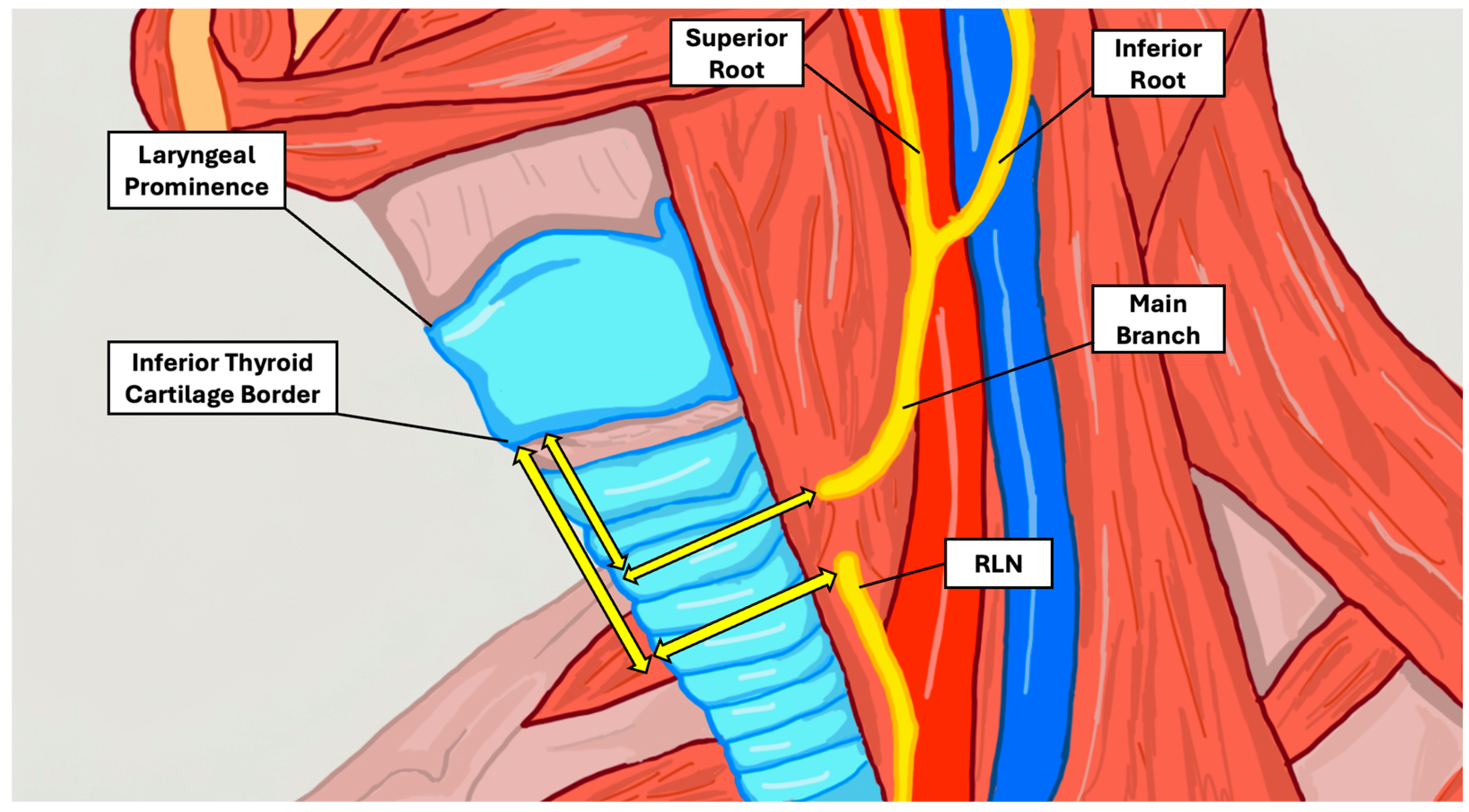
Dissection was performed along the course of the ansa cervicalis and its motor entry point into the infrahyoid musculature. Measurement landmarks are depicted in Figure 1. All dissections and measurements were performed with the neck in a neutral position without deliberate extension or lateral rotation. The overlying neck skin was removed, and blunt dissection was carried through subcutaneous tissues. The sternocleidomastoid muscle was reflected from its sternal and clavicular attachments. Deeper dissection identified the carotid sheath and its contents. The ansa cervicalis was traced from the descent of the superior root (C1) from the hypoglossal nerve, anterior to the internal jugular vein, to its loop with the inferior root (C2–C3). The superior root, inferior root, and main branch were individually identified and their course mapped relative to the infrahyoid muscles, common carotid artery, carotid bifurcation, and thyroid cartilage.
Nerve lengths were measured by placing a length of string along the nerve from the carotid bifurcation and then reading the string against a digital Vernier caliper (Mitutoyo, Kawasaki, Japan). This method was applied to the superior and inferior roots from the carotid bifurcation to their convergence and to the main branch from that convergence to its motor entry point in the infrahyoid musculature. Because the terminal branches enter the infrahyoid muscles together, the first point at which the main branch entered the musculature was designated the ansa cervicalis motor entry point. Each measurement was obtained by one observer while a second observer directly observed landmark placement and the caliper reading. A value was recorded only after both observers agreed. If they did not initially agree, the measurement was adjusted until consensus was reached. Each structure therefore contributed one consensus measurement and was not measured independently or repeatedly by either observer.
The primary anatomical measurements were the positions of the ansa cervicalis motor entry point and RLN laryngeal entry point relative to the inferior border of the thyroid cartilage at the midline laryngeal prominence. The ansa cervicalis motor entry point was defined as the first point at which the main branch entered the infrahyoid musculature. Because the RLN does not have a single discrete motor entry point comparable to that of the ansa cervicalis, its laryngeal entry point was defined as the point at which the nerve passed deep to the inferior border of the thyroid cartilage to enter the larynx. For each entry point, a horizontal mediolateral distance to the midline defined by the laryngeal prominence and a vertical superoinferior distance to the inferior thyroid cartilage border were measured. These perpendicular components were combined as a Euclidean landmark-referenced straight-line distance. The primary paired outcome was the within-side difference in landmark-referenced straight-line distance, calculated as the RLN distance minus the ansa cervicalis distance. This value represents a difference in distance from the shared landmark and not the direct point-to-point distance between the two nerve entry points. The laryngeal prominence was selected because it is externally palpable and consistently identifiable during neck surgery, and the distal entry points were selected because they represent clinically relevant targets during ARA dissection and mobilization.

2.3. Statistical Analysis

Analysis used SPSS version 29 (IBM, Chicago, IL, USA). Descriptive statistics included means, standard deviations, and ranges. Continuous comparisons by sex and laterality used analysis of variance (ANOVA) when assumptions of normality and equal variance were satisfied and the Mann–Whitney U test otherwise. Landmark-referenced straight-line distances for the ansa cervicalis and RLN were compared within sides using a paired-samples t-test, with the Wilcoxon signed-rank test as a nonparametric sensitivity analysis and the Shapiro–Wilk test used to assess normality of paired differences. Mediolateral and superoinferior coordinate differences were evaluated separately using the same paired approach, and the proportion of sides in which the RLN entry point lay farther from the landmark was recorded. The direct point-to-point distance between the two entry points was additionally calculated for each side as the Euclidean distance derived from the mediolateral and superoinferior coordinate differences. To account for the non-independence of bilateral sides contributed by the same donor, a linear mixed-effects model with a random intercept for donor was fitted to provide confirmatory analyses of the primary paired comparison, its mediolateral and superoinferior components, and the sex and laterality comparisons. A donor intraclass correlation coefficient (ICC) was derived from the model variance components. Statistical significance was defined as a two-tailed p < 0.05.

3. Results

Twenty-three donor bodies were included (mean age 77 ± 13 years; range 48–96). Nine were male (mean 80 ± 15 years), and 14 were female (mean 75 ± 11 years). Bilateral dissection was performed in 9 donors (18 ansa cervicales); unilateral dissection in 14 donors (6 right, 8 left), yielding 32 total ansa cervicales. Unilateral dissection was necessitated by prior student dissection of contralateral structures. Donor characteristics are presented in Table 1.

3.1. Landmark-Referenced Entry Point Distances

The primary finding was the simultaneous quantification of the ansa cervicalis motor entry point and RLN laryngeal entry point relative to the inferior thyroid cartilage border (Table 2, Figure 1). The mean landmark-referenced straight-line distance was 43.3 mm (SD 9.7; range 30.6–79.6 mm) for the ansa cervicalis and 55.5 mm (SD 11.7; range 38.2–82.1 mm) for the RLN. On paired within-side analysis, the mean difference in landmark-referenced straight-line distance (RLN minus ansa cervicalis) was 12.2 mm (SD 10.6; 95% CI 8.3–16.0 mm; paired t-test p < 0.001; Wilcoxon p < 0.001), with the RLN farther from the landmark in 29 of 32 sides (91%). The RLN laryngeal entry point was also a mean of 13.6 mm farther from the midline than the ansa cervicalis motor entry point (p < 0.001). In contrast, the mean superoinferior coordinate difference was 2.2 mm and was not statistically significant (p = 0.23). Thus, the landmark-referenced difference was driven primarily by mediolateral rather than superoinferior position. A confirmatory linear mixed-effects model with a random intercept for donor yielded a consistent estimate for the difference in landmark-referenced straight-line distance (12.9 mm; 95% CI 8.3–17.5; p < 0.001; donor ICC 0.82). The mediolateral coordinate difference remained significant (14.2 mm; 95% CI 10.4–17.9; p < 0.001), whereas the superoinferior coordinate difference did not (2.7 mm; p = 0.23). Sex and laterality comparisons remained nonsignificant in the mixed-effects models. The direct point-to-point distance between the ansa cervicalis motor entry point and the RLN laryngeal entry point averaged 17.8 mm (SD 8.1; 95% CI 14.8–20.7; range 5.8–35.1 mm); a confirmatory mixed-effects model gave a consistent estimate (18.8 mm; 95% CI 15.3–22.2; donor ICC 0.87).

3.2. Ansa Cervicalis Morphology

In 30 of 32 dissections (94%), the ansa cervicalis formed from a superior root (C1) descending from the hypoglossal nerve and an inferior root (C2–C3) from the cervical plexus, converging to form the main branch. Mean lengths of the superior root, inferior root, and main branch were 34.7 ± 13.4 mm, 38.6 ± 15.3 mm, and 42.6 ± 17.1 mm, respectively. No statistically significant differences were found between right and left sides or between male and female donors (Table 3).
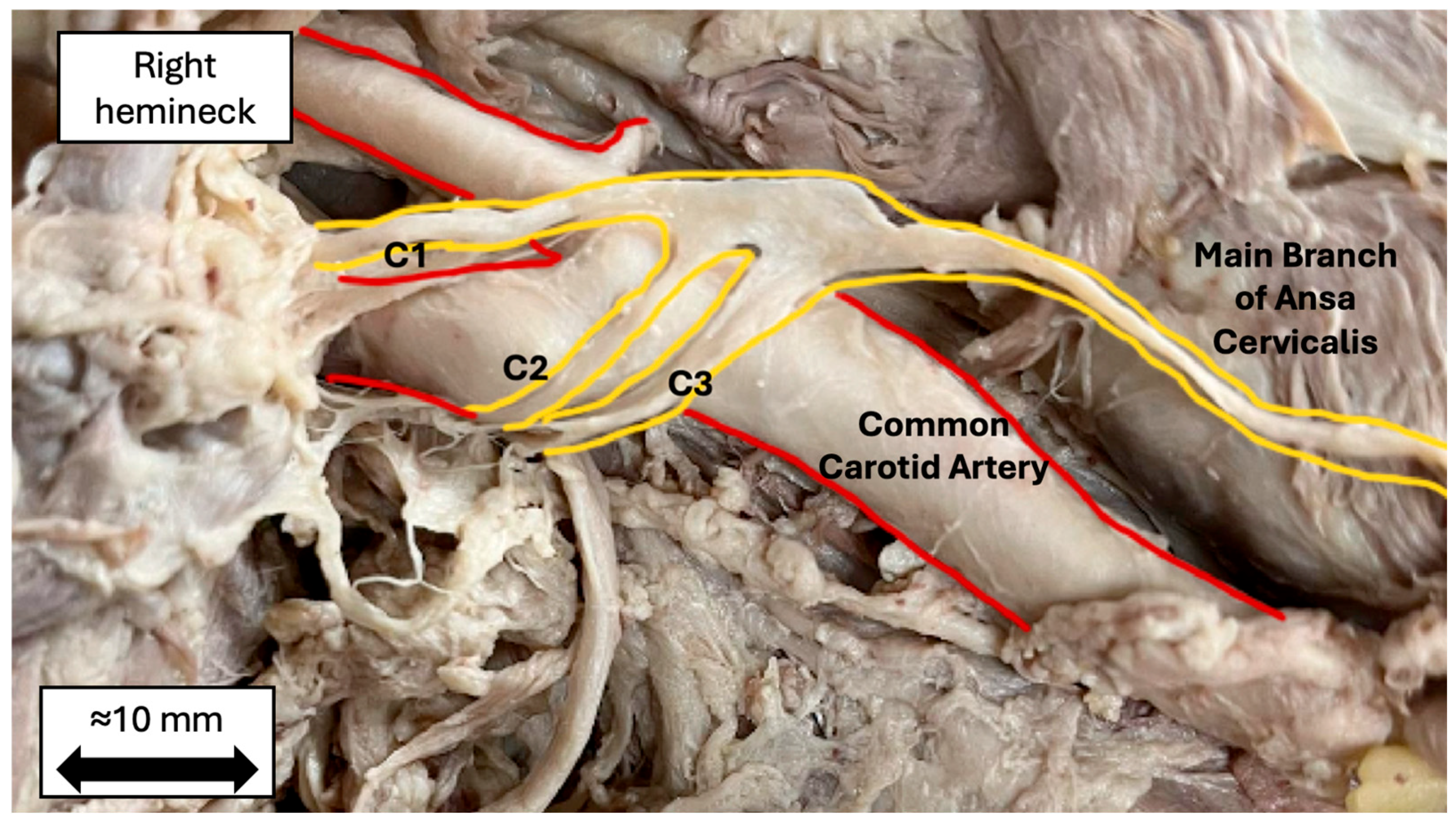
In two dissections (6.25%), the main branch arose from three distinct roots (C1, C2, and C3) rather than the typical two-root configuration (Figure 2). This pattern is consistent with multi-root variants described in prior anatomical literature [22,23]. These three-root variants were excluded from the root- and main-branch length analyses to maintain a consistent morphometric definition. In three additional sides, separately measurable superior and inferior root segments were not obtained. In one of these, the superior and inferior roots converged above the level of the carotid bifurcation. These three sides were retained for entry-point analysis but excluded from the root-length analyses, which therefore comprise 27 sides. The main-branch analysis excludes only the two three-root variants (30 sides).

4. Discussion

This study provides landmark-referenced measurements for both the ansa cervicalis motor entry point and the RLN laryngeal entry point using the inferior border of the thyroid cartilage at the midline laryngeal prominence as a shared reference. The mean within-side difference in landmark-referenced straight-line distance was 12.2 mm and was driven primarily by the mediolateral coordinate difference. This simultaneous landmark-based quantification has not been reported in prior cadaveric studies and may warrant evaluation as a supplemental spatial reference during ARA dissection.
Relative to the laryngeal prominence landmark, the RLN laryngeal entry point was, on average, approximately 12 mm farther from the landmark than the ansa cervicalis motor entry point. This value is a difference in landmark-referenced straight-line distance rather than a direct measurement of the distance between the two nerves. If validated intraoperatively, this relationship could serve as an additional mediolateral spatial cue after one nerve has been identified, including in fields altered by prior surgery, radiation, or fibrosis. The established RLN landmarks, including the cricothyroid joint and tracheoesophageal groove, remain the primary methods of intraoperative identification. The present reference is not intended to replace them. Instead, it may provide a basis for anticipating the relative positions of the distal targets and the mobilization geometry that must be assessed before transection. However, this potential application was not evaluated in the present cadaveric study. The feasibility of a tension-free anastomosis must still be determined intraoperatively. The direct separation between the two entry points averaged 17.8 mm. This value describes the anatomical gap between the donor and recipient targets more directly than the landmark-referenced difference and may be more relevant to the amount of mobilization required, although that relationship was not tested.
A tension-free anastomosis is fundamental to axonal regeneration because excess tension disrupts Schwann cell alignment and impairs functional recovery [26,27]. The mean main-branch length of 42.6 ± 17.1 mm describes the available donor length in this cadaveric sample. However, formal neurorrhaphy and standardized tension testing were not performed in this study. Therefore, individual donor nerves were not classified as adequate or inadequate for completed anastomosis. When ipsilateral donor length is insufficient in clinical practice, contralateral ansa cervicalis harvest may provide an alternative [25].
The three-root variant observed in 6.25% of dissections is consistent with multi-root ansa cervicalis patterns described in prior anatomical literature [23]. The variant may be relevant during initial exposure near the carotid bifurcation, where supernumerary roots may be mistaken for adjacent neural or vascular structures. Because ARA dissection generally proceeds distally toward the selected donor branch, such proximal variation may not alter the final neurorrhaphy, but awareness remains important during identification and mobilization.
No statistically significant differences in nerve dimensions were detected by sex or laterality, consistent with the observations of Prades et al. and Banneheka [23,24]. However, the absence of statistically significant differences does not establish anatomical equivalence, and the study was not powered for subgroup equivalence testing.
This study has several limitations. The sample included 23 donors and 32 sides, which was sufficient for the primary descriptive objective but limited subgroup analyses and may have underrepresented rare variants. Formalin fixation alters tissue properties relative to living patients, and dynamic nerve tension cannot be assessed cadaverically. Nine donors contributed bilateral sides; therefore, side-level observations were not fully independent. The primary paired result was confirmed using a donor-random-intercept mixed-effects model, which produced a consistent estimate. Each value was obtained as a single consensus measurement by two observers, without independent repeated measurements. Consequently, intraobserver and interobserver reliability statistics could not be calculated, and residual measurement variability remains unquantified. Future studies should use repeated overlapping measurements to permit formal reliability assessment. All dissections were performed with the neck neutral, whereas extension and lateral rotation may alter cervical spatial relationships in operative settings. Subcutaneous and other soft-tissue thicknesses also differ between embalmed cadavers and living patients, so absolute landmark distances may differ in vivo. The laryngeal prominence may be less distinct in patients with obesity, prior anterior neck surgery, or a less prominent thyroid cartilage angle. Future work should include larger and more diverse samples, intraoperative validation, and correlation with functional outcomes after ARA.

5. Conclusions

This cadaveric study found that the RLN laryngeal entry point was, on average, approximately 12 mm farther from the inferior thyroid cartilage border at the midline laryngeal prominence than the ansa cervicalis motor entry point. The 12.2 mm value represents a difference in landmark-referenced straight-line distance, not the direct distance between the two nerves, and was driven primarily by mediolateral position. The direct point-to-point distance averaged 17.8 mm. This relationship may be considered a candidate spatial reference for ARA dissection, pending validation in living surgical settings. No statistically significant sex- or laterality-associated differences were detected, although the study was not designed to establish subgroup equivalence. A three-root variant was observed in 2 of 32 dissections (6.25%), illustrating the anatomical variability that may be encountered near the carotid bifurcation. Future studies should evaluate these measurements intraoperatively and determine whether they correlate with surgical or functional outcomes.

Author Contributions

Conceptualization, J.H. and S.S.; methodology, J.H. and J.W.; investigation, J.H., J.W. and P.J.; formal analysis, J.H. and J.W.; data curation, J.H. and J.W.; visualization, J.H.; writing—original draft preparation, J.H. and J.W.; writing—review and editing, J.H., J.W., P.J., K.S., J.B. and S.S.; supervision, S.S. All authors have read and agreed to the published version of the manuscript.

Funding

This research received no external funding.

Institutional Review Board Statement

The study was conducted according to the guidelines of the Declaration of Helsinki, and approved by the Institutional Biosafety Committee at Kansas City University (IBC #2215767-1), approval date: 22 August 2024.

Informed Consent Statement

Informed consent, including permission to publish images for research and/or educational purposes, was obtained from all subjects involved in the study through their participation in the Gift Body Program at Kansas City University. All procedures performed in this study were in accordance with the ethical standards of the institutional and/or national research committee and with the tenets of the Declaration of Helsinki and its later amendments.

Data Availability Statement

The deidentified data supporting the findings of this study are available from the corresponding author upon reasonable request.

Acknowledgments

The authors sincerely thank Brock Halling, Elliot Parens, and Jason Schwab for their assistance with dissection and data collection. We also thank the individuals who donated their bodies to science and their families. Their generosity advances anatomical knowledge and may ultimately improve patient care.

Conflicts of Interest

The authors declare no conflicts of interest.

Abbreviations

The following abbreviations are used in this manuscript:
ARAAnsa cervicalis-to-recurrent laryngeal nerve anastomosis
ANOVAAnalysis of variance
RLNRecurrent laryngeal nerve
UVFPUnilateral vocal fold paralysis
SDStandard deviation
CIConfidence interval
IBCInstitutional Biosafety Committee
ICCIntraclass correlation coefficient

References

  1. Pomal, P.; Bhalodiya, N.; Mishra, S. Effects of voice therapy in early onset unilateral vocal fold paralysis. Indian J. Otolaryngol. Head Neck Surg. 2022, 74, 5075–5081. [Google Scholar] [PubMed]
  2. Kletzien, H.; Macdonald, C.L.; Orne, J.; Francis, D.O.; Leverson, G.; Wendt, E.; Sippel, R.S.; Connor, N.P. Comparison between patient-perceived voice changes and quantitative voice measures in the first postoperative year after thyroidectomy. JAMA Otolaryngol. Head Neck Surg. 2018, 144, 995. [Google Scholar] [CrossRef] [Scilit] [PubMed]
  3. Brisebois, S.; Vahabzadeh-Hagh, A.M.; Zubiaur, F.; Merati, A. Management of recurrent laryngeal nerve paralysis. In Surgery of the Thyroid and Parathyroid Glands; Elsevier: Amsterdam, The Netherlands, 2021; pp. 410–418. [Google Scholar]
  4. Kono, T.; Tomisato, S.; Ozawa, H. Effectiveness of vocal fold medialization surgery on the swallowing function of patients with unilateral vocal fold paralysis. Laryngoscope Investig. Otolaryngol. 2023, 8, 1007–1013. [Google Scholar] [CrossRef] [Scilit] [PubMed]
  5. Feinstein, A.J.; Long, J.L. Glottic insufficiency. In Dysphagia Evaluation and Management in Otolaryngology; Elsevier: Amsterdam, The Netherlands, 2019; pp. 137–142. [Google Scholar]
  6. Ortega Beltrá, N.; Martínez Ruíz De Apodaca, P.; Matarredona Quiles, S.; Nieto Curiel, P.; Dalmau Galofre, J. Parálisis unilateral de cuerda vocal de causa extralaríngea: Etiología y pronóstico. Acta Otorrinolaringol. Esp. 2022, 73, 376–383. [Google Scholar] [CrossRef] [Scilit]
  7. Mau, T.; Husain, S.; Sulica, L. Pathophysiology of iatrogenic and idiopathic vocal fold paralysis may be distinct. Laryngoscope 2020, 130, 1520–1524. [Google Scholar] [PubMed]
  8. Laccourreye, O.; Malinvaud, D.; Ménard, M.; Bonfils, P. Immobilités laryngées unilatérales après chirurgie de la glande thyroïde. J. Chir. 2009, 146, 553–558. [Google Scholar] [CrossRef] [Scilit]
  9. Husain, S.; Sadoughi, B.; Mor, N.; Levin, A.M.; Sulica, L. Time course of recovery of idiopathic vocal fold paralysis. Laryngoscope 2018, 128, 148–152. [Google Scholar] [CrossRef] [Scilit] [PubMed]
  10. Safia, A.; Abd Elhadi, U.; Roch, M.; Kassem, K.; Safiya, R.; Zubeidat, A.; Nofal, N.; Heib, A.; Merchavy, S.; Bishara, T. Determinants of clinical response to injection laryngoplasty in unilateral vocal fold paralysis: A systematic review and meta-analysis. Int. J. Surg. 2024, 110, 7281–7310. [Google Scholar] [CrossRef] [Scilit] [PubMed]
  11. Valley, Z.A.; Karp, A.; Garber, D. Safety and adverse events of medialization thyroplasty: A systematic review. Laryngoscope 2024, 134, 1994–2004. [Google Scholar] [PubMed]
  12. Wang, W.; Chen, D.; Chen, S.; Li, D.; Li, M.; Xia, S.; Zheng, H. Laryngeal reinnervation using ansa cervicalis for thyroid surgery-related unilateral vocal fold paralysis: A long-term outcome analysis of 237 cases. PLoS ONE 2011, 6, e19128. [Google Scholar] [CrossRef] [Scilit] [PubMed]
  13. Lee, W.T.; Milstein, C.; Hicks, D.; Akst, L.M.; Esclamado, R.M. Results of ansa to recurrent laryngeal nerve reinnervation. Otolaryngol. Head Neck Surg. 2007, 136, 450–454. [Google Scholar] [CrossRef] [Scilit] [PubMed]
  14. Lee, Y.J.; Lee, S.W. Ten-year outcomes of recurrent laryngeal nerve reinnervation for thyroidectomy-related unilateral vocal fold paralysis. Am. J. Otolaryngol. 2024, 45, 104242. [Google Scholar] [CrossRef] [Scilit] [PubMed]
  15. Li, M.; Liu, F.; Shi, S.; Chen, S.; Chen, D.; Zheng, H. Bridging gaps between the recurrent laryngeal nerve and ansa cervicalis using autologous nerve grafts. J. Voice 2013, 27, 381–387. [Google Scholar] [CrossRef] [Scilit] [PubMed]
  16. Li, M.; Chen, S.; Wang, W.; Chen, D.; Zhu, M.; Liu, F.; Zhang, C.; Li, Y.; Zheng, H. Effect of duration of denervation on outcomes of ansa-recurrent laryngeal nerve reinnervation. Laryngoscope 2014, 124, 1900–1905. [Google Scholar] [CrossRef] [Scilit] [PubMed]
  17. Zur, K.B.; Carroll, L.M. Recurrent laryngeal nerve reinnervation for management of aspiration in a subset of children. Int. J. Pediatr. Otorhinolaryngol. 2018, 104, 104–107. [Google Scholar] [CrossRef] [Scilit] [PubMed]
  18. Lorenz, R.R.; Esclamado, R.M.; Teker, A.M.; Strome, M.; Scharpf, J.; Hicks, D.; Milstein, C.; Lee, W.T. Ansa cervicalis-to-recurrent laryngeal nerve anastomosis for unilateral vocal fold paralysis: Experience of a single institution. Ann. Otol. Rhinol. Laryngol. 2008, 117, 40–45. [Google Scholar] [CrossRef] [Scilit] [PubMed]
  19. Miyauchi, A.; Matsusaka, K.; Kihara, M.; Matsuzuka, F.; Hirai, K.; Yokozawa, T.; Kobayashi, K.; Kobayashi, A.; Kuma, K. The role of ansa-to-recurrent-laryngeal nerve anastomosis in operations for thyroid cancer. Eur. J. Surg. 1998, 164, 927–933. [Google Scholar] [PubMed]
  20. Olson, D.E.L.; Goding, G.S.; Michael, D.D. Acoustic and perceptual evaluation of laryngeal reinnervation by ansa cervicalis transfer. Laryngoscope 1998, 108, 1767–1772. [Google Scholar] [CrossRef] [Scilit] [PubMed]
  21. Fadhil, M.; Havas, T.; Jacobson, I. Timing of ansa cervicalis-to-recurrent laryngeal nerve reinnervation: A systematic review. J. Voice 2024, 38, 1484–1497. [Google Scholar] [CrossRef] [Scilit] [PubMed]
  22. Malkidou, N.; Papadopoulos, V.; Fiska, A. Anatomical variations of the infrahyoid muscles and ansa cervicalis: A systematic review and an updated classification system for the omohyoid muscle. Anat. Cell Biol. 2025, 58, 322–333. [Google Scholar] [CrossRef] [Scilit] [PubMed]
  23. Banneheka, S. Morphological study of the ansa cervicalis and the phrenic nerve. Anat. Sci. Int. 2008, 83, 31–44. [Google Scholar] [CrossRef] [Scilit] [PubMed]
  24. Prades, J.M.; Gavid, M.; Dubois, M.D.; Dumollard, J.M.; Timoshenko, A.T.; Peoc’h, M. Surgical anatomy of the ansa cervicalis nerve: Which branch to use for laryngeal reinnervation in humans? Surg. Radiol. Anat. 2015, 37, 139–145. [Google Scholar] [PubMed]
  25. Wang, W.; Chen, S.; Chen, D.; Xia, S.; Qiu, X.; Liu, Y.; Zheng, H. Contralateral ansa cervicalis-to-recurrent laryngeal nerve anastomosis for unilateral vocal fold paralysis: A long-term outcome analysis of 56 cases. Laryngoscope 2011, 121, 1027–1034. [Google Scholar] [CrossRef] [Scilit] [PubMed]
  26. Grosu-Bularda, A.; Vancea, C.-V.; Hodea, F.-V.; Cretu, A.; Bordeanu-Diaconescu, E.-M.; Dumitru, C.-S.; Ratoiu, V.-A.; Teodoreanu, R.-N.; Lascar, I.; Hariga, C.-S. Optimizing peripheral nerve regeneration: Surgical techniques, biomolecular and regenerative strategies-a narrative review. Int. J. Mol. Sci. 2025, 26, 3895. [Google Scholar] [CrossRef] [Scilit] [PubMed]
  27. Yi, C.; Dahlin, L.B. Impaired nerve regeneration and Schwann cell activation after repair with tension. Neuroreport 2010, 21, 958–962. [Google Scholar] [CrossRef] [Scilit] [PubMed]
Figure 1. Anatomical illustration of the lateral neck showing the measurement landmarks. The superior root, inferior root, and main branch of the ansa cervicalis are shown in relation to the recurrent laryngeal nerve (RLN), laryngeal prominence, and thyroid cartilage. For each entry point, two perpendicular distances were measured (yellow double-headed arrows): a mediolateral distance to the midline defined by the laryngeal prominence and a superoinferior distance to the inferior border of the thyroid cartilage. The landmark-referenced straight-line distance was derived from these components (ansa cervicalis motor entry point, mean 43.3 mm; RLN laryngeal entry point, mean 55.5 mm). The mean within-side difference in landmark-referenced straight-line distance was 12.2 mm and was driven primarily by the mediolateral coordinate difference. Arrows are schematic and not drawn to scale. Original illustration by J.H.
Anatomia 05 00020 g001
Figure 2. Cadaveric photograph of a variant ansa cervicalis (6.25% incidence). Yellow outlines trace three distinct root contributions (C1, C2, and C3) converging to form the main branch; red outlines indicate the common carotid artery. The side and anatomical orientation are labeled. The approximate 10 mm scale bar was retrospectively calibrated to the measured main-branch length in the pictured specimen and is provided for general orientation only, not for quantitative measurement.
Anatomia 05 00020 g002
Table 1. Donor age and height overall and stratified by sex.
CharacteristicAge (Years)Height (m)n
Mean ± SDRangeMean ± SDRange
Overall77 ± 1348–961.7 ± 0.11.5–2.023
Male80 ± 1555–961.8 ± 0.11.7–2.09
Female75 ± 1148–901.6 ± 0.11.5–1.814
SD, standard deviation.
Table 2. Mediolateral, superoinferior, and derived straight-line distances from the ansa cervicalis and RLN entry points to the inferior thyroid cartilage border at the midline.
MeasurementMean (mm)SD (mm)Range (mm)
Ansa cervicalis: mediolateral (entry point to midline)34.58.315.7–57.0
Ansa cervicalis: superoinferior (to inferior thyroid border)23.812.04.9–61.2
Ansa cervicalis: straight-line distance (derived)43.39.730.6–79.6
RLN: mediolateral (entry point to midline)48.211.931.9–78.7
RLN: superoinferior (to inferior thyroid border)26.08.78.7–47.0
RLN: straight-line distance (derived)55.511.738.2–82.1
Mediolateral and superoinferior distances were measured directly, and straight-line distances were derived per side as the hypotenuse of these two components. The paired within-side difference in landmark-referenced straight-line distance (RLN minus ansa cervicalis) was 12.2 mm (SD 10.6; 95% CI 8.3–16.0; paired t-test p < 0.001; Wilcoxon p < 0.001), with the RLN farther from the landmark in 29 of 32 sides. This difference was predominantly mediolateral (13.6 mm; p < 0.001; 30 of 32 sides) rather than superoinferior (2.2 mm; p = 0.23; 20 of 32 sides). All measurements are reported in millimeters.
Table 3. Lengths of the superior root, inferior root, and main branch of the ansa cervicalis stratified by sex and laterality. All data are presented as mean ± SD. No statistically significant differences were detected between groups. Root-length analyses exclude the two three-root variants and three sides without separately measurable root segments (27 sides); the main-branch analysis excludes the two three-root variants (30 sides). All p > 0.05.
Measurement (mm)OverallMaleFemaleRightLeft
Superior Root34.7 ± 13.4 (n = 27)34.5 ± 15.4 (n = 11)34.8 ± 12.4 (n = 16)34.9 ± 15.7 (n = 13)34.4 ± 11.6 (n = 14)
Inferior Root38.6 ± 15.3 (n = 27)37.8 ± 17.0 (n = 11)39.1 ± 14.5 (n = 16)38.9 ± 18.8 (n = 13)38.3 ± 11.9 (n = 14)
Main Branch42.6 ± 17.1 (n = 30)46.7 ± 17.6 (n = 12)39.9 ± 16.8 (n = 18)39.9 ± 17.1 (n = 14)45.0 ± 17.3 (n = 16)
Disclaimer/Publisher’s Note: The statements, opinions and data contained in all publications are solely those of the individual author(s) and contributor(s) and not of MDPI and/or the editor(s). MDPI and/or the editor(s) disclaim responsibility for any injury to people or property resulting from any ideas, methods, instructions or products referred to in the content.