


Abstract
This study aimed to optimize pigment production from Monascus purpureus TISTR3090 using Khao Klong broken rice (brown rice) as a solid substrate and to evaluate the antioxidant and anti-aging activities of the resulting pigment extracts. Solid-state fermentation showed a progressive increase in red pigment production, reaching a maximum on day 8 (OD680 = 0.10). The water extract (WAE) exhibited a higher total phenolic content (148.86 mg GAE/g) than the ethanol extract (ETE) and demonstrated stronger antioxidant activities, including DPPH radical inhibition (74.66%) and ABTS scavenging activity (65.53%). In addition, WAE showed superior anti-aging potential, with greater inhibition of tyrosinase (75.29%) and collagenase (32.88%) compared to ETE. The MP extract exhibited the presence of R-NH2 functional groups, identified by FTIR, indicating the primary criterion for fungal red pigment. Gas chromatography–mass spectrometry (GC-MS) analysis identified 25 bioactive compounds, mainly fatty acid methyl esters and aromatic derivatives, which may contribute to the observed bioactivities. Liquid chromatography–mass spectrometry (LC-MS) further confirmed higher levels of gallic acid, chlorogenic acid, epigallocatechin gallate, and oroxylin A in WAE. Thus, Monascus pigments derived from broken brown rice-based fermentation represent a promising natural colorant and functional bioingredient with potential applications in nutraceutical and cosmeceutical industries, warranting further studies on formulation, stability, and in vivo validation.
1. Introduction
Microbial pigments, especially those generated by Monascus fungi, have drawn considerable interest due to their vivid colors and potential health advantages [1]. Historically, Monascus species have been employed in Asian nations for ages, chiefly in the manufacture of red yeast rice, a fermented product recognized for its use as a natural food dye and its therapeutic attributes [2,3]. Normally, the agroindustry and food industry byproducts have been used as a carbon source for microbial pigment production such as broken rice hydrolysate [4], corn residues [5], orange processing waste [6], corncob hydrolysate [7], and sugarcane bagasse hydrolysate [8], which are widely used as cost-effective and nutrient-rich substrates for microbial pigment production, primarily through fermentation processes.
The pigments derived from Monascus fermentation are not only valued for their coloring properties but also for their biological activities [9]. Notably, these pigments exhibit significant antioxidant activities, which is crucial in preventing cellular damage. Additionally, the total phenolic content of these pigments correlates with their antioxidant capacity, as phenolic compounds are known for their free radical scavenging abilities. The relationship between total phenolic content and antioxidant activity is pivotal, as higher phenolic content often translates to enhanced antioxidant capacity [9,10]. This increased antioxidant activity can improve cell viability by protecting cells from oxidative stress-induced damage, thereby reducing the risk of chronic diseases and supporting overall cellular health [1]. In addition to their antioxidant properties, Monascus pigments (MPs) have been investigated for their cytotoxic effects against various cancer and normal cell lines to evaluate their potential biological activities and safety profiles. Previous studies have reported differential cytotoxic effects of certain MPs among cell lines, with greater inhibitory effects observed in some cancer cell lines than in normal cells. These findings suggest that MPs may warrant further investigation as potential candidates for anticancer drug development [11,12]. MPs are aphilone derivatives, including substances like monascin, ankaflavin, rubropunctatin, and monascorubrin [13]. Although these chemicals provide advantageous qualities, safety issues have been highlighted, especially with the synthesis of citrinin, a mycotoxin that may arise during fermentation [14]. Citrinin poses nephrotoxic risks, and its presence necessitates stringent control measures in the production process to ensure the safety of Monascus-derived products. Monascus species can synthesize monacolin K, a secondary metabolite structurally similar to lovastatin, via the polyketide biosynthetic pathway during submerged or solid-state fermentation. Monacolin K has garnered significant attention in the health sector for its cholesterol-lowering properties through the inhibition of HMG-CoA reductase; however, excessive intake or uncontrolled fermentation conditions may result in detrimental effects, such as hepatotoxicity and myopathy, especially when produced alongside citrinin, a nephrotoxic mycotoxin. Consequently, optimization and safety surveillance are crucial for the industrial use of Monascus-derived products [15,16].
Despite the extensive research on MPs, there remains a gap in understanding how different fermentation substrates, such as various rice varieties, influence the production of novel compounds and their associated biological activities [17,18]. The utilization of broken brown rice as a substrate for fungal fermentation has garnered significant interest as a novel and sustainable method for producing natural colors and high-value bioproducts for the food, nutraceutical, and cosmetic sectors. Although its economic value is diminished by physical damage incurred during milling, broken brown rice maintains substantial concentrations of carbs, proteins, lipids, vitamins, minerals, and bioactive phytochemicals, rendering it an exceptional nutrient-dense substrate for microbial bioprocesses [19]. Filamentous fungi, specifically Monascus purpureus, Aspergillus spp., and Talaromyces spp., exhibit significant potential for pigment biosynthesis through solid-state fermentation on rice-based substrates, yielding bioactive natural colorants such as red, orange, and yellow pigments with antioxidant and antimicrobial attributes [20,21]. Exploring the fermentation of Khao Klong broken rice (brown rice) with Monascus fungi could lead to the discovery of unique pigments with specific bioactivities, paving the way for their application in health care and cosmetic products. Hence, the objective of this study was to optimize the MPs production from M. purpureus TISTR3090, which was fermented by Khao Klong broken rice (brown rice) as solid substrate and antioxidant activity of MPs.
2. Materials and Methods
2.1. Fungal Strain and Culture Condition
Test fungal-free citrinin strain, Monascus purpureus TISTR3090, was obtained from Thailand Institute of Scientific and Technology Research (Bangkok, Thailand), Thailand. The fungal was cultured on Potato dextrose agar (PDA) and incubated at 30 °C for 30 days; additionally, there was maintenance of fungal culture at 4 °C before use.
2.2. Fungal Pigment Production by Solid-State Fermentation (SSF)
Khao Klong broken rice (brown rice) was carefully cleaned with distilled water. The solid rice medium was added to a 250 mL Erlenmeyer flask by immersing 20 g of rice in 47 mL of distilled water to set a beginning moisture level of 70%. The pH was adjusted to pH 6 before sterilization at 121 °C and 15 psi for 15 min. After cooling spontaneously to the ambient temperature, the flask was moving around evenly. The fungal spore suspension (4.5 × 10⁶ spores) was injected onto the solid medium and incubated for 30 days at 30 °C.
2.3. Extraction of MPs
The fermented rice solids were oven-dried at 60–65 °C overnight to remove moisture and blended. The dried material was then subjected to extraction using the following two different solvents for comparison: distilled water (as the control) called WAE and 95% ethanol called ETE. Each extraction process utilized 1 g of the dried fermented rice solids per 160 mL of the respective solvent, following the methodology described by Shaleha et al. [22]. The percent yield of MP extracts was calculated using following formula:
Yield (%) = [Weights of solvent free extract (g)/Dried fermented rice] × 100
2.4. Total Phenolic Content (TPC)
The TPC was determined using the Folin–Ciocalteu reagent (FC). Basically, 0.5 mL of the extract was mixed with 0.5 mL of FC. The solution was maintained at 25 °C for exactly 5 min before adding 2 mL of 7.5% sodium carbonate mixture and mixing it with 8 mL of water. The absorbance at 725 nm was measured two hours later. The calibration curve used gallic acid as its standard. The TPC was determined using UV spectrometer (Tecan Sunrise Basic, Groedig, Austria) in milligrams of gallic acid equivalent per gram of sample [23]. The TPC result was expressed as mg gallic acid equivalent per g of extract (mg GAE/g sample).
2.5. 1,1-Diphenyl-2-Picrylhydrazyl (DPPH) Assay
The radical scavenging activity was measured using the DPPH assay, which had minor changes as opposed to Lee et al.’s approach [24]. To summarize, 40 μL of MPs (10 mg/mL in DMSO) was combined with 2.96 mL of DPPH (0.1 mM) solution. The combination of reactions was rapidly agitated before the incubation process for 30 min in a dark place at an ambient temperature. The absorbance at 517 nm was measured using microplate reader (Tecan Sunrise Basic, Groedig, Austria). DPPH was used as a control. Ascorbic acid was employed as a means of control. The percentage of inhibitory radical scavenging activity was determined by applying a particular formula:
% Inhibition = [(Abs Control − Abs Sample)/Abs Control] × 100
2.6. Ferric-Reducing Antioxidant Power (FRAP) Assay
The FRAP assay was carried out in accordance with Wu et al. [25], with a few modifications. The stock solutions included 300 mM acetate buffer at pH 3.6, 10 mM TPTZ (2-, 4-, and 6-tripyridyl-s-triazine), and 20 mM FeCl3 solution. MPs (50 μL, 1 mg/mL) were mixed with 1.5 mL of the FRAP solution and incubated for 5 min in dark circumstances. The colorful product was then measured using UV spectrometer (Tecan Sunrise Basic, Groedig, Austria) at 593 nm and reported as milligram equivalents of FeSO4 per milligram of dry weight. The calibration line was created employing the following subsequent values of FeSO4: 0.0025, 0.005, 0.01, and 0.02 mg/mL.
2.7. 2,2′-Azino-Bis(3-Ethylbenzothiazoline-6-Sulfonic Acid) (ABTS) Assay
The ABTS stock solution was prepared by mixing 7 mM ABTS in water with 2.45 mM potassium persulfate in an equal volume. After diluting the initial solution with water, the working solution had an absorbance of 0.706 ± 0.02 at 734 nm. Each experiment used a freshly generated ABTS+ solution. In darkness, 20 µL of the substance was mixed with 180 µL of ABTS solution for 30 min. The wavelength of the absorbance was measured using microplate reader (Tecan Sunrise Basic, Groedig, Austria) at 734 nm with a microplate spectrophotometer reader. The formula for calculating ABTS radical scavenging was as follows:
ABTS radical scavenging = [(AB − AA)/AB] × 100
When
AB = absorbance of ABTS radical + distilled water;
AA = absorbance of ABTS radical + sample extract [25].
2.8. Anti-Tyrosinase Determination
Jiang et al. [26] demonstrated that this approach was used under specific conditions to block tyrosinase. The mixture included 50 μL of WAE and ETE (2.0 mg/mL), 50 μL of 0.2 M sodium phosphate buffer (pH 6.6), and 50 μL of tyrosinase (50 units/mL). After 10 min of incubation at 37 °C, 50 μL of a 2.5 mM L-DOPA solution was added. The reaction was finally observed using microplate reader (Tecan Sunrise Basic, Groedig, Austria) at 492 nm. The proportion of tyrosinase inhibition was approximated using the following equation:
Inhibition is calculated as (A − B)/A × 100
When
A = serving as the control slope at 492 nm;
B = representing the test sample slope.
2.9. Anti-Collagenase Determination
The reaction mixture contained 20 μL of each sample and 20 μL of an enzyme mixture (0.8 units/mL in 50 mM Tricine buffer, pH 7.5, and incubated at 37 °C for 10 min before use). After applying 200 μL of N-[3-(2-furyl)acryloyl]-L-leucyl-glycyl-L-prolyl-L-alanine (FALGPA) substrate (0.5 mM in buffer), the mixture was incubated for 10 min and measured using microplate reader (Tecan Sunrise Basic, Groedig, Austria) at 340 nm for 1 min intervals by a spectrophotometer. The enzyme activity was determined by lowering kinetic absorbance, and the percentage of collagenase inhibition was calculated using the following equation [27]:
Inhibition (%) = [Activity (con) − Activity (In)]/[Activity (con)] × 100.
2.10. Flavonoid and Phenolic Compound Determination
Flavonoid compounds, including oroxin B (purity ≥ 98%, HPLC), oroxylin A (purity ≥ 98%, HPLC), baicalin (purity ≥ 95%, HPLC), and baicalein (purity ≥ 98%), purchased from Sigma-Aldrich (St. Louis, MO, USA), were quantified using high-performance liquid chromatography with photodiode array detection (HPLC-PDA). The analysis was performed using an Accela HPLC system (Thermo Fisher Scientific, San Jose, CA, USA) equipped with an Accela 600 Pump, an Accela Autosampler (AS), and an Accela Photodiode Array (PDA) Detector. The PDA Detector Conditions (Accela PDA) were executed for 35 min at a discrete wavelength of 280 nm. The Autosampler Conditions (Accela AS) were configured with a volume injection of 2 μL, a tray temperature of 25 °C, and a column oven temperature of 35 °C. Chromatographic separation was performed on a Luna® C8 column (150 mm × 2.0 mm, 5 μm particle size; Phenomenex (Torrance, CA, USA). The evaporated dried extract was scraped and accurately weighed (1 mg) and then dissolved in acetonitrile (ACN) to a final volume of 1 mL.
The solution was sonicated at 30 °C for 5 min and subsequently filtered through a 0.45 μm syringe filter prior to chromatographic analysis. All reference standards of phenolic compounds, namely gallic acid (GA) (purity ≥ 97.5%), catechin (CAT) (purity ≥ 98%), epicatechin (EPI) (purity ≥ 99%), vanillic acid (VA) (purity ≥ 97%), p-coumaric acid (PCA) (purity ≥ 98%), quercetin (QUE) (purity ≥ 95%), kaempferol (KAE) (purity ≥ 97%), chlorogenic acid (CLA) (purity ≥ 97%), and epigallocatechin gallate (EGCG) (purity ≥ 95%), were purchased from Sigma-Aldrich (St. Louis, MO, USA) and analyzed using liquid chromatography–mass spectrometry (LC-MS). The mobile phase consisted of 0.1% (v/v) acetic acid in water (Solvent A) and 0.1% (v/v) acetic acid in acetonitrile (Solvent B). Chromatographic separation was performed on a Luna® C8 column (150 mm × 2.0 mm, 5 μm particle size; Phenomenex). The injection volume was 2.5 μL, with the autosampler tray maintained at 25 °C and the column oven set at 35 °C. Mass spectrometric detection was carried out using a Surveyor MSQ Plus system (Thermo Scientific Surveyor MSQ Plus) equipped with an electrospray ionization (ESI) source operating in negative ion mode. The probe temperature was set at 450 °C, the needle voltage at 4.5 kV, and nitrogen was used as the nebulizing gas at a pressure of 5 bar. Data acquisition was performed over a scan time of 0–19 min [28].
2.11. Chemical Characterization
2.11.1. GC–MS Analysis
The Red pigment was conducted using the same conditions and protocol as previously reported by Girija et al. [29] with some modifications. Briefly, the pigment (ETE) was dissolved in hexane (1:20 v/v) and was injected into the capillary column (30 m × 0.25 mm diameter × 0.25 µm) of an Agilent 7890 gas chromatograph. The injection temperature was 280 °C, the column oven temperature was 45 °C, and the helium flow rate was 1.40 mL/min. Also, 50 min is the total holding time. Conditions for the mass spectra were electron impact (40 eV, 200 °C ion source temperature, and 240 °C interface temperature). The MPs’ chemical profiles were analyzed using a GC–MS (Agilent, CA, USA). Mass-spectrum interpretation was measured using the National Institute of Standards and Technology (NIST) database, which had over 62,000 patterns. The spectra of unknown components were compared to those of known components contained in the NIST library. The names, molecular formulas, weights, and chemical structures of the substances were determined.
2.11.2. LC–MS Analysis
The chemical constituents of the Monascus purpureus TISTR 3090 pigment extract were analyzed using an Agilent 1290 Infinity II UHPLC system coupled to an Agilent 6545 Q-TOF mass spectrometer (Agilent, Santa Clara, CA, USA) equipped with a Dual Agilent Jet Stream electrospray ionization (Dual AJS-ESI) source operated in positive ion mode. Chromatographic separation was performed on a reversed-phase C18 column at 25 °C using 0.1% formic acid in water (A) and acetonitrile (B) as the mobile phases. The gradient was 5% B (0–5 min), increased linearly to 100% B (35–40 min), maintained at 100% B (40–45 min), and returned to 5% B for re-equilibration until 50 min. The flow rate and injection volume were 0.20 mL min−1 and 10 μL, respectively. Mass spectra were acquired using Auto MS/MS over m/z 100–1200 for full-scan MS and m/z 50–1200 for MS/MS. MS and MS/MS acquisition rates were 1.0 and 2.0 spectra s−1, respectively. Data-dependent MS/MS was performed for up to three precursor ions per cycle using an approximately 4 amu isolation width and collision energies of 10, 20, and 40 eV. The source parameters were: capillary voltage, 4000 V; nozzle voltage, 2000 V; fragmentor voltage, 175 V; skimmer voltage, 65 V; octopole RF peak, 750 V; drying gas, 325 °C at 13 L min−1; sheath gas, 275 °C at 12 L min−1; and nebulizer pressure, 35 psig. Continuous mass calibration was performed using reference ions at m/z 121.0509 and 922.0098. Putative compound identification was based on accurate mass, isotope distribution, and MS/MS fragmentation patterns, with comparison against available spectral databases and published data on Monascus azaphilone pigments. In the absence of authentic reference standards, compound assignments were considered tentative.
2.11.3. FTIR-ATR Analysis
The functional groups and chemical characteristics of the Monascus purpureus TISTR 3090 pigment extract were characterized using a Thermo Scientific Nicolet Summit X FTIR spectrometer equipped (Thermo Scientific, Madison, WI, USA) with a DTGS KBr detector and an Everest attenuated total reflectance (ATR) accessory (Smart Accessory ID: B124649). The instrument was equipped with a KBr beamsplitter and an IR source. The pigment extract was directly placed onto the ATR accessory and analyzed over the wavenumber range of 4000–450 cm−1. Spectral acquisition was performed using the instrument’s standard optical configuration, with an aperture setting of 100 and 24-bit digitization. The resulting spectra were evaluated based on characteristic absorption bands to identify the major chemical bonds and functional groups present in the pigment extract [22].
2.12. Statistical Analysis
All data were collected in triplicate and shown as means ± standard deviation. The statistical analysis was performed using GraphPad Prism Version 5.01 (San Diego, CA, USA) to determine significant variations between extracts at p < 0.05.
3. Results and Discussion
3.1. Production of Fungal Red Pigment on Solid-State Fermentation
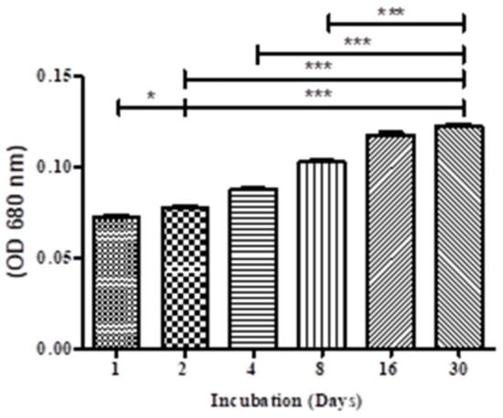
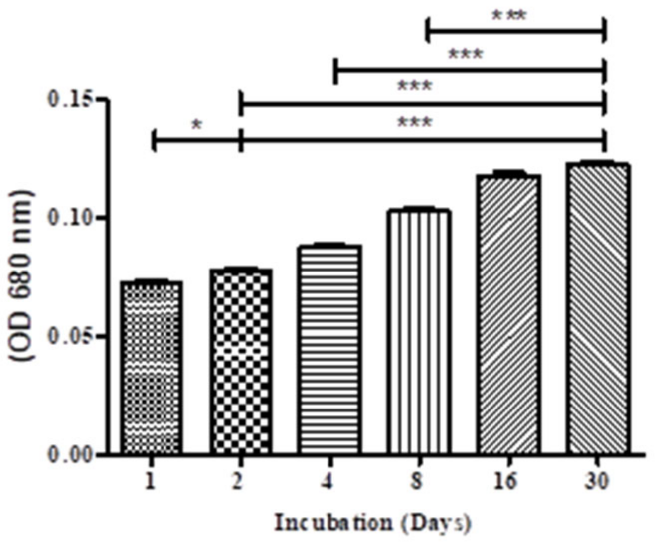
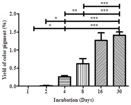
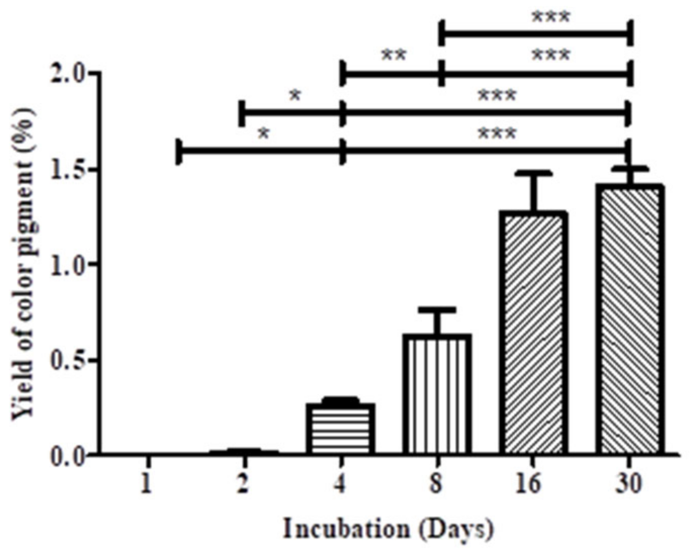
Figure 1 and Figure 2 displayed the MP production by M. purpureus TISTR3090 under solid-state fermentation conditions using brown rice as a carbon source. The pigment product was measured by UV-spectrophotometer (680 nm) as an indicator of pigment concentration. The initial day recorded a minimal average value of the percentage of red pigment extract yield around 0.26 ± 0.03, which progressively increased until day 30, reaching a maximum output value of 1.41 ± 0.09 (p < 0.001). The results demonstrate that the development and production of secondary metabolites in M. purpureus TISTR3090 correlate with fermentation duration, as pigment accumulation escalates over time, aligning with the fungi’s advancement into the stationary phase typically linked to secondary metabolite synthesis. Contrary to comparable studies, research by Ren et al. [23] demonstrated that mannitol supplementation combined with cellulase pretreatment of a substrate can promote M. purpureus growth and improve MP biosynthesis with rice bran as the substrate. Optimization of Monascus purpureus culture conditions in rice bran for enhanced Monascus pigment biosynthesis was conducted with rice bran as the substrate. The concentration stabilizes or reduces prior to any pigment degradation or modifications. The pH of the system, aligned with the study’s results indicating maximum output on day 30, confirmed that the fermentation conditions were appropriate and markedly improved pigment production from Monascus fungi.
Figure 1.
The red pigment production of M. purpureus TISTR3090 under solid-state fermentation. * p < 0.05, *** p < 0.001.
Figure 2.
The percentage of red pigment produced from M. purpureus TISTR3090 under solid-state fermentation. * p < 0.05, ** p < 0.01, *** p < 0.001.
3.2. Antioxidant Activity and Total Phenolic Content
According to the results obtained, the rice extracts exhibited potent antioxidant activities. Among them, the WAE demonstrated the most potent antioxidant capacity and exhibited the highest TPC, which was calculated at 148.86 ± 0.40 mg GAE/g sample. The TPC of ETE was calculated at 71.68 ± 0.24 mg GAE/g sample. The statistical analysis revealed significant differences (p < 0.001) between the WAE and ETE. The DPPH radical scavenging assay—WAE exhibited the highest DPPH inhibition, which was calculated at 74.66 ± 1.47%. The DPPH inhibition of ETE was calculated at 56.79 ± 0.82%. The statistical analysis revealed significant differences (p < 0.01) between the WAE and ETE. The FRAP value—WAE exhibited the highest FRAP value, which was calculated at 0.36 ± 0.00 mM. The FRAP value of ETE was calculated at 0.17 ± 0.01 mM. The statistical analysis revealed significant differences (p < 0.001) between the WAE and ETE. The ABTS—WAE exhibited the highest ABTS, which was calculated at 65.53 ± 1.03%. The ABTS of ETE was calculated at 21.61 ± 1.05%. The statistical analysis revealed significant differences (p < 0.001) between the WAE and ETE (Table 1). The findings indicated that the aqueous extract of Monascus exhibited a superior DPPH scavenging action compared to the ethanolic extract, consistent with the observations of Abd Raza et al. [30] about the influence of polarity on the compound. The red pigment extracted by Rhizopus oligosporus and M. purpureus utilizing solid-state fermentation exhibited a phenolic content and antioxitant activity, which was consistent with the results reported in Abd Razak et al. [30]. Moreover, the red pigments of Monascus species, notably monascorubramine and rubropunctamine, have acknowledged antioxidant effects, as well as other health-enhancing attributes [31].
Table 1.
TPC and antioxidant activities of MPs extract.
3.3. Anti-Tyrosinase and Anti-Collagenase Activity
The anti-aging properties of MP extracts were assessed in vitro. The findings demonstrated a favorable efficacy of fungal extract at a dosage of 2.0 mg/mL against the enzymes tyrosinase and collagenase. The extracts of WAE and ETE showed inhibitory action against two enzymes (Table 2). The WAE demonstrated significant anti-tyrosinase and anti-collagenase activity, with inhibition percentages of 75.29 ± 0.09 and 32.88 ± 0.54, respectively. In contrast, ETE had a lower inhibition of tyrosinase and collagenase, measuring 56.57 ± 0.55 and 19.27 ± 0.54, respectively. Previous research has demonstrated the solid and liquid fermentation of chestnut inner shell utilizing M. kaoliang. The ethanol extracts from submerged fermentation exhibited superior levels of phenolic compounds, antioxidant activity, tyrosinase activity, and elastase activity compared to solid-state fermentation on day 3 [18]. It indicates that fungal species significantly contribute to the generation of bioactive chemicals, even within the same genus, together with the fermentation techniques and substrates employed in the fermentation process [12,24]. However, this study conducted a single concentration as a preliminary assessment of the potency of MP extract, necessitating further in-depth investigations with several concentrations for confirmation.
Table 2.
Anti-aging potential of MP extract.
3.4. HPLC Phenolic Compounds of MPs
The MPs demonstrated the presence of flavonoid and phenolic compounds in the extract when compared with phenolic compound standard (Figure 3). Among the flavonoids, Oroxylin A was detected at a concentration of 0.24 mg/kg. Regarding phenolic compounds, gallic acid (GA) was present at 0.38 mg/kg, while chlorogenic acid (CLA) was the predominant phenolic compound, with a concentration of 9.69 mg/kg. In addition, epigallocatechin gallate (EGCG) was quantified at 3.34 mg/kg. Our study was comparable to that by Husakova and Patakova [12], which revealed that antioxidants in substrate—such as phenolic and flavonoids in rice—enhance monascus pigments in fermented food. Duarte et al. [32] also found that brown seaweed fermented with M. purpureus contained phenolic substances.
Figure 3.
HPLC chromatogram of phenolic compounds of MPs.
3.5. Chemical Analysis of Ethanolic Extract of MPs
GC–MS Analysis of Red Pigment
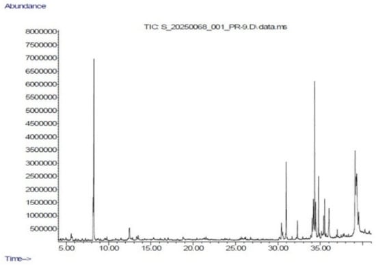
The chemical analysis of the MP extract was conducted utilizing the GC-MS technique. The outcome of the chromatogram is distinctly depicted in Figure 4 and Table 3. Twenty-five molecules were discovered in the MP extract. The main components included cis-13-octadecenoic acid, methyl ester (14.50%), methyl palmitate (8.55%), p-xylene (8.18%), 11-octadecenoic acid, methyl ester (7.56%), ethylbenzene (5.56%), methyl 9-cis,11-trans-octadecadienoate (4.05%), methyl stearate (3.87%), ethyl oleate (3.38%), 1,1′-biphenyl, 2-methyl- (3.44%), and benzene, 1-ethyl-2-methyl- (3.43%), respectively. Previous investigations have indicated that cis-13-octadecenoic acid methyl ester possesses antioxidant properties [25,33] and functions as an emollient in cosmetics [34]. Moreover, additional chemical compounds, such as methyl palmitate [35] and methyl 9-cis,11-trans-octadecadienoate, exhibited effectiveness in antioxidant activity. In addition, certain scientific data revealed that the ethanol extraction of M. purpureus BCRC 38110 produced the following eight chemical compounds: 5S,6S-monaspurpyridine A, 5R,6R-monaspurpyridine A, monasxanthones A and B, monasnaphthalenone, monapurpurin, and two identified compounds, exhibiting anti-melanogenic potential in mice [29]. Consequently, M. purpureus demonstrated potential for development as a cosmeceutical application.
Figure 4.
GC–MS chromatogram of Monascus purpureus-derived red pigment extract.
Table 3.
Chemical components from ethanolic extract detected with GC–MS of Monascus extract by NIST11.
3.6. FTIR Determination of Red Pigment
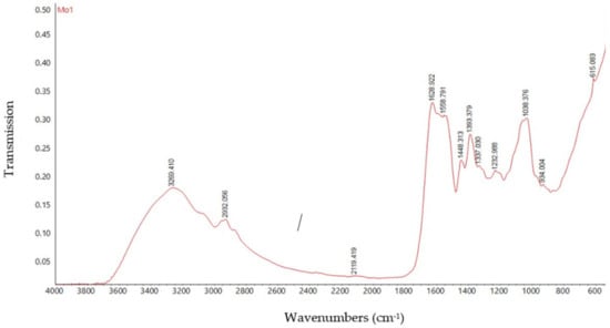
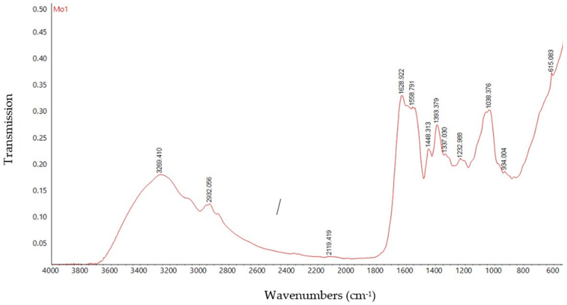
FTIR spectra of MP extract from Monascus purpureus TISTR3090 was represented in Figure 5, indicating comparable chemical structures with variations in functional group intensities. The spectra exhibited broad absorption bands in the region of 3269–3296 cm−1, corresponding to O– H stretching vibrations, suggesting the presence of hydroxyl groups [36,37,38], and hydrogen bonding. Peaks around 2932–2936 cm−1 were attributed to aliphatic C–H stretching, confirming the presence of alkyl side chains characteristic of polyketide compounds [36]. A prominent absorption band at approximately 1629 cm−1 was observed in both samples, corresponding to conjugated C=O or C=C stretching vibrations, which are essential components of the chromophore system responsible for pigment coloration [39]. Notably, this sample exhibited absorption bands in the region of 1530–1550 cm−1, indicative of N–H bending or C–N stretching vibrations, suggesting a higher concentration of nitrogen-containing compounds. The presence of a R–NH2 stretching at 1558 cm−1 indicates that the MP pigment was red pigment [40]. Typically, the red pigments generated by Monascus purpureus are monascorubramine (C23H27NO4) and rubropunctamine (C21H23NO4) [41]. Additional bands observed at 1227–1233 cm−1 and 1038–1082 cm−1 correspond to C–O stretching vibrations, confirming the presence of ester or lactone groups [42], which are typical structural features of Monascus pigments.
Figure 5.
FTIR spectrum of MPs extracted by ethanol of Monascus purpureus TISTR3090. The spectrum shows characteristic absorption bands including O–H stretching (~3296 cm−1), aliphatic C–H (~2932 cm−1), conjugated C=O/C=C (~1629 cm−1), and C–O stretching (~1233 and 1038 cm−1). The presence of a band near 1537 cm−1 suggests nitrogen-containing functional groups, indicating the presence of red pigment derivatives.
3.7. LC–MS Profiling of Compounds Detected in Negative and Positive ESI Mode
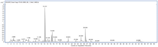
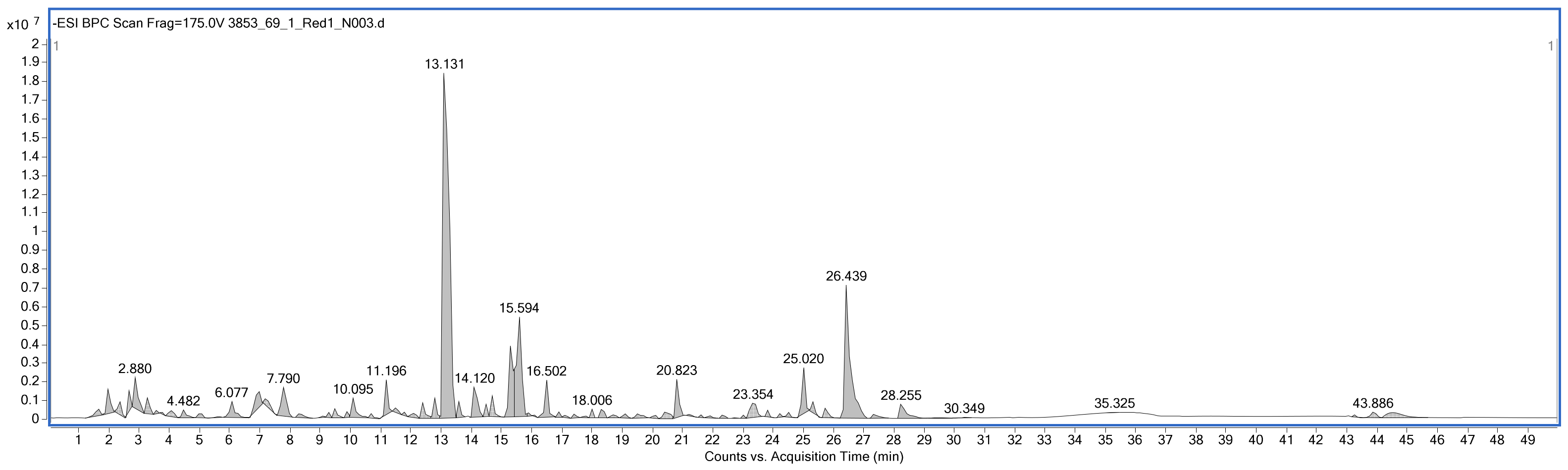
LC–MS analysis of the Monascus purpureus TISTR 3090 sample was performed using positive electrospray ionization (ESI) mode to characterize the detected metabolites and pigment-related compounds. Several molecular features were detected across the chromatographic profile and were tentatively assigned by comparison with the mass spectral library based on their retention times and observed m/z values.
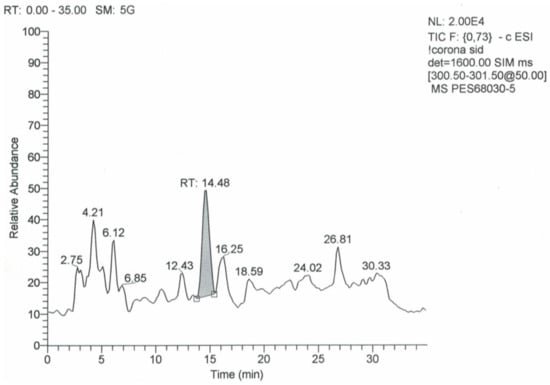
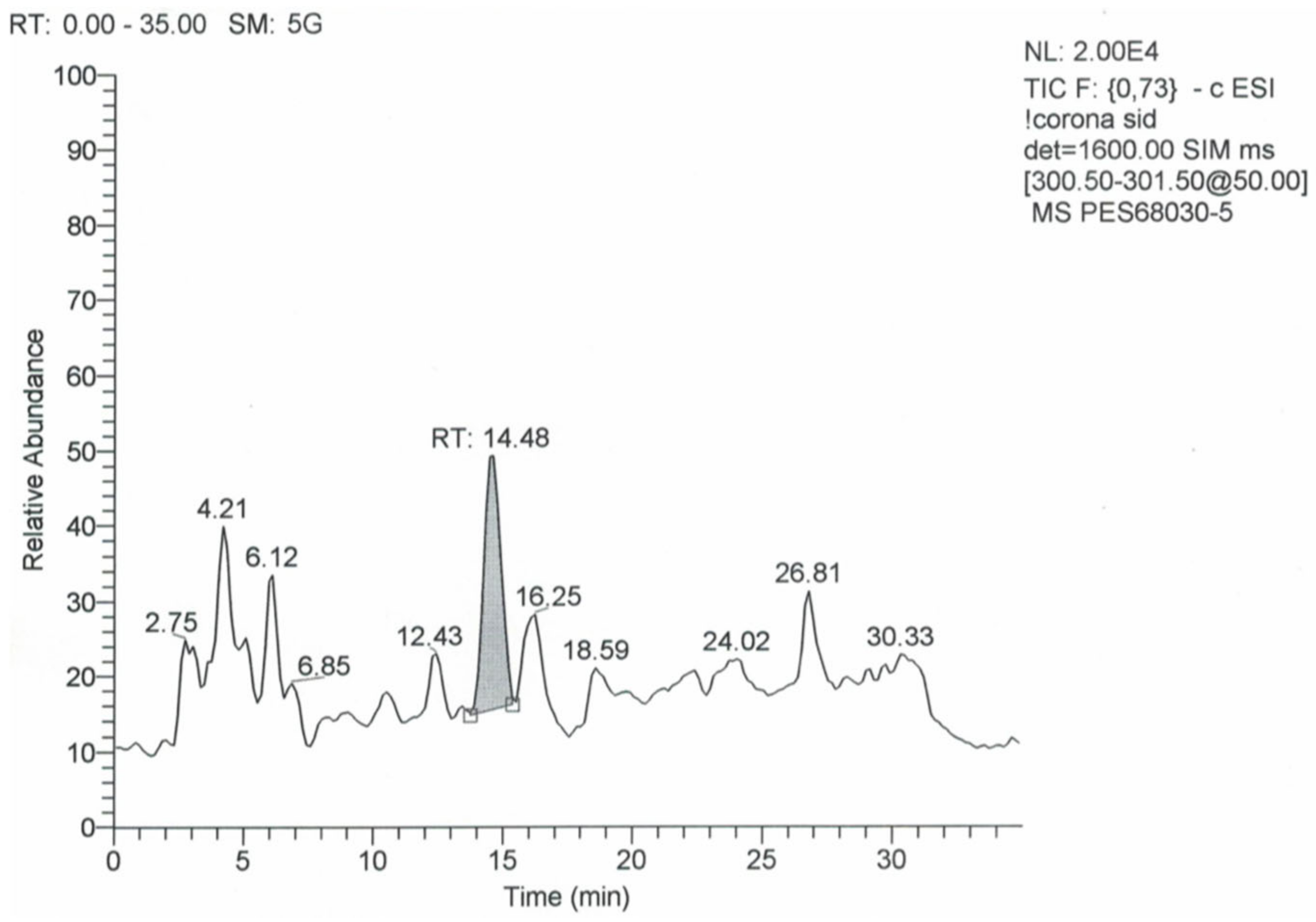
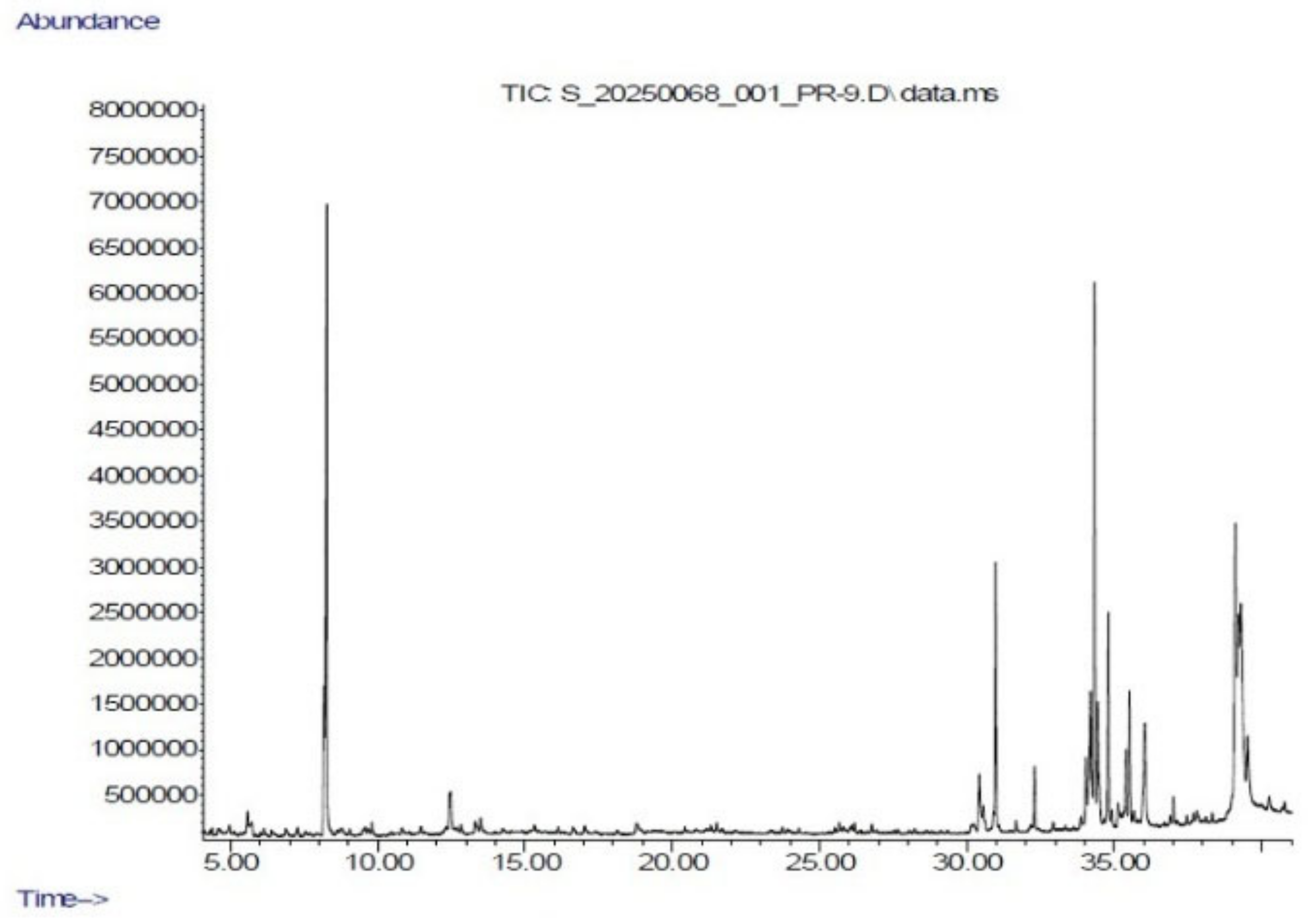
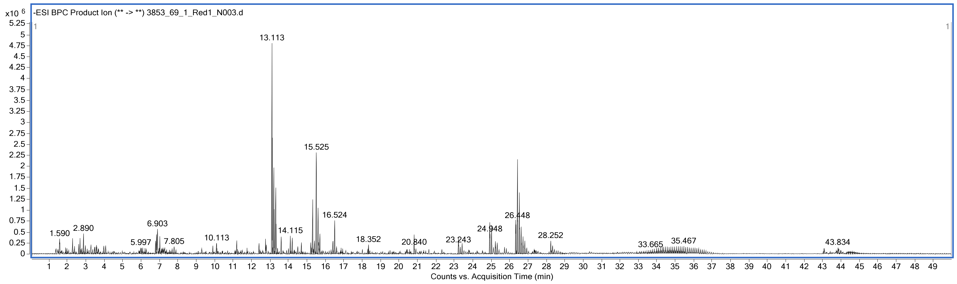
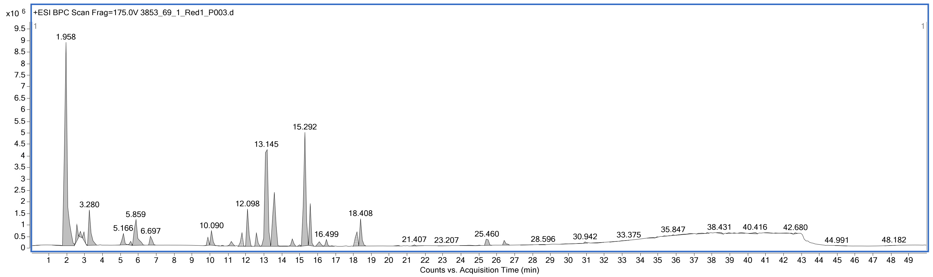
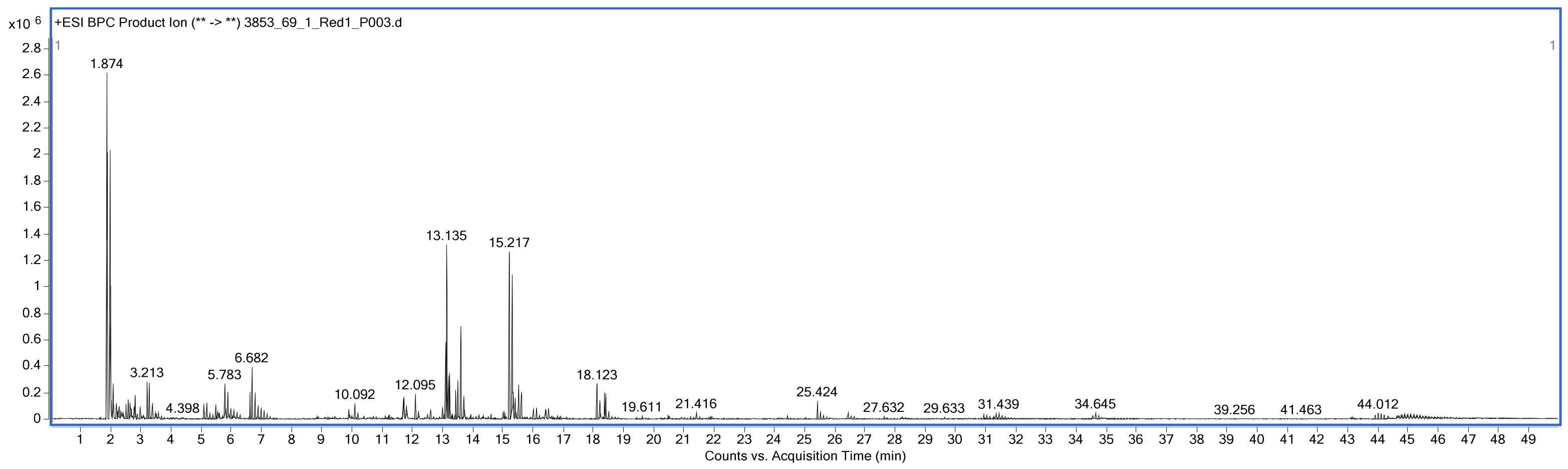
In negative ESI mode (Figure 6 and Figure 7), the detected 38 compounds and positive ESI mode (Figure 8 and Figure 9), the detected 21 compounds (Table 4), the compounds detected at the later retention times, particularly those observed at RT 11.814, 17.175, 23.865, and 24.265 min, represent relatively higher-molecular-weight components of the sample. However, their library-based assignments should be considered tentative because library matching alone does not provide definitive structural confirmation. In particular, the compounds tentatively assigned as piperpenone, bisindolylmaleimide I, desoxycorticosterone acetate, and 3-hydroxyquinidine cannot be conclusively classified as Monascus pigments based solely on the available LC–MS data.
Figure 6.
Negative electrospray ionization (ESI) BPC scan chromatogram: chromatogram created by plotting the base peak ion on current in a series of MS scan records as a function of retention time.
Figure 7.
Negative electrospray ionization (ESI) BPC product ion chromatogram: chromatogram created by plotting the base peak ion in a series of product ions records as a function of retention time.
Figure 8.
Positive electrospray ionization (ESI) BPC scan chromatogram: chromatogram created by plotting the base peak ion on current in a series of MS scan records as a function of retention time.
Figure 9.
Positive electrospray ionization (ESI) BPC product ion chromatogram: chromatogram created by plotting the base peak ion in a series of product ions records as a function of retention time.
Table 4.
Liquid chromatography–positive and negative electrospray ionization mass spectrometry of MPs.
Additional production measurements of the positive ESI chromatographic profile showed prominent features at RT 13.191 min with an observed m/z of 431.2399 (neutral mass 430.2327) and RT 15.208 min with an observed m/z of 251.0918 (neutral mass 250.0846). These molecular features were not directly assigned to specific Monascus pigments from the available library-based identification and should therefore be considered additional pigment-associated or metabolite-associated features requiring further structural characterization.
Overall, positive ESI LC–MS analysis demonstrated a complex chemical profile in the M. purpureus TISTR 3090 sample, comprising amino acids, vitamins, nucleobases, organic acids, and higher-molecular-weight metabolites. The detected molecular features provide evidence of a chemically diverse metabolite profile; however, definitive identification of Monascus pigment compounds requires confirmation using MS/MS fragmentation patterns, accurate molecular formulas, authentic reference standards, and/or complementary spectroscopic techniques.
4. Conclusions
This study reveals that M. purpureus TISTR3090 can take advantage of Khao Klong broken rice (brown rice) as a substrate, yielding maximal secondary metabolites at 30 days. MP extract possesses potential as a natural source of antioxidants and anti-aging properties. The aqueous extract (WAE) exhibited greater antioxidant, anti-tyrosinase, and anti-collagenase activities compared to the ethanol extract (ETE). The MP extract exhibited the presence of R–NH2 functional groups, identified by FTIR, indicating the primary criterion for fungal red pigment. The GC–MS analysis found 25 bioactive molecules, primarily fatty acid methyl esters and aromatic substances. The WAE exhibited elevated levels of phenolic compounds (gallic acid, chlorogenic acid, and epigallocatechin gallate) and flavonoids (oroxylin A). Subsequent research will concentrate on elucidating the structure of the red pigment utilizing nuclear magnetic resonance (NMR) and examining the stability of the compounds. This study illustrates the enhanced utilization of agricultural by-products using microbial biotechnology and emphasizes their potential as future raw materials for cosmetics and health applications.
Author Contributions
S.T.: conceptualization; methodology; formal analysis; production of the original draft. T.D., W.N. and K.J.: antioxidant, antityrosinase, and anti-collagenase analysis. W.K.: statistics; data curation and review; editing of the manuscript. All authors have read and agreed to the published version of the manuscript.
Funding
This research received no external funding.
Institutional Review Board Statement
Not applicable.
Informed Consent Statement
Not applicable.
Data Availability Statement
The original contributions presented in this study are included in the article. Further inquiries can be directed to the corresponding author.
Acknowledgments
The authors express their gratitude to the Faculty of Integrative Medicine at Rajamangala University of Technology Thanyaburi, Pathum Thani, Thailand, for providing valuable bioactivity and all necessary facilities.
Conflicts of Interest
The authors declare no conflicts of interest.
Abbreviations
The following abbreviations are used in this manuscript:
| M. purpureus | Monascus purpureus |
| MPs | Monascus pigments |
| GC-MS | Gas chromatography–mass spectrometry |
| PDA | Potato dextrose agar |
| SSF | Solid-state fermentation |
| DPPH | 1,1-diphenyl-2-picrylhydrazyl |
| TPTZ | 2, 4, 6-tripyridyl-s-triazine |
| GA | Gallic acid |
| CLA | Chlorogenic acid |
| EGCG | Epigallocatechin gallate |
References
- Feng, S.S.; Li, W.; Hu, Y.J.; Feng, J.X.; Deng, J. The biological activity and application of Monascus pigments: A mini review. Int. J. Food Eng. 2022, 18, 253–266. [Google Scholar] [CrossRef] [Scilit]
- Gong, P.; Shi, R.; Liu, Y.; Luo, Q.; Wang, C.; Chen, W. Recent advances in Monascus pigments produced by Monascus purpureus: Biosynthesis, fermentation, function, and application. LWT-Food Sci. Technol. 2023, 185, 115162. [Google Scholar] [CrossRef] [Scilit]
- Lin, L.; Xu, J. Fungal pigments and their roles associated with human health. J. Fungi 2020, 6, 280. [Google Scholar] [CrossRef] [Scilit]
- Cai, Y.; Liu, Y.; Liu, T.; Gao, K.; Zhang, Q.; Cao, L.; Wang, L.; Wu, X.; Zheng, H.; Peng, H.; et al. Heterotrophic cultivation of Chlorella vulgaris using broken rice hydrolysate as carbon source for biomass and pigment production. Bioresour. Technol. 2021, 323, 124607. [Google Scholar] [CrossRef] [Scilit]
- Li, P.; Zhao, G.; Zhang, K.; Wang, H. Improvement of red pigment production during biomembrane surface cultivation of Penicillium novaezelandiae by supplementing with corn straw. BioResources 2017, 12, 7680–7691. [Google Scholar] [CrossRef] [Scilit]
- Kantifedaki, A.; Kachrimanidou, V.; Mallouchos, A.; Papanikolaou, S.; Koutinas, A.A. Orange processing waste valorisation for the production of bio-based pigments using the fungal strains Monascus purpureus and Penicillium purpurogenum. J. Clean. Prod. 2018, 185, 882–890. [Google Scholar] [CrossRef] [Scilit]
- Zhou, Z.; Yin, Z.; Hu, X. Corncob hydrolysate, an efficient substrate for Monascus pigment production through submerged fermentation. Biotechnol. Appl. Biochem. 2014, 61, 716–723. [Google Scholar] [CrossRef] [Scilit]
- Hilares, R.T.; de Souza, R.A.; Marcelino, P.F.; da Silva, S.S.; Dragone, G.; Mussatto, S.I.; Santos, J.C. Sugarcane bagasse hydrolysate as a potential feedstock for red pigment production by Monascus ruber. Food Chem. 2018, 245, 786–791. [Google Scholar] [CrossRef] [Scilit]
- Jiang, S.; Yuan, S.; Zuo, Y.; Liu, T.; Zhou, Y.; Wu, D.; Chen, Q.; Shu, X. The functional and nutritional properties of a novel Monascus fermented rice rich in resistant starch. J. Cereal Sci. 2024, 119, 103999. [Google Scholar] [CrossRef] [Scilit]
- Chaudhary, V.; Katyal, P.; Panwar, H.; Kaur, J.; Aluko, R.E.; Puniya, A.K.; Poonia, A.K. Antioxidative, anti-inflammatory, and anticancer properties of the red biopigment extract from Monascus purpureus (MTCC 369). J. Food Biochem. 2022, 46, e14249. [Google Scholar] [CrossRef] [Scilit]
- Srianta, I.; Kusdiyantini, E.; Zubaidah, E.; Ristiarini, S.; Nugerahani, I.; Alvin, A.; Iswanto, N.; Zhang, B.B. Utilization of agro-industrial by-products in Monascus fermentation: A review. J. Biosci. Bioeng. 2021, 8, 129. [Google Scholar] [CrossRef] [Scilit]
- Husakova, M.; Patakova, P. Purified Monascus pigments: Biological activities and mechanisms of action. J. Nat. Prod. 2025, 88, 607–615. [Google Scholar] [CrossRef] [Scilit]
- Chen, W.; Chen, R.; Liu, Q.; He, Y.; He, K.; Ding, X.; Kang, L.; Guo, X.; Xie, N.; Zhou, Y.; et al. Orange, red, yellow: Biosynthesis of azaphilone pigments in Monascus fungi. Chem. Sci. 2017, 8, 4917–4925. [Google Scholar] [CrossRef] [Scilit]
- Chen, Q.; Wang, W.; Zhou, J.; Shen, W.; Jia, X.; Xiang, L.; Chen, X. Analysis on the antioxidant capacities of four Monascus pigment components and their binding mechanisms with bovine serum albumin. LWT-Food Sci. Technol. 2025, 230, 118259. [Google Scholar] [CrossRef] [Scilit]
- Zhang, Y.; Chen, Z.; Wen, Q.; Xiong, Z.; Cao, X.; Zheng, Z.; Zhang, Y.; Huang, Z. An overview on the biosynthesis and metabolic regulation of monacolin K/lovastatin. Food Funct. 2020, 11, 5738–5748. [Google Scholar] [CrossRef] [Scilit]
- Patakova, P. Monascus secondary metabolites: Production and biological activity. J. Ind. Microbiol. Biotechnol. 2013, 40, 169–181. [Google Scholar] [CrossRef] [Scilit]
- Chaves, N.; Santiago, A.; Alías, J.C. Quantification of the antioxidant activity of plant extracts: Analysis of sensitivity and hierarchization based on the method used. Antioxidants 2020, 9, 76. [Google Scholar] [CrossRef] [Scilit]
- Jin, Y.Y.; Ritthibut, N.; Lim, S.T.; Oh, S.J. Antioxidant and in vitro cosmeceutical activities of chestnut inner shell fermented by Monascus kaoliang. Food Sci. Biotechnol. 2023, 32, 813–822. [Google Scholar] [CrossRef] [Scilit]
- Gul, K.; Yousuf, B.; Singh, A.K.; Singh, P.; Wani, A.W. A rice bran: Nutritional values and its emerging potential for development of functional food—A review. Bioact. Carbohydr. Diet. Fibre. 2015, 6, 24–30. [Google Scholar] [CrossRef] [Scilit]
- Afroz Toma, M.; Rahman, M.H.; Rahman, M.S.; Arif, M.; Nazir, K.N.H.; Dufossé, L. Fungal pigments: Carotenoids, riboflavin, and polyketides with diverse applications. J. Fungi 2023, 9, 454. [Google Scholar] [CrossRef] [Scilit]
- Mapari, S.A.; Meyer, A.S.; Thrane, U.; Frisvad, J.C. Identification of potentially safe promising fungal cell factories for the production of polyketide natural food colorants using chemotaxonomic rationale. Microb. Cell Fact. 2009, 8, 24. [Google Scholar] [CrossRef] [Scilit]
- Shaleha, R.R.; Yuliana, A.; Rahmiyani, I.; Pebiansyah, A.; Indriyanti, I. Bioactive pigments of Monascus purpureus: Identification and characterization. J. Food Pharm. Sci. 2025, 13, 356–366. [Google Scholar] [CrossRef] [Scilit]
- Ren, R.; Zeng, H.; Mei, Q.; Xu, Z.; Mazhar, M.; Qin, L. Effects of Monascus purpureus-fermented tartary buckwheat extract on the blood lipid profile, glucose tolerance and antioxidant enzyme activities in KM mice. J. Cereal Sci. 2022, 105, 103465. [Google Scholar] [CrossRef] [Scilit]
- Lee, Y.L.; Yang, J.H.; Mau, J.L. Antioxidant properties of ethanolic and methanolic extracts from Monascus-fermented soybeans. J. Food Biochem. 2009, 33, 707–727. [Google Scholar] [CrossRef] [Scilit]
- Wu, L.; Zhou, K.; Chen, F.; Chen, G.; Yu, Y.; Lv, X.; Zhang, W.; Rao, P.; Ni, L. Comparative study on the antioxidant activity of Monascus yellow pigments from two different types of Hongqu—Functional Qu and coloring Qu. Front. Microbiol. 2021, 12, 715295. [Google Scholar] [CrossRef] [Scilit]
- Jiang, F.; Li, W.; Huang, Y.; Chen, Y.; Jin, B.; Chen, N.; Ding, Z.; Ding, X. Antioxidant, antityrosinase and antitumor activity comparison: The potential utilization of fibrous root part of Bletilla striata (Thunb.) Reichb. f. PLoS ONE 2013, 8, e58004. [Google Scholar] [CrossRef] [Scilit]
- Thring, T.S.; Hili, P.; Naughton, D.P. Anti-collagenase, anti-elastase and anti-oxidant activities of extracts from 21 plants. BMC Complement. Altern. Med. 2009, 9, 27. [Google Scholar] [CrossRef] [Scilit]
- Khalid, A.; Abdalgadir, E.A.; Abdel Gadir, I.K.; Abdalla, A.N.; Homeida, H.E.; Sultana, S.; Rehman, Z.; Hassani, R.; Alqahtani, A.S.; Alhazmi, H.A. GC-MS profiling and therapeutic potentials of Prosopis juliflora (Sw.) DC: Cytotoxic and antimicrobial insights. J. Spectrosc. 2024, 2024, 1121745. [Google Scholar] [CrossRef] [Scilit]
- Girija, S.; Duraipandiyan, V.; Kuppusamy, P.S.; Gajendran, H.; Rajagopal, R. Chromatographic characterization and GC-MS evaluation of the bioactive constituents with antimicrobial potential from the pigmented ink of Loligo duvauceli. Int. Sch. Res. Not. 2014, 2014, 820745. [Google Scholar] [CrossRef] [Scilit]
- Abd Razak, D.L.; Abd Rashid, N.Y.; Jamaluddin, A.; Sharifudin, S.A.; Long, K. Enhancement of phenolic acid content and antioxidant activity of rice bran fermented with Rhizopus oligosporus and Monascus purpureus. Biocatal. Agric. Biotechnol. 2015, 4, 33–38. [Google Scholar] [CrossRef] [Scilit]
- Dishliyska, V.; Miteva-Staleva, J.; Gocheva, Y.; Stoyancheva, G.; Yovchevska, L.; Abrashev, R.; Spasova, B.; Angelova, M.; Krumova, E. Biological potential of extremophilic filamentous fungi for the production of new compounds with antimicrobial effect. Fermentation 2025, 11, 347. [Google Scholar] [CrossRef] [Scilit]
- Duarte, A.M.; Guarino, M.P.; Barroso, S.; Gil, M.M. Phytopharmacological strategies in the management of type 2 diabetes mellitus. Foods 2020, 9, 271. [Google Scholar] [CrossRef] [Scilit]
- Ayoola, A.A.; Ekunseitan, D.A.; Muhammad, S.B.; Oguntoye, M.A.; Adejola, Y.A. Phytochemicals analysis and GC-MS determination of ethanolic extracts of Azadirachta indica and Mangifera indica stem bark and their biological potentials. Pac. J. Sci. Technol. 2020, 21, 219–229. [Google Scholar]
- Tene, S.T.; Natarajan, V. Metabolomics profile of two germinated maize flours (Atp-Y and Coca-sr) using gas chromatography-mass spectrometry (GC-MS). Explor. Foods Foodomics 2025, 3, 101087. [Google Scholar] [CrossRef] [Scilit]
- Hamed, A.; Mantawy, E.; El-Bakly, W.; Abdel-Mottaleb, Y.; Azab, S. Methyl palmitate: The naturally occurring cardioprotective agent. Arch. Pharm. Sci. Ain Shams Univ. 2020, 4, 47–62. [Google Scholar] [CrossRef] [Scilit]
- Hsiao, K.Y.; Chung, R.J.; Chang, P.P.; Tsai, T.H. Identification of hydroxyl and polysiloxane compounds via infrared absorption spectroscopy with targeted noise analysis. Polymers 2025, 17, 1533. [Google Scholar] [CrossRef] [Scilit]
- Jonoobi, M.; Harun, J.; Tahir, P.M.; Shakeri, A.; SaifulAzry, S.; Makinejad, M.D. Physicochemical characterization of pulp and nanofibers from kenaf stem. Mater. Lett. 2011, 65, 1098–1100. [Google Scholar] [CrossRef] [Scilit]
- Silbir, S.; Goksungur, Y. Natural red pigment production by Monascus purpureus in submerged fermentation systems using a food industry waste: Brewer’s spent grain. Foods 2019, 8, 161. [Google Scholar] [CrossRef] [Scilit]
- Bhushan, B.; Bibwe, B.; Pal, A.; Mahawar, M.K.; Dagla, M.C.; Yathish, K.R.; Jat, B.S.; Kumar, P.; Aggarwal, S.K.; Singh, A.; et al. FTIR spectra, antioxidant capacity and degradation kinetics of maize anthocyanin extract under variable process conditions. Appl. Food Res. 2023, 3, 100282. [Google Scholar] [CrossRef] [Scilit]
- Vendruscolo, F.; Schmidell, W.; Moritz, D.E.; Bühler, R.M.M.; de Oliveira, D.; Ninow, J.L. Isoelectric point of amino acid: Importance for monascus pigment production. Biocatal. Agric. Biotechnol. 2016, 5, 179–185. [Google Scholar] [CrossRef] [Scilit]
- Shetty, A.V.K.; Dave, N.; Murugesan, G.; Pai, S.; Pugazhendhi, A.; Varadavenkatesan, T.; Vinayagam, R.; Selvaraj, R. Production and extraction of red pigment by solid-state fermentation of broken rice using Monascus sanguineus NFCCI 2453. Biocatal. Agric. Biotechnol. 2021, 33, 101964. [Google Scholar] [CrossRef] [Scilit]
- Borel, L.D.M.S.; Lira, T.S.; Ribeiro, J.A.; Ataíde, C.H.; Barrozo, M.A.S. Pyrolysis of brewer’s spent grain: Kinetic study and products identification. Ind. Crops Prod. 2018, 121, 388–395. [Google Scholar] [CrossRef] [Scilit]

Disclaimer/Publisher’s Note: The statements, opinions and data contained in all publications are solely those of the individual author(s) and contributor(s) and not of MDPI and/or the editor(s). MDPI and/or the editor(s) disclaim responsibility for any injury to people or property resulting from any ideas, methods, instructions or products referred to in the content. |
© 2026 by the authors. Licensee MDPI, Basel, Switzerland. This article is an open access article distributed under the terms and conditions of the Creative Commons Attribution (CC BY) license.