


Abstract
Genodermatoses are rare hereditary skin disorders, most of which still lack effective curative treatment, and progress toward modern therapy depends on experimental models that capture both the genetic defect and its tissue context. This review examines the main modeling platforms (patient-derived induced pluripotent stem cells (iPSC), 3D organotypic skin equivalents, and animal models), with emphasis on ichthyoses and epidermolysis bullosa, and considers their role in the development of gene therapy and genome editing, as well as patent activity in the field. The platforms are not hierarchical; they differ in how they balance genetic fidelity against physiological context and are best viewed as complementary. Common limitations include reliance on qualitative readouts and the lack of developmental and chronic dimensions, which restricts how well the models reproduce lifelong diseases with graded severity. Translational barriers include the high cost and limited scalability of clinical-grade iPSC products, reliance on individually prepared patient material, small patient cohorts, and the absence of qualified genodermatosis-specific endpoints. Emerging technologies, such as skin-on-chip systems, spatial omics, and base/prime editing, could help address some of these gaps. This review outlines how common quantitative criteria, shared reference material, and agreed qualification pathways could make existing models comparable and link them to therapeutic development.
1. Introduction
Genodermatoses constitute a group of hereditary skin disorders whose primary manifestations include pronounced dryness, peeling, and thickening of the epidermis. The course of these diseases is associated with a high risk of disability, which necessitates the development of effective therapies. One of the most promising avenues is causal treatment, employing genetic technologies with viral and non-viral vectors [1]. However, progress in this field is hampered by the complexity of preclinical studies, which stems from the rarity of genodermatoses, their genetic heterogeneity, and their complex pathogenesis. In this regard, a key task is to create adequate experimental models (in vitro and in vivo) that reproduce the key molecular-genetic, morphological, and clinical features of the diseases. Experimental models of genodermatoses include cellular (primary keratinocytes and patient-derived stem cells), tissue (3D skin equivalents), and organismal (transgenic animals and xenografts) systems.
Among cellular models, induced pluripotent stem cells (iPSCs) represent a powerful tool for investigating genodermatoses and developing therapeutic strategies [2]. The ability of iPSCs to differentiate into various skin cell populations (epidermal keratinocytes and dermal fibroblasts) enables the creation of personalized in vitro disease models and the development of cell replacement therapies. Despite this potential, the translation of iPSC-based technologies into clinical practice is constrained by numerous technical, regulatory, and ethical issues that require comprehensive resolution [3].
A promising direction is the creation of three-dimensional (3D) organotypic skin models (skin equivalents). These complex in vitro models, which recapitulate skin architecture, provide a unique platform for analyzing molecular pathogenesis, conducting high-throughput drug screening, and developing innovative non-pharmacological therapeutic strategies [4].
Animal models play a key role in preclinical studies of genodermatoses, as they allow the reproduction of characteristic clinical and morphological manifestations of diseases and the evaluation of the efficacy of potential therapeutic interventions. Of these, xenotransplantation is particularly important; in this method, fragments of the patient’s affected skin are transplanted into laboratory animals. This is made possible by the good engraftment of human skin on immunodeficient animals. This strategy provides conditions that closely mimic physiological ones, enabling a comprehensive study of pathogenesis and therapeutic response in vivo [5].
An analysis of patent activity in the field of genodermatoses therapy confirms the promise of the approaches being developed [6]. The substantial number of patent applications for gene therapy and skin bioengineering technologies reflects sustained scientific and commercial interest while enabling the identification of key technological trends.
Thus, progress in the study of genodermatoses is characterized by the development of a wide range of experimental approaches, from personalized models based on iPSCs to complex bioengineering constructs and xenograft systems. The integration of these methods facilitates not only the elucidation of disease mechanisms but also the development of therapeutic strategies that significantly improve patient prognosis. This review summarizes current advances in the modeling of genodermatoses and assesses the potential for their clinical translation (Figure 1). Literature for this review was identified through searches of PubMed, Scopus, Web of Science and Google Patents/Espacenet for patents, using combinations of the terms “genodermatoses”, “ichthyosis”, “ARCI”, “iPSC”, “organotypic skin equivalent”, “xenograft”, “gene therapy” and “genome editing”. Original studies and reviews were included. Genodermatoses comprise a large and diverse group of genetic disorders with diverse pathogenic mechanisms, so this review focuses on the best-characterised groups in terms of models and therapy: ichthyoses (mainly autosomal recessive congenital ichthyoses—ARCI) and epidermolysis bullosa (mainly dystrophic). Other genodermatoses are mentioned as promising directions for future research.
Figure 1.
Approaches to modeling genodermatoses. From cell technologies to animal models. Schematic representation of experimental systems used to study genetically determined skin diseases. In vitro models: (1) suppression of TGM1 expression in rat keratinocytes; (2) use of primary and immortalized human keratinocytes (HaCaT, FEPE1L-8, NM1, NIKS, etc.); (3) use of induced pluripotent stem cells (iPSCs) in the creation of artificial epidermis and keratinocyte-like cells; (4) reprogramming of fibroblasts into iPSCs. Ex vivo models: (5) 3D models using DED. In vivo models: (6) xenotransplantation of human RDEB skin to mice; (7) transgenic model mice; (8–10) models in dogs with spontaneous mutations in COL7A1, NIPAL4, and TGM1; (11) transplantation of skin from patients with LI to immunodeficient mice. Abbreviations: ARCI, autosomal recessive congenital ichthyosis; CIE, congenital ichthyosiform erythroderma; DED, de-epidermized dermis; HI, harlequin ichthyosis; LI, lamellar ichthyosis; RDEB, recessive dystrophic epidermolysis bullosa; SCID, severe combined immunodeficiency; SE, skin equivalent.
2. Genodermatoses: Genetics and Pathogenesis
Genodermatoses encompass a large and clinically heterogeneous group of hereditary skin diseases. In addition to hereditary forms, there are also acquired, phenotypically similar disorders [7,8]. Characteristic manifestations of genodermatoses encompass hyperkeratosis, inflammation, erythema, impaired sweating and thermoregulation, recurrent infections, xerosis, and skin barrier dysfunction. The pathology is often systemic, complicated by involvement of the eyes, ears, and gastrointestinal tract [9]. Acquired forms of ichthyosis, in turn, are associated with malignant neoplasms, as well as autoimmune, metabolic, endocrine, and infectious diseases.
Owing to the inherent genetic nature of genodermatoses, the pathology manifests early in life, from birth or early infancy, and persists throughout the patient’s lifespan. The severity of the disease varies from mild to severe depending on the genotype. There are known cases of subclinical disease, where patients are unaware that they are carriers of the pathogenic gene variant. Severe forms are associated with a significantly reduced quality of life, a high risk of disability, and increased mortality [10,11,12].
A distinct subgroup of genodermatoses comprises ARCI—predominantly monogenic disorders caused by mutations in at least ten genes [12,13,14]. The ARCI group includes lamellar (non-bullous) ichthyosis (LI), non-bullous congenital ichthyosiform erythroderma (NBICE), and the most severe and rare form—Harlequin ichthyosis (HI), which is caused by homozygous or compound heterozygous pathogenic variants of the ABCA12 gene. The product of this gene is responsible for lipid transport in stratum corneum keratinocytes for the formation of extracellular lipid layers, which is critical for skin barrier function [15]. Despite the genetic heterogeneity of LI associated with mutations in at least eight genes (TGM1, ABCA12, ALOXE3, ALOX12B, CERS3, CYP4F22, NIPAL4, and PNPLA1), pathogenic variants of the TGM1 gene remain the most common cause of the disease. This gene encodes transglutaminase 1, a key Ca2+-dependent enzyme involved in the formation of the stratum corneum. The degree of enzyme function determines phenotypic severity. Complete loss of TGM1 activity leads to the classic LI phenotype, while partial dysfunction is usually associated with milder forms of LI or NBICE. The latter phenotype may also be caused by mutations in other genes, such as ALOX12B, ALOXE3, NIPAL4, PNPLA1, and CYP4F22 [16,17,18].
Hereditary dermatoses associated with immune dysregulation include a wide range of pathologies, from rare syndromes to common inflammatory diseases. Their clinical manifestations are diverse and often systemic. An example of such a pathology is trichothiodystrophy type 1 (TTD1), a rare autosomal recessive disease caused by homozygous or compound heterozygous mutations in the ERCC2 gene. Key features include increased hair fragility (due to a deficiency of sulfur-containing proteins), photosensitivity, an increased risk of skin cancer, and delayed physical and intellectual development. The diversity of TTD1 symptoms necessitates a comprehensive management approach [19]. Ectrodactyly, ectodermal dysplasia, and cleft lip/palate (EEC) syndrome is a multisystem disorder characterized by predominantly autosomal dominant inheritance (including sporadic cases). Key clinical manifestations include ectrodactyly (hypoplasia/absence of fingers), ectodermal dysplasia (abnormal development of skin, hair, and nails), and cleft lip and palate. The pathology may also affect other organs derived from various germ layers. Genetic diagnosis, including exome sequencing, allows the identification of pathogenic variants of the TP63 gene (locus 3q28), providing opportunities for early confirmation of diagnosis, prenatal diagnosis, and the use of assisted reproductive technologies [20,21].
Psoriasis and atopic dermatitis, unlike rare syndromes, are among the most common chronic inflammatory dermatoses. Their pathogenesis is determined by a complex interaction of genetic predisposition, immune system dysregulation, and environmental factors. Psoriasis is characterized by keratinocyte hyperproliferation coupled with impaired differentiation, as well as the activation of immune pathways with a key role played by type 17 T-helper (Th17) cells and the cytokine cascade of interleukin (IL)-23 and IL-17. These processes lead to the formation of typical inflammatory plaques with scaling. The disease is often associated with systemic manifestations and significantly reduces the quality of life of patients [21,22,23].
Atopic dermatitis usually manifests in childhood in individuals with a hereditary predisposition to atopy. A key link in the pathogenesis is disruption of the skin barrier function, often caused by mutations in the FLG gene, which encodes the filaggrin protein. Variations in genes that regulate the immune response (e.g., IL-4, IL-13, and tumor necrosis factor (TNF)) also play a significant role. The combination of these factors determines the variability of clinical manifestations and the severity of atopic dermatitis in different patients [23,24].
Xeroderma pigmentosum (XP) is an autosomal recessive disease caused by defective DNA repair after damage by ultraviolet radiation. The clinical presentation includes heightened photosensitivity, xerosis, the appearance of freckle-like pigment spots, and a significantly elevated risk of skin cancer. In some cases, neurological and ophthalmological complications are observed. The disease is associated with mutations in one of eight genes encoding proteins of the nucleotide excision repair system (XPA–XPG and XPV), which determines its genetic heterogeneity. The diversity of phenotypic manifestations depending on the genotype continues to be actively studied, underscoring the need for further research into the molecular basis of XP [24,25].
Genodermatoses affecting pigmentation form a separate group that is only briefly covered in this review. Oculocutaneous albinism (OCA) is an autosomal recessive disorder of melanin synthesis caused mainly by variants in TYR, OCA2, TYRP1 and SLC45A2, and at least 20 genes have been implicated in albinism overall. Modeling of these disorders relies mainly on iPSC-derived cells: iPSC-derived retinal pigment epithelium reproduces the pigmentation defect of OCA1A and OCA2 [26], and isogenic CRISPR-corrected iPSC lines have recently been generated for TYR variants [27]. However, these models mostly reproduce the ocular rather than the cutaneous phenotype, and skin equivalents containing patient-derived melanocytes remain to be developed. Therefore, pigmentation disorders are not analysed in detail here and represent an important direction for future work.
Almost all genodermatoses currently remain incurable diseases, and existing therapies are palliative, aimed at relieving symptoms. The development of causal treatment methods is hampered by the rarity of these pathologies, their genetic heterogeneity, and complex pathogenesis. Nevertheless, the severity of the disease and the significant reduction in patients’ quality of life, and the associated disability necessitate the creation of relevant in vitro, ex vivo, and in vivo models that will allow the development and testing of new pathogenetic therapies.
3. Modeling of Genodermatoses
3.1. In Vitro Models of Genodermatoses: The Use of Induced Pluripotent Stem Cells
Research into the molecular mechanisms of skin diseases typically begins with establishing in vitro cell models. Clinically significant findings at this stage provide the rationale for progressing to preclinical studies in animal models. Cell cultures, most often two-dimensional (2D) monolayers of primary and immortalized keratinocytes and fibroblasts, are widely used for modeling genodermatoses. Among them, induced pluripotent stem cells represent a particularly promising direction. iPSCs are a class of pluripotent cells that combine the advantages of embryonic stem cells (ESCs), such as unlimited self-renewal and the capacity to differentiate into derivatives of all three germ layers. They can also be derived from a patient’s own somatic cells [28,29]. iPSCs are obtained by reprogramming somatic cells (most often skin fibroblasts) through the induction of a specific set of Yamanaka transcription factors (OCT3/4, SOX2, KLF4, and C-MYC). These factors induce a radical reprogramming of cell identity, reverting mature cells to a pluripotent state akin to that of ESCs. OCT3/4 and SOX2 support self-renewal and pluripotency, KLF4 inhibits differentiation, and C-MYC enhances proliferative activity, which together leads to a fundamental restructuring of gene expression profiles and cell functions [30].
Clonal colonies of iPSCs typically form within 14–21 days after the start of reprogramming. They are identified based on their characteristic morphology (dense cell formations with clear boundaries and a high nuclear-to-cytoplasmic ratio) and the expression of specific pluripotency markers (e.g., NANOG, OCT3/4, and stage-specific embryonic antigens 3/4 (SSEA-3/4)) [31]. The methodology for obtaining iPSCs has been tested on various types of fibroblasts [32], including adult human dermal fibroblasts, neonatal foreskin fibroblasts, and fetal lung fibroblasts [25].
The differentiation potential of iPSCs into diverse cell types is harnessed in both fundamental and applied research. In particular, iPSCs are used to obtain cell lines with specific mutations and to create disease-specific models [33]. A key advantage of iPSCs is their capacity for unlimited in vitro proliferation, enabling the generation of substantial cell numbers from small skin biopsies [3]. Furthermore, unlike ESCs, the use of iPSCs is not subject to the same ethical and legal constraints, which makes them preferable for research [3].
For example, they can be used to derive keratinocytes for the reconstruction of artificial epidermis to model pathological processes and screen drug compounds [34]. A four-stage protocol for differentiating iPSCs into keratinocytes has been developed, allowing the formation of a multilayered epidermal equivalent with a structurally and functionally complete stratum corneum (patent WO2016061071A1). The resulting cells expressed characteristic markers (KRT14 and ITGB4) and demonstrated a profile close to that of primary keratinocytes. The barrier function of the model corresponded to that of native skin, confirming its suitability for studying the pathogenesis of dermatoses and screening drugs [35].
In addition, 3D skin equivalents containing both keratinocytes and fibroblasts differentiated from iPSCs were created. In such models, the expression of key structural proteins was observed, in particular type VII collagen, the deficiency of which underlies recessive dystrophic epidermolysis bullosa (RDEB) [4].
However, the widespread adoption of iPSC technology for creating skin equivalents faces several limitations. These include the risk of genetic abnormalities accumulating during reprogramming and long-term cultivation, as well as insufficient standardization of protocols for obtaining, maintaining, and differentiating iPSCs [36].
First of all, iPSCs can serve as an intermediate step in generating models based on differentiated cells, such as keratinocytes. Methods for differentiating iPSCs into keratinocyte-like cells from patients with various dermatological diseases have been successfully described in the literature [34,36,37]. To identify genetic defects associated with psoriasis, control and psoriatic iPSC lines were differentiated into keratinocytes. The results showed the expected significant decrease in the expression of pluripotency markers as differentiation progressed. At the same time, by day 30 of culture, increased expression of keratinocyte markers, including KRT18, KRT14, KRT1, and TP63, was observed in both cell groups, confirming their successful differentiation into mature keratinocytes [38].
In a study on obtaining keratinocytes from iPSCs, fibroblasts from patients with various forms of hereditary dermatoses were used: ARCI (mutations in the TGM1 and PNPLA1 genes) and TTD1 (mutations in the ERCC2 gene) [39,40]. Keratinocyte growth factor (KGF) was used to stimulate the proliferation of basal keratinocytes during differentiation. The differentiation of iPSCs into basal keratinocytes reproduced the key stages of epidermal development, as confirmed by the sequential expression of TP63 and KRT18 markers. At the same time, pluripotency markers (OCT3/4, NANOG, and SOX2) were no longer detectable as early as day 25. Comparative analysis revealed disease-specific features: keratinocytes derived from TTD1 iPSCs showed reduced expression of FLG, SPRR2B, and ALOX genes, while keratinocytes obtained from ARCI iPSCs were characterized by significant changes in cell morphology and expression of keratin genes (KRT5 and KRT14) [34].
It was demonstrated that mutated iPSCs derived from fibroblasts of patients with EEC syndrome failed to differentiate into epithelial cells in vitro [41]. mRNA analysis revealed increased expression of mesodermal genes regulated by TP63, including vimentin (VIM) and matrix metalloproteinases (MMPs). This expression profile is characteristic of non-epithelial cells, such as myoblasts, lung fibroblasts, and human umbilical vein endothelial cells (HUVECs) [42]. The data obtained indicate a dual role for the TP63 gene: in addition to regulating epithelial and epidermal genes, it is capable of activating mesodermal genes, which may explain the disruption of epithelial differentiation in EEC syndrome [41].
Mesodermal induction inhibitors such as heparin, suramin, and valproic acid can be used to correct impaired epidermal differentiation. Treatment with these compounds led to the activation of the expression of epidermal differentiation genes KRT5, EDAR2, DSG3, and LAMA3 [41]. In addition, differentiation of iPSCs into keratinocytes can be achieved with retinoic acid and bone morphogenetic protein 4 (BMP4). It has been shown that BMP4 in combination with retinoic acid increases chromatin accessibility at loci associated with epidermal differentiation, while retinoic acid modulates TP63 expression in keratinocytes [43,44]. The critical role of both factors is underscored by the finding that the absence of BMP4 disrupted p63 DNA binding and maintained pluripotency, whereas p63 deficiency blocked epidermal differentiation and multilayered epithelium formation even with BMP4 present [44].
In another study, the use of a similar protocol made it possible to obtain p63-positive epidermal cells that expressed basal keratinocyte markers (ITGA6, ITGB4, and COL17A1) for 20 days and maintained proliferative activity for four passages. The resulting cells demonstrated the ability to form a multilayered epidermis with expression of terminal differentiation markers—TGM1 and TGM2 [45,46]. To maintain the proliferative capacity of keratinocytes obtained from iPSCs, the Rho kinase inhibitor Y-27632 can be used. When the Ca2+ concentration in the culture medium was increased to >1.2 mM, the expression of terminal differentiation markers KRT1, FLG, and loricrin (LOR) was activated in these cells [45]. The key marker of basal keratinocytes is ITGB4, on which the survival of proliferating cells depends. Deficiency of this protein increases the sensitivity of cells to degeneration [47].
Skin equivalents created using 3D bioprinting technologies and cells, including those derived from iPSCs, are widely used in pharmacology, toxicology, and cosmetology. A promising application is their use for modeling dermatological diseases and preclinical testing of therapeutic and cosmetic strategies [35]. The generation of 3D skin models using iPSC-derived keratinocytes to replicate genodermatosis-specific defects is associated with several challenges. When non-mutant iPSCs are used, the disease-causing mutation has to be introduced by gene knockout, which requires the delivery of recombinant DNA into the cells and therefore necessitates the development of effective and safe delivery methods. In addition, prolonged cultivation of iPSCs can potentially lead to the accumulation of mutations in the genome [34]. Innovative approaches are being developed to overcome these limitations. In particular, a selective medium enriched with L-alanine can be used to obtain pure populations of keratinocytes completely free of undifferentiated iPSCs [48]. In addition, deep sequencing methods allow mutations in iPSCs to be detected before they are used in research [49].
Despite the significant potential of iPSCs as a tool for creating 3D skin models, such models have so far been used rarely and insufficiently to study keratinization disorders in psoriasis, ichthyosis vulgaris, and atopic dermatitis. The development and detailed characterization of such systems will allow for a deeper understanding of the molecular mechanisms of genodermatoses and the creation of new approaches to their treatment.
3.2. Modeling of Skin Equivalents In Vitro and Ex Vivo: Approaches, Possibilities and Limitations
3.2.1. General Principles and Classification of Skin Equivalents
Pharmacological responses in animal models do not always predict human responses. In addition, there is a growing trend in the scientific community to reduce the use of laboratory animals (3R principles) [50].
An alternative is skin equivalents—3D organotypic constructs consisting of ex vivo keratinocytes or differentiated iPSCs (described above) and fibroblasts that allow in vitro modeling of processes occurring in native skin. However, artificial skin models have limitations; they do not fully recapitulate the structural organization and physiological functions of human skin. As a rule, they lack vascularization, immune components, interactions with adipocytes, and the influence of systemic factors. In addition, their biochemical profile (proteins, sphingolipids, and fatty acids) differs from that of native skin [51]. A number of techniques for creating such models will be discussed below. The models are described below in order of increasing structural complexity—epidermal models (Section 3.2.2), full-thickness models (Section 3.2.3), and advanced multicomponent models (Section 3.2.4)—followed by their commercial implementations (Section 3.2.5) and by their applications in disease modeling, inflammation, wound healing, and aging research (Section 3.2.6).
3.2.2. Epidermal Skin Equivalents: Cell Sources, Culture Protocols, and Differentiation Control
Epidermal models without a dermal or feeder layer are often created based on primary cell cultures obtained from patients with diseases such as atopic dermatitis and ichthyosis, which allows for the study of specific pathological mechanisms [52]. Key parameters in constructing such models include donor age and cell passage number, as these directly influence the gene expression profile [53]. To overcome the limitation associated with the rapid senescence of primary cells, immortalized cell lines such as HaCaT, FEPE1L-8, NM1, and NIKS are used. The HaCaT cell line consists of spontaneously immortalized human epidermal keratinocytes. FEPE1L-8 is an immortalized human keratinocyte line generated by transfection with HPV-16 E6/E7 and described as non-tumorigenic with extended proliferative capacity [54,55]. NM1 is a stable human keratinocyte line characterized cytogenetically and biochemically in the original description. NIKS is a spontaneously immortalized near-diploid human keratinocyte line.
The simplest skin equivalent models are created by seeding keratinocytes onto a porous membrane placed in a special insert of a culture plate. The membrane is pre-coated with collagen or fibronectin to enhance cell adhesion [56]. The membrane allows bidirectional nutrient access through its pores while preventing cell migration into the underlying medium. In the initial stage, keratinocytes are cultured in a low-calcium medium to maintain a proliferative basal phenotype [57]. Differentiation induction is initiated by replacing the medium with a high-calcium medium. The final stage involves air–liquid interface (ALI) culture, where the basal side contacts the nutrient medium and the apical cell layers are exposed to air, stimulating cornification. The formation of a functional stratum corneum is critical for reproducing the skin’s barrier function in vitro, as complete cornification substantially affects the response of skin equivalents to drug application and determines their properties, in particular transepidermal water loss.
Artificial skin equivalents created according to the basic protocol often demonstrate limited reliability in assessing substance penetration due to excessive fragility and high permeability of cell layers. The stability of the model can be improved by adding components such as clofibrate, ascorbic acid, and fatty acids to the culture medium: this promotes the formation of a lipid profile close to that of native skin and induces the formation of a more durable and functional stratum corneum [58]. The duration of cultivation is also an important parameter. It has been established that prolonged cultivation promotes the self-organization of cells into more distinct, histologically identifiable layers. Thus, by the 13th day in vitro, distinct basal, intermediate, and stratum corneum layers are formed, which significantly increases the histomorphological similarity of the model to natural epidermis [58,59].
A key advantage of keratinocytes in skin equivalents is the ability to precisely control their state, from proliferative basal to terminally differentiated. This can be achieved by regulating the calcium concentration in the medium. This allows specific conditions to be created for modeling various stages of epidermal homeostasis. It has been shown that cultivation in a medium with a low calcium concentration (0.03–0.05 mM) induces the formation of a basal phenotype within 12–24 h. When the calcium concentration is increased to 0.05 mM or higher, the sequential activation of differentiation markers is observed. Expression of KRT1 and KRT10 begins, and after 24 h of culture in a medium containing 0.1 mM calcium, KRT10 and FLG expression is additionally detected, indicating the initiation of terminal differentiation processes [57].
3.2.3. Multilayer Skin Equivalents: Dermal Scaffolds, Co-Culture, and Epidermal Organization
There are more complex 3D skin models that include not only the epidermis but also the dermal component with its extracellular matrix (ECM) and basement membrane. The dermal compartment can be generated by culturing fibroblasts on a porous substrate or by using engineered scaffolds such as collagen gels, polymer hydrogels, decellularized matrices, or other biomimetic materials [60,61,62]. Keratinocytes seeded onto such dermal equivalents form a multilayered epidermis with a basement membrane, which markedly improves the histological and functional resemblance of the construct to native skin [63].
One of the most popular methods for creating full-thickness skin equivalents, including both epidermis and dermis, is to mix live fibroblasts with a solution of type I collagen, often in combination with other ECM components such as fibronectin, glycosaminoglycans, hyaluronic acid, and elastin. This approach allows control over the thickness and basic mechanical properties of the model and is widely used as a platform for wound healing and disease-oriented studies [60,63]. Within the collagen matrix, fibroblasts migrate, proliferate, and secrete their own ECM components, establishing a dynamic microenvironment and paracrine signaling with keratinocytes that supports the formation of a multilayered, morphologically organized epidermis [60,63].
A drawback of fibroblast-populated collagen dermal equivalents is their propensity for contracture—a reduction in size due to cell-mediated ECM reorganization [64,65]. To overcome this, stabilized collagen matrices with a nanofibrillar structure, resistant to fibroblast-driven degradation, or self-assembling fibroblast-derived matrices have been used, which reduce or prevent contracture, although they require prolonged cultivation in ascorbic acid-enriched medium to build up sufficient dermal volume [66,67,68]. Another option is the layer-by-layer coating of fibroblasts with a fibronectin–gelatin hydrogel, in which IL-17A stimulation induces a psoriatic phenotype without contracture [69,70].
An alternative to collagen-fibroblast scaffolds is the de-epidermized dermis (DED)—a cell-free dermis obtained from native skin by removing the epidermis and completely eliminating cellular components [71]. The advantage of DED is the ability to create a skin equivalent with the natural thickness and structure of the dermis. In clinical practice, cell-free dermal scaffolds obtained from human, porcine, bovine, and fish skin are used to treat burns and wounds as a basis for subsequent autologous skin grafting [61]. The co-culture of mesenchymal stem cells (MSCs) and keratinocytes on DED not only allowed the formation of a multilayered epidermis but also modeled the wound healing process. At the same time, in the absence of an extracellular matrix, MSCs spontaneously differentiated into keratinocyte-like cells with the expression of corresponding markers, but the skin models created on their basis did not acquire a multilayered organization [72,73].
Keratinocytes seeded onto the surface of the dermal equivalent adhere to the matrix and form a basal layer containing proliferative stem and progenitor cells, which maintain epidermal renewal and provide a cellular source for re-epithelialization during wound healing [74]. As keratinocytes move from the basal to the upper layers, their keratin profile changes from KRT5, KRT14, and KRT15 to KRT1, KRT2, and KRT10, followed by late differentiation markers (IVL, FLG, and LOR) [75,76]. A full list of biomarkers used for skin cell and extracellular matrix components phenotyping is presented in Table 1 [74].
Table 1.
List of biomarkers used for skin cell phenotyping.
Despite the extensive list of known keratinocyte differentiation markers, their quantitative expression largely depends on culture conditions and cell type. For example, in the immortalized HaCaT line, the marker profile varies with differentiation stage and monolayer confluence and differs significantly from primary keratinocytes due to acquired mutations, which manifests, in particular, in the inability to form a complete stratum corneum [93]. Similar difficulties arise when phenotyping fibroblasts, since many markers such as VIM and PDGFR are not highly specific and are expressed by MSCs or adipocytes, so panels including antigens such as TE-7 and 1B10 are often used for more accurate identification in skin tissue [84,94].
Long-term co-culture of fibroblasts and keratinocytes is a key condition for the formation of a multilayered epidermis that is as close as possible in structure and function to native skin [94]. To form a complete equivalent with clearly distinguishable layers of the epidermis, a long cultivation period of up to 42 days may be required [95]. The fundamental advantage of such co-cultures lies in the active paracrine interaction between cells of different types. The exchange of signaling molecules allows a complex cellular response to external stimuli to be modeled, reproducing the reaction of living tissue much more accurately than a monoculture [51,67]. This is especially important for studying wound healing processes, since fibroblasts migrate to the wound area in response to damage and, by actively secreting growth factors and matrix components, critically contribute to re-epithelialization [96,97].
Morphologically, the structure of skin equivalents demonstrates a similar but more simplified organization of the epidermis compared to native tissue. Nevertheless, the multilayered organization of keratinocytes in them is much more realistic than in monolayer cultures and provides cellular responses close to in vivo conditions. In multilayer models, dividing cells of the basal layer retain apical-basal polarity, which manifests itself in the specific spatial distribution of adhesive molecules, signaling complexes, organelles, and membrane transport systems [68,98]. ITGB1 is essential for basal layer polarity, ensuring contact with the basement membrane, while tight junctions form in the suprabasal layers and increase in number as differentiation progresses [99]. Cell polarity plays a key role in establishing the calcium gradient in the epidermis, characterized by low concentration in the basal layer and its increase in differentiated layers. In contrast, cells in two-dimensional monolayer cultures show an altered expression profile and response to toxic effects [100].
3.2.4. Advanced Skin Equivalents: Vascularization, Adipose Tissue, and Hair Follicle-Like Structures
To further increase physiological relevance, several approaches aim to recreate vascularization and subcutaneous adipose tissue in skin equivalents. Endothelial cells from human umbilical cord blood or HUVECs co-cultured with fibroblasts in collagen matrices form capillary-like structures, especially after implantation, while advanced three-layer models combining a collagen–fibroblast dermis, an adipocyte layer, and a keratinocyte epidermis reduce gel contracture and show more physiological responses to irritants [101,102,103,104]. Hair follicle-like structures can be mimicked by implanting dermal papilla cells into preformed pits within the dermal equivalent, generated by casting collagen–fibroblast gels into 3D molds [105].
Beyond conventional skin equivalents, hair-bearing skin organoids derived from human pluripotent stem cells self-organize into stratified epidermis, dermis, and pigmented hair follicles with sebaceous glands, and match second-trimester fetal skin on single-cell transcriptomics [106]. Protocols for iPSC-derived skin organoids have since been refined, including planar air–liquid interface formats [107], and their application to epidermolysis bullosa modeling has been explored, with attention to the organization of the dermal–epidermal junction [108]. These systems add a developmental and appendage-bearing dimension that conventional equivalents lack, but their long culture time (months) and variability limit their use for screening.
3.2.5. Commercial Skin Equivalents: An Overview of Ready-Made Solutions
There are many standardized skin equivalents available as commercial products for scientific research. Among them are various products from EpiSkin, containing natural human keratinocytes and fibroblasts, as well as models with melanocytes and Langerhans cells, designed to study irritation, tissue damage, and immune response, as well as for DNA studies [109].
Commercial systems include EpiDerm™ (MatTek Corporation, Ashland, MA, USA), EpiCS® (Phenion, Düsseldorf, Germany), ZenSkin RHE model, LabCyte EPI-MODE, KeraSkin, Skin+™ (Sterlab, Saint Bernard, France), and SkinEthic™ (EpiSkin, Lyon, France); a complete list is provided in reviews [109,110]. These commercial skin equivalents are standardized and widely used at the preclinical stage of research; several of them are validated for testing skin irritation and corrosion [111,112]. These models usually consist of normal human keratinocytes grown on a collagen base, sometimes with the addition of fibroblasts and melanocytes. They are considered an intermediate step between cell culture testing and animal experiments. However, these models have certain limitations, including the absence of the complete cellular composition of native skin, the inability to perform intercellular molecular exchange, and the inability to mount a systemic immune response. Nevertheless, depending on the research objectives, the models can be adapted by including specific components, for example, to study wound healing processes or simulate skin pathologies.
3.2.6. Applications of Skin Equivalents: Genodermatoses Modeling, Inflammation, Wound Healing, and Senescence
Skin equivalents are widely used for testing the toxicity of drugs and cosmetics, modeling rare and complex dermatological diseases, including malignant skin tumors, and studying fundamental cellular processes and wound healing mechanisms in vitro [113,114,115,116]. The main applications are considered below.
Genetically defined organotypic systems have been developed to reproduce specific genodermatoses. For instance, ABCA12 knockout keratinocytes generated by CRISPR/Cas9 technology were used to create a 3D model of ichthyosis, which recapitulated key pathological features such as disturbed epidermal differentiation, an altered lipid profile, and loss of glucosylceramides in the upper epidermal layers, while the absence of pronounced hyperkeratosis likely reflected an early disease stage [51]. Similarly, culturing skin equivalents in the presence of IL-4 and IL-13 in skin-on-a-chip setups is used to model atopic dermatitis, leading to reduced expression of terminal differentiation markers (IVL, FLG, and LOR), thinning of the stratum corneum, and impaired barrier function [117].
Many genodermatoses extend beyond their primary genetic defect. Psoriasis, atopic dermatitis, and several forms of ichthyosis are characterized by prominent inflammatory components and accelerated epidermal aging [9,21,22,23]. These processes significantly determine disease severity and clinical progression. Organotypic models capable of reproducing inflammation and aging therefore represent an essential complement to genotype-specific systems.
Burn injury can be modeled in 3D skin equivalents via controlled thermal exposure, enabling standardized analysis of tissue damage, regeneration, and post-burn inflammation [118]. For this purpose, monocytes were applied to the burn area to study the immune response, and T lymphocytes were activated before incorporation into the dermal equivalent [119]. An alternative approach involves applying cytokines (TNF-α, IL-1α, IL-22, IL-6, and IL-17A) to the surface of the keratinocyte layer to mimic immune responses [120].
Conducting tests with mechanical scarification is complicated by the fragility of the models and the difficulty of standardizing the depth of damage, which affects the reproducibility of the results. As an alternative, standardized damage can be created by applying an electric current with specified parameters [121], adhesive tape stripping, or chemical injury, for example, with acetone [122]. At the same time, in wound models based on artificial scaffolds or DED, the application of collagen gel or hydrogel stimulated the migration of fibroblasts and keratinocytes to the defect area, leading to the closure of the wound site. In comparative studies, re-epithelialization was observed exclusively in artificial models, while ex vivo explants did not demonstrate the ability to heal [119].
Age-related changes are investigated in organotypic models in which fibroblast senescence is induced pharmacologically, for example by sublethal doses of doxorubicin, mitomycin C, or hydrogen peroxide [121,123]. Such models reproduce age-related changes characteristic of in vivo conditions, including decreased collagen, elastin, and filaggrin synthesis, impaired barrier function, and alterations in the interleukin profile [121,123,124]. Thus, modern organotypic skin models are a prospective tool for studying genodermatoses, as they allow key aspects of morphogenesis and cellular responses to be reproduced in vitro, provided that the models are carefully adapted to more accurately mimic the complex cellular interactions of the whole organism.
3.3. Animal Models in Genodermatoses Research
3.3.1. Transgenic and Xenotransplantation Models
Genetically modified mice are widely used to study pathogenesis and test therapeutic strategies. For example, a transgenic model was created with a phenotype similar to epidermolytic palmoplantar keratoderma (EPK) caused by a heterozygous mutation c.434delAinsGGCT in the Krt9 gene. This model recapitulated key pathological features, including hyperkeratosis and impaired epidermal differentiation on the footpads. To correct the phenotype, CRISPR/Cas9 lentiviral vector technology was used for delivery to the affected areas. Three injections resulted in a significant reduction in mutant Krt9 expression (~14.6%), confirming the effectiveness of the approach [125].
In another study, a Col7a1 knockout model mimicked DEB, demonstrating skin fragility, blistering, and impaired dermo-epidermal adhesion. The use of newborn mice (because they die within the first week of life without intervention) allowed the evaluation of the efficacy of systemic therapy with recombinant human type VII collagen. Treatment resulted in the restoration of skin structural integrity and improved survival, without eliciting an immune response to the administered protein [126].
Bioengineered grafts were used for preclinical evaluation of therapy for ARCI associated with TGM1 deficiency. Keratinocytes and fibroblasts from patients with mutations in the TGM1 gene were cultured on a fibrin dermal equivalent and then implanted into immunodeficient mice. After 4–6 weeks, the transplants developed an ARCI phenotype, including hyperkeratosis and impaired barrier function. The model was used to test topical enzyme replacement therapy with a liposomal form of recombinant human TGM1, which showed dose-dependent restoration of enzyme activity, normalization of epidermal differentiation, and improvement of barrier function [5]. Thus, modern genetically engineered and bioengineered animal models can offer an adequate platform for the preclinical evaluation of pathogenetic therapy not only for DEB and ARCI, but also for other genodermatoses.
3.3.2. Large Laboratory Animal Models
The mechanisms of pathogenesis of skin diseases show significant similarities in humans and animal models (mice, dogs, and pigs) [33], and existing models adequately reproduce most of the morphological and histological features of ARCI. However, the degree of structural similarity to human skin differs between species. Porcine skin is a highly relevant model: the epidermal thickness in pigs (~75 μm) and humans (~52 μm), as well as the stratum corneum thickness and hair follicle histology, show significant comparability [127]. In contrast, the epidermis of mice and rats is considerably thinner (~10–20 μm), and the murine water barrier is more permeable. This leads to increased transdermal absorption of substances and limits the applicability of mouse models for evaluating the effectiveness of topical drug delivery [127].
On this basis, a line of pigs with an HI model was developed using N-ethyl-N-nitrosourea (ENU) mutagenesis of the Abca12 gene. The study was conducted on a line of miniature Bama pigs, in which third-generation individuals with a recessive phenotype characterized by generalized hyperkeratosis, impaired stratum corneum formation, and increased transepidermal water loss were identified [33]. To evaluate the therapeutic potential, systemic therapy with acitretin (a retinoid) was performed. The treatment resulted in a significant increase in survival, normalization of epidermal differentiation, and a decrease in keratinocyte apoptosis [33].
Despite fewer available genetic tools and greater logistical challenges compared to mice, dogs are valuable for advanced preclinical studies due to the physiological and structural similarities of their skin to human skin. Jack Russell Terriers with a natural mutation in the Tgm1 gene caused by the insertion of a LINE transposon into an intron were used as a model of recessive LI. Similar retrotransposon insertions have previously been identified as the cause of hereditary pathologies in dogs [128,129,130]. The mutation led to impaired pre-mRNA splicing, its subsequent degradation, and a significant decrease in Tgm1 expression [131]. The animals exhibited generalized hyperkeratosis without signs of epidermolysis, as well as the formation of thin keratinized membranes, which together reproduced the key phenotypic manifestations of LI in humans.
A model of RDEB has been described in Central Asian Shepherd Dogs with a mutation in the Col7a1 gene, causing a premature stop codon and loss of functional type VII collagen. The clinical and histological manifestations in dogs corresponded to the human RDEB phenotype, making this breed promising for modeling this disease [132]. American Bulldogs with clinical signs of ARCI are considered a model for a disease of the Nipal4 gene. Animals under 6 weeks of age showed characteristic manifestations of ichthyosis, including orthokeratotic hyperkeratosis, hypergranulosis, and disruption of the lipid layers of the stratum corneum [133]. Although canine models with spontaneous mutations have been used in preclinical studies of therapy for dystrophic epidermolysis bullosa, namely genetic correction of canine cells with retroviral vectors [134], their use for evaluating therapy of ichthyosis has not been reported to date. This makes dogs with spontaneous Tgm1 and Nipal4 mutations a promising direction for preclinical evaluation of gene and protein replacement therapy.
Of greatest interest is the first reported model of epidermolysis bullosa simplex (EBS) in primates that are as close as possible to those in humans. A spontaneous model of EBS in rhesus macaques (Macaca mulatta) has been described, caused by a homozygous insertion mutation in the 5th exon of the Krt5 gene. Homozygous individuals were stillborn and exhibited a severe phenotype, including generalized loss of epidermis, formation of blisters and skin ulcerations, and mucosal involvement. Histological analysis revealed intraepidermal delamination, vacuolization of basal keratinocytes, and a complete absence of immunohistochemical staining for Krt5 in all layers of the epidermis. Genetic analysis confirmed the presence of a mutation leading to a frameshift and premature termination of protein synthesis, and loss of the functional domain of Krt5. The mutation is inherited in an autosomal recessive manner, with no clinical signs of disease in heterozygous animals [135].
A comparative table of genodermatosis models, assessing their advantages, limitations, reproducibility, predictive value, scalability, cost, regulatory acceptance, translational readiness, and field of application, is provided in Appendix A. This makes the translation of experimental results into clinical practice, namely gene therapy, genome editing, and RNA-based approaches, the logical next step, which is considered in the next section.
3.4. Prospects and Translation of Models into Therapy
The transition from experimental research to clinical application is accompanied by the active development of biomedical technologies, which is reflected in the dynamics of patent activity. In recent years, growing insights into the genetic mechanisms of genodermatoses have stimulated the development of innovative therapeutic strategies, including gene therapy, pluripotent stem cell applications, RNA interference, and other promising technologies [136]. This section systematizes key patents and inventions that propose solutions for correcting genetic defects and implementing personalized therapy, highlighting their translational potential for clinical application in treating rare skin diseases (Table 2).
Table 2.
Therapeutic approaches for genodermatoses: from patents and preclinical studies to approved therapy.
Patent US11717547 proposes a strategy for treating LI using a recombinant herpes simplex virus type 1 (HSV-1) encoding the human TGM1 gene. It has been shown that HSV-1-TGM1 induces the synthesis of functional protein in keratinocytes of patients with TGM1 deficiency, cultured in a 3D organotypic system. In addition, in vivo experiments in animal models confirmed the successful expression of TGM1 in the skin after local or intradermal administration of the recombinant virus. Krystal Biotech has developed Vyjuvek (beremagene geperpavec, B-VEC; STN: 125774), a topical gene therapy for the treatment of wounds in patients with DEB from birth. The drug is based on a replication-deficient, non-integrating HSV-1-based vector expressing human type VII collagen. Despite the lack of a sustained long-term effect, the therapy has received FDA approval, representing a significant milestone in gene therapy development for genodermatoses and beyond.
Patent EP1976870 proposes a method for treating ichthyosis vulgaris and associated conditions (eczema, asthma, and psoriasis) based on the use of compounds that induce suppression of nonsense mutations in the FLG gene. Aminoglycoside antibiotics (gentamicin, paromomycin, neomycin, and tobramycin) and negamycin are indicated as such agents. The use of mutant transfer RNAs (tRNAs) with altered anticodons capable of recognizing stop codons resulting from mutation is also proposed. In addition, the patent includes a method for identifying new nonsense mutation suppressors. The technique involves creating a reporter construct containing a mutant FLG gene with a premature stop codon fused in-frame with a reporter gene. Exposing this construct to a test compound allows its ability to induce stop codon readthrough to be assessed based on the level of reporter gene expression.
Patent EP3279324 describes a strategy for treating DEB based on the targeted skipping of exons of the COL7A1 gene that carry frequent mutations. Exons 73, 74, and 80 are proposed as targets because they contain a significant number of recessive and dominant mutations associated with the disease. To implement this approach, the authors used antisense oligonucleotides (ASOs). They are complementary to key regulatory elements of pre-mRNA splicing: donor and acceptor sites, branch points, and exon or intron splicing enhancers. ASO binding to the sequences blocks spliceosome recognition of target exons, leading to their exclusion from the mature mRNA. As a result, a shortened but functionally active form of type VII collagen is synthesized, which retains key domains and is able to partially compensate for the defect caused by the mutation. This strategy allows various types of mutations in these exons to be bypassed, making it a potentially universal approach for a significant group of patients with DEB and other genodermatoses.
The invention presented in patent EP1045902 offers a promising approach for gene therapy of diseases caused by nonsense mutations, such as XP. The development is based on the creation of synthetic oligonucleotide sequences encoding suppressor tRNAs or their functional analogues. When introduced into cells with nonsense mutations, these constructs suppress premature stop codons, allowing the synthesis of full-length functional proteins to be restored. Key features of the system are its compact size (less than 150 nucleotides), the presence of a human tRNA structural gene sequence with a limited number of 3′-flanking residues (no more than 20) and the absence of 5′-flanking sequences, as well as a modified anticodon region capable of recognizing alternative codons. This development is designed for use in genetic engineering protocols and holds significant potential for treating diseases caused by nonsense mutations, including XP and ichthyosis vulgaris.
Adenoviral (Ad) vectors are another platform for in vivo CRISPR/Cas9 delivery to the skin. Their large cargo capacity allows the entire editing cassette to be packaged in a single particle: for RDEB, a helper-dependent adenoviral (HDAd) vector carrying Staphylococcus aureus Cas9 (SaCas9) and two single-guide RNAs (sgRNAs) flanking COL7A1 exon 80 (the site of the prevalent c.6527dupC mutation) was constructed and, unlike a first-generation Ad vector, did not reduce the viability of patient keratinocytes [137]. In cultured patient cells, the exon was deleted in ~85–95% of alleles. When applied in fibrin gel to wounds of regenerated RDEB skin in mice, the vector restored continuous type VII collagen (C7) deposition at the basement membrane zone for 8 weeks and protected the skin from suction-induced blistering [137]. In JEB, adenoviral delivery of Cas9/sgRNA combined with a lentiviral LAMB3 donor enabled HDR-mediated correction in patient keratinocytes [138]. Nevertheless, Ad-based CRISPR/Cas9 delivery remains preclinical: acute wounds do not model chronic RDEB wounds, and pre-existing immunity to adenovirus serotype 5 (Ad5) or Cas9 may significantly limit efficacy [137].
Current developments in this field mainly use recombinant HSV-1, owing to its natural tropism for epithelial cells; promising alternatives are modified AAV vectors with selective affinity for the epithelium [136]; AAV-mediated delivery of TGM1 has been evaluated in rat and pig skin [139]. The examples considered above show that the models reviewed in this article are not successive stages of a single pipeline but complementary platforms, and the choice among them depends on the disease and on the question being addressed. For LI, HSV-1-mediated TGM1 delivery was tested in patient keratinocytes in a 3D organotypic system and in animal models, and topical TGM1 enzyme replacement was evaluated in a xenograft model [5]. For RDEB, HDAd-based CRISPR/Cas9 editing was tested in patient keratinocytes and in humanized skin mice [137], and for JEB, correction was performed ex vivo in patient keratinocytes followed by grafting of corrected clones [138]. Thus, models are compared and selected by their relevance to a particular disease and therapeutic approach rather than by their position in a hierarchy from cells to animals. No single model can address mechanism, barrier function, and therapeutic durability at once, so combining several platforms gives the most complete assessment of a therapy. Vyjuvek shows that such an approach can lead to an approved therapy, although the effect of its non-integrating vector is transient and requires repeated application. This explains the interest in targeted gene modification aimed at correcting mutations in specific genes, which could potentially provide a more stable therapeutic effect than the delivery of a functional copy of the gene [140]. Therefore, the further development of therapy for genodermatoses depends on choosing the models most relevant to a specific disease and therapeutic approach, and on how well they reproduce both the genetic defect and the tissue context in which the therapy must act.
Together, these examples indicate that the main bottlenecks of clinical translation [141] are not the lack of therapeutic concepts but the durability of the effect (transient expression with non-integrating vectors), immunogenicity of vectors and editing nucleases, manufacturing cost and scalability for ultra-rare diseases, and the absence of qualified clinical endpoints (Section 4).
4. Discussion
The models surveyed above are usually presented as a sequential development, starting from two-dimensional cultures and proceeding through three-dimensional organotypic equivalents to animal models. However, the available evidence does not support such a hierarchy. What distinguishes these systems is the trade-off each makes between genetic fidelity and tissue context.
iPSC-derived keratinocytes reproduce a patient’s exact genotype but lack the mechanical, vascular, immune, and neurosensory environment in which a barrier defect becomes a clinical phenotype [34]. 3D organotypic equivalents restore stratification and cornification but remain avascular and immunologically silent [51]. Spontaneous large-animal mutants provide full physiological context at the cost of genetic tractability and of any guarantee that the orthologous mutation acts as it does in human skin [133,142]. Appendix A should therefore be read as a fit-for-purpose matrix rather than a ranking: the question dictates the platform, and no single system can address molecular mechanism, barrier physiology and therapeutic durability at once.
Across model platforms, the studies converge on one mechanistic principle. In ARCI, severity tracks residual enzymatic activity rather than mutation presence: complete loss of TGM1 function yields classic LI, whereas partial dysfunction produces milder LI or NBICE [16,17,18]. The same graded logic applies to ABCA12, where lipid-transport capacity, not genotype class, determines whether the phenotype is harlequin or lamellar [15]. Yet the dominant readouts (presence or absence of a marker, qualitative histology, and descriptive lipid profiles) are qualitative rather than quantitative in nature and do not allow the differences in disease severity to be assessed. This explains why models described as recapitulating a genodermatosis often reproduce only part of it, as when an ABCA12-knockout equivalent showed altered lipid composition and loss of glucosylceramides but no pronounced hyperkeratosis [51]. Such a result may reflect early stage disease, but it is equally consistent with the absence of a quantitative scale on which model and patient can be compared. The field needs continuous, dimensioned endpoints (enzyme activity per unit protein, transepidermal water loss, quantitative lipidomics of the cornified envelope, and dye-penetration kinetics) reported in the same units across platforms.
A second gap concerns timing and chronicity. Genodermatoses manifest at birth or in early infancy and persist for life, yet almost all models are built from adult donor cells, assessed over days to weeks and read out at a single time point. Neonatal barrier adaptation, postnatal establishment of the skin microbiome and cumulative photodamage in repair-deficient disorders such as XP therefore lie outside the window that current models interrogate [25,28]. Where a developmental dimension has been captured, it has proved informative: the failure of EEC-syndrome iPSCs to undergo epithelial differentiation, with activation of TP63-dependent mesodermal programmes, is a phenotype that no adult-cell or endpoint assay would have revealed [41].
Several technologies that have so far received little attention in genodermatosis research could help close these gaps. Skin-on-chip systems with dynamic perfusion improve epidermal morphogenesis and barrier function relative to static inserts and can in principle incorporate the vascular and immune components that conventional equivalents lack [117,143]. Single-cell and spatial transcriptomics have resolved disease-associated cell states and spatially defined immune and stromal niches in developing, healthy, and inflamed human skin [144,145], and, when applied to iPSC-derived and organotypic models, they could show how closely these constructs reproduce patient skin. Genome editing without double-strand breaks is advancing in parallel: prime editing enables targeted sequence changes without donor DNA [146], and adenine base editing and prime editing have corrected COL7A1 mutations in RDEB fibroblasts, with edited skin equivalents showing type VII collagen deposition and anchoring fibril formation after grafting onto immunodeficient mice [140]. Machine-learning analysis of high-content cell images and clinical skin images could further standardize phenotypic readouts and accelerate drug screening [147,148], although small cohorts limit training data, and combining patient-derived models, molecular readouts and precise genome editing in iterative pipelines is a logical next step toward personalized therapies.
Translational constraints, rarely addressed in the modelling literature, further determine whether any platform reaches patients. A GMP-compatible iPSC product costs roughly an order of magnitude more than a research line (itself tens of thousands of dollars), and scale-up of differentiation to clinical quantities remains unresolved [149,150,151]; xenograft and graft models are limited by individually prepared patient material and surgery [5,152], and large-animal models by the small colonies of animals carrying rare spontaneous mutations [142,153]. For an autologous therapy in an ultra-rare disease, these constraints compound, as already seen with approved RDEB gene therapy, where high per-patient cost coexists with limited durability.
Standardisation is equally lacking: reconstructed human epidermis is validated for irritation and corrosion (OECD TG 439/431) [111,112], but no genodermatosis endpoint has comparable qualification, and harmonised quality-control criteria for iPSC products [154] and specific standards for dermatological xenografts [155] are absent. Ethical issues, including the 3R framework [50], consent for indefinitely propagated identifiable lines, the re-identification risks associated with small cohorts, and equitable access to individually priced therapies, limit what the models can deliver.
Three priorities follow. First, a minimum reporting standard should be established for genodermatoses models, specifying the genotype and residual protein function of the source cells, passage number, donor age, culture duration, calcium regime, and a core set of quantitative barrier endpoints; without it, results from different laboratories remain formally incomparable. Second, a shared benchmark panel of well-characterised patient-derived lines spanning the genotype–severity ranges of TGM1, ABCA12, NIPAL4, PNPLA1, and COL7A1, distributed through a common biobank, would enable platforms to be compared using identical genetic material. Third is the prospective co-development of model qualification and therapeutic development: genome-editing strategies, skin-on-chip platforms, and single-cell and spatial readouts will shorten the path to the clinic only if the endpoints they generate are agreed with regulators in advance and are usable in a small, biomarker-anchored trial. Given cohorts of tens or hundreds of patients worldwide for the rarest ARCI genotypes, data sharing and natural-history data are preconditions for this programme, not optional accompaniments.
Thus, models and therapeutic technologies exist, and several have reached or approached the clinic. What does not yet exist is the comparability infrastructure (common endpoints, shared reference material, agreed qualification pathways, and a manufacturing route compatible with ultra-rare disease economics) that would allow results from one platform to inform decisions on another. Building it, rather than adding further model systems, is the key step for the next phase of genodermatoses research.
5. Conclusions
This review shows that the main experimental platforms for genodermatoses (patient-derived iPSCs, 3D organotypic skin equivalents, and animal models), considered mainly for ichthyoses and epidermolysis bullosa, are complementary rather than hierarchical: each balances genetic fidelity against physiological context, and the choice among them should be dictated by the disease and the research question. Together they have clarified disease mechanisms and supported preclinical testing of gene, protein, and RNA-based approaches, and the approval of a gene therapy for dystrophic epidermolysis bullosa confirms that this route can lead to clinical treatment. However, existing models share common limitations: qualitative readouts, no developmental or chronic dimension, and, for translation, the cost and limited scalability of clinical-grade iPSC products, reliance on individually prepared patient material, small patient cohorts, and the lack of qualified genodermatosis-specific endpoints.
Skin-on-chip systems, spatial omics and base/prime editing may help address some of these gaps. We suggest that the next step for the field is not the creation of further models, but the development of infrastructure for comparability: common quantitative endpoints, shared reference patient-derived lines, and qualification pathways agreed upon with regulators. This would make existing models comparable and link them more directly to the development of therapies for these rare diseases.
Author Contributions
Conceptualization, A.S.P., A.A.R. and V.V.S.; writing—original draft preparation, A.S.P., R.R.N., L.A.K., L.M.Y. and M.D.D.; writing—review and editing, V.V.S. and A.A.R.; visualization, M.D.D.; supervision, A.S.P., V.V.S. and A.A.R. All authors have read and agreed to the published version of the manuscript.
Funding
This work was supported by the Kazan Federal University Strategic Academic Leadership Program (PRIORITY-2030).
Institutional Review Board Statement
Not applicable.
Informed Consent Statement
Not applicable.
Data Availability Statement
No new data were created or analyzed in this study. Data sharing is not applicable to this article.
Acknowledgments
During the preparation of this manuscript, the authors used Claude Opus 5 (Anthropic) for the purposes of translation the text into English and language clarity. The authors have reviewed and edited the output and take full responsibility for the content of this publication.
Conflicts of Interest
The authors declare no conflicts of interest.
Abbreviations
AAV, adeno-associated virus; Ad, adenoviral; ALI, air-liquid interface; ARCI, autosomal recessive congenital ichthyosis; ASO, antisense oligonucleotide; BMP4, bone morphogenetic protein 4; B-VEC, beremagene geperpavec; C7, type VII collagen; CRISPR/Cas9, clustered regularly interspaced short palindromic repeats/CRISPR-associated protein 9; DEB, dystrophic epidermolysis bullosa; DED, de-epidermized dermis; EBS, epidermolysis bullosa simplex; ECM, extracellular matrix; EEC, ectrodactyly ectodermal dysplasia-cleft lip/palate; ENU, N-ethyl-N-nitrosourea; EPK, epidermolytic palmoplantar keratoderma; ESC, embryonic stem cell; FDA, Food and Drug Administration; FLG, filaggrin; HDAd, helper-dependent adenoviral; HDR, homology-directed repair; HI, harlequin ichthyosis; HSV-1, herpes simplex virus type 1; HUVEC, human umbilical vein endothelial cells; IL, interleukin; iPSC, induced pluripotent stem cell; ITG, integrin; IVL, involucrin; JEB, junctional epidermolysis bullosa; KGF, keratinocyte growth factor; KRT, keratin; LI, lamellar ichthyosis; LOR, loricrin; MMP, matrix metalloproteinase; MSC, mesenchymal stem cell; NBICE, non-bullous congenital ichthyosiform erythroderma; NHEJ, non-homologous end joining; PDGFR, platelet-derived growth factor receptor; RDEB, recessive dystrophic epidermolysis bullosa; REK, rat epidermal keratinocytes; SaCas9, Staphylococcus aureus Cas9; SCID, severe combined immunodeficiency; SE, skin equivalent; sgRNA, single-guide RNA; TGM1, transglutaminase 1; Th17, type 17 T helper; TNF, tumor necrosis factor; TTD1, trichothiodystrophy type 1; VIM, vimentin; XP, xeroderma pigmentosum.
Appendix A
| Model | Advantages | Limitations | Reproducibility | Predictive Value | Scalability | Cost | Regulatory Acceptance | Translational Readiness | Field of Application |
| 1. iPSC-derived models | Derived from the patient’s own somatic cells (most often fibroblasts); unlimited in vitro proliferation from small biopsies; free of the ethical and legal constraints associated with ESCs; enable the creation of personalized, disease-specific models (ARCI, TTD1, etc.) [3,28,29,33,34,39,40] | Risk of accumulating genetic abnormalities during reprogramming and long-term culture; insufficient standardization of protocols for derivation, maintenance, and differentiation; need for safe delivery of recombinant DNA [34,36] | Limited by insufficient standardization of differentiation protocols between laboratories [36] | According to reviews of GMP manufacturing of iPSC products, the reproducibility of differentiation and the functional identity of the cells remain the main limitation to the predictive validity of such models [149] | iPSCs have unlimited proliferative capacity, but scaling up GMP-compatible differentiation protocols is technically challenging and not fully resolved [150] | According to specialized reviews, deriving and characterizing a single research iPSC line costs on the order of tens of thousands of US dollars, and a GMP-compatible cell product costs an order of magnitude more [151] | The lack of unified international standards and stringent quality-control requirements considerably hinder regulatory approval of iPSC products [154] | The technology is still rarely and insufficiently used to study keratinization disorders (psoriasis, ichthyosis, atopic dermatitis)—low (Section 3.1) | Reconstruction of artificial epidermis, study of pathological processes, screening of drug compounds [34] |
| 2. 3D organotypic skin equivalents (in vitro/ex vivo) | Modeling of native skin processes in vitro modeling of rare dermatoses, study of wound healing [51,52,53,156] | immune components, adipocytes, and systemic factors; different biochemical profile [51] | Complicated by the fragility of the models and the variability of injury depth (shown in burn and wound models) | on, but their predictive value for genodermatoses pathogenesis has not been validated [111] | their routine large-scale use is limited by cost and labor intensity [110] | widespread routine use [112] | EURL ECVAM and included in OECD TG 439/431 for assessing skin irritation and corrosion [111] | therapies—relatively high [35] | Toxicity testing; modeling of psoriasis, atopic dermatitis, burns, wound healing, and skin aging |
| 3. Genetically modified (transgenic/knockout) mouse models | Reproduce key pathological features (hyperkeratosis, blistering, impaired dermo-epidermal adhesion); allow testing of gene therapy (CRISPR/Cas9) and protein replacement therapy [125,126] | Structural differences between mouse and human skin (higher permeability of the water barrier) limit the assessment of topical drug delivery [126,128] | Genetically homogeneous mouse lines ensure high phenotypic reproducibility and are a standard tool in preclinical research [157] | Considered an “adequate platform” for the preclinical evaluation of pathogenetic therapy of genodermatoses (summary of Section 3.3.1) | Well-established breeding and genome-editing methods (including CRISPR/Cas9) ensure high scalability of mouse models [158] | Housing and breeding of mice are considerably cheaper than those of large animals, which explains their wide use in preclinical studies [159] | Mouse models are the standard of preclinical regulatory practice, including lines validated for mutagenicity assessment [160] | Used to demonstrate the efficacy of CRISPR/Cas9 correction and replacement therapy with improved survival [125,126] | Preclinical evaluation of the efficacy of gene therapy and protein replacement therapy [125,126] |
| 4. Large animals with spontaneous mutations (dogs, pigs, primates) | Reproduce the phenotype; the primate model is the closest to humans [127] | Fewer genetic tools and greater logistical challenges than in mice (dogs) [130,161]; in general, large-animal models require higher housing costs and specialized | Depends on the availability and breeding of animals carrying a rare spontaneous mutation; the small size of colonies limits reproducibility [153] | The models reliably reproduce the key clinical and morphological features of the human disease (porcine HI model) [33] | Limited by slow reproduction and small colonies of mutation-carrying animals [142] | Housing large animals and the need for specialized veterinary/surgical facilities make them considerably more expensive than rodents [162] | Products (ATMPs), but require additional ethical justification under the 3R principles [142] | Efficacy of systemic acitretin therapy with improved survival demonstrated in a pig model [33] | Preclinical validation of systemic therapy; study of pathogenesis under conditions as close as possible to clinical ones [33,129,130,133,135,161] |
| 5. Xenotransplantation/bioengineered graft models | Combine patient-specific cells with in vivo conditions; allow testing of topical enzyme replacement therapy under physiological conditions close to clinical ones [5] | Technically complex (culture on a fibrin dermal equivalent followed by transplantation into immunodeficient mice); 4–6 weeks are required for the phenotype to develop [5] | Reproducibility depends on optimization of the transplantation technique: with an optimized protocol, skin graft survival on immunodeficient mice reaches high efficiency, but without standardization the results are variable [163] | A dose-dependent therapeutic response was demonstrated (restoration of enzymatic activity, normalization of epidermal differentiation) [5] | Individual preparation and transplantation of patient-specific material limit the scalability of the method [152] | The combined costs of cell culture, housing of immunodeficient animals, and surgical transplantation make this one of the most expensive of the methods reviewed [164] | Such models are widely used as a preclinical tool in translational medicine (primarily in oncology), but no specific regulatory standards have been developed for dermatological xenograft models [155] | Directly used for the preclinical evaluation of topical TGM1 enzyme replacement therapy [5] | Preclinical evaluation of enzyme replacement/topical therapy using patient-specific material [5] |
References
- Chulpanova, D.S.; Shaimardanova, A.A.; Ponomarev, A.S.; Elsheikh, S.; Rizvanov, A.A.; Solovyeva, V.V. Current Strategies for the Gene Therapy of Autosomal Recessive Congenital Ichthyosis and Other Types of Inherited Ichthyosis. Int. J. Mol. Sci. 2022, 23, 2506. [Google Scholar] [CrossRef] [Scilit] [PubMed]
- van den Akker, P.C.; Bolling, M.C.; Pasmooij, A.M.G. Revertant Mosaicism in Genodermatoses: Natural Gene Therapy Right Before Your Eyes. Biomedicines 2022, 10, 2118. [Google Scholar] [CrossRef] [Scilit] [PubMed]
- Moradi, S.; Mahdizadeh, H.; Šarić, T.; Kim, J.; Harati, J.; Shahsavarani, H.; Greber, B.; Moore, J.B., IV. Research and Therapy with Induced Pluripotent Stem Cells (iPSCs): Social, Legal, and Ethical Considerations. Stem Cell Res. Ther. 2019, 10, 341. [Google Scholar] [CrossRef] [Scilit] [PubMed]
- Khurana, P.; Kolundzic, N.; Flohr, C.; Ilic, D. Human Pluripotent Stem Cells: An Alternative for 3D In Vitro Modelling of Skin Disease. Exp. Dermatol. 2021, 30, 1572–1587. [Google Scholar] [CrossRef] [Scilit] [PubMed]
- Aufenvenne, K.; Rice, R.H.; Hausser, I.; Oji, V.; Hennies, H.C.; Del Rio, M.; Traupe, H.; Larcher, F. Long-Term Faithful Recapitulation of Transglutaminase 1-Deficient Lamellar Ichthyosis in a Skin-Humanized Mouse Model, and Insights from Proteomic Studies. J. Investig. Dermatol. 2012, 132, 1918–1921. [Google Scholar] [CrossRef] [Scilit] [PubMed]
- Morren, M.A.; Legius, E.; Giuliano, F.; Hadj-Rabia, S.; Hohl, D.; Bodemer, C. Challenges in Treating Genodermatoses: New Therapies at the Horizon. Front. Pharmacol. 2022, 12, 746664. [Google Scholar] [CrossRef] [Scilit] [PubMed]
- Vahlquist, A.; Törmä, H. Ichthyosis: A Road Model for Skin Research. Acta Derm. Venereol. 2020, 100, 197–206. [Google Scholar] [CrossRef] [Scilit] [PubMed]
- Alkhalili, M.E.; Bashir, A.; Said, H.Z.B.; Alakhras, A.F.; Ashour, L.; Hani, O.B. Case Report: Neonatal Ichthyosis Presenting as Collodion Baby. Afr. J. Biomed. Res. 2024, 27, 6988–6990. [Google Scholar] [CrossRef] [Scilit]
- Süßmuth, K.; Traupe, H.; Metze, D.; Oji, V. Ichthyoses in Everyday Practice: Management of a Rare Group of Diseases. JDDG J. Dtsch. Dermatol. Ges. 2020, 18, 225–243. [Google Scholar] [CrossRef] [Scilit] [PubMed]
- Akdeniz, N.; Karadağ, A.S.; Çalka, Ö.; Ediz, L.; Karadağ, R.; Çeçen, İ. A Case Report of Ichthyosis Vulgaris with Arthropathy and Ophthalmic Findings. J. Turk. Acad. Dermatol. 2011, 5, 1153c3. [Google Scholar]
- Patel, N.; Spencer, L.A.; English, J.C.; Zirwas, M.J. Acquired Ichthyosis. J. Am. Acad. Dermatol. 2006, 55, 647–656. [Google Scholar] [CrossRef] [Scilit] [PubMed]
- Xie, Y.; Rizzi, S.C.; Dawson, R.; Lynam, E.; Richards, S.; Leavesley, D.I.; Upton, Z. Development of a Three-Dimensional Human Skin Equivalent Wound Model for Investigating Novel Wound Healing Therapies. Tissue Eng. Part C Methods 2010, 16, 1111–1123. [Google Scholar] [CrossRef] [Scilit] [PubMed]
- Elkhatib, A.M.; Omar, M. Ichthyosis Fetalis. In StatPearls; StatPearls Publishing: Treasure Island, FL, USA, 2025. [Google Scholar]
- Fukaura, R.; Tanahashi, K.; Omi, M.; Takeichi, T.; Akiyama, M. Nonsense Variant in CYP4F22 Causes Malformation of Corneocyte Lipid Envelopes in a Lamellar Ichthyosis Patient. Acta Derm. Venereol. 2025, 105, adv41072. [Google Scholar] [CrossRef] [Scilit] [PubMed]
- Akiyama, M. The Roles of ABCA12 in Epidermal Lipid Barrier Formation and Keratinocyte Differentiation. Biochim. Biophys. Acta (BBA)-Mol. Cell Biol. Lipids 2014, 1841, 435–440. [Google Scholar] [CrossRef] [Scilit] [PubMed]
- Becker, K.; Csikós, M.; Sárdy, M.; Szalai, Z.S.; Horváth, A.; Kárpáti, S. Identification of Two Novel Nonsense Mutations in the Transglutaminase 1 Gene in a Hungarian Patient with Congenital Ichthyosiform Erythroderma. Exp. Dermatol. 2003, 12, 324–329. [Google Scholar] [CrossRef] [Scilit] [PubMed]
- Zeng, J.; Shan, B.; Guo, L.; Lv, S.; Li, F. Compound Heterozygous Mutations in TGM1 Causing a Severe Form of Lamellar Ichthyosis: A Case Report. Pharmacogenomics Pers. Med. 2022, 15, 583–588. [Google Scholar] [CrossRef] [Scilit] [PubMed]
- Haneda, T.; Imai, Y.; Uchiyama, R.; Jitsukawa, O.; Yamanishi, K. Activation of Molecular Signatures for Antimicrobial and Innate Defense Responses in Skin with Transglutaminase 1 Deficiency. PLoS ONE 2016, 11, e0159673. [Google Scholar] [CrossRef] [Scilit] [PubMed]
- Cagdas, D.; Ayasun, R.; Gulseren, D.; Sanal, O.; Tezcan, I. Cutaneous Findings in Inborn Errors of Immunity: An Immunologist’s Perspective. J. Allergy Clin. Immunol. Pract. 2023, 11, 3030–3039. [Google Scholar] [CrossRef] [Scilit] [PubMed]
- Bartalis, K.; Kálmán, B.; Kisely, M. Ectrodactyly-Ectodermal Dysplasia-Clefting (EEC) Syndrome. Orv. Hetil. 2023, 164, 1831–1837. [Google Scholar] [CrossRef] [Scilit] [PubMed]
- Sharma, D.; Kumar, C.; Bhalerao, S.; Pandita, A.; Shastri, S.; Sharma, P. Ectrodactyly, Ectodermal Dysplasia, Cleft Lip, and Palate (EEC Syndrome) with Tetralogy of Fallot: A Very Rare Combination. Front. Pediatr. 2015, 3, 51. [Google Scholar] [CrossRef] [Scilit] [PubMed]
- Mateu-Arrom, L.; Puig, L. Genetic and Epigenetic Mechanisms of Psoriasis. Genes 2023, 14, 1619. [Google Scholar] [CrossRef] [Scilit] [PubMed]
- Calabrese, L.; D’Onghia, M.; Lazzeri, L.; Rubegni, G.; Cinotti, E. Blocking the IL-4/IL-13 Axis versus the JAK/STAT Pathway in Atopic Dermatitis: How Can We Choose? J. Pers. Med. 2024, 14, 775. [Google Scholar] [CrossRef] [Scilit] [PubMed]
- Thyssen, J.P.; Halling, A.-S.; Schmid-Grendelmeier, P.; Guttman-Yassky, E.; Silverberg, J.I. Comorbidities of Atopic Dermatitis—What Does the Evidence Say? J. Allergy Clin. Immunol. 2023, 151, 1155–1162. [Google Scholar] [CrossRef] [Scilit] [PubMed]
- Garcia-Moreno, H.; Langbehn, D.R.; Abiona, A.; Garrood, I.; Fleszar, Z.; Manes, M.A.; Morley, A.M.S.; Craythorne, E.; Mohammed, S.; Henshaw, T.; et al. Neurological Disease in Xeroderma Pigmentosum: Prospective Cohort Study of Its Features and Progression. Brain 2023, 146, 5044–5059. [Google Scholar] [CrossRef] [Scilit] [PubMed]
- George, A.; Sharma, R.; Pfister, T.; Abu-Asab, M.; Hotaling, N.; Bose, D.; DeYoung, C.; Chang, J.; Adams, D.R.; Cogliati, T.; et al. In Vitro Disease Modeling of Oculocutaneous Albinism Type 1 and 2 Using Human Induced Pluripotent Stem Cell-Derived Retinal Pigment Epithelium. Stem Cell Rep. 2022, 17, 173–186. [Google Scholar] [CrossRef] [Scilit] [PubMed]
- Downton, P.; Bates, N.; Woods, S.; Adamson, A.; Sergouniotis, P.I. Genome Editing of a Low-Penetrance Albinism-Associated Variant in TYR in Patient-Derived Pluripotent Stem Cells. Stem Cell Res. 2025, 89, 103855. [Google Scholar] [CrossRef] [Scilit] [PubMed]
- Piccione, M.; Fortina, A.B.; Ferri, G.; Andolina, G.; Beretta, L.; Cividini, A.; De Marni, E.; Caroppo, F.; Citernesi, U.; Di Liddo, R. Xeroderma Pigmentosum: General Aspects and Management. J. Pers. Med. 2021, 11, 1146. [Google Scholar] [CrossRef] [Scilit] [PubMed]
- Tian, M.Y. Induced Pluripotent Stem Cell (iPSC) Therapy: A Novel Method to Treat Degenerative Diseases? Sch. Res. Prog. 2024, 8, 37–45. [Google Scholar]
- Silva, B.; Bragança, J. Induced Pluripotent Stem Cell-Derived Mesenchymal Stem Cells for Modeling and Treating Metabolic Associated Fatty Liver Disease and Metabolic Associated Steatohepatitis: Challenges and Opportunities. World J. Stem Cells 2025, 17, 99331. [Google Scholar] [CrossRef] [Scilit] [PubMed]
- Takahashi, K.; Yamanaka, S. Induction of Pluripotent Stem Cells from Mouse Embryonic and Adult Fibroblast Cultures by Defined Factors. Cell 2006, 126, 663–676. [Google Scholar] [CrossRef] [Scilit] [PubMed]
- Ackermann, M.; Hashtchin, A.R.; Manstein, F.; Oliveira, M.C.; Kempf, H.; Zweigerdt, R.; Lachmann, N. Continuous Human iPSC-Macrophage Mass Production by Suspension Culture in Stirred Tank Bioreactors. Nat. Protoc. 2022, 17, 513–539. [Google Scholar] [CrossRef] [Scilit] [PubMed]
- Wang, X.; Cao, C.; Li, Y.; Hai, T.; Jia, Q.; Zhang, Y.; Zheng, Q.; Yao, J.; Qin, G.; Zhang, H.; et al. A Harlequin Ichthyosis Pig Model with a Novel ABCA12 Mutation Can Be Rescued by Acitretin Treatment. J. Mol. Cell Biol. 2019, 11, 1029–1041. [Google Scholar] [CrossRef] [Scilit] [PubMed]
- Cunha, D.L.; Oram, A.; Gruber, R.; Plank, R.; Lingenhel, A.; Gupta, M.K.; Altmüller, J.; Nürnberg, P.; Schmuth, M.; Zschocke, J.; et al. HiPSC-Derived Epidermal Keratinocytes from Ichthyosis Patients Show Altered Expression of Cornification Markers. Int. J. Mol. Sci. 2021, 22, 1785. [Google Scholar] [CrossRef] [Scilit] [PubMed]
- Petrova, A.; Celli, A.; Jacquet, L.; Dafou, D.; Crumrine, D.; Hupe, M.; Arno, M.; Hobbs, C.; Cvoro, A.; Karagiannis, P.; et al. 3D In Vitro Model of a Functional Epidermal Permeability Barrier from Human Embryonic Stem Cells and Induced Pluripotent Stem Cells. Stem Cell Rep. 2014, 2, 675–689. [Google Scholar] [CrossRef] [Scilit] [PubMed]
- Chu, G.Y.; Chen, Y.F.; Chen, H.Y.; Chan, M.H.; Gau, C.S.; Weng, S.M. Stem Cell Therapy on Skin: Mechanisms, Recent Advances and Drug Reviewing Issues. J. Food Drug Anal. 2018, 26, 14–20. [Google Scholar] [CrossRef] [Scilit] [PubMed]
- Oceguera-Yanez, F.; Avila-Robinson, A.; Woltjen, K. Differentiation of Pluripotent Stem Cells for Modeling Human Skin Development and Potential Applications. Front. Cell Dev. Biol. 2022, 10, 1030339. [Google Scholar] [CrossRef] [Scilit] [PubMed]
- Ali, G.; Elsayed, A.K.; Nandakumar, M.; Bashir, M.; Younis, I.; Abu Aqel, Y.; Memon, B.; Temanni, R.; Abubaker, F.; Taheri, S.; et al. Keratinocytes Derived from Patient-Specific Induced Pluripotent Stem Cells Recapitulate the Genetic Signature of Psoriasis Disease. Stem Cells Dev. 2020, 29, 383–400. [Google Scholar] [CrossRef] [Scilit] [PubMed]
- Lambert, W.C.; Gagna, C.E.; Lambert, M.W. Trichothiodystrophy: Photosensitive, TTD-P, TTD, Tay Syndrome. In Advances in Experimental Medicine and Biology; Springer Nature: Berlin/Heidelberg, Germany, 2010; Volume 685, pp. 106–110. [Google Scholar] [CrossRef] [Scilit] [PubMed]
- Zhang, H.; Ericsson, M.; Weström, S.; Vahlquist, A.; Virtanen, M.; Törmä, H. Patients with Congenital Ichthyosis and TGM1 Mutations Overexpress Other ARCI Genes in the Skin: Part of a Barrier Repair Response? Exp. Dermatol. 2019, 28, 1164–1171. [Google Scholar] [CrossRef] [Scilit] [PubMed]
- Soares, E.; Xu, Q.; Li, Q.; Qu, J.; Zheng, Y.; Raeven, H.H.M.; Brandao, K.O.; Petit, I.; Akker, W.M.R.v.D.; van Heeringen, S.J.; et al. Single-Cell RNA-Seq Identifies a Reversible Mesodermal Activation in Abnormally Specified Epithelia of P63 EEC Syndrome. Proc. Natl. Acad. Sci. USA 2019, 116, 17361–17370. [Google Scholar] [CrossRef] [Scilit] [PubMed]
- Yang, J.; Antin, P.; Berx, G.; Blanpain, C.; Brabletz, T.; Bronner, M.; Campbell, K.; Cano, A.; Casanova, J.; Christofori, G.; et al. Author Correction: Guidelines and Definitions for Research on Epithelial–Mesenchymal Transition. Nat. Rev. Mol. Cell Biol. 2021, 22, 834. [Google Scholar] [CrossRef] [Scilit] [PubMed]
- Bamberger, C.; Schmale, H.; Pollet, D. Retinoic Acid Inhibits Downregulation of ΔNp63α Expression during Terminal Differentiation of Human Primary Keratinocytes. J. Investig. Dermatol. 2002, 118, 133–138. [Google Scholar] [CrossRef] [Scilit] [PubMed]
- Pattison, J.M.; Melo, S.P.; Piekos, S.N.; Torkelson, J.L.; Bashkirova, E.; Mumbach, M.R.; Rajasingh, C.; Zhen, H.H.; Li, L.; Liaw, E.; et al. Retinoic Acid and BMP4 Cooperate with P63 to Alter Chromatin Dynamics during Surface Epithelial Commitment. Nat. Genet. 2018, 50, 1658–1665. [Google Scholar] [CrossRef] [Scilit] [PubMed]
- Sah, S.K.; Kanaujiya, J.K.; Chen, I.P.; Reichenberger, E.J. Generation of Keratinocytes from Human Induced Pluripotent Stem Cells under Defined Culture Conditions. Cell. Reprogramming 2021, 23, 1–10. [Google Scholar] [CrossRef] [Scilit] [PubMed]
- Teshima, H.; Kato, M.; Tatsukawa, H.; Hitomi, K. Analysis of the Expression of Transglutaminases in the Reconstructed Human Epidermis Using a Three-Dimensional Cell Culture. Anal. Biochem. 2020, 603, 113606. [Google Scholar] [CrossRef] [Scilit] [PubMed]
- Oldak, M.; Smola-Hess, S.; Maksym, R. Integrin β4, Keratinocytes and Papillomavirus Infection (Review). Int. J. Mol. Med. 2006, 17, 195–202. [Google Scholar] [CrossRef] [Scilit]
- Nagashima, T.; Shimizu, K.; Matsumoto, R.; Honda, H. Selective Elimination of Human Induced Pluripotent Stem Cells Using Medium with High Concentration of L-Alanine. Sci. Rep. 2018, 8, 12427. [Google Scholar] [CrossRef] [Scilit] [PubMed]
- Quaid, K.; Xing, X.; Chen, Y.-H.; Miao, Y.; Neilson, A.; Selvamani, V.; Tran, A.; Cui, X.; Hu, M.; Wang, T. iPSCs and iPSC-Derived Cells as a Model of Human Genetic and Epigenetic Variation. Nat. Commun. 2025, 16, 1750. [Google Scholar] [CrossRef] [Scilit] [PubMed]
- Niehues, H.; Bouwstra, J.A.; El Ghalbzouri, A.; Brandner, J.M.; Zeeuwen, P.L.J.M.; van den Bogaard, E.H. 3D Skin Models for 3R Research: The Potential of 3D Reconstructed Skin Models to Study Skin Barrier Function. Exp. Dermatol. 2018, 27, 501–511. [Google Scholar] [CrossRef] [Scilit] [PubMed]
- Enjalbert, F.; Dewan, P.; Caley, M.P.; Jones, E.M.; Morse, M.A.; Kelsell, D.P.; Enright, A.J.; O’tOole, E.A. 3D Model of Harlequin Ichthyosis Reveals Inflammatory Therapeutic Targets. J. Clin. Investig. 2020, 130, 4798–4810. [Google Scholar] [CrossRef] [Scilit] [PubMed]
- Vaughan, M.B.; Ramirez, R.D.; Brown, S.A.; Yang, J.C.; Wright, W.E.; Shay, J.W. A Reproducible Laser-Wounded Skin Equivalent Model to Study the Effects of Aging In Vitro. Rejuvenation Res. 2004, 7, 99–110. [Google Scholar] [CrossRef] [Scilit] [PubMed]
- Jang, H.J.; Lee, J.B.; Yoon, J.K. Advanced In Vitro Three-Dimensional Skin Models of Atopic Dermatitis. Tissue Eng. Regen. Med. 2023, 20, 539–552. [Google Scholar] [CrossRef] [Scilit] [PubMed]
- Fujisaki, H.; Futaki, S.; Yamada, M.; Sekiguchi, K.; Hayashi, T.; Ikejima, T.; Hattori, S. Respective Optimal Calcium Concentrations for Proliferation on Type I Collagen Fibrils in Two Keratinocyte Line Cells, HaCaT and FEPE1L-8. Regen. Ther. 2018, 8, 73–79. [Google Scholar] [CrossRef] [Scilit] [PubMed]
- Choi, M.; Lee, C. Immortalization of Primary Keratinocytes and Its Application to Skin Research. Biomol. Ther. 2015, 23, 391–399. [Google Scholar] [CrossRef] [Scilit] [PubMed]
- Bush, K.A.; Pins, G.D. Carbodiimide Conjugation of Fibronectin on Collagen Basal Lamina Analogs Enhances Cellular Binding Domains and Epithelialization. Tissue Eng. Part A 2010, 16, 829–838. [Google Scholar] [CrossRef] [Scilit] [PubMed]
- Yuspa, S.H.; Kilkenny, A.E.; Steinert, P.M.; Roop, D.R. Expression of Murine Epidermal Differentiation Markers Is Tightly Regulated by Restricted Extracellular Calcium Concentrations In Vitro. J. Cell Biol. 1989, 109, 1207–1217. [Google Scholar] [CrossRef] [Scilit] [PubMed]
- Batheja, P.; Song, Y.; Wertz, P.; Michniak-Kohn, B. Effects of Growth Conditions on the Barrier Properties of a Human Skin Equivalent. Pharm. Res. 2009, 26, 1689–1700. [Google Scholar] [CrossRef] [Scilit] [PubMed]
- Zhuang, L.; Gu, H.; Huang, Y.; Li, X.; Lu, Y.; Kaku, K. Development of a New Diaper Dermatitis-Like Reconstructed Skin Equivalent for Testing Children Atopic Dermatitis Relieving Cosmetics. Ski. Res. Technol. 2019, 25, 839–845. [Google Scholar] [CrossRef] [Scilit] [PubMed]
- Reuter, C.; Walles, H.; Groeber, F. Preparation of a Three-Dimensional Full Thickness Skin Equivalent. In Methods in Molecular Biology; Springer: New York, NY, USA, 2017; Volume 1612, pp. 199–206. [Google Scholar]
- Dussoyer, M.; Michopoulou, A.; Rousselle, P. Decellularized Scaffolds for Skin Repair and Regeneration. Appl. Sci. 2020, 10, 3435. [Google Scholar] [CrossRef] [Scilit]
- Kremer, M.; Lang, E.; Berger, A. Evaluation of Dermal—Epidermal Skin Equivalents (‘Composite-Skin’) of Human Keratinocytes in a Collagen-Glycosaminoglycan Matrix (Integra™ Artificial Skin). Br. J. Plast. Surg. 2000, 53, 459–465. [Google Scholar] [CrossRef] [Scilit] [PubMed]
- Bellas, E.; Seiberg, M.; Garlick, J.; Kaplan, D.L. In Vitro 3D Full-Thickness Skin-Equivalent Tissue Model Using Silk and Collagen Biomaterials. Macromol. Biosci. 2012, 12, 1627–1636. [Google Scholar] [CrossRef] [Scilit] [PubMed]
- Rhee, S. Fibroblasts in Three Dimensional Matrices: Cell Migration and Matrix Remodeling. Exp. Mol. Med. 2009, 41, 858–865. [Google Scholar] [CrossRef] [Scilit] [PubMed]
- Schafer, I.A.; Shapiro, A.; Kovach, M.; Lang, C.; Fratianne, R.B. The Interaction of Human Papillary and Reticular Fibroblasts and Human Keratinocytes in the Contraction of Three-Dimensional Floating Collagen Lattices. Exp. Cell Res. 1989, 183, 112–125. [Google Scholar] [CrossRef] [Scilit] [PubMed]
- Rimann, M.; Jüngel, A.; Mousavi, S.; Moeschlin, N.; Calcagni, M.; Wuertz-Kozak, K.; Brunner, F.; Dudli, S.; Distler, O.; Adlhart, C. Acrylonitrile and Pullulan Based Nanofiber Mats as Easily Accessible Scaffolds for 3D Skin Cell Models Containing Primary Cells. Cells 2022, 11, 445. [Google Scholar] [CrossRef] [Scilit] [PubMed]
- Heraud, S.; Delalleau, A.; Houcine, A.; Guiraud, B.; Bacqueville, D.; Payre, B.; Delisle, M.-B.; Bessou-Touya, S.; Damour, O. Structural and Biomechanical Characterization of a Scaffold-Free Skin Equivalent Model via Biophysical Methods. Ski. Pharmacol. Physiol. 2020, 33, 17–29. [Google Scholar] [CrossRef] [Scilit] [PubMed]
- Cvetkovska, B.; Islam, N.; Goulet, F.; Germain, L. Identification of Functional Markers in a Self-Assembled Skin Substitute In Vitro. Vitr. Cell Dev. Biol. Anim. 2008, 44, 444–450. [Google Scholar] [CrossRef] [Scilit] [PubMed]
- Takami, A.; Mayuka, N.; Ayami, H.; Tomoki, Y.; Michiya, M.; Sachiko, T.; Masanori, B.; Mitsuru, A. Development of Three-Dimensional Human Skin Models by Cell Coating Technology for Alternative Systems to Animal Testing. Front. Bioeng. Biotechnol. 2016, 4, 1. [Google Scholar] [CrossRef]
- Singh, S.; Marquardt, Y.; Rimal, R.; Nishiguchi, A.; Huth, S.; Akashi, M.; Moeller, M.; Baron, J.M. Long-Term and Clinically Relevant Full-Thickness Human Skin Equivalent for Psoriasis. ACS Appl. Bio Mater. 2020, 3, 6639–6647. [Google Scholar] [CrossRef] [Scilit] [PubMed]
- Ojeh, N.O.; Frame, J.D.; Navsaria, H.A. In Vitro Characterization of an Artificial Dermal Scaffold. Tissue Eng. 2001, 7, 457–472. [Google Scholar] [CrossRef] [Scilit] [PubMed]
- Ojeh, N.O.; Navsaria, H.A. An In Vitro Skin Model to Study the Effect of Mesenchymal Stem Cells in Wound Healing and Epidermal Regeneration. J. Biomed. Mater. Res. Part A 2014, 102, 2785–2792. [Google Scholar] [CrossRef] [Scilit] [PubMed]
- Dos Santos, J.F.; Freitas-Marchi, B.L.; Reigado, G.R.; de Assis, S.R.; Engler, S.S.M.; Alcalde, F.S.C.; Nunes, V.A. Mesenchymal Stem Cells Express Epidermal Markers in an In Vitro Reconstructed Human Skin Model. Front. Cell Dev. Biol. 2023, 10, 1012637. [Google Scholar] [CrossRef] [Scilit] [PubMed]
- Peltonen, J.; Larjava, H.; Jaakkola, S.; Gralnick, H.; Akiyama, S.K.; Yamada, S.S.; Yamada, K.M.; Uitto, J. Localization of Integrin Receptors for Fibronectin, Collagen, and Laminin in Human Skin: Variable Expression in Basal and Squamous Cell Carcinomas. J. Clin. Investig. 1989, 84, 1916–1923. [Google Scholar] [CrossRef] [Scilit] [PubMed]
- Czyz, C.M.; Kunth, P.W.; Gruber, F.; Kremslehner, C.; Hammers, C.M.; Hundt, J.E. Requisite instruments for the establishment of three-dimensional epidermal human skin equivalents—A methods review. Exp. Dermatol. 2023, 32, 1870–1883. [Google Scholar] [CrossRef] [Scilit] [PubMed]
- Cohen, E.; Johnson, C.; Redmond, C.J.; Nair, R.R.; Coulombe, P.A. Revisiting the Significance of Keratin Expression in Complex Epithelia. J. Cell Sci. 2022, 135, jcs260594. [Google Scholar] [CrossRef] [Scilit] [PubMed]
- Lemaître, G.; Gonnet, F.; Vaigot, P.; Gidrol, X.; Martin, M.T.; Tortajada, J.; Waksman, G. CD98, a Novel Marker of Transient Amplifying Human Keratinocytes. Proteomics 2005, 5, 3637–3645. [Google Scholar] [CrossRef] [Scilit] [PubMed]
- Aumailley, M. Laminins and Interaction Partners in the Architecture of the Basement Membrane at the Dermal-Epidermal Junction. Exp. Dermatol. 2021, 30, 17–24. [Google Scholar] [CrossRef] [Scilit] [PubMed]
- Méhul, B.; Bernard, D.; Schmidt, R. Calmodulin-Like Skin Protein: A New Marker of Keratinocyte Differentiation. J. Investig. Dermatol. 2001, 116, 905–909. [Google Scholar] [CrossRef] [Scilit] [PubMed]
- Alam, H.; Sehgal, L.; Kundu, S.T.; Dalal, S.N.; Vaidya, M.M. Novel Function of Keratins 5 and 14 in Proliferation and Differentiation of Stratified Epithelial Cells. Mol. Biol. Cell 2011, 22, 4068–4078. [Google Scholar] [CrossRef] [Scilit] [PubMed]
- Stefanovic, N.; Irvine, A.D. Filaggrin and Beyond: New Insights into the Skin Barrier in Atopic Dermatitis and Allergic Diseases, from Genetics to Therapeutic Perspectives. Ann. Allergy Asthma Immunol. 2024, 132, 187–195. [Google Scholar] [CrossRef] [Scilit] [PubMed]
- Watt, F.M. Involucrin and Other Markers of Keratinocyte Terminal Differentiation. J. Investig. Dermatol. 1983, 81, S100–S103. [Google Scholar] [CrossRef] [Scilit] [PubMed]
- Soares, E.; Zhou, H. Master Regulatory Role of P63 in Epidermal Development and Disease. Cell. Mol. Life Sci. 2018, 75, 1179–1190. [Google Scholar] [CrossRef] [Scilit] [PubMed]
- Łuszczyński, K.; Soszyńska, M.; Komorowski, M.; Lewandowska, P.; Zdanowski, R.; Sobiepanek, A.; Brytan, M.; Malejczyk, J.; Lutyńska, A.; Ścieżyńska, A. Markers of Dermal Fibroblast Subpopulations for Viable Cell Isolation via Cell Sorting: A Comprehensive Review. Cells 2024, 13, 1206. [Google Scholar] [CrossRef] [Scilit] [PubMed]
- Soare, A.; Györfi, H.A.; Matei, A.E.; Dees, C.; Rauber, S.; Wohlfahrt, T.; Chen, C.-W.; Ludolph, I.; Horch, R.E.; Baeuerle, T.; et al. Dipeptidylpeptidase 4 as a Marker of Activated Fibroblasts and a Potential Target for the Treatment of Fibrosis in Systemic Sclerosis. Arthritis Rheumatol. 2020, 72, 137–149. [Google Scholar] [CrossRef] [Scilit] [PubMed]
- Agorku, D.J.; Langhammer, A.; Heider, U.; Wild, S.; Bosio, A.; Hardt, O. CD49b, CD87, and CD95 Are Markers for Activated Cancer-Associated Fibroblasts Whereas CD39 Marks Quiescent Normal Fibroblasts in Murine Tumor Models. Front. Oncol. 2019, 9, 716. [Google Scholar] [CrossRef] [Scilit] [PubMed]
- Zittermann, S.I.; Issekutz, A.C. Basic Fibroblast Growth Factor (bFGF, FGF-2) Potentiates Leukocyte Recruitment to Inflammation by Enhancing Endothelial Adhesion Molecule Expression. Am. J. Pathol. 2006, 168, 835–846. [Google Scholar] [CrossRef] [Scilit] [PubMed]
- Theocharidis, G.; Connelly, J.T. Minor Collagens of the Skin with Not So Minor Functions. J. Anat. 2019, 235, 418–429. [Google Scholar] [CrossRef] [Scilit] [PubMed]
- Maarof, M.; Lokanathan, Y.; Ruszymah, H.I.; Saim, A.; Chowdhury, S.R. Proteomic Analysis of Human Dermal Fibroblast Conditioned Medium (DFCM). Protein J. 2018, 37, 589–607. [Google Scholar] [CrossRef] [Scilit] [PubMed]
- Trębacz, H.; Barzycka, A. Mechanical Properties and Functions of Elastin: An Overview. Biomolecules 2023, 13, 574. [Google Scholar] [CrossRef] [Scilit] [PubMed]
- Smith, M.M.; Melrose, J. Proteoglycans in Normal and Healing Skin. Adv. Wound Care 2015, 4, 152–173. [Google Scholar] [CrossRef] [Scilit] [PubMed]
- Castro-Muñozledo, F.; Meza-Aguilar, D.G.; Domínguez-Castillo, R.; Hernández-Zequinely, V.; Sánchez-Guzmán, E. Vimentin as a Marker of Early Differentiating, Highly Motile Corneal Epithelial Cells. J. Cell. Physiol. 2017, 232, 818–830. [Google Scholar] [CrossRef] [Scilit] [PubMed]
- Boelsma, E.; Verhoeven, M.C.H.; Ponec, M. Reconstruction of a Human Skin Equivalent Using a Spontaneously Transformed Keratinocyte Cell Line (HaCaT). J. Investig. Dermatol. 1999, 112, 489–498. [Google Scholar] [CrossRef] [Scilit] [PubMed]
- Goodpaster, T.; Legesse-Miller, A.; Hameed, M.R.; Aisner, S.C.; Randolph-Habecker, J.; Coller, H.A. An Immunohistochemical Method for Identifying Fibroblasts in Formalin-Fixed, Paraffin-Embedded Tissue. J. Histochem. Cytochem. 2008, 56, 347–358. [Google Scholar] [CrossRef] [Scilit] [PubMed]
- Şenkal, S.; Burukçu, D.; Hayal, T.B.; Kiratli, B.; Şişli, H.B.; Sağraç, D.; Asutay, B.; Sümer, E.; Şahin, F.; Doğan, A. 3D Culture of HaCaT Keratinocyte Cell Line as an In Vitro Toxicity Model. Trak. Univ. J. Nat. Sci. 2022, 23, 211–220. [Google Scholar] [CrossRef] [Scilit]
- El Ghalbzouri, A.; Hensbergen, P.; Gibbs, S.; Kempenaar, J.; van der Schors, R.; Ponec, M. Fibroblasts Facilitate Re-Epithelialization in Wounded Human Skin Equivalents. Lab. Investig. 2004, 84, 102–112. [Google Scholar] [CrossRef] [Scilit] [PubMed]
- Spiekstra, S.W.; Breetveld, M.; Rustemeyer, T.; Scheper, R.J.; Gibbs, S. Wound-Healing Factors Secreted by Epidermal Keratinocytes and Dermal Fibroblasts in Skin Substitutes. Wound Repair Regen. 2007, 15, 708–717. [Google Scholar] [CrossRef] [Scilit] [PubMed]
- van Leen, E.V.; di Pietro, F.; Bellaïche, Y. Oriented Cell Divisions in Epithelia: From Force Generation to Force Anisotropy by Tension, Shape and Vertices. Curr. Opin. Cell Biol. 2020, 62, 9–16. [Google Scholar] [CrossRef] [Scilit] [PubMed]
- Tellkamp, F.; Vorhagen, S.; Niessen, C.M. Epidermal Polarity Genes in Health and Disease. Cold Spring Harb. Perspect. Med. 2014, 4, a015255. [Google Scholar] [CrossRef] [Scilit] [PubMed]
- Chen, L.; Wu, M.; Jiang, S.; Zhang, Y.; Li, R.; Lu, Y.; Liu, L.; Wu, G.; Liu, Y.; Xie, L.; et al. Skin Toxicity Assessment of Silver Nanoparticles in a 3D Epidermal Model Compared to 2D Keratinocytes. Int. J. Nanomed. 2019, 14, 9707–9719. [Google Scholar] [CrossRef] [Scilit] [PubMed]
- Workman, V.L.; Giblin, A.-V.; Green, N.H.; MacNeil, S.; Hearnden, V. Development of a Tissue-Engineered Skin Model with Epidermal, Dermal and Hypodermal Components. Vitr. Models 2023, 2, 297–306. [Google Scholar] [CrossRef] [Scilit] [PubMed]
- Baltazar, T.; Merola, J.; Catarino, C.; Xie, C.B.; Kirkiles-Smith, N.C.; Lee, V.; Hotta, S.Y.K.; Dai, G.; Xu, X.; Ferreira, F.C.; et al. Three-Dimensional Bioprinting of a Vascularized and Perfusable Skin Graft Using Human Keratinocytes, Fibroblasts, Pericytes, and Endothelial Cells. Tissue Eng. Part A 2020, 26, 227–238. [Google Scholar] [CrossRef] [Scilit] [PubMed]
- Chen, X.; Aledia, A.S.; Ghajar, C.M.; Griffith, C.K.; Putnam, A.J.; Hughes, C.C.W.; George, S.C. Prevascularization of a Fibrin-Based Tissue Construct Accelerates the Formation of Functional Anastomosis with Host Vasculature. Tissue Eng. Part A 2009, 15, 1363–1371. [Google Scholar] [CrossRef] [Scilit] [PubMed]
- Schmidt, F.F.; Nowakowski, S.; Kluger, P.J. Improvement of a Three-Layered In Vitro Skin Model for Topical Application of Irritating Substances. Front. Bioeng. Biotechnol. 2020, 8, 388. [Google Scholar] [CrossRef] [Scilit] [PubMed]
- Abaci, H.E.; Coffman, A.; Doucet, Y.; Chen, J.; Jacków, J.; Wang, E.; Guo, Z.; Shin, J.U.; Jahoda, C.A.; Christiano, A.M. Tissue Engineering of Human Hair Follicles Using a Biomimetic Developmental Approach. Nat. Commun. 2018, 9, 5301. [Google Scholar] [CrossRef] [Scilit] [PubMed]
- Lee, J.; Rabbani, C.C.; Gao, H.; Steinhart, M.R.; Woodruff, B.M.; Pflum, Z.E.; Kim, A.; Heller, S.; Liu, Y.; Shipchandler, T.Z.; et al. Hair-Bearing Human Skin Generated Entirely from Pluripotent Stem Cells. Nature 2020, 582, 399–404. [Google Scholar] [CrossRef] [Scilit] [PubMed]
- De Henau, C.M.S.; Lorrain, V.; Flesseman, M.P.; Ramovs, V.; Raymond, K. Generation of Human Induced Pluripotent Stem Cell-Derived Planar Hair-Bearing Skin Organoids Using an Air-Liquid Interface Culture System. J. Vis. Exp. 2025, 224, e69088. [Google Scholar] [CrossRef] [Scilit] [PubMed]
- Ramovs, V.; Janssen, H.; Fuentes, I.; Pitaval, A.; Rachidi, W.; Chuva de Sousa Lopes, S.M.; Freund, C.; Gidrol, X.; Mummery, C.L.; Raymond, K. Characterization of the Epidermal-Dermal Junction in hiPSC-Derived Skin Organoids. Stem Cell Rep. 2022, 17, 1279–1288. [Google Scholar] [CrossRef] [Scilit] [PubMed]
- Netzlaff, F.; Lehr, C.M.; Wertz, P.W.; Schaefer, U.F. The Human Epidermis Models EpiSkin®, SkinEthic® and EpiDerm®: An Evaluation of Morphology and Their Suitability for Testing Phototoxicity, Irritancy, Corrosivity, and Substance Transport. Eur. J. Pharm. Biopharm. 2005, 60, 167–178. [Google Scholar] [CrossRef] [Scilit] [PubMed]
- Randall, M.J.; Jüngel, A.; Rimann, M.; Wuertz-Kozak, K. Advances in the Biofabrication of 3D Skin in Vitro: Healthy and Pathological Models. Front. Bioeng. Biotechnol. 2018, 6, 154. [Google Scholar] [CrossRef] [Scilit] [PubMed]
- Spielmann, H.; Hoffmann, S.; Liebsch, M.; Botham, P.; Fentem, J.H.; Eskes, C.; Roguet, R.; Cotovio, J.; Cole, T.; Worth, A.; et al. The ECVAM International Validation Study on In Vitro Tests for Acute Skin Irritation: Report on the Validity of the EPISKIN and EpiDerm Assays and on the Skin Integrity Function Test. Altern. Lab. Anim. 2007, 35, 559–601. [Google Scholar] [CrossRef] [Scilit] [PubMed]
- Mathes, S.H.; Ruffner, H.; Graf-Hausner, U. The Use of Skin Models in Drug Development. Adv. Drug Deliv. Rev. 2014, 69–70, 81–102. [Google Scholar] [CrossRef] [Scilit] [PubMed]
- Vičanová, J.; Ponec, M.; Weerheim, A.; Swope, V.; Westbrook, M.; Harriger, D.; Boyce, S. Epidermal Lipid Metabolism of Cultured Skin Substitutes during Healing of Full-Thickness Wounds in Athymic Mice. Wound Repair Regen. 1997, 5, 329–338. [Google Scholar] [CrossRef] [Scilit] [PubMed]
- Yee, C.H.; Aoki, S.; Uchihashi, K.; Matsunobu, A.; Yamasaki, F.; Misago, N.; Piao, M.; Tetsuji, U.; Yonemitsu, N.; Sugihara, H.; et al. The Air Liquid-Interface, a Skin Microenvironment, Promotes Growth of Melanoma Cells, but Not Their Apoptosis and Invasion, through Activation of Mitogen-Activated Protein Kinase. Acta Histochem. Cytochem. 2010, 43, 83–90. [Google Scholar] [CrossRef] [Scilit] [PubMed]
- Rossi, A.; Appelt-Menzel, A.; Kurdyn, S.; Walles, H.; Groeber, F. Generation of a Three-Dimensional Full Thickness Skin Equivalent and Automated Wounding. J. Vis. Exp. 2015, 96, e52571. [Google Scholar] [CrossRef] [Scilit] [PubMed]
- Hofmann, E.; Schwarz, A.; Fink, J.; Kamolz, L.P.; Kotzbeck, P. Modelling the Complexity of Human Skin In Vitro. Biomedicines 2023, 11, 794. [Google Scholar] [CrossRef] [Scilit] [PubMed]
- Risueño, I.; Valencia, L.; Jorcano, J.L.; Velasco, D. Skin-on-a-Chip Models: General Overview and Future Perspectives. APL Bioeng. 2021, 5, 030901. [Google Scholar] [CrossRef] [Scilit] [PubMed]
- Mulder, P.P.G.; Raktoe, R.S.; Vlig, M.; Elgersma, A.; Middelkoop, E.; Boekema, B.K.H.L. Full Skin Equivalent Models for Simulation of Burn Wound Healing, Exploring Skin Regeneration and Cytokine Response. J. Funct. Biomater. 2023, 14, 29. [Google Scholar] [CrossRef] [Scilit] [PubMed]
- Mulder, P.P.G.; Vlig, M.; Elgersma, A.; Rozemeijer, L.; Mastenbroek, L.S.; Middelkoop, E.; Joosten, I.; Koenen, H.J.; Boekema, B.K. Monocytes and T Cells Incorporated in Full Skin Equivalents to Study Innate or Adaptive Immune Reactions after Burn Injury. Front. Immunol. 2023, 14, 1264716. [Google Scholar] [CrossRef] [Scilit] [PubMed]
- Tjabringa, G.; Bergers, M.; van Rens, D.; de Boer, R.; Lamme, E.; Schalkwijk, J. Development and Validation of Human Psoriatic Skin Equivalents. Am. J. Pathol. 2008, 173, 815–823. [Google Scholar] [CrossRef] [Scilit] [PubMed]
- Keese, C.R.; Wegener, J.; Walker, S.R.; Giaever, I. Electrical Wound-Healing Assay for Cells In Vitro. Proc. Natl. Acad. Sci. USA 2004, 101, 1554–1559. [Google Scholar] [CrossRef] [Scilit] [PubMed]
- Freedman, J.C.; Parry, T.J.; Zhang, P.; Majumdar, A.; Krishnan, S.; Regula, L.K.; O’Malley, M.; Coghlan, S.; Yogesha, S.D.; Ramasamy, S.; et al. Preclinical Evaluation of a Modified Herpes Simplex Virus Type 1 Vector Encoding Human TGM1 for the Treatment of Autosomal Recessive Congenital Ichthyosis. J. Investig. Dermatol. 2021, 141, 874–882.e6. [Google Scholar] [CrossRef] [Scilit] [PubMed]
- Iljas, J.D.; Röhl, J.; McGovern, J.A.; Moromizato, K.H.; Parker, T.J.; Cuttle, L. A Human Skin Equivalent Burn Model to Study the Effect of a Nanocrystalline Silver Dressing on Wound Healing. Burns 2021, 47, 417–429. [Google Scholar] [CrossRef] [Scilit] [PubMed]
- Lacroix, S.; Bouez, C.; Vidal, S.; Cenizo, V.; Reymermier, C.; Justin, V.; Vičanová, J.; Damour, O. Supplementation with a Complex of Active Nutrients Improved Dermal and Epidermal Characteristics in Skin Equivalents Generated from Fibroblasts from Young or Aged Donors. Biogerontology 2007, 8, 97–109. [Google Scholar] [CrossRef] [Scilit] [PubMed]
- Luan, X.R.; Chen, X.L.; Tang, Y.X.; Zhang, J.Y.; Gao, X.; Ke, H.P.; Lin, Z.-Y.; Zhang, X.-N. CRISPR/Cas9-Mediated Treatment Ameliorates the Phenotype of the Epidermolytic Palmoplantar Keratoderma-Like Mouse. Mol. Ther. Nucleic Acids 2018, 12, 220–228. [Google Scholar] [CrossRef] [Scilit] [PubMed]
- Hou, Y.; Guey, L.T.; Wu, T.; Gao, R.; Cogan, J.; Wang, X.; Hong, E.; Ning, W.V.; Keene, D.; Liu, N.; et al. Intravenously Administered Recombinant Human Type VII Collagen Derived from Chinese Hamster Ovary Cells Reverses the Disease Phenotype in Recessive Dystrophic Epidermolysis Bullosa Mice. J. Investig. Dermatol. 2015, 135, 3060–3067. [Google Scholar] [CrossRef] [Scilit] [PubMed]
- Ardicli, S.; Ardicli, O.; Yazici, D.; Pat, Y.; Babayev, H.; Xiong, P.; Zeyneloglu, C.; Garcia-Sanchez, A.; Shi, L.; Viscardi, O.G.; et al. Epithelial Barrier Dysfunction and Associated Diseases in Companion Animals: Differences and Similarities between Humans and Animals and Research Needs. Allergy 2024, 79, 3238–3268. [Google Scholar] [CrossRef] [Scilit] [PubMed]
- Brooks, M.B.; Gu, W.; Barnas, J.L.; Ray, J.; Ray, K. A LINE-1 Insertion in the Factor IX Gene Segregates with Mild Hemophilia B in Dogs. Mamm. Genome 2003, 14, 788–795. [Google Scholar] [CrossRef] [Scilit] [PubMed]
- Clark, L.A.; Wahl, J.M.; Rees, C.A.; Murphy, K.E. Retrotransposon Insertion in SILV Is Responsible for Merle Patterning of the Domestic Dog. Proc. Natl. Acad. Sci. USA 2006, 103, 1376–1381. [Google Scholar] [CrossRef] [Scilit] [PubMed]
- Pelé, M.; Tiret, L.; Kessler, J.L.; Blot, S.; Panthier, J.J. SINE Exonic Insertion in the PTPLA Gene Leads to Multiple Splicing Defects and Segregates with the Autosomal Recessive Centronuclear Myopathy in Dogs. Hum. Mol. Genet. 2005, 14, 1417–1427. [Google Scholar] [CrossRef] [Scilit] [PubMed]
- Credille, K.M.; Minor, J.S.; Barnhart, K.F.; Lee, E.; Cox, M.L.; Tucker, K.A.; Diegel, K.L.; Venta, P.J.; Hohl, D.; Huber, M.; et al. Transglutaminase 1-Deficient Recessive Lamellar Ichthyosis Associated with a LINE-1 Insertion in Jack Russell Terrier Dogs. Br. J. Dermatol. 2009, 161, 265–272. [Google Scholar] [CrossRef] [Scilit] [PubMed]
- Niskanen, J.; Dillard, K.; Arumilli, M.; Salmela, E.; Anttila, M.; Lohi, H.; Hytönen, M.K. Nonsense Variant in COL7A1 Causes Recessive Dystrophic Epidermolysis Bullosa in Central Asian Shepherd Dogs. PLoS ONE 2017, 12, e0177527. [Google Scholar] [CrossRef] [Scilit] [PubMed]
- Mauldin, E.A.; Wang, P.; Evans, E.; Cantner, C.A.; Ferracone, J.D.; Credille, K.M.; Casal, M.L. Autosomal Recessive Congenital Ichthyosis in American Bulldogs Is Associated with NIPAL4 (ICHTHYIN) Deficiency. Vet. Pathol. 2015, 52, 654–662. [Google Scholar] [CrossRef] [Scilit] [PubMed]
- Baldeschi, C.; Gache, Y.; Rattenholl, A.; Bouillé, P.; Danos, O.; Ortonne, J.-P.; Bruckner-Tuderman, L.; Meneguzzi, G. Genetic Correction of Canine Dystrophic Epidermolysis Bullosa Mediated by Retroviral Vectors. Hum. Mol. Genet. 2003, 12, 1897–1905. [Google Scholar] [CrossRef] [Scilit] [PubMed]
- Johnson, A.L.; Peterson, S.M.; Terry, M.M.L.; Ferguson, B.; Colgin, L.M.; Lewis, A.D. Spontaneous KRT5 Gene Mutation in Rhesus Macaques (Macaca mulatta): A Novel Nonhuman Primate Model of Epidermolysis Bullosa Simplex. Vet. Pathol. 2020, 57, 344–348. [Google Scholar] [CrossRef] [Scilit] [PubMed]
- Issa, S.S.; Shaimardanova, A.A.; Solovyeva, V.V.; Rizvanov, A.A. Various AAV Serotypes and Their Applications in Gene Therapy: An Overview. Cells 2023, 12, 785. [Google Scholar] [CrossRef] [Scilit] [PubMed]
- García, M.; Bonafont, J.; Martínez-Palacios, J.; Xu, R.; Turchiano, G.; Svensson, S.; Thrasher, A.J.; Larcher, F.; Del Rio, M.; Hernández-Alcoceba, R.; et al. Preclinical Model for Phenotypic Correction of Dystrophic Epidermolysis Bullosa by In Vivo CRISPR-Cas9 Delivery Using Adenoviral Vectors. Mol. Ther. Methods Clin. Dev. 2022, 27, 96–108. [Google Scholar] [CrossRef] [Scilit] [PubMed]
- Benati, D.; Miselli, F.; Cocchiarella, F.; Patrizi, C.; Carretero, M.; Baldassarri, S.; Ammendola, V.; Has, C.; Colloca, S.; Del Rio, M.; et al. CRISPR/Cas9-Mediated In Situ Correction of LAMB3 Gene in Keratinocytes Derived from a Junctional Epidermolysis Bullosa Patient. Mol. Ther. 2018, 26, 2592–2603. [Google Scholar] [CrossRef] [Scilit] [PubMed]
- Ponomarev, A.; Ganiev, I.; Aimaletdinov, A.; Mansurova, M.; Titova, A.; Rizvanov, A.; Solovyeva, V. Evaluation of the Efficacy of Transglutaminase 1 Gene Delivery by Adeno-Associated Virus into Rat and Pig Skin and Safety of ARCI Gene Therapy. Int. J. Mol. Sci. 2025, 26, 9976. [Google Scholar] [CrossRef] [Scilit] [PubMed]
- Hong, S.A.; Kim, S.E.; Lee, A.Y.; Hwang, G.H.; Kim, J.H.; Iwata, H.; Kim, S.C.; Bae, S.; Lee, S.E. Therapeutic Base Editing and Prime Editing of COL7A1 Mutations in Recessive Dystrophic Epidermolysis Bullosa. Mol. Ther. 2022, 30, 2664–2679. [Google Scholar] [CrossRef] [Scilit] [PubMed]
- Kohn, D.B.; Chen, Y.Y.; Spencer, M.J. Successes and Challenges in Clinical Gene Therapy. Gene Ther. 2023, 30, 738–746. [Google Scholar] [CrossRef] [Scilit] [PubMed]
- Ribitsch, I.; Baptista, P.M.; Lange-Consiglio, A.; Melotti, L.; Patruno, M.; Jenner, F.; Schnabl-Feichter, E.; Dutton, L.C.; Connolly, D.J.; van Steenbeek, F.G.; et al. Large Animal Models in Regenerative Medicine and Tissue Engineering: To Do or Not to Do. Front. Bioeng. Biotechnol. 2020, 8, 972. [Google Scholar] [CrossRef] [Scilit] [PubMed]
- Sriram, G.; Alberti, M.; Dancik, Y.; Wu, B.; Wu, R.; Feng, Z.; Ramasamy, S.; Bigliardi, P.L.; Bigliardi-Qi, M.; Wang, Z. Full-Thickness Human Skin-on-Chip with Enhanced Epidermal Morphogenesis and Barrier Function. Mater. Today 2018, 21, 326–340. [Google Scholar] [CrossRef] [Scilit]
- Reynolds, G.; Vegh, P.; Fletcher, J.; Poyner, E.F.M.; Stephenson, E.; Goh, I.; Botting, R.A.; Huang, N.; Olabi, B.; Dubois, A.; et al. Developmental Cell Programs Are Co-Opted in Inflammatory Skin Disease. Science 2021, 371, eaba6500. [Google Scholar] [CrossRef] [Scilit] [PubMed]
- Castillo, R.L.; Sidhu, I.; Dolgalev, I.; Chu, T.; Prystupa, A.; Subudhi, I.; Yan, D.; Konieczny, P.; Hsieh, B.; Haberman, R.H.; et al. Spatial Transcriptomics Stratifies Psoriatic Disease Severity by Emergent Cellular Ecosystems. Sci. Immunol. 2023, 8, eabq7991. [Google Scholar] [CrossRef] [Scilit] [PubMed]
- Anzalone, A.V.; Randolph, P.B.; Davis, J.R.; Sousa, A.A.; Koblan, L.W.; Levy, J.M.; Chen, P.J.; Wilson, C.; Newby, G.A.; Raguram, A.; et al. Search-and-Replace Genome Editing without Double-Strand Breaks or Donor DNA. Nature 2019, 576, 149–157. [Google Scholar] [CrossRef] [Scilit] [PubMed]
- Chandrasekaran, S.N.; Ceulemans, H.; Boyd, J.D.; Carpenter, A.E. Image-Based Profiling for Drug Discovery: Due for a Machine-Learning Upgrade? Nat. Rev. Drug Discov. 2021, 20, 145–159. [Google Scholar] [CrossRef] [Scilit] [PubMed]
- Esteva, A.; Kuprel, B.; Novoa, R.A.; Ko, J.; Swetter, S.M.; Blau, H.M.; Thrun, S. Dermatologist-Level Classification of Skin Cancer with Deep Neural Networks. Nature 2017, 542, 115–118. [Google Scholar] [CrossRef] [Scilit] [PubMed]
- Haase, A.; Ruhparwar, A.; Martin, U. GMP-Compliant Manufacturing of iPSC-Derived Therapeutic Cell Products: Technologies, Applications, Risks and Limitations. Adv. Drug Deliv. Rev. 2026, 229, 115744. [Google Scholar] [CrossRef] [Scilit] [PubMed]
- Rivera-Ordaz, A.; Peli, V.; Manzini, P.; Barilani, M.; Lazzari, L. Critical Analysis of cGMP Large-Scale Expansion Process in Bioreactors of Human Induced Pluripotent Stem Cells in the Framework of Quality by Design. BioDrugs 2021, 35, 693–714. [Google Scholar] [CrossRef] [Scilit] [PubMed]
- Madrid, M.; Lakshmipathy, U.; Zhang, X.; Bharti, K.; Wall, D.M.; Sato, Y.; Muschler, G.; Ting, A.; Smith, N.; Deguchi, S.; et al. Considerations for the Development of iPSC-Derived Cell Therapies: A Review of Key Challenges by the JSRM-ISCT iPSC Committee. Cytotherapy 2024, 26, 1382–1399. [Google Scholar] [CrossRef] [Scilit] [PubMed]
- Cristóbal, L.; Asúnsolo, Á.; Sánchez, J.; Ortega, M.A.; Álvarez-Mon, M.; García-Honduvilla, N.; Buján, J.; Maldonado, A.A. Mouse Models for Human Skin Transplantation: A Systematic Review. Cells Tissues Organs 2021, 210, 250–259. [Google Scholar] [CrossRef] [Scilit] [PubMed]
- Ellinwood, N.M.; Ausseil, J.; Desmaris, N.; Bigou, S.; Liu, S.; Jens, J.K.; Snella, E.M.; Mohammed, E.E.; Thomson, C.B.; Raoul, S.; et al. Safe, Efficient, and Reproducible Gene Therapy of the Brain in the Dog Models of Sanfilippo and Hurler Syndromes. Mol. Ther. 2011, 19, 251–259. [Google Scholar] [CrossRef] [Scilit] [PubMed]
- Sullivan, S.; Stacey, G.N.; Akazawa, C.; Aoyama, N.; Baptista, R.; Bedford, P.; Griscelli, A.B.; Chandra, A.; Elwood, N.; Girard, M.; et al. Quality Control Guidelines for Clinical-Grade Human Induced Pluripotent Stem Cell Lines. Regen. Med. 2018, 13, 859–866. [Google Scholar] [CrossRef] [Scilit] [PubMed]
- Brooks, I.R.; Sheriff, A.; Moran, D.; Wang, J.; Jacków, J. Challenges of Gene Editing Therapies for Genodermatoses. Int. J. Mol. Sci. 2023, 24, 2298. [Google Scholar] [CrossRef] [Scilit] [PubMed]
- Mewes, K.R.; Raus, M.; Bernd, A.; Zöller, N.N.; Sättler, A.; Graf, R. Elastin Expression in a Newly Developed Full-Thickness Skin Equivalent. Ski. Pharmacol. Physiol. 2007, 20, 85–95. [Google Scholar] [CrossRef] [Scilit] [PubMed]
- Yoshiki, A.; Ballard, G.; Perez, A.V. Genetic Quality: A Complex Issue for Experimental Study Reproducibility. Transgenic Res. 2022, 31, 413–430. [Google Scholar] [CrossRef] [Scilit] [PubMed]
- Wang, H.; Yang, H.; Shivalila, C.S.; Dawlaty, M.M.; Cheng, A.W.; Zhang, F.; Jaenisch, R. One-Step Generation of Mice Carrying Mutations in Multiple Genes by CRISPR/Cas-Mediated Genome Engineering. Cell 2013, 153, 910–918. [Google Scholar] [CrossRef] [Scilit] [PubMed]
- Zuberi, A.; Lutz, C. Mouse Models for Drug Discovery. Can New Tools and Technology Improve Translational Power? ILAR J. 2016, 57, 178–185. [Google Scholar] [CrossRef] [Scilit] [PubMed]
- Rülicke, T.; Montagutelli, X.; Pintado, B.; Thon, R.; Hedrich, H.J. FELASA Guidelines for the Production and Nomenclature of Transgenic Rodents. Lab. Anim. 2007, 41, 301–311. [Google Scholar] [CrossRef] [Scilit] [PubMed]
- Francisco, L.V.; Langsten, A.A.; Mellersh, C.S.; Neal, C.L.; Ostrander, E.A. A Class of Highly Polymorphic Tetranucleotide Repeats for Canine Genetic Mapping. Mamm. Genome 1996, 7, 359–362. [Google Scholar] [CrossRef] [Scilit] [PubMed]
- Vallender, E.J.; Hotchkiss, C.E.; Lewis, A.D.; Rogers, J.; Stern, J.A.; Peterson, S.M.; Ferguson, B.; Sayers, K. Nonhuman Primate Genetic Models for the Study of Rare Diseases. Orphanet J. Rare Dis. 2023, 18, 20. [Google Scholar] [CrossRef] [Scilit] [PubMed]
- Guerrero-Aspizua, S.; García, M.; Murillas, R.; Retamosa, L.; Illera, N.; Duarte, B.; Holguín, A.; Puig, S.; Hernández, M.I.; Meana, A.; et al. Development of a Bioengineered Skin-Humanized Mouse Model for Psoriasis: Dissecting Epidermal-Lymphocyte Interacting Pathways. Am. J. Pathol. 2010, 177, 3112–3124. [Google Scholar] [CrossRef] [Scilit] [PubMed]
- Auxenfans, C.; Valencia, R.G.; Abdel-Sayed, P.; Alaminos, M.; Brunet, J.F.; Campos, F.; Chato-Astrain, J.; Carmona, G.; de Buys Roessingh, A.; Droz-Georget, S.; et al. Laboratory-Generated Autologous Skin Substitutes for Burn Treatment in Europe: Narrative Review, Experts’ Opinion, and Legal Considerations. Eur. Burn J. 2025, 6, 30. [Google Scholar] [CrossRef] [Scilit] [PubMed]
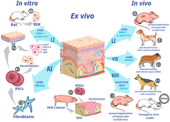
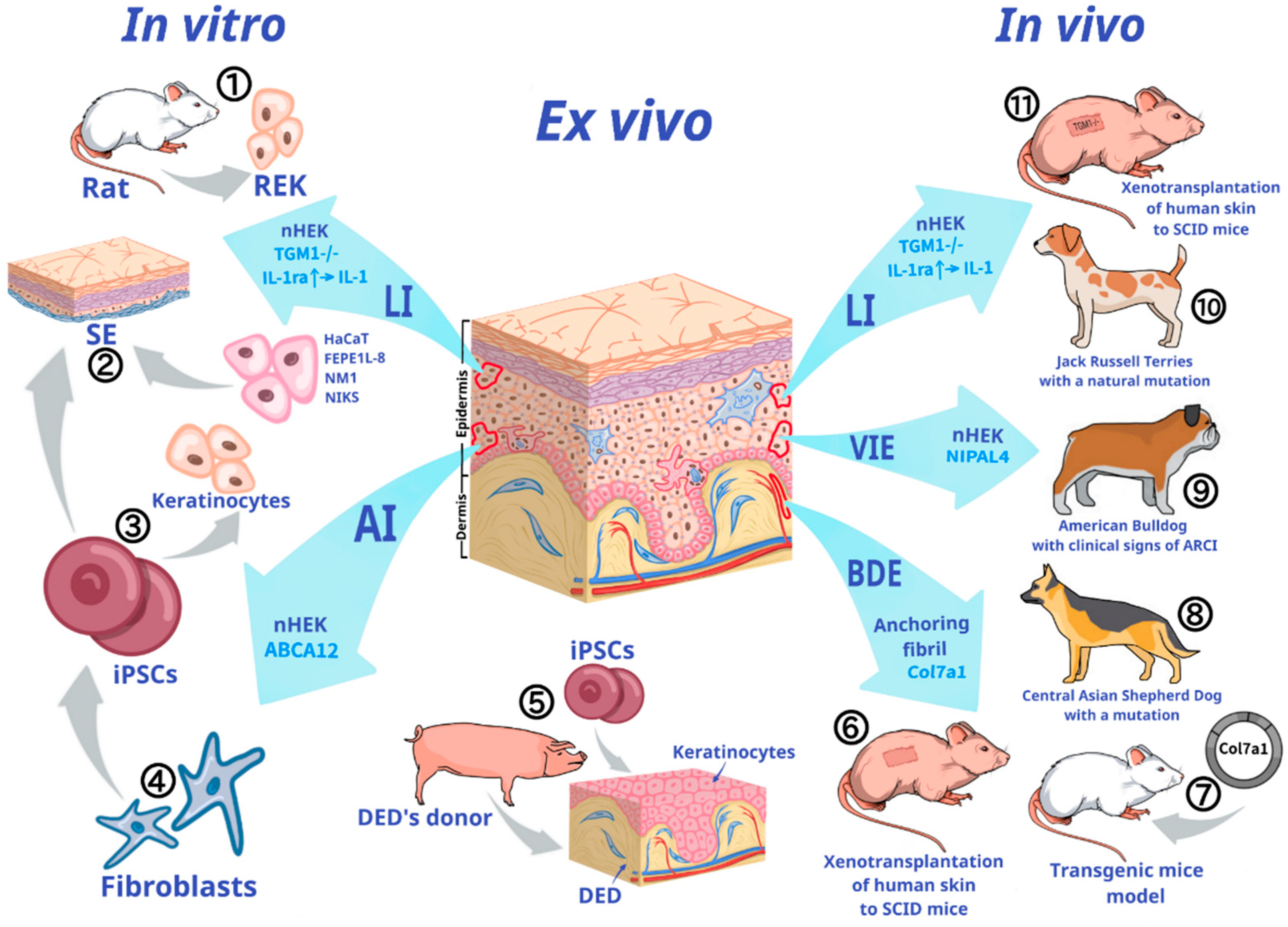

Disclaimer/Publisher’s Note: The statements, opinions and data contained in all publications are solely those of the individual author(s) and contributor(s) and not of MDPI and/or the editor(s). MDPI and/or the editor(s) disclaim responsibility for any injury to people or property resulting from any ideas, methods, instructions or products referred to in the content. |
© 2026 by the authors. Licensee MDPI, Basel, Switzerland. This article is an open access article distributed under the terms and conditions of the Creative Commons Attribution (CC BY) license.