


Abstract
Low levels of high-density lipoprotein cholesterol (HDL-C) are strongly associated with increased cardiovascular risk. However, under various pathological conditions, high-density lipoprotein (HDL) particles may undergo structural and functional modifications, leading to a progressive loss of antioxidant capacity and a shift from a cardioprotective to a proatherogenic phenotype. In this cross-sectional study, we investigated differences in HDL particle distribution and antioxidant function between individuals with elevated lipoprotein(a) [Lp(a)] levels (≥30 mg/dL) and those with low Lp(a) levels (<10 mg/dL). Serum low-density lipoprotein (LDL) and HDL subfractions were analyzed in twenty subjects with high Lp(a) and ten low-Lp(a) controls using non-denaturing polyacrylamide gel electrophoresis (PAGE, Lipoprint system). Enzymatic activities of paraoxonase-1 (PON1) and HDL-associated lipoprotein-associated phospholipase A2 (HDL-Lp-PLA2) were measured. Electrophoretic analysis revealed a significant increase in small HDL (S-HDL) in the high-Lp(a) group compared to the controls (34.1 ± 13.2% vs. 21.5 ± 2.7%, p = 0.01), alongside a reduction in large HDL (L-HDL) (19.6 ± 9.9% vs. 33.4 ± 3.8%, p < 0.001). Furthermore, the high Lp(a) group exhibited significantly lower HDL-PON1 activity (55 ± 12 vs. 67 ± 7 U/mL, p < 0.001) and HDL-Lp-PLA2 activity (2.6 ± 1.0 vs. 3.6 ± 1.2 nmol/min/mL, p < 0.02) compared with the controls. These findings suggest that markedly elevated Lp(a) levels are associated with a shift toward a more proatherogenic HDL subfraction profile and impaired antioxidant functionality, which may reflect mechanisms linked to increased atherosclerotic cardiovascular disease (ASCVD) risk.
1. Introduction
Low levels of high-density lipoprotein cholesterol (HDL-C) have been established as a robust risk factor for atherosclerotic cardiovascular disease (ASCVD), primarily due to the central role of high-density lipoprotein (HDL) in reverse cholesterol transport (RCT) and plaque regression [1]. However, the persistence of significant residual cardiovascular risk, even in individuals with optimal HDL-C levels, has shifted research interest from the quantitative measurement of cholesterol to the qualitative and functional assessment of HDL particles [2]. In this context, the term “HDL quality” refers to structural and functional characteristics of HDL particles beyond the HDL-C concentration. In the present study, HDL quality is defined by particle size distribution (large, intermediate, and small HDL subfractions), HDL-associated antioxidant enzyme activities [paraoxonase-1 (PON1) and HDL-associated lipoprotein-associated phospholipase A2 (HDL-Lp-PLA2)], and oxidative susceptibility assessed through copper-induced oxidation kinetics.
HDL represents a highly heterogeneous population of lipoproteins with an atheroprotective capacity that is attributed to pleiotropic properties, including antioxidant, anti-inflammatory, and cytoprotective effects that inhibit the oxidative modification of low-density lipoprotein (LDL) [2,3]. This functionality is closely linked to the structural integrity and chemical composition of HDL particles. Under conditions of oxidative stress, chronic inflammation, or aging, HDL undergoes extensive remodeling, transitioning from large, spherical, cholesterol-rich particles to small, dense, and dysfunctional subfractions [3,4]. A central mechanism underlying this transition is the loss of enzymatic activity of key HDL-associated antioxidant enzymes, particularly paraoxonase-1 (PON1) and HDL-associated lipoprotein-associated phospholipase A2 (HDL-Lp-PLA2) [5]. Genetic polymorphisms, dietary patterns, and environmental stressors may further modulate these enzymatic activities [6], ultimately transforming HDL from a protective particle into a pro-oxidative and pro-inflammatory carrier that exacerbates atherogenesis [5,7].
Lipoprotein(a) [Lp(a)] is a well-established, genetically determined independent risk factor for ASCVD, myocardial infarction, and aortic stenosis [8]. Structurally, Lp(a) consists of an LDL-like particle in which apolipoprotein B-100 (apoB) is covalently linked via a single disulfide bridge to a unique, highly hydrophilic glycoprotein, apolipoprotein(a) [apo(a)] [8]. Plasma Lp(a) concentration is predominantly determined by the LPA gene locus, which exhibits marked size polymorphism due to variations in the number of Kringle IV type 2 (KIV-2) repeats [8]. This genetic architecture gives rise to a broad spectrum of apo(a) isoforms; notably, smaller (low-molecular-weight) isoforms are inversely associated with plasma Lp(a) concentrations and confer higher cardiovascular risk [9].
Unlike LDL, Lp(a) has a prolonged residence time in circulation, largely attributable to its reduced affinity for the LDL receptor (LDLR), thereby limiting LDLR-mediated clearance. Alternative pathways, including scavenger receptor class B type I (SR-BI) and possibly renal excretion, have been proposed to contribute to its catabolism [10]. The apo(a) moiety of Lp(a) interferes with LDLR-mediated clearance, further contributing to its accumulation.
The heightened atherogenicity of Lp(a) compared with LDL is largely attributed to its role as the primary carrier of oxidized phospholipids (OxPL) in human plasma, transporting approximately 85% of circulating OxPLs [11]. These OxPLs bind to the Kringle IV type 10 (KIV-10) and protease domains of apo(a) and act as potent pro-inflammatory mediators that promote endothelial dysfunction and foam cell formation. Specifically, Lp(a) induces vascular injury by inhibiting endothelial nitric oxide synthase (eNOS) and increasing the expression of adhesion molecules, including intercellular adhesion molecule-1 (ICAM-1) and vascular cell adhesion molecule-1 (VCAM-1), via activation of the Rho/Rho kinase signaling pathway [8,12,13]. By functioning both as a carrier of OxPL and as a driver of vascular inflammation, Lp(a) represents a critical link between lipid metabolism, thrombosis, and the development of unstable atherosclerotic plaques [14].
Despite the well-established independent roles of Lp(a) and HDL in cardiovascular pathophysiology, the potential crosstalk between these lipoprotein systems remains incompletely understood [15]. Prior research has extensively explored the quantitative relationship between Lp(a) and total HDL-C, but data regarding the impact of elevated Lp(a) on HDL quality (subfraction profile) and antioxidant functionality are limited. Emerging evidence suggests a complex metabolic interplay, possibly involving shared clearance pathways such as SR-BI, which may facilitate lipid exchange or the transfer of pro-inflammatory cargo between particles [16]. Clinical observations have frequently demonstrated a paradoxical positive correlation between Lp(a) and HDL-C levels; however, elevated Lp(a) appears to attenuate the protective capacity of HDL, resulting in increased cardiovascular risk even in the presence of high HDL-C concentrations [15,17]. This phenomenon may be related to the pro-inflammatory systemic and vascular milieu associated with elevated Lp(a), which promotes the transformation of HDL into a dysfunctional phenotype, thereby neutralizing its anti-atherogenic properties [17].
Furthermore, this interaction appears to be influenced by triglyceride levels, suggesting that the metabolic interplay between Lp(a) and HDL subfractions (HDL2 and HDL3) is strongly dependent on the overall lipid environment [18,19]. As conventional quantitative lipid measurements fail to capture these qualitative alterations, high-resolution studies are needed to determine whether marked elevations in Lp(a) are associated with structural and functional shifts in the HDL profile [20].
Although several clinical studies have reported a paradoxically positive correlation between Lp(a) and HDL-C levels, most prior studies have relied primarily on quantitative lipid measurements rather than qualitative or functional assessments of HDL particles. Specifically, previous studies have largely examined HDL-C concentrations without evaluating HDL subfraction distribution, antioxidant enzyme activity, or oxidative susceptibility. Consequently, it remains unclear whether elevated Lp(a) levels are associated with structural remodeling and functional impairment of HDL beyond simple changes in HDL-C concentration.
To address this gap, a multiparametric evaluation of HDL structural and functional properties is required. High-resolution HDL subfraction analysis using the Lipoprint system allows particle size distribution to be assessed, distinguishing large, intermediate, and small HDL particles, which differ in cardioprotective capacity. Measurement of HDL-associated paraoxonase-1 (PON1) and HDL-bound lipoprotein-associated phospholipase A2 (HDL-Lp-PLA2) activities provides insight into HDL’s antioxidant defense mechanisms. In addition, copper-induced oxidation kinetics can provide a functional assessment of HDL oxidative susceptibility. Together, these complementary approaches enable a more comprehensive evaluation of HDL quality than HDL-C concentration alone.
Therefore, the aim of the present study was to evaluate the qualitative and functional integrity of HDL in individuals with significantly elevated Lp(a) levels compared with a normo-Lp(a) control group. To this end, we employed a multiparametric approach, including high-resolution analysis of HDL subfraction distribution using polyacrylamide gel electrophoresis, measurement of the enzymatic activities of PON1 and HDL-Lp-PLA2, and assessment of the oxidative susceptibility of HDL particles through copper-induced oxidation kinetics. Through this comprehensive evaluation, we sought to explore whether the cardiovascular risk associated with high Lp(a) levels may be linked to alterations in HDL structural and functional properties.
2. Materials and Methods
2.1. Study Population and Design
This cross-sectional study followed a consecutive recruitment strategy. The study cohort comprised 20 patients with elevated Lp(a) levels (≥30 mg/dL), aged 51 ± 15 years [Lp(a) group], and 10 healthy volunteers with low Lp(a) levels (<10 mg/dL), aged 50 ± 10 years (control group). All participants were of Caucasian ethnicity, were treatment-naïve regarding lipid-lowering agents (statins, ezetimibe, or PCSK9 inhibitors), antihypertensive medications, antidiabetic drugs, and antioxidant supplements for at least 3 months prior to enrollment, and were recruited from the Outpatient Lipid Clinic of the University Hospital of Ioannina, Greece. Exclusion criteria included diabetes mellitus, chronic kidney disease (eGFR < 60 mL/min/1.73m2), active liver disease, clinical evidence of acute infection, diagnosed autoimmune or inflammatory diseases, and thyroid dysfunction (abnormal TSH levels). The ≥30 mg/dL threshold, regarding Lp(a) levels, was selected based on established evidence suggesting increased cardiovascular risk above this concentration.
The study was conducted in accordance with the Declaration of Helsinki and was approved by the Institutional Ethics Committee of the University General Hospital of Ioannina (Protocol No. 896/21-12-2020). Written informed consent was obtained from all participants prior to enrollment.
A comprehensive clinical and laboratory evaluation was performed in all subjects. Recorded demographic and clinical characteristics included age, sex, smoking status, and comorbidities [with particular emphasis on ASCVD, body mass index (BMI), and blood pressure (BP)].
Venous blood samples were collected after a 12 h overnight fast into sterile Vacutainer SST™ II Advance tubes and K2-EDTA tubes (Becton Dickinson, Plymouth, UK). Serum and plasma aliquots were stored at −80 °C until analysis (storage duration: 1–8 months).
Serum concentrations of total cholesterol, triglycerides, HDL-C, and Lp(a) were measured using enzymatic colorimetric assays (Roche Diagnostics, Mannheim, Germany) on a Cobas analyzer (Roche Diagnostics, Mannheim, Germany). Lp(a) was quantified by immunonephelometry (Siemens Healthineers, Erlangen, Germany) at the Biochemical Laboratory of the University General Hospital of Ioannina. Low-density lipoprotein cholesterol (LDL-C) levels were calculated using the Friedewald formula, provided that triglyceride concentrations were <400 mg/dL. To account for the cholesterol content within Lp(a) particles, corrected LDL-C (LDL-C_corr) was calculated using the formula: LDL-C_corr = LDL-C − [0.3 × Lp(a) mass].
The baseline clinical and biochemical characteristics of the study population are presented in Table 1.
Table 1.
Clinical and Biochemical Characteristics of the Study Population.
2.2. Serum Lipoprotein Subfraction Analysis
Serum lipoprotein subfractions were analyzed using the Lipoprint System (Quantimetrix Corp., Redondo Beach, CA, USA), a high-resolution, non-denaturing polyacrylamide gel electrophoresis (PAGE) method. This system enables the quantitative distribution of LDL and HDL particles based on size and electrophoretic mobility.
Electrophoretic separation and phenotypic characterization (Phenotype A and Phenotype B) were performed using the LDL Subfractions Kit (Lipoprint LDL Kit; Quantimetrix Corp.) according to the manufacturer’s instructions and as previously described [21]. Briefly, serum samples were pre-stained with a lipophilic dye that binds proportionally to the cholesterol content of lipoprotein particles. Samples were then loaded onto precast polyacrylamide gradient gels.
Electrophoresis allowed the separation of lipoproteins into distinct bands: very low-density lipoprotein (VLDL) [origin, relative mobility Rf = 0], midbands corresponding to intermediate-density lipoprotein (IDL), up to seven LDL subfractions (LDL-1 to LDL-7), and HDL (fastest migrating fraction, Rf = 1).
LDL particles were classified into two phenotypes: Phenotype A, characterized by a predominance of large, buoyant LDL subfractions (LDL-1 and LDL-2), and Phenotype B, characterized by the predominance of small, dense LDL (sdLDL) particles (LDL-3 to LDL-7). Following electrophoresis, gels were scanned, and the cholesterol concentration of each subfraction (expressed in mg/dL) was calculated by multiplying the relative area under the curve of each band by the total serum cholesterol concentration of the corresponding sample [21].
2.3. HDL Subfraction Analysis
Serum HDL subfractions were characterized and quantified using the HDL Subfractions Kit (Lipoprint HDL Kit; Quantimetrix Corp., Redondo Beach, CA, USA) on the Lipoprint system, according to the manufacturer’s instructions. The analytical principle of this method is consistent with the LDL subfractionation described above and is based on high-resolution, non-denaturing PAGE.
The system separates HDL particles into ten distinct subfractions according to their size and electrophoretic mobility. These are further categorized into three major groups: Large HDL (L-HDL) (subfractions 1–3), Intermediate HDL (I-HDL) (subfractions 4–7), and Small HDL (S-HDL) (subfractions 8–10). This classification corresponds to the HDL2 and HDL3 subclasses traditionally identified by analytical ultracentrifugation [22].
Following electrophoresis, the relative area under the curve for each HDL band was determined and multiplied by the total serum HDL-C concentration to calculate the cholesterol content (mg/dL) of each subfraction. This detailed distribution analysis allows a precise assessment of the HDL subfraction profile and its potential association with cardiovascular risk [22].
2.4. Measurement of PON1 Arylesterase Activities
The arylesterase activity of PON1 was quantified spectrophotometrically using phenylacetate as the substrate, as previously described [23]. Briefly, the hydrolysis of phenylacetate to phenol was monitored by measuring the increase in absorbance at 270 nm over a 1.5 min interval at 25 °C.
To ensure analytical precision and reproducibility, all assays were performed under strictly controlled conditions in accordance with optimized laboratory protocols [23].
2.5. Measurement of HDL-Associated Lp-PLA2 Activity
The activity of HDL-Lp-PLA2 was determined in the apoB-depleted plasma fraction. ApoB-containing lipoproteins were precipitated by the addition of dextran sulfate and magnesium chloride (MgCl2) to plasma samples, followed by centrifugation, as previously described [21].
Lp-PLA2 activity in the resulting supernatant, containing HDL particles, was assessed by measuring the hydrolysis of the tritiated platelet-activating factor ([3H]-PAF) used as the substrate. The reaction was performed at 37 °C for 30 min, and the generated [3H]-acetate was separated from the unreacted substrate by centrifugal vacuum evaporation or liquid–liquid extraction. Enzymatic activity was quantified using a liquid scintillation counter and expressed as nanomoles of platelet-activating factor (PAF) hydrolyzed per minute per milliliter of plasma (nmol/min/mL).
2.6. Isolation of HDL and Protein Determination
HDL fractions were isolated from plasma by sequential preparative ultracentrifugation (100,000× g at 4 °C), as previously described [22]. Following isolation, the HDL fraction underwent extensive dialysis against a large volume of phosphate-buffered saline (PBS). Dialysis was performed at 4 °C for 24 h using appropriate dialysis membranes, with repeated buffer changes to ensure complete removal of potassium bromide (KBr) and ethylenediaminetetraacetic acid (EDTA).
The removal of EDTA was essential, as its presence would inhibit the subsequent copper-induced oxidation assay. Protein concentration was determined using the bicinchoninic acid (BCA) protein assay kit. To preserve particle integrity and minimize ex vivo oxidative modification—a recognized limitation of this methodology—samples were stored under a nitrogen (N2) atmosphere at 4 °C and were used for functional assays within 48 h of isolation.
2.7. Susceptibility of HDL to Copper-Induced Oxidation
The oxidative susceptibility of the isolated HDL fraction was assessed by monitoring the formation of conjugated dienes at 234 nm. Oxidation kinetics were continuously recorded spectrophotometrically. In brief, samples were standardized to a final protein concentration of 100 μg/mL, as determined by the Pierce™ BCA Protein Assay Kit (Thermo Fisher Scientific, Waltham, MA, USA), in 10 mM PBS (pH 7.4). Oxidation was initiated by the addition of CuSO4 at a final concentration of 10 μM, in a total reaction volume of 250 μL. The procedure was performed in UV-transparent microplates (Costar®, Corning Inc., Corning, NY, USA), and kinetics were recorded every 10 min for 6 h at 37 °C using an Infinite® 200 PRO microplate reader (Tecan Group Ltd., Männedorf, Switzerland). Given that HDL does not exhibit a distinct lag phase during copper-induced oxidation, oxidative susceptibility was evaluated using the slope of the oxidation curve (SOC), expressed as ΔA/min [24].
2.8. Statistical Analysis
Statistical analysis was performed using SPSS Statistics software version 26.0 (IBM Corp., Armonk, NY, USA). Data distribution was assessed using the Shapiro–Wilk test to determine the appropriate parametric or non-parametric statistical approach.
Continuous variables are presented as mean ± standard deviation (SD) for normally distributed data and as median [interquartile range (IQR)] for non-normally distributed variables. Categorical variables are expressed as frequencies and percentages.
Comparisons between the two study groups were performed using the unpaired Student’s t-test or the Mann–Whitney U test, as appropriate. Fisher’s exact test was used for categorical variables. Due to the exploratory nature of this study, a matching strategy was employed during recruitment to ensure that the groups were comparable in terms of age, sex, smoking status, and baseline lipid profile (as shown in Table 1), thereby minimizing the need for multivariable adjustment. Furthermore, since the evaluated HDL structural and functional parameters are biologically highly interrelated, no formal correction for multiple comparisons was applied to avoid the risk of Type II errors and the potential omission of clinically relevant trends. Correlations between variables were evaluated using Pearson’s or Spearman’s correlation coefficients, depending on data distribution.
3. Results
3.1. Clinical and Biochemical Characteristics of the Study Population
The study population consisted of 20 individuals [High Lp(a) group] with a median Lp(a) concentration of 66 mg/dL (range: 45–95 mg/dL) and 10 healthy volunteers (Control group) with Lp(a) levels <10 mg/dL. All participants were of Caucasian ethnicity, were treatment-naive at the time of recruitment, and were followed at the Outpatient Lipid Clinic of the University Hospital of Ioannina. A comprehensive clinical and laboratory assessment was performed in all subjects. Demographic and clinical characteristics, including age, sex, smoking status, BMI, and BP, are summarized in Table 1. The two groups were well matched for age (51 ± 15 vs. 50 ± 10 years) and BMI (26 ± 4 vs. 23 ± 5 kg/m2), with no statistically significant differences observed. Regarding the lipid profile, although minor differences in mean values were noted, no statistically significant differences were detected between the High Lp(a) group and the Control group in total cholesterol (155 ± 18 vs. 170 ± 15 mg/dL), triglycerides (105 ± 45 vs. 110 ± 30 mg/dL), LDL-C (80 ± 26 vs. 93 ± 23 mg/dL), or HDL-C (54 ± 14 vs. 55 ± 17 mg/dL). These findings indicate that the primary distinguishing feature between the two groups was the concentration of Lp(a).
3.2. Antioxidant Capacity of HDL
3.2.1. Evaluation of HDL-Associated Enzymatic Activities (PON1 and HDL-Lp-PLA2)
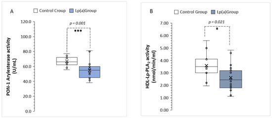
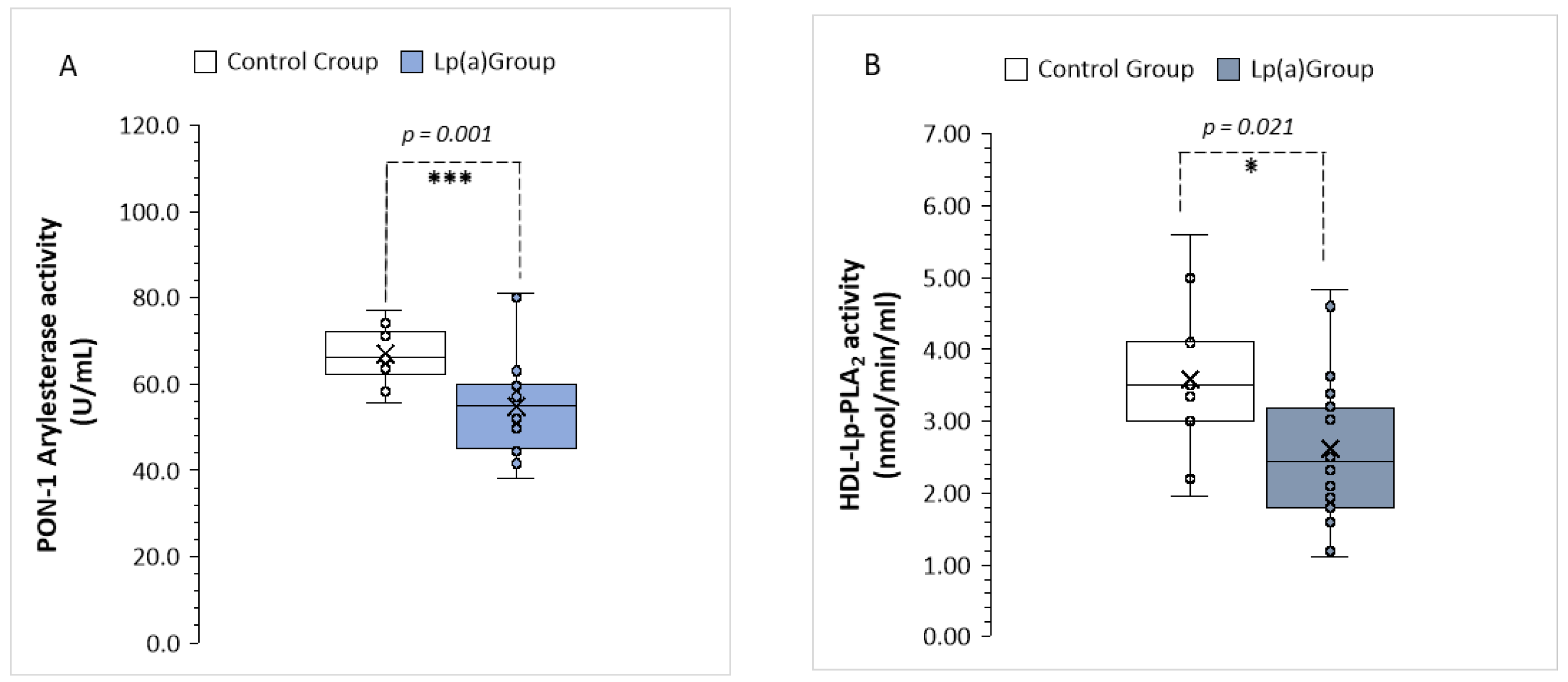
The enzymatic activities of PON1 and HDL-associated Lp-PLA2 were measured in serum samples. PON1 arylesterase activity was significantly reduced in the High Lp(a) group compared with the Control group (55 ± 12 vs. 67 ± 7 U/mL, p = 0.001; Figure 1A). Similarly, HDL-Lp-PLA2 activity was significantly lower in subjects with high Lp(a) levels compared with controls (2.6 ± 1.0 vs. 3.6 ± 1.2 nmol/min/mL, p = 0.021; Figure 1B).
Figure 1.
Comparison of PON1 and HDL-Lp-PLA2 enzymatic activities between the study groups (Control group, n = 10, High Lp(a) group, n = 20). (A) PON-1 Arylesterase activity levels (U/mL). (B) HDL-Lp-PLA2 activity (nmol/min/mL). Data are presented as box-and-whisker plots/(median, interquartile range, and min–max). Statistical significance was determined by unpaired Student’s t-test (* p < 0.05, *** p < 0.001).
3.2.2. Oxidative Susceptibility of the HDL Fraction
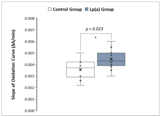
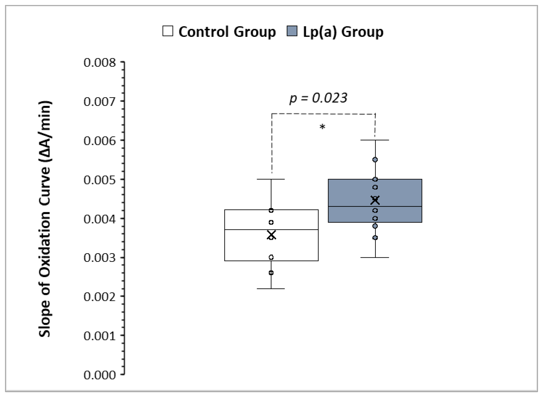
To evaluate the antioxidant capacity of HDL, Cu2+-induced oxidation kinetics were assessed. As HDL particles typically lack a distinct lag phase during copper-induced oxidation, oxidative susceptibility was quantified using the SOC, expressed as the change in absorbance per minute (ΔA/min). The HDL oxidation slope was significantly higher in the High Lp(a) group compared with the Control group (0.000479 ± 0.000097 vs. 0.000326 ± 0.000095 ΔA/min, p = 0.023) (Figure 2). This finding demonstrates an association between elevated Lp(a) levels and an accelerated HDL oxidation rate, reflecting impaired antioxidant capacity and increased oxidative susceptibility of the HDL fraction in these individuals.
Figure 2.
Comparison of HDL oxidation rates between the study groups (Control group, n = 10, High Lp(a) group, n = 20). The slope of the oxidation curve (SOC) is expressed as ΔA/min. Data are presented as box-and-whisker plots (median, interquartile range, and min–max). Statistical significance was determined by an unpaired t-test (* p < 0.05).
3.3. Evaluation of Lipoprotein Particle Distribution (Lipoprint Analysis)
3.3.1. LDL Particle Distribution
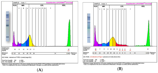
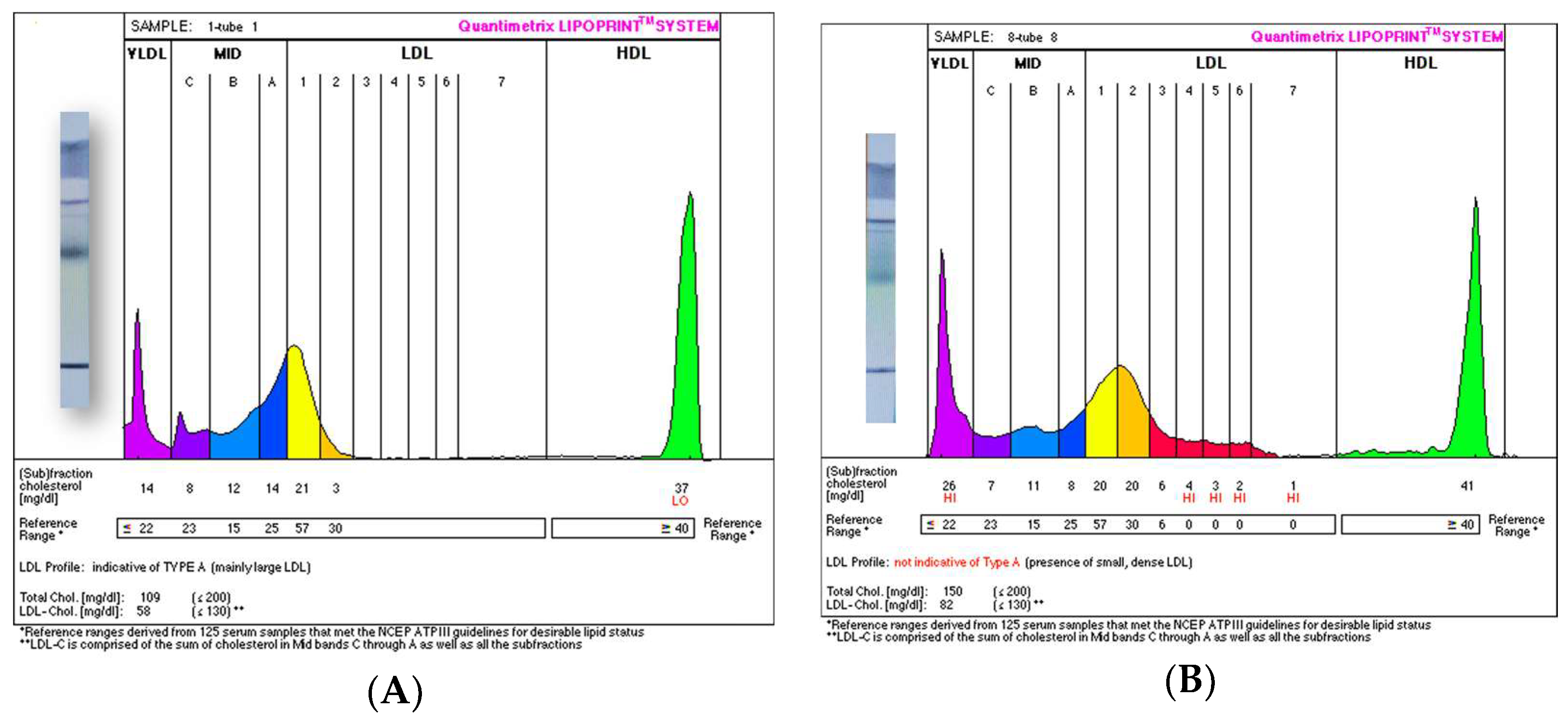
Electrophoretic analysis of LDL subfractions revealed significant differences in the lipoprotein profile between the study groups. Subjects in the High Lp(a) group exhibited a significantly higher proportion of small, dense LDL (sdLDL) particles (LDL-3 to LDL-7) compared to the Control group (4.35 ± 3.34% vs. 0.89 ± 0.44%, respectively; p = 0.001). These quantitative findings corresponded to a categorical classification of Phenotype B for all subjects in the High Lp(a) group (Figure 3B) and Phenotype A for all subjects in the Control group (Figure 3A). It should be noted that the elevated Lp(a) mass likely contributes to the measured sdLDL signal in the High Lp(a) group due to electrophoretic co-migration in this region.
Figure 3.
Representative LDL subfraction profiles and particle size distribution obtained via high-resolution electrophoresis (Lipoprint System). Εlectrophoretic profiles with their corresponding gel images (left) are shown for: (A) the Control group (n = 10), showing a predominance of large, buoyant LDL particles (Phenotype A), and (B) the High Lp(a) group (n = 20), showing a distribution shift towards the small, dense LDL region (Phenotype B). These profiles are representative examples of the phenotypic patterns observed in each study group. The potential contribution of Lp(a) mass to the signal measured within the sdLDL region is a characteristic of this electrophoretic methodology and is considered in the interpretation of these profiles.
3.3.2. HDL Subfraction Distribution
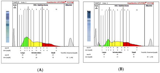
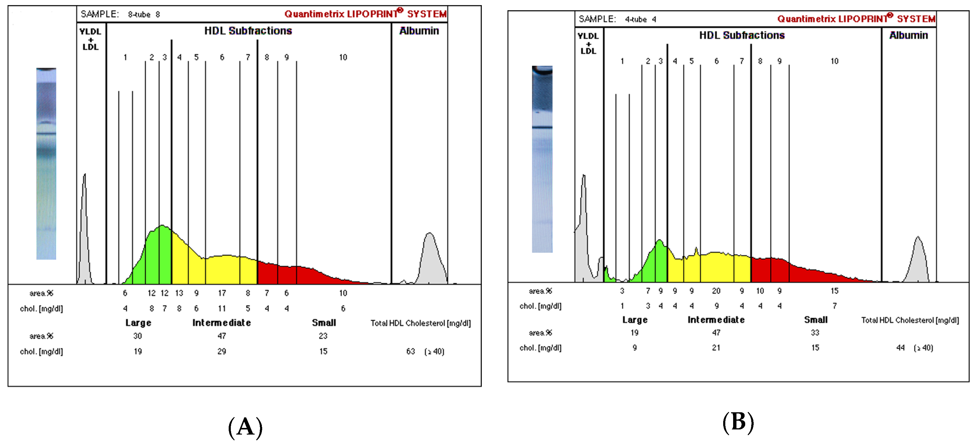
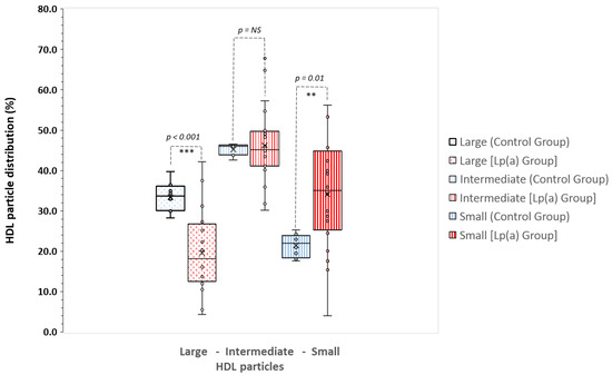
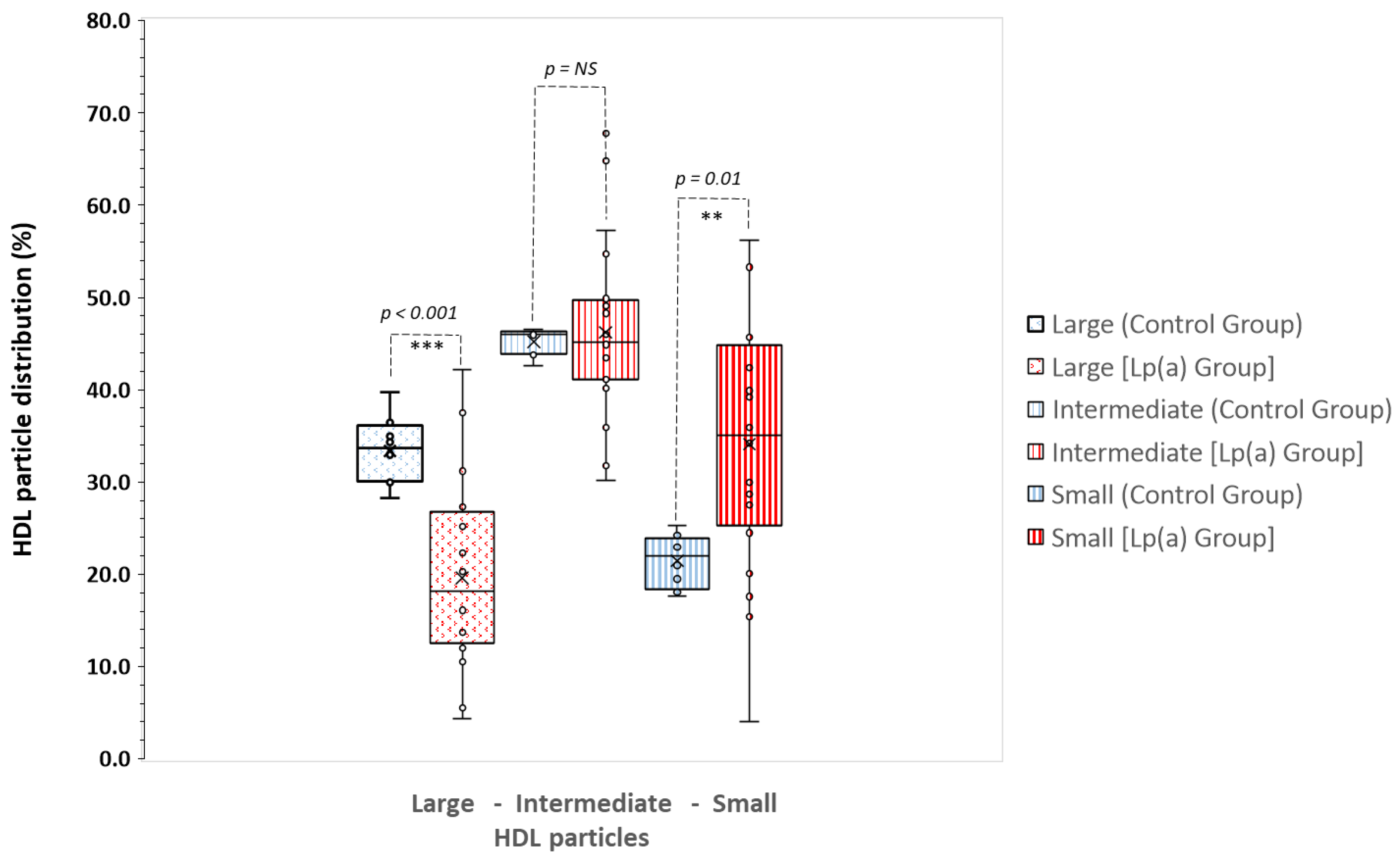
High-resolution electrophoresis revealed significant structural differences in the HDL subfraction profile between the two groups (Figure 4A,B). Compared with the Control group, subjects in the High Lp(a) group exhibited a significant reduction in the proportion of large HDL (L-HDL) particles (19.6 ± 9.9 vs. 33.4 ± 3.8%, p < 0.001) and a significant increase in small HDL (S-HDL) particles (34.1 ± 13.2% vs. 21.5 ± 2.7%, p < 0.01). No significant differences were observed in intermediate HDL (I-HDL) subfractions between the groups (Figure 5).
Figure 4.
Representative HDL subfraction profiles and particle size distribution obtained via high-resolution electrophoresis (Lipoprint System). Εlectrophoretic profiles with their corresponding gel images (left) are shown for: (A) the Control group (n = 10), showing a predominance of large HDL (L-HDL) subfractions, and (B) the High Lp(a) group (n = 20), showing a distribution shift towards smaller HDL particles (S-HDL).
Figure 5.
Comparative analysis of HDL subfraction distribution between the study groups (Control group, n = 10, High Lp(a) group, n = 20) via high-resolution electrophoresis (Lipoprint System). Data are presented as box-and-whisker plots (median, IQR, and range); the “×” indicates the mean value, and individual data points are superimposed. Statistical significance was determined by unpaired Student’s t-test (** p < 0.01, *** p < 0.001, NS: non-significant). Abbreviations: large-sized HDL particles;, intermediate-sized HDL particles;, small-sized HDL particles.
3.4. Correlation Analysis
Correlation analysis was performed to explore the association between Lp(a) levels and lipoprotein functional markers. A significant negative correlation was observed between Lp(a) concentration and PON1 arylesterase activity (Spearman’s rho = −0.44, p < 0.001). Additionally, Lp(a) levels showed a positive correlation with the percentage of small HDL particles (Spearman’s rho = 0.53, p = 0.016).
4. Discussion
This study investigated the association between Lp(a) levels and HDL quality, focusing on subfraction distribution and functional markers. Our results demonstrate that subjects with elevated Lp(a) exhibit a more fragmented HDL profile and compromised antioxidant enzyme activities compared to controls. While HDL remodeling is a recognized feature of various inflammatory and metabolic states [7], our findings specifically highlight the correlation between elevated Lp(a) and the transition of HDL toward a dysfunctional, potentially pro-atherogenic state, independent of traditional lipid metrics [3,7].
The pathogenicity of elevated Lp(a) is multifaceted and significantly linked to its role as the principal circulating reservoir for oxidized phospholipids (OxPL) [9,11]. Our findings suggest that elevated Lp(a) levels are associated with a broader remodeling of the lipoprotein milieu. While all subjects in the High Lp(a) group were classified as LDL Phenotype B, this observation reflects a high-risk lipid environment rather than a definitive isolated increase in small-dense LDL particles. As noted, the potential co-migration of Lp(a) mass within the sdLDL region during electrophoresis is a known characteristic of the Lipoprint system. However, this finding remains confirmatory of the complex atherogenic profile seen in these subjects, which is further characterized by the concurrent impairment of HDL structure and function observed in our study.
Data examining the interaction between Lp(a) and HDL functionality remain limited. Our results demonstrate that a high Lp(a) environment is associated with a significant compromise in HDL functional integrity. Specifically, the increased susceptibility of the HDL fraction to Cu2+-induced oxidation suggests a diminished protective capacity in subjects with elevated Lp(a). This impairment was paralleled by a marked reduction in the activities of key antioxidant enzymes, namely PON1 arylesterase and HDL–Lp-PLA2. These findings align with prior observations suggesting that the OxPL cargo of Lp(a) may influence the enzymatic repertoire of HDL, potentially by overwhelming its antioxidant defenses [25]. The observed decline in enzymatic activity reflects a substantial loss of HDL’s ability to limit lipid peroxidation, a finding consistent with reports where heightened oxidative stress leads to a concomitant reduction in PON1 functionality [3,7].
Beyond enzymatic activity, HDL particle size and distribution are critical determinants of functional capacity. Functional HDL particles are typically large, spherical, and enriched in cholesterol and apolipoprotein A-I (apoA-I). However, under conditions of metabolic stress or systemic inflammation, HDL morphology may shift toward smaller, denser, and potentially dysfunctional particles [3,7]. In our study, elevated Lp(a) was associated with a significant reduction in L-HDL particles and an increase in S-HDL subfractions. While a shift toward smaller, triglyceride-rich HDL variants is often observed in conditions of systemic inflammation or in specific populations such as smokers [26], our study groups were matched for age, BMI, and smoking status. This suggests that the observed remodeling may reflect the specific impact of the high-Lp(a) environment on HDL maturation or stability. The synergy between diminished PON1 activity and an unfavorable HDL subfraction profile points toward a substantial reduction in the overall antioxidant capacity within the lipoprotein environment, further compromising the ability of HDL to inhibit LDL oxidation [7,25]. Consequently, the combination of enzymatic and structural markers may serve as a more sensitive indicator of cardiovascular risk than HDL-C levels alone.
In subjects with elevated Lp(a), the observed shift from L-HDL toward S-HDL subfractions is characterized by a more fragmented HDL phenotype. This distribution pattern is particularly relevant as smaller HDL particles have been associated with increased ASCVD risk, potentially due to reduced reverse cholesterol transport capacity and altered protein cargo [3,7]. Our findings demonstrate that HDL-C concentration alone is an insufficient marker of cardioprotective potential in the context of High Lp(a). Specifically, HDL particles may be quantitatively preserved but functionally impaired, rendering traditional lipid metrics potentially misleading for these individuals. Therefore, incorporating HDL quality markers—such as PON1 activity and subfraction distribution—could complement standard lipid assessments and allow for more accurate cardiovascular risk stratification. The observed associations in our study further support this notion, as the progressive decline in antioxidant enzymatic activity and the concomitant shift toward smaller HDL subfractions as Lp(a) levels rise, point towards a systematic deterioration of the lipoprotein’s protective role. These correlations suggest that the impact of Lp(a) may extend beyond its inherent pathogenicity by potentially compromising the endogenous antioxidant defenses provided by the HDL pool.
It is important to note that, given the cross-sectional design of the present study, the observed relationships between elevated Lp(a) levels and alterations in HDL structure and function should be interpreted as associative rather than causal. It remains unclear whether elevated Lp(a) directly influences HDL remodeling or whether both reflect shared underlying inflammatory, oxidative, or metabolic mechanisms. Longitudinal and mechanistic studies are required to clarify these pathways.
Study Limitations
Several limitations of the present study should be acknowledged. First, the relatively small sample size limits statistical power and may increase the risk of both type I and type II errors. This investigation was designed as an exploratory, cross-sectional, hypothesis-generating study in a highly selected, treatment-naïve cohort, and no a priori power calculation was performed. Second, multiple mechanistically related biochemical and lipoprotein endpoints were evaluated. Although these outcomes were predefined and biologically interconnected, formal correction for multiple comparisons (e.g., Bonferroni adjustment) was not applied to avoid the potential omission of biologically meaningful trends in this exploratory setting. Therefore, the findings should be viewed as hypothesis-generating and confirmed in larger independent cohorts. Third, the cross-sectional design precludes causal inference and does not allow determination of whether elevated Lp(a) directly induces HDL dysfunction or whether both reflect a shared inflammatory or oxidative milieu. Fourth, certain biochemical and functional parameters, such as apolipoprotein A-I (apoA-I) levels, high-sensitivity C-reactive protein (hsCRP), and reverse cholesterol transport (RCT) capacity, were not measured. While the lack of hsCRP data is a limitation, the strict clinical exclusion of subjects with active infections or inflammatory diseases aimed to minimize the impact of systemic inflammation. Furthermore, although RCT is a hallmark of HDL function, this study specifically focused on the antioxidant repertoire and structural remodeling of HDL. Future research incorporating a broader panel of apolipoproteins and cholesterol efflux assays is needed to provide a more comprehensive characterization of HDL’s vasoprotective role in the context of high Lp(a). In addition, although the Lipoprint system provides high-resolution subfraction analysis, potential co-migration of Lp(a) particles within the small dense LDL region may influence density-based distribution patterns. Functional assays such as copper-induced oxidation kinetics and enzymatic activity measurements represent ex vivo assessments and may not fully reflect in vivo HDL functionality. Finally, clinical outcome data were not available; therefore, the observed alterations in HDL structure and function cannot be directly linked to incident cardiovascular events. Larger prospective studies incorporating outcome measures are warranted to validate the clinical relevance of these findings.
5. Conclusions
In conclusion, our findings suggest that elevated Lp(a) levels are associated with significant alterations in both the structure and functional quality of HDL particles. This study highlights a potential link between high Lp(a) concentrations and a shift toward smaller, more dysfunctional HDL subfractions, accompanied by reduced PON1 and HDL-Lp-PLA2 enzymatic activities. Given the exploratory nature and limited sample size of the present study, the results should be considered hypothesis-generating. Further longitudinal and mechanistic studies are required to clarify whether these associations reflect causal pathways and to determine their clinical significance. Nevertheless, our findings underscore the importance of assessing HDL functional markers alongside traditional lipid profiles to better evaluate the complex cardiovascular risk associated with elevated Lp(a).
Author Contributions
Conceptualization, C.T. (Constantinos Tellis) and A.D.T.; methodology, C.T. (Constantinos Tellis), H.M. and G.T.; validation, A.D.K., G.T. and C.T. (Constantinos Tellis); formal analysis, A.D.K., G.T. and C.T. (Constantinos Tellis); investigation, A.D.K., G.T. and C.T. (Constantinos Tellis); resources, A.D.T., K.S. and C.T. (Constantinos Tellis); data curation, E.L., A.D.T. and C.T. (Constantinos Tellis); writing—original draft preparation, A.D.K., G.T. and C.T. (Christina Telli); writing—review and editing, A.D.K., G.T., C.T. (Christina Telli) and K.S.; visualization, C.T. (Constantinos Tellis), G.T. and H.M.; supervision, C.T. (Constantinos Tellis), E.L., H.M. and A.D.T.; project administration, K.S. and C.T. (Constantinos Tellis); funding acquisition, A.D.T., E.L. and C.T. (Constantinos Tellis). All authors have read and agreed to the published version of the manuscript.
Funding
This research received no external funding.
Institutional Review Board Statement
The study protocol conforms to the ethical guidelines of the 1975 Declaration of Helsinki, and was approved by the Ethics Committee of the University General Hospital of Ioannina, Greece (896/21-12-2020).
Informed Consent Statement
Informed consent was obtained from all subjects involved in the study. Written informed consent has been obtained from the patients to publish this paper.
Data Availability Statement
The data presented in this study are not publicly available due to privacy and ethical restrictions. De-identified data may be made available from the corresponding author upon reasonable request and with permission of the institutional review board.
Conflicts of Interest
The authors declare no conflicts of interest.
Abbreviations
The following abbreviations are used in this manuscript:
| HDL-C | High-density lipoprotein cholesterol |
| HDL | High-density lipoprotein |
| Lp(a) | Lipoprotein(a) |
| LDL | Low-density lipoprotein |
| PON1 | Paraoxonase-1 |
| HDL-Lp-PLA2 | HDL-associated lipoprotein-associated phospholipase A2 |
| S-HDL | Small HDL |
| L-HDL | Large HDL |
| ASCVD | Atherosclerotic cardiovascular disease |
| RCT | Reverse cholesterol transport |
| apoB | Apolipoprotein B-100 |
| apo(a) | Apolipoprotein(a) |
| KIV-2 | Kringle IV type 2 |
| LDLR | LDL receptor |
| SR-BI | Scavenger receptor class B type I |
| OxPL | Oxidized phospholipids |
| KIV-10 | Kringle IV type 10 |
| eNOS | Endothelial nitric oxide synthase |
| ICAM-1 | Intercellular adhesion molecule-1 |
| VCAM | Vascular cell adhesion molecule-1 |
| BMI | Body mass index |
| BP | Blood pressure |
| LDL-C | Low-density lipoprotein cholesterol |
| PAGE | Polyacrylamide gel electrophoresis |
| VLDL | Very low-density lipoprotein |
| IDL | Intermediate-density lipoprotein |
| sdLDL | Small, dense LDL |
| I-HDL | Intermediate HDL |
| MgCl2 | Magnesium chloride |
| [3H]-PAF | Tritiated platelet-activating factor |
| PAF | Platelet-activating factor |
| PBS | Phosphate-buffered saline |
| KBr | Potassium bromide |
| EDTA | Ethylenediaminetetraacetic acid |
| BCA | Bicinchoninic acid |
| N2 | Nitrogen |
| SOC | Slope of the oxidation curve |
| SD | Standard deviation |
| IQR | Interquartile range |
| ApoA-I | Apolipoprotein A-I |
| SSA | Serum amyloid A |
| ApoC-III | apolipoprotein C-III |
References
- Castelli, W.P.; Garrison, R.J.; Wilson, P.W.F.; Abbott, R.D.; Kalousdian, S.; Kannel, W.B. Incidence of coronary heart disease and lipoprotein cholesterol levels: The Framingham Study. JAMA 1986, 256, 2835–2838. [Google Scholar] [CrossRef] [Scilit]
- Rohatgi, A.; Khera, A.; Berry, J.D.; Givens, E.G.; Ayers, C.R.; Wedin, K.E.; Neeland, I.J.; Yuhanna, I.S.; Rader, D.R.; de Lemos, J.A.; et al. HDL cholesterol efflux capacity and incident cardiovascular events. N. Engl. J. Med. 2014, 371, 2383–2393. [Google Scholar] [CrossRef] [Scilit] [PubMed]
- Kontush, A.; Chapman, M.J. Functionally defective high-density lipoprotein: A new therapeutic target at the crossroads of dyslipidemia, inflammation, and atherosclerosis. Pharmacol. Rev. 2006, 58, 342–374. [Google Scholar] [CrossRef] [Scilit]
- Brites, F.; Martin, M.; Guillas, I.; Kontush, A. Antioxidative activity of high-density lipoprotein (HDL): Mechanistic insights into potential clinical benefit. BBA Clin. 2017, 8, 66–77. [Google Scholar] [CrossRef] [Scilit]
- Rosenson, R.S.; Brewer, H.B.; Ansell, B.J.; Barter, P.; Chapman, M.J.; Heinecke, J.W.; Kontush, A.; Tall, A.R.; Webb, N.R. Dysfunctional HDL and atherosclerotic cardiovascular disease. Nat. Rev. Cardiol. 2016, 13, 48–60. [Google Scholar] [CrossRef] [Scilit] [PubMed]
- Mackness, M.; Mackness, B. Human paraoxonase-1 (PON1): Gene structure and expression, promiscuous activities and multiple physiological roles. Gene 2015, 567, 12–21. [Google Scholar] [CrossRef] [Scilit]
- Cho, K.H. The current status of research on high-density lipoproteins (HDL): A paradigm shift from HDL quantity to HDL quality and HDL functionality. Int. J. Mol. Sci. 2022, 23, 3967. [Google Scholar] [CrossRef] [Scilit] [PubMed]
- Tsimikas, S. A test in context: Lipoprotein(a): Diagnosis, prognosis, controversies, and emerging therapies. J. Am. Coll. Cardiol. 2017, 69, 692–711. [Google Scholar] [CrossRef] [Scilit]
- Kronenberg, F.; Mora, S.; Stroes, E.S.G.; Ference, B.A.; Arsenault, B.J.; Berglund, L.; Dweck, M.R.; Koschinsky, M.; Lambert, G.; Mach, F.; et al. Lipoprotein(a) in atherosclerotic cardiovascular disease and aortic stenosis: A European Atherosclerosis Society consensus statement. Eur. Heart J. 2022, 43, 3925–3946. [Google Scholar] [CrossRef] [Scilit] [PubMed]
- Kozyraki, R.; Fyfe, J.; Kristiansen, M.; Gerdes, C.; Jacobsen, C.; Cui, S.; Christensen, E.I.; Aminoff, M.; de la Chapelle, A.; Krahe, R.; et al. The intrinsic factor-vitamin B12 receptor, cubilin, is a high-affinity apolipoprotein A-I receptor facilitating endocytosis of high-density lipoprotein. Nat. Med. 1999, 5, 656–661. [Google Scholar] [CrossRef] [Scilit]
- Koutsogianni, A.D.; Liberopoulos, E.; Tellis, K.; Tselepis, A.D. Oxidized phospholipids and lipoprotein(a): An update. Eur. J. Clin. Investig. 2022, 52, e13710. [Google Scholar] [CrossRef] [Scilit]
- Yeang, C.; Wilkinson, M.J.; Tsimikas, S. Lipoprotein(a) and oxidized phospholipids in calcific aortic valve stenosis. Curr. Opin. Cardiol. 2016, 31, 440–450. [Google Scholar] [CrossRef] [Scilit]
- Boffa, M.B.; Koschinsky, M.L. Lipoprotein(a) and cardiovascular disease. Biochem. J. 2024, 481, 1277–1296. [Google Scholar] [CrossRef] [Scilit]
- Tsimikas, S.; Brilakis, E.S.; Miller, E.R.; McConnell, J.P.; Lennon, R.J.; Kornman, K.S.; Witztum, J.L.; Berger, P.B. Oxidized phospholipids, Lp(a) lipoprotein, and coronary artery disease. N. Engl. J. Med. 2005, 353, 46–57. [Google Scholar] [CrossRef] [Scilit] [PubMed]
- Johansen, O.E.; Neubacher, D.; von Eynatten, M.; Patel, S.; Woerle, H.J. Cardiovascular safety with linagliptin in patients with type 2 diabetes mellitus: A pre-specified, prospective, and adjudicated meta-analysis of a phase 3 programme. Cardiovasc. Diabetol. 2012, 11, 3. [Google Scholar] [CrossRef] [Scilit]
- Yang, X.P.; Amar, M.J.; Vaisman, B.; Bocharov, A.V.; Vishnyakova, T.G.; Freeman, L.A.; Kurlander, R.J.; Patterson, A.P.; Becker, L.C.; Remaley, A.T. Scavenger receptor-BI is a receptor for lipoprotein(a). J. Lipid Res. 2013, 54, 2450–2457. [Google Scholar] [CrossRef] [Scilit]
- Al Hageh, C.; Chacar, S.; Ghassibe-Sabbagh, M.; Platt, D.E.; Henschel, A.; Hamdan, H.; Gauguier, D.; Murr, Y.E.; Alefishat, E.; Chammas, E.; et al. Elevated Lp(a) levels correlate with severe and multiple coronary artery stenotic lesions. Vasc. Health Risk Manag. 2023, 19, 31–41. [Google Scholar] [CrossRef] [Scilit] [PubMed]
- Konerman, M.; Kulkarni, K.; Toth, P.P.; Jones, S.R. Evidence of dependence of lipoprotein(a) on triglyceride and high-density lipoprotein metabolism. J. Clin. Lipidol. 2012, 6, 27–32. [Google Scholar] [CrossRef] [Scilit]
- von Eckardstein, A.; Nordestgaard, B.G.; Remaley, A.T.; Catapano, A.L. High-density lipoprotein revisited: Biological functions and clinical relevance. Eur. Heart J. 2023, 44, 1394–1407. [Google Scholar] [CrossRef] [Scilit]
- Sheashaa, H.; Mousa, H.; Abbas, M.T.; Farina, J.M.; Awad, K.; Pereyra, M.; Scalia, I.G.; Ali, N.B.; Javadi, N.; Bismee, N.N.; et al. Interaction between lipoprotein(a) and other lipid molecules: A review of the current literature. Biomolecules 2025, 15, 162. [Google Scholar] [CrossRef] [Scilit] [PubMed]
- Gazi, I.; Lourida, E.S.; Filippatos, T.; Tsimihodimos, V.; Elisaf, M.; Tselepis, A.D. Lipoprotein-associated phospholipase A2 activity is a marker of small, dense LDL particles in human plasma. Clin. Chem. 2005, 51, 2264–2273. [Google Scholar] [CrossRef] [Scilit]
- Dafnis, I.; Tsouka, A.N.; Gkolfinopoulou, C.; Tellis, C.C.; Chroni, A.; Tselepis, A.D. PCSK9 is minimally associated with HDL but impairs the anti-atherosclerotic HDL effects on endothelial cell activation. J. Lipid Res. 2022, 63, 100272. [Google Scholar] [CrossRef] [Scilit]
- Gan, K.N.; Smolen, A.; Eckerson, H.W.; La Du, B.N. Purification of human serum paraoxonase/arylesterase. Evidence for one esterase appearing to be identical with paraoxonase. Drug Metab. Dispos. 1991, 19, 100–106. [Google Scholar] [CrossRef] [Scilit] [PubMed]
- Krzesińska, A.; Marlęga-Linert, J.; Chyła-Danił, G.; Marcinkowska, M.; Rogowska, P.; Stumska, K.; Fijałkowski, M.; Gruchała, M.; Jankowski, M.; Mickiewicz, A.; et al. Reduced oxidative susceptibility of Lp(a) and LDL fractions as a pleiotropic effect of lipoprotein apheresis in patients with elevated Lp(a) and ASCVDs. Int. J. Mol. Sci. 2024, 25, 13597. [Google Scholar] [CrossRef] [Scilit] [PubMed]
- Khalil, A.; Fulop, T.; Berrougui, H. Role of Paraoxonase1 in the Regulation of High-Density Lipoprotein Functionality and in Cardiovascular Protection. Antioxid. Redox Signal. 2021, 34, 191–200. [Google Scholar] [CrossRef] [Scilit] [PubMed]
- He, B.M.; Zhao, S.P.; Peng, Z.Y. Effects of cigarette smoking on HDL quantity and function: Implications for atherosclerosis. J. Cell. Biochem. 2013, 114, 2431–2436. [Google Scholar] [CrossRef] [Scilit]

Disclaimer/Publisher’s Note: The statements, opinions and data contained in all publications are solely those of the individual author(s) and contributor(s) and not of MDPI and/or the editor(s). MDPI and/or the editor(s) disclaim responsibility for any injury to people or property resulting from any ideas, methods, instructions or products referred to in the content. |
© 2026 by the authors. Licensee MDPI, Basel, Switzerland. This article is an open access article distributed under the terms and conditions of the Creative Commons Attribution (CC BY) license.