


Abstract
Streptococcus agalactiae is a major contagious pathogen that causes bovine mastitis and substantial economic losses in the dairy industry. This study investigated the capsular genotypes, antimicrobial susceptibility, and virulence-associated phenotypes of S. agalactiae isolated from bovine mastitis cases in northeastern Thailand. A total of 177 S. agalactiae isolates were identified, and characterized. Antimicrobial susceptibility was determined by broth microdilution. Biofilm formation was evaluated using Congo red agar and crystal violet staining, whereas adhesion, invasion, and cytotoxicity were assessed using A549 and HeLa epithelial cell lines using representative isolates. Capsular genotype Ia predominated (95.48%), followed by genotype IX (3.39%), and the remaining were non-typeable (1.13%). All isolates were susceptible to penicillin, vancomycin, erythromycin, and clindamycin, whereas resistance to ceftriaxone (1.13%) and levofloxacin (0.56%) was detected. Among the three representative isolates, the non-typeable isolate exhibited the strongest biofilm-forming ability, followed by the genotype IX and genotype Ia isolates. The genotype IX isolate showed the highest mean adhesion, invasion, and cytotoxicity in both A549 and HeLa cell lines, although not all pairwise differences were statistically significant. This isolate also exhibited higher cyl and bac mRNA levels than the other two isolates. These findings indicate that S. agalactiae isolated in bovine mastitis in this area remained susceptible to most antimicrobials tested and that virulence-associated phenotypes varied among the representative bovine isolates. Epidemiological surveillance, responsible antimicrobial use, farmer education, and improved farm remain important priorities for the prevention and control of bovine mastitis and antimicrobial resistance.
1. Introduction
S. agalactiae is a major bacterial pathogens associated with intramammary infections and, bovine mastitis [1]. The bacterium enters bovine mammary gland through the teat canal and cistern, colonizes the mammary gland ducts, and proliferates, leading to intramammary infection and mastitis [2]. Mastitis caused by S. agalactiae is generally a chronic disease characterized by occasional acute outbreaks and few overt clinical signs, but it reduces milk yield and causes substantial economic losses for dairy farms [3,4]. These losses arise from treatment costs, discarded milk, additional labor, reduced milk production, and costs associated with culling and replacement of affected animals [5]. Over the past few decades, S. agalactiae has remained a significant contagious mastitis pathogen, particularly in countries with emerging dairy industries. High prevalence has been reported in several countries, including Colombia, where herd-level prevalence ranges from 11.1% to 58.3% [6], as well as Algeria (37.66%) [7], Ethiopia (39.67%) [8], and Kenya (80%) [9]. In China, S. agalactiae is among the predominant contagious pathogens associated with bovine mastitis and has been detected in 92.2% of bulk tank milk samples collected from dairy herds [10]. These reports highlight the importance of bovine mastitis caused by S. agalactiae on dairy farms worldwide [11]. In Thailand, S. agalactiae remains a prevalent contagious pathogen in dairy farms, with reported animal-level prevalence ranging from 20.0% to 46.3% and herd-level prevalence of 21.8% in Khon Kaen Province [2,12]. Furthermore, a study conducted in Sakon Nakhon Province reported that S. agalactiae accounted for 33.3% of bacterial isolates recovered from bovine mastitis cases, underscoring its importance as one of the major mastitis pathogens in northeastern Thailand [13].
The pathogenicity of S. agalactiae depends on multiple virulence factors that mediate adhesion, invasion, and infection [14] and facilitate colonization, survival, and persistence within the host [15]. These virulence factors include mechanisms of resistance to phagocytosis and immune evasion [15], and biofilm formation [16]. Their characterization is therefore important for understanding the pathogenic potential and clinical significance of S. agalactiae [2]. Beyond bovine mastitis, S. agalactiae is also a major cause of pneumonia, sepsis, and meningitis in human newborns [17]. Neonates born to colonized mothers may acquire the bacterium by aspirating infected amniotic fluid or vaginal secretions during birth. Subsequent adherence to pulmonary epithelial cells can facilitate invasion of deeper tissues and dissemination into the bloodstream [17]. However, the pathogenesis of S. agalactiae to the epithelial cells remain incompletely understood.
Treatment aims to eliminate existing infections and reduce sources of new infection within herds. Intramammary antimicrobial therapy can reduce S. agalactiae infections and limit transmission [18]. Antimicrobial susceptibility surveillance is therefore important for guiding appropriate treatment and control strategies. Studies in Thailand have reported variation in the resistance profiles of bovine S. agalactiae. Wataradee et al. reported that among 100 S. agalactiae isolates obtained from 13 dairy herds in central Thailand, resistance was observed to tetracycline (33%) and sulfadimethoxine (50%), whereas most isolates remained susceptible to β-lactam antimicrobials, including ampicillin, cephalothin, and oxacillin [19]. A subsequent study using the same collection of 100 isolates reported resistance to tetracycline (32%), kanamycin (100%), and gentamicin (100%), with seven multidrug-resistant isolates identified, including two isolates resistant to vancomycin [2]. More recently, Camsing et al. investigated bovine mastitis on 35 dairy farms in Sakon Nakhon and found that 77.03% (57/74) of S. agalactiae isolates were resistant to tetracycline and 13.51% (10/74) were resistant to clindamycin, whereas all isolates remained susceptible to penicillin and levofloxacin [13]. These findings indicate substantial variation in antimicrobial resistance patterns among bovine S. agalactiae isolates in Thailand. Because antimicrobial resistance (AMR) among animal-associated bacteria has the potential to spread to humans, it raises growing public health concerns and highlights the urgent need for comprehensive measures to mitigate the emergence and dissemination of AMR [20]. Dairy farmers, veterinarians, and animal handlers may be exposed to resistant bacteria through direct contact with infected animals [20], and resistant bacteria can also persist in the environment [21].
Molecular methods complement phenotypic antimicrobial susceptibility testing by enabling the detection of resistance genes and other genetic determinants associated with AMR [20,22]. Molecular and cellular approaches can also help elucidate the mechanisms underlying cytotoxicity and tissue damage. Previous studies have demonstrated cytotoxic effects of S. agalactiae isolates from bovine mastitis, while studies in A549 epithelial cells have implicated oxidative stress and apoptosis-related pathways in S. agalactiae-induced cellular injury [23,24]. Thus, combining phenotypic assays with molecular characterization can provide a more comprehensive assessment of antimicrobial resistance, virulence, and cytotoxicity in S. agalactiae.
This study aimed to identify and characterize S. agalactiae isolates associated with bovine mastitis in northeastern Thailand, determine their antimicrobial susceptibility profiles, and evaluate their virulence-associated phenotypes, including adhesion, invasion, and cytotoxicity in human epithelial cell lines. In addition, the molecular characterization of virulence-associated genes was assessed by the quantitative reverse transcription polymerase chain reaction (RT-PCR). By examining phenotypic variation among representative S. agalactiae isolates from different capsular categories, this study sought to improve understanding of their pathogenic potential. The findings may inform mastitis management, support prevention and control strategies, and promote responsible antimicrobial use on Thai dairy farms.
2. Materials and Methods
2.1. Sample Collection and Isolation of S. agalactiae
Between May and October 2024, 177 S. agalactiae isolates were recovered from milk samples collected from 90 crossbred Holstein cows with clinical (n = 48) and subclinical (n = 42) mastitis on 24 dairy farms in Sakon Nakhon, Thailand. Mastitis was classified based on the clinical examination of milk and udder characteristics, together with the California Mastitis Test (CMT) [13,19]. Quarter-level milk cultures detected S. agalactiae in one quarter of 34 cows, two quarters of 30 cows, three quarters of 21 cows, and all four quarters of 5 cows (Table 1). Milk was collected aseptically by hand into sterile tubes, transported on ice, and processed for microbiological analysis within 12 h. Samples were cultured on CHROMagar™ StrepB (CHROMagar, Paris, France) and incubated at 37 °C for 72 h, with plates examined every 24 h. Ten colonies exhibiting the characteristic mauve color on CHROMagar™ StrepB were selected as presumptive Group B Streptococcus [13].
Table 1.
Characteristics of the 177 S. agalactiae isolates recovered from cows with mastitis.
2.2. Identification of S. agalactiae
DNA was extracted from presumptive S. agalactiae colonies grown on CHROMagar™ StrepB as previously described [25]. Species identity was confirmed by multiplex polymerase chain reaction (PCR) (Table 2). Each 15 µL reaction contained 7.5 µL of 2 × Taq PCR Master Mix (TIANGEN, Beijing, China), 0.45 µL of AGA and 0.6 µL of Sag primer (10 µM), 2 µL of template DNA, and nuclease-free water to the final volume. DNA from S. agalactiae ATCC 12400 served as the positive control, and nuclease-free water served as the negative control. The amplification program consisted of initial denaturation at 95 °C for 3 min, followed by 35 cycles of denaturation at 95 °C for 30 s, annealing at 37 °C for 60 s, and elongation at 72 °C for 45 s [26].
Table 2.
Multiplex PCR identification of S. agalactiae species.
The S. agalactiae isolates were further characterized by multiplex PCR to determine their capsular polysaccharide type (Ia, Ib, II, III, IV, V, VI, VII, VIII, and IX) using multiplex PCR (Table 3). The capsular typing PCR was performed in a total volume of 15 µL, containing 7.5 µL of 2 × Taq PCR Master Mix (TIANGEN, Beijing, China), primer(s) at the specified concentration (10 µM), 2 µL of template DNA, and nuclease-free water to the final volume. The amplification program consisted of initial denaturation at 95 °C for 5 min, followed by 15 cycles of denaturation at 95 °C for 60 s, annealing at 54 °C for 60 s, and extension at 72 °C for 2 min; this was followed by an additional 25 cycles of 95 °C for 60 s, 56 °C for 60 s, and extension at 72 °C for 2 min. A final extension was performed at 72 °C for 10 min [29].
Table 3.
Multiplex PCR identification of S. agalactiae serogroup.
2.3. Antimicrobial Susceptibility Testing
Antimicrobial susceptibility was assessed by broth microdilution according to Clinical and Laboratory Standards Institute (CLSI) guidelines [30]. The S. agalactiae isolates were tested against six antimicrobial agents commonly used to treat bovine mastitis, including penicillin, ceftriaxone, vancomycin, erythromycin, levofloxacin, and clindamycin. Two-fold serial dilutions of each antimicrobial agent, ranging from 0.03125 to 64 µg/mL, were prepared in 96-well microtiter plates (Thermo Scientific™, Thermo Fisher Scientific, Waltham, MA, USA). The plates were inoculated with the bacterial isolates and incubated at 37 °C for 20–24 h. S. pneumoniae ATCC 49619 served as the antimicrobial-susceptible control strain. Minimum inhibitory concentrations (MICs) were determined and interpreted according to CLSI recommendations.
2.4. Biofilm Formation Assay
Three representative S. agalactiae isolates representing capsular genotypes Ia (ID: E09), IX (ID: Q02), and non-typable (ID: U03) were selected to evaluate biofilm formation using Congo red agar (CRA) and crystal violet assays. CRA was prepared from brain heart infusion (BHI) agar (Difco™, BD, Sparks, MD, USA) supplemented with 0.8 g/L Congo red dye (Loba Chemie Pvt. Ltd., Mumbai, Maharashtra, India) and 36 g/L sucrose (Kemaus Pty Ltd., Dandenong, VIC, Australia). Colony appearance was assessed after 24, 48, and 72 h. Biofilm production was scored using six colony color tones. Very black, black, and almost black colonies indicated strong, moderate, and weak biofilm production, respectively, whereas Bordeaux, red, and very red colonies indicated no biofilm production [31].
For the crystal violet assay, cultures were adjusted to an optical density at 600 nm (OD600) of 0.1 in brain heart infusion (BHI) broth (HiMedia Laboratories Pvt. Ltd., Thane, Maharashtra, India) and incubated for 24 h at 37 °C. Non-adherent bacteria were removed by aspiration, and the wells were washed three times with phosphate-buffered saline (PBS; pH 7.4). Adherent cells were stained with 0.1% crystal violet for 10 min. The wells were washed three times with PBS, and the remaining liquid was aspirated. Bound dye was solubilized in 200 µL of 33% (v/v) glacial acetic acid (RCI Labscan Limited, Bangkok, Thailand) for 15 min, and absorbance was measured at 595 nm (OD595) using a Multiskan SkyHigh full-wavelength microplate reader (A51119500C; Thermo Fisher Scientific; Waltham, MA, USA). The mean sample absorbance (ODs) was compared with the mean negative control absorbance (ODnc). Biofilm production was classified as follows: no biofilm production (ODs ≤ ODnc), weak (ODnc < ODs ≤ 2 ODnc), moderate (2 ODnc < ODs ≤ 4 ODnc), and strong (4 ODnc < ODs) [31]. All assays were performed using three independent biological replicates.
2.5. Adhesion and Invasion Assay
Adhesion to and invasion of A549 and HeLa cells by representative S. agalactiae isolates Ia (ID: E09), IX (ID: Q02), and non-typable (ID: U03) were assessed as previously described [31]. The human lung epithelial cell line A549 (ATCC; Manassas, VA, USA) was cultured in RPMI 1640 medium (Gibco, Thermo Fisher Scientific, Waltham, MA, USA) supplemented with 10% heat-inactivated fetal bovine serum (FBS; Gibco, Thermo Fisher Scientific, Paisley, UK), 100 units/mL penicillin, and 100 µg/mL streptomycin (Sigma-Aldrich, Grand Island, NY, USA) [32]. HeLa cells, kindly provided by Khon Kaen University, Khon Kaen, Thailand, were cultured in Dulbecco’s Modified Eagle Medium (DMEM; Gibco, Sigma-Aldrich, Grand Island, NY, USA) supplemented with 15% FBS, 100 units/mL penicillin, and 100 µg/mL streptomycin (Sigma-Aldrich, Grand Island, NY, USA). Both cell lines were maintained at 37 °C in a humidified atmosphere containing 5% CO2 [33].
For the adhesion assay, A549 and HeLa cells were seeded in 6-well tissue culture plates (Corning Incorporated, Corning, NY, USA) at a density of 1 × 10⁵ cells/well and grown to confluence. Representative S. agalactiae isolates Ia (ID: E09), IX (ID: Q02), and non-typable (ID: U03) were cultured in brain heart infusion (BHI) broth. The cultures were then harvested by centrifugation at 2000 rpm for 5 min, washed twice with 1× phosphate-buffered saline (PBS), and resuspended in complete culture medium. The bacterial suspensions were co-cultured with A549 cells at multiplicities of infection (MOI) of 1 (approximately 1 × 10⁵ CFU/mL) or 10 (approximately 1 × 10⁶ CFU/mL) for 2 h at 37 °C in 5% CO2. Monolayers were then washed five times with 1× PBS to remove non-adherent bacteria, and lysed with 0.1% Triton X-100 (Merck Millipore, Billerica, MA, USA) for 3 min to release adherent and internalized bacteria. The lysates were serially diluted and spread-plated on BHI agar (Becton Dickinson and Company, Le Pont de Claix, France), followed by incubation at 37 °C for 24 h. Bacterial colonies were enumerated to determine colony-forming units (CFU). Adhesion efficiency was calculated as the percentage of recovered bacteria from the cell lysates relative to the initial bacterial inoculum. All assays were performed using three independent biological replicates.
For the invasion assay, A549 and HeLa cells were exposed to isolates E09, Q02, and U03 at MOIs of 1 and 10, as described above. After 4 h at 37 °C in 5% CO2 of co-infection, the supernatant and extracellular bacteria were gently aspirated, and the monolayers were washed three times. Monolayers were then treated with 100 µg/mL gentamicin and 5 µg/mL penicillin G for 2 h at 37 °C in 5% CO2 to eliminate residual extracellular bacteria. After washing, the cell monolayers were lysed with 0.1% Triton X-100 for 3 min with shaking at 125 rpm. The lysates were serially diluted and plated on BHI agar using the drop- plate method. Invasion efficiency was calculated as the percentage of recovered bacteria after antibiotic treatment and cell lysis relative to the initial bacterial inoculum. All assays were performed using three independent biological replicates.
2.6. Cytotoxicity Assay
The cytotoxicity of representative S. agalactiae isolates of capsular types Ia (ID: E09), IX (ID: Q02), and non-typable (ID: U03) was assessed in A549 and HeLa cell lines. Briefly, human lung epithelial A549 and cervical epithelial HeLa cells were seeded at approximately 1 × 10⁴ cells/well in 100 µL of culture medium and allowed to attach before bacterial exposure. The cells were then infected with each S. agalactiae isolates at MOIs of 1, 10, and 100, corresponding to approximately 1 × 10⁴ CFU/well, 1 × 10⁵ CFU/well, and 1 × 10⁶ CFU/well, respectively, and incubated for 2, 4, or 18 h. Subsequently, cell viability was assessed by adding 10 µL of CCK-8 reagent (Dojindo Laboratories, Kumamoto, Japan) according to the manufacturer’s instructions, and measuring absorbance at 450 nm using a Multiskan SkyHigh full-wavelength microplate reader (A51119500C; Thermo Fisher Scientific; Waltham, MA, USA). Untreated cells served as the cell control, and wells containing culture medium and CCK-8 reagent without cells served as the blank control. Cell viability was calculated as follows: Cell viability (%) = (A_infected − A_blank)/(A_control − A_blank) × 100, where A_infected, A_control, and A_blank denote the absorbance of infected cells, untreated control cells, and the blank control, respectively. Cytotoxicity (%) was calculated as 100 − cell viability (%). This calculation was adapted from previously described CCK-8 methods [34]. The assays were performed using three independent biological replicates.
2.7. Quantitative Real-Time Polymerase Chain Reaction (RT-PCR)
The assay was performed as previously described, with minor modifications [35]. Total RNA was extracted from representative S. agalactiae isolates of capsular types Ia (ID: E09), IX (ID: Q02), and non-typeable (NT; ID: U03) using the easy-BLUE™ Total RNA Extraction Kit (iNtRON Biotechnology, Seongnam, Gyeonggi-do, Republic of Korea). Complementary DNA (cDNA) was synthesized by reverse transcription using the Maxime™ RT PreMix Kit (iNtRON Biotechnology, Seongnam, Gyeonggi-do, Republic of Korea), according to the manufacturer’s instructions. Specific primers targeting cyl and bac genes are listed in Table 4. Quantitative reverse transcription PCR (qRT-PCR) was performed to measure transcript levels of the adhesion-associated gene bac and the infection- and tissue-damage-associated gene cyl, using a QuantStudio™ 1 Real-Time PCR System (Applied Biosystems™, Life Technologies Holdings Pte Ltd., Singapore) with qPCRBIO SyGreen Mix Lo-ROX (PCR Biosystems Ltd., London, UK). For cyl, the amplification consisted of an initial denaturation at 95 °C for 2 min, followed by 40 cycles of denaturation at 95 °C for 15 s, annealing at 55 °C for 15 s, and extension at 72 °C for 45 s. For bac, the program consisted of an initial denaturation at 95 °C for 2 min, followed by 40 cycles of denaturation at 95 °C for 15 s, annealing at 50 °C for 15 s, and extension at 72 °C for 45 s. Melting curve analysis was subsequently performed at 95 °C for 15 s, 60 °C for 1 min, and 95 °C for 15 s. The 16S rRNA gene was used as the internal reference to normalize target gene expression, as it is commonly used for bacterial gene-expression analysis [36]. However, the stability of 16S rRNA should be demonstrated under the specific experimental conditions used, depending on the strain, growth phase, and temperature [36]. The difference between the threshold cycle (Ct) values of each target gene and the reference 16S rRNA was calculated as ΔCt = Ct_target − Ct_16S rRNA. Relative transcript abundance was then calculated using the 2−ΔCt method [36,37].
Table 4.
List of primers used to detect virulence factor-associated genes.
2.8. The Ethical Approval Statement
All experimental protocols were reviewed and approved by the Kasetsart University Institutional Animal Care and Use Committee (ACKU66-ETC-006).
2.9. Statistical Analyses
All experiments were performed in triplicate. Statistical analyses were conducted using SPSS version 26.0 (IBM Corp., Armonk, NY, USA). Data are presented as mean ± standard deviation (SD). Differences among groups were assessed by one-way analysis of variance (ANOVA). Statistical significance was defined as p < 0.05 (*), p < 0.01 (**), and p < 0.001 (***).
3. Results
3.1. Characterization of S. agalactiae
Species-specific PCR confirmed that all 177 isolates collected from 24 dairy farms in Sakon Nakhon, Thailand, between May and October 2024 were S. agalactiae. Multiplex PCR classified the isolates into three capsular genotypes: Ia (95.48%, 169/177), IX (3.39%, 6/177), and non-typable (NT, 1.13%, 2/177).
3.2. Antimicrobial Susceptibility
Antimicrobial susceptibility of S. agalactiae was assessed for six antimicrobial agents based on MIC values. All 177 isolates had an MIC of 0.03 µg/mL for penicillin and vancomycin and 0.12 µg/mL for erythromycin and clindamycin. For levofloxacin, 176 isolates had an MIC of 0.5 µg/mL, while one isolate had an MIC of 16 µg/mL. For ceftriaxone, 175 isolates had an MIC of 0.06 µg/mL, whereas two isolates had an MIC of 8 µg/mL. Based on the interpretive breakpoints presented in Table 5, all isolates were susceptible to penicillin, vancomycin, erythromycin, and clindamycin. Resistance to levofloxacin was observed in one isolate (0.56%, 1/177), and resistance to ceftriaxone was observed in two isolates (1.13%, 2/177).
Table 5.
Antimicrobial susceptibility and resistance profiles of S. agalactiae isolates.
3.3. Biofilm Formation Ability
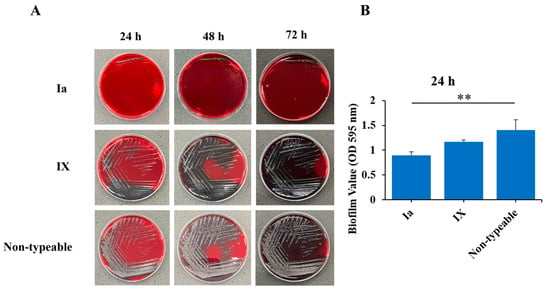
Biofilm-forming ability of selective S. agalactiae capsular genotypes including Ia, IX, and non-typeable strains was evaluated using Congo red agar (CRA) and the crystal violet staining assay. As shown in Figure 1A, the representative non-typeable isolate exhibited the strongest biofilm formation on CRA, followed by the representative genotype IX isolate, whereas the representative genotype Ia isolate showed the weakest biofilm-forming phenotype. Non-typeable isolate colonies displayed a typical black, dry, crystalline morphology indicative of strong biofilm production. In contrast, genotype IX isolate produced dark red to black colonies, suggesting moderate biofilm formation, while genotype Ia isolate formed smooth pink-to-red colonies, indicating weak biofilm formation (Figure 1A). Consistent with these observations, the crystal violet staining assay (Figure 1B) showed differences in biofilm biomass among the three representative isolates. The representative non-typeable isolate produced the highest biofilm biomass, with a mean OD value of 1.41 ± 0.21, followed by the representative genotype IX isolate (1.17 ± 0.03) and genotype Ia isolate (0.90 ± 0.07). Biofilm biomass was significantly higher in non-typeable isolate than in genotype Ia (p < 0.01), whereas no significant difference was observed between non-typeable and genotype IX isolates. These findings indicate that all three representative isolates were capable of biofilm formation, with the representative non-typeable isolate showing the highest mean biofilm biomass, although the difference compared with the genotype IX isolate was not statistically significant (Figure 1B).
Figure 1.
Biofilm formation by selected S. agalactiae capsular types Ia, IX, and non-typeable (NT). (A) Biofilm formation on Congo red agar after 24, 48, and 72 h of incubation. (B) Biofilm biomass was determined by crystal violet staining of biofilms formed in 96-well microplates after 24 h of incubation and quantified by measuring the absorbance at 595 nm (OD595). Data represent three independent experiments. Statistical significance was defined as ** p < 0.01.
3.4. Adhesion Ability to A549 Cells and HeLa Cells
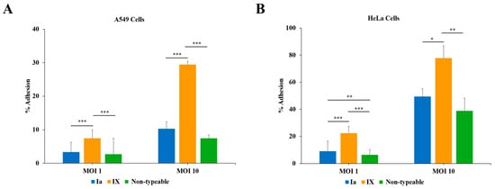
Adhesion of representative S. agalactiae isolates belonging to capsular types Ia, IX, and non-typeable was evaluated in A549 and HeLa cells at MOIs of 1 and 10. Adhesion efficiency increased with increasing MOI in both cell lines. The representative genotype IX isolate consistently exhibited significantly higher adhesion than the representative genotypes Ia and non-typeable isolates.
At MOI 1, the representative genotype Ia isolate adhered to A549 cells at 3.33 ± 2.89%, while the representative non-typeable isolate showed an adhesion efficiency of 2.72 ± 4.81%. In contrast, the representative genotype IX isolate showed significantly greater adhesion (7.44 ± 2.55%) than both genotype Ia and non-typeable isolates (p < 0.001) (Figure 2A). Similar results were observed in HeLa cells, where adhesion efficiencies were 9.00 ± 7.64%, 22.33 ± 5.00%, and 6.44 ± 3.85% for the representative genotypes Ia, IX, and the non-typeable isolates, respectively. The representative genotype IX isolate again exhibited significantly greater adhesion than the representative genotype Ia and non-typeable isolates (p < 0.001), while the representative genotype Ia isolate showed significantly adhesion than the non-typeable isolate (p < 0.01) (Figure 2B).
Figure 2.
Adhesion of selected S. agalactiae isolates of capsular types Ia, IX, and non-typeable (NT) to A549 and HeLa cells. (A) Adhesion of S. agalactiae to A549 cells at MOIs of 1 and 10. (B) Adhesion of S. agalactiae to HeLa cells at MOIs of 1 and 10. Adhesion was expressed as the percentage of cell-associated bacteria recovered 2 h post-infection relative to the initial inoculum. Data are presented as the mean ± SD of three independent experiments. Statistical significance is indicated as * p < 0.05, ** p < 0.01, and *** p < 0.001.
At an MOI of 10, the adhesion rates in A549 cells were 10.28 ± 2.08% for the representative genotype Ia isolate, 29.44 ± 0.96% for the representative genotype IX isolate, and 7.44 ± 1.00% for the representative non-typeable isolate. The representative genotype IX isolate showed significantly greater adhesion than the representative genotype Ia and non-typeable isolates (p < 0.001) (Figure 2A). In HeLa cells, adhesion was 49.44 ± 5.85%, 77.78 ± 9.18%, and 38.89 ± 9.18% for the representative genotypes Ia, IX, and non-typeable isolates, respectively. The representative genotype IX isolate showed significantly greater adhesion than genotype Ia (p < 0.05) and non-typeable isolates (p < 0.01) (Figure 2B).
3.5. Invasion Ability in A549 Cells and HeLa Cells
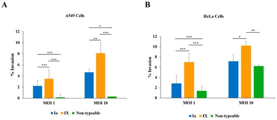
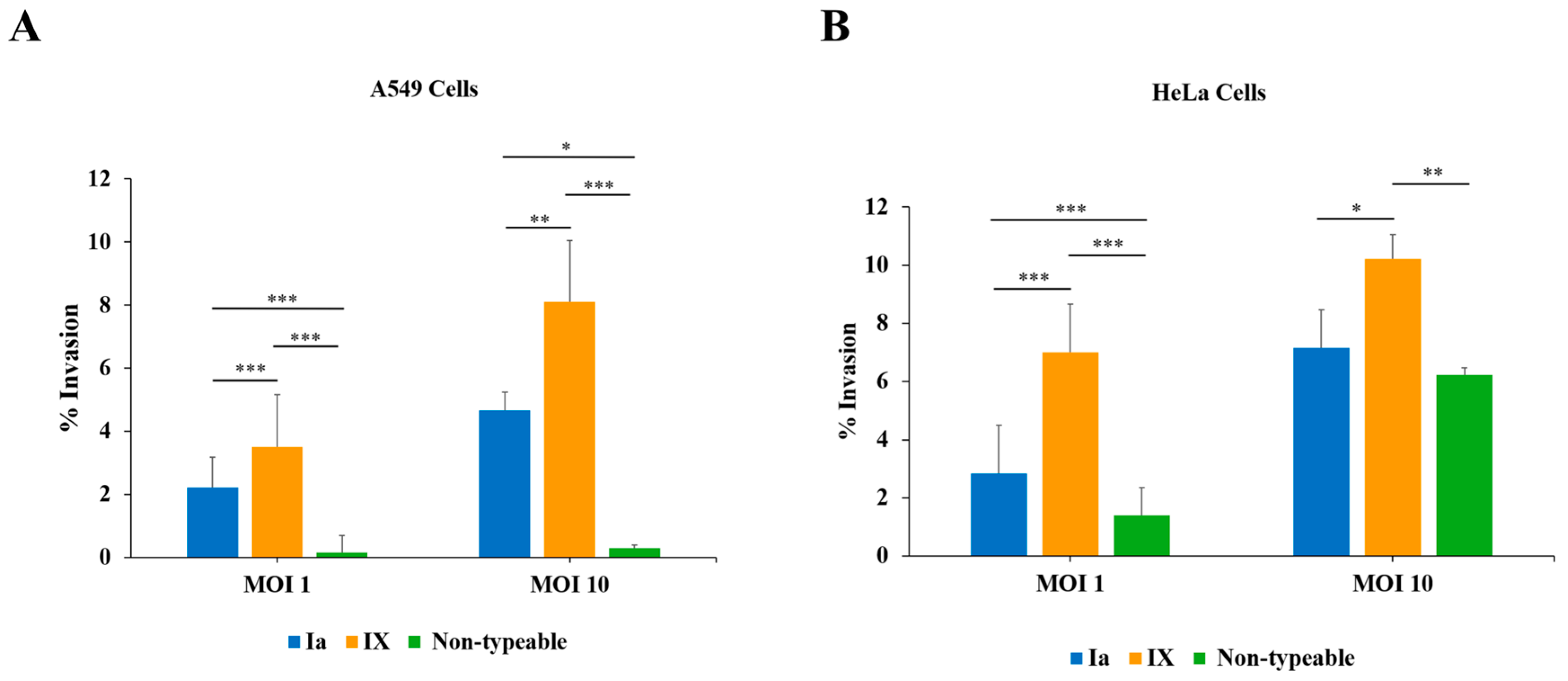
The invasion ability of representative S. agalactiae isolates belonging to capsular types Ia, IX, and non-typeable (NT) was evaluated in A549 and HeLa cells at MOIs of 1 and 10. The representative genotype IX isolate exhibited significantly higher invasion efficiency than the other isolates across both cell lines and MOIs. However, the magnitude and statistical significance of the differences among genotypes varied depending on the cell line and MOI.
At MOI of 1, in A549 cells, invasion efficiencies were 2.22 ± 0.96%, 3.50 ± 1.67%, and 0.16 ± 0.54% for the representative genotypes Ia, IX, and non-typeable, respectively. The representative genotype IX isolate exhibited significantly greater invasion than the representative genotype Ia and non-typeable isolates (p < 0.001). Additionally, the representative genotype Ia isolate showed significantly higher invasion than the representative genotype non-typeable isolate (p < 0.001) (Figure 3A). In HeLa cells, invasion efficiencies were 2.83 ± 1.67%, 7.00 ± 1.67%, and 1.39 ± 0.96% for the representative genotypes Ia, IX, and non-typeable, respectively. The representative genotype IX isolate exhibited significantly greater invasion than the representative genotype Ia and non-typeable isolates (p < 0.001). Similarly, the representative genotype Ia isolate exhibited significantly higher invasion than the representative genotype non-typeable isolate (p < 0.001) (Figure 3B).
Figure 3.
Invasion of selected S. agalactiae isolates of capsular types Ia, IX, and non-typeable (NT) in A549 and HeLa cells. (A) Invasion of S. agalactiae capsular types Ia, IX, and non-typeable (NT) in A549 cells at MOIs of 1 and 10. (B) Invasion of S. agalactiae capsular types Ia, IX, and non-typeable (NT) in HeLa cells at MOIs of 1 and 10. Invasion was expressed as the percentage of intracellular bacteria recovered 4 h post-infection relative to the initial inoculum. Data are presented as the mean ± SD of three independent experiments. Statistical significance is indicated as * p < 0.05, ** p < 0.01, and *** p < 0.001.
At MOI of 10, the invasion efficiencies in A549 cells were 4.67 ± 0.58%, 8.11 ± 1.93%, and 0.30 ± 0.10% for the representative genotypes Ia, IX, and non-typeable, respectively. The representative genotype IX isolate exhibited significantly greater invasion than the representative genotype Ia (p < 0.01) and non-typeable isolates (p < 0.001). Additionally, the representative genotype Ia isolate exhibited significantly greater invasion than the representative non-typeable isolate (p < 0.05) (Figure 3A). In HeLa cells, invasion efficiencies were 7.17 ± 1.30%, 10.22 ± 0.84%, and 6.22 ± 0.25% for the representative genotypes Ia, IX, and non-typeable isolates, respectively. The representative genotype IX isolate showed significantly greater invasion than the representative genotype Ia (p < 0.05) and non-typeable isolates (p < 0.01) (Figure 3B).
3.6. Cytotoxicity to A549 Cells and HeLa Cells
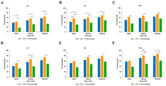
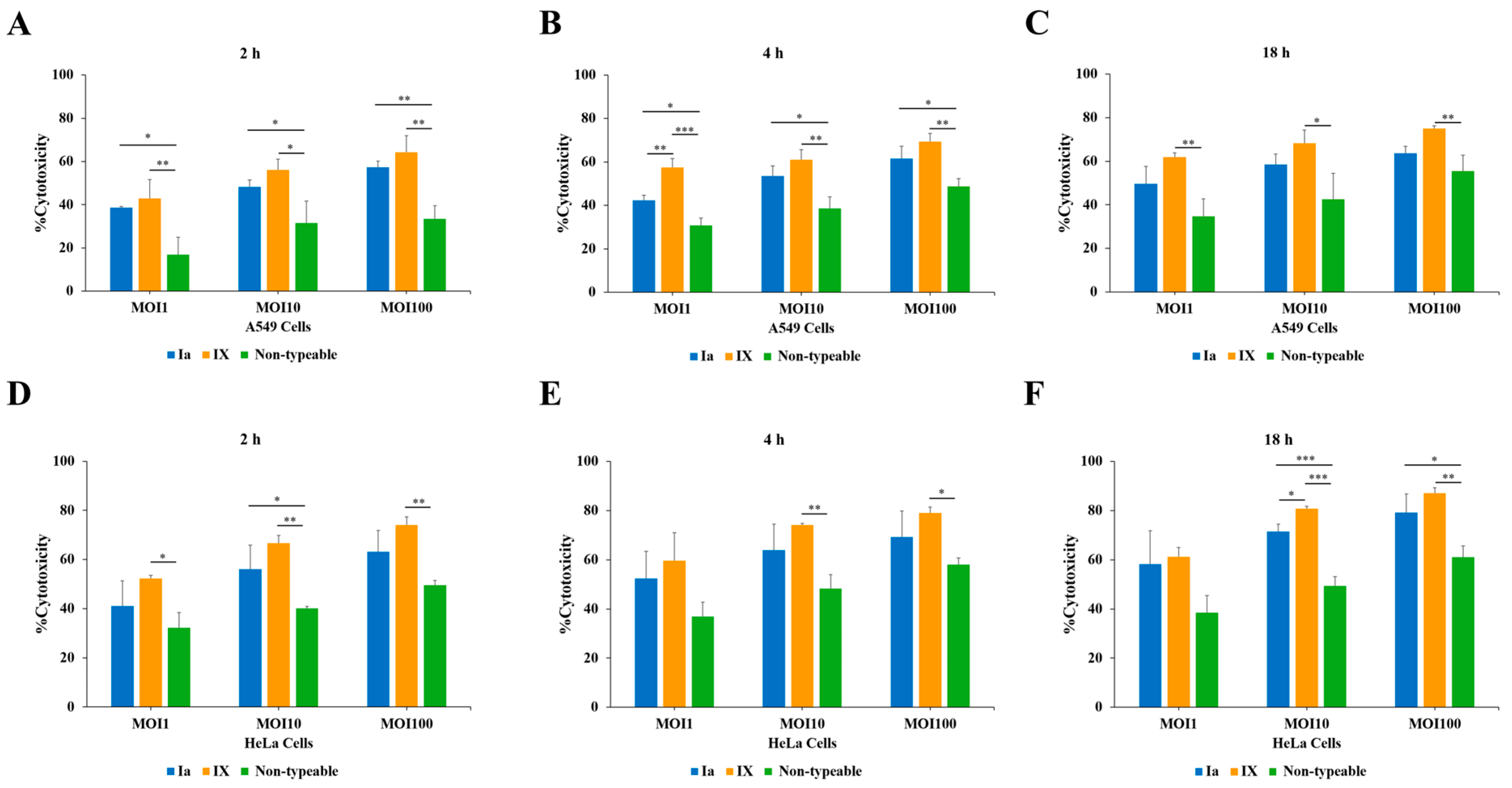
Cytotoxicity of representative S. agalactiae isolates of genotype types Ia, IX, and non-typeable was assessed using the CCK-8 assay at MOIs of 1, 10, and 100 for 2, 4, and 18 h of incubation. Cytotoxicity increased in a time- and MOI-dependent manner in both cell lines. Although the representative genotype IX isolate generally exhibited the highest cytotoxicity, the differences among the isolates were not statistically significant under all tested conditions (Figure 4).
Figure 4.
Cytotoxicity of selected S. agalactiae isolates of capsular types Ia, IX, and non-typeable (NT) toward A549 and HeLa cells determined using the CCK-8 assay. A549 cells were infected with each capsular type at MOIs of 1, 10, and 100. Cytotoxicity of A549 cells was determined after (A) 2 h, (B) 4 h, and (C) 18 h of incubation. Cytotoxicity of HeLa cells was determined after (D) 2 h, (E) 4 h, and (F) 18 h of incubation. Data are presented as the mean ± SD from three independent experiments. Statistical significance is indicated as * p < 0.05, ** p < 0.01, and *** p < 0.001.
In A549 cells after 2 h of incubation, the representative genotype Ia isolate induced cytotoxicity levels of 38.56 ± 0.53%, 48.22 ± 3.22%, and 57.27 ± 2.90%, while the representative non-typeable isolate induced cytotoxicity levels of 16.93 ± 7.91%, 31.48 ± 10.13%, and 33.53 ± 6.06%, at MOIs of 1, 10, and 100, respectively. The representative type IX isolate showed the highest cytotoxicity at MOIs of 1, 10, and 100 (42.89 ± 8.80%, 56.15 ± 4.98%, and 64.17 ± 7.70%, respectively). The representative genotype IX isolate exhibited significantly greater cytotoxicity than the representative non-typeable isolate at MOIs of 1 (p < 0.01), 10 (p < 0.05), and 100 (p < 0.01). Similarly, the representative genotype Ia isolate exhibited significantly greater cytotoxicity than the representative non-typeable isolate at MOIs of 1 (p < 0.05), 10 (p < 0.05), and 100 (p < 0.01) (Figure 4A).
After 4 h of infection in A549 cells, the representative genotype Ia isolate induced cytotoxicity levels of 42.41 ± 2.18%, 53.47 ± 4.72%, and 61.54 ± 5.66%, whereas the representative non-typeable isolate induced cytotoxicity levels of 30.75 ± 3.41%, 38.67 ± 5.34%, and 48.79 ± 3.52%, at MOIs of 1, 10, and 100, respectively. The representative genotype IX isolate exhibited higher cytotoxicity levels of 57.42 ± 4.18%, 60.92 ± 4.63%, and 69.30 ± 3.87% at MOIs of 1, 10, and 100, respectively. Its cytotoxicity were significantly higher than that of the representative genotype Ia isolate at MOIs (p < 0.01) and those of the representative non-typeable isolate at MOI of 1 (p < 0.001), and MOIs of 10 and 100 (p < 0.01). The representative genotype Ia isolate also exhibited significantly greater cytotoxicity than the representative non-typeable isolate at MOIs of 1, 10, and 100 (p < 0.05) (Figure 4B).
After 18 h of infection in A549 cells, the representative genotype Ia isolate induced cytotoxicity levels of 49.70 ± 7.94%, 58.48 ± 4.75%, and 63.64 ± 3.20% at MOIs of 1, 10, and 100, respectively. The representative non-typeable isolate induced cytotoxicity level of 34.67 ± 8.12%, 42.48 ± 11.91%, and 55.48 ± 7.39% at MOIs of 1, 10, and 100, respectively. In comparison, the representative type IX isolate induced significantly higher cytotoxicity level of 61.88 ± 1.92%, 68.23 ± 5.99%, and 75.02 ± 1.25% at the corresponding MOIs. Cytotoxicity induced by the representative IX isolate was significantly higher than that induced by the non-typeable isolate at an MOI of 1 (p < 0.01), 10 (p < 0.05), and 100 (p < 0.01) (Figure 4C).
In HeLa cells after 2 h of incubation, the representative genotype Ia isolate induced cytotoxicity levels of 41.12 ± 10.17%, 56.14 ± 9.70%, and 63.14 ± 8.76% at MOIs of 1, 10, and 100, respectively. In contrast, the representative non-typeable isolate induced cytotoxicity levels 32.25 ± 6.22%, 40.09 ± 0.89%, and 49.55 ± 1.94%, respectively. The representative genotype IX isolate exhibited the highest cytotoxicity at MOIs of 1, 10, and 100 with values of 52.30 ± 1.20%, 66.69 ± 3.12%, and 74.03 ± 3.34%, respectively. At an MOI of 1, the representative type IX isolate showed significantly higher cytotoxicity than the representative non-typeable isolate (p < 0.05). At an MOI of 10, the representative type IX isolate showed significantly higher cytotoxicity than the representative non-typeable isolate (p < 0.01). The representative genotype Ia isolate also induced significantly higher cytotoxicity than the representative non-typeable isolate (p < 0.05). At an MOI of 100, the representative IX isolate showed significantly higher cytotoxicity than the representative non-typeable isolate (p < 0.01) (Figure 4D).
At 4 h of incubation, the representative genotype Ia isolate induced cytotoxicity levels of 52.41 ± 11.03%, 63.93 ± 10.63%, and 69.25 ± 10.54% at MOIs of 1, 10, and 100, respectively. In contrast, the representative non-typeable isolate induced cytotoxicity levels in HeLa cells of 37.00 ± 5.74%, 48.25 ± 5.79%, and 58.07 ± 2.66%, respectively. The representative type IX isolate induced cytotoxicity levels of 59.66 ± 11.28%, 74.14 ± 0.68%, and 79.11 ± 2.29% at MOIs of 1, 10, and 100, respectively (Figure 4E). No significant differences in cytotoxicity were observed among the three representative isolates at an MOI of 1. At MOIs of 10 and 100, the representative type IX isolate exhibited significantly higher cytotoxicity than the representative non-typeable isolate (p < 0.01 and p < 0.05, respectively). However, no significant differences between the representative genotype IX and Ia isolates or between the representative genotype Ia and non-typeable isolates (Figure 4E).
After 18 h of incubation in HeLa cells, the representative genotype Ia isolate induced cytotoxicity levels of 58.20 ± 13.56%, 71.55 ± 2.93%, and 79.28 ± 7.45% at MOIs of 1, 10, and 100, respectively. The representative type IX isolate showed cytotoxicity levels of 61.27 ± 3.83%, 80.77 ± 0.95%, and 87.11 ± 2.13%, respectively, whereas the representative non-typeable isolate induced cytotoxicity levels of 38.55 ± 6.93%, 49.46 ± 3.67%, and 61.08 ± 4.55%, respectively. At an MOI of 1, no significant differences in cytotoxicity were observed among the three representative isolates. At an MOI of 10, the representative genotype IX isolate exhibited significantly greater cytotoxicity than the representative genotype Ia (p < 0.05) and non-typeable isolates (p < 0.001). Additionally, the representative genotype Ia isolate also showed significantly greater cytotoxicity than the representative non-typeable isolate (p < 0.001). At an MOI of 100, the representative genotype IX isolate exhibited significantly greater cytotoxicity than the representative non-typeable (p < 0.01). Similarly, the representative genotype Ia isolate also showed significantly higher cytotoxicity than the representative non-typeable isolate (p < 0.05) (Figure 4F).
3.7. Expression Profiling of the Virulence Factor Genes
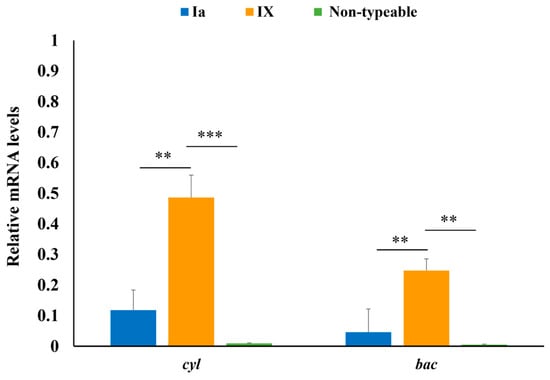
Transcript levels of cyl and bac genes were analyzed by qRT-PCR in the representative S. agalactiae isolates of genotype Ia (ID: E09), IX (ID: Q02), and non-typeable (ID: U03). The expression levels of both genes varied among the three representative isolates. For cyl, relative mRNA expression was highest in the representative genotype IX (0.486 ± 0.077), followed by the representative genotype Ia isolate (0.118 ± 0.066) and non-typeable isolate (0.009 ± 0.002). The representative genotype cyl transcript level in the representative genotype IX isolate was significantly higher than that in the genotype Ia isolate (p < 0.01) and the non-typeable isolate (p < 0.001). Similarly, bac expression was highest in the representative genotype IX isolate (0.247 ± 0.038), followed by the genotype Ia isolate (0.045 ± 0.073) and the non-typeable isolate (0.005 ± 0.001). The bac transcript level in the representative genotype IX isolate was significantly higher than those in the representative genotype Ia and non-typeable isolates (p < 0.01). These gene expression patterns were consistent with the observed cytotoxicity and adhesion profiles of the representative isolates (Figure 5).
Figure 5.
Relative mRNA expression levels of the cyl and bac genes in selected S. agalactiae isolates of capsular types Ia (ID: E09), IX (ID: Q02), and non-typeable (ID: U03). qRT-PCR analyzed gene expression. The results from three independent assays are presented as means ± SD; ** p < 0.01 and *** p < 0.001.
4. Discussion
Capsular typing revealed that genotype Ia was the predominant genotype in this study, accounting for 95.48% of isolates, followed by genotype IX (3.39%) and non-typable isolates (1.13%). Similar distributions have been reported in China. Lin et al. found that genotype Ia was the predominant genotype, accounting for 57.1% of isolates [39], and Yang et al. reported genotype Ia as predominant, accounting for 89% of isolates [1]. In contrast, a study conducted in Iran found that all 29 isolates identified as S. agalactiae were dominated by serotype III (41.37%, 12/29), followed by serotypes Ia, IV, and V, each accounting for 17.24% (5/29) of isolates [40]. In Brazil, six genotypes were identified among 59 S. agalactiae strains isolated from bovine mastitis. Genotype III was the most prevalent (38.98%, 23/59), followed by genotypes II (20.33%, 12/59), Ib (15.25%, 9/59), Ia (8.47%, 5/59), and IV (8.47%, 5/59); the remaining 8.47% (5/59) were non-typeable [41]. Among bovine S. agalactiae isolates from Germany, serotype Ia in combination with surface protein X was the predominant type (49.4%), followed by non-typeable isolates (40.5%) [42]. These comparisons indicate substantial geographic variation in the capsular genotype distribution of bovine S. agalactiae. Antibiotics remain the primary treatment for bovine mastitis. However, widespread and inappropriate use has increased concerns about antimicrobial residues and resistance, creating challenges for public health and mastitis management [43]. Intramammary administration of β-lactam antimicrobials has achieved high bacteriological cure rates [44,45], and the antimicrobial susceptibility profile of S. agalactiae requires continuous monitoring [12]. In the present study, S. agalactiae exhibited low resistance rates to ceftriaxone (1.13%) and levofloxacin (0.56%), and all isolates were susceptible (100%) to penicillin, vancomycin, erythromycin, and clindamycin. These findings suggest that the tested agents remain largely effective against S. agalactiae isolates from dairy cattle in Sakon Nakhon Province. Camsing et al. also reported complete susceptibility to levofloxacin and penicillin among isolates from Sakon Nakhon, but found resistance to tetracycline (77.03%, 57/74), and clindamycin (13.51%, 10/74), together with intermediate susceptibility to erythromycin (4.05%, 3/74) [13]. In contrast, Han et al. reported that 98.1% of bovine S. agalactiae isolates in China were resistant to penicillin, amoxicillin, ceftazidime, and ceftriaxone [46]. Taken together, the present findings and most previous reports indicate that bovine S. agalactiae generally remains highly susceptible to penicillin, vancomycin, erythromycin, and clindamycin, supporting their continued use as therapeutic options for bovine mastitis, although regional variation.
Antimicrobial resistance among bovine mastitis pathogens is an increasing concern because extensive antimicrobial use in dairy production may select for resistant bacteria and compromise treatment efficacy [47,48]. Biofilm formation is another important virulence mechanism that enhances bacterial survival by limiting antimicrobial penetration and protecting bacteria from host immune defenses. Consequently, biofilm-producing bacteria are therefore more likely to establish chronic and recurrent mastitis [49]. In the present study, the non-typable genotype isolate U03 exhibited resistance to levofloxacin and ceftriaxone and demonstrated a strong biofilm-forming capacity. Similar biofilm-forming capacity has been reported in bovine S. agalactiae isolates from other regions. In Poland, Kaczorek et al. found that some S. agalactiae isolates were predominantly weak biofilm producers (74%), with fewer moderate (11%) and strong (7.5%) producers [50]. Whereas in Brazil, Bonsaglia et al. found that all 145 S. agalactiae isolates from bovine subclinical mastitis produced biofilms, including 58.6% strong, 29.0% moderate, and 12.4% weak biofilm producers [51]. Another study similarly showed that biofilm production varied among strains and is influenced by environmental conditions such as pH, reflecting substantial phenotypic diversity in biofilm-forming capacity [52]. Consistent with these reports, all isolates in the present study formed biofilms, although biofilm biomass differed significantly among capsular genotypes. Non-typeable isolates exhibited the strongest biofilm-forming ability, followed by genotype IX and genotype Ia, supporting biofilm formation as a conserved virulence trait that may facilitate bacterial persistence in bovine intramammary infections [50].
The adhesion and invasion abilities of the representative S. agalactiae isolates also varied among the capsular categories tested. The genotype IX isolate exhibited the greatest adhesion and invasion in both A549 and HeLa cells, followed by the non-typable and genotype Ia isolates. This pattern was consistent with the bac mRNA transcript level of the adhesion gene by qPCR in S. agalactiae; type IX was significantly higher than the Ia and non-typable strains. Previous studies have demonstrated that adhesion of S. agalactiae is a multifactorial process mediated by various virulence factor genes, including bibA, bca, and bac [2]. Pang et al. reported that S. agalactiae isolates from cows with clinical and subclinical mastitis in China adhered strongly to bovine mammary epithelial cells (BMECs), with adhesion rates of 10.6–12.7% [53]. Similarly, Bonsaglia et al. reported that all representative bovine S. agalactiae isolates adhered to BMECs [51], although none showed detectable invasion under their experimental conditions. Regarding invasion, Guo et al. demonstrated that bovine mastitis-associated S. agalactiae invaded BMECs, with intracellular bacterial numbers increasing with MOI [54]. Sukhnanand et al. similarly reported that bovine S. agalactiae isolates invaded HeLa cells with no significant difference in invasion efficiency between bovine and human isolates [55]. Moreover, Tyrrell et al. reported that S. agalactiae exhibited greater invasion of HeLa epithelial cells than MRC-5 fibroblasts, with a serotype V strain showing particularly high invasive capacity [56].
Regarding cytotoxicity, the present study found that the representative genotype IX isolate exhibited significantly higher cytotoxicity toward both A549 and HeLa cells than the representative genotype Ia and non-typeable isolates. Previous studies have demonstrated that purified lipoteichoic acid (LTA) from a type III S. agalactiae strain induced cytotoxic effects on several human cell monolayers, including HeLa cells [57]. Additionally, S. agalactiae induced cytotoxicity and apoptosis in A549 cells through mechanisms involving reactive oxygen species generation, mitochondrial dysfunction, and caspase activation [24]. A study of bovine mastitis-associated isolates from Poland reported variable cytotoxicity across isolates and cell lines, including SK and Vero cells [23]. In the present study, the cytotoxicity pattern was consistent with the expression of the virulence-associated gene cyl, with significantly higher transcript levels in the genotype IX isolate than in the genotype Ia and non-typable isolates. Previous studies have implicated virulence-associated genes, including cyl, in S. agalactiae infection and tissue damage [2]. Previous functional studies have identified β-hemolysin/cytolysin encoded by cylE an important contributor to GBS-associated cellular injury [58]. Additionally, β-hemolysin/cytolysin, encoded within the cyl locus, contributes to epithelial cell injury [59], suggesting that differences in cyl expression may represent one possible mechanism underlying the variation in cytotoxicity observed among genotypes. Nevertheless, the present study did not directly evaluate apoptosis- or necrosis-associated markers, oxidative stress, downstream signaling pathways, or global changes in host-cell gene and protein expression. Further studies should therefore incorporate apoptosis and necrosis assays, oxidative-stress markers, time-resolved gene-expression analysis, transcriptomic or proteomic profiling, and pathway-specific functional experiments are warranted to determine whether increased cyl expression directly contributes to the enhanced cytotoxic phenotype of genotype IX representative isolates and to elucidate the underlying molecular mechanisms. Further studies involving multiple isolates from each capsular type, comprehensive sample collection not only in northeastern Thailand, perform additional validation of the reference gene for real-time expression studies in S. agalactiae, the use of bovine mammary epithelial cells to investigate virulence traits, and additional molecular approaches are also needed to clarify the mechanisms underlying S. agalactiae pathogenicity in bovine mastitis.
5. Conclusions
This study demonstrated that S. agalactiae isolates recovered from bovine mastitis in northeastern Thailand were susceptible to most of the antimicrobial agents tested but exhibited considerable variation in virulence-associated phenotypes among the three representative isolates examined. Capsular genotyping revealed that genotype Ia was predominant, followed by genotype IX and non-typeable isolates. Most isolates were susceptible to the antimicrobials tested, including penicillin, vancomycin, erythromycin, and clindamycin, with resistance detected only to ceftriaxone and levofloxacin at low frequencies. The representative non-typeable isolate exhibited the strongest biofilm-forming ability, followed by the representative genotype IX and type Ia isolates. The representative genotype IX isolate exhibited the highest cytotoxic, adhesion, and invasion capacities in epithelial cells, consistent with its higher mRNA expression levels of cyl and bac compared with those of the representative Ia and non-typable isolates. The contrasting phenotypic profiles indicate variation in virulence-associated characteristics among the three representative isolates examined. These findings provide baseline data on antimicrobial susceptibility and virulence-associated phenotypes of bovine S. agalactiae and support further investigation into the pathogenic characteristics of these isolates.
Author Contributions
T.B., A.A. and P.B.: Collected samples, Performed experiments, Investigation, Formal analysis; E.K. and S.Y.: Collected samples, Methodology, Validation; P.C., R.H. and A.K.: Formal analysis, Validation, Visualization; P.B.: Conceptualization, Funding acquisition, Supervision, Formal analysis, Validation, Writing—original draft, Writing—review and editing. All authors have read and agreed to the published version of the manuscript.
Funding
Funding for this research was provided in part by the Research Administration and Academic Service Division, Kasetsart University Chalermphrakiat Sakon Nakhon Province Campus, Sakon Nakhon, Thailand (Fiscal Year 2024) and the Kasetsart University Research and Development Institute [FF (KU) 11.70], Thailand.
Institutional Review Board Statement
The study was approval by the Kasetsart University Institutional Animal Care and Use Committee (ACKU66-ETC-006, 1 March 2024).
Informed Consent Statement
Not applicable.
Data Availability Statement
Data is contained within the article. Further inquiries can be directed to the corresponding author.
Conflicts of Interest
The authors declare no conflicts of interest.
References
- Yang, Y.; Liu, Y.; Ding, Y.; Yi, L.; Ma, Z.; Fan, H.; Lu, C. Molecular characterization of Streptococcus agalactiae isolated from bovine mastitis in Eastern China. PLoS ONE 2013, 8, e67755. [Google Scholar] [CrossRef] [Scilit] [PubMed]
- Wataradee, S.; Boonserm, T.; Samngamnim, S.; Ajariyakhajorn, K. Characterization of virulence factors and antimicrobial susceptibility of Streptococcus agalactiae associated with bovine mastitis cases in Thailand. Animals 2024, 14, 447. [Google Scholar] [CrossRef] [Scilit] [PubMed]
- Cobo-Ángel, C.; Jaramillo-Jaramillo, A.S.; Lasso-Rojas, L.M.; Aguilar-Marin, S.B.; Sanchez, J.; Rodriguez-Lecompte, J.C.; Ceballos-Márquez, A.; Zadoks, R.N. Streptococcus agalactiae is not always an obligate intramammary pathogen: Molecular epidemiology of GBS from milk, feces and environment in Colombian dairy herds. PLoS ONE 2018, 13, e0208990. [Google Scholar] [CrossRef] [Scilit] [PubMed]
- Seligsohn, D.; Crestani, C.; Gitahi, N.; Lejon Flodin, E.; Chenais, E.; Zadoks, R.N. Investigation of extramammary sources of Group B Streptococcus reveals its unusual ecology and epidemiology in camels. PLoS ONE 2021, 16, e0252973. [Google Scholar] [CrossRef] [Scilit] [PubMed]
- El Garch, F.; Youala, M.; Simjee, S.; Moyaert, H.; Klee, R.; Truszkowska, B.; Rose, M.; Hocquet, D.; Valot, B.; Morrissey, I. Antimicrobial susceptibility of nine udder pathogens recovered from bovine clinical mastitis milk in Europe 2015–2016: VetPath results. Vet. Microbiol. 2020, 245, 108644. [Google Scholar] [CrossRef] [Scilit] [PubMed]
- Keefe, G.; Chaffer, M.; Ceballos, A.; Londoño, M.; Jaramillo, M.; Toro, M. Effects of Streptococcus agalactiae on the Colombian dairy industry. In 3rd International Symposium on Mastitis and Milk Quality; National Mastitis Council: St. Louis, MO, USA, 2011; pp. 156–159. [Google Scholar]
- Zaatout, N.; Ayachi, A.; Kecha, M. Epidemiological investigation of subclinical bovine mastitis in Algeria and molecular characterization of biofilm-forming Staphylococcus aureus. Trop. Anim. Health Prod. 2020, 52, 283–292. [Google Scholar] [CrossRef] [Scilit] [PubMed]
- Kitila, G.; Kebede, B.; Wakgari, M. Prevalence, aetiology and risk factors of mastitis of dairy cows kept under extensive management system in west Wollega, western Oromia, Ethiopia. Vet. Med. Sci. 2021, 7, 1593–1599. [Google Scholar] [CrossRef] [Scilit] [PubMed]
- Mbindyo, C.M.; Gitao, G.C.; Mulei, C.M. Prevalence, etiology, and risk factors of mastitis in dairy cattle in Embu and Kajiado Counties, Kenya. Vet. Med. Int. 2020, 2020, 8831172. [Google Scholar] [CrossRef] [Scilit] [PubMed]
- Bi, Y.; Wang, Y.J.; Qin, Y.; Guix Vallverdú, R.; Maldonado García, J.; Sun, W.; Li, S.; Cao, Z. Prevalence of Bovine Mastitis Pathogens in Bulk Tank Milk in China. PLoS ONE 2016, 11, e0155621. [Google Scholar] [CrossRef] [Scilit] [PubMed]
- Almeida, A.; Alves-Barroco, C.; Sauvage, E.; Bexiga, R.; Albuquerque, P.; Tavares, F.; Santos-Sanches, I.; Glaser, P. Persistence of a dominant bovine lineage of group BS treptococcus reveals genomic signatures of host adaptation. Environ. Microbiol. 2016, 18, 4216–4229. [Google Scholar] [CrossRef] [Scilit] [PubMed]
- Kampa, J.; Sukolapong, V.; Buttasri, A.; Charoenchai, A. Prevalence of Mycoplasma bovis and other contagious bovine mastitis pathogens in bulk tank milk of dairy cattle herds in Khon Kaen Province, Thailand. Thai J. Vet. Med. 2009, 39, 275–280. [Google Scholar] [CrossRef] [Scilit]
- Camsing, A.; Phetburom, N.; Chopjitt, P.; Pumhirunroj, B.; Patikae, P.; Watwiengkam, N.; Yongkiettrakul, S.; Kerdsin, A.; Boueroy, P. Occurrence of antimicrobial-resistant bovine mastitis bacteria in Sakon Nakhon, Thailand. Vet. World 2024, 17, 1202–1209. [Google Scholar] [CrossRef] [Scilit] [PubMed]
- Kulkarni, A.G.; Kaliwal, B. Bovine mastitis: A review. Int. J. Recent Sci. Res. 2013, 4, 543–548. [Google Scholar]
- Liu, Y.; Liu, J. Group B Streptococcus: Virulence factors and pathogenic mechanism. Microorganisms 2022, 10, 2483. [Google Scholar] [CrossRef] [Scilit] [PubMed]
- Di Xia, F.; Mallet, A.; Caliot, E.; Gao, C.; Trieu-Cuot, P.; Dramsi, S. Capsular polysaccharide of Group B Streptococcus mediates biofilm formation in the presence of human plasma. Microbes Infect. 2015, 17, 71–76. [Google Scholar] [CrossRef] [Scilit] [PubMed]
- Schubert, A.; Zakikhany, K.; Pietrocola, G.; Meinke, A.; Speziale, P.; Eikmanns, B.J.; Reinscheid, D.J. The fibrinogen receptor FbsA promotes adherence of Streptococcus agalactiae to human epithelial cells. Infect. Immun. 2004, 72, 6197–6205. [Google Scholar] [CrossRef] [Scilit] [PubMed]
- Keefe, G. Update on control of Staphylococcus aureus and Streptococcus agalactiae for management of mastitis. Vet. Clin. Food Anim. Pr. 2012, 28, 216. [Google Scholar] [CrossRef] [Scilit] [PubMed]
- Wataradee, S.; Samngamnim, S.; Boonserm, T.; Ajariyakhajorn, K. Genotypic and antimicrobial susceptibility of Streptococcus agalactiae causing bovine mastitis in the central region of Thailand. Front. Vet. Sci. 2023, 10, 1250436. [Google Scholar] [CrossRef] [Scilit] [PubMed]
- Lechner, I.; Freivogel, C.; Stärk, K.D.; Visschers, V.H. Exposure pathways to antimicrobial resistance at the human-animal interface—A qualitative comparison of Swiss expert and consumer opinions. Front. Public Health 2020, 8, 558177. [Google Scholar] [CrossRef] [Scilit] [PubMed]
- Serwecińska, L. Antimicrobials and antibiotic-resistant bacteria: A risk to the environment and to public health. Water 2020, 12, 3313. [Google Scholar] [CrossRef] [Scilit]
- Hattab, S.; Ma, A.H.; Tariq, Z.; Vega Prado, I.; Drobish, I.; Lee, R.; Yee, R. Rapid Phenotypic and Genotypic Antimicrobial Susceptibility Testing Approaches for Use in the Clinical Laboratory. Antibiotics 2024, 13, 786. [Google Scholar] [CrossRef] [Scilit] [PubMed]
- Zastempowska, E.; Twarużek, M.; Grajewski, J.; Lassa, H. Virulence Factor Genes and Cytotoxicity of Streptococcus agalactiae Isolated from Bovine Mastitis in Poland. Microbiol. Spectr. 2022, 10, e02224-21. [Google Scholar] [CrossRef] [Scilit] [PubMed]
- Ferreira Eduardo da Costa, A.; Alfredo Moraes, J.; Silva Santos de Oliveira, J.; Hanthequeste Bittencourt dos Santos, M.; da Silva Santos, G.; Barja-Fidalgo, C.; Luiza Mattos-Guaraldi, A.; Emy Nagao, P. Reactive oxygen species involved in apoptosis induction of human respiratory epithelial (A549) cells by Streptococcus agalactiae. Microbiology 2016, 162, 94–99. [Google Scholar] [CrossRef] [Scilit] [PubMed]
- Barbosa, C.; Nogueira, S.; Gadanho, M.; Chaves, S. DNA extraction: Finding the most suitable method. In Molecular Microbial Diagnostic Methods; Elsevier: Amsterdam, The Netherlands, 2016; pp. 135–154. [Google Scholar]
- Kansaen, R.; Boueroy, P.; Hatrongjit, R.; Kamjumphol, W.; Kerdsin, A.; Chopjitt, P. The occurrence and characteristics of methicillin-resistant Staphylococcal isolates from foods and containers. Antibiotics 2023, 12, 1287. [Google Scholar] [CrossRef] [Scilit] [PubMed]
- Dmitriev, A.; Bhide, M.; Mikula, I. cpn60 gene-based multiplex-PCR assay for simultaneous identification of Streptococcal species. Acta Vet. Brno 2006, 75, 235–240. [Google Scholar] [CrossRef] [Scilit]
- Ke, D.; Ménard, C.; Picard, F.J.; Boissinot, M.; Ouellette, M.; Roy, P.H.; Bergeron, M.G. Development of conventional and real-time PCR assays for the rapid detection of group B streptococci. Clin. Chem. 2000, 46, 324–331. [Google Scholar] [CrossRef] [Scilit]
- Imperi, M.; Pataracchia, M.; Alfarone, G.; Baldassarri, L.; Orefici, G.; Creti, R. Amultiplex PCRassay for the direct identification of the capsular type (Ia to IX) of Streptococcus agalactiae. J. Microbiol. Methods 2010, 80, 212–214. [Google Scholar] [CrossRef] [Scilit] [PubMed]
- CLSI M100; Performance Standards for Antimicrobial Susceptibility Testing, 36th ed. Clinical and Laboratory Standards Institute: Wayne, PA, USA, 2026.
- Boueroy, P.; Phetburom, N.; Chareonsudjai, S.; Chopjitt, P.; Hatrongjit, R.; Payen, S.; Segura, M.; Gottschalk, M.; Kerdsin, A. Pathogenic characteristics of an unencapsulated Streptococcus suis serotype 31 strain isolated from a patient in Thailand. Sci. Rep. 2025, 15, 37788. [Google Scholar] [CrossRef] [Scilit] [PubMed]
- Bunthiang, T.; Sunontarat, S.; Phetburom, N.; Dulyasucharit, R.; Intharaksa, O.; Boonmars, T.; Kanokmedhakul, S.; Lekphrom, R.; Chopjitt, P.; Kerdsin, A. Antimicrobial Activity of Aurisin A Against Streptococcus suis and Its Protective Effect on Epithelial Cells. Int. J. Mol. Sci. 2026, 27, 1798. [Google Scholar] [CrossRef] [Scilit] [PubMed]
- Boueroy, P.; Brizuela, J.; Roodsant, T.J.; Wongsurawat, T.; Jenjaroenpun, P.; Chopjitt, P.; Hatrongjit, R.; Phetburom, N.; Chareonsudjai, S.; Boonmars, T.; et al. Genomic analysis and virulence of human Streptococcus suis serotype 14. Eur. J. Clin. Microbiol. Infect. Dis. 2025, 44, 639–651. [Google Scholar] [CrossRef] [Scilit] [PubMed]
- Yuan, F.; Zheng, L.; Wang, M.; Liu, W.; Li, X.; Gao, T.; Guo, R.; Liu, Z.; Yang, K.; Li, C.; et al. Study on the Effect of Phillyrin on Streptococcus suis In Vivo and In Vitro. Biomolecules 2024, 14, 1542. [Google Scholar] [CrossRef] [Scilit] [PubMed]
- Sunontarat, S.; Bunthiang, T.; Phetburom, N.; Chopjitt, P.; Saiboonjan, B.; Tippayawat, P.; Buddhisa, S.; Hatrongjit, R.; Nuanualsuwan, S.; Kerdsin, A.; et al. The sbp3 gene of Streptococcus suis plays an important role in biofilm formation, adhesion, invasion, and cytotoxicity in the human intestinal epithelial cell line. Microb. Pathog. 2026, 216, 108496. [Google Scholar] [CrossRef] [Scilit] [PubMed]
- Florindo, C.; Ferreira, R.; Borges, V.; Spellerberg, B.; Gomes, J.P.; Borrego, M.J. Selection of reference genes for real-time expression studies in Streptococcus agalactiae. J. Microbiol. Methods 2012, 90, 220–227. [Google Scholar] [CrossRef] [Scilit] [PubMed]
- Huang, R.; Li, M.; Ye, M.; Yang, K.; Xu, X.; Gregory, R.L. Effects of Nicotine on Streptococcus gordonii Growth, Biofilm Formation, and Cell Aggregation. Appl. Environ. Microbiol. 2014, 80, 7212–7218. [Google Scholar] [CrossRef] [Scilit] [PubMed]
- Li, S.; Li, W.; Liang, Q.; Cao, J.; Li, H.; Li, Z.; Li, A. Characterization and virulence of Streptococcus agalactiae deficient in SaeRS of the two-component system. Front. Microbiol. 2023, 14, 1121621. [Google Scholar] [CrossRef] [Scilit] [PubMed]
- Lin, L.; Huang, X.; Yang, H.; He, Y.; He, X.; Huang, J.; Li, S.; Wang, X.; Tang, S.; Liu, G.; et al. Molecular epidemiology, antimicrobial activity, and virulence gene clustering of Streptococcus agalactiae isolated from dairy cattle with mastitis in China. J. Dairy Sci. 2021, 104, 4893–4903. [Google Scholar] [CrossRef] [Scilit] [PubMed]
- HajiAhmadi, P.; Momtaz, H.; Tajbakhsh, E. Capsular Typing and Molecular Characterization of Streptococcus agalactiae Strains Isolated From Bovine Mastitis in Iran. Vet. Med. Sci. 2025, 11, e70275. [Google Scholar] [CrossRef] [Scilit] [PubMed]
- Carvalho-Castro, G.A.; Silva, J.R.; Paiva, L.V.; Custódio, D.A.; Moreira, R.O.; Mian, G.F.; Prado, I.A.; Chalfun-Junior, A.; Costa, G.M. Molecular epidemiology of Streptococcus agalactiae isolated from mastitis in Brazilian dairy herds. Braz. J. Microbiol. 2017, 48, 551–559. [Google Scholar] [CrossRef] [Scilit] [PubMed]
- Merl, K.; Abdulmawjood, A.; Lämmler, C.; Zschöck, M. Determination of epidemiological relationships of Streptococcus agalactiae isolated from bovine mastitis. FEMS Microbiol. Lett. 2003, 226, 87–92. [Google Scholar] [CrossRef] [Scilit] [PubMed]
- Hayes, K.; O’Halloran, F.; Cotter, L. A review of antibiotic resistance in Group B Streptococcus: The story so far. Crit. Rev. Microbiol. 2020, 46, 253–269. [Google Scholar] [CrossRef] [Scilit] [PubMed]
- Kaczorek, E.; Małaczewska, J.; Wójcik, R.; Rękawek, W.; Siwicki, A. Phenotypic and genotypic antimicrobial susceptibility pattern of Streptococcus spp. isolated from cases of clinical mastitis in dairy cattle in Poland. J. Dairy Sci. 2017, 100, 6442–6453. [Google Scholar] [CrossRef] [Scilit] [PubMed]
- Tomazi, T.; de Souza Filho, A.F.; Heinemann, M.B.; Santos, M.V.d. Molecular characterization and antimicrobial susceptibility pattern of Streptococcus agalactiae isolated from clinical mastitis in dairy cattle. PLoS ONE 2018, 13, e0199561. [Google Scholar] [CrossRef] [Scilit] [PubMed]
- Han, G.; Zhang, B.; Luo, Z.; Lu, B.; Luo, Z.; Zhang, J.; Wang, Y.; Luo, Y.; Yang, Z.; Shen, L.; et al. Molecular typing and prevalence of antibiotic resistance and virulence genes in Streptococcus agalactiae isolated from Chinese dairy cows with clinical mastitis. PLoS ONE 2022, 17, e0268262. [Google Scholar] [CrossRef] [Scilit] [PubMed]
- Bengtsson, B.; Unnerstad, H.E.; Ekman, T.; Artursson, K.; Nilsson-Öst, M.; Waller, K.P. Antimicrobial susceptibility of udder pathogens from cases of acute clinical mastitis in dairy cows. Vet. Microbiol. 2009, 136, 142–149. [Google Scholar] [CrossRef] [Scilit] [PubMed]
- Oliver, S.P.; Murinda, S.E. Antimicrobial Resistance of Mastitis Pathogens. Vet. Clin. N. Am. Food Anim. Pract. 2012, 28, 165–185. [Google Scholar] [CrossRef] [Scilit] [PubMed]
- Pedersen, R.R.; Krömker, V.; Bjarnsholt, T.; Dahl-Pedersen, K.; Buhl, R.; Jørgensen, E. Biofilm Research in Bovine Mastitis. Front. Vet. Sci. 2021, 8, 656810. [Google Scholar] [CrossRef] [Scilit] [PubMed]
- Kaczorek, E.; Małaczewska, J.; Wójcik, R.; Siwicki, A.K. Biofilm production and other virulence factors in Streptococcus spp. isolated from clinical cases of bovine mastitis in Poland. BMC Vet. Res. 2017, 13, 398. [Google Scholar] [CrossRef] [Scilit] [PubMed]
- Bonsaglia, E.C.R.; Rossi, R.S.; Latosinski, G.; Rossi, B.F.; Campos, F.C.; Junior, A.F.; Pantoja, J.C.F.; Rall, V.L.M. Relationship between Biofilm Production and High Somatic Cell Count in Streptococcus agalactiae Isolated from Milk of Cows with Subclinical Mastitis. Pathogens 2023, 12, 311. [Google Scholar] [CrossRef] [Scilit] [PubMed]
- Miranda, P.S.D.; Lannes-Costa, P.S.; Pimentel, B.A.S.; Silva, L.G.; Ferreira-Carvalho, B.T.; Menezes, G.C.; Mattos-Guaraldi, A.L.; Hirata, R., Jr.; Mota, R.A.; Nagao, P.E. Biofilm formation on different pH conditions by Streptococcus agalactiae isolated from bovine mastitic milk. Lett. Appl. Microbiol. 2018, 67, 235–243. [Google Scholar] [CrossRef] [Scilit] [PubMed]
- Pang, M.; Sun, L.; He, T.; Bao, H.; Zhang, L.; Zhou, Y.; Zhang, H.; Wei, R.; Liu, Y.; Wang, R. Molecular and virulence characterization of highly prevalent Streptococcus agalactiae circulated in bovine dairy herds. Vet. Res. 2017, 48, 65. [Google Scholar] [CrossRef] [Scilit] [PubMed]
- Guo, Z.; Ma, Y.; Jia, Z.; Wang, L.; Lu, X.; Chen, Y.; Wang, Y.; Hao, H.; Yu, S.; Wang, Z. Crosstalk between integrin/FAK, Crk/Vps25 governs invasion of bovine mammary epithelial cells by S. agalactiae. Iscience 2023, 26, 107884. [Google Scholar] [CrossRef] [Scilit] [PubMed]
- Sukhnanand, S.; Dogan, B.; Ayodele, M.O.; Zadoks, R.N.; Craver, M.P.; Dumas, N.B.; Schukken, Y.H.; Boor, K.J.; Wiedmann, M. Molecular subtyping and characterization of bovine and human Streptococcus agalactiae isolates. J. Clin. Microbiol. 2005, 43, 1177–1186. [Google Scholar] [CrossRef] [Scilit] [PubMed]
- Tyrrell, G.J.; Kennedy, A.; Shokoples, S.E.; Sherburne, R.K. Binding and invasion of HeLa and MRC-5 cells by Streptococcus agalactiae. Microbiology 2002, 148, 3921–3931. [Google Scholar] [CrossRef] [Scilit] [PubMed][Green Version]
- Goldschmidt, J.C.; Panos, C. Teichoic acids of Streptococcus agalactiae: Chemistry, cytotoxicity, and effect on bacterial adherence to human cells in tissue culture. Infect. Immun. 1984, 43, 670–677. [Google Scholar] [CrossRef] [Scilit] [PubMed]
- Doran, K.S.; Chang, J.C.W.; Benoit, V.M.; Eckmann, L.; Nizet, V. Group B Streptococcal β-Hemolysin/Cytolysin Promotes Invasion of Human Lung Epithelial Cells and the Release of Interleukin-8. J. Infect. Dis. 2002, 185, 196–203. [Google Scholar] [CrossRef] [Scilit] [PubMed]
- Pritzlaff, C.A.; Chang, J.C.; Kuo, S.P.; Tamura, G.S.; Rubens, C.E.; Nizet, V. Genetic basis for the beta-haemolytic/cytolytic activity of group B Streptococcus. Mol. Microbiol. 2001, 39, 236–247. [Google Scholar] [CrossRef] [Scilit] [PubMed]

Disclaimer/Publisher’s Note: The statements, opinions and data contained in all publications are solely those of the individual author(s) and contributor(s) and not of MDPI and/or the editor(s). MDPI and/or the editor(s) disclaim responsibility for any injury to people or property resulting from any ideas, methods, instructions or products referred to in the content. |
© 2026 by the authors. Licensee MDPI, Basel, Switzerland. This article is an open access article distributed under the terms and conditions of the Creative Commons Attribution (CC BY) license.