


Abstract
In recent years, outbreaks of inclusion body hepatitis (IBH) have been reported with increasing frequency worldwide. In Austria, the presence of fowl aviadenoviruses (FAdVs) and/or IBH has been documented sporadically. The present report describes a surge of natural IBH cases in fifteen Ross 308 broiler flocks. The clinical picture was characterized by the sudden onset of increased mortality in birds 3 to 15 days of age, persisting for approximately one week, resulting in cumulative flock mortality ranging from 2% to 27%. The main pathological findings were hepatitis with diffuse necrotic foci and petechial hemorrhages with intranuclear inclusion bodies by histology. FAdV was detected in liver samples from all affected flocks. Based upon partial hexon sequence analysis, FAdV serotype-11, species-D (FAdV-11, FAdV-D) was identified, showing complete sequence identity among broiler-derived strains and high identity with previous isolates from Austria and globally IBH-associated isolates. Furthermore, FAdV-11 DNA was detected in dead-in-shell embryos collected during the ongoing outbreak, originating from one suspect broiler breeder flock, confirming vertical transmission. Serological investigations of the suspect breeders confirmed the FAdV-11 infections within the studied timeframe. In conclusion, investigations confirmed vertical transmission of FAdV-11 and subsequent manifestation of IBH in broilers under field conditions, highlighting the epidemiological importance of broiler breeders in IBH outbreaks.
1. Introduction
Fowl aviadenoviruses (FAdV) of the family Adenoviridae and genus Aviadenovirus are grouped into five distinct species (FAdV-A to FAdV-E) based on molecular criteria, and into twelve serotypes (FAdV-1 to -8a, and -8b to -11) based on cross-neutralization tests [1,2]. The role of FAdVs as primary pathogens has been documented in the context of natural outbreaks of inclusion body hepatitis (IBH), hepatitis-hydropericardium syndrome, and adenoviral gizzard erosion [3].
IBH is mainly caused by the serotypes FAdV-2, FAdV-11 (species Fowl aviadenovirus D), FAdV-8a, and FAdV-8b (species Fowl aviadenovirus E), that characteristically affect broilers up to five weeks of age [3,4,5]. Historically, IBH was first described in poultry in the USA by Helmboldt and Frazier [6]. Although IBH has been associated with immunosuppressive co-infections such as infectious bursal disease virus (IBDV) or chicken anemia virus (CAV) [7,8], disease outbreaks resulting in significant economic losses, as well as successful experimental reproduction of IBH, have demonstrated the virulence and primary causative role of certain FAdVs [9,10,11,12,13,14]. Furthermore, field studies have highlighted the negative impact of subclinical FAdV infections in poultry [15,16].
Horizontal transmission of FAdV, which occurs mainly via the fecal-oral route, plays an important role in the spread of FAdV within and between flocks in combination with inadequate on-farm biosecurity measures [12,16]. However, vertical transmission of FAdV, which was originally discovered and described as an endogenous virus of the egg based on its isolation from embryos inoculated with an unrelated pathogen, is one of its main biological characteristics [17]. The occurrence of transovarian transmission of FAdV-8 was confirmed by direct detection of viral antigen in fertile eggs from a previously IBH-infected and sero-positive broiler breeder flock [13]. Up to now, there have been several reports of IBH outbreaks in very young chicks, hypothesizing that vertical transmission of FAdV and induction of the disease must have occurred [18,19,20]. Unfortunately, in many cases, there is no substantial evidence to confirm this phenomenon. However, as early horizontal exposure cannot be ruled out under field conditions, direct evidence of the presence of FAdV in fertilized eggs or dead-in-shell embryos, together with molecular comparison of breeder- and broiler-derived strains, is crucial to verify vertical transmission.
In recent years, reports of IBH outbreaks have increased worldwide [3]. As a prophylactic measure, broiler breeders around the world are frequently vaccinated against prevalent FAdV serotypes to prevent vertical transmission and to protect their progenies against clinical disease outbreaks via maternal antibodies [3]. In Austria, the presence of FAdV-D and -E strains has been documented by molecular investigations, and IBH has been sporadically diagnosed on the basis of pathohistological investigations, with the first cases already published in 1974 [3,11,21]. However, it is important to note that the local parent flocks are generally not vaccinated against FAdV. Therefore, they and their progeny may be susceptible to infection throughout production.
The present study investigated the role of vertical transmission during natural outbreaks of IBH in fifteen flocks of young broiler chickens, mainly linked to the same hatchery and the same parent flock. Clinical data were analyzed, and necropsy, histological, virological, and serological investigations were performed.
2. Materials and Methods
2.1. Case History and Flock Data
From December 2023 to February 2024, a sudden onset of increased mortality that lasted for approximately one week was noticed in 15 broiler flocks (Ross 308) between 3 and 15 days of age. Clinically, birds presented with non-specific clinical signs, in particular apathy, ruffled feathers, and huddling. No additional clinical signs were observed. Broilers in the affected flocks originated from the same commercial hatchery, and most of the birds could be traced back to one or more of seven suspect parent flocks (Table 1). The broilers had received a standard vaccination program at the hatchery with an infectious bronchitis vaccine (Nobilis® IB Ma5; MSD, Munich, Germany) via coarse spray. Additionally, several flocks received vaccination with infectious bursal disease vaccine (AviPro Gumboro vac., Lohmann Animal Health GmbH, Cuxhaven, Germany—flocks 5, 8, 10, 12, 13; Nobilis® D78, MSD—flock 11; or Hipragumboro®, Hipra Laboratories, Amer, Spain—flock 14) via drinking water between day 14 and 16 of life; in general, after the onset of IBH. Birds (flocks 2, 4, 6, 7, and 9) or organ samples (flocks 1, 3, 5, 8, 10 to 15) were submitted to the Clinic for Poultry and Fish Medicine, University of Veterinary Medicine, Vienna, Austria, for routine diagnostics.
Table 1.
Broiler flocks and their relation to parent stock involved in the IBH outbreak in Austria between December 2023 and February 2024.
2.2. Broiler: Post-Mortem Examinations, Sampling, and Histopathological Investigations
From flocks 2, 4, 6, 7, and 9, up to seven dead birds were submitted to the Clinic for Poultry for post-mortem examination, and routine necropsy was performed. Gross pathological changes were documented, and liver samples were aseptically collected for laboratory examinations.
For bacteriological analysis, liver samples were plated directly onto Columbia agar containing 5% sheep blood (bioMérieux, Vienna, Austria) and MacConkey agar (Neogen®, Lansing, MI, USA), then incubated at 37 °C for 24 h.
Furthermore, liver samples were kept frozen at −20 °C for virus isolation and PCR and were fixed for 24 h in 10% buffered formalin before being embedded in paraffin. Sections 3 µm thick were prepared using a Microm HM 360 microtome (Microm Laborgeräte GmbH, Walldorf, Germany). Subsequently, samples were mounted on glass slides, and routine staining with hematoxylin and eosin was performed. In addition, thymus, spleen, bone marrow, and/or bursa were collected and pooled for PCR detection of immunosuppressive viruses (see molecular diagnostics).
On the remaining farms, necropsy and sampling (liver swabs) were carried out on-site. All samples were obtained during post-mortem investigations or as part of routine veterinary practice and surveillance of flocks. No animals were euthanized specifically for this study, and according to national legislation, ethical approval was not required for such kind of samples.
2.3. Sampling of Broiler Breeder Flocks
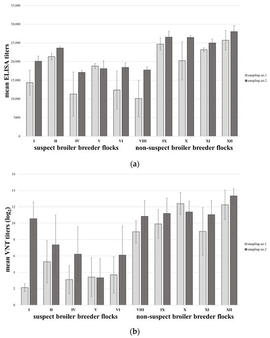
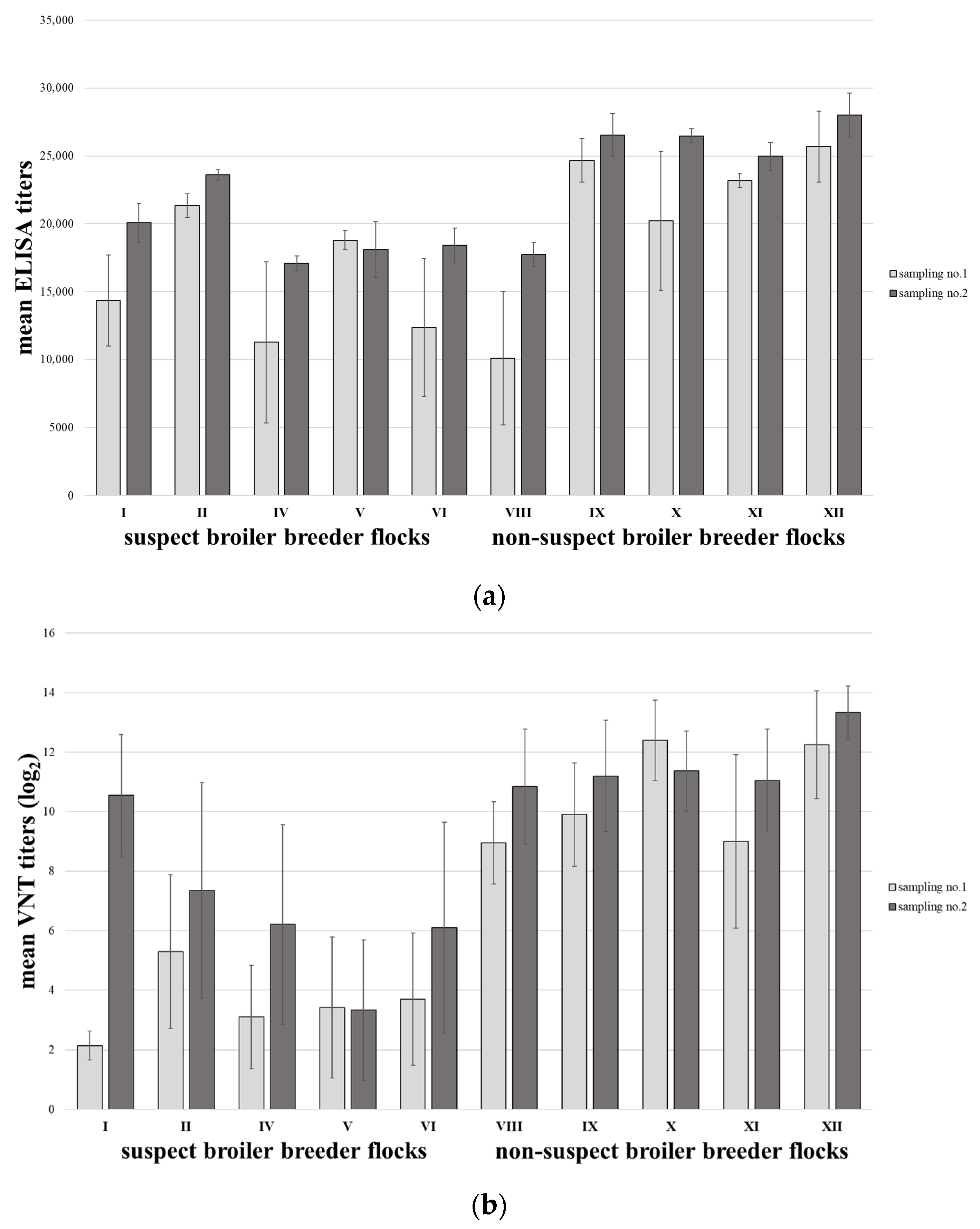
Paired blood samples were collected for serological testing from five of the suspect broiler breeder flocks and five contemporary non-suspect broiler breeder flocks with no connection to the outbreak. The initial samples were taken up to five months before (flocks I, IV, V, and VI as well as non-suspect flocks VIII to XII) or early during the onset of IBH in the broilers (flock II). The second sampling took place contemporaneously with the end of the IBH outbreak. Blood samples were tested by commercial enzyme-linked immunosorbent assay (ELISA) and by virus neutralization test (VNT).
To investigate vertical transmission, cloacal swabs were taken at the hatchery during the ongoing outbreak from day-old chicks of the suspected broiler breeder flocks I to VII. In addition, dead-in-shell (DIS) embryos from the suspected breeder flocks I and III were available for virus detection. They were subjected to post-mortem examination, and liver samples were taken, pooled, and kept frozen at −20 °C until virus isolation and PCR were performed.
2.4. Virus Isolation
Virus isolation was performed on primary chicken embryo liver (CEL) cells, prepared from 14-day-old SPF chicken embryos (VALO; Lohmann Tierzucht GmbH, Cuxhaven, Germany) according to a method described by Sellers & Schat [22].
For this purpose, 20% homogenates of liver samples (from broilers and dead-in-shell chicks) were prepared with an antibiotic-phosphate-buffered saline solution (1 mg/mL streptomycin and 100,000 IU/mL penicillin). Cloacal swabs (from day-old progeny chicks) were placed in 1 mL of an antibiotic-phosphate-buffered saline solution. Samples were filter sterilized using syringe filters with a pore size of 0.2 μm (VWR, Vienna, Austria). Nearly confluent CEL cells were inoculated with filtered sample material, and the cultures were monitored for up to 5 days post-infection. A sample was considered positive if a cytopathic effect was observed within three passages.
2.5. DNA Extraction, PCR, and Sequence Analysis
Viral DNA was extracted from liver swabs, liver samples, and cell culture supernatants using the IndiSpin Pathogen Kit (Indical Bioscience, Leipzig, Germany), according to the manufacturer’s instructions. For amplification of the FAdV hexon gene loop 1 region, a protocol described by Meulemans et al. [23] was followed. Selected liver samples (n = 10) were then subjected to fiber gene amplification with FAdV-D-specific primer sets in accordance with the protocol of Schachner et al. [21].
The amplification products from both PCRs were analyzed by gel electrophoresis using a 1.5% agarose gel (Biozym, Vienna, Austria). Fragments of the correct length were excised from the gel, purified using the QIAquick gel extraction kit (Qiagen, Hilden, Germany), and Sanger sequenced by LGC Genomics GmbH (Berlin, Germany). Assembly and analyses were performed with Accelrys Gene, version 2.5 (Accelrys Inc., San Diego, CA, USA) and DNASTAR Lasergene v18 (DNASTAR, Madison, WI, USA) software. Multiple-sequence alignments of Hex L1 and FAdV-D fiber nucleotide sequences were performed using ClustalW in Accelrys Gene, v2.5 software. Molecular typing of the FAdV field strains was accomplished by phylogenetic analysis based on a hexon gene segment corresponding to nt172-857 of AF339914, together with FAdV reference strains representing all species and serotypes. Phylogeny was computed by the Neighbor Joining (BIONJ) approach as implemented in the MegAlign Pro module of Lasergene v18 software (DNASTAR).
Additional investigations included the screening of broiler samples for chicken anemia virus (CAV) and infectious bursal disease virus (IBDV) using standard laboratory RT-real-time and conventional PCR protocols [24,25].
2.6. Serological Investigations
Investigations with ‘‘FAdV ELISA’’ (BioChek Bv, Reeuwijk, The Netherlands) were carried out and evaluated according to the manufacturer’s instructions. For the VNT, sera were inactivated for 30 min at 56 °C in a thermomixer (Eppendorf, Hamburg, Germany). The VNT was performed according to a constant virus diluted serum method using 100 µL of 100 median tissue culture infective dose/100 µL FAdV-11 (strain 380) [26]. The cells were incubated for 5 days at 37 °C under 5% CO2 conditions and were investigated for a cytopathic effect. An antibody titer ≤ 3 log2 was regarded as negative.
3. Results
3.1. Broiler: Production Data
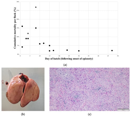
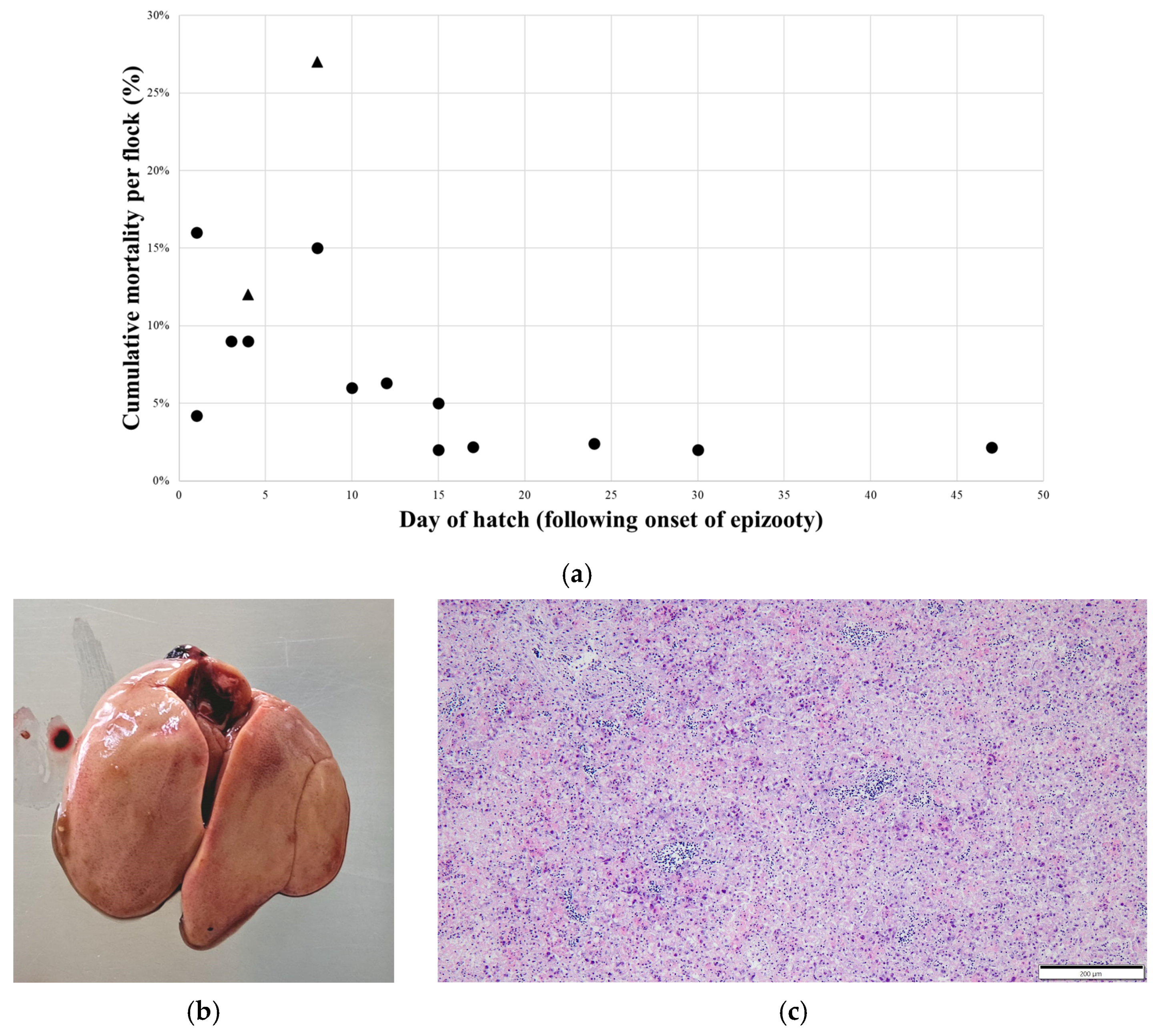
During the IBH outbreak, an increased mortality rate was observed in the affected broiler flocks over a period of approximately 6 to 8 days. Cumulative flock mortality varied from 2% to 27% among the 15 flocks investigated. As shown in Figure 1a, higher cumulative mortality rates were observed in the flocks that hatched soon after the onset of the IBH outbreak, compared to the flocks that were affected at a later stage. Overall, surviving birds from affected flocks were found to be on target body weight for slaughter, and condemnation rates at slaughterhouses were not significantly increased (≤1%).
Figure 1.
Clinical data and pathologic presentation of IBH cases. (a) Chronological representation of cumulative flock mortality of IBH outbreak (triangles represent flocks with concurrent IBDV infection). (b) Broiler liver showing a mottled surface with diffuse necrotic foci and petechial hemorrhages. (c) Histopathology of a liver sample showing multifocal areas of degeneration, inflammation, and necrosis with intranuclear inclusion bodies in hepatocytes (bar = 200 µm).
3.2. Broiler: Pathomorphological Changes, Microbiological and Molecular Diagnostics
Swollen and friable livers with a mottled surface of diffuse necrotic foci and petechial hemorrhages were observed on post-mortem examination of broilers (Figure 1b). Some broilers had necrotic foci on the surface of the pancreas and pale, edematous kidneys. There were no significant lesions noted in other organs.
Histopathology showed multifocal areas of degeneration and necrosis in the liver samples of affected broilers. Large intranuclear inclusion bodies were observed in the nuclei of degenerating and necrotizing hepatocytes (Figure 1c). Additionally, focal areas of inflammation demonstrated by the accumulation of mononuclear cells and heterophils were detected in the liver parenchyma.
No pathogenic bacteria were isolated from the liver samples.
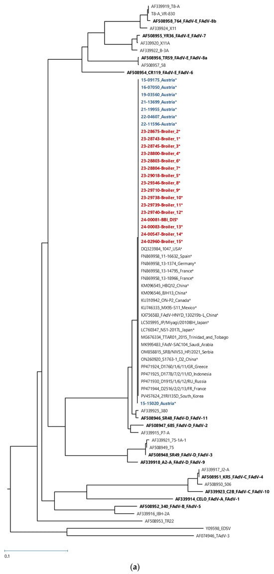
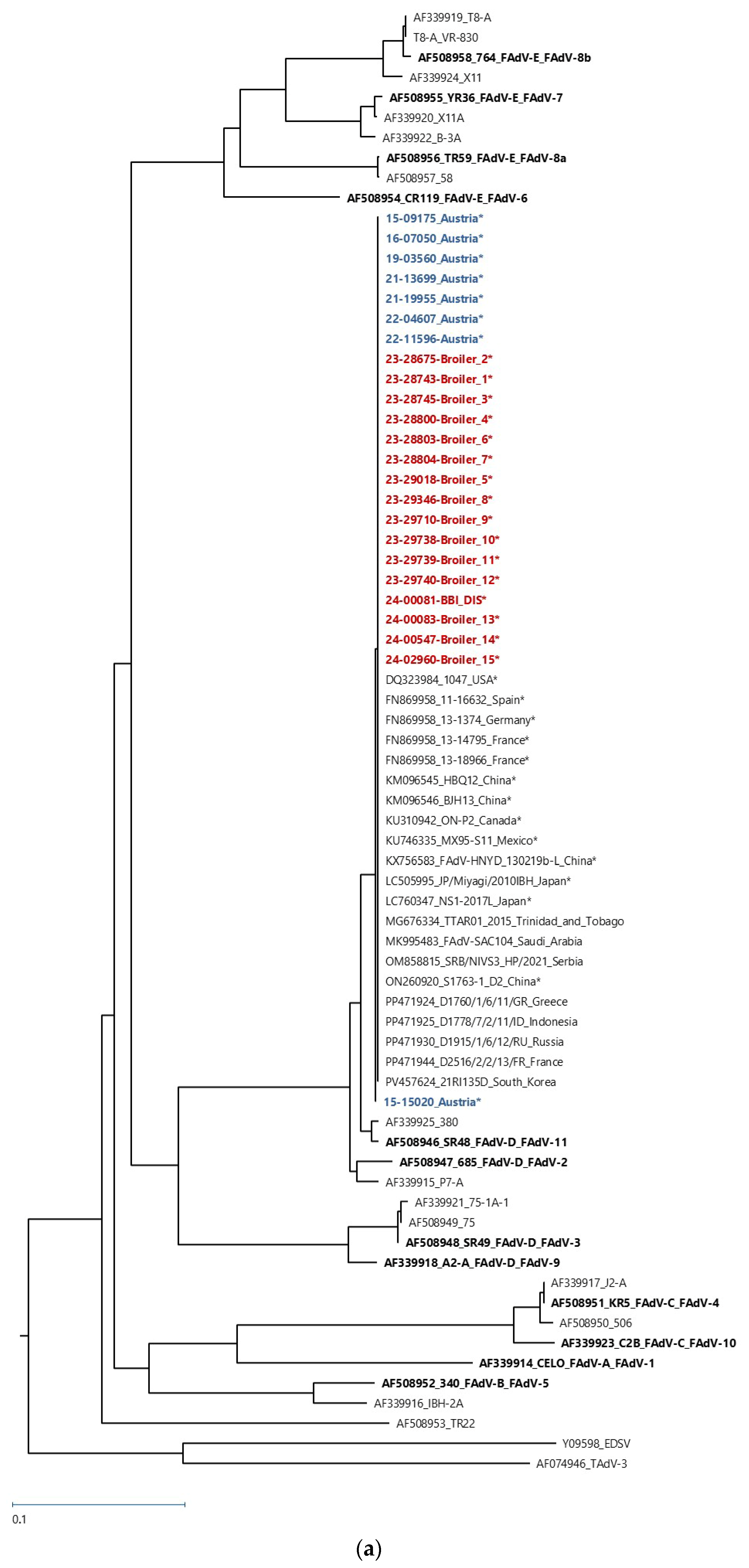
In all 15 affected broiler flocks, FAdV was detected in the liver by virus isolation and/or PCR. Sequencing of positive PCR products, including the L1 loop region of the hexon gene, showed nucleotide sequences of the broiler-derived strains genetically identical. Phylogenetic analysis based on the hexon gene using available GenBank sequences of reference strains FAdV-1 to -8a and -8b to -11 identified them as FAdV-D, serotype 11, sharing 97.6% nucleotide identity with the reference strain 380 available from GenBank (Figure 2a). Inclusion of eight additional FAdV-D hexon sequences from Austrian broiler farms (2015–2022), suffering from IBH, revealed complete sequence identity, demonstrating persistent circulation of a single FAdV-D serotype 11 strain in Austrian flocks since at least 2015. Overall, these findings confirm that FAdV-11 was the causative agent of the investigated IBH outbreaks and that the detected isolates were genetically related to previously identified regional isolates.
Figure 2.
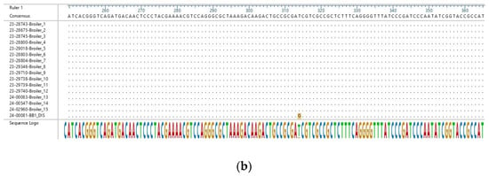
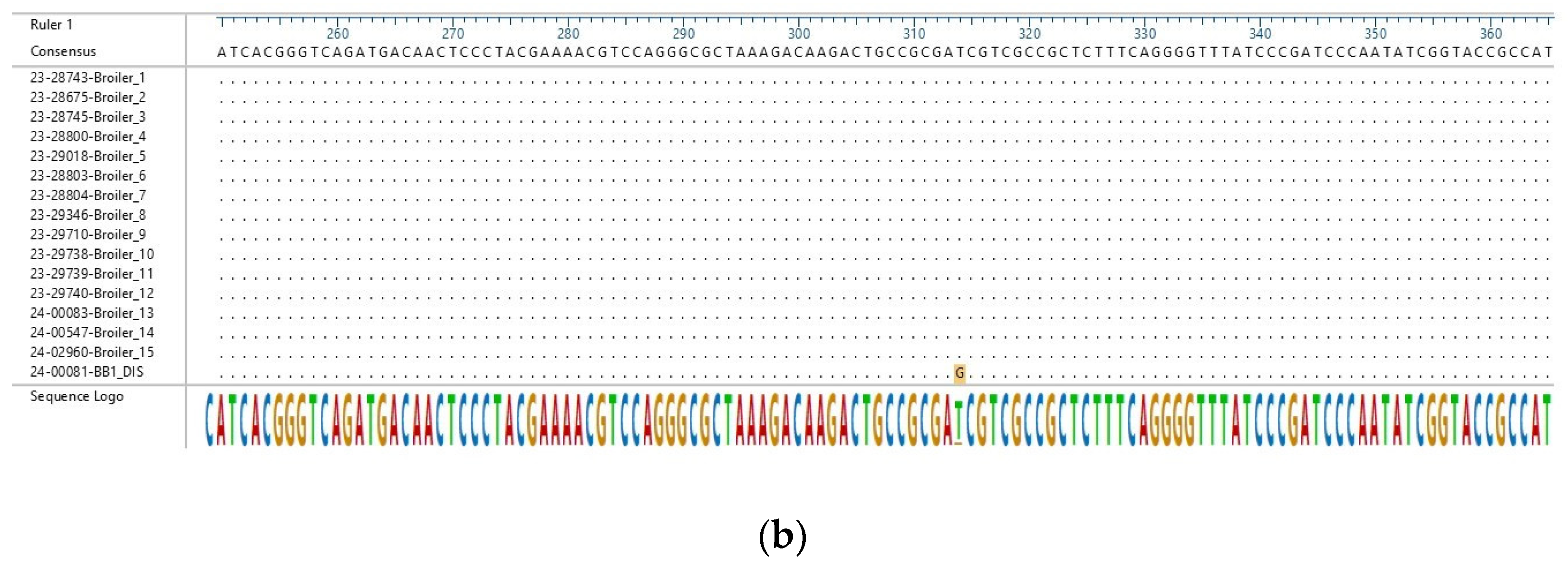
Molecular typing of FAdV in broiler flocks 1 to 15 and in dead-in-shell (DIS) embryos from broiler breeder flock I. (a) The phylogenetic analysis was performed by the Neighbor Joining approach (BIONJ) and was based on the alignment of 583 nucleotides of the hexon gene (L1 loop). The unrooted tree is shown. This analysis included 70 nucleic acid sequences. All sequences not derived in the present study were downloaded from the NCBI GenBank and are labeled with the accession number followed by the name of the strain and the geographical location in which the isolate was obtained. Sequences serving as references for each FAdV species and sero-/genotype are labeled in bold and corresponding FAdV species/FAdV-sero-/genotype designation. All sequences derived in the present study are labeled in bold; samples colored in red originate from broiler flocks 1 to 15 and DIS embryos from broiler breeder flock I (BBI_DIS). Samples colored in blue originate from previous FAdV-D-positive diagnostic samples from Austria. Sequences from isolates connected with IBH are labeled with an asterisk (*). (b) Comparison of the partial hexon nucleic acid sequences of fowl adenovirus D serotype 11 detected in the present study in broiler flocks 1 to 15 and in dead-in-shell (DIS) embryos from broiler breeder flock I. The partial alignment showing the position of the single-nucleotide polymorphism is presented. The ruler is based on the AF339925.
The analyzed portions of the fiber gene shared 99.9–100% nucleotide homology with each other and 95.1% with the nucleotide sequence of the FAdV-11 reference strain 380 (Supplementary Figure S1). However, a slightly higher nucleotide sequence identity range of 95.6–95.8% was noticed with the FAdV-2 fiber gene. Both serotypes belong to species FAdV-D, and a high level of cross-neutralization, in addition to high sequence identity, supports their close evolutionary relationship [27].
Testing for viral co-infections revealed two flocks positive for IBDV (nos.4 and 6). CAV was not detected in samples taken from IBH-affected broilers of any of the 15 farms.
3.3. Broiler Breeder: Results of Hatchery Samples
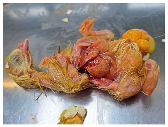
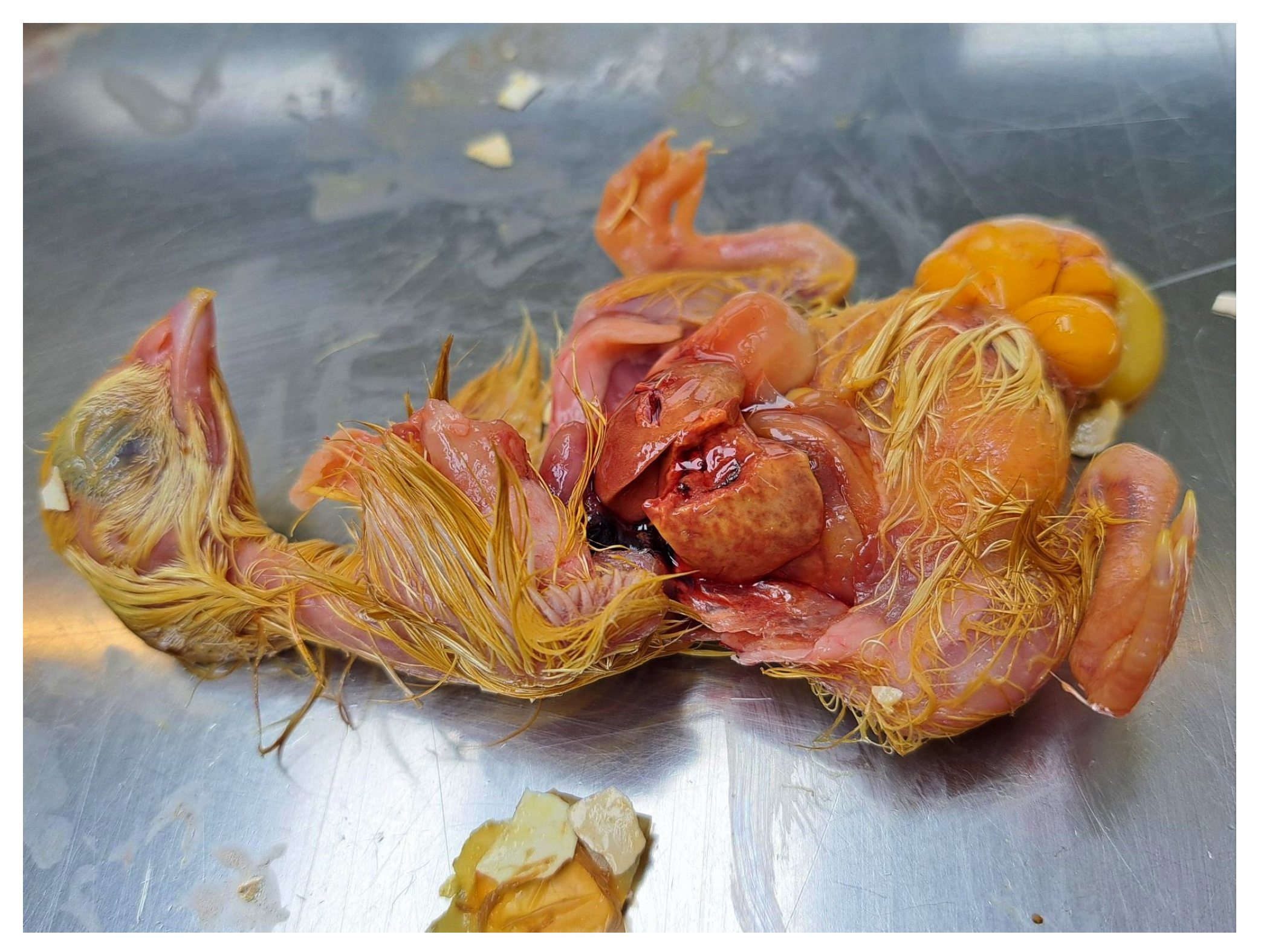
Some investigated DIS embryos had friable livers with a mottled surface of diffuse necrotic foci and petechial hemorrhages (Figure 3). While virus isolation was negative, FAdV was detected by PCR from liver samples of DIS chicks originating from broiler breeder flock I. Sequence analysis of the hexon gene PCR product identified the sequence as FAdV-D, serotype-11. The obtained sequence showed a single nucleotide polymorphism at position 314 when compared to the broiler samples (99.9% sequence identity) (Figure 2b). The presence of FAdV-11 DNA in DIS embryos is indicative of vertical transmission, linking breeder infection to disease in the progeny.
Figure 3.
Dead-in-shell embryo originating from breeder flock I. Liver showed a mottled surface with diffuse necrotic foci and petechial hemorrhages.
Samples from DIS embryos from breeder flock III and the cloacal swabs from day-old progeny chicks from suspected broiler breeder flocks were all negative.
3.4. Broiler Breeder: Serology Results
All serum samples investigated by ELISA were positive with high titers (Supplementary Table S1). When comparing the mean ELISA titers from all suspected and non-suspected local broiler breeders, an increase was generally observed between the first and second sampling. However, suspect broiler breeders had lower mean titers compared with non-suspect flocks (15,629 to 19,454 and 20,781 to 24,753, respectively; Figure 4a).
Figure 4.
Bar plot presentation of serology results from broiler breeder flocks at sampling 1 and 2. (a) Mean ELISA titers per flock and (b) mean VNT titers per flock. VNT titers ≤ 3 log2 are considered negative. Broiler breeder flocks I, II, IV, V, and VI were suspected to be involved in vertical transmission in the IBH outbreak. Non-suspect broiler breeder flocks VIII to XII were contemporary local flocks with no connection to the outbreak.
Using VNT, 24 out of 80 serum samples from suspected broiler breeder flocks were positive for neutralizing antibodies against FAdV-11 at the first sampling. One-third of these (eight out of ten positive samples with low neutralizing titers against FAdV-11) were detected in broiler breeder flock II, which was initially sampled early during the IBH outbreak. The overall mean titers of suspected broiler breeder flocks at the first sampling were 3.53 ± 1.02. At the second sampling, 70 out of 100 samples from suspected breeder flocks were positive with an overall mean titer of 6.71 ± 2.33; however, broiler breeder flock V remained largely negative (Figure 4b). These serological findings indicate an ongoing infection in suspect breeder flocks during the outbreak period.
In contrast, virus-neutralizing antibodies against FAdV-11 were detected in 164 out of 165 samples from the non-suspect broiler breeder flocks. Overall, mean titers were consistently high with 10.50 log2 and 11.56 log2 at the first and second sampling, respectively.
4. Discussion
The present study describes IBH outbreaks over an eight-week period in fifteen broiler flocks in Austria. Consistent with observations from previous outbreaks of IBH, the main pathological findings were severe hepatitis with swollen, friable livers showing diffuse necrotic foci and petechial hemorrhages. Additionally, some broilers presented necrotic foci on the surface of the pancreas and pale, edematous kidneys; both pathological changes were reported from previous outbreaks [3,5]. As described by Hemboldt et al. [6], histopathology confirmed the presence of intranuclear inclusion bodies in the nuclei of degenerating and necrotizing hepatocytes. Molecular investigations revealed the presence of FAdV-11 (FAdV-D) in liver samples from all the affected flocks. The broiler-derived strains showed complete sequence identity, confirming that a FAdV-11 strain was the causative agent of the IBH outbreak. Moreover, the finding that all Austrian FAdV-D hexon sequences typed since 2015 were identical strongly suggests the persistence of a single viral strain in Austrian broiler populations circulating for several years. The BLAST +2.17.0 search results, which revealed over 70 sequences with 100% nucleotide identity, further highlight the global distribution of this strain. Importantly, metadata associated with these sequences consistently linked FAdV-11 to naturally occurring IBH outbreaks, indicating that the same or a closely related strain may be responsible for disease cases across different geographic regions [28,29]. These observations underscore both the epidemiological relevance and the potential transboundary nature of FAdV-D infections in poultry production. Based on previously published data from natural outbreaks of IBH, strains of FAdV-D appear to be among the most important pathogens involved in the epidemiology of this disease, along with strains of FAdV-E [3,4,5].
The first evidence of vertical transmission of FAdV was published for CELO (a strain of FAdV-1) [17] and was later also confirmed in cases of adenoviral gizzard erosion in broilers [30]. Vertical transmission has frequently been implicated as the cause of reported IBH outbreaks due to the young age of the affected birds and/or a common origin of several flocks. Thus, Bertran et al. [19] suggested that vertical transmission was the main route of infection, as evidenced by the majority of IBH outbreaks in their study occurring in broilers and broiler breeders less than 21 days old. Batista et al. [20] even documented an outbreak of IBH with onset of mortality in a flock of broiler breeders at three days of age. In Iran, IBH outbreaks were investigated on two separate farms with progeny from a single breeder farm, which were caused by genetically identical FAdV-11 [18]. Additional reports from several other concurrent progeny flocks showed similar scenarios, suggesting that the breeder farm was involved in the spreading of the virus. While such epidemiological data indicate vertical transmission, these criteria alone cannot be regarded as conclusive under field conditions, as FAdVs are generally highly prevalent in commercial poultry [3,16], which may facilitate horizontal transmission at a very early stage. Unfortunately, confirming laboratory results, including virus isolation and/or serology from parent stock and diseased progeny for undoubted confirmation of vertical transmission, is very limited, if available at all.
Similarly to the above-mentioned reports, birds affected in the present study were generally less than 2 weeks old and originated from the same hatchery with overlapping parent flocks involved in their production. As FAdV vaccination was not practiced among Austrian broiler breeders (at the time outbreaks occurred), birds were susceptible to natural infection throughout their lives. It is suspected that following IBH in early life, latent infection can be reactivated in breeders during peak egg production, causing vertical transmission to the progeny [31]. However, this was not the case in the current study for the suspect broiler breeder flocks, as they had no clinical history of adenoviral disease and no FAdV-11 infection during rearing, as indicated by the serological investigations.
Vertical transmission may occur as a result of an acute FAdV infection in naïve breeders during production. In the actual study, no clinical IBH was reported in the broiler breeders, which is consistent with the literature describing no obvious signs of disease in mature birds [30,32]. Previously, some reports have indicated that hatchability may be adversely affected in FAdV-infected egg-producing birds [30,32]. A retrospective assessment of the suspect flocks by the attending veterinarians revealed no obvious negative changes in hatchability. However, necropsy of available DIS embryos from a suspect breeder flock demonstrated macroscopic changes in the liver, similar to those observed in the affected broilers. Furthermore, FAdV-11 was detected in one pool of livers taken from the DIS embryos. This FAdV-11 strain showed near complete sequence identity in the L1 loop of the hexon gene to FAdV-11 strains obtained from broilers during the outbreak, thereby confirming vertical transmission of the causative FAdV. Unfortunately, re-isolation of FAdV-11 was unsuccessful from additional liver samples from DIS embryos and from cloacal swab samples from day-old progeny chicks sampled at the hatchery, which might be due to the time point of sampling. Experimental studies have shown that vertical transmission is intermittent, limiting virus detection either from cloacal swabs or organs of progeny chicks [33,34]. Furthermore, some authors have noted that vertical induction of adenoviral disease is time-limited and restricted to the period of seroconversion of breeder flocks [30,31]. Examination of paired serological samples from suspect parent flocks showed low or absent FAdV-11-specific neutralizing antibody titers in the initial sampling. An increase in both the number of reagents as well as the mean antibody titers against FAdV-11 was demonstrated at the second sampling, confirming an acute, ongoing infection in the investigated period. Vertical transmission of FAdV-4 from experimentally infected breeders to progenies was reported for up to 5 weeks [32,34], which is consistent with the overall timeline of the outbreaks in the affected broiler flocks of the actual study. Furthermore, longitudinal serological monitoring of commercial broiler breeders showed a negative correlation between the number of FAdV serotypes isolated from breeders and their levels of neutralizing antibodies, highlighting the role of serotype-specific immunity in the epidemiology of FAdV infections in breeder flocks [35].
The presence of a single IBH-inducing strain within a certain region also highlights the continuous risks of horizontal introduction and the need to establish a targeted protection strategy. Horizontal dissemination of FAdV by feces has been demonstrated in experimental studies [36,37]. Accordingly, contaminated fomites, including farm equipment and personnel, have been identified as a potential source of infection between birds [16,38]. In addition, the high resistance of adenoviruses to environmental conditions, including heat and commonly used disinfectants, facilitates horizontal spread under field conditions even in the presence of standard cleaning protocols [2,39]. Interestingly, in the present case, broiler flocks that could not be traced back to the involved breeder flocks either hatched contemporaneously and/or were mixed with chicks from the involved breeder flocks. Therefore, cross-contamination between chicks in the incubator and/or in the hatchery may have been a contributing factor in the early horizontal spread of FAdV and the outbreak due to the chicks’ proximity and handling prior to transportation to the broiler farms. Similarly, Troxler et al. [40] observed cross-contamination between chicks of different origins that hatched on the same day as the source of viral infection and spread in an outbreak of reovirus tenosynovitis.
Overall, the observed clinical signs and the progression of the disease were in line with previous observations of IBH outbreaks. Clinical signs were characterized by a sudden increase in mortality, which started as early as three days of age and persisted for approximately six to eight days. Cumulative mortality over this timeframe ranged from 2% to 27%. In the past, outbreaks in young chicks similarly described mortality rates between 2% and 40% [13,18,25]. Generally, variation in disease development and resulting mortalities is influenced by factors such as the age and breed of the affected chickens, the birds’ health and immune condition, the virus infection load, and/or concurrent infection with other infectious agents [3]. In the present study, birds were of the same breed and were kept under similar husbandry conditions. The absence of typical macroscopic pathological changes in the lymphatic organs of affected broilers, as well as the vaccination programs applied to the broiler breeders to protect the young chicks against IBDV and CAV, suggests that immunosuppressive diseases did not play a substantial role in the present outbreak. Nevertheless, IBDV was detected in two flocks, which were in the higher range of documented mortalities. This suggests that, while FAdV-11 was identified as the primary causative agent in all outbreaks, co-infection with IBDV likely contributed to the most severe outbreaks. Similarly, Senties-Cué et al. [41] reported higher mortality rates from some field outbreaks of IBH concurrent with immunosuppressive infections. Interestingly, there was a tendency for lower mortality in flocks affected later during the epizooty, suggestive of lower viral infection dose and the starting protective effect of maternal antibodies during the vertically induced outbreak. In agreement with this, Mirzazadeh et al. [35] reported a higher amount of FAdV-DNA excreted by breeders in flocks with lower antibodies. However, the link between virus excretion of breeders and vertical transmission deserves much more attention.
In summary, the present study documents outbreaks of IBH in fifteen contemporary broiler flocks linked to the same hatchery and parent flock(s). The data confirm the general assumption that vertical transmission of FAdV-11, a strain reported over the years from different areas, and the subsequent manifestation of disease occur within broiler production. This highlights the importance of targeted control and prevention strategies against FAdV-11 at the breeder and hatchery levels. Vaccination of broiler breeders against circulating FAdV serotypes, combined with strict hatchery hygiene and monitoring programs, may significantly reduce the risk of vertical transmission and subsequent IBH outbreaks in broiler flocks.
Supplementary Materials
The following supporting information can be downloaded at: https://www.mdpi.com/article/10.3390/poultry5020025/s1, Figure S1: Phylogenetic analysis of FAdV-D fiber gene; Table S1: Serology results from broiler breeders.
Author Contributions
Conceptualization, B.G. and M.H.; methodology, B.G. and M.H.; validation, B.G., D.L. and I.B. formal analysis, B.G., D.L. and I.B.; investigation, B.G., J.Z., P.M., E.W., C.H., D.L. and I.B.; resources, M.H.; data curation, B.G.; writing—original draft preparation, B.G.; writing—review and editing, B.G., J.Z., P.M., E.W., I.B., D.L., C.H. and M.H.; visualization, B.G.; supervision, M.H.; project administration, B.G. and M.H.; funding acquisition, M.H. All authors have read and agreed to the published version of the manuscript.
Funding
The authors declare that this study received funding from ‘Österreichischen Qualitätsgeflügelvereinigung (QGV)’, the Austrian poultry health service.
Institutional Review Board Statement
Not applicable.
Informed Consent Statement
Not applicable.
Data Availability Statement
The original data are included in the manuscript and the Supplementary Files. Further inquiries can be directed to the corresponding author.
Acknowledgments
The authors would like to thank Ardita Ferizi, Nouman Haider, Irina Prokofieva, and Patricia Wernsdorf for their technical support. The authors also thank all involved Austrian poultry farmers for their contribution.
Conflicts of Interest
Author Peter Mitsch is the head of Tierarzt GmbH Dr. Mitsch. Author Ernst Wiesinger is the head of Tierarztpraxis Wiesinger GmbH. Author Josef Ziegerhofer is an attending veterinarian. The remaining authors declare that the research was conducted in the absence of any commercial or financial relationships that could be construed as a potential conflict of interest. The authors declare that this study received funding from ‘Österreichischen Qualitätsgeflügelvereinigung (QGV)’, the Austrian poultry health service. The funder was not involved in the study design, collection, analysis, interpretation of data, the writing of this article, or the decision to submit it for publication.
References
- Benkő, M.; Aoki, K.; Arnberg, N.; Davison, A.J.; Echavarría, M.; Hess, M.; Jones, M.S.; Kaján, G.L.; Kajon, A.E.; Mittal, S.K.; et al. ICTV Virus Taxonomy Profile: Adenoviridae 2022. J. Gen. Virol. 2022, 103, 001721. [Google Scholar] [CrossRef] [Scilit]
- El-Shall, N.A.; El-Hamid, H.S.A.; Elkady, M.F.; Ellakany, H.F.; Elbestawy, A.R.; Gado, A.R.; Geneedy, A.M.; Hasan, M.E.; Jaremko, M.; Selim, S.; et al. Epidemiology, pathology, prevention, and control strategies of inclusion body hepatitis and hepatitis-hydropericardium syndrome in poultry: A comprehensive review. Front. Vet. Sci. 2022, 9, 963199. [Google Scholar] [CrossRef] [Scilit]
- Schachner, A.; Matos, M.; Grafl, B.; Hess, M. Fowl adenovirus-induced diseases and strategies for their control—A review on the current global situation. Avian Pathol. 2018, 47, 111–126. [Google Scholar] [CrossRef] [Scilit] [PubMed]
- Kiss, I.; Homonnay, Z.G.; Mató, T.; Bányai, K.; Palya, V. Research Note: An overview on distribution of fowl adenoviruses. Poult. Sci. 2021, 100, 101052. [Google Scholar] [CrossRef] [Scilit] [PubMed]
- Niu, D.; Feng, J.; Duan, B.; Shi, Q.; Li, Y.; Chen, Z.; Ma, L.; Liu, H.; Wang, Y. Epidemiological survey of avian adenovirus in China from 2015 to 2021 and the genetic variability of highly pathogenic Fadv-4 isolates. Infect. Genet. Evol. 2022, 101, 105277. [Google Scholar] [CrossRef] [Scilit]
- Helmboldt, C.F.; Frazier, M.N. Avian Hepatic Inclusion Bodies of Unknown Significance. Avian Dis. 1963, 7, 446–450. [Google Scholar] [CrossRef] [Scilit] [PubMed]
- Fadly, A.M.; Winterfield, R.W.; Olander, H.J. Role of the Bursa of Fabricius in the Pathogenicity of Inclusion Body Hepatitis and Infectious Bursal Disease Viruses. Avian Dis. 1976, 20, 467–477. [Google Scholar] [CrossRef] [Scilit]
- Bülow, V.v.; Rudolph, R.; Fuchs, B. Folgen der Doppelinfektion von Küken mit Adenovirus oder Reovirus und dem Erreger der aviären infektiösen Anämie (CAA). J. Vet. Med. Ser. B 1986, 33, 717–726. [Google Scholar] [CrossRef] [Scilit]
- Gomis, S.; Goodhope, A.R.; Ojkic, A.D.; Willson, P. Inclusion body hepatitis as a primary disease in broilers in Saskatchewan, Canada. Avian Dis. 2006, 50, 550–555. [Google Scholar] [CrossRef] [Scilit]
- Christensen, N.H.; Saifuddin, M. A Primary Epidemic of Inclusion Body Hepatitis in Broilers. Avian Dis. 1989, 33, 622–630. [Google Scholar] [CrossRef] [Scilit]
- Köhler, H.; Hromatka-Vasicek, L. Einschlußkörperchen-Hepatitis bei Broilern in Österreich. Wien. Tierärztliche Monatsschrift 1974, 61, 90–95. [Google Scholar]
- Tsiouris, V.; Mantzios, T.; Kiskinis, K.; Guérin, J.-L.; Croville, G.; Brellou, G.D.; Apostolopoulou, E.P.; Petridou, E.J.; Georgopoulou, I. First Detection and Identification of FAdV-8b as the Causative Agent of an Outbreak of Inclusion Body Hepatitis in a Commercial Broiler Farm in Greece. Vet. Sci. 2022, 9, 160. [Google Scholar] [CrossRef] [Scilit] [PubMed]
- Pilkington, P.; Brown, T.; Villegas, P.; McMurray, B.; Page, R.K.; Rowland, G.N.; Thayer, S.G. Adenovirus-Induced Inclusion Body Hepatitis in Four-Day-Old Broiler Breeders. Avian Dis. 1997, 41, 472–474. [Google Scholar] [CrossRef] [Scilit]
- Steer, P.A.; Sandy, J.R.; O’Rourke, D.; Scott, P.C.; Browning, G.F.; Noormohammadi, A.H. Chronological analysis of gross and histological lesions induced by field strains of fowl adenovirus serotypes 1, 8b and 11 in one-day-old chickens. Avian Pathol. 2015, 44, 106–113. [Google Scholar] [CrossRef] [Scilit] [PubMed]
- De Derdt, P.; Timmerman, T.; Defoort, P.; Lycke, K.; Jaspers, R. Fowl adenovirus infections in Belgian broilers: A ten-year survey. Vlaams Diergeneeskd. Tijdschr. 2013, 82, 125–132. [Google Scholar] [CrossRef] [Scilit]
- Grafl, B.; Gaußmann, B.; Bilic, I.; Folkertsma, R.; Hess, M. Influence of biosecurity on the occurrence of various enteric viruses in broiler flocks. Avian Pathol. 2024, 54, 50–61. [Google Scholar] [CrossRef] [Scilit]
- Yates, V.J.; Fry, D.E. Observations on a chicken embryo lethal orphan (CELO) virus. Am. J. Vet. Res. 1957, 18, 657–660. [Google Scholar]
- Mirzazadeh, A.; Asasi, K.; Mosleh, N.; Abbasnia, M.; Abdi-Hachesoo, B. A primary occurrence of inclusion body hepatitis in absence of predisposing agents in commercial broilers in Iran: A case report. Iran. J. Vet. Res. 2020, 21, 314–318. [Google Scholar]
- Bertran, K.; Blanco, A.; Antilles, N.; Nofrarías, M.; Valle, R.M.; Cobos, À.; Ramis, A.; Biarnés, M.; Majó, N. A 10-Year Retrospective Study of Inclusion Body Hepatitis in Meat-Type Chickens in Spain (2011–2021). Viruses 2021, 13, 2170. [Google Scholar] [CrossRef] [Scilit]
- Batista, E.B.; Kunert Filho, H.C.; Withoeft, J.A.; de Oliveira Cunha, A.L.; Fonseca, A.; Casagrande, R.A. Fowl Aviadenovirus (FAdV-11) as the causative agent of a vertical outbreak of inclusion body hepatitis in commercial broiler breeders in Brazil. Microbe 2024, 3, 100102. [Google Scholar] [CrossRef] [Scilit]
- Schachner, A.; Marek, A.; Grafl, B.; Hess, M. Detailed molecular analyses of the hexon loop-1 and fibers of fowl aviadenoviruses reveal new insights into the antigenic relationship and confirm that specific genotypes are involved in field outbreaks of inclusion body hepatitis. Vet. Microbiol. 2016, 186, 13–20. [Google Scholar] [CrossRef] [Scilit]
- Sellers, H.; Schat, K.A. Cell-culture methods. In A Laboratory Manual for the Isolation, Identification and Characterization of Avian Pathogens, 6th ed.; Williams, S.M., Dufour-Zavala, L., Jackwood, M.W., Lee, M.D., Lupiani, B., Reed, W.M., Spackman, E., Woolcock, P.R., Eds.; American Association of Avian Pathologists: Athens, GA, USA, 2016; pp. 327–338. [Google Scholar]
- Meulemans, G.; Boschmans, M.; Berg, T.P.; Decaesstecker, M. Polymerase chain reaction combined with restriction enzyme analysis for detection and differentiation of fowl adenoviruses. Avian Pathol. 2001, 30, 655–660. [Google Scholar] [CrossRef] [Scilit]
- Escaffre, O.; Queguiner, M.; Eterradossi, N. Development and validation of four Real-Time quantitative RT-PCRs specific for the positive or negative strands of a bisegmented dsRNA viral genome. J. Virol. Methods 2010, 170, 1–8. [Google Scholar] [CrossRef] [Scilit] [PubMed]
- Zadravec, M.; Slavec, B.; Krapez, U.; Kaján, G.; Racnik, J.; Juntes, P.; Jursic-Cizerl, R.; Benkõ, M.; Zorman-Rojs, O. Inclusion body hepatitis (IBH) outbreak associated with fowl adenovirus type 8b in broilers. Acta Vet. 2013, 63, 101–110. [Google Scholar] [CrossRef] [Scilit]
- Wakenell, P.S.; Thayer, S.G.; Beard, C.W. Serologic Procedures. In A Laboratory Manual for the Isolation, Identification and Characterization of Avian Pathogens, 6th ed.; Williams, S.M., Dufour-Zavala, L., Jackwood, M.W., Lee, M.D., Lupiani, B., Reed, W.M., Spackman, E., Eds.; American Association of Avian Pathologists: Athens, GA, USA, 2016; pp. 343–354. [Google Scholar]
- Steer, P.A.; O’Rourke, D.; Ghorashi, S.A.; Noormohammadi, A.H. Application of high-resolution melting curve analysis for typing of fowl adenoviruses in field cases of inclusion body hepatitis. Aust. Vet. J. 2011, 89, 184–192. [Google Scholar] [CrossRef] [Scilit] [PubMed]
- Mase, M.; Nakamura, K.; Minami, F. Fowl Adenoviruses Isolated from Chickens with Inclusion Body Hepatitis in Japan, 2009–2010. J. Vet. Med. Sci. 2012, 74, 1087–1089. [Google Scholar] [PubMed]
- Zhao, J.; Zhong, Q.; Zhao, Y.; Hu, Y.; Zhang, G. Pathogenicity and Complete Genome Characterization of Fowl Adenoviruses Isolated from Chickens Associated with Inclusion Body Hepatitis and Hydropericardium Syndrome in China. PLoS ONE 2015, 10, e0133073. [Google Scholar] [CrossRef] [Scilit]
- Grafl, B.; Aigner, F.; Liebhart, D.; Marek, A.; Prokofieva, I.; Bachmeier, J.; Hess, M. Vertical transmission and clinical signs in broiler breeders and broilers experiencing adenoviral gizzard erosion. Avian Pathol. 2012, 41, 599–604. [Google Scholar] [CrossRef] [Scilit]
- Grgić, H.; Philippe, C.; Ojkić, D.; Nagy, É. Study of vertical transmission of fowl adenoviruses. Can. J. Vet. Res. 2006, 70, 230–233. [Google Scholar]
- Toro, H.; González, O.; Escobar, C.; Cerda, L.; Morales, M.A.; Gonzalez, C. Vertical induction of the inclusion body hepatitis/hydropericardium syndrome with fowl adenovirus and chicken anemia virus. Avian Dis. 2001, 45, 215–222. [Google Scholar] [CrossRef] [Scilit]
- Reece, R.L.; Barr, D.A.; Grix, D.C. An investigation of vertical transmission of a fowl adenovirus serotype 8. Aust. Vet. J. 1985, 62, 136–137. [Google Scholar] [CrossRef] [Scilit]
- Mazaheri, A.; Prusas, C.; Voß, M.; Hess, M. Vertical transmission of fowl Adenovirus serotype 4 investigated in specified pathogen-free birds after experimental infection. Arch. Geflügelkunde 2003, 67, 6–10. [Google Scholar] [CrossRef] [Scilit]
- Mirzazadeh, A.; Grafl, B.; Berger, E.; Schachner, A.; Hess, M. Longitudinal serological monitoring of commercial broiler breeders for fowl adenoviruses (FAdVs)—Presence of antibodies is linked with virus excretion. Avian Dis. 2020, 65, 177–187. [Google Scholar] [CrossRef] [Scilit]
- Ono, M.; Okuda, Y.; Shibata, I.; Sato, S.; Okada, K. Reproduction of adenoviral gizzard erosion by the horizontal transmission of fowl adenovirus serotype 1. J. Vet. Med. Sci. 2007, 69, 1005–1008. [Google Scholar] [CrossRef] [Scilit]
- Li, G.; Yu, G.; Niu, Y.; Cai, Y.; Liu, S. Airborne Transmission of a Serotype 4 Fowl Adenovirus in Chickens. Viruses 2019, 11, 262. [Google Scholar] [CrossRef] [Scilit] [PubMed]
- Akhtar, S.; Zahid, S.; Khan, M.I. Risk factors associated with hydropericardium syndrome in broiler flocks. Vet. Rec. 1992, 131, 481–484. [Google Scholar] [CrossRef] [Scilit] [PubMed]
- Inoue, D.; Hayashima, A.; Tanaka, T.; Ninomiya, N.; Tonogawa, T.; Nakazato, S.; Mase, M. Virucidal effect of commercial disinfectants on fowl adenovirus serotype 1 strains causing chicken gizzard erosion in Japan. J. Appl. Poult. Res. 2020, 29, 383–390. [Google Scholar] [CrossRef] [Scilit]
- Troxler, S.; Rigomier, P.; Bilic, I.; Liebhart, D.; Prokofieva, I.; Robineau, B.; Hess, M. Identification of a new reovirus causing substantial losses in broiler production in France, despite routine vaccination of breeders. Vet. Rec. 2013, 172, 556. [Google Scholar] [CrossRef] [Scilit]
- Sentíes-Cué, C.G.; Wills, R.W.; Stayer, P.A.; Burleson, M.A.; Magee, D.L. Epidemiology and effect on production parameters of an outbreak of inclusion body hepatitis in broilers. Avian Dis. 2010, 54, 74–78. [Google Scholar] [CrossRef] [Scilit] [PubMed]

Disclaimer/Publisher’s Note: The statements, opinions and data contained in all publications are solely those of the individual author(s) and contributor(s) and not of MDPI and/or the editor(s). MDPI and/or the editor(s) disclaim responsibility for any injury to people or property resulting from any ideas, methods, instructions or products referred to in the content. |
© 2026 by the authors. Licensee MDPI, Basel, Switzerland. This article is an open access article distributed under the terms and conditions of the Creative Commons Attribution (CC BY) license.