


Abstract
Background and Aims: Proper sagittal alignment and postural control are important for functional independence. This exploratory cross-sectional study evaluated the sagittal vertical axis (SVA), as measured by spinal radiography, in individuals with axial spondyloarthritis (iwaxSpA), and examined its associations with postural sway parameters, derived from plantar pressure via baropodometry, as well as with handgrip strength (HGS). Methods: Twenty-one iwaxSpA and 20 controls underwent whole-spine lateral radiography for SVA measurement, baropodometric assessment, and HGS testing. Results: An SVA ≥ 50 mm was observed in 9/21 (42.8%) iwaxSpA and 4/20 (20.0%) controls. Median SVA was higher in iwaxSpA than in controls (31 [16–53] vs. 26.5 [9.50–36.7] mm), but the between-group comparison was not statistically significant after correction for multiple testing (adjusted p = 0.802). No baropodometric parameter or mean HGS differed significantly between groups after multiplicity correction. Within the iwaxSpA group, SVA showed nominal positive correlations with age (ρ = 0.511, unadjusted p = 0.017) and maximal mediolateral displacement (ρ = 0.452, unadjusted p = 0.039), but neither association remained statistically significant after Benjamini–Hochberg adjustment (adjusted p = 0.356 and p = 0.395, respectively). Conclusions: In iwaxSpA, there are numerical trends toward poorer postural assessment and balance control, as measured via spinal radiography and baropodometry, though no significant differences remain after adjusting for multiple comparisons. No significant associations were found between spinal alignment (as assessed by spinal radiography), baropodometric parameters, and HGS. Due to the exploratory nature of the study and its limited statistical power, larger controlled multicenter trials are needed to better define the roles of spinal radiography and baropodometry in this population.
1. Introduction
Axial spondyloarthritis (axSpA) is a chronic immune-mediated inflammatory rheumatic disease that predominantly affects the sacroiliac joints and the axial skeleton, in which sacroiliitis may be detected on magnetic resonance imaging before structural damage becomes visible on plain radiographs [1,2]. AxSpA belongs to the broader spondyloarthritis family, which also includes psoriatic arthritis, arthritis associated with inflammatory bowel disease, and reactive arthritis, and it is associated with extra-musculoskeletal manifestations such as uveitis, psoriasis, and inflammatory bowel disease [2]. Sacroiliitis is therefore not specific to axSpA: it may occur across the spondyloarthritis spectrum and has several non-inflammatory mimics, so its presence on imaging does not, by itself, establish the diagnosis, which relies on the clinician’s integration of clinical, laboratory, and imaging findings [3]. For classification purposes, axSpA is subdivided into radiographic axSpA, the term now preferred to ankylosing spondylitis [4], and non-radiographic axSpA, according to the presence or absence of definite sacroiliitis on plain radiographs as defined by the modified New York criteria [5]; these are classification rather than diagnostic criteria. The global prevalence of axSpA is unclear, ranging from 0.3% to 1.4% [6]. The condition is sometimes associated with human leukocyte antigen B27 (HLA-B27) positivity and has a similar prevalence between sexes, with an onset before the age of 45 [2,7]. AxSpA progresses slowly with recurrent periods of remission over the years [6]. In advanced stages, the inflammatory process can progress to fibrosis and calcification, which can impair mobility and cause spinal fusion. In addition to inflammation of the hips, shoulders, peripheral joints, and digits of the hands and feet, individuals with axSpA (iwaxSpA) typically present with lower back pain and progressive spinal stiffness [8]. In recent years, faster access to contemporary treatments, particularly biological disease-modifying antirheumatic drugs (bDMARDs) [9,10,11,12], can prevent structural damage, which in turn is effective in preserving posture and balance control (BC).
IwaxSpa tend to develop vertebral fusion, which results in progressive spinal stiffness and worsening lower back pain [13]. These changes, combined with the antalgic posture adopted by these individuals, contribute to reduced mobility and flexibility. This leads to a shift in the center of mass (CoM) and a forward-leaning posture [14]. In advanced stages, sagittal misalignment occurs due to inadequate correction of the kyphotic curve [15,16]. This leads to worsening thoracic hyperkyphosis, alters the body’s vertical axis, and impairs biomechanical balance. Furthermore, this misalignment creates disharmony between the vertebral segments and the pelvis, hindering the body’s ability to maintain its center of gravity (CoG) within the base of support (BoS) [17]. Although there are now modern methods of assessing spinopelvic imbalance in axSpA, such as the videorasterstereography, computed tomography (CT), and magnetic resonance imaging, the panoramic lateral radiograph remains one of the most common methods used to determine sagittal vertical axis (SVA) in iwaxSpA [18].
BC involves the coordination of muscle activation and the rapid adjustment of motor responses to environmental demands. This process requires complex interactions among the somatosensory, vestibular, and visual systems [19]. In iwaxSpA, the musculoskeletal system, which is responsible for the motor responses involved in BC, is the most prominently affected: joint restriction, muscle shortening, and atrophy lead to a loss of range of motion, reduced flexibility, and altered BC [20,21]. However, somatosensory deficits, particularly impaired proprioceptive acuity, have also been documented in iwaxSpA [22]. Furthermore, impaired BC and body stability can increase the risk of falls, a phenomenon observed in up to 35% of iwaxSpA [23]. A forward shift in the CoM occurs as axSpA progresses, driven by abnormalities in the neuromuscular and proprioceptive systems. Consequently, iwaxSpA may adopt compensatory mechanisms, such as posterior pelvic tilt, hip extension, knee flexion, and ankle dorsiflexion, to counteract shifts in the CoM [21,24]. Studies on BC in iwaxSpA have produced inconsistent findings, partly due to the use of different assessment methods [18]. Despite advancements in modern methods for assessing BC, such as computerized posturography and smartphone–based gyroscopes, baropodometry has yet to be investigated in iwaxSpA, even though it has been used for nearly two decades. Compared to the force plate, baropodometry uses plantar pressure devices and is advantageous in terms of cost savings and portability [25].
Some iwaxSpA may experience functional limitations that are partially explained by a progressive loss of muscle mass. They may exhibit reduced muscle strength in their upper and lower limbs, which suggests generalized musculoskeletal system involvement. CT scans of iwaxSpA reveal a decrease in total muscle mass, particularly in key areas such as the thighs. These scans also reveal changes in muscle structure, such as quadriceps fibrosis, indicating that axSpA alters muscle composition and quality rather than merely reducing volume [26]. Despite these findings, there has been considerable debate in recent years regarding whether sarcopenia is a complication of axSpA [27].
In axSpA, postural assessment is important because it reflects physical performance and quality of life [28]. Patients with axSpA who develop thoracolumbar kyphosis may experience changes in sagittal balance, which can compromise their ability to gaze at the horizon and perform activities of daily living (ADLs) and work-related tasks [29]. Furthermore, entheses are richly innervated structures with a presumed mechanosensory role [30], and enthesitis is a hallmark of axSpA. In inflammatory joint disease, mechanoreceptor damage combined with muscle weakness is thought to impair joint position sense [31], and muscle weakness per se impairs the proprioceptive control of upright stance [32]. Consistent with this, higher enthesitis scores have been associated with greater postural sway in patients with ankylosing spondylitis [33]. These mechanisms have been proposed to impair rapid muscle responses and alter BC [34], although direct evidence linking enthesitis to muscle response latency in axSpA remains limited. Consequently, iwaxSpA are believed to exhibit impaired BC, which is characterized by altered ankle-hip-step balance strategies, disordered muscle activation, and restricted trunk-righting reactions due to limited mobility. Reduced range of motion in the cervical spine and head can also affect postural adjustments and impair gaze stabilization [20,21]. A deeper understanding of postural alterations in axSpA and their relationship with BC and handgrip strength (HGS) is essential. Since BC and gait are among the most common ADLs, examining the relationships between posture, BC, and muscle strength using low-cost, easy-to-implement methods is crucial. This analysis is necessary to prevent losses in functionality and work productivity, avoid falls, and develop more effective rehabilitation strategies. Thus, this study aimed to evaluate the SVA, as measured by spinal radiography, in iwaxSpA, and to examine its associations with postural sway parameters, derived from plantar pressure via baropodometry, as well as with HGS.
2. Materials and Methods
2.1. Subjects and Ethics
From December 2025 to September 2026, a cross-sectional study was conducted with 21 iwaxSpA at the Policlínica Tereza Rambaldi, Campos dos Goytacazes, Brazil. Eligibility for the axSpA group was based on previously proposed classification criteria [35]. Inclusion criteria for the axSpA group comprised adult individuals (18 years or older) of either sex with clinically stable axSpA for at least six months and without hip or knee prostheses. Exclusion criteria comprised systemic musculoskeletal diseases, vestibular and/or cerebellar disorders, cognitive limitations that prevented comprehension of the instructions, pregnancy, previous or current psychiatric impairment, and a history of hip surgery. A control group of 20 adult individuals (18 years or older) without previous neurological or orthopedic disease and without ambulation disability was also evaluated. The control group consisted of healthy companions of patients without axSpA who were seen at the outpatient clinic. Full-spine radiographs of the control subjects were obtained for research purposes, in order to rule out neurological and orthopedic conditions that could affect SVA. All radiographs were obtained in accordance with standard radiation protection procedures.
The Research Ethics Committee of Centro Universitário Augusto Motta approved the protocol (CAAE-92856525.6.0000.5235; approval date: 24 November 2025). The study adhered to the principles of the Declaration of Helsinki, and all patients provided written informed consent.
2.2. Assessments
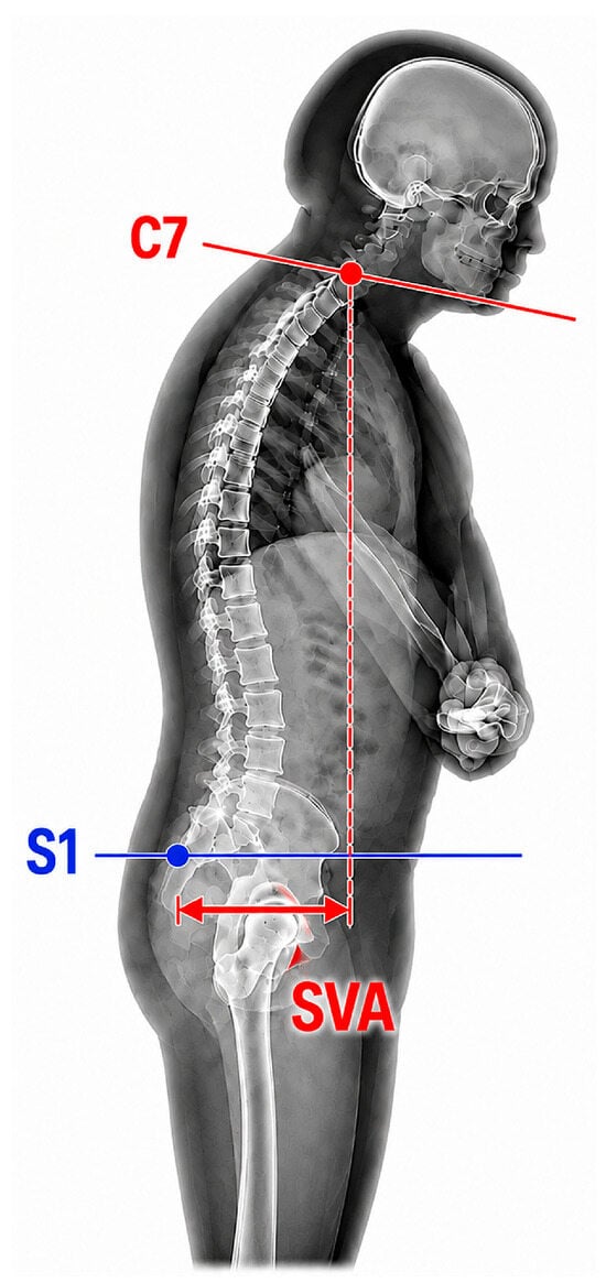
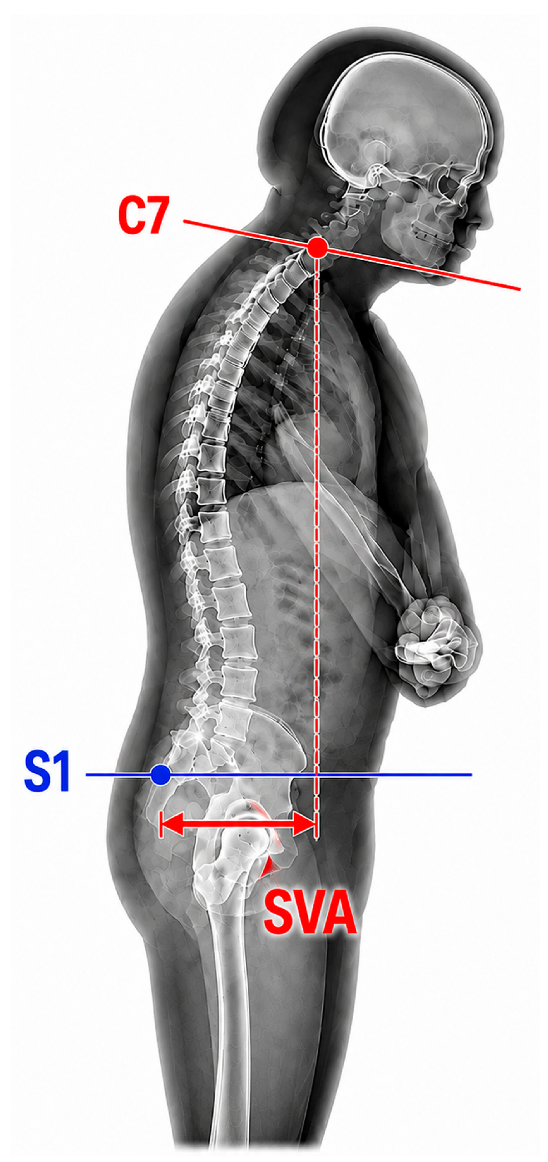
SVA was assessed using a panoramic lateral radiograph encompassing the cervical, thoracic, and lumbar spine and the pelvis. The SVA was defined as the horizontal distance between the C7 plumb line and the posterosuperior aspect of S1 (Figure 1) [36]. Participants were positioned standing upright and barefoot with the feet parallel. The upper limbs were flexed to 90°, with the hands resting lightly on supports positioned anteriorly. One evaluator (P.F.F.B.) analyzed the images without access to the participants’ clinical or demographic data. The radiographs were analyzed using Surgimap Spine software (Nemaris Inc., New York, NY, USA). This system has been proven to be reliable and valid for measuring spinopelvic alignment, with inter- and intraobserver reliability analyses revealing good to excellent agreement [37,38]. Prior to measurement, each image was calibrated individually based on the grid markers present on the radiograph itself. Since the distance between the markers was actually 10 cm, the image scale was adjusted within the software so that subsequent distances would be expressed in millimeters. SVA was analyzed as a continuous measure of global sagittal alignment. For descriptive categorical analyses, values from 0 to 49 mm were considered within the reference range and values ≥ 50 mm were classified as anterior sagittal displacement [39,40].
Figure 1.
Illustration showing how to measure the sagittal vertical axis (SVA). The SVA corresponds to the horizontal distance between a vertical plumb line drawn from the center of the C7 vertebral body and a vertical line drawn at the posterosuperior aspect of S1.
We evaluated plantar load distribution and postural sway using a BaroScan baropodometer (HS Technology, Londrina, Brazil). The platform’s dimensions are 46 cm in length and 51 cm in width, and it is equipped with 4096 pressure sensors. The acquisition rate was 10 frames per second. Participants stood barefoot with their arms relaxed at their sides, gaze fixed on a point ahead, and body in an upright position. These conditions were standardized to reduce procedural variability without suppressing the physiological postural sway of interest. Participants remained on the platform for 30 s in a quiet, level environment with controlled temperature and lighting. Before acquiring valid data, a familiarization trial was conducted to allow the participant to adapt to the experimental protocol. Data obtained during this initial trial were excluded from the analyses. A second attempt was then performed, and the resulting data were used for analysis. Data were processed using BaroSys software version 3.8.2.2 (BaroScan, Londrina, Brazil). The following variables were obtained: forefoot and rearfoot load distribution; right and left lower-limb load distribution (LRLL and LLLL); maximal amplitude, mean velocity, and standard deviation of anteroposterior displacement (MADAP, MVAP, and SDDAP); maximal amplitude, mean velocity, and standard deviation of mediolateral displacement (MADML, MVML, and SDDML); maximal center-of-pressure displacement (MDCOP); total center-of-pressure displacement area (MADCOP); and the coefficient of maximal displacement divided by area (CMD/A) [41].
HGS was measured using a digital dynamometer (Electronic Hand Dynamometer, SKU A1754, São Paulo, Brazil). Participants were seated on an armless chair with the elbow flexed to 90°, forearm in neutral position, and wrist extended between 0° and 30° [42]. Maximal HGS of the dominant hand was assessed during a 3-s sustained contraction. Three trials were performed, separated by 1-min intervals, and the highest value was retained for analysis. The observed values were compared to the HGS reference values for healthy Brazilian adults [43].
2.3. Statistical Analysis
Sample size calculation was performed using G*Power software version 3.1.9.7 (Heinrich Heine University, Düsseldorf, Germany). The study was designed to compare two independent groups of participants (iwaxSpA and controls). The primary outcome variable selected was the SVA, based on results from the study by de Oliveira and colleagues [29]. Considering a significance level of 5% and a statistical power of 80%, the sample size calculation indicated a requirement of 20 participants per group.
We assessed all quantitative variables for normality of distribution separately within each applicable group using the Shapiro–Wilk test. We complemented this analysis with visual inspection of histograms and Q–Q plots. Comparisons between the axSpA and control groups for each quantitative variable were performed using a Welch’s t-test if both groups met the criteria for a parametric approach and a Mann–Whitney test if not. Effect size was measured using Hedges’ g for parametric comparisons and rank-biserial correlation for nonparametric comparisons, both with 95% confidence intervals (95% CI). For categorical variables, we tested for associations with the group using the chi-square test; when an expected cell count fell below five, we used Fisher’s exact test. We measured effect size using the odds ratio. We defined the event as “male” for sex and “left-handed” for dominant side. We compared the odds in the axSpA group relative to the control group. Associations between postural assessment (SVA) and other quantitative variables in the axSpA group were measured using Pearson’s coefficient if the relationship between the two variables was approximately linear and neither showed evidence of deviation from normality. Otherwise, Spearman’s coefficient was used. The 95% CI for the Pearson coefficient was obtained using the standard closed-form formula. The 95% CI for the Spearman coefficient was obtained via percentile bootstrapping with 2000 resamples and a fixed seed of 123 for reproducibility.
The p-values from the between-group comparisons and the SVA association tests formed two distinct families of tests. Each family underwent a separate Benjamini–Hochberg adjustment to control the false discovery rate. The significance level adopted prior to adjustment was 5%. The reported 95% CIs were individual and were not adjusted for multiple analyses. Therefore, conclusions regarding statistical significance after controlling for multiplicity were based on p-values adjusted using the Benjamini–Hochberg method rather than on the 95% CIs directly. The analyses were performed in the R environment, version 4.3.3 (R package Bibliometrix).
3. Results
3.1. Patient Characteristics
Of the 22 iwaxSpA who were evaluated for inclusion in the study, only one was excluded due to pregnancy. Of those patients, 11 were male and 10 were female. The mean age was 48.8 ± 14.1 years, and the median time since diagnosis was 11 (6–18.2) years. Seven participants had radiographic axSpA, while 14 had non-radiographic axSpA. Eleven participants tested positive for HLA-B27. Comparisons of anthropometric data between the axSpA group and the control group showed no significant differences, except for larger foot size in the axSpA group (unadjusted p-value = 0.045) (Table 1). However, this difference in foot size between the two groups was not significant after adjusting for multiple comparisons (adjusted p-value = 0.796).
Table 1.
Anthropometry data of the studied sample (n = 41).
The medications used by the patients are listed below: 12 (57.1%) iwaxSpA were on bDMARDs, 10 (47.6%) iwaxSpA were on non-steroidal anti-inflammatory drugs (NSAIDs), and 3 (14.3%) were on salazopyrin. Of the patients using bDMARDs, 6, 5, and 1 were receiving adalimumab, golimumab, and infliximab, in that order. Elevated erythrocyte sedimentation rates and C-reactive protein levels were observed in 10 (47.6%) and 11 (52.4%) patients, respectively.
3.2. Comparisons Between the Two Groups Regarding Sagittal Alignment Baropodometric Parameters, and Handgrip Strength
An SVA ≥ 50 mm was observed in 9/21 (42.8%) iwaxSpA and 4/20 (20.0%) controls. Median SVA was higher in iwaxSpA than in controls (31 [16–53] vs. 26.5 [9.50–36.7] mm), but the difference was not statistically significant after multiplicity correction (adjusted p = 0.802). Point estimates for several load-distribution and CoP-derived variables differed between groups, but none remained statistically significant after correction for multiple testing. Nine iwaxSpA (42.8%) and four controls (20%) had HGS values below the adopted sex-specific cutoffs [43]; mean HGS did not differ significantly between groups in the original unadjusted comparison. Because these analyses were not adjusted for age and sex, the estimates should be considered exploratory pending adjusted modeling. Table 2 summarizes the original between-group comparisons.
Table 2.
Postural assessment, baropodometer parameters, and handgrip strength in the studied sample (n = 41).
3.3. Correlations Between Sagittal Vertical Alignment, Baropodometric Parameters, and Handgrip Strength
Within the iwaxSpA group, SVA showed a nominal positive correlation with age (ρ = 0.511, 95% CI 0.019–0.813; unadjusted p = 0.017), but the association was not statistically significant after Benjamini–Hochberg correction (adjusted p = 0.356). SVA also showed a nominal positive correlation with MADML (ρ = 0.452, 95% CI −0.006 to 0.799; unadjusted p = 0.039), which likewise did not remain statistically significant after multiplicity adjustment (adjusted p = 0.395). No other SVA correlation remained statistically significant after multiplicity correction. These bivariate associations should be interpreted cautiously because of the small sample and potential confounding, particularly by age and sex. Table 3 presents the original correlation analyses.
Table 3.
Spearman’s correlation coefficients between postural assessment, clinical data, baropodometer parameters, and handgrip strength measurements in individuals with axial spondyloarthritis (n = 21).
4. Discussion
This exploratory study examined SVA, baropodometric measures of plantar load distribution and postural sway, and HGS in iwaxSpA compared with controls. The principal observation is not that the groups were equivalent, but that the available sample produced imprecise estimates and not statistically robust between-group differences after correction for multiple testing. SVA was numerically higher in iwaxSpA, several baropodometric point estimates suggested greater sway or altered load distribution, and mean HGS was lower; however, the corresponding confidence intervals were wide. Within the axSpA group, the nominal associations of SVA with age and MADML did not remain statistically significant after multiplicity correction. These findings should therefore be interpreted as exploratory estimates rather than evidence of absence of clinically relevant differences.
AxSpA primarily affects the sacroiliac joints and spine and may produce progressive restriction of spinal mobility and altered sagittal alignment [44,45]. In the present sample, 42.8% of iwaxSpA and 20% of controls had SVA ≥ 50 mm. Although the continuous SVA comparison was not statistically significant after multiplicity correction, the approximately twofold difference in the proportion above the descriptive threshold is clinically noteworthy and warrants more precise estimation in a larger cohort [44,45]. Importantly, a non-significant result in this small sample should not be interpreted as proof of equivalence. SVA captures one component of global sagittal alignment and may not fully characterize pelvic, thoracic, lumbar, cervical, or lower-limb compensatory strategies [29]. Thus, future studies should consider a broader set of sagittal alignment parameters when feasible.
Importantly, the design of these new trials should focus on the impact of new axSpA therapies, particularly bDMARDs [9,10,11,12]. In fact, Son and colleagues [9] demonstrated that anti-TNF-α therapy improves lumbar lordosis and clinical outcomes in axSpA. In the inflamed sacroiliac joint of iwaxSpA, dense mononuclear infiltrates invade the cartilage in the presence of TNF-α mRNA [46]. Along the same lines, it is worth noting that nearly 60% of our patients were using bDMARDs, and this may have affected the lack of statistical differences between the two groups regarding SVA and baropodometric parameters. By controlling the inflammation that precedes pathological new bone formation, bDMARDs help prevent the rigid, stooped posture that severely impairs balance later in the disease’s progression.
Postural control depends on the integration of sensory information and coordinated neuromuscular responses that maintain the center of pressure within the BoS [20,21]. In the present study, several baropodometric point estimates differed between groups, but none remained statistically significant after multiplicity correction. These null inferential findings may reflect limited statistical precision, phenotypic heterogeneity, the measurement properties of the baropodometric protocol, or genuinely small between-group effects. Accordingly, the data do not establish that baropodometry is insensitive to axSpA-related postural changes. Load-distribution variables should also be distinguished conceptually from CoP-derived sway variables because they represent related but non-equivalent constructs. Previous studies have likewise reported heterogeneous findings depending on the balance task, instrument, and disease phenotype [18,20,21]. It is worth noting that the predominance of females in the control group may pose a challenge to interpreting our findings. Osama [47] recently demonstrated that males tend to have better postural stability when their eyes are open. In contrast, females have a lower risk of falling and better postural stability than males when their eyes are closed. This suggests that males may rely more on vision to maintain balance.
In the original bivariate analyses, SVA showed nominal positive associations with age and MADML, but neither association remained statistically significant after false-discovery-rate correction. The association with age is nevertheless clinically plausible and relevant to interpretation because age also differed between groups. Thus, future analyses should evaluate the SVA–postural-sway relationship using parsimonious adjusted models rather than relying exclusively on unadjusted bivariate correlations. Previous work using clinical postural measures has reported associations between sagittal alignment and balance-related outcomes, whereas more recent studies have demonstrated that dynamic balance and gait may be affected even when static sway variables are similar [18,35].
Individuals with axSpA may exhibit reduced muscle performance because of systemic inflammation, physical inactivity, metabolic alterations, pain, and structural impairment [48]. In the present study, HGS was used as a pragmatic marker of muscle function; it should not be interpreted as a direct measure of lower-limb or postural muscle strength [49]. Mean HGS was numerically lower in iwaxSpA, but the original between-group comparison was not statistically significant after multiplicity correction. Interpretation is further complicated by the unequal sex distribution between groups because sex is strongly associated with HGS [49]. Accordingly, age- and sex-adjusted analyses are needed before attributing HGS differences to axSpA. Future studies should additionally assess lower-limb muscle groups that contribute more directly to postural control.
From a clinical perspective, SVA, plantar-pressure-derived postural sway, and HGS should be viewed as complementary rather than interchangeable domains [32]. The present data do not support using a single normal SVA value to exclude balance impairment or muscle-function deficits in axSpA. A multimodal assessment integrating disease activity, spinal mobility, structural damage, BC, and task-relevant muscle performance is likely to be more informative for rehabilitation planning.
This study has several limitations. First, the sample was small and produced wide 95% CIs, limiting precision and the ability to detect small-to-moderate effects; in this scenario, for example, due to the concomitant use of NSAIDs in the majority of patients using bDMARDs, comparisons between drug subgroups were not possible, which prevents the evaluation of the effects of different treatment combinations. Second, the single-center design and the recruitment strategy may limit external validity and should be described explicitly. Third, the axSpA group was heterogeneous, including 7 radiographic and 14 non-radiographic cases, and the study did not provide detailed measures of disease activity, spinal mobility, functional status, pain, physical activity, or structural damage that could explain variation in SVA and BC. Fourth, the groups were not matched for age and sex, and the original inferential analyses were not adjusted for these potential confounders. Although appropriate statistical tests revealed no significant differences in age or sex between the two groups, it must be acknowledged that an imbalance in these two variables complicates the interpretation of our findings. Fifth, sagittal alignment was represented by SVA alone, rather than by a broader set of spinopelvic parameters; additionally, a single evaluator identified the milestones for the Surgimap Spine software measurements. Sixth, baropodometry was based on a brief static assessment and important acquisition/processing details were not fully reported; the validity and reliability of the specific device and protocol for all analyzed CoP variables should be documented. Finally, HGS was the only muscle-strength measure and does not directly quantify the lower-limb muscle groups most relevant to postural control. Despite these limitations, our study can serve as a starting point for larger studies to define the role of user-friendly, low-cost BC assessment tools and evaluate the impact of bDMARDs on posture and balance in the new era of axSpA treatment.
5. Conclusions
In iwaxSpA, there are numerical trends toward poorer postural assessment and BC, as measured by spinal radiography and baropodometry, respectively; however, no significant differences remain after adjusting for multiple comparisons. No significant associations were found between spinal alignment (as assessed by spinal radiography), baropodometric parameters, and HGS. These findings should be interpreted with caution, especially considering the small sample size and possible influence of age and sex. Given the exploratory nature of this study and its limited statistical power, controlled multicenter trials involving larger cohorts of iwaxSpA are needed to better define the roles of spinal radiography and baropodometry in this population.
Author Contributions
Conceptualization, K.R.R.d.S. and A.J.L.; methodology, G.S.d.E.G., L.M.C., C.A.L. and E.R.M.; formal analysis, K.R.R.d.S. and A.J.L.; investigation, K.R.R.d.S., G.S.d.E.G., L.M.C., P.J.F.B., C.A.L. and E.R.M.; data curation, A.J.L.; writing—original draft preparation, A.J.L.; writing—review and editing, K.R.R.d.S., G.S.d.E.G., L.M.C., P.J.F.B., C.A.L. and E.R.M.; supervision, K.R.R.d.S.; project administration, K.R.R.d.S. and A.J.L.; funding acquisition, A.J.L. All authors have read and agreed to the published version of the manuscript.
Funding
This work was supported by the Conselho Nacional de Desenvolvimento Científico e Tecnólogico (CNPq) under Grant number #401633/2023-3, Brazil, the Fundação Carlos Chagas Filho de Amparo à Pesquisa do Estado do Rio de Janeiro (FAPERJ) under Grant number #E-26/200.037/2026, Brazil, and the Coordenação de Aperfeiçoamento de Pessoal de Nível Superior (CAPES) under Grant number Finance Code 001, Brazil.
Institutional Review Board Statement
The study was conducted in accordance with the Declaration of Helsinki. All patients provided written informed consent. The protocol was approved by the Research Ethics Committee of the Centro Universitário Augusto Motta (CAAE-92856525.6.0000.5235; 24 November 2025).
Informed Consent Statement
Informed consent was obtained from all subjects involved in the study.
Data Availability Statement
Data is contained within the article.
Conflicts of Interest
The authors declare no conflicts of interest.
Abbreviations
The following abbreviations are used in this manuscript:
| axSpA | Axial spondyloarthritis |
| HLA-B27 | Human leukocyte antigen B27 |
| iwaxSpA | Individuals with axial spondyloarthritis |
| bDMARD | Biological disease-modifying antirheumatic drug |
| BC | Balance control |
| CoM | Center of mass |
| ADL | Activity of daily living |
| CoG | Center of gravity |
| BoS | Base of support |
| CT | Computed tomography |
| SVA | Sagittal vertical axis |
| ADL | Activities of daily living |
| LLRL | Percentage of load on the right lower limb |
| LLLL | Percentage of load on the left lower limb |
| MADAP | Maximal amplitude of anteroposterior displacement |
| MVAP | Mean velocity of anteroposterior displacement |
| SDDAP | Standard deviation of anteroposterior displacement |
| MADML | Maximal amplitude of mediolateral displacement |
| MVML | Mean velocity of mediolateral displacement |
| SDDML | Standard deviation of mediolateral displacement |
| MDCOP | Maximal displacement of center of pressure |
| MADCOP | Maximal total area of displacement of center of pressure |
| CMD/A | Coefficient of maximal displacement divided by the area |
| HGS | Handgrip strength |
| 95% CI | 95% confidence interval |
References
- Rudwaleit, M.; Jurik, A.G.; Hermann, K.G.; Landewé, R.; van der Heijde, D.; Baraliakos, X.; Marzo-Ortega, H.; Ostergaard, M.; Braun, J.; Sieper, J. Defining active sacroiliitis on magnetic resonance imaging (MRI) for classification of axial spondyloarthritis: A consensual approach by the ASAS/OMERACT MRI group. Ann. Rheum. Dis. 2009, 68, 1520–1527. [Google Scholar] [CrossRef] [Scilit] [PubMed]
- Bittar, M.; Deodhar, A. Axial spondyloarthritis: A review. JAMA 2025, 333, 408–420. [Google Scholar] [PubMed]
- Mease, P.J.; Deodhar, A. Differentiating nonradiographic axial spondyloarthritis from its mimics: A narrative review. BMC Musculoskelet. Disord. 2022, 23, 240. [Google Scholar] [CrossRef] [Scilit] [PubMed]
- van der Heijde, D.; Molto, A.; Ramiro, S.; Braun, J.; Dougados, M.; van Gaalen, F.A.; Gensler, L.S.; Inman, R.D.; Landewé, R.B.M.; Marzo-Ortega, H.; et al. Goodbye to the term ‘ankylosing spondylitis’, hello ‘axial spondyloarthritis’: Time to embrace the ASAS-defined nomenclature. Ann. Rheum. Dis. 2024, 83, 547–549. [Google Scholar] [CrossRef] [Scilit] [PubMed]
- van der Linden, S.; Valkenburg, H.A.; Cats, A. Evaluation of diagnostic criteria for ankylosing spondylitis. A proposal for modification of the New York criteria. Arthritis Rheum. 1984, 27, 361–368. [Google Scholar] [PubMed]
- Bohn, R.; Cooney, M.; Deodhar, A.; Curtis, J.R.; Golembesky, A. Incidence and prevalence of axial spondyloarthritis: Methodologic challenges and gaps in the literature. Clin. Exp. Rheumatol. 2018, 36, 263–274. [Google Scholar] [PubMed]
- Atar, E.; Askin, A. Somatosensory dysfunction related neuropathic pain component affects disease activity, functional status and quality of life in ankylosing spondylitis. Int. J. Rheum. Dis. 2020, 23, 1656–1663. [Google Scholar] [CrossRef] [Scilit] [PubMed]
- Moltó, A.; Nikiphorou, E. Comorbidities in spondyloarthritis. Front. Med. 2018, 5, 62. [Google Scholar] [CrossRef] [Scilit] [PubMed]
- Son, S.M.; Choi, S.H.; Shin, J.K.; Goh, T.S.; Lee, J.S. Radiologic parameters of ankylosing spondylitis patients treated with anti-TNF-α versus nonsteroidal anti-inflammatory drugs and sulfasalazine. Eur. Spine J. 2019, 28, 649–657. [Google Scholar] [CrossRef] [Scilit] [PubMed]
- Zhu, J.; Zhao, C.; Mi, C.; Liu, S.; Wu, R.; Li, T.; Duan, X.; Sun, L.; Yang, L.; He, X.; et al. Vunakizumab for radiographic axial spondyloarthritis: A randomized clinical trial. JAMA Netw. Open 2026, 9, e2611632. [Google Scholar] [PubMed]
- Zhang, S.; Xu, D.; Liu, S.; Li, S.; Wang, X.; Yuan, F.; Gou, W.; Xu, B.; Sun, L.; Gu, J.; et al. Xeligekimab, an interleukin-17a an-tagonist for active radiographic axial spondyloarthritis in chinese patients: 16- and 48-week results from a phase III, ran-domized, double-blind, placebo-controlled study. BioDrugs 2026, 40, 151–162. [Google Scholar] [CrossRef] [Scilit] [PubMed]
- Baraliakos, X.; Deodhar, A.; van der Heijde, D.; Van den Bosch, F.; Magrey, M.; Maksymowych, W.P.; Tomita, T.; Xu, H.; Coarse, J.; Prajapati, C.; et al. Bimekizumab long-term safety and efficacy across the spectrum of axial spondyloarthritis over 3 years: Results from 2 phase III studies. J. Rheumatol. 2026, 53, 1071–1082. [Google Scholar] [CrossRef] [Scilit] [PubMed]
- Nam, B.; Jo, S.; Bang, S.; Bang, S.Y.; Park, Y.; Shin, J.H.; Park, Y.S.; Lee, S.; Joo, K.B.; Kim, T.H. Clinical and genetic factors associated with radiographic damage in patients with ankylosing spondylitis. Ann. Rheum. Dis. 2022, 82, 527–532. [Google Scholar] [CrossRef] [Scilit] [PubMed]
- Çınar, E.; Akkoç, Y.; Karapolat, H.; Durusoy, R.; Keser, G. Postural deformities: Potential morbidities to cause balance problems in patients with ankylosing spondylitis? Eur. J. Rheumatol. 2016, 3, 5–9. [Google Scholar] [PubMed]
- Jalai, C.M.; Cruz, D.L.; Diebo, B.G.; Poorman, G.; Lafage, R.; Bess, S.; Ramchandran, S.; Day, L.M.; Vira, S.; Liabaud, B.; et al. Full-body analysis of age-adjusted alignment in adult spinal deformity patients and lower-limb compensation. Spine 2017, 42, 653–661. [Google Scholar] [CrossRef] [Scilit] [PubMed]
- Batur, E.B.; Karataş, G.K. Do postural changes affect balance in patients with ankylosing spondylitis? J. Rehabil. Med. 2017, 49, 437–440. [Google Scholar] [CrossRef] [Scilit] [PubMed]
- Schwab, F.; Lafage, V.; Boyce, R.; Skalli, W.; Farcy, J.P. Gravity line analysis in adult volunteers: Age-related correlation with spinal parameters, pelvic parameters, and foot position. Spine 2006, 31, E959–E967. [Google Scholar] [PubMed]
- Türk, E.; Yurdakul, F.G.; Güler, T.; Bodur, H. Posture, balance and gait in axial spondyloarthritis: A case-control study. Rheumatol. Int. 2024, 44, 2527–2538. [Google Scholar] [CrossRef] [Scilit] [PubMed]
- Horak, F.B. Postural orientation and equilibrium: What do we need to know about neural control of balance to prevent falls? Age Ageing 2006, 35, ii7–ii11. [Google Scholar] [CrossRef] [Scilit] [PubMed]
- Aydoğ, E.; Depedibi, R.; Bal, A.; Eksioglu, E.; Unlü, E.; Cakci, A. Dynamic postural balance in ankylosing spondylitis patients. Rheumatology 2006, 45, 445–448. [Google Scholar] [CrossRef] [Scilit] [PubMed]
- Murray, H.C.; Elliott, C.; Barton, S.E.; Murray, A. Do subjects with ankylosing spondylitis have poorer balance than normal subjects? Rheumatology 2000, 39, 497–500. [Google Scholar] [CrossRef] [Scilit] [PubMed]
- Ozen, T.; Tonga, E.; Polat, M.G.; Bayraktar, D.; Akar, S. Cervical proprioception accuracy is impaired in patients with axial spondyloarthritis. Musculoskelet. Sci. Pract. 2021, 51, 102304. [Google Scholar] [CrossRef] [Scilit] [PubMed]
- El Miedany, Y.; El Gaafary, M.; Youssef, S.; Palmer, D. Towards a multidimensional assessment of patient-reported outcome measures: Development and validation of a questionnaire for patients with ankylosing spondylitis/spondyloarthritis. Jt. Bone Spine 2010, 77, 575–581. [Google Scholar] [CrossRef] [Scilit] [PubMed]
- Bot, S.D.M.; Caspers, M.; Van Royen, B.J.; Toussaint, H.M.; Kingma, I. Biomechanical analysis of posture in patients with spinal kyphosis due to ankylosing spondylitis: A pilot study. Rheumatology 1999, 38, 441–443. [Google Scholar] [CrossRef] [Scilit] [PubMed]
- Nomura, K.; Fukada, K.; Azuma, T.; Hamasaki, T.; Sakoda, S.; Nomura, T. A quantitative characterization of postural sway during human quiet standing using a thin pressure distribution measurement system. Gait Posture 2009, 29, 654–657. [Google Scholar] [CrossRef] [Scilit] [PubMed]
- Faus-Riera, S.; Martínez-Pardo, S.; Blanch-Rubió, J.; Benito-Ruiz, P.; Duró-Pujol, J.C.; Corominas-Torres, J.M. Muscle pathology in ankylosing spondylitis: Clinical, enzymatic, electromyographic and histologic correlation. J. Rheumatol. 1991, 18, 1368–1371. [Google Scholar] [PubMed]
- Ceolin, C.; Bindoli, S.; Cozzi, G.; di Marzio, B.; Lorenzin, M.; De Rui, M.; Sfriso, P.; Doria, A.; Sergi, G.; Ramonda, R. Body composition, bone mineral density, and functional impairment in axial spondyloarthritis: A 36-month longitudinal study. BMC Musculoskelet. Disord. 2025, 26, 878. [Google Scholar] [CrossRef] [Scilit] [PubMed]
- Magrey, M.; Wei, J.C.C.; Yndestad, A.; Bushmakin, A.G.; Cappelleri, J.C.; Dina, O.; Deodhar, A. Relationships of work productivity and activity impairment with patient-reported outcomes in ankylosing spondylitis: Results from two trials. Arthritis Care Res. 2004, 76, 359–365. [Google Scholar]
- de Oliveira, T.L.; Silva, F.D.; Ormond Filho, A.G.; Nico, M.A.C.; da Rocha Correa Fernandes, A.; Ramiro, S.; de Medeiros Pinheiro, M. Exploring the influence of sagittal imbalance on spinal mobility, function and quality of life in axial spondyloarthritis: A novel comprehensive compensatory mechanism of adaptation. Adv. Rheumatol. 2025, 65, 60. [Google Scholar] [CrossRef] [Scilit] [PubMed]
- Benjamin, M.; Redman, S.; Milz, S.; Büttner, A.; Amin, A.; Moriggl, B.; Brenner, E.; Emery, P.; McGonagle, D.; Bydder, G. Adipose tissue at entheses: The rheumatological implications of its distribution. A potential site of pain and stress dissipation? Ann. Rheum. Dis. 2004, 63, 1549–1555. [Google Scholar] [CrossRef] [Scilit] [PubMed]
- Knoop, J.; Steultjens, M.P.M.; van der Leeden, M.; van der Esch, M.; Thorstensson, C.A.; Roorda, L.D.; Lems, W.F.; Dekker, J. Proprioception in knee osteoarthritis: A narrative review. Osteoarthr. Cartil. 2011, 19, 381–388. [Google Scholar] [CrossRef] [Scilit] [PubMed]
- Butler, A.A.; Lord, S.R.; Rogers, M.W.; Fitzpatrick, R.C. Muscle weakness impairs the proprioceptive control of human standing. Brain Res. 2008, 1242, 244–251. [Google Scholar] [CrossRef] [Scilit] [PubMed]
- Tolu, S.; Hindioğlu, N.; Kiraz, İ.E.; Calkin Korkmaz, M. Postural stability and the relationship with enthesitis in ankylosing spondylitis: A cross-sectional study. Med. Sci. 2019, 8, 613–619. [Google Scholar]
- Pompeu, J.E.; Romano, R.S.L.; Pompeu, S.M.A.A.; Lima, S.M. Static and dynamic balance in subjects with ankylosing spondylitis: Literature review. Rev. Bras. Reumatol. 2012, 52, 409–416. [Google Scholar] [PubMed]
- Rudwaleit, M.; van der Heijde, D.; Landewé, R.; Listing, J.; Akkoc, N.; Brandt, J.; Braun, J.; Chou, C.T.; Collantes-Estevez, E.; Dougados, M.; et al. The development of Assessment of SpondyloArthritis international Society classification criteria for axial spondyloarthritis (part II): Validation and final selection. Ann. Rheum. Dis. 2009, 68, 777–783. [Google Scholar] [CrossRef] [Scilit] [PubMed]
- Gueugnon, M.; Riglet, L.; Fournel, I.; Ksiazek, E.; Beaurain, J.; Chapon, R.; Ornetti, P.; Laroche, D. Evaluation of the sagittal vertical axis with postural and 3D motion analyses in lumbar spinal stenosis. BMC Musculoskelet. Disord. 2024, 25, 827. [Google Scholar] [CrossRef] [Scilit] [PubMed]
- Lafage, R.; Ferrero, E.; Henry, J.K.; Challier, V.; Diebo, B.; Liabaud, B.; Lafage, V.; Schwab, F. Validation of a new computer-assisted tool to measure spino-pelvic parameters. Spine J. 2015, 15, 2493–2502. [Google Scholar] [CrossRef] [Scilit] [PubMed]
- Helmya, N.A.; El-Sayyadb, M.M.; Kattabeib, O.M. Intra-rater and inter-rater reliability of Surgimap Spine software for measuring spinal postural angles from digital photographs. Bull. Fac. Phys. Ther. 2015, 20, 193–199. [Google Scholar] [CrossRef] [Scilit]
- Lafage, R.; Schwab, F.; Challier, V.; Henry, J.K.; Gum, J.; Smith, J.; Hostin, R.; Shaffrey, C.; Kim, H.J.; Ames, C.; et al. Defining spino-pelvic alignment thresholds: Should operative goals in adult spinal deformity surgery account for age? Spine 2016, 41, 62–68. [Google Scholar] [PubMed]
- Liu, C.; Zhu, W.; Li, Y.; Li, X.; Shi, B.; Kong, C.; Lu, S. How does cervical sagittal profile change after the spontaneous compensation of global sagittal imbalance following one- or two-level lumbar fusion. BMC Musculoskelet. Disord. 2024, 25, 387. [Google Scholar] [CrossRef] [Scilit] [PubMed]
- Pena Junior, P.M.; de Sá Ferreira, A.; Telles, G.; Lemos, T.; Nogueira, L.A.C. Concurrent validation of the centre of pressure displacement analyzed by baropodometry in patients with chronic non-specific low back pain during functional tasks. J. Bodyw. Mov. Ther. 2021, 28, 489–495. [Google Scholar] [CrossRef] [Scilit] [PubMed]
- Nonato, C.P.; Azevedo, B.L.P.A.; Oliveira, J.G.M.; Gardel, D.G.; de Souza, D.C.N.; Lopes, A.J. The Glittre Activities of Daily Living Test in women with scleroderma and its relation to hand function and physical capacity. Clin. Biomech. 2020, 73, 71–77. [Google Scholar] [CrossRef] [Scilit] [PubMed]
- Schlüssel, M.M.; dos Anjos, L.A.; de Vasconcellos, M.T.L.; Kac, G. Reference values of handgrip dynamometry of healthy adults: A population-based study. Clin. Nutr. 2008, 27, 601–607. [Google Scholar] [CrossRef] [Scilit] [PubMed]
- Braun, J.; Sieper, J. Ankylosing spondylitis. Lancet 2007, 369, 1379–1390. [Google Scholar] [CrossRef] [Scilit] [PubMed]
- Wanders, A.J.; Landewe, R.B.; Spoorenberg, A.; Dougados, M.; Van Der Linden, S.; Mielants, H.; van der Tempel, H.; van der Heijde, D.M.F.M. What is the most appropriate radiologic scoring method for ankylosing spondylitis? A comparison of the available methods based on the Outcome Measures in Rheumatology Clinical Trials filter. Arthritis Rheum. 2004, 50, 2622–2632. [Google Scholar] [PubMed]
- Wang, X.B.; Ellis, J.J.; Pennisi, D.J.; Song, X.; Batra, J.; Hollis, K.; Bradbury, L.A.; Li, Z.; Kenna, T.J.; Brown, M.A. Transcriptome analysis of ankylosing spondylitis patients before and after TNF-α inhibitor therapy reveals the pathways affected. Genes Immun. 2017, 18, 184–190. [Google Scholar] [CrossRef] [Scilit] [PubMed]
- Osama, M. Gender-based differences in postural stability, sensory integration of balance and fall risk between healthy young male and female adults: The GENAB study. J. Back Musculoskelet. Rehabil. 2025, 38, 829–837. [Google Scholar] [CrossRef] [Scilit] [PubMed]
- Sardoo, A.M.; Neto, A.; Pinheiro Torres, R.; Rodrigues-Manica, S.; Domingues, L.; Lage Crespo, C.; Lagoas-Gomes, J.; Mascarenhas, V.; Mendes, C.S.; Galzerano, A.; et al. The role of muscle in the susceptibility and progression of axial spondyloarthritis: The myospa study protocol. Acta Reumatol. Port. 2021, 46, 342–349. [Google Scholar] [PubMed]
- Lima, T.R.L.; Almeida, V.P.; Ferreira, A.S.; Guimarães, F.S.; Lopes, A.J. Handgrip strength and pulmonary disease in the elderly: What is the link? Aging Dis. 2019, 10, 1109–1129. [Google Scholar] [CrossRef] [Scilit] [PubMed]

Disclaimer/Publisher’s Note: The statements, opinions and data contained in all publications are solely those of the individual author(s) and contributor(s) and not of MDPI and/or the editor(s). MDPI and/or the editor(s) disclaim responsibility for any injury to people or property resulting from any ideas, methods, instructions or products referred to in the content. |
© 2026 by the authors. Licensee MDPI, Basel, Switzerland. This article is an open access article distributed under the terms and conditions of the Creative Commons Attribution (CC BY) license.