


Abstract
Bacterial toxins, such as Staphylococcal Enterotoxin B (SEB) and Shiga Toxins (STX1, STX2), pose severe public health risks and significant biological threats, demanding rapid and precise qPCR detection. This study reports the initial stages of standardization for uniplex qPCR components, serving as a foundational step toward a future multiplex detection system. The annealing temperature was successfully optimized for the stx1, seb, and 16S rRNA targets, showing high consistency in Cq values and fluorescence intensities at 58.4 °C. The optimized primer ratios ensure maximum amplification efficiency while minimizing potential molecular competition. These optimized assays provide a robust foundation for the multiplex qPCR platform. Once fully validated, this protocol will enhance timely and effective responses, as well as improve preparedness and readiness within the Brazilian biodefense system.
1. Introduction
Bacterial toxins such as Staphylococcal Enterotoxin B (SEB) and Shiga toxins (STX1 and STX2) represent serious public health and biothreat risks due to their extreme potency, rapid onset and thermal stability [1,2]. SEB (from Staphylococcus aureus) is a potent superantigen causing toxic shock syndrome through overproduction of cytokines [3]. STX1 and STX2 (Shiga toxin-producing Escherichia coli—STEC) inhibit protein synthesis, leading to hemolytic-uremic syndrome [4]. Furthermore, both SEB and Shiga toxins are critical virulence factors frequently implicated in severe cases of foodborne intoxication, often leading to serious clinical complications and life-threatening conditions [3,4].
These toxins are classified as Category B bioterrorism agents by the U.S. Centers for Disease Control and Prevention [5], owing to their ease of dissemination and the current lack of effective specific therapies. Consequently, rapid and accurate detection is crucial for both epidemiological surveillance and biological defense emergency response [6]. The Brazilian Army plays a strategic biosafety and biodefense role, using mobile and fixed laboratories [7] to identify and mitigate these threats. Developing sensitive molecular methods, like real-time PCR (qPCR) [6], is vital for strengthening national detection and response capabilities.
Therefore, the present study focused on the initial development and standardization of the uniplex qPCR components for the detection of seb, stx1, and 16S rRNA genes, as a crucial step towards the final multiplex assay enabling rapid and precise identification of toxin-producing (TP) bacterial strains associated with foodborne and other biological risks.
2. Methods
2.1. Bacterial Strains and Culture Conditions
All bacteria used in the study are detailed in Table 1 (including the genotypes of both toxin-producing and non-toxin-producing strains. Bacterial cultures were grown in Brain Heart Infusion medium (Oxoid™, Hampshire, UK) and incubated overnight at 37 °C [8].
Table 1.
Bacterial strains used in this study and their toxin genotypes.
2.2. DNA Extraction and Quantification
DNA was extracted from 200 µL of bacterial culture broth using the MPTA016 Pathogen Kit on the Extracta 32 system (Loccus, São Paulo, Brazil) followed by quality control [13,14]. DNA purity was assessed using a NanoDrop 2000c spectrophotometer (Thermo Fisher Scientific, Waltham, MA, USA). Samples with 260/280 absorbance ratios between 1.8 and 2.0 were considered suitable for downstream assays. DNA concentration was quantified using the QuantiFluor® dsDNA System Kit on a Quantus Fluorometer (Promega, Madison, WI, USA), and the samples were then stored at −20 °C until analysis.
2.3. qPCR Amplification and Optimization
The qPCR assays were run on the Amplio96™ platform (Loccus, São Paulo, Brazil). 20 µL reaction contained 10 µL of TaqMan Fast Advanced Master Mix for qPCR (Applied Biosystems, Foster City, CA, USA), forward/reverse primers for each gene toxin (350 nM), DNA template (10 ng), and probes (250 nM). All uniplex reactions were performed in duplicate, including non-template controls (NTCs). Probes and internal quenching used were 6-FAM/Zen/IABkFQ for seb and HEX/Zen/IABkFQ for stx1. The 16S rRNA primer and probe sequences used were, Fw-5’TGGAGCATGTGGTTTAATTCGA3’, P-5’CY5.5/CACGAGCTGACGACACCCATGCA/IAbRQSp3’ and Rev-5’TGCGGGACTTAACCCAACA3’ [15]. Oligonucleotide sets for seb and stx1 were developed by our research group (unpublished data), with their respective melting temperatures (Tm) for forward (Fw) primers, reverse (Rev) primers, and probes characterized as: stx1 (62, 62, and 68 °C), seb (63, 62, and 66 °C), and 16S rRNA (62.6, 63.8, and 70.1 °C). Thermal cycling conditions consisted of a pre-incubation at 58 °C for 2 min, 95 °C for 2 min, followed by 40 cycles of 95 °C denaturation for 15 s, and the annealing/extension step at the optimized temperature for 1 min. Optimization assays were conducted using a gradient of temperature (56 °C to 62.4 °C) [16], and primer concentrations (200 nM to 500 nM) [17]. Optimal conditions were selected based on the lowest quantification cycle (Cq) values and fluorescence intensity. The Cq variation (∆Cq) was calculated as the absolute difference between the highest and lowest Cq values obtained among the tested primer concentrations for each target gene.
3. Results
3.1. Uniplex qPCR Assessment and Specificity
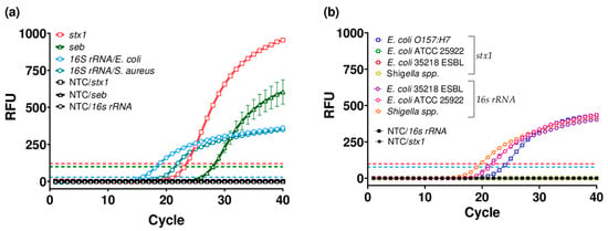
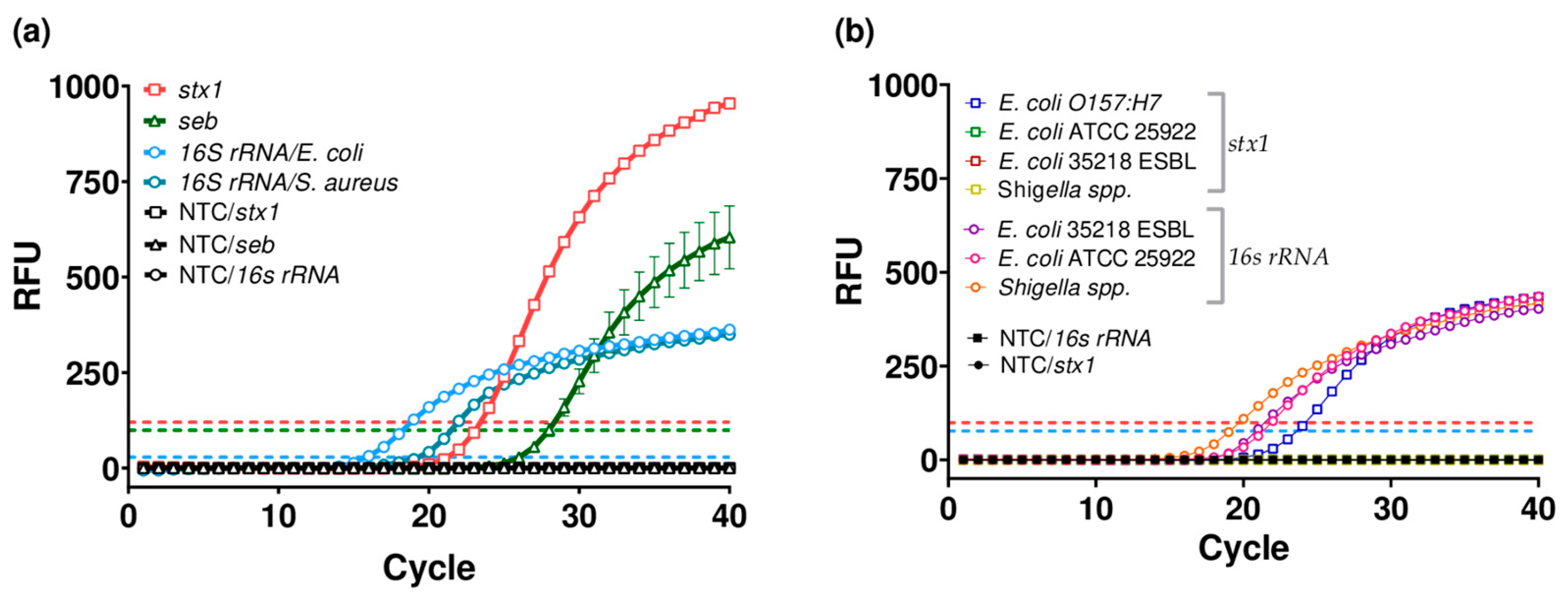
Prior to optimizing the qPCR assays for each target gene, we conducted an initial evaluation to assess primer and probe performance and to verify their specificity. For this purpose, uniplex reactions were tested for the stx1, seb and 16S rRNA and the results confirmed the assays’ viability (Figure 1a). The stx1 assay showed the highest relative fluorescence unit (RFU) signal (~1000), followed by the seb assay (~600), while the 16s rRNA curve, despite having the lowest RFU (~350), reached the earliest plateau. The corresponding Cq values were 23.46, 27.90, and 15.75/19.23 for stx1, seb, and 16S rRNA, respectively. The amplification profile of the internal control differed from that previously observed, yet it fulfilled its intended purpose [15]. The earlier amplification of the 16S rRNA control is biologically expected due to its multicopy nature, and the amplification profiles varied according to the target species, as anticipated. This interspecies Cq variation is attributed to inherent differences in 16S rRNA gene copy number across bacterial genomes, as described by Větrovský T. et al. [18], resulting in the distinct Cq ranges observed for E. coli and S. aureus and confirming that these differences reflect intrinsic genomic characteristics rather than methodological bias.
Figure 1.
Uniplex qPCR amplification curves for stx1, seb, and 16S rRNA genes. Parameter settings were 350 nM primers, 250 nM probe, and 60.4 °C annealing temperature. DNA detection was performed by monitoring fluorescence (RFU) as a function of cycle number. E. coli O157:H7 and S. aureus ATCC 25923 reference strains served as positive controls for the stx1 and seb targets, respectively. Dashed lines represent the amplification thresholds for the stx1 (red), seb (green), and 16S rRNA (blue) genes. (a) Functionality test. All three targets were successfully amplified, with no detectable signal in any of the no-template controls (NTC). (b) Specificity test. Assessment of the stx1 target against non-toxigenic E. coli and Shigella spp. strains showed exclusive stx1 amplification in E. coli O157:H7.
The subsequent analytical specificity for the stx1 primers was confirmed against a panel of reference strains (Table 1). The stx1 gene was successfully amplified in the TP E. coli O157:H7 strain (indicated by the red line and open red square markers) Cq of 24.2, whereas no amplification signal was observed in any non-TP E. coli strains or related Shigella species (Figure 1b). Notably, the 16S rRNA internal control was amplified in all tested strains, with the Cq value of 19.0 for Shigella spp., 20.9 for E. coli ATCC 35218, and 21.3 for E. coli ATCC 25922., confirming DNA integrity and absence of PCR inhibition. These results indicate the high specificity for the stx1 primers within the current panel, although further studies are required to evaluate a broader range of species.
These findings are particularly relevant considering the documented genetic proximity between E. coli and Shigella spp., as well as the reported horizontal transfer and variability of stx genes among enteric bacteria, described by Lamba K. et al. [11] and Eklund M. et al. [19]. In this context, the absence of cross-amplification reinforces the analytical specificity of the assay and reduces the risk of false-positive detection in closely related species. Furthermore, although most recent studies, such as Yun Y.S. et al. [4] and Li B. et al. [8], focus on multiplex platforms, they emphasize that rigorous singleplex validation is essential to ensure specificity and balanced amplification. Although broader validation across additional strains would further strengthen specificity assessment, the stable Cq values and consistent internal control performance support the robustness of the assay and its suitability for subsequent multiplex development.
3.2. Optimization of Annealing Temperature and Primer Concentration
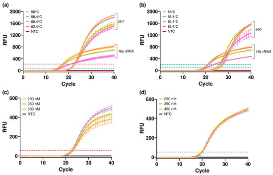
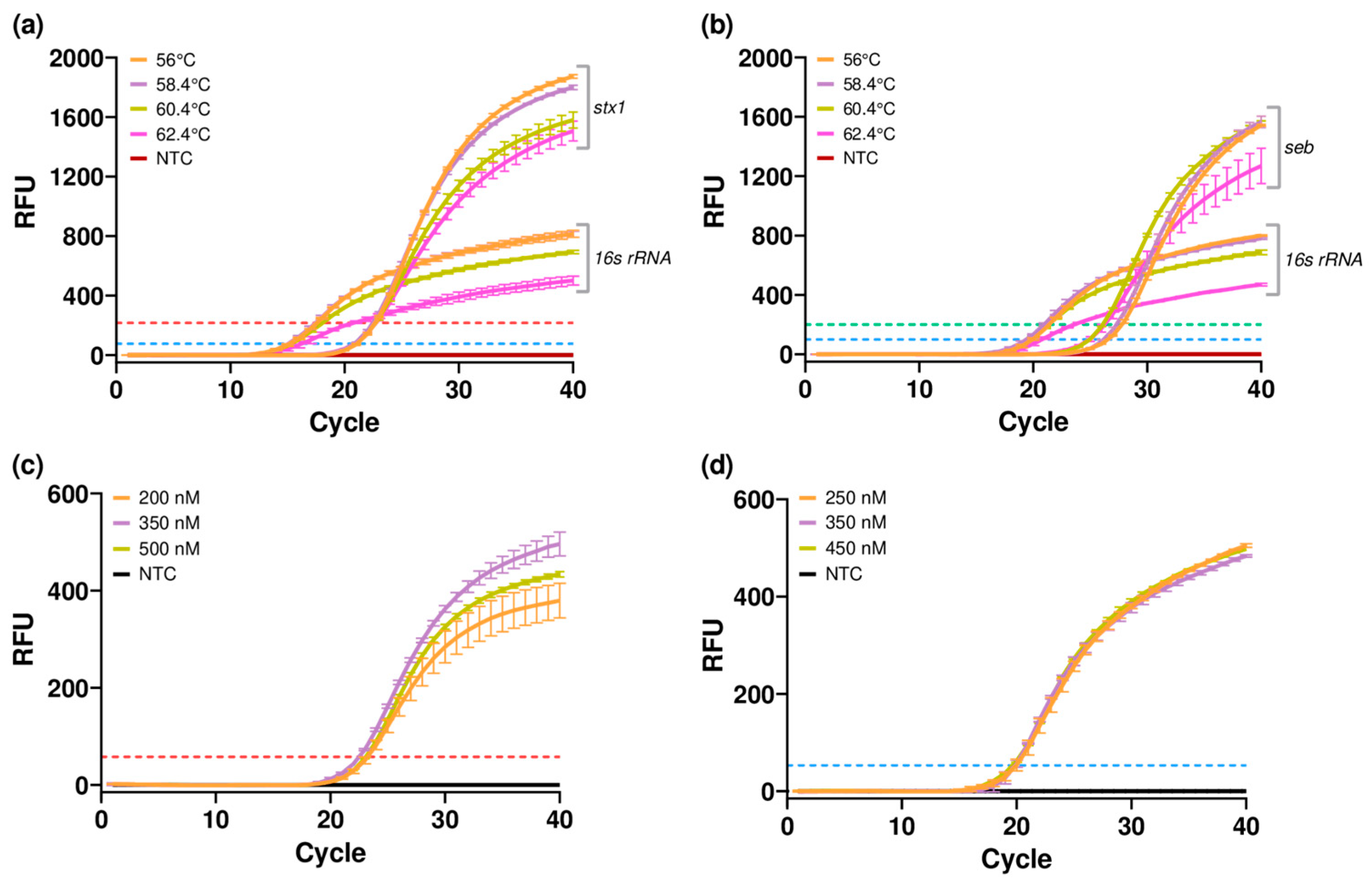
To enable future multiplexing, annealing temperature and primer concentrations were optimized under uniplex reactions. Regarding the evaluation of annealing temperatures, amplification was observed for all targets throughout the tested range (Figure 2a,b). Both Cq values and RFU remained highly consistent across the thermal gradient. Cq values for stx1 showed high stability, ranging from 22.49 to 22.75 (ΔCq = 0.26), while seb ranged from 26.10 to 27.8 (ΔCq = 1.7). In relation to 16S rRNA, values varied between 14.91 and 16.35 (ΔCq = 1.34; Figure 2a) and between 19.78 and 21.77 (ΔCq = 1.49; Figure 2b). These results demonstrate the consistency and robustness of the optimized uniplex assays, confirming their suitability as a reliable foundation. At 58.4 °C, Cq values were 22.49 (stx1), 27.33 (seb), 15.07 (16S rRNA-E. coli O157:H7), and 19.78 (16S rRNA-S. aureus), with RFU signals reaching peak or near-peak levels. Consequently, 58.4 °C was selected as the optimal temperature across all targets to minimize potential non-specific amplification or primer-dimer formation, a critical requirement for the future transition from uniplex to a multiplex qPCR configuration.
Figure 2.
Optimization of qPCR parameters for stx1, seb and 16S rRNA assays. Annealing temperature gradient curves for stx1 (a) and seb (b) using 350 nM primer and 250 nM probe. Primer concentration gradient curves for stx1 (c) and 16S rRNA (d) using 250 nM probe at 58.4 °C. Robust amplification was observed across all tested conditions. E. coli O157:H7 and S. aureus ATCC 25923 reference strains were used as positive controls for the stx1 and seb targets, respectively. Dashed lines represent the amplification thresholds for the stx1 (red), seb (green), and 16S rRNA (blue) genes.
Building on these results, a primer concentration gradient was subsequently tested for stx1 and 16s rRNA. For the stx1 target, the Cq values showed minimal variation between the tested concentrations (ΔCq = 0.72), indicating highly consistent performance. Although the 350 nM concentration yielded a slightly higher RFU, with a Cq of 22.53 (Figure 2c), the 200 nM concentration was selected as optimal. Similarly, for the 16S rRNA gene, no significant differences in amplification were observed among the concentrations (ΔCq = 0,31), with Cq values remaining consistently around 20.0 (Figure 2d). Consequently, 250 nM (Cq = 19.98) was selected to minimize potential primer competition in the subsequent multiplex assay.
The limited Cq variation observed across the annealing temperature gradient (<1.7 cycles for all targets) demonstrates the thermal robustness of the assays and supports efficient primer–probe interaction within a harmonized melting temperature range. According to established qPCR optimization guidelines Zhao F. et al. [16]; Rodríguez A. et al. [17], minimal Cq fluctuation across temperature gradients and the use of the lowest effective primer concentrations are key indicators of appropriate primer design, reaction stability, and amplification specificity. In this context, the consistent performance observed across temperatures and reduced primer concentrations confirms stable amplification kinetics, minimizes the risk of non-specific interactions, and supports the overall reliability of the optimized assay conditions.
4. Conclusions
The qPCR assay developed in this study demonstrated robust amplification performance and analytical specificity for detecting the stx1, seb, and 16S rRNA genes. Based on the optimization results, an annealing temperature of 58.4 °C and primer concentrations of 200 nM (for stx1) and 250 nM (for 16S rRNA) were established to ensure maximum efficiency. Future work will focus on optimizing seb primer concentrations and broadening the specificity testing against a wider panel of non-target strains. Additionally, complete multiplex validation will be conducted to assess analytical sensitivity and performance in complex matrices such as environmental, clinical, and food samples. This multiplex configuration is critical for reducing time and resource requirements by enabling the simultaneous detection of multiple toxins, which is a capability especially valuable for outbreak investigations and biothreat monitoring. Such implementation may contribute to enhanced diagnostic preparedness within the Brazilian Army’s laboratory network, supporting outbreak response and biological threat surveillance initiatives.
Author Contributions
Writing—original draft preparation, V.H.G.P., V.S.G.d.A. and B.d.P.M.; writing—review and editing, V.H.G.P., B.d.P.M., V.H.G.D., M.D.R., V.d.S.S., C.G.M.d.S. and V.S.G.d.A.; and supervision, V.S.G.d.A. and T.L.S.N.; investigation, V.H.G.P. and S.D.d.S. All authors have read and agreed to the published version of the manuscript.
Funding
This work was supported by FAPERJ (39/2021) and DECEX/CADESM (341/22).
Institutional Review Board Statement
The study was conducted in accordance with the Declaration of Helsinki, and approved by the Institutional Review Board of the Fundação de Amparo a Pesquisa do Estado do Rio de Janeiro (protocol code E-26/210.343/2022 in 4 April 2022).
Informed Consent Statement
Not applicable.
Data Availability Statement
Certain primer sequences are omitted due to intellectual property protection. Further details and raw qPCR data are available from the corresponding author upon reasonable request, subject to institutional and legal restrictions.
Acknowledgments
We would like to thank Samuel Dias, Beatriz Paiva, Victor Hugo Giordano, and Virginia Grancieri for their valuable contributions and insightful discussions. In particular, we extend our deepest gratitude to Virginia, whose support and permission made this study possible.
Conflicts of Interest
The authors declare no conflicts of interest.
Abbreviations
| qPCR | Quantitative Polymerase Chain Reaction |
| SEB | Staphylococcal Enterotoxin B |
| STX | Shiga toxin |
References
- Janik, E.; Ceremuga, M.; Saluk-Bijak, J.; Bijak, M. Biological Toxins as the Potential Tools for Bioterrorism. Int. J. Mol. Sci. 2019, 20, 1181. [Google Scholar] [CrossRef] [Scilit] [PubMed]
- Liu, Y.; Tian, S.; Thaker, H.; Dong, M. Shiga Toxins: An Update on Host Factors and Biomedical Applications. Toxins 2021, 13, 222. [Google Scholar] [CrossRef] [Scilit] [PubMed]
- Bae, J.; Da, F.; Liu, R.; He, L.; Lv, H.; Fisher, E.; Rajagopalan, G.; Li, M.; Cheung, G.; Otto, M. Contribution of Staphylococcal Enterotoxin B to Staphylococcus aureus Systemic Infection. J. Infect. Dis. 2020, 223, 1766–1775. [Google Scholar] [CrossRef] [Scilit] [PubMed]
- Yun, Y.S.; Park, D.Y.; Oh, I.; Shin, W.R.; Ahn, G.; Ahn, J.Y.; Kim, Y.H. Pathogenic Factors and Recent Study on the Rapid Detection of Shiga Toxin-Producing Escherichia coli (STEC). Mol. Biotechnol. 2023, 67, 16–26. [Google Scholar] [CrossRef] [Scilit] [PubMed]
- Centers for Disease Control and Prevention (CDC). Bioterrorism Agents/Diseases (by Category) | Emergency Preparedness & Response. Available online: https://emergency.cdc.gov/agent/agentlist-category.asp (accessed on 13 October 2025).
- Organisation for the Prohibition of Chemical Weapons, Scientific Advisory Board’s Temporary Working Group. Analysis of Biotoxins, Report of the Scientific Advisory Board’s Temporary Working Group; SAB/REP/1/23; OPCW: The Hague, The Netherlands, 2023; Available online: https://www.opcw.org/sites/default/files/documents/2023/04/Analysis%20of%20Biotoxins%20Final%20Report.pdf (accessed on 13 October 2025).
- Exército Brasileiro. Diretriz de Biossegurança, Bioproteção e Defesa Biológica do Exército. Portaria EME/C Ex No. 1.240. 7 February 2024. Available online: https://www.dsau.eb.mil.br/index.php?view=article&layout=edit&id=191:biosseguranca-bioprotecao-e-defesa-biologica&catid=21 (accessed on 1 March 2026).
- Duffy, G.; Whiting, R.C.; Sheridan, J.J. The Effect of a Competitive Microflora, pH and Temperature on the Growth Kinetics of Escherichia coli O157:H7. Food Microbiol. 1999, 16, 299–307. [Google Scholar] [CrossRef] [Scilit]
- Li, B.; Liu, H.; Wang, W. Multiplex Real-Time PCR Assay for Detection of Escherichia coli O157:H7 and Screening for Non-O157 Shiga Toxin-Producing E. coli. BMC Microbiol. 2017, 17, 215. [Google Scholar] [CrossRef] [Scilit]
- Enriquez-Gómez, E.; Acosta-Dibarrat, J.; Talavera-Rojas, M.; Soriano-Vargas, E.; Navarro, A.; Morales-Espinosa, R.; Velázquez-Ordoñez, V.; Cal-Pereyra, L. Serotypes, Pathotypes, Shiga Toxin Variants and Antimicrobial Resistance in Diarrheagenic Escherichia coli Isolated from Rectal Swabs and Sheep Carcasses in an Abattoir in Mexico. Agriculture 2023, 13, 1604. [Google Scholar] [CrossRef] [Scilit]
- Ludwig, S.; Jimenez-Bush, I.; Brigham, E.; Bose, S.; Diette, G.; McCormack, M.C.; Matsui, E.C.; Davis, M.F. Analysis of home dust for Staphylococcus aureus and staphylococcal enterotoxin genes using quantitative PCR. Sci. Total Environ. 2017, 581–582, 750–755. [Google Scholar] [CrossRef] [Scilit] [PubMed]
- Lamba, K.; Nelson, J.A.; Kimura, A.C.; Poe, A.; Collins, J.; Kao, A.S.; Cruz, L.; Inami, G.; Vaishampayan, J.; Garza, A.; et al. Shiga Toxin 1-Producing Shigella sonnei Infections, California, United States, 2014–2015. Emerg. Infect. Dis. 2016, 22, 679–686. [Google Scholar] [CrossRef] [Scilit] [PubMed]
- Lucena-Aguilar, G.; Sánchez-López, A.M.; Barberán-Aceituno, C.; Carillo-Ávila, J.A.; López-Guerero, J.A.; Aguilar-Quesada, R. DNA Source Selection for Downstream Applications Based on DNA Quality Indicators Analysis. Biopreserv. Biobank 2016, 14, 264–270. [Google Scholar] [CrossRef] [Scilit] [PubMed]
- Versmessen, N.; Van Simaey, L.; Negash, A.A.; Vandekerckhove, M.; Hulpiau, P.; Vaneechoutte, M.; Cools, P. Comparison of DeNovix, NanoDrop and Qubit for DNA Quantification and Impurity Detection of Bacterial DNA Extracts. PLoS ONE 2024, 19, e0305650. [Google Scholar] [CrossRef] [Scilit] [PubMed]
- Alexandrino, F.; Malgarin, J.S.; Krieger, M.A.; Morello, L.G. Optimized broad-range real-time PCR-based method for bacterial screening of platelet concentrates. Braz. J. Biol. 2021, 81, 692–700. [Google Scholar] [CrossRef] [Scilit] [PubMed]
- Zhao, F.; Maren, N.A.; Kosentka, P.Z.; Liao, Y.Y.; Lu, H.; Duduit, J.R.; Huang, D.; Ashrafi, H.; Zhao, T.; Huerta, A.I.; et al. An Optimized Protocol for Stepwise Optimization of Real-Time RT-PCR Analysis. Hortic. Res. 2021, 8, 179. [Google Scholar] [CrossRef] [Scilit] [PubMed]
- Rodríguez, A.; Rodríguez, M.; Córdoba, J.J.; Andrade, M.J. Design of Primers and Probes for Quantitative Real-Time PCR Methods. Methods Mol. Biol. 2015, 1275, 31–56. [Google Scholar] [CrossRef] [Scilit] [PubMed]
- Větrovský, T.; Baldrian, P. The variability of the 16S rRNA gene in bacterial genomes and its consequences for bacterial community analyses. PLoS ONE 2013, 8, e57923. [Google Scholar] [CrossRef] [Scilit] [PubMed]
- Eklund, M.; Leino, K.; Siitonen, A. Clinical Escherichia coli Strains Carrying stx Genes: Stx Variants and stx-Positive Virulence Profiles. J. Clin. Microbiol. 2002, 40, 4585–4593. [Google Scholar] [CrossRef] [Scilit] [PubMed]

Disclaimer/Publisher’s Note: The statements, opinions and data contained in all publications are solely those of the individual author(s) and contributor(s) and not of MDPI and/or the editor(s). MDPI and/or the editor(s) disclaim responsibility for any injury to people or property resulting from any ideas, methods, instructions or products referred to in the content. |
© 2026 by the authors. Licensee MDPI, Basel, Switzerland. This article is an open access article distributed under the terms and conditions of the Creative Commons Attribution (CC BY) license (https://creativecommons.org/licenses/by/4.0/).