


Abstract
Pancreatic ductal adenocarcinoma (PDAC) remains one of the deadliest solid tumours, driven by late diagnosis, early metastatic dissemination, and profound resistance to systemic therapies. Increasing evidence indicates that these hallmarks are not solely tumour cell intrinsic but are critically orchestrated by a complex and highly dynamic tumour microenvironment (TME) composed of pancreatic stellate cells (PSCs), cancer-associated fibroblast (CAF) subtypes, immune cells, endothelial and neuronal elements, and a dense extracellular matrix (ECM). This review provides an integrated overview of the cellular and acellular components of the PDAC TME and delineates how their reciprocal crosstalk drives desmoplasia, immune suppression, metabolic reprogramming, epithelial–mesenchymal transition (EMT), pre-metastatic niche formation, and metastatic outgrowth. Particular emphasis is placed on the context-dependent roles of stromal and immune niches in modulating drug delivery, chemoresistance, and failure of immunotherapy, highlighting why indiscriminate stromal depletion has yielded paradoxical outcomes. Building on these mechanistic insights, the review critically examines emerging therapeutic strategies targeting PSCs, CAF subsets, ECM components, myeloid and lymphoid populations, and key signalling pathways, including approaches that normalize stroma, reprogram immunity, or exploit nanocarrier-based delivery systems. Finally, a structured framework is proposed for rational TME-targeted combination regimens that integrate cytotoxic, targeted, and immunotherapeutic agents to overcome current therapeutic barriers in PDAC.
1. Introduction
Pancreatic cancer remains one of the most lethal malignancies worldwide. According to GLOBOCAN 2022, approximately 511,000 new cases of pancreatic cancer and 467,000 related deaths were reported globally. Owing to its extremely poor prognosis, pancreatic cancer ranks as the sixth leading cause of cancer-related mortality in both sexes combined and accounts for nearly 5% of all cancer deaths worldwide [1]. Alarmingly, by 2030, pancreatic ductal adenocarcinoma (PDAC) is projected to surpass breast, prostate, and colorectal cancers to become the second leading cause of cancer-related deaths in the United States, preceded only by lung cancer [2]. Most malignant tumours of the pancreas are adenocarcinomas, while rarer forms include neuroendocrine tumours, acinar cell carcinomas, colloid carcinomas, pancreatoblastomas, and solid pseudopapillary neoplasms. Among these, PDAC represents the most prevalent and clinically aggressive subtype. Multi-omic and spatial profiling studies have also recently radically altered the concept of PDAC as an immunologically cold malignancy. Instead of being marked by absolute lack of immune infiltration, PDAC is marked by severe immune malfunction, T-cell exhaustion, defective antigen presentation, and overshadowing immunosuppressive stromal and myeloid compartments [3,4,5,6].
One major reason for a very high death rate in PDAC is that it develops silently in its early stages without noticeable symptoms. Diagnosis at a curable stage remains exceedingly difficult, as patients rarely present with early symptoms and tumours lack sensitive and specific diagnostic markers [7,8]. Owing to its highly aggressive nature, PDAC often spreads to distant organs such as the liver, lungs, and peritoneum. PDAC is characterized by a dense and fibrotic stromal structure, comprising collagen, fibronectin, hyaluronan, and PSCs, which collectively impede proper drug penetration and diminish the effectiveness of chemotherapy. The immune landscape of PDAC is also hostile, rendering immunotherapy largely unsuccessful [9]. Emerging studies demonstrate that PDAC failure to immunotherapy is caused more by functional depletion of infiltrating T cells rather than impaired trafficking. Worn-out CAR-T and endogenous T cells survive in tumours but gradually acquire incapacity to execute their effector functions through metabolic pressure, checkpoint stimulation, and ongoing antigen exposure [5,6,10]. Reduced activity of CD8+ T cells and the dominance of myeloid-derived suppressor cells weaken the body’s immune response, facilitating immune evasion and allowing unchecked tumour growth.
The TME of PDAC consists of cancer-associated fibroblasts (CAFs), extracellular matrix (ECM), immune cells, cytokines, and growth factors. The dynamic and reciprocal interactions between cancer cells and these microenvironmental components drive stromal activation, immune suppression, desmoplasia, tumour invasion, and metastasis [11]. Considering the multifactorial and highly dynamic nature of pancreatic cancer progression, increasing emphasis has been placed on targeting the TME and its components as a therapeutic strategy. Recent reports also indicate that PDAC cells make use of chemokine signalling pathways to impose immune exclusion. Tumour-secreted CCL20 attracts CCR6+ suppressive T cells, mutated p53 enhances immunosuppressive chemokine expression via enhancer reprogramming, and the combination of the two inhibits T-cell penetration and suppresses checkpoint inhibitor activity [12,13].
At the molecular level, pancreatic cancer generally shows a small set of repeated genetic changes, most commonly involving Kirsten Rat Sarcoma Viral Oncogene Homologue (KRAS), Cyclin-Dependent Kinase Inhibitor 2A (CDKN2A) (encoding p16), Tumour Protein p53 (TP53), and SMAD Family Member 4 (SMAD4) [14,15,16]. On top of stromal barriers, the immune microenvironment of PDAC is actively modelled by tumour-intrinsic genetic changes. Massive transcriptomic studies reveal that specific KRAS mutation subtypes, especially KRASG12D, are linked to a smaller tumour mutational burden, macrophage polarization, and decreased CD8+ T-cell infiltration, all of which contribute to immune resistance [3]. Pancreatic cancer is also highly resistant to standard treatment options, including chemotherapy, radiation therapy, and targeted drugs, thus limiting their overall effectiveness. PDAC is highly invasive, as cancer cells invade nearby nerves and blood vessels at a relatively early stage, and then spread to distant organs [17]. Total surgical tumour excision is not feasible due to the early spread and aggressiveness of the disease [18]. Concurrently, stromal elements in the non-cellular form play a role in immune evasion. The cross-linked fibrin-laden stromal matrixes and desmoplasia tissue serve as physical and biochemical barriers that limit immune cell infiltration and promote an immunosuppressive niche in PDAC tumours [4,19].
In this review, the focus is on integrating the roles of key cellular and acellular components of the PDAC TME with the processes of metastasis and treatment failure, and on critically examining how these insights can be translated into rational therapeutic strategies. Specifically, the review aims to delineate how distinct TME niches and their crosstalk drive PDAC invasion, pre-metastatic niche formation, and immune escape; highlight emerging pharmacological approaches that modulate stromal, vascular, and immune elements to overcome drug resistance; and propose a structured framework for designing TME-targeted combination therapies that more effectively integrate cytotoxic, targeted, and immunotherapeutic modalities. By emphasizing the nexus between TME biology, metastasis, and therapeutic response, this work intends to move beyond descriptive TME overviews and provide a mechanistically grounded roadmap for developing and optimizing TME-directed interventions in pancreatic cancer.
2. Mechanisms and Pathophysiology of Pancreatic Cancer
2.1. Carcinogenesis and Molecular Mechanism
Pancreatic intraepithelial neoplasia (PanIN) is the most common pre-neoplastic ductal lesion that causes pancreatic cancer, whereas larger cystic precursor lesions, such as intraductal papillary mucinous neoplasms (IPMNs) and mucinous cystic neoplasms, are less prevalent [20]. It is estimated that progression of PanIN to invasive PDAC depends on grade of PanIN—PanIN-1 is very low (approximately 1–1.5%); the lifetime progression of PanIN-2 is intermediate (approximately 5%); and PanIN-3/high-grade PanIN carries a very high risk (approximately 28–33%) [21]. Pancreatic carcinogenesis is driven by a highly complex and interdependent signalling network involving activation of several tyrosine kinase receptors, including epidermal growth factor receptor (EGFR), hepatocyte growth factor receptor (HGFR), and KRAS- and transforming growth factor-β (TGF-β)-associated pathways (Figure 1) [22,23,24].
Figure 1.
Aberrant growth factor and TGF-β signalling in PDAC. Frequent loss or mutation of SMAD4 in PDAC disrupts canonical TGF-β–SMAD tumour-suppressive signalling, abrogating transcriptional programmes such as p21 induction. Consequently, signalling is redirected toward non-canonical pathways that cooperate with RAS–RAF–MAPK, HIF-1α, and WNT signalling to promote proliferation, metabolic reprogramming, fibrosis, invasion, and therapeutic resistance. Created using Adobe Illustrator 2024_v28.2.0.532.
The limited efficacy of conventional chemotherapy in pancreatic cancer can be partly attributed to the redundancy and crosstalk among these signalling pathways. The inhibition of a single pathway is often insufficient to suppress tumour growth, as multiple protumourigenic signals operate concurrently. Pancreatic cancer is characterized by the overexpression and excessive ligand simulation of EGFR and HGFR, resulting in heterodimer formation between EGFR and HGFR [25,26,27]. This heterodimerization leads to the activation of multiple downstream signalling pathways, further driving tumour growth, invasion, and drug resistance. Genetic alterations, such as oncogenic KRAS mutations, enable tumour cells to proliferate independently of external growth factor signals, while the loss of the p16 tumour suppressor disrupts cell cycle regulation, resulting in uncontrolled cell division [28]. Oncogenic KRAS mutations in addition to their autonomous proliferation functions actively reprogram the tumour immune microenvironment in PDAC. A recent genomic and transcriptomic study indicates that KRAS-mutated PDACs, and specifically KRASG12D, have a lessened tumor mutational burden, distorted polarization of macrophages, decreased CD8+ T-cell infiltration, and even increased occupation of immunosuppressive stromal factors, which further support immune evasion and therapeutic resistance [3]. In addition, adhesion molecules and cell surface co-receptors, such as neuropilin-1, CD44, and β1-integrin, facilitate cancer cell attachment to the ECM, thereby enhancing invasive behaviour [29,30].
TGFβ typically functions as a tumour suppressor; however, in pancreatic cancer, its role is altered due to KRAS mutations and defective SMAD signalling [31]. Under such circumstances, TGFβ predominantly activates Mitogen-Activated Protein Kinase (MAPK), Proto-oncogene tyrosine-protein kinase Src (SRC), and Protein Kinase B (AKT) pathways, which promote tumour growth and invasion rather than inhibition [32,33]. These cancerous signalling programmes are also connected with the action of inflammation and chemokines, enhancing stromal engagement and immune suppression, which subsequently combine to produce an inviting niche that facilitates PDAC progression and metastatic dissemination [12,13].
2.2. Metabolic Reprogramming in Pancreatic Cancer
By reprogramming their metabolic pathways, malignant tumour cells can multiply quickly despite hypoxia and the limited presence of nutrients. Factors such as a hypoxic microenvironment, reduced nutrient supply, dense stromal architecture, and frequent KRAS mutations collectively drive this metabolic rewiring [34]. Hypoxia-inducible factor-1α (HIF-1α) and oncogenic KRAS are the primary regulators of this process [35,36]. HIF-1α becomes activated under hypoxic conditions, while KRAS enhances glucose uptake and promotes glycolysis to support tumour growth. A recent study demonstrates that metabolic reprogramming in PDAC not only sustains tumour cell survival but also directly contributes to immune dysfunction. Hypoxia-driven metabolic enzymes such as ENO1 promote PD-L1 expression through HIF-1α signalling, thereby impairing CD8+ T-cell infiltration and fostering an immunosuppressive tumour immune microenvironment [37].
In addition to altered glucose metabolism, pancreatic cancer cells rely on autophagy as a survival mechanism under metabolic stress [38]. Through autophagy, cells degrade damaged organelles and intracellular proteins, supplying essential nutrients required for survival. For metabolic adaptation in PDAC with KRAS mutations, macropinocytosis is crucial [39]. At the same time, the metabolic crosstalk between tumour cells and immune cells triggers dysfunction in T cells. The mitochondrial metabolism and redox changes induced by tumours produce metabolites promoting epigenetic restructuring of T cells that result in permanent PD-1 and functional exhaustion [10]. Notably, macropinocytosis in tumour cells does not imply being limited only to that, but also the central role in sustaining cancer-associated fibroblast (CAF) phenotypes in metabolic stress. Macropinocytosis inhibition causes CAF subtype reprogramming, decreases collagen deposition, increases immune cell intracellular uptake, and sensitizes tumours of PDAC to chemotherapy and immunotherapy [40]. Through this process, cancer cells absorb extracellular proteins and break them down within lysosomes to generate amino acids, particularly glutamine, which supports continued growth under nutrient-limited conditions.
3. Overview of the Pancreatic Tumour Microenvironment
3.1. General Characteristics of the PDAC Tumour Microenvironment
Pancreatic cancer grows within a thick, fibrotic, and poorly oxygenated environment known as the TME or stroma. This stromal compartment protects cancer cells, suppresses antitumour immune responses, and actively promotes tumour invasion, metastasis, chemoresistance, and recurrence. The microenvironment is characterized by an abundant collagen-rich matrix that renders the tumour stiff and fibrotic [41]. Such excessive stromal deposition compresses intratumoral blood vessels, leading to reduced blood perfusion and oxygen deprivation, thereby creating a hypoxic environment that therapeutic drugs are unable to penetrate effectively [42]. In addition to these mechanical properties, the PDAC stroma actively determines immune exclusion. Recent spatial and multiplex studies reveal that stromal architecture inhibits cytotoxic CD8+ T-cell infiltration and supports cytotoxic myeloid cell compartmentalization, which is advantageous in supporting immune escape and suppressive effects relevant to immunotherapy [4,19].
In PDAC, the dense desmoplastic stroma constitutes a significant proportion of the tumour mass and acts as a physical and functional barrier to chemotherapy. This stromal barrier not only limits drug diffusion but also contributes to an aggressive tumour phenotype by sustaining hypoxia and metabolic stress [43,44]. As a result, conventional chemotherapeutic approaches often fail, prompting increased interest in stroma- and TME-targeted therapeutic strategies. These observations have shifted therapeutic paradigms from indiscriminate stromal depletion toward rational stromal reprogramming strategies aimed at improving drug delivery, restoring immune surveillance, and sensitizing tumours to combination therapies [40,45].
3.2. Cellular Components of the Tumour Microenvironment
The pancreatic TME is composed of a varied population of cellular components that collectively support tumour progression. These comprise pancreatic cancer cells (PCCs), PSCs, CAFs, immune cells, endothelial cells, and neuronal cells (Table 1) [46]. Among these, PSCs play a central role in shaping the stromal architecture through excessive extracellular matrix production [47]. Recent single-cell and spatial transcriptomic studies have revealed marked heterogeneity among CAF populations, including inflammatory CAFs (iCAFs), myofibroblastic CAFs (myCAFs), and antigen-presenting CAFs (apCAFs), each exerting distinct effects on immune modulation, fibrosis, and therapeutic response in PDAC [40,45,48].
Table 1.
Cellular Components of the Pancreatic Tumour Microenvironment.
Beyond matrix production, stromal cells actively communicate with cancer cells, immune cells, and endothelial cells to modulate tumour behaviour. Endothelial cells create an abnormal, compressed, and poorly perfused vascular network in PDAC which maintains an environment of hypoxia, impairs the immune cell trafficking, and severely impairs the delivery of chemotherapeutic drugs [54]. Vascular compression hypoxia provides additional stimulation to immunosuppressive signalling cascades, such as HIF-1a-based metabolic and transcriptional regulation that limits T-cell infiltration and activity within the PDAC TME [10,37]. Simultaneously, neural elements are coming to be considered as contributors to tumour progression, since PDAC has a high rate of perineural invasion [55,56]. Recent findings suggest that the immune dysfunction in PDAC is predominated by regulatory T cells, tumour-associated macrophages (TAMs), and myeloid-derived suppressor cells (TAMS), rather than by mere immunological exclusion. Recruitment of CCR6+ Tregs with the help of chemokines and metabolic reprogramming of TAMs also support the process of immune suppression and are associated with poor patient prognosis [4,13,57]. The two-way communication between tumour cells, stromal cells, and neurons through neurotrophic factors enhances invasion, metastatic dissemination, and immune modulation. PSCs and CAFs secrete growth factors and cytokines that promote angiogenesis, tumour cell survival, and immune modulation. In parallel, immune cell populations within the TME are skewed toward immunosuppressive phenotypes, which limits cytotoxic T-cell infiltration and function [58,59]. Together, these cellular interactions establish a tumour-supportive microenvironment that facilitates disease progression and therapeutic resistance.
3.3. Non-Cellular Components of the Tumour Microenvironment
The non-cellular components of the pancreatic TME primarily consist of ECM proteins and soluble mediators that collectively provide a protective framework around cancer cells. The ECM is composed of collagen, fibronectin, laminin, glycoproteins, proteoglycans, and glycosaminoglycans, which together form a thick physical barrier that supports tumour invasion and survival [60,61]. In addition to the classical ECM proteins, new information is emerging that fibrin-rich stromal networks are also important regulators of immune cell trafficking. The role of crosslinked fibrin matrices as biochemical and physical barriers limits understanding of immune cell infiltration into tumour nests and facilitates immune evasion in PDAC [19].
In addition to structural components, several ECM-associated mediators, including growth factors, periostin, and osteopontin, regulate interactions between cancer and ECM. These molecules enhance tumour cell migration, survival, and invasive capacity while reinforcing the desmoplastic response [62,63]. Dynamic ECM remodelling also produces bioactive fragments and mechanical cues that activate integrin- and FAK-mediated signalling, also increasing the invasiveness of tumour cells, stromal activation, and chemotherapy/immunotherapy resistance [64,65]. The continuous remodelling of ECM components and signalling molecules highlights the dynamic nature of the pancreatic TME, which evolves in response to tumour progression and therapeutic pressure.
The role of the stroma is context- and stage-dependent, as revealed by research. In early stages, it acts as a restraining barrier, but in advanced stages, it primarily promotes tumour growth [66]. This dynamic behaviour underscores the complexity of the TME and the need for carefully designed therapeutic approaches that take into account both its protective and tumour-supportive roles.
4. Pancreatic Stellate Cells (PSCs) and Cancer-Associated Fibroblasts (CAFs) in PDAC
Although PSCs are a significant resident mesenchymal population in the normal pancreas, there is growing evidence to indicate that activated PSCs can be converted to CAF-like phenotypes in response to tumour-related cues [49,67,68]. On the other hand, CAFs are a heterogeneous population of stromal cells that develops as a result of various cell types, such as PSCs, endothelial cells, and mesothelial cells [46]. In such a way, instead of being rigidly differentiated entities, PSCs and CAFs are progressively coming to be considered dynamic and functionally plastic stromal states the phenotypes of which are dictated by spatial and signalling contexts in PDAC.
Under healthy circumstances, PSCs maintain connective tissue architecture in the pancreas and remain dormant. PSCs make up 4–7% of the total cell population in the normal pancreas and are distinguished by their lipid droplets that are rich in vitamin A [49,67]. However, under pathological conditions, such as chronic pancreatitis, chronic inflammation, and pancreatic cancer, PSCs become activated and change into a phenotype that actively promotes the advancement of disease [69]. Upon activation, PSCs become less vitamin A-rich, show increased expression of α-smooth muscle actin (α-SMA), and produce more ECM components, which results in desmoplasia [70].
Activated PSCs are responsible for excessive deposition of collagen, fibronectin, laminin, and other ECM components, resulting in pancreatic fibrosis and the formation of a dense desmoplastic stroma [71]. Importantly, PSC activation occurs at an early stage of disease development, even during the PanIN stage, indicating that stromal formation initiated alongside tumour induction rather than being restricted to advanced disease. Apte et al. demonstrated colocalization of α-SMA, a marker of activated PSCs, with procollagen mRNA, confirming that activated PSCs constitute a significant source of collagen in human PDAC [67].
Apart from ECM synthesis, active interaction among PSCs, cancer cells, and other stromal components supports tumour growth, survival, and invasion. Activated PSCs exhibit elevated expression of remodelling genes, such as matrix metalloproteinase-3 (MMP-3) and collagen type IVα1, which contribute to ECM degradation, remodelling, and weakening of the basement membrane [72,73,74]. This remodelling process rattles tissue barriers and facilitates cancer cell invasion. Additionally, periostin, a stromal protein secreted by PSCs, enhances cancer cell growth, hypoxia tolerance, and resistance to nutrient deprivation. Periostin also establishes an autocrine feedback loop by acting on PSCs themselves to increase the expression of collagen I, TGF-β1, and fibronectin, thus further densifying the stroma and impairing drug delivery [75,76]. Beyond matrix remodelling, activated PSCs actively suppress antitumour immunity by secreting cytokines and metabolic mediators that impair CD8+ T-cell function and promote recruitment of regulatory T cells and myeloid-derived suppressor cells, thereby coupling fibrosis with immune escape [13,57].
In the TME, elevated levels of platelet-derived growth factor (PDGF) promote the proliferation of PSCs, while TGF-β1 and fibroblast growth factor-2 (FGF-2) stimulate the increased production of fibronectin and collagen, intensifying desmoplasia and reducing therapeutic penetration [77]. Cyclooxygenase-2 (COX-2) is also expressed in PSCs, and its expression is upregulated in pancreatic cancer. Inhibition of COX-2 has been shown to suppress PSC proliferation, highlighting its role in sustaining the activated PSC phenotype [78]. Notably, these programmes driven by growth factor also contribute to chemotherapy and immunotherapy resistance by maintaining stroma-derived survival signals, and thus the importance of targeting cancer cells alone is not adequate in PDAC [79,80].
In PDAC, Trefoil Factor Family 1 (TFF1) and Trefoil Factor Family 2 (TFF2) are overexpressed, contributing to PSC-mediated tumour progression [81]. TFF1 enhances PSC proliferation and migration, whereas TFF2 is responsible for signalling through chemokine receptor 4, which promotes PDAC cell migration. Downstream signalling activated by TFF1 and TFF2 involves the AKT and Extracellular Signal-Regulated Kinase (ERK) pathways, which collectively promote cell migration and metastasis [82,83].
In this stimulated stromal environment, the progenitor and non-progenitor fibroblasts of PSCs originate homogeneous and heterogeneous populations of cancer-associated fibroblasts, further diversifying stromal roles in PDAC.
CAFs and PSCs are the central architects of the fibrotic and desmoplastic TME in PDAC. Beyond matrix deposition already driven by activated PSCs, CAFs further contribute to stromal expansion and tissue stiffness through dynamic remodelling of the collagen-rich extracellular matrix [84]. CAFs originate from multiple cellular sources, including endothelial cells through endothelial–mesenchymal transition (EMT), mesothelial cells, macrophages, resident/circulating progenitor cells, and even epithelial cells. The divergent origin of CAFs contributes to its heterogeneous population, whose function is dependent on the tumour stage, spatial localization, and microenvironmental context [85].
Pancreatic cancer cells secrete signalling molecules such as TGF-β, PDGF, IL-6, and Sonic Hedgehog (SHH), which actively reprogram the surrounding stromal environment and drive stromal cells toward CAF-like states. Several markers are commonly used to identify CAFs, including collagen type I alpha 1 (COL1A1), fibroblast activation protein (FAP), podoplanin (PDPN), and decorin (DCN) [86,87,88,89].
Based on transcriptional and functional differences, CAFs can be categorized into several subtypes, including myCAFs, iCAFs, apCAFs, complement-secreting CAFs (csCAFs), and metabolic CAFs (meCAFs) (Table 2) [90,91]. myCAFs are primarily produced through TGF-β signalling and are closely associated with activated PSCs. They have a contractile and matrix-depositing phenotype due to high expression of α-SMA. myCAFs produce a dense extracellular matrix in bulk, promoting desmoplasia and restricting drug penetration and immune cell infiltration [92]. IL-1-driven signalling pathways enrich iCAFs at the tumour periphery, where they secrete cytokines such as interleukin-6 (IL-6), interleukin-8 (IL-8), C-X-C motif chemokine ligand 1 (CXCL1), C-X-C motif chemokine ligand 2 (CXCL2), C-X-C motif chemokine ligand 9 (CXCL9), and C-X-C motif chemokine ligand 12 (CXCL12), promoting recruitment of immunosuppressive immune populations [93]. The relative balance between myCAFs and iCAFs significantly influences tumour behaviour, with iCAF-dominant microenvironments associated with more aggressive disease. apCAFs represent a distinct stromal subset characterized by expression of major histocompatibility complex (MHC) class II molecules and enrichment at the tumour–immune interface. Although apCAFs lack classical co-stimulatory signals, their interaction with CD4+ T cells promote regulatory T-cell (Tregs) differentiation, thereby contributing to immune tolerance and impaired antitumour immunity in PDAC [94].
csCAFs express complement proteins including C3, C7, CFB, CFD, CFH, and CFI, and are predominantly observed in early-stage PDAC. Antigen-presenting CAFs express MHC class II molecules but lack co-stimulatory signals, resulting in preferential differentiation of CD4+ T cells into Tregs and contributing to immune tolerance [50,95]. meCAFs represent a distinct subset associated with poor prognosis yet paradoxically linked to enhanced responsiveness to immunotherapy [96,97,98].
When cocultured with specific CAFs subpopulations, such as CD10+GPR77+ CAFs and CD44+ CAFs, they have demonstrated the ability to support cancer stem cell maintenance. Such interactions enhance tumorigenesis, chemoresistance, and disease recurrence, establishing CAFs as key supporters, progressors, and preservers of PDAC [99].
Importantly, the stromal compartment in PDAC is highly dynamic, and indiscriminate stromal targeting can yield paradoxical outcomes. Inhibition of SHH signalling increases the iCAF-to-myCAF ratio, generating a cytokine-rich inflammatory milieu associated with poorer survival [100,101,102]. Collectively, these observations support a paradigm shift from stromal ablation toward selective stromal reprogramming, in which tumour-promoting PSC and CAF states are functionally redirected while preserving restraining stromal elements to improve therapeutic response in PDAC [40,45,79].
Table 2.
Cancer-Associated Fibroblast (CAF) Subtypes in Pancreatic Cancer.
5. Immune Landscape of Pancreatic Cancer TME
The PDAC TME is not only fibrotic but is also highly infiltrated by immune cells, which promote cancer progression and immune suppression through chronic inflammation [106]. During chronic inflammatory situations, mediators such as tumour necrosis factor-alpha (TNF-α) and IL-6 are generated, which activate immune cells, and a protumour milieu is established in succession [107,108]. Initially, immune cells attempt to repair tissue damage; however, over time, their roles change, and instead of suppressing the tumour, they begin to support tumour growth and invasion [109]. Immunoprofiling studies have in recent times shown that PDAC does not result in immune failure because of a lack of immune cells in the disease, but instead a deep-seated functional deficiency. Chronic exposure to antigens and metabolic and prolonged inhibitory signalling often render tumour-infiltrating immune cells dysfunctional, leading to a tumour microenvironment that produces immune exhaustion [5,6,10]. Key immunosuppressive immune cell populations in PDAC include M2 tumour-associated macrophages (TAMs), which promote tumour growth and angiogenesis, myeloid-derived suppressor cells (MDSCs), which suppress T-cell activation and enhance immune evasion; and Tregs, which inhibit cytotoxic T-cell responses.
5.1. Tumour-Associated Macrophages (TAMs)
In PDAC, cancer cells reprogram macrophages from their normal role of pathogen clearance and tissue repair to become TAMs. These TAMs express markers such as CD163 and CD204, actively supporting tumour development, angiogenesis, and metastasis [51]. Due to their central involvement in PDAC, they are a desirable therapeutic target. Macrophages also express colony-stimulating factor-1 receptor (CSF-1R), and blocking this has been shown to reprogram TAMs toward a more antitumour phenotype, with restored T-cell-mediated immunity [110]. Recent work also shows that specific TAM subsets actively induce T-cell exhaustion through the release of immunosuppressive cytokines and metabolic mediators, whereas checkpoint signalling pathways like PD-1/PD-L1 are functionally dominant in spite of macrophage reprogramming, which partially accounts on the limited clinical efficacy of TAM-targeted monotherapies [10,111]. In addition, decoy receptor 3 has been identified as another potential immunomodulatory target, as it suppresses antigen presentation by macrophages and further dampens antitumour immunity [112].
5.2. Myeloid-Derived Suppressor Cells (MDSCs)
MDSCs are immature myeloid cells; their levels are markedly increased in PDAC. Two major subsets of MDSCs are present in PDAC, namely polymorphonuclear MDSCs (PMN-MDSCs) and monocytic MDSCs (M-MDSCs). PDAC cells secrete granulocyte-macrophage colony-stimulating factor (GM-CSF), which recruits immature myeloid cells and skews their differentiation toward an MDSC phenotype rather than normal myeloid lineages [52]. Ultimately, these MDSCs suppress both CD4+ T cells and CD8+ cytotoxic T cells, thus protecting tumour cells from immune-mediated eradication. As PanIN lesions advance toward PDAC, MDSC levels increase; a phenomenon confirmed in mouse models. Similarly, in human patients with advanced PDAC, levels of MDSCs were found to be significantly elevated in systemic circulation [113]. Therapeutic inhibition of MDSCs has shown promising results in preclinical models; tumour cell apoptosis significantly increased with enhanced CD8+ T-cell infiltration [114].
5.3. Tumour-Infiltrating T Cells
Tumour-infiltrating T lymphocytes are abundant within the PDAC stroma and primarily consist of CD4+ helper T cells, CD8+ cytotoxic T cells, and Tregs [115]. CD4+ T cells promote PanIN formation and KRAS-driven PDAC progression by secreting IL-17 and suppressing CD8+ T-cell-mediated antitumour activity. CD4+ T cells can be broadly classified into Th1 and Th2 subsets. Th1 cells secrete interleukin-2 (IL-2) and interferon-gamma (IFN-γ), and are associated with antitumour immune responses, whereas Th2 cells produce IL-4, IL-5, IL-9, IL-10, and IL-13, and support tumor progression [116,117]. In PDAC, Th2 cells are more abundant than Th1 cells, and a higher Th2/Th1 ratio is associated with worse clinical outcomes. De Monte et al. demonstrated that advanced PDAC patients with a high Th2/Th1 ratio exhibit significantly poorer survival [118]. On the other hand, tumours from long-term PDAC survivors exhibit a higher neoantigen load and increased CD8+ T-cell infiltration, suggesting that neoantigen-directed immunotherapy may have therapeutic potential in a specific subset of patients [119].
Under normal physiological conditions, the immune system is capable of eliminating newly formed cancer cells. However, when a small number of cancer cells escape immune surveillance, the tumour can grow slowly and remain clinically undetectable. Over time, pancreatic cancer cells acquire the ability to evade immune recognition, allowing disease progression. This process demonstrates how the development of cancer is not only driven by genetic mutations but also by immune suppression or immunoediting. Immune cells infiltrate the tumour during this phase, but become stuck in the TME or functionally impaired. Collectively, these cells are known as tumour-infiltrating cells, which are crucial prognostic indicators for pancreatic cancer.
Clinical evidence suggests that patients with pancreatic cancer who have a higher presence of both CD4 T lymphocytes and CD8 T lymphocytes at the time of surgery have a better chance of survival than those who do not have these cells present [120]. In addition to overall T-cell abundance, the functional balance between immunosuppressive and cytotoxic immune cells is critical for patient outcomes. The ratio of Foxp3+ regulatory T cells to granzyme B-expressing cytotoxic cells correlates with survival, where a high Foxp3+/low granzyme B ratio reflects a highly immunosuppressive environment and poor prognosis, while a low Foxp3+/high granzyme B ratio indicates effective tumour cell killing and better survival outcomes [121].
In addition to lymphocytic activity, mast cell activity contributes to the immune environment of pancreatic carcinoma. Mast cells enhance angiogenesis, promote the proliferation of cancerous cells, invade adjacent tissues, and ultimately facilitate the development of metastases [122].
6. Extracellular Matrix and Stromal Barriers
Approximately 90% of the tumour mass in PDAC is composed of the ECM, which is an acellular, stiff, fibrotic, and highly resistant to drug treatment [123]. Structural proteins, such as collagen and fibronectin, along with soluble factors secreted byPSCs, including cytokines, chemokines, and growth factors, make up the ECM (Table 3) [43]. Among these components, collagen I is the most significant ECM component as it functions as a tumour-promoting matrix. Collagen I also interacts with collagen IV and integrin receptors to promote tumour cell growth and prevent apoptosis [124]. As a result of these functions, ECM and its components have become significant therapeutic targets for tumour regression and improvement of patient survival.
Desmoplasia is present in both primary and metastatic pancreatic tumours; it surrounds cancer cells with a dense mechanical barrier. Acting as a barrier, it restricts blood perfusion, limits the delivery of chemotherapeutic agents, and impairs immune cell infiltration into the tumour (Figure 2). Earlier, it was thought that targeting desmoplasia alone might be sufficient to control tumour progression. However, it is now understood that desmoplasia can also act as a physical constraint that limits early tumour spread, and its total removal may paradoxically promote more aggressive and invasive tumour behaviour.
Figure 2.
Schematic illustration of desmoplasia-induced vascular compression and impaired chemotherapeutic drug delivery in pancreatic cancer. Created using Adobe Illustrator 2024_v28.2.0.532.
MMPs are enzymes responsible for ECM degradation and remodelling and were believed to be the key regulators of the TME in PDAC [125,126]. Among them, MMP-2, MMP-7, and MMP-14 are frequently linked to stromal remodelling and tumour invasion, while MMP-9’s role remains context-dependent. MMP-9 expression elevates during PDAC progression. In clinical studies, broad-spectrum MMP inhibitors showed poorer survival outcomes, indicating the failure of non-selective MMP targeting [127,128]. The immune response is impaired, and tumour invasion increases due to systemic inhibition of MMP-9, as demonstrated in preclinical studies. In contrast, MMP-14, a membrane-type MMP predominantly expressed in fibroblasts and cancer cells, facilitates ECM degradation and metastatic spread through the activation of TGF-β signalling [129]. These findings highlight that localized and selective modulation of MMP activity offers a promising therapeutic benefit rather than systemic inhibition [130]. Recent evidence indicates that non-selective MMP inhibition disrupts immune surveillance by impairing antigen processing and immune cell migration, thereby exacerbating immune suppression and tumour invasion. These findings support spatially restricted or context-dependent targeting of specific MMPs, particularly fibroblast-associated MMP-14, as a more viable therapeutic strategy in PDAC [65,80].
Table 3.
Extracellular Matrix Components in Pancreatic Cancer.
Hyaluronan, a non-sulfated glycosaminoglycan and a major component of the pancreatic cancer ECM deposits in excessive amounts within the stroma and leads to tissue swelling and increased stiffness, which is associated with poor prognosis [16]. Hyaluronan’s strong affinity for water causes it to increase interstitial fluid pressure within the tumour, which compresses blood vessels and lowers blood perfusion, thereby restricting drug access to tumour cells [131]. Beyond vascular compression, excessive hyaluronan accumulation also impairs immune cell infiltration by increasing interstitial fluid pressure and physically segregating cytotoxic lymphocytes from tumour nests, thereby compounding resistance to immunotherapy [4,19]. To exploit this feature therapeutically, human recombinant PH20 hyaluronidase (PEGPH20) was developed, which enzymatically degrades hyaluronan. PEGylation of the enzyme was employed to achieve a prolonged circulation time and reduce immune reactions, thereby allowing adequate accumulation within the TME [132].
Hyaluronan depletion improves vascular permeability and enhances gemcitabine delivery, as demonstrated in preclinical studies. However, clinical outcomes have been mixed. In clinical trials combining PEGPH20 with gemcitabine and nab-paclitaxel, progression-free survival was improved, suggesting enhanced drug penetration and slowed tumour growth. On the other hand, when PEGPH20 was combined with FOLFIRINOX (folinic acid, 5-fluorouracil, irinotecan, and oxaliplatin), overall survival was reduced, likely as a result of vascular instability induced by hyaluronan depletion [133,134]. These results suggest that targeting hyaluronan alone is ineffective in PDAC, due to the multicomponent nature of the desmoplastic barrier, which involves dense collagen networks, PSCs, and immune cells. Only a part of the barrier is disrupted upon removal of a single ECM component, while its overall functionality remains unaffected. Therefore, rather than single-component targeting, stromal modulation in PDAC likely requires a coordinated, multi-targeted therapeutic strategy.
7. Immune-Suppressive TME in Pancreatic Cancer
As the immune system fails to show an effective antitumour response, TME in PDAC is widely considered to be immune-quiescent. The low expression of MHC class I molecules in PCCs is responsible for inefficient antigen presentation and reduced activation of cytotoxic T cells [135]. Moreover, immune checkpoint mediation becomes exhausted, compromising T-cell functions and resulting in the loss of perforin- and granzyme-mediated tumour cell apoptosis [136,137]. However, mutations in PCCs generate neoantigens that can be presented through MHC class I molecules and elicit strong immune responses, but these responses often fail due to pre-existing T-cell dysfunction, reduced MHC class I expression, and immune checkpoint exhaustion [138,139]. Complicating this further is the dense stromal barrier that limits T-cell infiltration and restricts effective antitumour immune activity.
PCCs and the surrounding extracellular matrix actively release TGF-β, a cytokine that plays a central role in immune suppression within the TME, while physiologically regulating inflammation, tissue repair, and immune homeostasis [140]. TGF-β weakens cytotoxic activity by inhibiting CD8+ T-cell proliferation and reducing perforin and granzyme expression. Further antitumour immunity is compromised as TGF-β impairs antigen presentation and inhibits proper maturation in dendritic cells [141]. Overall, such actions establish TGF-β as a key immunosuppressive mediator in pancreatic cancer, as it significantly reduces immune attack and promotes immune tolerance.
PCCs also express indoleamine 2,3-dioxygenase (IDO), an enzyme that degrades the essential amino acid tryptophan [142]. Depletion of tryptophan disrupts protein synthesis, cell cycle progression, and survival signalling in T cells, ultimately rendering them anergic and driving them toward apoptosis [143]. Chemically, cancer cells utilize this metabolic programming to manipulate the immune system and promote immunological tolerance. Importantly, immune suppression in pancreatic cancer is not a late-stage event but an early driving force in disease progression, as demonstrated in multiple mouse models [53,144,145].
The TME further recruits Tregs and promotes their expansion and activation. Tregs sequester IL-2, depriving cytotoxic T cells of critical growth factors, rendering effector T cells incapable [146]. Additionally, granzyme B is released from Tregs, which directly induces apoptosis in effector T cells and inhibits dendritic cell maturation, resulting in weakened antigen presentation. TAMs are another significant immunosuppressive population within the pancreatic TME; cytokines, such as CC chemokines and vascular endothelial growth factor (VEGF) actively recruit TAMs into the tumour. TAMs secrete multiple growth factors, including IL-10, which suppresses dendritic cell-mediated antitumour immune responses and reinforces immune tolerance [53,147].
7.1. Antigenicity and Immunogenicity in PDAC: Implications for Checkpoint Inhibition
The relationship between antigenicity, which is the presence and the quality of tumour-associated neoantigens, and immunogenicity, which is the ability of antigens to induce a productive antitumour immune response, is a critical determinant of tumour response to immune checkpoint inhibition [148]. Whereas PDAC carries oncogenic mutations that enable the production of neoantigens, its total antigenicity is low relative to cancers with a large tumour mutational burden, such as melanoma or non-small-cell lung cancer [119]. In addition, PDAC is often characterized by the downregulation of MHC class I molecules, which further restrict effective antigen presentation to cytotoxic T cells [149].
Notably, antigenicity is not adequate as a predictor of therapeutic responsiveness. PDAC is an immunologic disease with deeply dysfunctional immunogenicity which is the consequence of overwhelming immunosuppressive signals in the TME, such as TGF-b signalling, metabolic inhibition via IDO-induced immunosuppressive mechanisms, expansion of regulatory T-cells, myeloid-derived suppressor cell presence, and stromal rejection of effector lymphocytes [150]. These processes combined suppress T-cell priming, trafficking, and effector activity, respectively, rendering immune checkpoint blockade largely ineffective in the presence of potentially targetable antigens.
Recent research has indicated that rare groups of patients with PDAC that have a higher quality of neoantigens and have retained antigen presentation and pre-existing intratumoral infiltration by CD8+ T cells, have a better response to immunotherapy [119]. These findings indicate that restoring immunogenicity, and not simply boosting the antigen content, is a key requirement of effective immune checkpoint therapy in PDAC and charges the necessity of combination regimens that facilitate concomitant amplification of antigen presentation and alleviation of immune suppression by the TME.
7.2. T-Cell Exhaustion and Failure of Immune Checkpoint Therapy in PDAC
The immunosuppressive TME triggers gradual T-cell exhaustion, a pathological condition of the persistent expression of inhibitory receptors, dysfunctional cytokine production, and cytotoxic defect [151]. In PDAC, tumour-infiltrating CD8+ T cells are often highly expressed in exhaustion markers, which include programmed cell death-1 (PD-1), cytotoxic T-lymphocyte-associated protein-4 (CTLA-4), lymphocyte activation gene-3 (LAG-3), T-cell immunoglobulin and mucin-domain containing-3 (TIM-3), and T-cell immunoreceptor with Ig, and this depleted phenotype is compounded by continued TGF-b signalling, survival metabolic stress, and hypoxia and immunosuppressive populations of myeloid and stromal cells [152,153,154].
Terminal exhaustion of T cells in PDAC, as opposed to acute exhaustion, can be irreversible, with the fixation of epigenetics and decreased growth capacity [136]. Consequently, immune checkpoint blockade by itself is not enough to reinstate successful antitumour immunity, which accounts for the little clinical advantage for PD-1 or CTLA-4 blockers in unselected PDAC patients. Moreover, the thick stromal architecture physically limits T-cell infiltration, and antigen-presenting dysfunction suppresses enough T-cell priming, reinforcing resistance associated with exhaustion [155].
Emerging clinical and preclinical evidence demonstrates that CXCR4 blockade or TGF-β inhibition can partially reverse T-cell exhaustion and enhance immune checkpoint efficacy by improving intratumoral T-cell trafficking and function [45,111]. Therefore, T-cell exhaustion is a key mechanistic roadblock connecting immune suppression and checkpoint inhibitor failure as well as the TME in general in pancreatic cancer.
8. TME-Driven Metastasis in Pancreatic Cancer
Morbidity and mortality in pancreatic cancer are the result of metastasis and are responsible for life-threatening complications such as liver and lung failure. As early symptoms are absent, pancreatic cancer is diagnosed late, contributing to its aggressive and metastatic nature. As primary tumours directly invade surrounding pancreatic tissues, blood vessels, and nerves, this makes surgical resection unfeasible in most patients. Cancer cells disseminate through both hematogenous and lymphatic routes, leading to distant metastasis in organs such as the liver (via portal circulation), lungs, and peritoneum [156,157]. Metastasis is a multistep process requiring continuous interaction between cancer cells and the TME, which actively supports tumour dissemination, survival, and colonization at distant sites.
8.1. Epithelial–Mesenchymal Transition (EMT) in Pancreatic Cancer
In metastasis of pancreatic cancer, EMT is a key initiating event. During EMT, epithelial cancer cells gradually lose their epithelial identity and acquire mesenchymal-like properties, which include increased motility and invasiveness. In the EMT process, cell–cell junctions are dismantled, and epithelial adhesion is lost; the cytoskeleton reorganizes. Clinical specimens of pancreatic cancer indicate that EMT is associated with aggressive disease features such as portal vein invasion and lymph node metastasis [158].
Importantly, EMT begins during early pancreatic tumorigenesis, rather than being restricted to advanced disease stages. Tag-and-track experiments have shown that pancreatic epithelial cells can cross the basement membrane during premalignant stages, with circulating epithelial cells maintaining a mesenchymal phenotype even at the PanIN stage [159]. The TME actively promotes EMT through interactions with immune cells, PSCs, and stromal components. MMP-2 and MMP-9 activity is increased by TAMs, which facilitates EMT. This results in ECM degradation and a reduction in E-cadherin expression, a hallmark of EMT.
Galectin-1 secreted from PSCs further contributes to the EMT process. It is a potent immunosuppressive protein that promotes the progression and metastasis of pancreatic cancer [160]. Additionally, the recruitment of MDSCs and activation of Nrf2-dependent EMT-associated transcriptional programmes through IL-6/STAT signalling within PSCs indirectly support EMT [161].
8.2. Invasion and Migration
Detachment of PCCs from the primary tumour leads to migration and invasion in surrounding tissues. After local invasion, PCCs can enter tumour capillaries and lymphatic vessels, facilitating systemic dissemination. Circulating tumour cells commonly enter the portal venous system, which makes the liver a frequent site of metastasis, followed by spread to distant organs, such as the lungs.
These processes are strongly supported by the TME along with the intrinsic properties of PCCs. The earliest regulators of invasion are TAMs, as they secrete matrix-degrading enzymes and proteolytic factors to loosen the ECM, making it more permissive for cancer cell migration. In PDAC, TAM-derived macrophage inflammatory protein-3α (MIP-3α) binds to the CCR6 receptor on cancer cells, leading to an upregulation of MMP-9 expression [162,163]. MMP-9 is responsible for the degradation of basement membrane components, such as collagen IV, which weakens the physical barrier and enhances tumour cell motility [164].
CAFs remodel the ECM both mechanically and biochemically, playing a significant role in invasion. Higher levels of paladin, an actin-associated protein that reorganizes the cytoskeleton and remodels the ECM through GTPase Cdc42 signalling, are expressed in CAFs [165,166]. Continuous ECM remodelling and paracrine signalling mediated by PSCs further support invasion,
Invasion and migration are accelerated within the dense pancreatic stroma due to hypoxic conditions. Oxygen deprivation stabilizes HIF-1, induces transcription of multiple prometastatic genes, including NF-κB, MMP-2, quiescin-sulfhydryl-oxidase-1 (QSOX1), CX3CR1, and lysyl oxidase (LOX) [167,168]. Together, these factors promote ECM remodelling, chemotactic migration, and metastatic niche formation. Hypoxia has been demonstrated to induce SHH signalling in a ligand-independent manner, resulting in PSC activation and the secretion of perineural invasion-associated molecules that guide cancer cell migration along nerves [169,170].
PSCs secrete galectin-1, further reinforcing invasion and migration by inducing stromal-derived factor-1 (SDF-1) secretion through NF-κB signalling [171]. Activation of the SDF-1/CXCR4 axis results in enhanced migratory and invasive capacity of PCCs [172]. Moreover, PSC-derived HGF binds to c-Met receptors on cancer cells, activating the HGF/c-Met/survivin signalling cascade, which promotes tumour cell survival, migration, and invasion. This pathway is physiologically suppressed by the p53/p21 axis; however, p53 dysfunction in pancreatic cancer allows unchecked HGF signalling, making it a potential therapeutic target [173].
Expression of profibrotic and proinvasive molecules, such as α-SMA, collagen I, fibronectin, and TGF-β, is increased by periostin, another stromal protein that is aberrantly upregulated in PSCs. Additionally, periostin binds to EGFR and activates the AKT and ERK signalling pathways, conferring starvation resistance and enhancing the invasive potential of PCCs [174].
8.3. Angiogenesis
Angiogenesis becomes essential for continued tumour survival, as tumours grow within a dense and poorly oxygenated stroma. Sustained hypoxia due to the lack of formation of blood vessels results in tumour necrosis [22]. Multiple cells drive angiogenesis in pancreatic cancer, such as cancer cells, TAMs, MDSCs, and endothelial cells, through complex and continuous signalling networks.
VEGF activates VEGFR-2 on endothelial cells, triggering the MAPK and PI3K/Akt pathways, which lead to endothelial proliferation, migration, tube formation, and increased vessel permeability [175]. Abnormal and leaky tumour vasculature is the result that facilitates cancer cell intravasation and metastasis. STAT3 remains a key oncogenic node linking angiogenesis, immune suppression, and tumour survival, as it is constitutively active in pancreatic cancer, transcriptionally regulating VEGF, HIF-1α, and IL-6 [176]. Further angiogenesis is enhanced as overexpressed cell surface glycoprotein MUC1 aggravates hypoxia [177]. Hypoxic conditions stabilize HIF-1α, which in turn induces the expression of VEGF-A and PDGF-B.
Newly formed vessels get stabilized by pericytes recruited by PDGF-B. VEGF secreted by TAMs further amplifies angiogenesis. Recent studies demonstrated the role of IL-35 secreted from PCCs, which recruits TAMs and activates CXCL1 and CXCL8 signalling to stimulate angiogenesis. Combination treatment with IL-35 neutralizing antibodies and gemcitabine significantly reduces monocyte infiltration and microvessel density [178].
8.4. Lymphangiogenesis and Lymphatic Metastasis
Lymphangiogenesis provides an additional route for pancreatic cancer dissemination by enabling tumour cells to spread to regional lymph nodes. A prolymphangiogenic niche is established as PCCs actively reprogram the surrounding microenvironment, involving TAMs and PSCs.
In pancreatic cancer, tumour-draining lymph nodes develop into immunosuppressive niches enriched with Tregs, MDSCs, and immunosuppressive cytokines. Elevated Treg levels facilitate cancer cell survival within lymph nodes by suppressing CD8+ T-cell activity and blocking dendritic cell maturation [179]. PSCs contribute to desmoplasia, cytokine secretion, and ECM remodelling, thereby supporting lymphatic invasion.
Lymphangiogenesis is supported by M2-polarized TAMs, which release VEGF-C, VEGF-D, and MMPs, stimulating lymphatic endothelial cell proliferation and lymphatic sprouting. VEGF-C and VEGF-D activate VEGFR-3 signalling, leading to lymphatic vessel enlargement and increased lymphatic metastasis. Clinically, elevated VEGF-C/D expression in PDAC patients correlates with increased lymph node involvement and reduced survival [180].
8.5. Pre-Metastatic Niche Formation
Metastatic colonization does not occur randomly but requires prior conditioning of distant organs to support the survival and outgrowth of disseminated tumour cells. This process, termed pre-metastatic niche formation, represents a critical extension of TME-driven metastasis beyond the primary tumour site. Preparation of secondary sites that become permissive for colonization by metastatic PCC is termed pre-metastatic niche formation. Since the growth and survival of circulating tumour cells (CTCs) depend heavily on the local microenvironment of distant organs, these sites must shift from a hostile to a supportive state before tumour cell arrival [181].
8.5.1. Systemic Conditioning by the Primary Tumour
The primary pancreatic tumour initiates systemic conditioning of distant organs well before actual tumour cell dissemination. This is achieved through the continuous release of cytokines, growth factors, chemokines, matrix-modifying proteins, and exosomes into the circulation by tumour cells and PSCs [182]. During pre-metastatic niche formation, coordinated changes occur at distant sites, including immune suppression, ECM remodelling, recruitment of bone marrow-derived suppressive cells, and increased vascular permeability, collectively creating a permissive environment for metastatic seeding.
Importantly, the role of the pre-metastatic niche is not limited to immediate tumour outgrowth. It also facilitates tumour dormancy, allowing disseminated cancer cells to persist in a quiescent and clinically undetectable state. Upon acquisition of favourable microenvironmental cues, these dormant cells may reactivate and drive metastatic recurrence.
8.5.2. Liver Pre-Metastatic Niche Formation
The liver is the most common site of distant metastasis in pancreatic cancer. Soluble factors and extracellular vesicles released from primary pancreatic tumours activate liver-resident stromal cells, particularly hepatic stellate cells (HSCs), which are functionally analogous to PSCs. Activated HSCs promote fibrosis, ECM deposition, and immune suppression, thereby generating a tumour-supportive hepatic microenvironment (Figure 3).
Figure 3.
Schematic representation of liver metastasis in PDAC. Created using Adobe Illustrator 2024_v28.2.0.532.
Activated HSCs show increased proliferation and excessive production of ECM components such as collagen-I and cellular fibronectin [183,184,185].
Mutant p53 signalling further enhances metastatic potential by upregulating PDGF receptor-β signalling and promoting the release of prometastatic mediators, such as Col6A1 and Lcn2, facilitating colonization of portal veins and hepatic sinusoids [186]. Exosomes derived from primary tumours are preferentially taken up by Kupffer cells, inducing TGF-β secretion and subsequent HSC activation, fibronectin deposition, and immune suppression. Clinically, elevated circulating exosome levels in early-stage PDAC patients correlate with future liver metastasis [187]. Immune cells also contribute significantly to liver niche formation. Granulin secreted from TAMs activates HSCs, inducing a myofibroblast-like phenotype and fibrosis. CXCR2-mediated recruitment of MDSCs and neutrophils further establishes an immunosuppressive niche, and inhibition of CXCR2 in KPC models markedly reduces metastatic burden [188].
8.5.3. Dormancy and Metastatic Recurrence
At metastatic sites, PCCs frequently enter a dormant state characterized by survival without active proliferation. Dormant cancer cells exhibit downregulation of oncogenes such as KRAS and c-Myc, indicating that persistent oncogenic signalling is required for proliferation rather than survival [189]. Cancer stem cells (CSCs) in particular maintain a quiescent yet viable state, aided by TME components, including TAMs, cytokines, growth factors, and ECM components [190].
Thus, pre-metastatic niche formation represents a continuum of TME-driven metastatic progression, supporting tumour cell dissemination, long-term survival, and eventual metastatic recurrence.
9. Therapeutic Strategies and Challenges Modulating the TME in Pancreatic Cancer
PDAC does not respond well to standard treatments as the TME is very dense, fibrotic, and immunosuppressed; all this together blocks drug entry, helps the tumour evade immune attack, and supports tumour survival and proliferation. Recent translational and clinical studies further demonstrate that immune evasion and therapeutic resistance in PDAC are not solely cancer cell-intrinsic phenomena but are actively reinforced by stromal–immune–metabolic crosstalk within the TME [3,10,12] (Table 4).
For example, targeting integrin α5 with an engineered cyclic peptide disrupted the activity of cancer-associated fibroblasts, reduced excessive fibrous tissue formation, relieved pressure on blood vessels, increased infiltration of cytotoxic T cells, and improved the effectiveness of gemcitabine in both xenograft and genetically engineered mouse models, this suggests selective reprogramming of stromal interactions is more beneficial than complete removal of stroma [191]. Consistent with this concept, recent studies have shown that fibroblast-driven inflammatory signalling pathways critically determine whether stromal remodelling enhances or suppresses antitumor immunity, highlighting the need for precision stromal modulation rather than blanket stromal depletion [40,45,48]. In a similar approach, focal adhesion kinase inhibition diminished stromal density. When combined with oncolytic virus and immune checkpoint inhibitors, this intervention modulated the TME into a more immune-active state, increasing the efficacy of stroma-rich pancreatic cancer models. This highlights the importance of combination therapies in overcoming stroma-mediated resistance [192].
The development of drug delivery systems that can bypass the physical and biological barriers of the TME is also an important approach. Using nanocarriers to deliver the multi-targeted tyrosine kinase inhibitor nintedanib increased drug accumulation inside the tumour, reduced collagen buildup and abnormal blood vessels, enhanced CD8+ T-cell infiltration, and worked synergistically with PD-1 blockade [193,194,195,196]. Beyond improving drug penetration, emerging delivery platforms actively reprogram the tumour immune microenvironment by enhancing cytotoxic T-cell infiltration while simultaneously reducing CAF-driven desmoplasia, thereby converting immunologically ‘cold’ PDAC tumours into more therapy-responsive states [193,197,198].
However, recent evidence warns against broadly targeting TME components without precision. Genetic removal of specific macrophage populations, such as TREM2-positive macrophages, unexpectedly accelerated pancreatic cancer progression by increasing harmful inflammation. This indicates that some stromal and immune cells can restrain tumour growth depending on the context [199]. Additionally, CAFs can be reprogrammed, and chemotherapy resistance can be promoted by extracellular vesicles released from tumour cells that carry oncogenic KRAS proteins. This demonstrates a two-way communication between the stroma and tumour cell that can counteract therapeutic interventions [200]. These findings collectively reveal that the PDAC stroma harbours both tumour-restraining and tumour-promoting functions, and that indiscriminate elimination of stromal or immune compartments can paradoxically accelerate disease progression [4,19,201]. Accordingly, contemporary evidence supports a therapeutic paradigm in which biomarker-guided patient stratification is combined with rational multi-modal strategies that integrate stromal normalization, immune reprogramming, metabolic intervention, and advanced drug delivery systems to overcome PDAC therapeutic resistance [10,13,202].
Table 4.
Therapeutic Targeting of the Pancreatic Tumour Microenvironment.
10. Conclusions
PDAC remains one of the most lethal malignancies due to late diagnosis, rapid progression, aggressive biology, and pronounced resistance to current therapies. It is now evident that tumour cell-intrinsic alterations alone cannot explain these features; instead, the specialized TME plays a central role in shaping disease trajectory and therapeutic response.
The pancreatic TME is a highly complex and dynamic ecosystem dominated by dense desmoplastic stroma, activated PSCs, heterogeneous cancer-associated fibroblasts, immune cells, and a heavily remodelled extracellular matrix. Together, these components create formidable physical and biochemical barriers that impair drug delivery, sustain hypoxia, and enforce profound immune suppression, enabling tumour cells to survive and adapt under therapeutic pressure. Beyond local effects, continuous bidirectional signalling between cancer cells and stromal compartments actively promotes invasion, angiogenesis, lymphangiogenesis, pre-metastatic niche formation, and metastatic outgrowth, including tumour cell dormancy and later reactivation at distant sites.
This review underscores that indiscriminate or global depletion of stromal elements can yield paradoxical outcomes, accelerating disease by disrupting restraining components while failing to dismantle tumour-supportive niches adequately. Future progress will depend on precise, context-dependent reprogramming of the TME that distinguishes between protective and tumour-promoting stromal and immune states, coupled with simultaneous targeting of cancer cells and restoration of effective antitumour immunity. Rationally designed combination strategies that integrate cytotoxic, targeted, and immunotherapeutic agents with selective modulation of the TME are therefore essential to improve outcomes for patients with PDAC meaningfully.
Author Contributions
Conceptualization, P.P.; methodology P.P. and K.M.; formal analysis, K.M. and N.R.; investigation, K.M. and D.T.; writing—original draft preparation, K.M.; writing—review and editing, N.R., D.T. and P.S.R.; supervision, P.S.R. and P.P.; software, P.P.; project administration, P.P.; visualization, D.T. and P.P. All authors have read and agreed to the published version of the manuscript.
Funding
This research received no external funding.
Institutional Review Board Statement
Not applicable.
Informed Consent Statement
Not applicable.
Data Availability Statement
No new data were created or analyzed in this study.
Conflicts of Interest
The authors declare no conflicts of interest.
References
- Bray, F.; Laversanne, M.; Sung, H.; Ferlay, J.; Siegel, R.L.; Soerjomataram, I.; Jemal, A. Global cancer statistics 2022: GLOBOCAN estimates of incidence and mortality worldwide for 36 cancers in 185 countries. CA Cancer J. Clin. 2024, 74, 229–263. [Google Scholar] [CrossRef] [Scilit] [PubMed]
- Lafaro, K.J.; Melstrom, L.G. The Paradoxical Web of Pancreatic Cancer Tumor Microenvironment. Am. J. Pathol. 2019, 189, 44–57. [Google Scholar] [CrossRef] [Scilit] [PubMed]
- Faber, E.B.; Baca, Y.; Xiu, J.; Walker, P.; Manji, G.; Gholami, S.; Saeed, A.; Prakash, A.; Botta, G.P.; Sohal, D.; et al. Exploring the differences in the tumor microenvironment and immuno-oncologic targets in pancreatic ductal adenocarcinomas (PDAC) according to KRAS mutational status. ESMO Gastrointest. Oncol. 2024, 4, 100042. [Google Scholar] [CrossRef] [Scilit] [PubMed]
- Tomasich, E.; Mühlbacher, J.; Wöran, K.; Hatziioannou, T.; Herac, M.; Kleinberger, M.; Berger, J.M.; Dibon, L.K.; Berchtold, L.; Heller, G.; et al. Immune cell distribution and DNA methylation signatures differ between tumor and stroma enriched compartment in pancreatic ductal adenocarcinoma. Transl. Res. 2024, 271, 40–51. [Google Scholar] [CrossRef] [Scilit]
- Takeuchi, Y.; Wang, Y.; Sasaki, K.; Sato, O.; Tsuchikawa, T.; Wang, L.; Amaishi, Y.; Okamoto, S.; Mineno, J.; Hirokawa, Y.; et al. Exhaustion, rather than lack of infiltration and persistence, of CAR-T cells hampers the efficacy of CAR-T therapy in an orthotopic PDAC xenograft model. Biomed. Pharmacother. 2024, 170, 116052. [Google Scholar] [CrossRef] [Scilit]
- Zhang, S.; Daniels, E.R.; McGue, J.; Sudharshan, R.; Kim, H.C.; Thomas, D.G.; Thomas, D.G.; Krishnan, S.; Frankel, T.L.; Hissong, E.; et al. Elevated TIGIT Expression and Immune Cell Dysfunction Characterize COMPASS-like Complex Gene-mutated Pancreatic Ductal Adenocarcinoma. Mod. Pathol. 2026, 100972. [Google Scholar] [CrossRef] [Scilit]
- Chu, G.C.; Kimmelman, A.C.; Hezel, A.F.; DePinho, R.A. Stromal biology of pancreatic cancer. J. Cell Biochem. 2007, 101, 887–907. [Google Scholar] [CrossRef] [Scilit]
- Kota, J.; Hancock, J.; Kwon, J.; Korc, M. Pancreatic cancer: Stroma and its current and emerging targeted therapies. Cancer Lett. 2017, 391, 38–49. [Google Scholar] [CrossRef] [Scilit]
- Hartupee, C.; Nagalo, B.M.; Chabu, C.Y.; Tesfay, M.Z.; Coleman-Barnett, J.; West, J.T.; Moaven, O. Pancreatic cancer tumor microenvironment is a major therapeutic barrier and target. Front. Immunol. 2024, 15, 1287459. [Google Scholar] [CrossRef] [Scilit]
- Huang, F.; Li, C.; Chen, Y.; Lu, J.; Fang, J.; Zhang, Z.; Wei, J.; Zhang, C.; Hou, B.; Huang, S. Succinate dehydrogenase B palmitoylation promotes T cell exhaustion through the H3K27ac-PD1 axis in pancreatic cancer. Cancer Lett. 2026, 642, 218277. [Google Scholar] [CrossRef] [Scilit]
- Farrow, B.; Albo, D.; Berger, D.H. The Role of the Tumor Microenvironment in the Progression of Pancreatic Cancer. J. Surg. Res. 2008, 149, 319–328. [Google Scholar] [CrossRef] [Scilit] [PubMed]
- Mahat, D.B.; Kumra, H.; Castro, S.A.; Metcalf, E.; Nguyen, K.; Morisue, R.; Ho, W.W.; Chen, I.; Sullivan, B.; Yim, L.K.; et al. Mutant p53 Exploits Enhancers to Elevate Immunosuppressive Chemokine Expression and Impair Immune Checkpoint Inhibitors in Pancreatic Cancer. bioRxiv 2024. bioRxiv:08.28.609802. [Google Scholar] [CrossRef] [Scilit] [PubMed]
- Meng, L.D.; Jiang, L.Y.; Liang, Y.Y.; Zheng, Z.Z.; Chen, Q.; Li, D.R.; Wang, S.; Yaun, S.; Feng, X.; Jian, K.R. Tumor cell derived CCL20 exacerbates the immunosuppressive microenvironment by recruiting CCR6+ Tregs in pancreatic cancer. Cancer. Lett. 2026, 643, 218290. [Google Scholar] [CrossRef] [Scilit] [PubMed]
- DiGiuseppe, J.A.; Redston, M.S.; Yeo, C.J.; Kern, S.E.; Hruban, R.H. p53-independent expression of the cyclin-dependent kinase inhibitor p21 in pancreatic carcinoma. Am. J. Pathol. 1995, 147, 884–888. [Google Scholar]
- Wilentz, R.E.; Geradts, J.; Maynard, R.; Offerhaus, G.J.A.; Kang, M.; Goggins, M.; Yeo, C.J.; Kern, S.E.; Hruban, R.H. Inactivation of the p16 (INK4A) Tumor-suppressor Gene in Pancreatic Duct Lesions: Loss of Intranuclear Expression1. Cancer Res. 1998, 58, 4740–4744. [Google Scholar]
- Whatcott, C.J.; Diep, C.H.; Jiang, P.; Watanabe, A.; LoBello, J.; Sima, C.; Hostetter, G.; Shepard, H.M.; Von Hoff, D.D.; Han, H. Desmoplasia in Primary Tumors and Metastatic Lesions of Pancreatic Cancer. Clin. Cancer Res. 2015, 21, 3561–3568. [Google Scholar] [CrossRef] [Scilit]
- Collisson, E.A.; Sadanandam, A.; Olson, P.; Gibb, W.J.; Truitt, M.; Gu, S.; Cooc, J.; Weinkle, J.; Kim, G.E.; Jakkula, L.; et al. Subtypes of pancreatic ductal adenocarcinoma and their differing responses to therapy. Nat. Med. 2011, 17, 500–503. [Google Scholar] [CrossRef] [Scilit]
- Okasha, H.; Elkholy, S.; El-Sayed, R.; Wifi, M.N.; El-Nady, M.; El-Nabawi, W.; El-Dayem, W.A.; Radwan, M.I.; Farag, A.; El-sherif, Y.; et al. Real time endoscopic ultrasound elastography and strain ratio in the diagnosis of solid pancreatic lesions. World J. Gastroenterol. 2017, 23, 5962–5968. [Google Scholar] [CrossRef] [Scilit]
- Karim, M.; Hasan, M.M.; Kim, S.H.; Azam, Z.; Wahab, R.; Islam, T.; Alam, F.; Kim, Y.K.; Bae, D.J.; Roy, S.; et al. Stromal fibrin shapes immune infiltration landscape of pancreatic ductal adenocarcinoma. Biomaterials 2025, 320, 123280. [Google Scholar] [CrossRef] [Scilit]
- Scarpa, A.; Real, F.X.; Luchini, C. Genetic unrelatedness of co-occurring pancreatic adenocarcinomas and IPMNs challenges current views of clinical management. Gut 2018, 67, 1561–1563. [Google Scholar] [CrossRef] [Scilit]
- Peters, M.L.B.; Eckel, A.; Mueller, P.P.; Tramontano, A.C.; Weaver, D.T.; Lietz, A.; Hur, C.; Kong, C.Y.; Pandharipande, P.V. Progression to pancreatic ductal adenocarcinoma from pancreatic intraepithelial neoplasia: Results of a simulation model. Pancreatology 2018, 18, 928–934. [Google Scholar] [CrossRef] [Scilit] [PubMed]
- Erkan, M.; Reiser-Erkan, C.; Michalski, C.W.; Deucker, S.; Sauliunaite, D.; Streit, S.; Esposito, I.; Friess, H.; Kleeff, J. Cancer-Stellate Cell Interactions Perpetuate the Hypoxia-Fibrosis Cycle in Pancreatic Ductal Adenocarcinoma. Neoplasia 2009, 11, 497–508. [Google Scholar] [CrossRef] [Scilit] [PubMed]
- Rosenfeldt, M.T.; O’Prey, J.; Morton, J.P.; Nixon, C.; MacKay, G.; Mrowinska, A.; Au, A.; Rai, T.S.; Zheng, L.; Ridgway, R.; et al. p53 status determines the role of autophagy in pancreatic tumour development. Nature 2013, 504, 296–300. [Google Scholar] [CrossRef] [Scilit] [PubMed]
- Perera, R.M.; Stoykova, S.; Nicolay, B.N.; Ross, K.N.; Fitamant, J.; Boukhali, M.; Lengrand, J.; Deshpande, V.; Selig, M.K.; Ferrone, C.R.; et al. Transcriptional control of autophagy–lysosome function drives pancreatic cancer metabolism. Nature 2015, 524, 361–365. [Google Scholar] [CrossRef] [Scilit]
- Rescan, C.; Le Bras, S.; Lefebvre, V.H.; Frandsen, U.; Klein, T.; Foschi, M.; Pipeleers, D.G.; Scharfmann, R.; Madsen, O.L.; Heimberg, H. EGF-induced proliferation of adult human pancreatic duct cells is mediated by the MEK/ERK cascade. Lab. Investig. 2005, 85, 65–74. [Google Scholar] [CrossRef] [Scilit]
- Navas, C.; Hernández-Porras, I.; Schuhmacher, A.J.; Sibilia, M.; Guerra, C.; Barbacid, M. EGF Receptor Signaling Is Essential for K-Ras Oncogene-Driven Pancreatic Ductal Adenocarcinoma. Cancer Cell 2012, 22, 318–330. [Google Scholar] [CrossRef] [Scilit]
- Mustafa, M.; Abbas, K.; Alam, M.; Habib, S.; Zulfareen Hasan, G.M.; Islam, S.; Shamsi, A.; Hassan, I. Investigating underlying molecular mechanisms, signaling pathways, emerging therapeutic approaches in pancreatic cancer. Front. Oncol. 2024, 14, 1427802. [Google Scholar] [CrossRef] [Scilit]
- Dilly, J.; Hoffman, M.T.; Abbassi, L.; Li, Z.; Paradiso, F.; Parent, B.D.; Hennessey, C.J.; Jordan, A.C.; Morgado, M.; Dasgupta, S.; et al. Mechanisms of Resistance to Oncogenic KRAS Inhibition in Pancreatic Cancer. Cancer Discov. 2024, 14, 2135–2161. [Google Scholar] [CrossRef] [Scilit]
- Weniger, M.; Honselmann, K.C.; Liss, A.S. The Extracellular Matrix and Pancreatic Cancer: A Complex Relationship. Cancers 2018, 10, 316. [Google Scholar] [CrossRef] [Scilit]
- Ahmad, R.S.; Eubank, T.D.; Lukomski, S.; Boone, B.A. Immune Cell Modulation of the Extracellular Matrix Contributes to the Pathogenesis of Pancreatic Cancer. Biomolecules 2021, 11, 901. [Google Scholar] [CrossRef] [Scilit]
- Truty, M.J.; Urrutia, R. Basics of TGF-β and Pancreatic Cancer. Pancreatology 2007, 7, 423–435. [Google Scholar] [CrossRef] [Scilit] [PubMed]
- Shen, W.; Tao, G.Q.; Zhang, Y.; Cai, B.; Sun, J.; Tian, Z.Q. TGF-β in pancreatic cancer initiation and progression: Two sides of the same coin. Cell Biosci. 2017, 7, 39. [Google Scholar] [CrossRef] [Scilit] [PubMed]
- Luo, Q.; Hu, Z.; Zhao, H.; Fan, Y.; Tu, X.; Wang, Y.; Liu, X. The role of TGF-β in the tumor microenvironment of pancreatic cancer. Genes. Dis. 2023, 10, 1513–1524. [Google Scholar] [CrossRef] [Scilit] [PubMed]
- Eser, S.; Schnieke, A.; Schneider, G.; Saur, D. Oncogenic KRAS signalling in pancreatic cancer. Br. J. Cancer 2014, 111, 817–822. [Google Scholar] [CrossRef] [Scilit]
- Miyake, K.; Yoshizumi, T.; Imura, S.; Sugimoto, K.; Batmunkh, E.; Kanemura, H.; Yuji, M.; Mitsuo, S. Expression of Hypoxia-Inducible Factor-1α, Histone Deacetylase 1, and Metastasis-Associated Protein 1 in Pancreatic Carcinoma: Correlation with poor prognosis with Possible Regulation. Pancreas 2008, 36, e1. [Google Scholar] [CrossRef] [Scilit]
- Jin, X.; Dai, L.; Ma, Y.; Wang, J.; Liu, Z. Implications of HIF-1α in the tumorigenesis and progression of pancreatic cancer. Cancer Cell Int. 2020, 20, 273. [Google Scholar] [CrossRef] [Scilit]
- Tulamaiti, A.; Xiao, S.Y.; Yang, Y.; Mutailifu, M.; Li, X.Q.; Yin, S.Q.; Ma, H.T.; Yao, H.F.; Yao, L.L.; Hu, L.P.; et al. ENO1 promotes PDAC progression by inhibiting CD8+ T cell infiltration through upregulating PD-L1 expression via HIF-1α signaling. Transl. Oncol. 2025, 52, 102261. [Google Scholar] [CrossRef] [Scilit]
- Li, J.; Chen, X.; Kang, R.; Zeh, H.; Klionsky, D.J.; Tang, D. Regulation and function of autophagy in pancreatic cancer. Autophagy 2021, 17, 3275–3296. [Google Scholar] [CrossRef] [Scilit]
- Hobbs, G.A.; Baker, N.M.; Miermont, A.M.; Thurman, R.D.; Pierobon, M.; Tran, T.H.; Anderson, A.O.; Waters, A.M.; Diehl, J.N.; Papke, B.; et al. Atypical KRASG12R Mutant Is Impaired in PI3K Signaling and Macropinocytosis in Pancreatic Cancer. Cancer Discov. 2020, 10, 104–123. [Google Scholar] [CrossRef] [Scilit]
- Zhang, Y.; Ling, L.; Murad, R.; Maganti, S.; Manceau, A.; Hetrick, H.A.; Neff, M.; Galapte, C.M.; Grenier, S.F.; Carrette, F.; et al. Macropinocytosis maintains CAF subtype identity under metabolic stress in pancreatic cancer. Cancer Cell 2025, 43, 1677–1696.e15. [Google Scholar] [CrossRef] [Scilit]
- Shields, M.A.; Dangi-Garimella, S.; Redig, A.J.; Munshi, H.G. Biochemical role of the collagen-rich tumour microenvironment in pancreatic cancer progression. Biochem. J. 2011, 441, 541–552. [Google Scholar] [CrossRef] [Scilit] [PubMed]
- Gaianigo, N.; Melisi, D.; Carbone, C. EMT and Treatment Resistance in Pancreatic Cancer. Cancers 2017, 9, 122. [Google Scholar] [CrossRef] [Scilit] [PubMed]
- Gore, J.; Korc, M. Pancreatic Cancer Stroma: Friend or Foe? Cancer Cell 2014, 25, 711–712. [Google Scholar] [CrossRef] [Scilit] [PubMed]
- Hosein, A.N.; Brekken, R.A.; Maitra, A. Pancreatic cancer stroma: An update on therapeutic targeting strategies. Nat. Rev. Gastroenterol. Hepatol. 2020, 17, 487–505. [Google Scholar] [CrossRef] [Scilit]
- Korah, M.; Reveron-Thornton, R.F.; Fallah, M.; Xie, P.Y.; Gonçalves, A.; Guo, C.; Agloia, J.P.; Delitto, A.E.; Flojo, R.A.B.; Reddy, B.; et al. Disruption of fibroblast MYD88 signaling promotes antitumor immunity in pancreatic ductal adenocarcinoma. Cell Rep. 2025, 44, 116347. [Google Scholar] [CrossRef] [Scilit]
- Geng, X.; Chen, H.; Zhao, L.; Hu, J.; Yang, W.; Li, G.; Cheng, C.; Zhao, Z.; Zhang, T.; Li, L.; et al. Cancer-Associated Fibroblast (CAF) Heterogeneity and Targeting Therapy of CAFs in Pancreatic Cancer. Front. Cell. Dev. Biol. 2021, 9, 655152. [Google Scholar] [CrossRef] [Scilit]
- Vonlaufen, A.; Joshi, S.; Qu, C.; Phillips, P.A.; Xu, Z.; Parker, N.R.; Toi, C.S.; Pirola, R.C.; Wilson, J.S.; Goldstein, D.; et al. Pancreatic Stellate Cells: Partners in Crime with Pancreatic Cancer Cells. Cancer Res. 2008, 68, 2085–2093. [Google Scholar] [CrossRef] [Scilit]
- Guo, C.; Griffin, M.F.; Morgan, A.G.; Foster, D.S.; Parker, J.B.L.; Januszyk, M.; Lindsay, H.G.; Guardino, N.J.; Reveron-Thornton, R.; Xie, P.Y.; et al. NFAT mediates pro-tumorigenic inflammation in cancer-associated fibroblasts in pancreatic ductal adenocarcinoma. Cell Rep. 2026, 45, 116849. [Google Scholar] [CrossRef] [Scilit]
- Hrabákp, P.; Kalousová, M.; Krechlert, T. Zimat Pancreatic Stellate Cells—Rising Stars in Pancreatic Pathologies. Physiol. Res. 2021, 70, S597–S616. [Google Scholar] [CrossRef] [Scilit]
- Brichkina, A.; Polo, P.; Sharma, S.D.; Visestamkul, N.; Lauth, M. A Quick Guide to CAF Subtypes in Pancreatic Cancer. Cancers 2023, 15, 2614. [Google Scholar] [CrossRef] [Scilit]
- Karnevi, E.; Andersson, R.; Rosendahl, A.H. Tumour-educated macrophages display a mixed polarisation and enhance pancreatic cancer cell invasion. Immunol. Cell Biol. 2014, 92, 543–552. [Google Scholar] [CrossRef] [Scilit] [PubMed]
- Thyagarajan, A.; Alshehri, M.S.A.; Miller, K.L.R.; Sherwin, C.M.; Travers, J.B.; Sahu, R.P. Myeloid-Derived Suppressor Cells and Pancreatic Cancer: Implications in Novel Therapeutic Approaches. Cancers 2019, 11, 1627. [Google Scholar] [CrossRef] [Scilit] [PubMed]
- Jang, J.E.; Hajdu, C.H.; Liot, C.; Miller, G.; Dustin, M.L.; Bar-Sagi, D. Crosstalk between Regulatory T Cells and Tumor-Associated Dendritic Cells Negates Anti-tumor Immunity in Pancreatic Cancer. Cell Rep. 2017, 20, 558–571. [Google Scholar] [CrossRef] [Scilit] [PubMed]
- Kim, S.J.; Lee, Y.; Kim, N.Y.; Hwang, Y.; Hwang, B.; Min, J.K.; Koh, S.S. Pancreatic adenocarcinoma upregulated factor, a novel endothelial activator, promotes angiogenesis and vascular permeability. Oncogene 2013, 32, 3638–3647. [Google Scholar] [CrossRef] [Scilit]
- Demir, I.E.; Friess, H.; Ceyhan, G.O. Neural plasticity in pancreatitis and pancreatic cancer. Nat. Rev. Gastroenterol. Hepatol. 2015, 12, 649–659. [Google Scholar] [CrossRef] [Scilit]
- Takahashi, R.; Ijichi, H.; Fujishiro, M. The Role of Neural Signaling in the Pancreatic Cancer Microenvironment. Cancers 2022, 14, 4269. [Google Scholar] [CrossRef] [Scilit]
- Zhong, Z.; Yang, K.; Li, Y.; Zhou, S.; Yao, H.; Zhao, Y.; Huang, Y.; Zou, J.; Li, Y.; Li, J.; et al. Tumor-associated macrophages drive glycolysis through the IL-8/STAT3/GLUT3 signaling pathway in pancreatic cancer progression. Cancer Lett. 2024, 588, 216784. [Google Scholar] [CrossRef] [Scilit]
- Sunami, Y.; Häußler, J.; Zourelidis, A.; Kleeff, J. Cancer-Associated Fibroblasts and Tumor Cells in Pancreatic Cancer Microenvironment and Metastasis: Paracrine Regulators, Reciprocation and Exosomes. Cancers 2022, 14, 744. [Google Scholar] [CrossRef] [Scilit]
- Wang, Z.; He, R.; Dong, S.; Zhou, W. Pancreatic stellate cells in pancreatic cancer: As potential targets for future therapy. Front. Oncol. 2023, 13, 1185093. [Google Scholar] [CrossRef] [Scilit]
- Ferrara, B.; Pignatelli, C.; Cossutta, M.; Citro, A.; Courty, J.; Piemonti, L. The Extracellular Matrix in Pancreatic Cancer: Description of a Complex Network and Promising Therapeutic Options. Cancers 2021, 13, 4442. [Google Scholar] [CrossRef] [Scilit]
- Perez, V.M.; Kearney, J.F.; Yeh, J.J. The PDAC Extracellular Matrix: A Review of the ECM Protein Composition, Tumor Cell Interaction, and Therapeutic Strategies. Front. Oncol. 2021, 11, 751311. [Google Scholar] [CrossRef] [Scilit] [PubMed]
- Dorafshan, S.; Razmi, M.; Safaei, S.; Gentilin, E.; Madjd, Z.; Ghods, R. Periostin: Biology and function in cancer. Cancer Cell Int. 2022, 22, 315. [Google Scholar] [CrossRef] [Scilit] [PubMed]
- Kariya, Y.; Kariya, Y. Osteopontin in Cancer: Mechanisms and Therapeutic Targets. Int. J. Transl. Med. 2022, 2, 419–447. [Google Scholar] [CrossRef] [Scilit]
- Zhang, G.; Huang, X.; Li, R.; Hong, S.; Zheng, X.; Huang, K.; Wang, J.; Tao, Q.; Shi, X. Molecular and cellular mechanisms of PDAC progression based on RETN-CAP1-mediated macrophage-fibroblast crosstalk: Action of ITGB5 and ITGB1 recombinant proteins. Int. J. Biol. Macromol. 2025, 290, 139078. [Google Scholar] [CrossRef] [Scilit]
- Zhang, R.H.; Chen, T.; Xiong, Q.Q.; Wang, S.; Chen, G.Q.; Zhang, W.L.; Yaun, H.F.; Zhao, Y.L.; Liu, T.; Huang, Y.; et al. Discovery of a potent anticancer agent against pancreatic ductal adenocarcinoma targeting FAK with DFG-out state and JAK/Aurora kinases. Eur. J. Med. Chem. 2025, 282, 117059. [Google Scholar] [CrossRef] [Scilit]
- Kim, S.I.; Chaurasiya, S.; Sivanandam, V.; Kang, S.; Park, A.K.; Lu, J.; Yang, A.; Zhang, Z.; Bagdasarian, I.A.; Woo, Y.; et al. Priming stroma with a vitamin D analog to optimize viroimmunotherapy for pancreatic cancer. Mol. Ther. Oncolytics 2022, 24, 864–872. [Google Scholar] [CrossRef] [Scilit]
- Apte, M.V.; Haber, P.S.; Darby, S.J.; Rodgers, S.C.; McCaughan, G.W.; Korsten, M.A.; Pirola, R.C.; Wilson, J.S. Pancreatic stellate cells are activated by proinflammatory cytokines: Implications for pancreatic fibrogenesis. Gut 1999, 44, 534–541. [Google Scholar] [CrossRef] [Scilit]
- Öhlund, D.; Handly-Santana, A.; Biffi, G.; Elyada, E.; Almeida, A.S.; Ponz-Sarvise, M.; Corbo, V.; Oni, T.E.; Hearn, S.A.; Lee, E.J.; et al. Distinct populations of inflammatory fibroblasts and myofibroblasts in pancreatic cancer. J. Exp. Med. 2017, 214, 579–596. [Google Scholar] [CrossRef] [Scilit]
- Jin, G.; Hong, W.; Guo, Y.; Bai, Y.; Chen, B. Molecular Mechanism of Pancreatic Stellate Cells Activation in Chronic Pancreatitis and Pancreatic Cancer. J. Cancer 2020, 11, 1505–1515. [Google Scholar] [CrossRef] [Scilit]
- Thomas, D.; Radhakrishnan, P. Pancreatic Stellate Cells: The Key Orchestrator of The Pancreatic Tumor Microenvironment. In Tumor Microenvironment: Non-Hematopoietic Cells; Birbrair, A., Ed.; Springer International Publishing: Cham, Switzerland, 2020; pp. 57–70. [Google Scholar] [CrossRef] [Scilit]
- Schnittert, J.; Bansal, R.; Prakash, J. Targeting Pancreatic Stellate Cells in Cancer. Trends Cancer 2019, 5, 128–142. [Google Scholar] [CrossRef] [Scilit]
- Sri Manjari, K.; Nallari, P.; Vidyasagar, A.; Jyothy, A.; Venkateshwari, A. Plasma TGF-β1, MMP-1 and MMP-3 Levels in Chronic Pancreatitis. Indian. J. Clin. Biochem. 2012, 27, 152–156. [Google Scholar] [CrossRef] [Scilit] [PubMed]
- Drifka, C.R.; Loeffler, A.G.; Esquibel, C.R.; Weber, S.M.; Eliceiri, K.W.; Kao, W.J. Human pancreatic stellate cells modulate 3D collagen alignment to promote the migration of pancreatic ductal adenocarcinoma cells. Biomed. Microdevices 2016, 18, 105. [Google Scholar] [CrossRef] [Scilit] [PubMed]
- Wen, Z.; Liu, Q.; Wu, J.; Xu, B.; Wang, J.; Liang, L.; Guo, Y.; Peng, M.; Zhao, Y.; Liao, Q. Fibroblast activation protein α-positive pancreatic stellate cells promote the migration and invasion of pancreatic cancer by CXCL1-mediated Akt phosphorylation. Ann. Transl. Med. 2019, 7, 532. [Google Scholar] [CrossRef] [Scilit] [PubMed]
- Liu, Y.; Li, F.; Gao, F.; Xing, L.; Qin, P.; Liang, X.; Zhang, J.; Qiao, X.; Lin, L.; Zhao, Q.; et al. Role of microenvironmental periostin in pancreatic cancer progression. Oncotarget 2016, 8, 89552–89565. [Google Scholar] [CrossRef] [Scilit]
- Tsunetoshi, Y.; Sanada, F.; Kanemoto, Y.; Shibata, K.; Masamune, A.; Taniyama, Y.; Yamamoto, K.; Morishita, R. A Role for Periostin Pathological Variants and Their Interaction with HSP70-1a in Promoting Pancreatic Cancer Progression and Chemoresistance. Int. J. Mol. Sci. 2024, 25, 13205. [Google Scholar] [CrossRef] [Scilit]
- Kordes, C.; Brookmann, S.; Häussinger, D.; Klonowski-Stumpe, H. Differential and Synergistic Effects of Platelet-derived Growth Factor-BB and Transforming Growth Factor-β1 on Activated Pancreatic Stellate Cells. Pancreas 2005, 31, 156. [Google Scholar] [CrossRef] [Scilit]
- Yoshida, S.; Ujiki, M.; Ding, X.Z.; Pelham, C.; Talamonti, M.S.; Bell, R.H.; Denham, W.; Adrian, T.E. Pancreatic Stellate Cells (PSCs) express Cyclooxygenase-2 (COX-2) and pancreatic cancer stimulates COX-2 in PSCs. Mol. Cancer 2005, 4, 27. [Google Scholar] [CrossRef] [Scilit]
- Shah, A.; Jahan, R.; Kisling, S.G.; Atri, P.; Natarajan, G.; Nallasamy, P.; Cox, J.L.; Macha, M.A.; Sheikh, I.A.; Ponnusamy, M.P.; et al. Secretory Trefoil Factor 1 (TFF1) promotes gemcitabine resistance through chemokine receptor CXCR4 in Pancreatic Ductal Adenocarcinoma. Cancer Lett. 2024, 598, 217097. [Google Scholar] [CrossRef] [Scilit]
- Wu, Z.; Wan, Y.; Liu, H.; Liu, J.; Hu, X.; Meng, W.; Yang, X.; Pu, B.; Lin, Y. Nicotinamide riboside enhances the efficacy of gemcitabine and suppresses PDAC premalignant lesions via SIRT3 activation. Biochem. Pharmacol. 2026, 244, 117585. [Google Scholar] [CrossRef] [Scilit]
- Arumugam, T.; Brandt, W.; Ramachandran, V.; Moore, T.T.; Wang, H.; May, F.E.; Westley, B.R.; Hwang, R.F.; Logsdon, C.D. Trefoil Factor 1 Stimulates Both Pancreatic Cancer and Stellate Cells and Increases Metastasis. Pancreas 2011, 40, 815. [Google Scholar] [CrossRef] [Scilit]
- Sunagawa, M.; Yamaguchi, J.; Kokuryo, T.; Ebata, T.; Yokoyama, Y.; Sugawara, G.; Nagino, M. Trefoil factor family 1 expression in the invasion front is a poor prognostic factor associated with lymph node metastasis in pancreatic cancer. Pancreatology 2017, 17, 782–787. [Google Scholar] [CrossRef] [Scilit] [PubMed]
- Jahan, R.; Ganguly, K.; Smith, L.M.; Atri, P.; Carmicheal, J.; Sheinin, Y.; Rachagani, S.; Natarajan, G.; Brand, R.E.; Macha, M.A.; et al. Trefoil factor(s) and CA19.9: A promising panel for early detection of pancreatic cancer. eBioMedicine 2019, 42, 375–385. [Google Scholar] [CrossRef] [Scilit] [PubMed]
- Sherman, M.H.; di Magliano, M.P. Cancer-Associated Fibroblasts: Lessons from Pancreatic Cancer. Annu. Rev. Cancer Biol. 2023, 7, 43–45. [Google Scholar] [CrossRef] [Scilit]
- Norton, J.; Foster, D.; Chinta, M.; Titan, A.; Longaker, M. Pancreatic Cancer Associated Fibroblasts (CAF): Under-Explored Target for Pancreatic Cancer Treatment. Cancers 2020, 12, 1347. [Google Scholar] [CrossRef] [Scilit]
- Song, Y.; Wang, L.; Wang, K.; Lu, Y.; Zhou, P. COL12A1 Acts as a Novel Prognosis Biomarker and Activates Cancer-Associated Fibroblasts in Pancreatic Cancer through Bioinformatics and Experimental Validation. Cancers 2023, 15, 1480. [Google Scholar] [CrossRef] [Scilit]
- Cumming, J.; Maneshi, P.; Dongre, M.; Alsaed, T.; Dehghan-Nayeri, M.J.; Ling, A.; Pietras, K.; Patthey, C.; Ohlund, D. Dissecting FAP+ Cell Diversity in Pancreatic Cancer Uncovers an Interferon-Response Subtype of Cancer-Associated Fibroblasts with Tumor-Restraining Properties. Cancer Res. 2025, 85, 2388–2411. [Google Scholar] [CrossRef] [Scilit]
- Shindo, K.; Aishima, S.; Ohuchida, K.; Fujiwara, K.; Fujino, M.; Mizuuchi, Y.; Hattori, M.; Mizumoto, K.; Tanaka, M.; Oda, Y. Podoplanin expression in cancer-associated fibroblasts enhances tumor progression of invasive ductal carcinoma of the pancreas. Mol. Cancer 2013, 12, 168. [Google Scholar] [CrossRef] [Scilit]
- Ando, R.; Sakai, A.; Iida, T.; Kataoka, K.; Mizutani, Y.; Enomoto, A. Good and Bad Stroma in Pancreatic Cancer: Relevance of Functional States of Cancer-Associated Fibroblasts. Cancers 2022, 14, 3315. [Google Scholar] [CrossRef] [Scilit]
- Sunami, Y.; Häußler, J.; Kleeff, J. Cellular Heterogeneity of Pancreatic Stellate Cells, Mesenchymal Stem Cells, and Cancer-Associated Fibroblasts in Pancreatic Cancer. Cancers 2020, 12, 3770. [Google Scholar] [CrossRef] [Scilit]
- Menezes, S.; Okail, M.H.; Jalil, S.M.A.; Kocher, H.M.; Cameron, A.J.M. Cancer-associated fibroblasts in pancreatic cancer: New subtypes, new markers, new targets. J. Pathol. 2022, 257, 526–544. [Google Scholar] [CrossRef] [Scilit]
- Mucciolo, G.; Henríquez, J.A.; Jihad, M.; Teles, S.P.; Manansala, J.S.; Li, W.; Ashworth, S.; Lloyd, E.G.; Cheng, P.S.; Luo, W.; et al. EGFR-activated myofibroblasts promote metastasis of pancreatic cancer. Cancer Cell 2024, 42, 101–118.e11. [Google Scholar] [CrossRef] [Scilit] [PubMed]
- Zhang, Z.; Zhang, H.; Shi, L.; Wang, D.; Tang, D. Heterogeneous cancer-associated fibroblasts: A new perspective for understanding immunosuppression in pancreatic cancer. Immunology 2022, 167, 1–14. [Google Scholar] [CrossRef] [Scilit] [PubMed]
- Huang, H.; Wang, Z.; Zhang, Y.; Pradhan, R.N.; Ganguly, D.; Chandra, R.; Murimwa, G.; Wright, S.; Gu, X.; Maddipati, R.; et al. Mesothelial cell-derived antigen-presenting cancer-associated fibroblasts induce expansion of regulatory T cells in pancreatic cancer. Cancer Cell 2022, 40, 656–673.e7. [Google Scholar] [CrossRef] [Scilit] [PubMed]
- Shakiba, M.; Tuveson, D.A. Macrophages and fibroblasts as regulators of the immune response in pancreatic cancer. Nat. Immunol. 2025, 26, 678–691. [Google Scholar] [CrossRef] [Scilit]
- Wang, Y.; Liang, Y.; Xu, H.; Zhang, X.; Mao, T.; Cui, J.; Yao, J.; Wang, Y.; Jiao, F.; Xiao, X.; et al. Single-cell analysis of pancreatic ductal adenocarcinoma identifies a novel fibroblast subtype associated with poor prognosis but better immunotherapy response. Cell Discov. 2021, 7, 36. [Google Scholar] [CrossRef] [Scilit]
- Li, X.; Zhou, J.; Wang, X.; Li, C.; Ma, Z.; Wan, Q.; Peng, F. Pancreatic cancer and fibrosis: Targeting metabolic reprogramming and crosstalk of cancer-associated fibroblasts in the tumor microenvironment. Front. Immunol. 2023, 14, 1152312. [Google Scholar] [CrossRef] [Scilit]
- Niu, N.; Shen, X.; Wang, Z.; Chen, Y.; Weng, Y.; Yu, F.; Tang, Y.; Lu, P.; Liu, M.; Wang, L.; et al. Tumor cell-intrinsic epigenetic dysregulation shapes cancer-associated fibroblasts heterogeneity to metabolically support pancreatic cancer. Cancer Cell 2024, 42, 869–884.e9. [Google Scholar] [CrossRef] [Scilit]
- Schuth, S.; Le Blanc, S.; Krieger, T.G.; Jabs, J.; Schenk, M.; Giese, N.A.; Buchler, M.W.; Elis, R.; Conrad, C.; Strobel, O. Patient-specific modeling of stroma-mediated chemoresistance of pancreatic cancer using a three-dimensional organoid-fibroblast co-culture system. J. Exp. Clin. Cancer Res. 2022, 41, 312. [Google Scholar] [CrossRef] [Scilit]
- Targeting CXCL12 from FAP-expressing carcinoma-associated fibroblasts synergizes with anti–PD-L1 immunotherapy in pancreatic cancer. Proc. Natl. Acad. Sci. USA 2013, 110, 20212–20217. [CrossRef] [Scilit]
- Bockorny, B.; Semenisty, V.; Macarulla, T.; Borazanci, E.; Wolpin, B.M.; Stemmer, S.M.; Golan, T.; Geva, R.; Borad, M.J.; Pedersan, K.S.; et al. BL-8040, a CXCR4 antagonist, in combination with pembrolizumab and chemotherapy for pancreatic cancer: The COMBAT trial. Nat. Med. 2020, 26, 878–885. [Google Scholar] [CrossRef] [Scilit]
- Catenacci, D.V.T.; Junttila, M.R.; Karrison, T.; Bahary, N.; Horiba, M.N.; Nattam, S.R.; Marsh, R.; Wallace, J.; Kozloff, M.; Rajdev, L.; et al. Randomized Phase Ib/II Study of Gemcitabine Plus Placebo or Vismodegib, a Hedgehog Pathway Inhibitor, in Patients with Metastatic Pancreatic Cancer. J. Clin. Oncol. 2015, 33, 4284–4292. [Google Scholar] [CrossRef] [Scilit] [PubMed]
- Kearney, J.F.; Trembath, H.E.; Chan, P.S.; Morrison, A.B.; Xu, Y.; Luan, C.F.; McCabe, I.C.; Zarmer, S.A.; Kim, H.J.; Peng, X.L.; et al. Myofibroblastic cancer-associated fibroblast subtype heterogeneity in pancreatic cancer. J. Surg. Oncol. 2024, 129, 860–868. [Google Scholar] [CrossRef] [Scilit] [PubMed]
- Luo, W.; Zhang, T. Cancer-associated fibroblasts: A key target to snatch victory from defeat in therapy resistance associated with the pancreatic cancer stroma. Cancer Lett. 2023, 567, 216279. [Google Scholar] [CrossRef] [Scilit] [PubMed]
- Zhang, Q.; Cao, Z.; Yan, S.; Chen, B.; Wu, H.; Cao, H.; Lin, C.; Liu, Z. Metabolic and immune crosstalk between cancer-associated fibroblasts and pancreatic cancer cells. J. Transl. Med. 2025, 23, 1118. [Google Scholar] [CrossRef] [Scilit]
- Ju, Y.; Xu, D.; Liao Mmiao Sun, Y.; Bao Wdai Yao, F.; Ma, L. Barriers and opportunities in pancreatic cancer immunotherapy. Npj Precis. Oncol. 2024, 8, 199. [Google Scholar] [CrossRef] [Scilit]
- Tekin, C.; Aberson, H.L.; Bijlsma, M.F.; Spek, C.A. Early macrophage infiltrates impair pancreatic cancer cell growth by TNF-α secretion. BMC Cancer 2020, 20, 1183. [Google Scholar] [CrossRef] [Scilit]
- van Duijneveldt, G.; Griffin, M.D.W.; Putoczki, T.L. Emerging roles for the IL-6 family of cytokines in pancreatic cancer. Clin. Sci. 2020, 134, 2091–2115. [Google Scholar] [CrossRef] [Scilit]
- Morrison, A.H.; Byrne, K.T.; Vonderheide, R.H. Immunotherapy and Prevention of Pancreatic Cancer. Trends Cancer 2018, 4, 418–428. [Google Scholar] [CrossRef] [Scilit]
- Vázquez-Bellón, N.; Martínez-Bosch, N.; García de Frutos, P.; Navarro, P. Hallmarks of pancreatic cancer: Spotlight on TAM receptors. eBioMedicine 2024, 107, 105278. [Google Scholar] [CrossRef] [Scilit]
- Horvat, N.K.; Diab, M.; Phillips, M.J.; McCook-Veal, A.; Grundy, E.E.; Alese, O.B.; Greene, E.; Gbolahan, O.; Coleman, K.; Doxie, D.; et al. A phase Ib/II trial of XL888 (HSP90 inhibitor) and pembrolizumab in metastatic pancreatic cancer with translational immune profiling. Cancer Lett. 2026, 639, 218233. [Google Scholar] [CrossRef] [Scilit]
- Zhang, Y.; Li, D.; Zhao, X.; Song, S.; Zhang, L.; Zhu, D.; Wang, Z.; Chen, X.; Zhou, J. Decoy receptor 3 suppresses FasL-induced apoptosis via ERK1/2 activation in pancreatic cancer cells. Biochem. Biophys. Res. Commun. 2015, 463, 1144–1151. [Google Scholar] [CrossRef] [Scilit] [PubMed]
- Gabitass, R.F.; Annels, N.E.; Stocken, D.D.; Pandha, H.A.; Middleton, G.W. Elevated myeloid-derived suppressor cells in pancreatic, esophageal and gastric cancer are an independent prognostic factor and are associated with significant elevation of the Th2 cytokine interleukin-13. Cancer Immunol. Immunother. 2011, 60, 1419–1430. [Google Scholar] [CrossRef] [Scilit] [PubMed]
- Stromnes, I.M.; Brockenbrough, J.S.; Izeradjene, K.; Carlson, M.A.; Cuevas, C.; Simmons, R.M.; Greenberg, P.D.; Hingorani, S.R. Targeted depletion of an MDSC subset unmasks pancreatic ductal adenocarcinoma to adaptive immunity. Gut 2014, 63, 1769–1781. [Google Scholar] [CrossRef] [Scilit] [PubMed]
- Goulart, M.R.; Stasinos, K.; Fincham, R.E.A.; Delvecchio, F.R.; Kocher, H.M. T cells in pancreatic cancer stroma. World J. Gastroenterol. 2021, 27, 7956–7968. [Google Scholar] [CrossRef] [Scilit]
- Hall, M.; Liu, H.; Malafa, M.; Centeno, B.; Hodul, P.J.; Pimiento, J.; Pilon-Thomas, S.; Sarnaik, A.A. Expansion of tumor-infiltrating lymphocytes (TIL) from human pancreatic tumors. J. Immunother. Cancer 2016, 4, 61. [Google Scholar] [CrossRef] [Scilit]
- Orhan, A.; Vogelsang, R.P.; Andersen, M.B.; Madsen, M.T.; Hölmich, E.R.; Raskov, H.; Gogenur, I. The prognostic value of tumour-infiltrating lymphocytes in pancreatic cancer: A systematic review and meta-analysis. Eur. J. Cancer 2020, 132, 71–84. [Google Scholar] [CrossRef] [Scilit]
- De Monte, L.; Reni, M.; Tassi, E.; Clavenna, D.; Papa, I.; Recalde, H.; Braga, M.; Carlo, V.D.; Doglioni, C.; Protti, M.P. Intratumor T helper type 2 cell infiltrate correlates with cancer-associated fibroblast thymic stromal lymphopoietin production and reduced survival in pancreatic cancer. J. Exp. Med. 2011, 208, 469–478. [Google Scholar] [CrossRef] [Scilit]
- Balachandran, V.P.; Łuksza, M.; Zhao, J.N.; Makarov, V.; Moral, J.A.; Remark, R.; Herbst, B.; Askan, G.; Bhanot, U.; Senbabaoglu, Y.; et al. Identification of unique neoantigen qualities in long-term survivors of pancreatic cancer. Nature 2017, 551, 512–516. [Google Scholar] [CrossRef] [Scilit]
- Fukunaga, A.; Miyamoto, M.; Cho, Y.; Murakami, S.; Kawarada, Y.; Oshikiri, T.; Kentaor, K.; Takanori, K.; Masato, S.; Yoshihiro, N.; et al. CD8+ Tumor-Infiltrating Lymphocytes Together with CD4+ Tumor-Infiltrating Lymphocytes and Dendritic Cells Improve the Prognosis of Patients with Pancreatic Adenocarcinoma. Pancreas 2004, 28, e26. [Google Scholar] [CrossRef] [Scilit]
- Hwang, H.K.; Kim, H.I.; Kim, S.H.; Choi, J.; Kang, C.M.; Kim, K.S.; Lee, W.J. Prognostic impact of the tumor-infiltrating regulatory T-cell (Foxp3+)/activated cytotoxic T lymphocyte (granzyme B+) ratio on resected left-sided pancreatic cancer. Oncol. Lett. 2016, 12, 4477–4484. [Google Scholar] [CrossRef] [Scilit]
- Strouch, M.J.; Cheon, E.C.; Salabat, M.R.; Krantz, S.B.; Gounaris, E.; Melstrom, L.G.; Dangi-Garimella, S.; Wang, E.; Munshi, H.G.; Khazaie, K.; et al. Crosstalk between Mast Cells and Pancreatic Cancer Cells Contributes to Pancreatic Tumor Progression. Clin. Cancer Res. 2010, 16, 2257–2265. [Google Scholar] [CrossRef] [Scilit] [PubMed]
- Di Maggio, F.; El-Shakankery, K.H. Desmoplasia and Biophysics in Pancreatic Ductal Adenocarcinoma: Can We Learn from Breast Cancer? Pancreas 2020, 49, 313–325. [Google Scholar] [CrossRef] [Scilit] [PubMed]
- Hamada, S.; Masamune, A. Elucidating the link between collagen and pancreatic cancer: What’s next? Expert. Rev. Gastroenterol. Hepatol. 2018, 12, 315–317. [Google Scholar] [CrossRef] [Scilit] [PubMed]
- Mittal, R.; Patel, A.P.; Debs, L.H.; Nguyen, D.; Patel, K.; Grati, M.; Mittal, J.; Yan, D.; Chapagain, P.; Liu, X.Z. Intricate Functions of Matrix Metalloproteinases in Physiological and Pathological Conditions. J. Cell. Physiol. 2016, 231, 2599–2621. [Google Scholar] [CrossRef] [Scilit]
- Laronha, H.; Caldeira, J. Structure and Function of Human Matrix Metalloproteinases. Cells 2020, 9, 1076. [Google Scholar] [CrossRef] [Scilit]
- Tian, M.; Cui, Y.Z.; Song, G.H.; Zong, M.J.; Zhou, X.Y.; Chen, Y.; Han, J.X. Proteomic analysis identifies MMP-9, DJ-1 and A1BG as overexpressed proteins in pancreatic juice from pancreatic ductal adenocarcinoma patients. BMC Cancer 2008, 8, 241. [Google Scholar] [CrossRef] [Scilit]
- Moore, M.; Hamm, J.; Dancey, J.; Eisenberg, P.; Daganaeis, M.; Hagan, K.; Greenberg, B.; Colwell, B.; Zee, B.; Tu, D.; et al. Comparison of Gemcitabine Versus the Matrix Metalloproteinase Inhibitor BAY 12-9566 in Patients with Advanced or Metastatic Adenocarcinoma of the Pancreas: A Phase III Trial of the National Cancer Institute of Canada Clinical Trials Group. J. Clin. Oncol. 2003, 21, 3296–3302. [Google Scholar] [CrossRef] [Scilit]
- Qiang, L.; Cao, H.; Weller, S.; Krueger, S.; Zhang, L.; Razildo, G.; McNiven, M. Pancreatic tumor cell metastasis is restricted by MT1-MMP binding protein MTCBP-1. J. Cell Biol. 2019, 218, 317–332. [Google Scholar] [CrossRef] [Scilit]
- Benzing, C.; Lam, H.; Tsang, C.M.; Rimmer, A.; Arroyo-Berdugo, Y.; Calle, Y.; Wells, C.M. TIMP-2 secreted by monocyte-like cells is a potent suppressor of invadopodia formation in pancreatic cancer cells. BMC Cancer 2019, 19, 1214. [Google Scholar] [CrossRef] [Scilit]
- Toole, B.P.; Slomiany, M.G. Hyaluronan: A constitutive regulator of chemoresistance and malignancy in cancer cells. Semin. Cancer Biol. 2008, 18, 244–250. [Google Scholar] [CrossRef] [Scilit]
- Jacobetz, M.A.; Chan, D.S.; Neesse, A.; Bapiro, T.E.; Cook, N.; Frese, K.K.; Feig, C.; Nakagawa, T.; Caldwell, M.E.; Zecchini, H.I.; et al. Hyaluronan impairs vascular function and drug delivery in a mouse model of pancreatic cancer. Gut 2013, 62, 112–120. [Google Scholar] [CrossRef] [Scilit] [PubMed]
- Hingorani, S.R.; Zheng, L.; Bullock, A.J.; Seery, T.E.; Harris, W.P.; Sigal, D.S.; Braiteh, F.; Ritch, P.S.; Zalupski, M.M.; Bahary, N.; et al. HALO 202: Randomized Phase II Study of PEGPH20 Plus Nab-Paclitaxel/Gemcitabine Versus Nab-Paclitaxel/Gemcitabine in Patients with Untreated, Metastatic Pancreatic Ductal Adenocarcinoma. J. Clin. Oncol. 2018, 36, 359–366. [Google Scholar] [CrossRef] [Scilit] [PubMed]
- Ramanathan, R.K.; McDonough, S.L.; Philip, P.A.; Hingorani, S.R.; Lacy, J.; Kortmansky, J.S.; Thumar, J.; Chiorean, E.G.; Shields, A.F.; Behl, D.; et al. Phase IB/II Randomized Study of FOLFIRINOX Plus Pegylated Recombinant Human Hyaluronidase Versus FOLFIRINOX Alone in Patients with Metastatic Pancreatic Adenocarcinoma: SWOG S1313. J. Clin. Oncol. 2019, 37, 1062–1069. [Google Scholar] [CrossRef] [Scilit] [PubMed]
- Yamamoto, K.; Venida, A.; Yano, J.; Biancur, D.E.; Kakiuchi, M.; Gupta, S.; Sohn, A.S.W.; Mukopadhyay, S.; Lin, E.Y.; Parker, S.J.; et al. Autophagy promotes immune evasion of pancreatic cancer by degrading MHC-I. Nature 2020, 581, 100–105. [Google Scholar] [CrossRef] [Scilit]
- Saka, D.; Gökalp, M.; Piyade, B.; Cevik, N.C.; Arik Sever, E.; Unutmaz, D.; Ceyhan, G.O.; Demir, I.E.; Asimgli, H. Mechanisms of T-Cell Exhaustion in Pancreatic Cancer. Cancers 2020, 12, 2274. [Google Scholar] [CrossRef] [Scilit]
- Mishra, S.; Telang, G.; Bennur, D.; Chougule, S.; Dandge, P.B.; Joshi, S.; Vyas, N. T Cell Exhaustion and Activation Markers in Pancreatic Cancer: A Systematic Review. J. Gastrointest. Cancer 2024, 55, 77–95. [Google Scholar] [CrossRef] [Scilit]
- Bailey, P.; Chang, D.K.; Forget, M.A.; Lucas, F.A.S.; Alvarez, H.A.; Haymaker, C.; Chattopadhyay, C.; Kim, S.H.; Ekmekcioglu, S.; Grimm, E.A.; et al. Exploiting the neoantigen landscape for immunotherapy of pancreatic ductal adenocarcinoma. Sci. Rep. 2016, 6, 35848. [Google Scholar] [CrossRef] [Scilit]
- Chen, H.; Yang, G.; Xiao, J.; Zheng, L.; You, L.; Zhang, T. Neoantigen-based immunotherapy in pancreatic ductal adenocarcinoma (PDAC). Cancer Lett. 2020, 490, 12–19. [Google Scholar] [CrossRef] [Scilit]
- Hilbig, A.; Oettle, H. Transforming Growth Factor Beta in Pancreatic Cancer. Curr. Pharm. Biotechnol. 2011, 12, 2158–2164. [Google Scholar] [CrossRef] [Scilit]
- Principe, D.R.; DeCant, B.; Mascariñas, E.; Wayne, E.A.; Diaz, A.M.; Akagi, N.; Hwang, R.; Pasche, B.; Dawson, D.W.; Fang, D.; et al. TGFβ Signaling in the Pancreatic Tumor Microenvironment Promotes Fibrosis and Immune Evasion to Facilitate Tumorigenesis. Cancer Res. 2016, 76, 2525–2539. [Google Scholar] [CrossRef] [Scilit]
- Zhang, T.; Tan, X.L.; Xu, Y.; Wang, Z.Z.; Xiao, C.H.; Liu, R. Expression and Prognostic Value of Indoleamine 2, 3-dioxygenase in Pancreatic Cancer. Chin. Med. J. 2017, 130, 710–716. [Google Scholar] [CrossRef] [Scilit] [PubMed]
- Liang, H.; Zhan, J.; Chen, Y.; Xing, Z.; He, Z.N.T.; Liu, Y.; Li, X.; Chen, Y.; Li, Z.; Kuang, C.; et al. Tryptophan deficiency induced by indoleamine 2, 3-dioxygenase 1 results in glucose transporter 1-dependent promotion of aerobic glycolysis in pancreatic cancer. MedComm 2024, 5, e555. [Google Scholar] [CrossRef] [Scilit] [PubMed]
- Bayne, L.J.; Beatty, G.L.; Jhala, N.; Clark, C.E.; Rhim, A.D.; Stanger, B.Z.; Vonderheide, R.H. Tumor-Derived Granulocyte-Macrophage Colony-Stimulating Factor Regulates Myeloid Inflammation and T Cell Immunity in Pancreatic Cancer. Cancer Cell 2012, 21, 822–835. [Google Scholar] [CrossRef] [Scilit] [PubMed]
- Pylayeva-Gupta, Y.; Lee, K.E.; Hajdu, C.H.; Miller, G.; Bar-Sagi, D. Oncogenic Kras-Induced GM-CSF Production Promotes the Development of Pancreatic Neoplasia. Cancer Cell 2012, 21, 836–847. [Google Scholar] [CrossRef] [Scilit]
- Brouwer, T.; Ijsselsteijn, M.; Oosting, J.; Ruano, D.; Van Der Ploeg, M.; Dijk, F.; Bonsing, B.; Farina, A.; Morreau, H.; Vahrmeijer, A.; et al. A Paradoxical Role for Regulatory T Cells in the Tumor Microenvironment of Pancreatic Cancer. Cancers 2022, 14, 3862. [Google Scholar] [CrossRef] [Scilit]
- Zhang, Y.; Lazarus, J.; Steele, N.G.; Yan, W.; Lee, H.J.; Nwosu, Z.C.; Halbrook, C.J.; Menjivar, R.E.; Kemp, S.B.; Sirihorachai, V.R.; et al. Regulatory T-cell Depletion Alters the Tumor Microenvironment and Accelerates Pancreatic Carcinogenesis. Cancer Discov. 2020, 10, 422–439. [Google Scholar] [CrossRef] [Scilit]
- Binnewies, M.; Roberts, E.W.; Kersten, K.; Chan, V.; Fearon, D.F.; Merad, M.; Coussens, L.M.; Gabrilovich, D.I.; Ostren-Rosenberg, R.; Hedrick, C.C.; et al. Understanding the tumor immune microenvironment (TIME) for effective therapy. Nat. Med. 2018, 24, 541–550. [Google Scholar] [CrossRef] [Scilit]
- Deng, Y.; Xia, X.; Zhao, Y.; Zhao, Z.; Martinez, C.; Yin, W.; Yao, J.; Hang, Q.; Qu, W.; Zhang, J.; et al. Glucocorticoid receptor regulates PD-L1 and MHC-I in pancreatic cancer cells to promote immune evasion and immunotherapy resistance. Nat. Commun. 2021, 12, 7041. [Google Scholar] [CrossRef] [Scilit]
- Anu, R.I.; Shiu, K.K.; Khan, K.H. The immunomodulatory role of IDO1-Kynurenine-NAD+ pathway in switching cold tumor microenvironment in PDAC. Front. Oncol. 2023, 13, 1142838. [Google Scholar] [CrossRef] [Scilit]
- Bauer, C.; Kühnemuth, B.; Duewell, P.; Ormanns, S.; Gress, T.; Schnurr, M. Prevailing over T cell exhaustion: New developments in the immunotherapy of pancreatic cancer. Cancer Lett. 2016, 381, 259–268. [Google Scholar] [CrossRef] [Scilit]
- Wang, Z.; Xie, Z.; Mou, Y.; Geng, R.; Chen, C.; Ke, N. TIM-4 increases the proportion of CD4+CD25+FOXP3+ regulatory T cells in the pancreatic ductal adenocarcinoma microenvironment by inhibiting IL-6 secretion. Cancer Med. 2024, 13, e70110. [Google Scholar] [CrossRef] [Scilit] [PubMed]
- Seifert, L.; Plesca, I.; Müller, L.; Sommer, U.; Heiduk, M.; von Renesse, J.; Digomann, D.; Gluck, J.; Klimova, A.; Weitz, J.; et al. LAG-3-Expressing Tumor-Infiltrating T Cells Are Associated with Reduced Disease-Free Survival in Pancreatic Cancer. Cancers 2021, 13, 1297. [Google Scholar] [CrossRef] [Scilit] [PubMed]
- Danilova, L.; Ho, W.J.; Zhu, Q.; Vithayathil, T.; De Jesus-Acosta, A.; Azad, N.S.; Laheru, D.A.; Fertig, E.J.; Anders, R.; Jaffee, E.M.; et al. Programmed Cell Death Ligand-1 (PD-L1) and CD8 Expression Profiling Identify an Immunologic Subtype of Pancreatic Ductal Adenocarcinomas with Favorable Survival. Cancer Immunol. Res. 2019, 7, 886–895. [Google Scholar] [CrossRef] [Scilit] [PubMed]
- Ene–Obong, A.; Clear, A.J.; Watt, J.; Wang, J.; Fatah, R.; Riches, J.C.; Marshall, J.F.; Chin-Aleong, J.; Chelala, C.; Gribben, J.G.; et al. Activated Pancreatic Stellate Cells Sequester CD8+ T Cells to Reduce Their Infiltration of the Juxtatumoral Compartment of Pancreatic Ductal Adenocarcinoma. Gastroenterology 2013, 145, 1121–1132. [Google Scholar] [CrossRef] [Scilit]
- Keleg, S.; Büchler, P.; Ludwig, R.; Büchler, M.W.; Friess, H. Invasion and metastasis in pancreatic cancer. Mol. Cancer 2003, 2, 14. [Google Scholar] [CrossRef] [Scilit]
- Xu, Z.; Vonlaufen, A.; Phillips, P.A.; Fiala-Beer, E.; Zhang, X.; Yang, L.; Biankin, A.V.; Goldstein, D.; Pirola, R.C.; Wilson, J.S.; et al. Role of Pancreatic Stellate Cells in Pancreatic Cancer Metastasis. Am. J. Pathol. 2010, 177, 2585–2596. [Google Scholar] [CrossRef] [Scilit]
- Rhim, A.D.; Mirek, E.T.; Aiello, N.M.; Maitra, A.; Bailey, J.M.; McAllister, F.; Reichert, M.; Beatty, G.L.; Rustgi, A.K.; Vonderheide, R.H.; et al. EMT and Dissemination Precede Pancreatic Tumor Formation. Cell 2012, 148, 349–361. [Google Scholar] [CrossRef] [Scilit]
- Yamada, S.; Fuchs, B.C.; Fujii, T.; Shimoyama, Y.; Sugimoto, H.; Nomoto, S.; Takeda, S.; Tanabe, K.K.; Kodera, Y.; Nakao, A. Epithelial-to-mesenchymal transition predicts prognosis of pancreatic cancer. Surgery 2013, 154, 946–954. [Google Scholar] [CrossRef] [Scilit]
- Qian, D.; Lu, Z.; Xu, Q.; Wu, P.; Tian, L.; Zhao, L.; Cai, B.; Yin, J.; Wu, Y.; Staveley-O’Carroll, K.F.; et al. Galectin-1-driven upregulation of SDF-1 in pancreatic stellate cells promotes pancreatic cancer metastasis. Cancer Lett. 2017, 397, 43–51. [Google Scholar] [CrossRef] [Scilit]
- Cykowiak, M.; Krajka-Kuźniak, V. Role of Nrf2 in Pancreatic Cancer. Antioxidants 2022, 11, 98. [Google Scholar] [CrossRef] [Scilit]
- Kleeff, J.; Kusama, T.; Rossi, D.L.; Ishiwata, T.; Maruyama, H.; Friess, H.; Buchler, M.W.; Zlotnik, A.; Korc, M. Detection and localization of MIP-3α/LARC/exodus, a macrophage proinflammatory chemokine, and its CCR6 receptor in human pancreatic cancer. Int. J. Cancer 1999, 81, 650–657. [Google Scholar] [CrossRef] [Scilit]
- Kimsey, T.F.; Campbell, A.S.; Albo, D.; Wang, T.N. Co-localization of Macrophage Inflammatory Protein-3α (Mip-3α) and Its Receptor, CCR6, Promotes Pancreatic Cancer Cell Invasion. Cancer J. 2004, 10, 374. [Google Scholar] [CrossRef] [Scilit] [PubMed]
- Campbell, A.S.; Albo, D.; Kimsey, T.F.; White, S.L.; Wang, T.N. Macrophage inflammatory protein-3alpha (mip-3alpha) promotes pancreatic cancer cell invasion by upregulation of matrix metalloproteinase-9 (MMP-9) production. J. Surg. Res. 2003, 114, 250–251. [Google Scholar] [CrossRef] [Scilit]
- Alexander, J.I.; Vendramini-Costa, D.B.; Francescone, R.; Luong, T.; Franco-Barraza, J.; Shah, N.; Gardiner, J.C.; Nicolas, E.; Raghavan, K.S.; Cukierman, E. Palladin isoforms 3 and 4 regulate cancer-associated fibroblast pro-tumor functions in pancreatic ductal adenocarcinoma. Sci. Rep. 2021, 11, 3802. [Google Scholar] [CrossRef] [Scilit] [PubMed]
- Rademaker, G.; Costanza, B.; Pyr dit Ruys, S.; Peiffer, R.; Agirman, F.; Maloujahmoum, N.; Vertommen, D.; Turtoi, A.; Bellahcene, A.; Castronovo, V.; et al. Paladin, overexpressed in colon cancer, is required for actin polymerisation and liver metastasis dissemination. Oncogenesis 2022, 11, 42. [Google Scholar] [CrossRef] [Scilit]
- He, G.; Jiang, Y.; Zhang, B.; Wu, G. The effect of HIF-1alpha on glucose metabolism, growth and apoptosis of pancreatic cancerous cells. Asia Pac. J. Clin. Nutr. 2014, 23, 174–180. [Google Scholar] [CrossRef] [Scilit]
- Wang, M.; Chen, M.-Y.; Guo, X.-J.; Jiang, J.-X. Expression and significance of HIF-1α and HIF-2α in pancreatic cancer. J. Huazhong Univ. Sci. Technol. Med. Sci. 2015, 35, 874–879. [Google Scholar] [CrossRef] [Scilit]
- Onishi, H.; Kai, M.; Odate, S.; Iwasaki, H.; Morifuji, Y.; Ogino, T.; Morisaki, T.; Nakashima, Y.; Katano, M. Hypoxia activates the hedgehog signaling pathway in a ligand-independent manner by upregulation of Smo transcription in pancreatic cancer. Cancer Sci. 2011, 102, 1144–1150. [Google Scholar] [CrossRef] [Scilit]
- Spivak-Kroizman, T.R.; Hostetter, G.; Posner, R.; Aziz, M.; Hu, C.; Demeure, M.J.; Hoff, D.V.; Hingorani, S.R.; Palculict, T.B.; Izzo, J.; et al. Hypoxia Triggers Hedgehog-Mediated Tumor–Stromal Interactions in Pancreatic Cancer. Cancer Res. 2013, 73, 3235–3247. [Google Scholar] [CrossRef] [Scilit]
- Vera, R.E.; Lamberti, M.J.; Almada, L.L.; Tolosa, E.J.; Vrabel, A.M.; Sigafoos, A.N.; Toruner, M.D.; Flores, L.F.; Rivarola, V.A.; Vittar, N.B.R.; et al. GLI1 interaction with p300 modulates SDF1 expression in cancer-associated fibroblasts to promote pancreatic cancer cells migration. Biochem. J. 2023, 480, 225–241. [Google Scholar] [CrossRef] [Scilit]
- Li, X.; Ma, Q.; Xu, Q.; Liu, H.; Lei, J.; Duan, W.; Bhat, K.; Wang, F.; Wu, E.; Wang, Z. SDF-1/CXCR4 signaling induces pancreatic cancer cell invasion and epithelial–mesenchymal transition in vitro through non-canonical activation of Hedgehog pathway. Cancer Lett. 2012, 322, 169–176. [Google Scholar] [CrossRef] [Scilit] [PubMed]
- Yang, X.P.; Liu, S.L.; Xu, J.F.; Cao, S.G.; Li, Y.; Zhou, Y.B. Pancreatic stellate cells increase pancreatic cancer cells invasion through the hepatocyte growth factor/c-Met/survivin regulated by P53/P21. Exp. Cell. Res. 2017, 357, 79–87. [Google Scholar] [CrossRef] [Scilit] [PubMed]
- Liu, Y.; Du, L. Role of pancreatic stellate cells and periostin in pancreatic cancer progression. Tumor Biol. 2015, 36, 3171–3177. [Google Scholar] [CrossRef] [Scilit] [PubMed]
- Beckermann, B.M.; Kallifatidis, G.; Groth, A.; Frommhold, D.; Apel, A.; Mattern, J.; Salnikov, A.V.; Moldenhauer, G.; Wagner, W.; Diehlmann, A.; et al. VEGF expression by mesenchymal stem cells contributes to angiogenesis in pancreatic carcinoma. Br. J. Cancer 2008, 99, 622–631. [Google Scholar] [CrossRef] [Scilit]
- Corcoran, R.B.; Contino, G.; Deshpande, V.; Tzatsos, A.; Conrad, C.; Benes, C.H.; Levy, D.E.; Settleman, J.; Engelman, J.A.; Bardeesy, N. STAT3 Plays a Critical Role in KRAS-Induced Pancreatic Tumorigenesis. Cancer Res. 2011, 71, 5020–5029. [Google Scholar] [CrossRef] [Scilit]
- Roy, L.D.; Sahraei, M.; Subramani, D.B.; Besmer, D.; Nath, S.; Tinder, T.L.; Bajaj, E.; Shanmugam, K.; Lee, Y.Y.; Hwang, S.I.L.; et al. MUC1 enhances invasiveness of pancreatic cancer cells by inducing epithelial to mesenchymal transition. Oncogene 2011, 30, 1449–1459. [Google Scholar] [CrossRef] [Scilit]
- Huang, C.; Li, Z.; Li, N.; Li, Y.; Chang, A.; Zhao, T.; Wang, X.; Wang, H.; Gao, S.; Yang, S.; et al. Interleukin 35 Expression Correlates with Microvessel Density in Pancreatic Ductal Adenocarcinoma, Recruits Monocytes, and Promotes Growth and Angiogenesis of Xenograft Tumors in Mice. Gastroenterology 2018, 154, 675–688. [Google Scholar] [CrossRef] [Scilit]
- Morales-Oyarvide, V.; Rubinson, D.A.; Dunne, R.F.; Kozak, M.M.; Bui, J.L.; Yuan, C.; Qian, Z.R.; Babic, A.; Silva, A.D.; Nowak, J.A.; et al. Lymph node metastases in resected pancreatic ductal adenocarcinoma: Predictors of disease recurrence and survival. Br. J. Cancer 2017, 117, 1874–1882. [Google Scholar] [CrossRef] [Scilit]
- Kurahara, H.; Takao, S.; Maemura, K.; Shinchi, H.; Natsugoe, S.; Aikou, T. Impact of Vascular Endothelial Growth Factor-C and -D Expression in Human Pancreatic Cancer: Its Relationship to Lymph Node Metastasis. Clin. Cancer Res. 2004, 10, 8413–8420. [Google Scholar] [CrossRef] [Scilit]
- Gumberger, P.; Bjornsson, B.; Sandström, P.; Bojmar, L.; Zambirinis, C.P. The Liver Pre-Metastatic Niche in Pancreatic Cancer: A Potential Opportunity for Intervention. Cancers 2022, 14, 3028. [Google Scholar] [CrossRef] [Scilit]
- Houg, D.S.; Bijlsma, M.F. The hepatic pre-metastatic niche in pancreatic ductal adenocarcinoma. Mol. Cancer 2018, 17, 95. [Google Scholar] [CrossRef] [Scilit] [PubMed]
- Azizidoost, S.; Ahmadzadeh, A.; Rahim, F.; Shahjahani, M.; Seghatoleslami, M.; Saki, N. Hepatic metastatic niche: From normal to pre-metastatic and metastatic niche. Tumor Biol. 2016, 37, 1493–1503. [Google Scholar] [CrossRef] [Scilit] [PubMed]
- Weissmueller, S.; Manchado, E.; Saborowski, M.; Morris, J.P.; Wagenblast, E.; Davis, C.A.; Moon, S.H.; Pfister, N.T.; Tschaharganeh, D.F.; Kitzing, T.; et al. Mutant p53 Drives Pancreatic Cancer Metastasis through Cell-Autonomous PDGF Receptor β Signaling. Cell 2014, 157, 382–394. [Google Scholar] [CrossRef] [Scilit] [PubMed]
- Grünwald, B.; Harant, V.; Schaten, S.; Frühschütz, M.; Spallek, R.; Höchst, B.; Stutzer, K.; Berchtold, S.; Erkan, M.; Prokopchuk, O.; et al. Pancreatic Premalignant Lesions Secrete Tissue Inhibitor of Metalloproteinases-1, Which Activates Hepatic Stellate Cells Via CD63 Signaling to Create a Premetastatic Niche in the Liver. Gastroenterology 2016, 151, 1011–1024.e7. [Google Scholar] [CrossRef] [Scilit]
- Zhong, Y.; Macgregor-Das, A.; Saunders, T.; Whittle, M.C.; Makohon-Moore, A.; Kohutek, Z.A.; Poling, J.; Herbst, B.T.; Javier, B.M.; Cope, L.; et al. Mutant p53 Together with TGFβ Signaling Influence Organ-Specific Hematogenous Colonization Patterns of Pancreatic Cancer. Clin. Cancer Res. 2017, 23, 1607–1620. [Google Scholar] [CrossRef] [Scilit]
- Costa-Silva, B.; Aiello, N.M.; Ocean, A.J.; Singh, S.; Zhang, H.; Thakur, B.K.; Beckker, A.; Hoshino, A.; Mark, M.T.; Molina, H.; et al. Pancreatic cancer exosomes initiate pre-metastatic niche formation in the liver. Nat. Cell Biol. 2015, 17, 816–826. [Google Scholar] [CrossRef] [Scilit]
- Steele, C.W.; Karim, S.A.; Leach, J.D.G.; Bailey, P.; Upstill-Goddard, R.; Rishi, L.; Foth, M.; Bryson, S.; McDaid, K.; Wilson, Z.; et al. CXCR2 Inhibition Profoundly Suppresses Metastases and Augments Immunotherapy in Pancreatic Ductal Adenocarcinoma. Cancer Cell 2016, 29, 832–845. [Google Scholar] [CrossRef] [Scilit]
- Miquel, M.; Zhang, S.; Pilarsky, C. Pre-clinical Models of Metastasis in Pancreatic Cancer. Front. Cell. Dev. Biol. 2021, 9. [Google Scholar] [CrossRef] [Scilit]
- Liu, Y.; Cao, X. Characteristics and Significance of the Pre-metastatic Niche. Cancer Cell 2016, 30, 668–681. [Google Scholar] [CrossRef] [Scilit]
- Mardhian, D.F.; Pednekar, K.P.; Hemdan, A.G.; Kuninty, P.R.; Karim, S.A.; de Winter, S.; Metselaar, J.M.; Morton, J.P.; Prakash, J. Engineered cyclic peptide targeting ITGA5 disrupts tumor-stroma interaction to overcome desmoplasia and resistance in pancreatic ductal adenocarcinoma. Acta Pharm. Sin B 2025, 16, 305–321. [Google Scholar] [CrossRef] [Scilit]
- Yamada, T.; Tateishi, R.; Iwai, M.; Tanaka, M.; Ijichi, H.; Sano, M.; Koike, K.; Todo, T. Overcoming resistance of stroma-rich pancreatic cancer with focal adhesion kinase inhibitor combined with G47Δ and immune checkpoint inhibitors. Mol. Ther. Oncolytics 2023, 28, 31–43. [Google Scholar] [CrossRef] [Scilit] [PubMed]
- Luo, L.; Wang, X.; Liao, Y.P.; Xu, X.; Chang, C.H.; Nel, A.E. Reprogramming the pancreatic cancer stroma and immune landscape by a silicasome nanocarrier delivering nintedanib, a protein tyrosine kinase inhibitor. Nano Today 2024, 54, 102058. [Google Scholar] [CrossRef] [Scilit] [PubMed]
- Lavudi, K.; Nuguri, S.M.; Pandey, P.; Kokkanti, R.R.; Wang, Q.E. ALDH and cancer stem cells: Pathways, challenges, and future directions in targeted therapy. Life Sci. 2024, 356, 123033. [Google Scholar] [CrossRef] [Scilit] [PubMed]
- Pandey, P.; Chaudhary, R.; Tripathi, D.; Lavudi, K.; Dua, K.; Weinfeld, M.; Lavasanifar, A.; Rajinikanth, P.S. Personalized treatment approach for HER2-positive metastatic breast cancer. Med. Oncol. 2024, 41, 252. [Google Scholar] [CrossRef] [Scilit]
- Pandey, P.; Kumar Arya, D.; Kumar, A.; Kaushik, A.; Kumar Mishra, Y.; S Rajinikanth, P. Dual ligand functionalized pH-sensitive liposomes for metastatic breast cancer treatment: In vitro and in vivo assessment. J. Mater. Chem. B 2025, 13, 2682–2694. [Google Scholar] [CrossRef] [Scilit]
- Huang, H.C.; Chen, Y.J.; Lin, M.W.; Huang, C.I.; Hsiung, C.Y.; Yang, S.; Ke, Y.Y.; Yen, Y.T.; Hsieh, H.T.; Lu, Y.F.; et al. Stroma-targeted gene delivery for efficient immunogene therapy against pancreatic cancer. Mol. Ther. 2026, 34, 232–248. [Google Scholar] [CrossRef] [Scilit]
- Ghanwatkar, Y.; Mahto, S.; Chittipolu, A.; Ramanathan, R.; Shrestha, P.; Mahato, R.I. Dual Inhibition of KRAS G12D and PI3K/BRD4 signaling overcomes therapeutic resistance in pancreatic cancer. Acta Biomater. 2025, S1742706125009560. [Google Scholar] [CrossRef] [Scilit]
- Yang, D.; Sun, X.; Wang, H.; Wistuba, I.I.; Wang, H.; Maitra, A.; Chen, Y. TREM2 Depletion in Pancreatic Cancer Elicits Pathogenic Inflammation and Accelerates Tumor Progression via Enriching IL-1β+ Macrophages. Gastroenterology 2025, 168, 1153–1169. [Google Scholar] [CrossRef] [Scilit]
- Liu, X.; Yang, J.; Huang, S.; Hong, Y.; Zhu, Y.; Wang, J.; Wang, Y.; Liang, T.; Bai, X. Pancreatic cancer-derived extracellular vesicles enhance chemoresistance by delivering KRASG12D protein to cancer-associated fibroblasts. Mol. Ther. 2025, 33, 1134–1153. [Google Scholar] [CrossRef] [Scilit]
- Kang, Y.; Amoafo, E.B.; Entsie, P.; Layek, B.; Grandgenett, P.M.; Hollingsworth, M.A.; Magaliano, M.P.D.; Liverani, E. Co-treatment of ticagrelor and anti-PD-1 immunotherapy in tumor-associated macrophages reduces pancreatic cancer cell growth and migration through the TGF-β1/Smad2 pathway. Biomed. Pharmacother. 2025, 192, 118695. [Google Scholar] [CrossRef] [Scilit]
- Sun, R.; Sun, Y.; Wu, C.; Liu, Y.; Zhou, M.; Dong, Y.; Du, G.; Luo, H.; Shi, B.; Jiang, H.; et al. CXCR4-modified CAR-T cells suppresses MDSCs recruitment via STAT3/NF-κB/SDF-1α axis to enhance efficacy against pancreatic cancer. Mol. Ther. 2023, 31, 3193–3209. [Google Scholar] [CrossRef] [Scilit]
- Tempero, M.A.; Van Cutsem, E.; Sigal, D.; Oh, D.Y.; Fazio, N.; Macarulla, T.; Hitre, E.; Hammel, P.; Hendifar, A.E.; Bates, S.E.; et al. HALO 109-301: A randomized, double-blind, placebo-controlled, phase 3 study of pegvorhyaluronidase alfa (PEGPH20) + nab-paclitaxel/gemcitabine (AG) in patients (pts) with previously untreated hyaluronan (HA)-high metastatic pancreatic ductal adenocarcinoma (mPDA). J. Clin. Oncol. 2020, 38, 638. [Google Scholar] [CrossRef] [Scilit]
- Suzuki, N.; Hazama, S.; Iguchi, H.; Uesugi, K.; Tanaka, H.; Hirakawa, K.; Aruga, A.; Hatori, T.; Ishizaki, H.; Umeda, Y.; et al. Phase II clinical trial of peptide cocktail therapy for patients with advanced pancreatic cancer: VENUS-PC study. Cancer Sci. 2017, 108, 73–80. [Google Scholar] [CrossRef] [Scilit]
- Bockorny, B.; Macarulla, T.; Semenisty, V.; Borazanci, E.; Feliu, J.; Ponz-Sarvise, M.; Abad, D.G.; Oberstein, P.; Alistar, A.; Munoz, A.; et al. Motixafortide and Pembrolizumab Combined to Nanoliposomal Irinotecan, Fluorouracil, and Folinic Acid in Metastatic Pancreatic Cancer: The COMBAT/KEYNOTE-202 Trial. Clin. Cancer Res. 2021, 27, 5020–5027. [Google Scholar] [CrossRef] [Scilit]
- Kindler, H.L.; Ioka, T.; Richel, D.J.; Bennouna, J.; Létourneau, R.; Okusaka, T.; Funakoshi, A.; Furuse, J.; Park, Y.S.; Ohkawa, S.; et al. Axitinib plus gemcitabine versus placebo plus gemcitabine in patients with advanced pancreatic adenocarcinoma: A double-blind randomised phase 3 study. Lancet Oncol. 2011, 12, 256–262. [Google Scholar] [CrossRef] [Scilit]
- Alistar, A.; Morris, B.B.; Desnoyer, R.; Klepin, H.D.; Hosseinzadeh, K.; Clark, C.; Cameron, A.; Leyendecker, J.; D’Agostino, R.; Topaloglu, U.; et al. Safety and tolerability of the first-in-class agent CPI-613 in combination with modified FOLFIRINOX in patients with metastatic pancreatic cancer: A single-centre, open-label, dose-escalation, phase 1 trial. Lancet Oncol. 2017, 18, 770–778. [Google Scholar] [CrossRef] [Scilit]
- Patnaik, A.; Pelster, M.; Hong, D.S.; Strickler, J.H.; Garrido-Laguna, I.; Aguirre, A.; Curran, D.; Woo, T.; Spira, A.I. A phase 1 trial evaluating the safety, tolerability, PK, and preliminary efficacy of QTX3034, an oral G12D-preferring multi-KRAS inhibitor, in patients with solid tumors with KRASG12D mutation. J. Clin. Oncol. 2024, 42, TPS3172. [Google Scholar] [CrossRef] [Scilit]
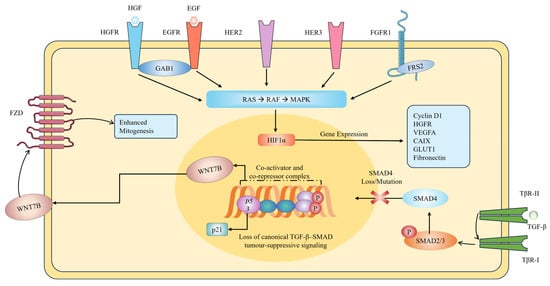
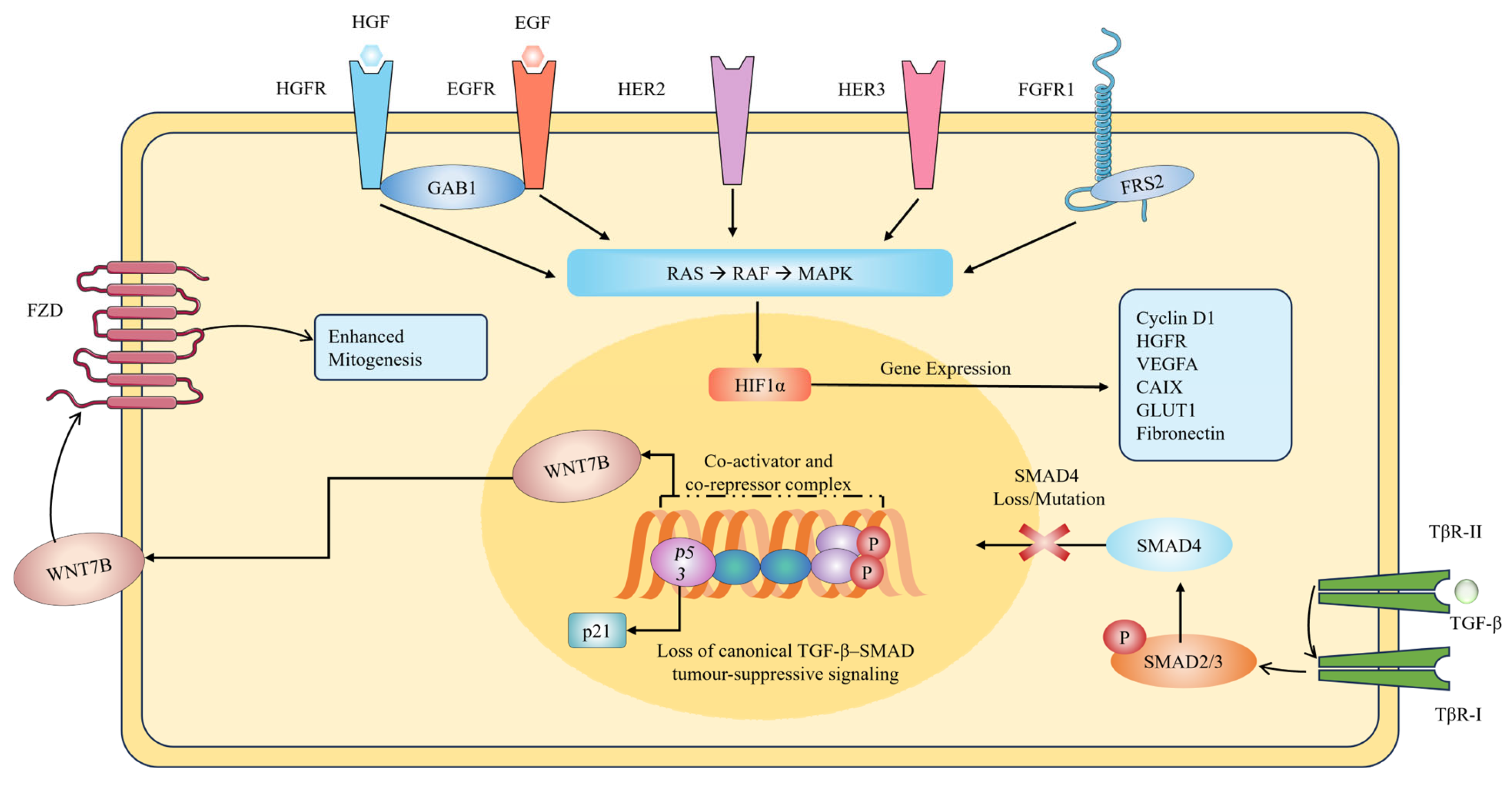
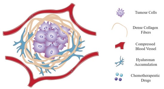
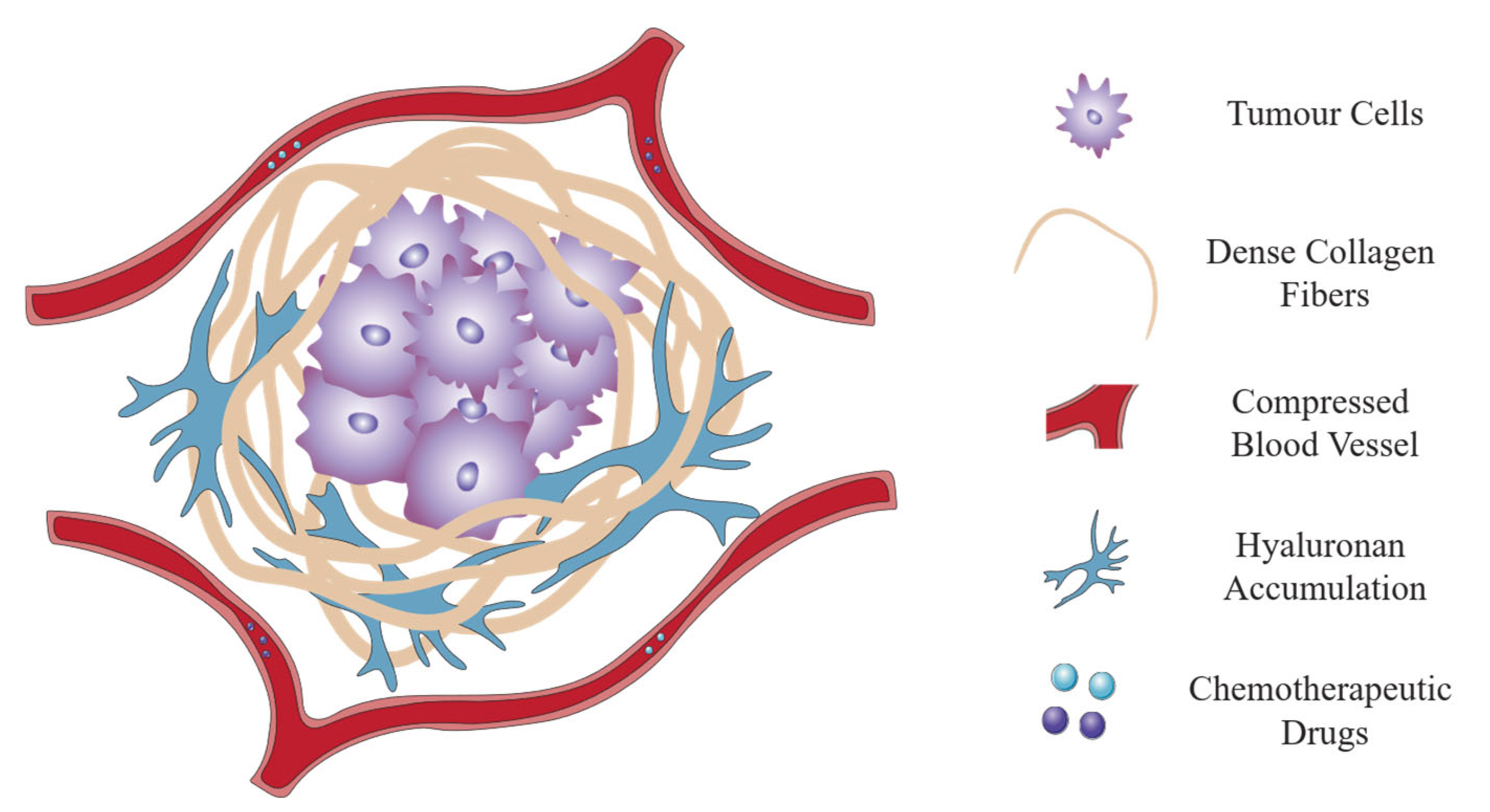
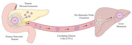
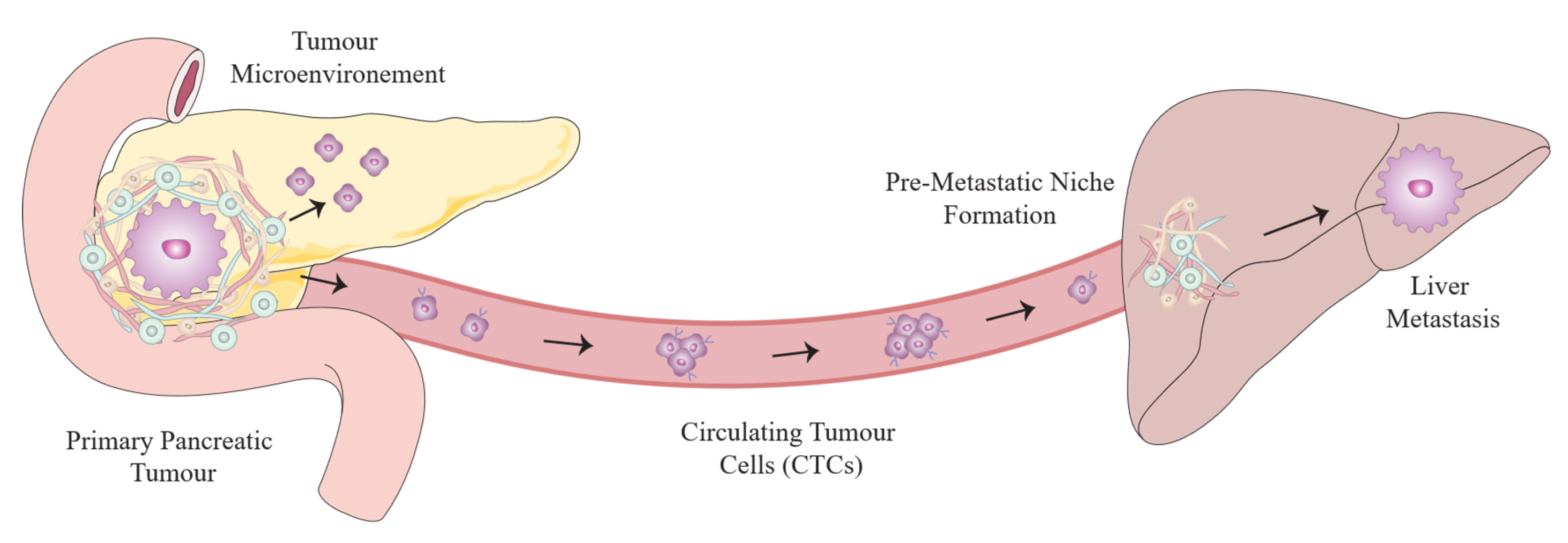

Disclaimer/Publisher’s Note: The statements, opinions and data contained in all publications are solely those of the individual author(s) and contributor(s) and not of MDPI and/or the editor(s). MDPI and/or the editor(s) disclaim responsibility for any injury to people or property resulting from any ideas, methods, instructions or products referred to in the content. |
© 2026 by the authors. Licensee MDPI, Basel, Switzerland. This article is an open access article distributed under the terms and conditions of the Creative Commons Attribution (CC BY) license.