


Abstract
Background: An estimated 28,900 deaths around the world in 2021 were attributed to unintentional CO poisoning. Following inhalation, CO binds to hemoglobin with an affinity 220–240 times greater than that of oxygen to form carboxyhemoglobin (COHb). While the constituents of CO exposure are known to determine CO uptake in the blood, much less is understood regarding the individual variability of the response to a given CO stimulus. Thus, the purpose of this paper was to explore the relationship between hemoglobin mass (HbM, a proxy for blood hemoglobin content) and the magnitude of the ensuing carboxyhemoglobinemia. Methods: This is a theoretical work based solely on considerations and published data. Discussion: Currently considered the gold standard for HbM assessment, the CO-rebreathing technique relies on the dilution principle, i.e., the lower the HbM value, the higher the ΔCOHb following a standardized CO bolus administration or an outdoor exposure. Accordingly, previously published prediction equations with HbM and ΔCOHb as the predictor and outcome variables, respectively, are reviewed with particular reference to the (confounding) factor of pulmonary ventilation. Screening procedures aiming to identify individuals susceptible to CO poisoning could henceforth include HbM assessments.
1. Introduction
In recent news, 14 people were hospitalized after exhibiting symptoms of carbon monoxide (CO) poisoning in Northern Greece; the inhalation was attributed to a gas leak from a hotel’s heating system. Common symptoms—widely reported in these cases of CO poisoning—include headache, weakness, dizziness, confusion, shortness of breath, and drowsiness that precede the loss of consciousness in victims. An estimated number of 28,900 deaths worldwide in 2021 were attributed to unintentional CO poisoning. The severity of CO toxicity symptoms may be reflected in the concomitant rise in carboxyhemoglobin (COHb, measured as a percentage) levels. COHb level has been repeatedly claimed to be an insufficient measure of the severity of CO poisoning. Such deviation observed between COHb levels and clinical severity might be attributed to the fact that the reduction in oxygen-carrying capacity—evidenced by increased COHb—might be a secondary mechanism by which CO causes toxicity. Goldbaum et al. [1] demonstrated in dogs that an increase in the partial pressure of CO in the blood seems to directly inhibit mitochondrial respiration as the gas binds with cytochrome oxidase. Specifically, dogs that had been transfused with blood with a COHb of 80% survived, whereas dogs that inhaled CO and reached average COHb values of 65% died within 65 min. The toxic effects of CO extend beyond the formation of COHb and include interactions with intracellular heme-containing proteins that contribute to tissue injury [2].
Other factors that may explain this discrepancy include the delay between the termination of CO exposure and the time blood is drawn for COHb measurement, as well as the administration of oxygen before the sample is obtained [3]. For the purposes of the present discussion, COHb refers to the actual in vivo COHb concentration rather than the measured blood COHb value. Notwithstanding relevant research work, COHb should still be measured when CO poisoning is suspected, as higher values have been associated with specific symptoms such as lower pH values and a higher frequency of loss of consciousness and death [3]. Previous attempts have been made to account for the kinetics of CO distribution inside the human body or even the diffusion of CO in compartments other than blood. For a comprehensive overview of the topic, the reader is further directed to the multi-compartmental model developed by Bruce and Bruce [4] that expanded the seminal work by Coburn et al. [5]. It is, therefore, important to investigate the overlooked determinants of individual variability in CO poisoning [6]. The purpose of this manuscript is to highlight a relatively underappreciated role of HbM as a physiological buffer against undesirable increases in ΔCOHb. High HbM levels may protect humans against acute CO exposure, at least under certain physiological conditions.
2. Exposure to Carbon Monoxide and Carboxyhemoglobinemia
2.1. Carboxyhemoglobinemia During CO-Rebreathing
The magnitude of the COHb increase is determined by not only the CO level and the duration of the exposure but also by other underlying factors that might demonstrate high inter-individual variability, with the first and foremost example being the total amount of hemoglobin available for CO binding. Though traditionally used, the concentration-based index of hemoglobin concentration ([Hb]) is dependent on plasma volume and, as such, might provide erroneous information on hemoglobin content, especially in cases of hemoconcentration (as occurs during exercise and dehydration, among other physiological instances). Yet, this is not the case for hemoglobin mass (HbM). Accordingly, individuals at the lowest end of the HbM spectrum should be characterized as a cohort at high risk for CO poisoning. Low CO doses are currently administered according to body mass (BM) and biological sex for diagnostic purposes [7]. The dilution principle implies that the higher the increase in COHb following a standardized CO bolus administration, the lower HbM values. Alternatively viewed, a higher baseline HbM might be suspected to protect the human body from a toxic (or even lethal) elevation in COHb. A COHb of 40% has been considered as a reasonable threshold for lethality, whereas excessive symptoms are experienced when COHb is >20% in healthy adults [8].
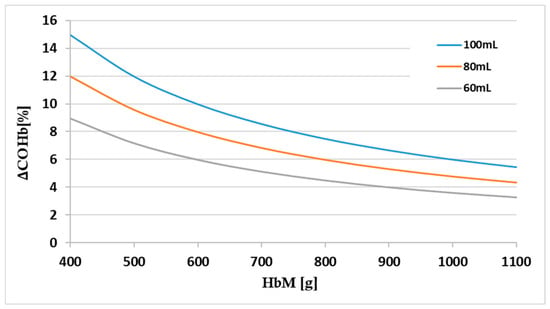
Despite the ever-increasing number of publications that rely on CO rebreathing to measure HbM and the resultant intravascular volumes, a scarcity of studies have published individual raw ΔCOHb data. As such, it remains tedious to extrapolate the highest COHb values attained from the reported HbM values. A relatively old formula [9] has recently resurfaced to estimate the CO bolus required to elevate ΔCOHb to a prespecified value [10]. The exact same equation could be modified to assess ΔCOHb in response to a CO bolus as a function of HbM and BM (Figure 1):
ΔCOHb [%] = 100 × CO [mL]/(HbM[g] × 1.34 [mL·g−1])
= 100 × CO [mL·kg−1] × BM [kg]/(HbM[g] × 1.34 [mL·g−1])
= 100 × CO [mL·kg−1] × BM [kg]/(HbM[g] × 1.34 [mL·g−1])
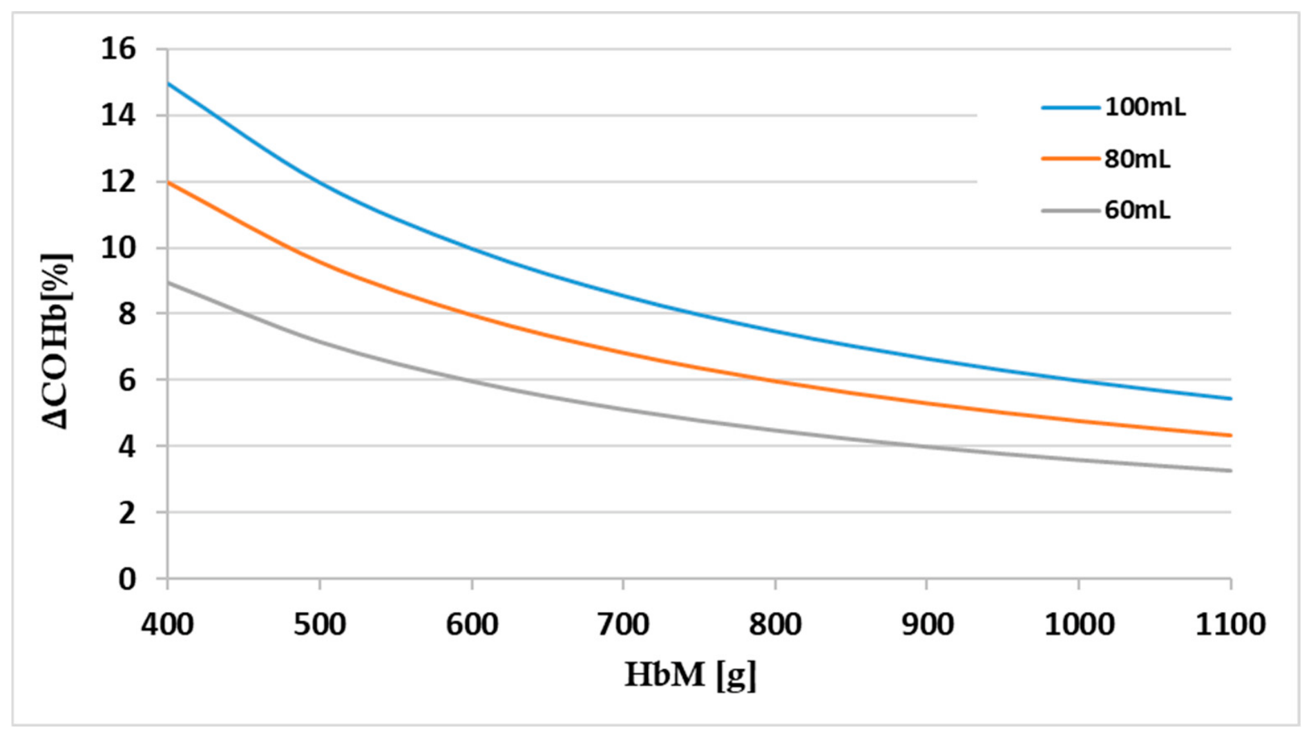
Figure 1.
The increase in carboxyhemoglobin (ΔCOHb, %) as a function of hemoglobin mass (HbM, g) (indicative values ranging between 400 and 1100 g) at different CO boluses (mL). Bolus stands for a CO dose administered via a closed rebreathing circuit under controlled laboratory conditions for a defined period (typically 2–10 min).
An illustrative example of the application of Equation (1) might be reflected in how closely reported COHb values lie to calculated values. Assuming back-to-back (duplicate) doses of 1.0 and 0.8 mL·kg−1 (for healthy men and women, respectively), accounting for total CO boluses of 2.0 and 1.6 mL·kg−1, the calculated ΔCOHb values actually coincide with the values reported [women: 12.2 vs. 12.5 ± 2.4%; men: 10.1 vs. 10.2 ± 1.1%] in the study by DiMarco et al. [11]. Marginal differences emerged when data from previous studies were also used [12]. It is imperative that researchers report separate ΔCOHb data for each time point (of HbM) so that this reverse calculation can be feasible.
2.2. Carboxyhemoglobinemia in Applied Environments
Equation (1) is only applicable in the case of CO bolus administration applied in controlled rebreathing procedures. Thus, minor differences in the COHb response should be viewed in light of the rather limited CO dose allowed to be provided in laboratory settings. From an occupational perspective, outdoor exposure will not be fitted to match the margin of safety for each individual. Notwithstanding the debilitating effect on exercise performance, COHb values as low as 5% over a 4 h exposure time have been shown to impair cognitive function [13] and increase the incidence of work accidents [14]. Exposure to CO should instead be expressed as ambient air CO concentration, which has been thought to be proportional to blood COHb concentration [5]. Forbes et al. [15] expressed ΔCOHb as a function of exposure time and air CO concentration for different physical activity levels. Their work was extended by Pace et al. [16], who highlighted that total blood volume would act as a governing factor of the increase in COHb:
where TBV: total blood volume to be measured using the CO-rebreathing technique; VT: pulmonary ventilation; t: exposure time; and k1: regression coefficient (equal to 4650 for the data in Pace, Consolazio, White Jr. and Behnke [16]).
ΔCOHb [%] = [CO] [ppm] × VT [L·min−1] × t [min]/k1 × TBV [L]
Equation (2) should be further viewed with caution since it is designed for lengthy exposures and applied to COHb values far from equilibrium with air CO concentration. A hyperbolic relationship between duration of exposure and ambient CO concentration is discernible in the standards set by respective occupational health bodies. The World Health Organization recommends short-term exposure limits of 25–35 ppm for up to 60 min or 90–100 ppm for 15 min. On the other end of the dose spectrum, a steady-state exposure to CO concentrations as low as 10 ppm is allowed for a work shift of 8 h. Simply put, the product of exposure duration and ambient CO concentration could serve as a proxy index of the environment-elicited exogenous CO strain ([CO] × t in ppm × min). Deciphering individual variability in CO poisoning demands that factors that differentiate the response to this standardized exogenous CO strain be elucidated.
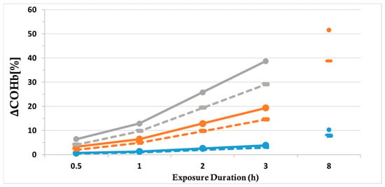
The CO-rebreathing technique allows for the valid assessment of total blood volume [7,17]. A group of researchers that have been assessing HbM and intravascular volumes for the last 15 years using this technique convincingly reported that differences in lean body mass, (endurance) training level and sex account for HbM and total blood volume heterogeneity at the population scale [18]. Accordingly, high inter-individual differences of up to 2.5 L in magnitude in total blood volume stores occurring between pupils of a younger age (9 yrs) and adults would result in COHb increases as high as 6.0% (19.71 vs. 13.68%) for the life-threatening CO exposure that has been historically described at a school facility [19]; representative values selected for [CO], VT, t and TBV were 500 ppm, 5.5/7 L·min−1, 100 min, and 3/5.5 L for children and adults, respectively. Along the same line of reasoning, Figure 2 demonstrates that for hour-long exposures to high ambient CO concentrations that could put human health in danger, the COHb increase might be altered by as much as ~10% for a TBV difference of 3–4 L (such as that reported between a trained 80 kg endurance athlete and a 60 kg non-exercising individual [18]).
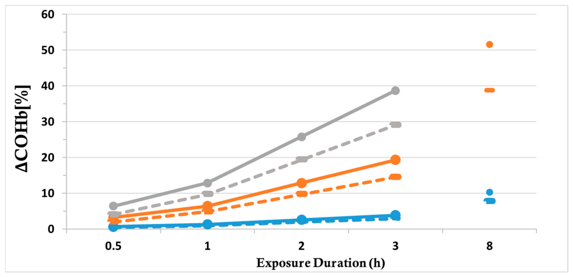
Figure 2.
The increase in carboxyhemoglobin (ΔCOHb, %) as a function of exposure duration (min) and ambient CO concentration (ppm) at different levels of total blood volume. Solid and dashed lines represent blood volumes of 4 L and 8 L, respectively. Blue, orange and gray lines represent ambient concentrations of 100, 500, and 1000 ppm, respectively. Values are derived using Equation (2).
Table 1 summarizes the predicted COHb responses across individuals representing the extremes of the age, sex, and hematological distributions.
Table 1.
Predicted ΔCOHb responses across individuals of different ages, sexes, and hematological characteristics. Results are shown for an exposure duration of 180 min and an ambient CO concentration of 500 ppm, as calculated using Equation (2) (see main text). Mean ± SD values were extracted from previously published studies: young adult males, young adult females, older adult males, older adult females [18], children [20], young anemic males [21], and young anemic females [22]. Estimated 25th and 75th percentile values were calculated assuming a normal distribution for HbM.
Equation (2) and Table 1 elegantly highlight that the VT/HbM ratio seems critical for ΔCOHb when humans are exposed to high-CO-concentration environments. Not surprisingly, the difference in ΔCOHb becomes marginal when males are compared to females (Table 3 in Zavorsky et al. [23]). Specifically, no difference in COHb was observed between males and females that inhaled a similar CO dose for a time interval of 30 min. Females are known to possess lower total blood volume values and HbM [18,24] and, thus, would be expected to experience a higher ΔCOHb for the same exposure. Even though there have been reports of a higher incidence of CO poisoning-induced syncope [25], recent data indicating a lower severity classification and higher cure and improvement rate in females [26] argue against this case. These findings may be accounted for by lower values of VT [23,27] and lung-diffusing (DLCO) capacity [23] in females that act to delay CO loading in the lungs. Yet, VT could be a double-edged sword. During uptake, it facilitates loading of the gas in the blood, whereas during the washout/elimination period or in cases of endogenous production, it has been modeled to remove CO into the environment [5]. Quite interestingly, HbM, but not alveolar ventilation rate, seems to explain sex differences in CO elimination [23]. Counterintuitively, a greater HbM has been shown to hasten CO removal once COHb has reached a threshold level of 10–15% [23]. The effect of HbM was speculated to be dampened at high alveolar ventilation rates even though the authors only calculated HbM using anthropometric data. We have now performed a sensitivity analysis to establish whether changes in HbM are comparable to those of other determinant factors of the COHb response. As can be seen in Table 2, a fixed change in the independent variable within a clinically realistic range yields a consistent effect on ΔCOHb (~±4–5%). Of note, high ventilation rates might allow for even higher ΔCOHb values (+38.7% for a VT of 10 L·min−1), in agreement with the findings of the previous study. Acute episodes of anxiety may similarly increase VT. However, given the expectedly limited duration of the response, it is not considered to contribute to a substantial degree to CO uptake. The kinetics of CO elimination will be subsequently addressed only in the context of breathing supplementary oxygen, since poisoned individuals are typically treated in this manner. Future studies are warranted to investigate sex-based differences in ΔCOHb while controlling for the plethora of earlier stated confounding factors. A particular reference needs to be made to pregnant women; an average TBV increase as high as ~1.7 L is reported (450 mL in red cell volume, 1250 mL in plasma volume) [28]. However, the fact that fetal hemoglobin binds CO more avidly than adult hemoglobin [29] renders the fetus particularly vulnerable to CO poisoning.
Table 2.
ΔCOHb response to fixed (±20%) within-individual alterations in HbM, VT and TBV. Reference values are 5 L, 500 ppm, 6 L·min−1, 180 min and 750 g for TBV, ambient CO concentration, VT, exposure time and HbM, respectively.
The previous discussion emphasizes a number of constraints associated with the use of Equation (2). The use of multiple linear regression might not be appropriate since inter-individual variation in the hemoglobin content of the blood is not accounted for; not all individuals possessing a standard volume of blood will demonstrate the same ΔCOHb for a given exposure and VT. By solving the differential equation first proposed by Coburn, Forster and Kane [5] to model CO removal into the environment over time, Tikuisis et al. [30] elegantly reflected the fact that the CO concentration in the blood mainly depends on HbM:
where PB is barometric pressure and β is inversely proportional to DLCO and alveolar ventilation.
ΔCOHb [%] = [CO] [ppm] × (PB − 47) [mmHg] × 10−2 × t [min]/(1.39 × β × HbM [g])
However, employing such an analytical approximation is valid only for short-duration CO exposures, where COHb kinetics can be assumed to be approximately linear. A thorough discussion on other determinant factors that drive CO absorption away from linearity is presented in Section 3. Another notable observation arising from Equation (3) is that ΔCOHb might be theoretically altered even within individuals as part of an adaptive response, i.e., repeated exposure to CO could augment HbM that would subsequently blunt the increase in COHb in response to a given stimulus. Indeed, an erythropoietic response has been shown to unfold in groups of individuals that are regularly exposed to considerable amounts of CO. Schmidt et al. [31] recently reported an erythropoiesis-related elevation in HbM following 3-week exposure of non-smoking non-athletes to multiple daily CO doses that would maintain their COHb levels at 8%. Remarkably, the “upscaling pattern” previously expected to unfold across the athletes [32] seemed to spread across the researchers. In an approach more oriented towards application, Urianstad, Villanova, Odden, Hansen, Molmen, Porcelli, Ronnestad and Cardinale [10] had elite cyclists live and train in natural hypoxia while the experimental group was additionally inhaling CO twice daily in the late afternoon/evening hours with a target COHb of 10%. In the most recent study, eight well-trained swimmers were inhaling three daily doses that would elevate their COHb to 15% for a time interval of four weeks [33]. From a cross-adaptation perspective, occupational groups such as chronic smokers [34], firefighters [35], workers that inhale exhaust fumes [36] and cooks that use “smoky” fires [37] might benefit from strategies aiming to enhance the buffering action of the additional hemoglobin molecules and including exercise training [38], hypoxic acclimatization [39] and long-term heat acclimation [40]. Interestingly, these strategies only allow for minor hematological gains but would act to reverse smoking-elicited cardiopulmonary disorders [41] and further elicit thermoregulatory adaptations [42,43,44] especially desirable in the occupational group of firefighters and endurance athletes exercising in polluted environments. Independent of the target COHb concentration and as far as intra-individual susceptibility to CO poisoning is concerned, the magnitude of the reduction in ΔCOHb is shown to be regrettably small for a HbM gain of 50 g (evident in the former studies), which could be already offset by an elevated baseline COHb as elicited by smoking or any other exogenous CO source. Indeed, the cited studies report that smoking workers demonstrated higher baseline COHb levels that would result in their COHb values ending up higher at the end of the work shift. Viewed from a different angle, workers should enter the work shift having abstained from smoking for >12 h [34] as the time elapsed from the previous shift has been shown to be adequate [35,45]. Notwithstanding the major assets associated with measures of HbM and intravascular volumes, the CO-rebreathing technique requires specialized equipment, trained personnel, and appropriate safety procedures [46]. An extensive network of healthcare facilities and the corresponding training of medical personnel could support the regular screening of occupational groups expected to be exposed to CO, not only as part of diagnosis and treatment but also for prevention purposes. Another group of interest might be individuals diagnosed with a cardiorespiratory disease. Heart failure patients have been reported to exhibit an inflammation-related increased baseline COHb [2], together with rather low values of HbM and plasma volume [47]. If not for the direct evaluation of HbM by means of the CO-rebreathing technique, self-reports of physical activity and health status might provide the foundation for evidence-based guidelines on the selection of individuals to be exposed to CO in occupational settings [18]. A combination of anthropometric (height, weight, and lean body mass) and demographic (age) variables has been suggested to adequately estimate TBV and HbM when the CO-rebreathing technique cannot be used in applied settings [18]. Supporting these models with routine complete blood count parameters such as [Hb]/Hct helps estimate individual HbM and might adequately anticipate the subsequent ΔCOHb response.
The proposed framework does not signify a new model; rather, it could be deemed as an extension of existing models that leverages methodological advancements already used for the precise determination of HbM and intravascular volumes. Such hematological parameters were only assigned reference values when ΔCOHb was to be assessed. Furthermore, the present manuscript encourages readers to reinterpret the well-established dilution technique as a reciprocal relationship between HbM and ΔCOHb.
3. Other Determinants of Susceptibility to Carbon Monoxide Poisoning
Beyond the described potential of HbM, other factors have been claimed to characterize individual susceptibility to CO poisoning, including VT, tissue diffusion characteristics, cardiac output, exposure duration and organ-specific oxygen demands. VT and HbM both scale with anthropometric characteristics such as body size and lean body mass. As such, larger individuals generally are characterized by higher VT [48] and possess a larger hemoglobin reservoir [18]. As previously discussed, the ratio between these variables is expected to partly determine the COHb response. The same applies for exposure dose expressed either as ambient CO concentration (ppm) or as a rebreathing bolus (mL). The term “bolus” refers to a body-mass-normalized volume of CO administered through a closed rebreathing circuit under strictly controlled laboratory conditions for a predefined period (typically 2–10 min). By contrast, occupational CO exposure is defined as the concentration of CO in ambient air (ppm). Consequently, these two metrics are not directly comparable: CO rebreathing delivers a relatively large amount of CO over a short period, whereas ambient exposure generally results in a much lower uptake rate, reaching a comparable absorbed CO dose only after prolonged exposure lasting several hours. Irrespective of the nature of the exposure, a biphasic increase in COHb will expectedly be observed with a steep increase during the early phase, subsequently followed by a leveling off [49]. As soon as individuals are withdrawn from the CO stimulus, COHb will be gradually reduced as they will start exhaling CO into the environment [50].
Indirect reference to tissue diffusion characteristics has already been made by virtue of Equation (3), which signifies that the higher the DLCO, the higher the ΔCOHb. This could explain why the increase in COHb seems similar for a given—short—CO exposure between children and adults if differences in DLCO (ranging between 15 and 40 mL·min−1·mmHg−1 for children and adult males, respectively) and VT are accounted for. The potential for a higher DLCO to elicit faster CO uptake is illustrated by the fact that COHb reaches a plateau within as little as 2 min in humans performing mild cycling exercise while connected to a closed CO-rebreathing system [51]. Stated differently, CO transfer across the lung is diffusion-limited. As such, increases in cardiac output and the concomitant pulmonary blood flow do not seem to directly affect CO uptake.
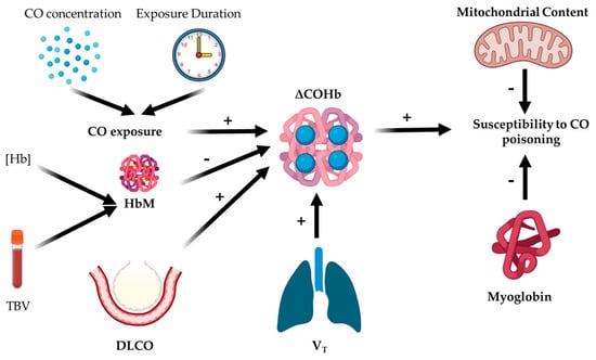
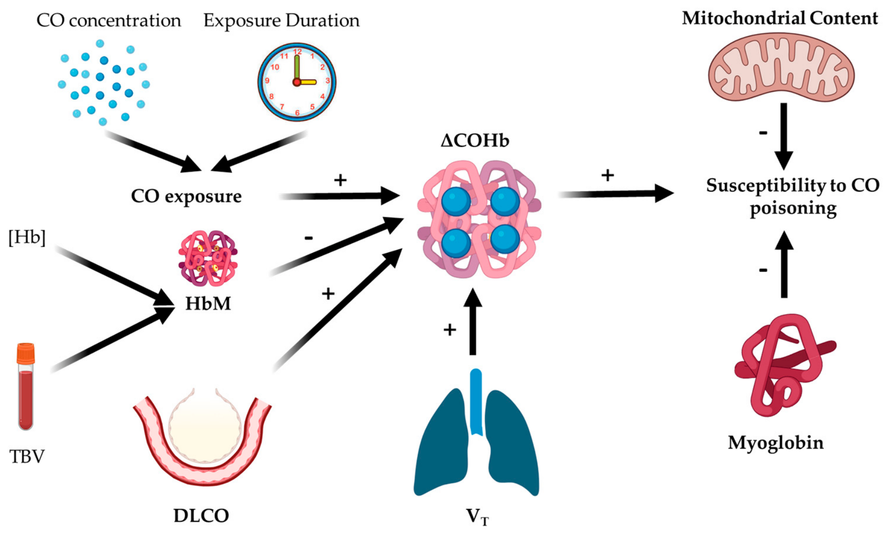
Interindividual variability in tissue susceptibility may depend not only on CO uptake kinetics but also on the size of the body heme reserves. Even though the binding capacity of hemoglobin remains much higher, myoglobin might act as an additional CO intracellular buffer. Individuals with greater skeletal muscle mass will further possess a higher mitochondrial content and could potentially demonstrate an increased tolerance to partial mitochondrial inhibition; such dysfunction has been shown in vitro after human mitochondria were incubated with CO [52] or in mitochondria extracted from individuals that suffered CO poisoning [53]. In this framework, greater HbM, skeletal muscle mass, myoglobin stores, and oxidative capacity could collectively increase the human body’s capacity to distribute, sequester, and tolerate a given CO burden. Figure 3 illustrates the proposed conceptual framework linking HbM, ΔCOHb and other physiological determinants of CO toxicity.
Figure 3.
Schematic illustration of the proposed determinants of the increase in ΔCOHb following CO exposure. CO toxicity is likely governed by additional physiological determinants beyond ΔCOHb. Positive (+) and negative (−) signs denote factors expected to increase susceptibility to CO poisoning, respectively. Abbreviations: CO, carbon monoxide; ΔCOHb, change in carboxyhemoglobin; HbM, hemoglobin mass; [Hb], hemoglobin concentration; TBV, total blood volume; DLCO, lung diffusing capacity for carbon monoxide; VT, pulmonary ventilation.
4. Prevention and Cure
In the undesirable event of CO poisoning, individuals should act to accelerate CO elimination. On-site access to 100% normobaric oxygen is deemed necessary to reduce the COHb half-life [54]—by increasing the partial pressure of oxygen—before professional medical care arrives. The use of oxygen concentrators renders oxygen therapy possible even on-field. In severe cases, even the application of hyperbaric oxygen therapy (2.5–3.0 ATA) might be enforced to drop COHb half-life down to ~20–25 min in healthy individuals [55]. Other countermeasures could be considered if the deleterious effect of hypocapnia on cerebral oxygen delivery is to be avoided. Specifically, normocapnic hyperoxic hyperpnea has been proposed to accelerate CO clearance (i.e., a rectangular hyperbolic relationship between CO half-life and minute ventilation applies) when compared to pure oxygen administration [56]. Regardless of the physiological mechanisms serving as guidelines for the selection of the most appropriate countermeasure, minimizing the time elapsing between exposure and treatment remains of the highest priority so long as CO poisoning-symptom severity is to be reduced [57]. The wide range of COHb measured in poisoned patients might have been mediated by differences in the time elapsing between withdrawal from the CO source and blood sampling [58].
Prevention dominates over cure; since CO is a colorless, odorless, tasteless gas, the precautionary measure of installing audible low-cost CO sensors should be enforced, especially in occupational areas where a CO leak might affect an excessive number of individuals. Despite showing limited precision [59], the non-invasive determination of COHb arising from the use of pulse CO-oximetry may facilitate the rapid identification of individuals with suspected CO poisoning in the field. However, given its suboptimal accuracy, it should be regarded as a complementary screening tool rather than a substitute for standard blood gas analysis.
Although CO is primarily considered in the present context because of its toxicity, endogenously generated CO also participates in physiological signaling. The binding of CO to heme-containing proteins may modify their activity and thereby regulate cellular processes. Indeed, CO-dependent regulation has been described for cystathione β-synthase and PGRMC1 [60,61]. Thus, the biological consequences of CO–heme interactions extend beyond the impaired oxygen delivery to tissues associated with COHb formation.
5. Conclusions
HbM has been mostly used as a surrogate index of aerobic performance [15]. Nevertheless, inter-individual susceptibility to carbon monoxide poisoning is currently investigated by revisiting (the implications of) equations that solidify the inverse relationship between HbM/TBV and ΔCOHb. Aiming at the expansion of this basis, future research needs to further elaborate on the potential buffering action of hemoglobin molecules against undesirable increases in COHb. To that end, prospective studies (that will apply the CO rebreathing technique coupled with measurements of pulmonary ventilation) could provide a screening method to identify population groups that are particularly vulnerable to CO poisoning. These groups include, but are not limited to, children, females, individuals with anemia/hypovolemia, and occupational populations exposed to elevated ambient CO concentrations. Given the inherent risks associated with CO inhalation in these specific groups, dedicated human modeling studies are warranted to better elucidate relevant physiological responses and inform safer screening protocols. Multilevel modeling could be considered in future empirical studies (particularly when repeated/hierarchically structured data are available) to assess the contribution of HbM in conjunction with other individual- and exposure-related determinants of CO susceptibility.
Author Contributions
Conception or design of the work: A.S., A.M., N.D.G. and M.K.; acquisition, analysis, or interpretation of data for the work: A.S.; drafting the work or revising it critically for important intellectual content: A.S., A.M., N.D.G. and M.K.; final approval of the version to be published: A.S., A.M., N.D.G. and M.K.; agreement to be accountable for all aspects of the work in ensuring that questions related to the accuracy or integrity of any part of the work are appropriately investigated and resolved: A.S., A.M., N.D.G. and M.K. All authors have read and agreed to the published version of the manuscript.
Funding
The authors report that there was no funding source for the work that resulted in the article or the preparation of the article.
Institutional Review Board Statement
An institutional review board decision is not included since no primary/secondary data were analyzed.
Informed Consent Statement
An informed consent is not included since no primary/secondary data were analyzed.
Data Availability Statement
No new data were created.
Conflicts of Interest
The authors declare there are no conflicts of interests.
Abbreviations
The following abbreviations are used in this manuscript:
| CO | Carbon monoxide |
| COHb | Carboxyhemoglobin |
| DLCO | Lung-diffusing capacity |
| [Hb] | Hemoglobin concentration |
| HbM | Hemoglobin mass |
| BM | Body mass |
| VT | Pulmonary ventilation |
| TBV | Total blood volume |
| PB | Barometric pressure |
| ppm | Parts per million |
| ATA | Atmospheres absolute |
| WHO | World Health Organization |
References
- Goldbaum, L.R.; Orellano, T.; Dergal, E. Mechanism of the toxic action of carbon monoxide. Ann. Clin. Lab. Sci. 1976, 6, 372–376. [Google Scholar] [PubMed]
- Foresti, R.; Bani-Hani, M.G.; Motterlini, R. Use of carbon monoxide as a therapeutic agent: Promises and challenges. Intensive Care Med. 2008, 34, 649–658. [Google Scholar] [CrossRef] [Scilit] [PubMed]
- Hampson, N.B.; Hauff, N.M. Carboxyhemoglobin levels in carbon monoxide poisoning: Do they correlate with the clinical picture? Am. J. Emerg. Med. 2008, 26, 665–669. [Google Scholar] [CrossRef] [Scilit] [PubMed]
- Bruce, M.C.; Bruce, E.N. Analysis of factors that influence rates of carbon monoxide uptake, distribution, and washout from blood and extravascular tissues using a multicompartment model. J. Appl. Physiol. 2006, 100, 1171–1180. [Google Scholar] [CrossRef] [Scilit] [PubMed]
- Coburn, R.F.; Forster, R.E.; Kane, P.B. Considerations of the physiological variables that determine the blood carboxyhemoglobin concentration in man. J. Clin. Investig. 1965, 44, 1899–1910. [Google Scholar] [CrossRef] [Scilit] [PubMed]
- Gatterer, H.; Dünnwald, T.; Woyke, S.; Faulhaber, M.; Schumacher, Y.O.; Schobersberger, W. Low-dose carbon monoxide inhalation to increase total hemoglobin mass and endurance performance: Scientific evidence and implications. Front. Physiol. 2024, 15, 1490205. [Google Scholar] [CrossRef] [Scilit] [PubMed]
- Schmidt, W.; Prommer, N. The optimised CO-rebreathing method: A new tool to determine total haemoglobin mass routinely. Eur. J. Appl. Physiol. 2005, 95, 486–495. [Google Scholar] [CrossRef] [Scilit] [PubMed]
- Nelson, G. Effects of carbon monoxide in man: Exposure fatality studies. In Carbon Monoxide and Human Lethality: Fire and Non-Fire Studies; CRC Press: Boca Raton, FL, USA, 2006; pp. 3–62. [Google Scholar]
- Ekblom, B.; Huot, R. Response to submaximal and maximal exercise at different levels of carboxyhemoglobin. Acta Physiol. Scand. 1972, 86, 474–482. [Google Scholar] [CrossRef] [Scilit] [PubMed]
- Urianstad, T.; Villanova, S.; Odden, I.; Hansen, J.; Molmen, K.S.; Porcelli, S.; Ronnestad, B.R.; Cardinale, D.A. Carbon monoxide Supplementation: Evaluating its Potential to Enhance Altitude Training Effects and Cycling Performance in Elite Athletes. J. Appl. Physiol. 2024, 137, 1092–1105. [Google Scholar] [CrossRef] [Scilit] [PubMed]
- DiMarco, K.G.; Chapman, C.L.; Weiser, N.E.; Matsell, E.R.; Lucernoni, K.M.; Chacon, S.; Grivette, M.M.B.; Halliwill, J.R.; Lovering, A.T.; Minson, C.T. Acute exposure to carbon monoxide inhalation and/or hot water immersion transiently increases erythropoietin in females but not in males. Exp. Physiol. 2024, 109, 1782–1795. [Google Scholar] [CrossRef] [Scilit] [PubMed]
- Naef, N.; Steiner, T.; Wehrlin, J.P. Replicate Measurements of Haemoglobin Mass during a Single Day are Feasible and Precise. Int. J. Sports Med. 2015, 36, e19–e23. [Google Scholar] [CrossRef] [Scilit] [PubMed]
- Putz, V.R. The effects of carbon monoxide on dual-task performance. Hum. Factors 1979, 21, 13–24. [Google Scholar] [CrossRef] [Scilit] [PubMed]
- Wadsworth, E.J.; Simpson, S.A.; Moss, S.C.; Smith, A.P. The Bristol Stress and Health Study: Accidents, minor injuries and cognitive failures at work. Occup. Med. 2003, 53, 392–397. [Google Scholar] [CrossRef] [Scilit] [PubMed]
- Forbes, W.H.; Sargent, F.; Roughton, F.J.W. The rate of carbon monoxide uptake by normal men. Am. J. Physiol. 1945, 143, 594–608. [Google Scholar] [CrossRef] [Scilit]
- Pace, N.; Consolazio, W.V.; White, W.A., Jr.; Behnke, A. Formulation of the principal factors affecting the rate of uptake of carbon monoxide by man. Am. J. Physiol. 1946, 147, 352–359. [Google Scholar] [CrossRef] [Scilit] [PubMed]
- Burge, C.M.; Skinner, S.L. Determination of hemoglobin mass and blood volume with CO: Evaluation and application of a method. J. Appl. Physiol. 1995, 79, 623–631. [Google Scholar] [CrossRef] [Scilit] [PubMed]
- Oberholzer, L.; Montero, D.; Robach, P.; Siebenmann, C.; Ryrsoe, C.K.; Bonne, T.C.; Breenfeldt Andersen, A.; Bejder, J.; Karlsen, T.; Edvardsen, E.; et al. Determinants and reference values for blood volume and total hemoglobin mass in women and men. Am. J. Hematol. 2024, 99, 88–98. [Google Scholar] [CrossRef] [Scilit] [PubMed]
- Burney, R.E.; Wu, S.C.; Nemiroff, M.J. Mass carbon monoxide poisoning: Clinical effects and results of treatment in 184 victims. Ann. Emerg. Med. 1982, 11, 394–399. [Google Scholar] [CrossRef] [Scilit] [PubMed]
- Mancera-Soto, E.M.; Ramos-Caballero, D.M.; Rojas, J.J.; Duque, L.; Chaves-Gomez, S.; Cristancho-Mejia, E.; Schmidt, W.F. Hemoglobin Mass, Blood Volume and VO2max of Trained and Untrained Children and Adolescents Living at Different Altitudes. Front. Physiol. 2022, 13, 892247. [Google Scholar] [CrossRef] [Scilit] [PubMed]
- Otto, J.M.; Plumb, J.O.M.; Clissold, E.; Kumar, S.B.; Wakeham, D.J.; Schmidt, W.; Grocott, M.P.W.; Richards, T.; Montgomery, H.E. Hemoglobin concentration, total hemoglobin mass and plasma volume in patients: Implications for anemia. Haematologica 2017, 102, 1477–1485. [Google Scholar] [CrossRef] [Scilit] [PubMed]
- Wachsmuth, N.B.; Aigner, T.; Volzke, C.; Zapf, J.; Schmidt, W.F. Monitoring recovery from iron deficiency using total hemoglobin mass. Med. Sci. Sports Exerc. 2015, 47, 419–427. [Google Scholar] [CrossRef] [Scilit] [PubMed]
- Zavorsky, G.S.; Tesler, J.; Rucker, J.; Fedorko, L.; Duffin, J.; Fisher, J.A. Rates of carbon monoxide elimination in males and females. Physiol. Rep. 2014, 2, e12237. [Google Scholar] [CrossRef] [Scilit] [PubMed]
- Falz, R.; Fikenzer, S.; Hoppe, S.; Busse, M. Normal Values of Hemoglobin Mass and Blood Volume in Young, Active Women and Men. Int. J. Sports Med. 2019, 40, 236–244. [Google Scholar] [CrossRef] [Scilit] [PubMed]
- Keles, A.; Demircan, A.; Kurtoglu, G. Carbon monoxide poisoning: How many patients do we miss? Eur. J. Emerg. Med. 2008, 15, 154–157. [Google Scholar] [CrossRef] [Scilit] [PubMed]
- Huijun, H.; Qiang, S.; Dazhi, G.; Yu, Z.; Yan, L.; Shuyi, P.; Xuejun, S. Sex differences may affect the severity of poisoning and prognosis after carbon monoxide poisoning: A retrospective study. Undersea Hyperb. Med. 2016, 43, 207–215. [Google Scholar] [PubMed]
- Sheel, A.W.; Richards, J.C.; Foster, G.E.; Guenette, J.A. Sex differences in respiratory exercise physiology. Sports Med. 2004, 34, 567–579. [Google Scholar] [CrossRef] [Scilit] [PubMed]
- Hytten, F. Blood volume changes in normal pregnancy. Clin. Haematol. 1985, 14, 601–612. [Google Scholar] [CrossRef] [Scilit]
- Engel, R.R.; Rodkey, F.L.; O’Neal, J.D.; Collison, H.A. Relative affinity of human fetal hemoglobin for carbon monoxide and oxygen. Blood 1969, 33, 37–45. [Google Scholar] [CrossRef] [Scilit]
- Tikuisis, P.; Kane, D.M.; McLellan, T.M.; Buick, F.; Fairburn, S.M. Rate of formation of carboxyhemoglobin in exercising humans exposed to carbon monoxide. J. Appl. Physiol. 1992, 72, 1311–1319. [Google Scholar] [CrossRef] [Scilit] [PubMed]
- Schmidt, W.F.J.; Hoffmeister, T.; Haupt, S.; Schwenke, D.; Wachsmuth, N.B.; Byrnes, W.C. Chronic Exposure to Low-Dose Carbon Monoxide Alters Hemoglobin Mass and V O2max. Med. Sci. Sports Exerc. 2020, 52, 1879–1887. [Google Scholar] [CrossRef] [Scilit] [PubMed]
- Lundby, C.; Robach, P. Toxic trend: Inhalation of the silent killer carbon monoxide by athletes to increase hemoglobin mass. J. Appl. Physiol. 2024, 137, 820. [Google Scholar] [CrossRef] [Scilit] [PubMed]
- Villanova, S.; Porcelli, S.; Ekstrom, L.; Cardinale, D.A. Effect of live-high, train-low strategy induced by chronic low-dose carbon monoxide exposure on haematological parameters and performance in trained individuals. Exp. Physiol. 2026, 111, 1891–1903. [Google Scholar] [CrossRef] [Scilit] [PubMed]
- Boehm, R.E.; Arbo, B.D.; Leal, D.; Hansen, A.W.; Pulcinelli, R.R.; Thiesen, F.V.; Balsan, A.M.; Onsten, T.G.H.; Gomez, R. Smoking fewer than 20 cigarettes per day and remaining abstinent for more than 12 h reduces carboxyhemoglobin levels in packed red blood cells for transfusion. PLoS ONE 2018, 13, e0204102. [Google Scholar] [CrossRef] [Scilit] [PubMed]
- Loke, J.; Farmer, W.C.; Matthay, R.A.; Virgulto, J.A.; Bouhuys, A. Carboxyhemoglobin levels in fire fighters. Lung 1976, 154, 35–39. [Google Scholar] [CrossRef] [Scilit] [PubMed]
- Bol, O.; Koyuncu, S.; Gunay, N. Prevalence of hidden carbon monoxide poisoning in auto service workers; a prospective cohort study. J. Occup. Med. Toxicol. 2018, 13, 35. [Google Scholar] [CrossRef] [Scilit] [PubMed]
- Neufeld, L.M.; Haas, J.D.; Ruel, M.T.; Grajeda, R.; Naeher, L.P. Smoky indoor cooking fires are associated with elevated hemoglobin concentration in iron-deficient women. Rev. Panam. Salud Publica 2004, 15, 110–118. [Google Scholar] [CrossRef] [Scilit] [PubMed]
- Montero, D.; Lundby, C. Refuting the myth of non-response to exercise training: ‘non-responders’ do respond to higher dose of training. J. Physiol. 2017, 595, 3377–3387. [Google Scholar] [CrossRef] [Scilit] [PubMed]
- Siebenmann, C.; Cathomen, A.; Hug, M.; Keiser, S.; Lundby, A.K.; Hilty, M.P.; Goetze, J.P.; Rasmussen, P.; Lundby, C. Hemoglobin mass and intravascular volume kinetics during and after exposure to 3,454-m altitude. J. Appl. Physiol. 2015, 119, 1194–1201. [Google Scholar] [CrossRef] [Scilit] [PubMed]
- Oberholzer, L.; Siebenmann, C.; Mikkelsen, C.J.; Junge, N.; Piil, J.F.; Morris, N.B.; Goetze, J.P.; Meinild Lundby, A.K.; Nybo, L.; Lundby, C. Hematological Adaptations to Prolonged Heat Acclimation in Endurance-Trained Males. Front. Physiol. 2019, 10, 1379. [Google Scholar] [CrossRef] [Scilit] [PubMed]
- Al-Eisa, E.; Alghadir, A.H.; Gabr, S.A.; Iqbal, Z.A. Exercise intervention as a protective modulator against metabolic disorders in cigarette smokers. J. Phys. Ther. Sci. 2016, 28, 983–991. [Google Scholar] [CrossRef] [Scilit] [PubMed]
- Sotiridis, A.; Debevec, T.; Ciuha, U.; Eiken, O.; Mekjavic, I.B. Heat acclimation does not affect maximal aerobic power in thermoneutral normoxic or hypoxic conditions. Exp. Physiol. 2019, 104, 345–358. [Google Scholar] [CrossRef] [Scilit] [PubMed]
- Sotiridis, A.; Debevec, T.; Ciuha, U.; McDonnell, A.C.; Mlinar, T.; Royal, J.T.; Mekjavic, I.B. Aerobic but not thermoregulatory gains following a 10-day moderate-intensity training protocol are fitness level dependent: A cross-adaptation perspective. Physiol. Rep. 2020, 8, e14355. [Google Scholar] [CrossRef] [Scilit] [PubMed]
- Sotiridis, A.; Debevec, T.; McDonnell, A.C.; Ciuha, U.; Eiken, O.; Mekjavic, I.B. Exercise cardiorespiratory and thermoregulatory responses in normoxic, hypoxic and hot environment following 10-day continuous hypoxic exposure. J. Appl. Physiol. 2018, 125, 1284–1295. [Google Scholar] [CrossRef] [Scilit] [PubMed]
- Topacoglu, H.; Katsakoglou, S.; Ipekci, A. Effect of exhaust emissions on carbon monoxide levels in employees working at indoor car wash facilities. Hippokratia 2014, 18, 37. [Google Scholar] [PubMed]
- Siebenmann, C.; Keiser, S.; Robach, P.; Lundby, C. CORP: The assessment of total hemoglobin mass by carbon monoxide rebreathing. J. Appl. Physiol. 2017, 123, 645–654. [Google Scholar] [CrossRef] [Scilit] [PubMed]
- Montero, D.; Haider, T.; Barthelmes, J.; Goetze, J.P.; Cantatore, S.; Sudano, I.; Ruschitzka, F.; Flammer, A.J. Hypovolemia and reduced hemoglobin mass in patients with heart failure and preserved ejection fraction. Physiol. Rep. 2019, 7, e14222. [Google Scholar] [CrossRef] [Scilit] [PubMed]
- Aitken, M.L.; Franklin, J.L.; Pierson, D.J.; Schoene, R.B. Influence of body size and gender on control of ventilation. J. Appl. Physiol. 1986, 60, 1894–1899. [Google Scholar] [CrossRef] [Scilit] [PubMed]
- Garvican, L.A.; Burge, C.M.; Cox, A.J.; Clark, S.A.; Martin, D.T.; Gore, C.J. Carbon monoxide uptake kinetics of arterial, venous and capillary blood during CO rebreathing. Exp. Physiol. 2010, 95, 1156–1166. [Google Scholar] [CrossRef] [Scilit] [PubMed]
- Prommer, N.; Schmidt, W. Loss of CO from the intravascular bed and its impact on the optimised CO-rebreathing method. Eur. J. Appl. Physiol. 2007, 100, 383–391. [Google Scholar] [CrossRef] [Scilit] [PubMed]
- Schmidt, W.F.J.; Hoffmeister, T.; Wachsmuth, N.B.; Byrnes, W.C. The effect of posture and exercise on blood CO kinetics during the optimized carbon monoxide rebreathing procedure. Scand. J. Clin. Lab. Investig. 2023, 83, 219–226. [Google Scholar] [CrossRef] [Scilit] [PubMed]
- Alonso, J.R.; Cardellach, F.; Lopez, S.; Casademont, J.; Miro, O. Carbon monoxide specifically inhibits cytochrome c oxidase of human mitochondrial respiratory chain. Pharmacol. Toxicol. 2003, 93, 142–146. [Google Scholar] [CrossRef] [Scilit] [PubMed]
- Miro, O.; Casademont, J.; Barrientos, A.; Urbano-Marquez, A.; Cardellach, F. Mitochondrial cytochrome c oxidase inhibition during acute carbon monoxide poisoning. Pharmacol. Toxicol. 1998, 82, 199–202. [Google Scholar] [CrossRef] [Scilit] [PubMed]
- Weaver, L.K.; Howe, S.; Hopkins, R.; Chan, K.J. Carboxyhemoglobin half-life in carbon monoxide-poisoned patients treated with 100% oxygen at atmospheric pressure. Chest 2000, 117, 801–808. [Google Scholar] [CrossRef] [Scilit] [PubMed]
- Peterson, J.E.; Stewart, R.D. Absorption and elimination of carbon monoxide by inactive young men. Arch. Environ. Health 1970, 21, 165–171. [Google Scholar] [CrossRef] [Scilit] [PubMed]
- Takeuchi, A.; Vesely, A.; Rucker, J.; Sommer, L.Z.; Tesler, J.; Lavine, E.; Slutsky, A.S.; Maleck, W.H.; Volgyesi, G.; Fedorko, L.; et al. A simple “new” method to accelerate clearance of carbon monoxide. Am. J. Respir. Crit. Care Med. 2000, 161, 1816–1819. [Google Scholar] [CrossRef] [Scilit] [PubMed][Green Version]
- Weaver, L.K.; Hopkins, R.O.; Chan, K.J.; Churchill, S.; Elliott, C.G.; Clemmer, T.P.; Orme, J.F., Jr.; Thomas, F.O.; Morris, A.H. Hyperbaric oxygen for acute carbon monoxide poisoning. N. Engl. J. Med. 2002, 347, 1057–1067. [Google Scholar] [CrossRef] [Scilit] [PubMed]
- Norkool, D.M.; Kirkpatrick, J.N. Treatment of acute carbon monoxide poisoning with hyperbaric oxygen: A review of 115 cases. Ann. Emerg. Med. 1985, 14, 1168–1171. [Google Scholar] [CrossRef] [Scilit] [PubMed]
- Lalande, S.; Kelsey, J.W.; Joyner, M.J.; Johnson, B.D. Determination of blood volume by pulse CO-oximetry. Physiol. Meas. 2012, 33, 19–27. [Google Scholar] [CrossRef] [Scilit] [PubMed]
- Fleischhacker, A.S.; Sarkar, A.; Liu, L.; Ragsdale, S.W. Regulation of protein function and degradation by heme, heme responsive motifs, and CO. Crit. Rev. Biochem. Mol. Biol. 2022, 57, 16–47. [Google Scholar] [CrossRef] [Scilit] [PubMed]
- Kabe, Y.; Yamamoto, T.; Kajimura, M.; Sugiura, Y.; Koike, I.; Ohmura, M.; Nakamura, T.; Tokumoto, Y.; Tsugawa, H.; Handa, H.; et al. Cystathionine beta-synthase and PGRMC1 as CO sensors. Free Radic. Biol. Med. 2016, 99, 333–344. [Google Scholar] [CrossRef] [Scilit] [PubMed]

Disclaimer/Publisher’s Note: The statements, opinions and data contained in all publications are solely those of the individual author(s) and contributor(s) and not of MDPI and/or the editor(s). MDPI and/or the editor(s) disclaim responsibility for any injury to people or property resulting from any ideas, methods, instructions or products referred to in the content. |
© 2026 by the authors. Licensee MDPI, Basel, Switzerland. This article is an open access article distributed under the terms and conditions of the Creative Commons Attribution (CC BY) license.