


Abstract
Introduction: The mechanism of delayed-onset muscle soreness (DOMS) has been far from entirely known for more than 120 years. A recent bi-phasic neurocentric DOMS theory proposes that DOMS is a non-contact acute compression axonopathy initiated on the intrafusal proprioceptive Type Ia neuron terminals by an acquired Piezo2 channelopathy under unaccustomed forced-lengthening contractions and allostatic stress. Methods: This study applied this theory as a working hypothesis and evaluated it by a dynamometer-based DOMS-inducing eccentric exercise protocol on 17 young elite male handball players in the Hungarian National Handball Academy. Stabilometry was used to measure postural sway right before and after the protocol. Results: The findings show that DOMS decreased sway with open eyes. Furthermore, closed eyes did not reduce antero-posterior postural sway due to the DOMS effect, in contrast to fatigue, but rather increased it. Therefore, both open- and closed-eye-related DOMS findings are opposed to fatigue-related ones, since fatigue increased sway when eyes were open and antero-posterior postural sway was reduced with closed eyes. Moreover, the closed-eye finding due to the DOMS effect is proposed to be an analogous tendency towards a positive Romberg test at a minuscule scale. Conclusions: A positive Romberg test is indicative of impaired proprioception, and Piezo2 is the principal mechanosensory ion channel responsible for proprioception. In summary, the authors theorize a Piezo2-induced reflex-like compensatory postural control enhancement initiation in response to closed eyes under fatigue, which is suggested to be driven by a cerebellar Piezo2-induced ultrafast intermittent postural control strategy and by the inactivation of the Piezo2 fine-regulated optokinetic reflex. However, this study shows that this reflex-like compensatory mechanism may be impaired due to the DOMS effect, resulting in antero-posterior and left–right postural sway when eyes are closed.
1. Introduction
Delayed-onset muscle soreness (DOMS) is a delayed onset of pain sensation often induced by unaccustomed and/or strenuous eccentric or isometric muscle contractions. No single theory or cause has fully explained the mechanism behind DOMS over more than 120 years, back to when this mysterious pain condition was first described by Theodore Hough [1]. Multiple theories aimed to describe DOMS, like the lactic acid, muscle spasm, inflammation, connective tissue damage, muscle damage and enzyme efflux theories [2], but none of them have provided the above-mentioned unequivocal scientific explanation. For the time being, the scientific community is in consensus in regard to the observation that DOMS comes with a higher risk of sport injuries [3].
A new neurocentric bi-phasic DOMS theory proposes that DOMS is a non-contact acute compression axonopathy initiated on the intrafusal proprioceptive Type Ia neuron terminal in the form of an autonomously acquired Piezo2 channelopathy under eccentric/isometric contractions and allostatic stress [4,5]. This neurocentric theory integrates all of the aforementioned theories with a neural explanation. One consequence of the initiating intrafusal acquired Piezo2 channelopathy, or the primary damage, is suggested to be secondary harsher tissue damage in the extrafusal space along the prolonged unaccustomed or strenuous DOMS-inducing eccentric contractions [4,5]. Therefore, the primary damage is proposed to be like a rupture of a protective “airbag” of the intact proprioception system, leading to the secondary injury phase of the extrafusal muscle space [5].
It is noteworthy that DOMS comes with impaired proprioception [2], and the stretch-gated excitatory Piezo2 mechanosensory ion channels were found to be the principle ones responsible for proprioception [6]. Moreover, the proposed acquired Piezo2 channelopathy on intrafusal proprioceptive terminals was theorized to come with autonomic disbalance due to the impaired crosstalk between proprioception and the autonomic nervous system (ANS) through Piezo2 channels [4,5]. Indeed Piezo2 has a role in autonomic regulation [7] and a pilot study demonstrated that DOMS impairs orthostasis, reflecting the dysregulated autonomic control that was devoted to acquired acute Piezo2 channelopathy [8]. The authors of this paper postulated that the detected underlying autonomic disbalance of DOMS may affect postural control and stability, hence contributing to the aforementioned higher risk of sport injuries due to the DOMS effect [8]. Very recent research in fact confirmed from the ANS side that knockout and/or ablation of PIEZO2-containing neurons in vagal ganglia resulted in orthostatic hypotension with the absence of reflex initiation that would have counterbalanced the decreased filling of the heart in response to upright posture change [9].
One study found that lumbar DOMS did not affect postural stability in contrast to chronic low back pain [10]. It is remarkable that fatigue is implicit of the unaccustomed and/or strenuous nature of DOMS-inducing eccentric or isometric contractions. Studies have demonstrated that muscle fatigue increases postural sway, also called postural oscillations [11,12,13,14]. Moreover, a recent study confirmed the increased postural sway due to fatiguing of the ankle but also showed a surprising reduced sway when eyes were closed [15]. It is also notable that this closed-eye fatigue-related finding is in contrast to a positive Romberg test when eyes are closed. Romberg test assesses proprioception in the absence of visual contribution and a positive Romberg test is when one cannot sustain balance with their eyes closed due to disruption in proprioception, in contrast to eyes open when one is capable of maintaining balance.
Extension of the non-contact acute compression proprioceptive axonopathy theory of DOMS posits that Piezo2-initiated ultrafast non-synaptic proton-based oscillatory neurotransmission to motoneurons and to the hippocampus is impaired due to the DOMS effect [16]. Additionally, fatigue is implicit of DOMS definition. Hence, both unaccustomed and strenuous eccentric contractions, as a prerequisite for DOMS inducement, are underpinned by fatigue but also suggested to be associated with proprioceptive terminal microdamage. Accordingly, our working hypothesis was that DOMS-inducing exercise protocol will decrease postural sway with eyes open due to exaggerated contractions and reduced range of motion, as cardinal symptoms of DOMS [4,17]. Moreover, we also hypothesized that DOMS-inducing exercise protocol will increase postural sway immediately post exercise when eyes are closed due to the theorized proprioceptive terminal microdamage. This hypothesis would be analogous to a tendency towards a positive Romberg test in a minuscule scale, reflecting transient proprioceptive impairment devoted to acquired proprioceptive Piezo2 channelopathy.
In summary, the goal of the current study was to examine how a dynamometer-based DOMS-inducing eccentric exercise protocol affects postural sway in junior elite handball players when we compare it right before and right after intervention, especially when their eyes were closed.
2. Results
2.1. Muscle Soreness Questionnaire
According to the results of the questionnaire performed on the VAS scale, as for all participants, the recorded data was in the medium or in the severe pain category. Therefore, we can conclude that DOMS occurred for all of the subjects who participated in this dynamometer-based DOMS-inducing eccentric exercise protocol.
2.2. Open Eyes Before and After Comparisons
In the comparison of the stabilometry data collection executed with open eyes before and after the dynamometer-based DOMS-inducing eccentric exercise protocol for the measured variables in the case of antero-posterior sway, the recorded data was smaller (22.4%) after the DOMS protocol than before. However, no significance could be detected statistically. For all the other variables, there was no significant difference in the comparison of the before–after open eyes values (Table 1).
Table 1.
Measured stabilometry values for the open eyes execution before the DOMS-inducing eccentric protocol and open eyes execution after the protocol. Asterisk marks significant differences between the before–after datasets (p < 0.05). (OE-pre: open eyes before; OE-post: open eyes after; CE-pre: closed eyes before; CE-post: closed eyes after).
2.3. Open Eyes Before and Closed Eyes After Data Comparisons
In the comparison of the open eyes before the DOMS-inducing protocol and closed eyes after the protocol measurements, all of the measured variables of the closed eyes after the DOMS-inducing protocol data measurements were significantly greater than the open eyes before the protocol measurements. To highlight the deviation between the two measured datasets the percentage of the difference between the closed eyes after and open eyes before the protocol values were determined: 303% for ellipse area; 178.1% for path length; 180.1% for velocity; 111.6% for left–right sway and 84.9% for antero-posterior sway, respectively (Table 2 and Table 3).
Table 2.
Measured stabilometry values for the open eyes execution before and after the DOMS protocol and closed eyes execution after the protocol. For all comparisons the closed eyes after protocol values were significantly greater than the open eyes before protocol values.
Table 3.
Exact p and effect size values for the measured stabilometry values.
3. Discussion
The finding of the current research shows that closing the eyes prior to the DOMS-inducing protocol increased postural sway (Table 1). Fatigue-related findings reported the same in this respect [15], and this phenomenon is a well-known physiological compensation at rest when closing the eyes removes visual input. Indeed, it has been long known that posture is under visual control when it comes to processing proprioceptive and vestibular stimuli [18].
However, DOMS-inducing protocol even decreased postural sway, meaning open-eyes (OE)-pre versus OE-post (Table 2 and Table 3), and it is suggested to represent DOMS-induced exaggerated contractions. This is opposite to fatigue-related findings where the exercise protocol significantly increased the postural sway [15].
Even more importantly, the DOMS-inducing protocol significantly increased the postural sway due to closing the eyes, meaning OE-post versus closed-eyes (CE)-post (Table 2). This finding is in contrast again to the fatigue-related one where closing the eyes after the exercise protocol reduced postural sway [15]. The authors of this paper translate these closed-eye-related DOMS findings as a tendency towards a positive Romberg test in a minuscule scale that may reflect our working hypothesis, namely DOMS might be initiated by a transient proprioceptive terminal microdamage in the form of an acquired acute Piezo2 channelopathy.
In a young healthy subject the antero-posterior postural sway was shown to be increased with open eyes but, surprisingly, it was reduced when eyes were closed after ankle muscle-fatiguing protocol [15]. This fatigue-induced increased oscillations with open eyes were devoted to the impairment of somatosensory input and resultant reduced position sense acuity [19], not to mention neural transmission delay [20]. However, the current authors rather devote the increased oscillation and neural transmission delay to the switch from monosynaptic Piezo2 containing Type Ia proprioceptive fibers to polysynaptic ASIC3 containing Type II fibers due to Piezo2 inactivation [21] and not to impairment of somatosensory input. Consequently, it seems that an ultrafast reflex-like compensatory postural control enhancement is initiated in response to closed eyes when fatigue is an underlying factor, since visual demand-induced neuro-energetic resources on somatosensory loading could be instantly reallocated in a reflex-like manner towards postural enhancement. In contrast, it seems that the DOMS effect cannot induce this proposed ultrafast reflex-like compensatory enhanced postural control with closed eyes, but the DOMS-induced exaggerated contractions and reduced range of motion provide some postural stability. Hence, the DOMS effect significantly increases postural sway with closed eyes in contrast to fatigue effect. Therefore, this finding mimics a tendency towards a positive Romberg test in a minuscule scale and the current authors devote this to acquired acute Piezo2 channelopathy, namely to the impairment of the Piezo2-initiated ultrafast long-distance non-synaptic proton-signaled synchronous neurotransmission, or the transient impairment of the principal proprioceptive ion channel. Moreover, the increased postural sway at rest when eyes are closed could be translated as the activation of excitatory proprioceptive Piezo2 may take time.
Romberg test was first used by a German neurologist, Moritz Heinrich Romberg, in the 19th century and he devoted the positive sign of gait insecurity of this test to impaired proprioception of his patients [22]. Indeed, this test assesses the spinal dorsal column functionally, the locus of the spinal cord that is responsible for conveying and integrating proprioceptive signaling. Not only the proprioceptive system but the visual and vestibular systems contribute to sustaining truncal stability [23]. It is important to note that lower and upper motor lesions, or the lesion of the basal ganglia or the cerebellum, causes impaired standing position sustainment even with eyes open. However, mild lesion of the vestibular or proprioceptive system is compensated with eyes and, as a result, a positive Romberg test could be induced when eyes are closed.
The authors translate the current findings of the study as follows. The acquired acute Piezo2 channelopathy theory, as the primary damage phase of DOMS, posits the impairment of the novel ultrafast non-synaptic long-range proton-coupled oscillatory neurotransmission to motoneurons and to the hippocampus [16]. This theory may support the earlier coupled oscillator model that theorizes the entrainment of the imposed forcing peripheral oscillator to the oscillator(s) in the central nervous system [24]. Furthermore, a body-wide Piezo2 system has been proposed earlier that is more than the proprioceptive system [25]. One axis of this Piezo2 system is the aforementioned ultrafast long-range Piezo2-initiated proton-coupled oscillatory neurotransmission to motoneurons and to the hippocampus that is proposed to contribute in the sustainment of truncal stability [16]. Indeed, Piezo2 is present in the visual [26], vestibular [27] and in the proprioceptive system [28], not to mention in the autonomous nervous system [7,8,21]. Moreover, it is important to highlight again that Piezo2 is found to be the principal ion channel responsible for proprioception along mechanotransduction [6].
Recent traumatic brain injury (TBI) research findings show that Piezo2′s role is critical in the defensive arousal response (DAR) [29]. It is also important to note that mild TBI and DOMS were paralleled earlier as an analogous bi-phasic non-contact injury mechanism on the periphery, and both may be initiated from the primary microdamage of proprioceptive terminals in the form of Piezo2 channelopathy under an acute stress response (ASR) [16,30]. DAR is an essential survival mechanism and invoked when an unpredictable threat is detected visually and/or auditorily in association with motor abilities [29]. Not only DOMS-associated ASR may be analogous to DAR [30] but exercise also decreases defensive responses to unpredictable threats at higher exercise intensities [31]. The current authors devote this transition to the inactivation of the ultrafast ultradian Piezo2-related sensory capabilities [30]. Correspondingly, the suggested acute Piezo2 channelopathy of proprioceptive terminals, as the primary damage of DOMS, may cause microdamage to defensive motor abilities [30] and may be reflected in altered responses to postural perturbations in DOMS [32] and in the significantly increased medium latency response of the stretch reflex [17]. Furthermore, the same TBI-related study showed that the restitution of Piezo2 function reduced escape latency and increased escape speed during DAR [29]. In fact, neural Piezo2 activates DAR that leads to elevated motor abilities [29].
The aforementioned features of Piezo2 may come from its principle ultradian ultrafast signaling initiation [30]. Indeed, a recent article verifies that PIEZO2 deletion results in a slight reduction in the rapidly adapting mechanosensitive currents with very fast activation in a way that the sensitivity or incidence of mechanosensitive currents are unaffected [33]. Moreover, the inactivation kinetics of the mechanosensitive currents was moderately reduced due to PIEZO2 deletion as well [33]. It has been postulated that these distinctive features elevate Piezo2 to be the principal mechanosensory channel responsible for proprioception [34], as the Nobel laureate Ardem Patapoutian and his team claimed earlier [6]. In addition, it has been theorized that these distinctive features of Piezo2 serve as anti-gravity non-linear stochastic entropic spring-like mechanics [35], as was put forward earlier and partially verified by later research system-wide [21] and on a cellular level as well [36]. Accordingly, Piezo2 channelopathy may not only explain the impaired ultradian ultrafast non-synaptic long-distance synchronous neurotransmission initiation by Piezo2 [34] but the impaired postural perturbation-response [32] and the significant delay of the MLR of the stretch reflex [17] as a result of DOMS. Correspondingly, impaired postural perturbation response is theorized to come from the impairment of the anti-gravity non-linear stochastic entropic spring-like mechanics of Piezo2 on glutamatergic proprioceptive terminals on a cellular level but leading to system-wide impairment due to impaired Piezo2-Piezo2 crosstalk. One more significant development that recent research mimicked with genetic manipulation is the existence of acquired Piezo channelopathy [37], as acquired Piezo channelopathy has been put forward first already in 2021 [4]. Accordingly, acquired acute Piezo2 channelopathy was theorized to be the primary damage phase of DOMS’s bi-phasic injury mechanism [4].
After all, the ultrafast matching of the Piezo2-initiated backbone of the eye–brain, auditory/vestibular–brain, and proprioceptive muscle–brain axes within the hippocampus [34] may sustain truncal stability under an ASR, as was theorized earlier [16]. These Piezo2-initiated underlying ultrafast backbone non-synaptic neurotransmission of the proposed brain axes may be provided by the principle ultradian-sensing capability of Piezo2 in order to respond readily to unpredictable changes [30]. However, acquired Piezo2 channelopathy and DOMS may induce a tendency to mimic a positive Romberg test in a minuscule scale, detected by stabilometry measurements, as an indication of proprioceptive impairment.
Research is emerging that the visual, vestibular and proprioceptive systems are linked to the hippocampus [38,39,40]. Furthermore, these systems are also linked to the neural oscillations-based theta rhythm observed in the hippocampus and other brain areas [16,41,42,43,44]. The phase-locked temporal correlation of visual and auditory discharges with the hippocampal theta rhythm, as part of ultradian brain rhythms, has been long known [45]. Correspondingly, it has been also postulated that the phase-locked temporal correlation of proprioceptive, visual and vestibular/auditory discharges with the hippocampal theta rhythm may prevail under ASR/DAR [21]. Therefore, the current authors suspect that the hippocampus is the ultrafast integrative hub of the Piezo2-initiated ultrafast proton-coupled somatosensory cues from the eyes, vestibular and proprioceptive systems. This is likely the locus where the abrupt ultrafast reflex-like compensatory reallocation could take place instigated by closed eyes after fatiguing exercise protocol. However, this proposed ultrafast reflex-like compensatory mechanism seems to be impaired in DOMS. Hence, it may reveal the underlying proprioceptive impairment or the impaired ultrafast long-range proton-coupled oscillatory neurotransmission to the hippocampus; therefore, it may not be able to integrate to the hippocampal hub in an ultrafast fashion. This is in contrast to fatigue where the ultrafast long-range proton-coupled oscillatory neurotransmission to the hippocampus is not impaired. In support of the suggested mechanism, it has been long known that a proprioceptive reflex-feedback mechanism exists that contributes significantly to maintain stand [46]. Hence, the neuro-energetic resources of Piezo2-related visual control induced neuro-energetic resources on somatosensory loading could be reallocated towards proprioception, leading to the observed decreased sway when eyes are closed. The impairment of the proposed ultrafast reflex-like compensatory mechanism may explain that DOMS alters the response to postural perturbations by reducing reflex activity in response to rapid destabilizing perturbations [32]. The molecular ground of this diminished reflex activity in DOMS is proposed to be the vesicular glutamate transporter 1 (VGLUT1) disconnection on motoneuron and vesicular glutamate transporter 2 (VGULT2) disconnection towards the hippocampus, arisen from microdamaged Piezo2 containing proprioceptive terminals [17]. Accordingly, acquired Piezo2 channelopathy-derived proprioceptive terminal microdamage is suggested to impair the static-phase firing encoding of the stretch reflex, as is reflected in the significantly delayed medium latency response of the stretch reflex [17]. Moreover, the reversible motor nerve impairment with increased M-wave latency [47] may be preceded by the proprioceptive impairment [30]. Hence, this proprioceptive microdamage might explain the exaggerated contractions, or the persistent inward current inducement on motoneurons, and reduced range of motion symptoms of DOMS [17] and may lead to the current observation of reduced antero-posterior sway at open eyes as a result of impaired Piezo2-initiated ultradian ultrafast long-range signaling.
In summary, the neuro-economics of scarce neuro-energy resource reallocation mechanism is still functional in an ultrafast manner within the body-wide Piezo2 system, including DAR [48] under fatigue in contrast to DOMS. Consequently, the free energy resources of lost visual energy demand due to closed eyes, more specifically due to the inactivation of the optokinetic reflex fine-regulated by Piezo2 (as a recent preprint study reports [49]), could be abruptly reallocated in an ultrafast reflex-like manner towards proprioception due to the unimpaired phase-locked temporal correlations to hippocampal theta rhythm. Under the fatigued circumstances, the recently proposed cerebellar Piezo2-driven ultrafast intermittent postural control strategy [48] could be exerted on unimpaired Piezo2-induced muscle spindle-derived proprioception due to long-distance Piezo2–Piezo2 crosstalk. This neural energy economics may highly support the quantum mechanical/molecular mechanical free energy stimulation aspect of the Piezo2-initiated ultrafast long-range proton-coupled oscillatory synchronous signaling theory through VGLUT1 and VGLUT2 [5,16]. In contrast, the aforementioned cerebellar Piezo2-driven ultrafast intermittent postural control strategy could not be exerted on impaired Piezo2-induced muscle spindle-derived proprioception due to impaired long-distance Piezo2–Piezo2 crosstalk under DOMS due to resultant impaired static phase firing encoding.
Indeed, Banks et al. has reported already in 1982 that Type Ia terminal branches supply separately the bag1 and bag2 and chain fibers within the muscle spindles [50], in response to an earlier finding that mechanical properties of intrafusal muscles convey the dynamic and static fusimotor effect on the primary response of the stretch reflex [51]. Accordingly, dynamic effect is mediated by the correspondingly segregated branch of Type Ia terminals innervating bag1 fibers, while the static fusimotor effect is mediated by the other correspondingly segregated branch of Type Ia terminals innervating the activated bag2 and chain fibers [50,52]. Therefore, the current authors propose that DOMS-inducing eccentric exercise-derived impaired static-phase firing encoding of intrafusal Type Ia proprioceptive fibers [17] may not let the cerebellar Piezo2-induced ultrafast intermittent postural control strategy to take place due to Piezo2 channelopathy. It is noteworthy that proprioceptive Piezo2-initiated static-phase encoding may be transmitted to cerebellar Piezo2-containing Purkinje cells (PC) through proton-based oscillatory signaling via VGLUT2 [48] and optokinetic signals have an input on PCs [53], not to mention that Piezo2 fine-modulates the optokinetic reflex [49]. Hence, we conclude that the free energy resources of inactivated visual demand due to closed eyes may not reallocate in a reflex-like manner in support of enhanced postural stability right after DOMS-inducing exercise protocol.
Finally, it is important to highlight that DOMS-related impaired proprioception cannot be devoted to nociceptive input that, in return, would reduce motor cortex excitability and motor output. Indeed, nociception was not a limiting factor since we executed the stabilometry measurements right after the DOMS-inducing exercise protocol when the delayed soreness sensation had not been evolved yet. It is important to note that we also detected DOMS-related non-linear heart rate variability alterations in a heart-rate-dependent manner prior to pain evolvement and devoted it to Piezo2 channelopathy [21]. Accordingly, we propose that we detected the intrafusal proprioceptive terminal microdamage that may be initiated by the autonomously acquired acute Piezo2 channelopathy right after DOMS-inducing exercise and it is theorized to be the pain-free primary damage phase of DOMS.
4. Materials and Methods
4.1. Participants
Seventeen healthy male adolescent U16 elite handball players were included in this experiment. Accordingly, Table 4 represents the participants’ basic anthropometrical characteristics. The subjects were athletes of the Hungarian National Academy of Handball (Balatonboglár, Hungary). Selection criteria were: minimal age of 13, male gender, and at least 8 h/week sport activity. Exclusion criteria were as follows: acute lower limb injury or acute lower limb pain during the examinations. The project received ethical approval from the Science Ethics Committee of the Hungarian University of Sports Science (TE-KEB/18/2022) and all experiments were performed in accordance with relevant guidelines and regulations. The subjects were informed about the DOMS-inducing exercise protocol and risks of the procedure prior to data collection. The parents of all participants signed a written informed consent for participating in the experiment.
Table 4.
Anthropometric values of the participants.
4.2. Procedure
The dominant leg was measured in the experiment, which in handball refers to the leg opposite the throwing hand. The participants were asked to fill out a simple visual analogue scale (VAS) questionnaire before the measurements and after. The measurement protocol was conducted in pre-defined time intervals. The sequence of events was started with stabilometry data recordings with open and closed eyes. A warmup session was implemented before the measurements for every participant, including a 5 min bicycle ergometer task with minimal load and 5 min of basic gymnastic exercise performed without any additional weight. Stabilometry measurements were carried out after the warm-up with a Humac NORM isokinetic dynamometer (CSMi, Computer Sports Medicine, Inc., Stoughton, MA, USA) by controlling the movement of the knee in the dominant leg of the participants. The participating athletes were familiar with the Humac NORM isokinetic dynamometer; hence, no prior training sessions were necessary. The participants were instructed to generate the maximal intensity of resistance while they carried out the dynamometer-based DOMS-inducing eccentric exercise protocol. Finally, the stabilometry data recordings were repeated after the dynamometer-based DOMS-inducing eccentric exercise protocol was performed.
4.3. Stabilometry Protocol
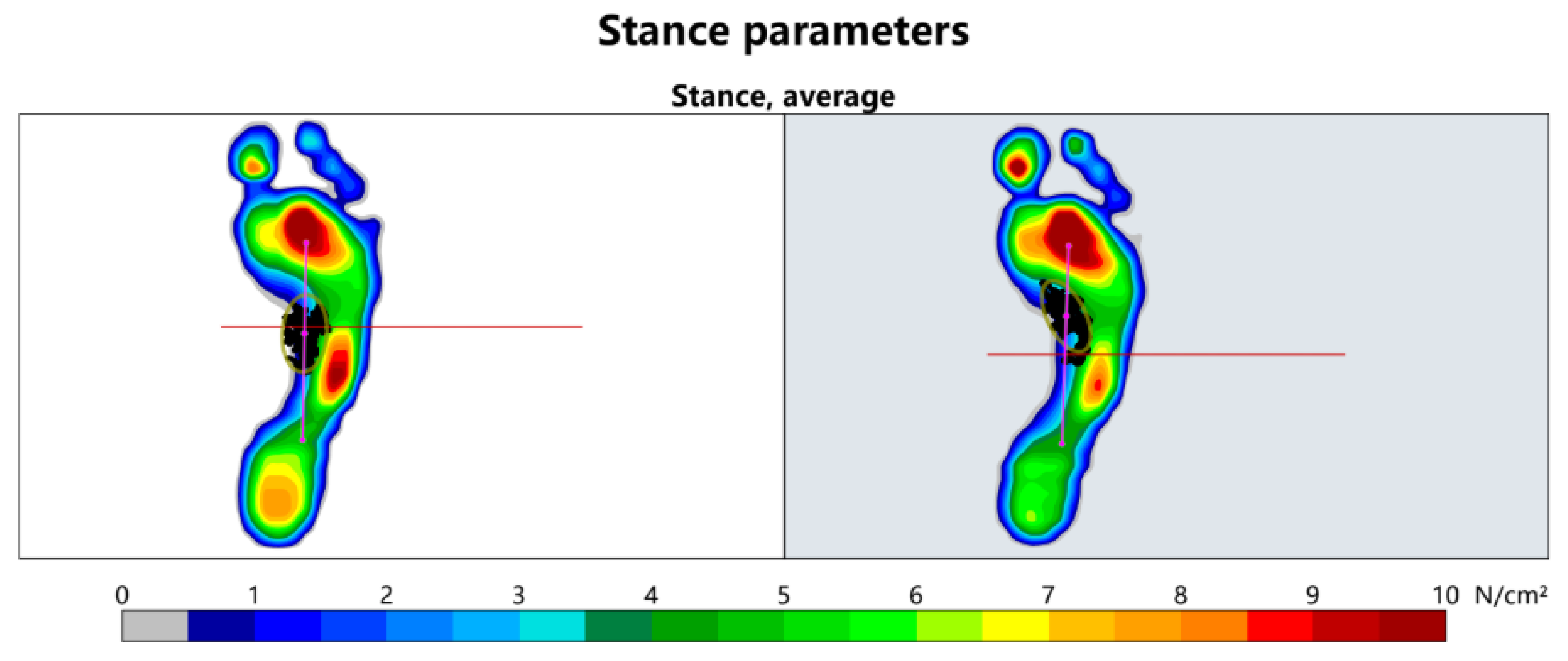
Participants stood upright and barefoot on a Zebris FDM-S force plate (Zebris Medical GmbH, Isny im Allgäu, Germany). They were positioned in a one-legged stance (with the dominant leg as the standing leg) with the dominant leg’s knee in an extended position and forefeet and heels aligned in a straight line (Figure 1). The other leg was elevated in a 90-degree angle flexed knee position. The arms were placed parallel to the body. Subjects performed the task twice, once with open eyes and once with closed eyes. Each trial lasted for 30 s. During stances with open eyes, participants were instructed to focus on the wall in front of them. Participants were specifically instructed not to use their arms for stabilization. Trials in which the arms were used for stabilization were considered as improper execution, and the subject had to repeat the test. Other improper executions included: loss of balance, stepping off or jumping off from the plate, and using the other leg for balance. Most of the subjects completed the tasks on their first attempt. The measured parameters that were included in the study were: 95% confidence ellipse area of the stabilogramm (mm2); path length of the Center Of Pressure (COP) (mm); velocity of the COP (mm/sec); length of the antero-posterior sway movement interval of the COP (mm); and length of the left–right sway movement interval of the COP (mm).
Figure 1.
Representative stabilometry measurement plot recorded using the Zebris FDM-S force plate. The black plot marks the trajectory of the COP. Left figure was captured before the DOMS protocol, while right one after the DOMS protocol with open eyes.
4.4. Dynamometer-Based DOMS-Inducing Eccentric Exercise Protocol
The Humac NORM isokinetic dynamometer was used for the DOMS-inducing protocol. A warm-up session was introduced before the first stabilometry measurement and prior to performing the DOMS-inducing protocol. This proper warm-up, corresponding to the specific load on the lower limbs, was carried out with the test subjects as follows: 5 min on a bicycle ergometer (rpm: 70–80) with light resistance and 5 min of exercises performed with their own body weight. Thereafter the participants took their places in the isokinetic dynamometer, and the dominant leg was measured. All subjects performed 5 trial repetitions. The measurement procedure in the isokinetic dynamometer was carried out as follows: 15 repetitions in six consecutive sets of eccentric contractions on the dominant leg while the motor moved the limb with an angular velocity of 60 °/s between a knee angle of 10 and 100 degrees. The angular velocity of 60 °/s and the number of sets were based on an earlier study [54]. A minute of rest was introduced between the sets. These repetitions were at maximum intensity. All subjects were familiar with the device, since they have had regular measurements taken on an isokinetic dynamometer formerly in their sports careers.
4.5. Muscle Soreness Questionnaire
Participants completed a VAS to rate their pain before the examination, immediately after the DOMS-inducing protocol and 24 h, 48 h and 72 h later. “0” means no pain and “10” means most severe pain.
4.6. Statistics
The Shapiro–Wilk’s W test of normality for the distribution of the within-subject differences with p < 0.05 to reject the null hypothesis concerning normality was used because of the limited sample size and for the purpose of selecting the adequate statistical procedure. Variables of the selected sample were presented by means and standard deviations. Parametric paired-samples T-tests were used to reveal differences on Statistica 12.0 software (TIBCO StatSoft Inc., Tulsa, OK, USA) as the appropriate statistical method of the before–after measurement experiment. A post hoc power analysis was conducted after the measurements to determine power values for the pre-determined alpha value. Only comparisons where the power value was greater than 0.8 were reported in this study. The power level was calculated using the G*Power version 3.1. 9.7 free software. Statistical significance level was set to p < 0.05.
5. Limitations
The current research team must comply with the athletic program set by the coaches. As a result, some athletes were exposed to the DOMS-inducing eccentric exercise protocol after days of training, in contrast to others who were exposed immediately after summer vacation. The repeated bout effect of DOMS with diminished symptoms [55] may have impacted the VAS score levels of those who were exposed to the study’s exercise protocol after days of training. Nevertheless, this did not alter the fact of DOMS evolvement. The absence of an appropriate control condition and the limited sample size is an acknowledged limitation of the study. As multiple comparisons were not performed, the analyses performed in the paper should be considered as explanatory results concerning the novel phenomenon.
6. Conclusions
In our study the antero-posterior postural sway was decreased right after DOMS-inducing exercise protocol at open eyes. This is in contrast to earlier observed fatigue-related findings. In addition, DOMS did not diminish postural sway with closed eyes regardless of whether fatigue is implicit of DOMS. Therefore, it seems that the DOMS effect cannot induce the ultrafast reflex-like compensatory enhanced intermittent postural control strategy at closed eyes and rather increased the postural sway due to the DOMS effect. This finding is suggested to mimic a tendency towards a positive Romberg test at a minuscule scale. However, the findings of this study should be re-confirmed on a larger sample size.
Author Contributions
Conceptualization, B.S.; methodology, L.R., B.K. and B.S.; validation, B.K.; formal analysis, G.L.; investigation B.F., T.S., G.L. and L.P.; resources, T.M.; data curation, G.L.; writing—original draft preparation, B.S.; writing—review and editing, L.R. and B.S.; visualization, B.K.; supervision, M.T. and P.Á.; project administration, G.L.; funding acquisition, T.M. All authors have read and agreed to the published version of the manuscript.
Funding
This study was supported under the TKP2021 funding scheme by the following grants from the Ministry of Culture and Innovation of Hungary and the National Research, Development and Innovation Fund: TKP-2021-EGA-10, TKP-2021-EGA-37, and 2020-1.1.2-PIACI-KFI-2021-00245.
Institutional Review Board Statement
This research was approved by the Science Committee of the Hungarian University of Sports Science (TE-KEB/18/2022), approval date 1 August 2022.
Informed Consent Statement
Informed consent was obtained from all subjects involved in this study.
Data Availability Statement
The data that supports the findings of this study are available on reasonable request from the corresponding author. The data are not publicly available due to containing information that could compromise the privacy of underaged research participants.
Acknowledgments
We would like to thank the Hungarian National Academy of Handball for their involvement in this work.
Conflicts of Interest
The authors declare no conflicts of interest.
References
- Hough, T. Ergographic Studies in Muscular Fatigue and Soreness. J. Boston Soc. Med. Sci. 1900, 5, 81–92. [Google Scholar] [CrossRef] [Scilit]
- Cheung, K.; Hume, P.; Maxwell, L. Delayed onset muscle soreness: Treatment strategies and performance factors. Sports Med. 2003, 33, 145–164. [Google Scholar] [CrossRef] [Scilit] [PubMed]
- Torres, R.; Vasques, J.; Duarte, J.A.; Cabri, J.M. Knee proprioception after exercise-induced muscle damage. Int. J. Sports Med. 2010, 31, 410–415. [Google Scholar] [CrossRef] [Scilit] [PubMed]
- Sonkodi, B.; Kopa, Z.; Nyirady, P. Post Orgasmic Illness Syndrome (POIS) and Delayed Onset Muscle Soreness (DOMS): Do They Have Anything in Common? Cells 2021, 10, 1867. [Google Scholar] [CrossRef] [Scilit] [PubMed]
- Sonkodi, B. Delayed-Onset Muscle Soreness Begins with a Transient Neural Switch. Int. J. Mol. Sci. 2025, 26, 2319. [Google Scholar] [CrossRef] [Scilit] [PubMed]
- Woo, S.H.; Lukacs, V.; de Nooij, J.C.; Zaytseva, D.; Criddle, C.R.; Francisco, A.; Jessell, T.M.; Wilkinson, K.A.; Patapoutian, A. Piezo2 is the principal mechanotransduction channel for proprioception. Nat. Neurosci. 2015, 18, 1756–1762. [Google Scholar] [CrossRef] [Scilit] [PubMed]
- Abboud, F.M. Four evolving concepts in molecular and clinical autonomic research. Clin. Auton. Res. 2021, 31, 467–471. [Google Scholar] [CrossRef] [Scilit] [PubMed]
- Sonkodi, B.; Radovits, T.; Csulak, E.; Kopper, B.; Sydo, N.; Merkely, B. Orthostasis Is Impaired Due to Fatiguing Intensive Acute Concentric Exercise Succeeded by Isometric Weight-Loaded Wall-Sit in Delayed-Onset Muscle Soreness: A Pilot Study. Sports 2023, 11, 209. [Google Scholar] [CrossRef] [Scilit] [PubMed]
- Liu, Z.; Lu, S.; Haskell, I.A.; Schappe, M.S.; Josipovic, M.; Min, S.; Alabi, A.A.; Chi, J.; Kim, M.; Liberles, S.D. Vagal blood volume receptors compensate for haemorrhage and posture change. Nature 2026, 651, 1068–1076. [Google Scholar] [CrossRef] [Scilit] [PubMed]
- Ducas, J.; Memari, S.; Houle, M.; Schwendenmann, Y.; Abboud, J.; Yiou, E.; Descarreaux, M. Impact of lumbar delayed-onset muscle soreness on postural stability in standing postures. Gait Posture 2024, 109, 201–207. [Google Scholar] [CrossRef] [Scilit] [PubMed]
- Vuillerme, N.; Nougier, V.; Prieur, J.M. Can vision compensate for a lower limbs muscular fatigue for controlling posture in humans? Neurosci. Lett. 2001, 308, 103–106. [Google Scholar] [CrossRef] [Scilit] [PubMed]
- Vuillerme, N.; Burdet, C.; Isableu, B.; Demetz, S. The magnitude of the effect of calf muscles fatigue on postural control during bipedal quiet standing with vision depends on the eye-visual target distance. Gait Posture 2006, 24, 169–172. [Google Scholar] [CrossRef] [Scilit] [PubMed]
- Boyas, S.; Hajj, M.; Bilodeau, M. Influence of ankle plantarflexor fatigue on postural sway, lower limb articular angles, and postural strategies during unipedal quiet standing. Gait Posture 2013, 37, 547–551. [Google Scholar] [CrossRef] [Scilit] [PubMed]
- Boyas, S.; Remaud, A.; Bisson, E.J.; Cadieux, S.; Morel, B.; Bilodeau, M. Impairment in postural control is greater when ankle plantarflexors and dorsiflexors are fatigued simultaneously than when fatigued separately. Gait Posture 2011, 34, 254–259. [Google Scholar] [CrossRef] [Scilit] [PubMed]
- Barbieri, F.A.; Penedo, T.; Simieli, L.; Barbieri, R.A.; Zagatto, A.M.; van Dieen, J.H.; Pijnappels, M.; Rodrigues, S.T.; Polastri, P.F. Effects of Ankle Muscle Fatigue and Visual Behavior on Postural Sway in Young Adults. Front. Physiol. 2019, 10, 643. [Google Scholar] [CrossRef] [Scilit] [PubMed]
- Sonkodi, B. Does Proprioception Involve Synchronization with Theta Rhythms by a Novel Piezo2 Initiated Ultrafast VGLUT2 Signaling? Biophysica 2023, 3, 695–710. [Google Scholar] [CrossRef] [Scilit]
- Sonkodi, B.; Hegedűs, Á.; Kopper, B.; Berkes, I. Significantly Delayed Medium-Latency Response of the Stretch Reflex in Delayed-Onset Muscle Soreness of the Quadriceps Femoris Muscles Is Indicative of Sensory Neuronal Microdamage. J. Funct. Morphol. Kinesiol. 2022, 7, 43. [Google Scholar] [CrossRef] [Scilit] [PubMed]
- Ivanenko, Y.P.; Grasso, R.; Lacquaniti, F. Effect of gaze on postural responses to neck proprioceptive and vestibular stimulation in humans. J. Physiol. 1999, 519, 301–314. [Google Scholar] [CrossRef] [Scilit] [PubMed]
- Bjorklund, M.; Crenshaw, A.G.; Djupsjobacka, M.; Johansson, H. Position sense acuity is diminished following repetitive low-intensity work to fatigue in a simulated occupational setting: A critical comment. Eur. J. Appl. Physiol. 2003, 88, 485–486. [Google Scholar] [CrossRef] [Scilit] [PubMed]
- Qu, X.; Nussbaum, M.A.; Madigan, M.L. Model-based assessments of the effects of age and ankle fatigue on the control of upright posture in humans. Gait Posture 2009, 30, 518–522. [Google Scholar] [CrossRef] [Scilit] [PubMed]
- Langmar, G.; Sumegi, T.; Fulop, B.; Pozsgai, L.; Mocsai, T.; Toth, M.; Racz, L.; Kopper, B.; Der, A.; Buzas, A.; et al. Heart Rate Variability Alterations During Delayed-Onset Muscle Soreness-Inducing Exercise-With Piezo2 Interpretation. Sports 2025, 13, 262. [Google Scholar] [CrossRef] [Scilit] [PubMed]
- Lanska, D.J.; Goetz, C.G. Romberg’s sign: Development, adoption, and adaptation in the 19th century. Neurology 2000, 55, 1201–1206. [Google Scholar] [CrossRef] [Scilit] [PubMed]
- Halmagyi, G.M.; Curthoys, I.S. Vestibular contributions to the Romberg test: Testing semicircular canal and otolith function. Eur. J. Neurol. 2021, 28, 3211–3219. [Google Scholar] [CrossRef] [Scilit] [PubMed]
- Cathers, I.; O’Dwyer, N.; Neilson, P. Entrainment to extinction of physiological tremor by spindle afferent input. Exp. Brain Res. 2006, 171, 194–203. [Google Scholar] [CrossRef] [Scilit] [PubMed]
- Sonkodi, B.; Resch, M.D.; Hortobagyi, T. Is the Sex Difference a Clue to the Pathomechanism of Dry Eye Disease? Watch out for the NGF-TrkA-Piezo2 Signaling Axis and the Piezo2 Channelopathy. J. Mol. Neurosci. 2022, 72, 1598–1608. [Google Scholar] [CrossRef] [Scilit] [PubMed]
- Zhu, L.; Ren, S.; He, S.; Liu, M.; Hou, F. Piezo in the eye: Expression, distribution and roles in ocular diseases. Front. Physiol. 2025, 16, 1651258. [Google Scholar] [CrossRef] [Scilit] [PubMed]
- Lee, J.H.; Perez-Flores, M.C.; Park, S.; Kim, H.J.; Chen, Y.; Kang, M.; Kersigo, J.; Choi, J.; Thai, P.N.; Woltz, R.L.; et al. The Piezo channel is a mechano-sensitive complex component in the mammalian inner ear hair cell. Nat. Commun. 2024, 15, 526. [Google Scholar] [CrossRef] [Scilit] [PubMed]
- Nagel, M.; Chesler, A.T. PIEZO2 ion channels in proprioception. Curr. Opin. Neurobiol. 2022, 75, 102572. [Google Scholar] [CrossRef] [Scilit] [PubMed]
- Zhang, Q.; Ma, H.; Huo, L.; Wang, S.; Yang, Q.; Ye, Z.; Cao, J.; Wu, S.; Ma, C.; Shang, C. Neural mechanism of trigeminal nerve stimulation recovering defensive arousal responses in traumatic brain injury. Theranostics 2025, 15, 2315–2337. [Google Scholar] [CrossRef] [Scilit] [PubMed]
- Sonkodi, B. Acquired Piezo2 Channelopathy is One Principal Gateway to Pathophysiology. Front. Biosci. (Landmark Ed) 2025, 30, 33389. [Google Scholar] [CrossRef] [Scilit] [PubMed]
- Lago, T.R.; Hsiung, A.; Leitner, B.P.; Duckworth, C.J.; Chen, K.Y.; Ernst, M.; Grillon, C. Exercise decreases defensive responses to unpredictable, but not predictable, threat. Depress. Anxiety 2018, 35, 868–875. [Google Scholar] [CrossRef] [Scilit] [PubMed]
- Hedayatpour, N.; Hassanlouei, H.; Arendt-Nielsen, L.; Kersting, U.G.; Falla, D. Delayed-onset muscle soreness alters the response to postural perturbations. Med. Sci. Sports Exerc. 2011, 43, 1010–1016. [Google Scholar] [CrossRef] [Scilit] [PubMed]
- Sanchez-Carranza, O.; Begay, V.; Chakrabarti, S.; Pampols-Perez, M.; Wang, L.; Garcia-Contreras, J.A.; Hammes, A.; Lewin, G.R. Mechanically-gated currents in mouse sensory neurons lacking PIEZO2. Biophys. J. 2025, 124, 4543–4550. [Google Scholar] [CrossRef] [Scilit] [PubMed]
- Sonkodi, B.; Nagy, Z.F.; Keller-Pintér, A.; Klivényi, P.; Molnár, M.J.; Széll, M. Genetic Variants in SDC3, KCNA2, KCNK1, KCNK16, and Heat Shock Transcription Factor-1 Genes: An Exploratory Analysis Supporting the Piezo2 Channelopathy Hypothesis in Amyotrophic Lateral Sclerosis Onset. Int. J. Mol. Sci. 2025, 26, 10218. [Google Scholar] [CrossRef] [Scilit] [PubMed]
- Sonkodi, B. Piezo2-Initiated Ultrafast Signaling and Its Acquired Channelopathy in Light of Quantum Gravity Theory. Int. J. Mol. Sci. 2026, 27, 4129. [Google Scholar] [CrossRef] [Scilit] [PubMed]
- Liu, W.; Zhang, X.; He, Q.; Meng, Y.; Tian, B.; Xiao, B. Spring-like mechanics enable rapid inactivation and stochastic single-channel gating of the mechanically activated PIEZO channel. Cell Rep. 2025, 44, 116615. [Google Scholar] [CrossRef] [Scilit] [PubMed]
- Hashad, A.M.; Abd-Alhaseeb, M.M.; Lim, X.R.; Mathieu, N.M.; Harraz, O.F. PIP(2) corrects an endothelial Piezo1 channelopathy. Proc. Natl. Acad. Sci. USA 2025, 122, e2522750122. [Google Scholar] [CrossRef] [Scilit] [PubMed]
- Castle, M.; Comoli, E.; Loewy, A.D. Autonomic brainstem nuclei are linked to the hippocampus. Neuroscience 2005, 134, 657–669. [Google Scholar] [CrossRef] [Scilit] [PubMed]
- Turk-Browne, N.B. The hippocampus as a visual area organized by space and time: A spatiotemporal similarity hypothesis. Vis. Res. 2019, 165, 123–130. [Google Scholar] [CrossRef] [Scilit] [PubMed]
- Smith, P.F. Vestibular-hippocampal interactions. Hippocampus 1997, 7, 465–471. [Google Scholar] [CrossRef] [Scilit]
- Aitken, P.; Zheng, Y.; Smith, P.F. The modulation of hippocampal theta rhythm by the vestibular system. J. Neurophysiol. 2018, 119, 548–562. [Google Scholar] [CrossRef] [Scilit] [PubMed]
- Kennedy, J.P.; Zhou, Y.; Qin, Y.; Lovett, S.D.; Cooper, T.; Sheremet, A.; Burke, S.N.; Maurer, A.P. Visual cortical LFP in relation to the hippocampal theta rhythm in track running rats. Front. Cell. Neurosci. 2023, 17, 1144260. [Google Scholar] [CrossRef] [Scilit] [PubMed]
- Mizelle, J.C.; Forrester, L.; Hallett, M.; Wheaton, L.A. Theta frequency band activity and attentional mechanisms in visual and proprioceptive demand. Exp. Brain Res. 2010, 204, 189–197. [Google Scholar] [CrossRef] [Scilit] [PubMed]
- Kubota, Y.; Sato, W.; Toichi, M.; Murai, T.; Okada, T.; Hayashi, A.; Sengoku, A. Frontal midline theta rhythm is correlated with cardiac autonomic activities during the performance of an attention demanding meditation procedure. Brain Res. Cogn. Brain Res. 2001, 11, 281–287. [Google Scholar] [CrossRef] [Scilit] [PubMed]
- Pedemonte, M.; Velluti, R.A. Sensory processing could be temporally organized by ultradian brain rhythms. Rev. Neurol. 2005, 40, 166–172. [Google Scholar] [PubMed]
- Fitzpatrick, R.C.; Gorman, R.B.; Burke, D.; Gandevia, S.C. Postural proprioceptive reflexes in standing human subjects: Bandwidth of response and transmission characteristics. J. Physiol. 1992, 458, 69–83. [Google Scholar] [CrossRef] [Scilit] [PubMed]
- Kouzaki, K.; Nosaka, K.; Ochi, E.; Nakazato, K. Increases in M-wave latency of biceps brachii after elbow flexor eccentric contractions in women. Eur. J. Appl. Physiol. 2016, 116, 939–946. [Google Scholar] [CrossRef] [Scilit] [PubMed]
- Sonkodi, B. Parkinson’s Disease Onset in Light of Acquired Irreversible Piezo2 Channelopathy. Preprints 2026. [Google Scholar] [CrossRef] [Scilit]
- Hamilton, N.R.; Neckles, V.N.; Al-Khindi, T.; Donthi, N.; Mizutori, S.; Fu, R.; Kiraly, J.K.; Winship, B.C.; Kolodkin, A.L.; Chaudhari, K. The mechanotransduction channel Piezo2 refines axonal projections to the accessory optic system and regulates the optokinetic reflex. bioRxiv 2026. [Google Scholar] [CrossRef] [Scilit] [PubMed]
- Banks, R.W.; Barker, D.; Stacey, M.J. Form and distribution of sensory terminals in cat hindlimb muscle spindles. Philos. Trans. R. Soc. Lond. B Biol. Sci. 1982, 299, 329–364. [Google Scholar] [CrossRef] [Scilit] [PubMed]
- Matthews, P.B. Evolving views on the internal operation and functional role of the muscle spindle. J. Physiol. 1981, 320, 1–30. [Google Scholar] [CrossRef] [Scilit] [PubMed]
- Banks, R.W. The motor innervation of mammalian muscle spindles. Prog. Neurobiol. 1994, 43, 323–362. [Google Scholar] [CrossRef] [Scilit] [PubMed]
- Wada, N.; Funabiki, K.; Nakanishi, S. Role of granule-cell transmission in memory trace of cerebellum-dependent optokinetic motor learning. Proc. Natl. Acad. Sci. USA 2014, 111, 5373–5378. [Google Scholar] [CrossRef] [Scilit] [PubMed]
- Vaczi, M.; Costa, A.; Racz, L.; Tihanyi, J. Effects of consecutive eccentric training at different range of motion on muscle damage and recovery. Acta Physiol. Hung. 2009, 96, 459–468. [Google Scholar] [CrossRef] [Scilit] [PubMed]
- Nosaka, K.; Clarkson, P.M.; McGuiggin, M.E.; Byrne, J.M. Time course of muscle adaptation after high force eccentric exercise. Eur. J. Appl. Physiol. Occup. Physiol. 1991, 63, 70–76. [Google Scholar] [CrossRef] [Scilit] [PubMed]

Disclaimer/Publisher’s Note: The statements, opinions and data contained in all publications are solely those of the individual author(s) and contributor(s) and not of MDPI and/or the editor(s). MDPI and/or the editor(s) disclaim responsibility for any injury to people or property resulting from any ideas, methods, instructions or products referred to in the content. |
© 2026 by the authors. Licensee MDPI, Basel, Switzerland. This article is an open access article distributed under the terms and conditions of the Creative Commons Attribution (CC BY) license.