


Abstract
Background/Objectives: This study investigated the preventive effects of apilarnil (APL) against copper nanoparticle (CuNPs)-induced liver and kidney injury in rats. Methods: Thirty-five rats were randomly allocated into five groups (n = 7): Control (physiological saline), APL (400 mg/kg), CuNPs (100 mg/kg), CuNPs + APL200 (100 mg/kg CuNPs + 200 mg/kg APL) and CuNPs + APL400 (100 mg/kg CuNPs + 400 mg/kg APL). All treatments were administered orally by gavage for 28 consecutive days. Results: CuNPs administration increased serum alanine aminotransferase (ALT), aspartate aminotransferase (AST), alkaline phosphatase (ALP), blood urea nitrogen (BUN) and creatinine levels, whereas APL attenuated these biochemical alterations. CuNPs also decreased superoxide dismutase (SOD), glutathione peroxidase (GPx) and catalase (CAT) activities together with reduced glutathione (GSH) levels while increasing malondialdehyde (MDA), whereas APL attenuated these oxidative alterations. Furthermore, CuNPs markedly upregulated the mRNA expression of endoplasmic reticulum (ER) stress-related genes, including activating transcription factor 6 (ATF6), glucose-regulated protein 78 (GRP78), protein kinase RNA-like ER kinase (PERK) and C/EBP homologous protein (CHOP). In parallel, CuNPs increased the protein levels of Toll-like receptor 4 (TLR4), high-mobility group box 1 (HMGB1), nuclear factor-κB (NF-κB), monocyte chemoattractant protein-1 (MCP-1) and tumor necrosis factor-α (TNF-α), whereas APL attenuated these inflammatory responses. Histopathological examination showed that APL markedly attenuated CuNPs-induced hepatic and renal lesions and reduced the immunoreactivity of caspase-3 and 8-hydroxy-2′-deoxyguanosine (8-OHdG). Conclusions: Overall, APL alleviated CuNPs-induced hepatorenal toxicity and was associated with improvements in oxidative stress parameters, ER stress-related gene expression, inflammatory protein concentrations, caspase-3 immunoreactivity, and oxidative DNA damage.
1. Introduction
Heavy metals are a class of metallic elements characterized by relatively high density and are released into the environment through both natural processes and anthropogenic activities. They are generally defined as metals with atomic weights ranging from 63.5 to 200.6 g/mol and densities exceeding 5 g/cm3 in their standard states [1]. Although trace amounts of heavy metals are essential for normal physiological processes, excessive exposure or accumulation can lead to toxic effects [2]. The widespread use of heavy metals, together with their ease of environmental dispersion and resistance to biodegradation, poses a significant threat to both human health and the environment [1].
CuNPs, which are composed of elemental copper, are widely used in various fields, including the electrical, electronics, construction, transportation, chemical, paint, jewelry and energy industries. Copper is an essential trace element required for numerous physiological processes; however, excessive exposure to elemental CuNPs has been associated with the generation of reactive oxygen species (ROS), oxidative stress and pathological alterations in multiple organs, particularly the liver and kidneys. However, the contribution of nanoscale copper particles to overall human copper exposure through drinking water remains uncertain. Drinking water may represent a route of oral copper exposure, as copper-containing materials in distribution and plumbing systems can release dissolved copper and nanoscale copper-containing particles into water [3]. Following oral ingestion, CuNPs can accumulate in biological tissues, particularly in the liver and kidneys, and excessive tissue accumulation is considered a key factor underlying their toxic effects [4,5].
The liver and kidneys are particularly susceptible to injury caused by various pathological conditions and environmental toxicants, including heavy metals, chemicals and other toxic agents [6,7]. Consequently, increasing attention has been directed toward the identification of protective strategies against toxicant-induced hepatic and renal injury. Among these, naturally derived bioactive compounds with antioxidant properties, particularly those of plant and animal origin, have attracted considerable interest because of their potential to alleviate toxicant-induced oxidative and inflammatory damage [8,9,10].
Apitherapy is a traditional therapeutic practice that has been used since ancient times to enhance immune function and manage a variety of diseases and inflammatory conditions through the use of bee-derived products, including honey, pollen, propolis, royal jelly and bee venom [11]. APL is a natural bee-derived product that has attracted increasing research interest because of its diverse biological properties. It is widely used in apitherapy and is produced by lyophilizing 3- to 7-day-old male honeybee larvae. APL contains vitamins A, B1, B6 and choline; minerals including calcium (Ca), phosphorus (P), sodium (Na), zinc (Zn), manganese (Mn), iron (Fe) and potassium (K); as well as polyphenols, which contribute to its antioxidant capacity and immunomodulatory properties. Owing to these bioactive constituents, APL has emerged as a promising natural agent for preventing and alleviating oxidative stress-related disorders [11,12].
The present study aimed to investigate the hepatotoxic and nephrotoxic effects of CuNPs and to evaluate the protective potential of APL against CuNPs-induced organ damage through the comprehensive assessment of oxidative stress, ER stress, inflammation, apoptosis, oxidative DNA damage, biochemical function markers and histopathological changes.
2. Results
2.1. Effects of CuNPs and APL Administrations on Serum ALT, AST, ALP, Urea, Creatinine and BUN Levels
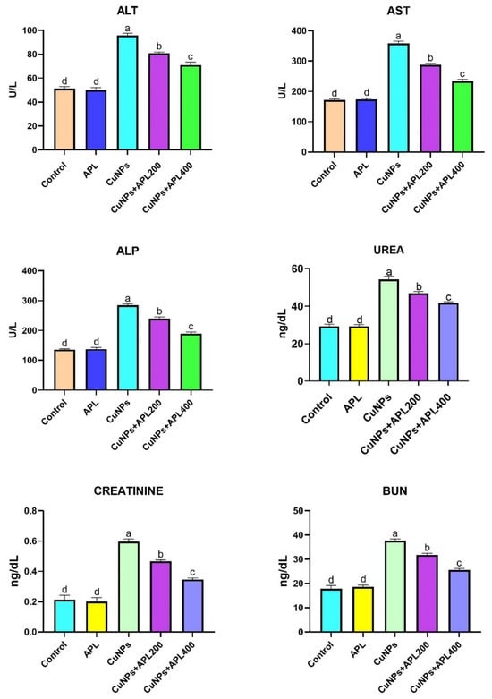
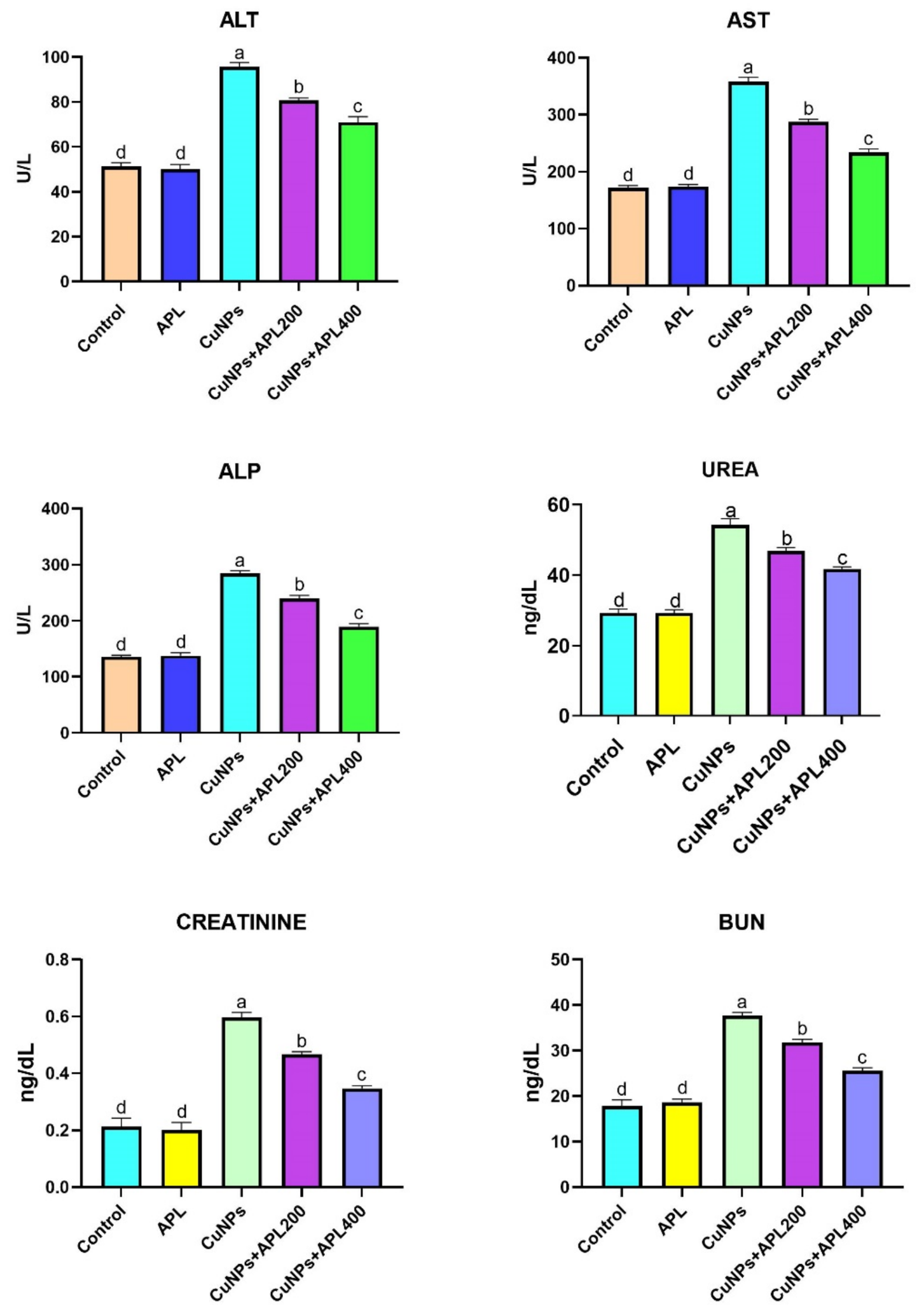
Serum ALT, AST, ALP, urea, creatinine and BUN levels are shown in Figure 1. While there was no difference in serum urea, creatinine and BUN levels between the control and APL groups (p > 0.05), the CuNPs group showed increased levels (p < 0.05) and the 400 mg and 200 mg/kg APL administration reduced these levels, with the 400 mg/kg dose being more effective (p < 0.05).
Figure 1.
Serum liver and kidney function tests were performed in the experimental groups. A substantial discrepancy was identified among the means designated by different superscripts (a, b, c and d) (p < 0.05).
2.2. Oxidative Stress Biomarkers in Liver and Kidney Tissues
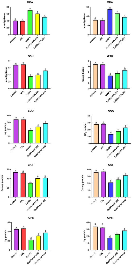
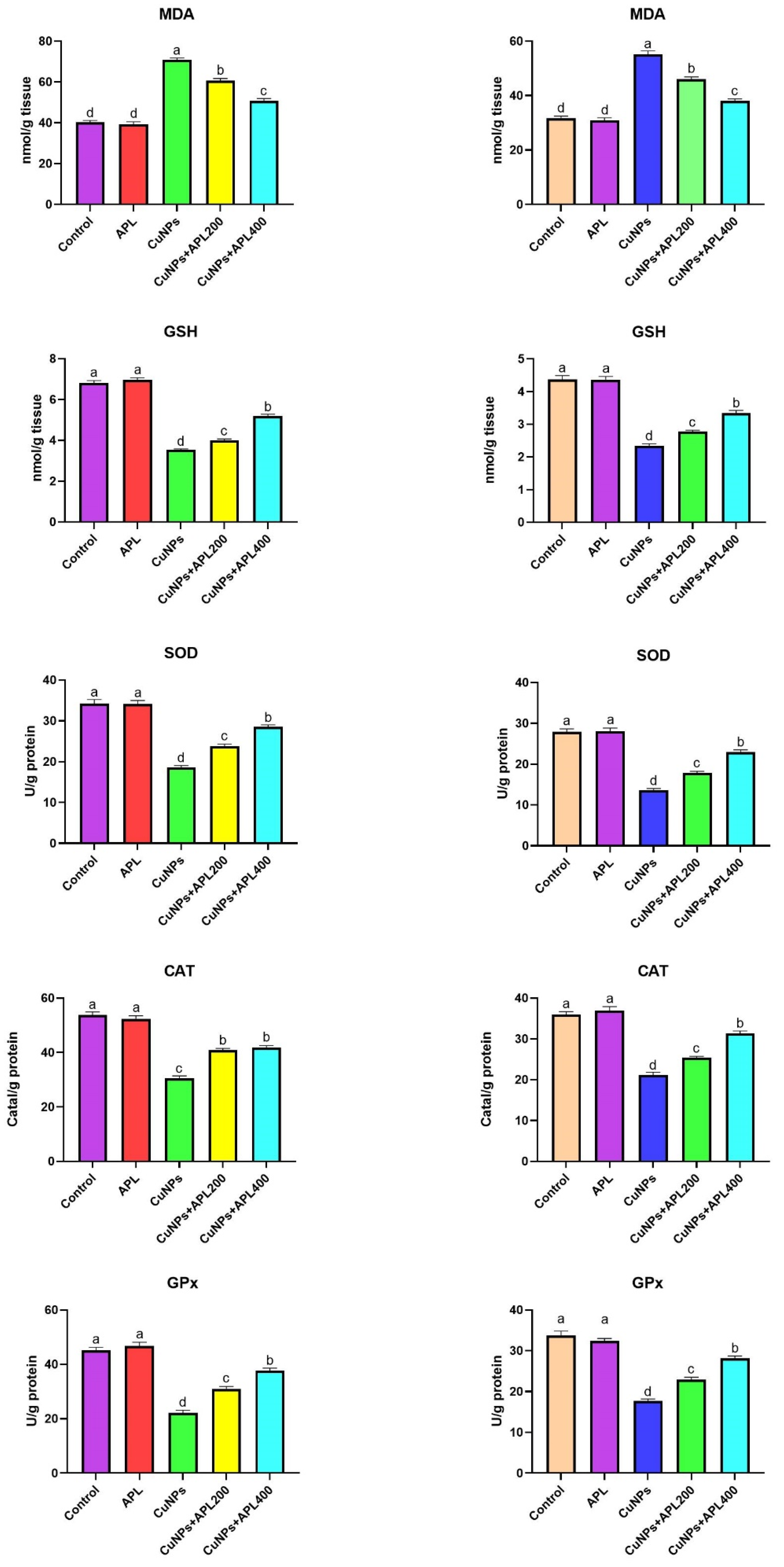
Oxidative stress parameters in the liver and kidney tissues of the experimental groups are presented in Figure 2. The data obtained demonstrated a significant increase in MDA levels in all groups following CuNPs application (p < 0.05). However, decrease in MDA levels was observed in the CuNPs + APL groups. The administration of CuNPs resulted in a substantial decline in GSH levels, as evidenced by a p-value less than 0.05. However, the administration of APL subsequent to CuNPs resulted in a substantial augmentation of GSH levels. Subsequent analysis of the SOD, CAT and GPx enzyme activities revealed a significant decrease in these enzymes in the presence of CuNPs (p < 0.05). APL administration resulted in increase in these enzyme activities (p < 0.05).
Figure 2.
Levels of oxidative stress and antioxidant parameters (MDA, GSH, SOD, CAT, and GPx) in liver (left panel) and kidney (right panel) tissues of the experimental groups. A substantial discrepancy was identified among the means designated by different superscripts (a, b, c and d) (p < 0.05).
2.3. Inflammation Biomarkers in Liver and Kidney Tissues
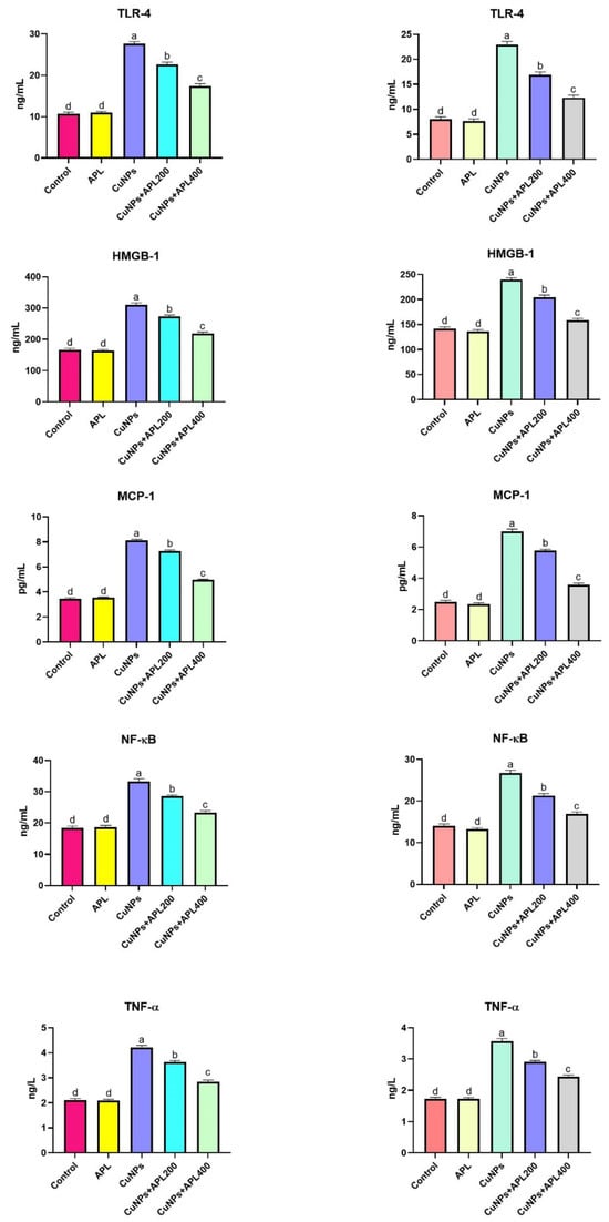
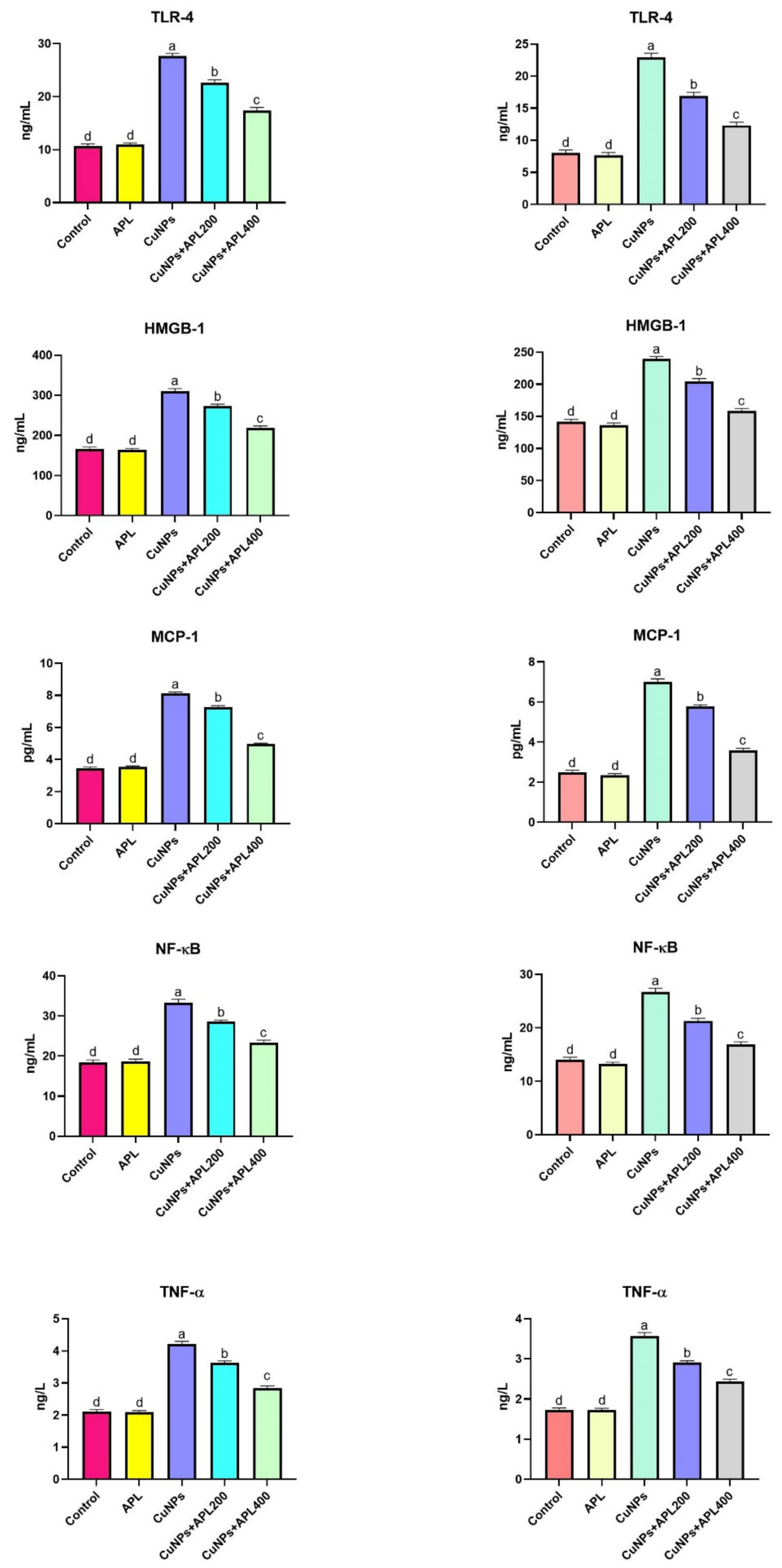
The tissue protein concentrations of inflammatory mediators were significantly elevated in liver and kidney tissues following CuNPs exposure. CuNPs administration markedly increased the tissue protein concentrations of TLR4, HMGB1, NF-κB, TNF-α and MCP-1 compared with the control group (p < 0.05) (Figure 3).
Figure 3.
Inflammation parameters in liver (left panel) and kidney (right panel) tissues across experimental groups. A substantial discrepancy was identified among the means designated by different superscripts (a, b, c and d) (p < 0.05).
APL supplementation significantly reduced the elevated tissue protein concentrations of these inflammatory mediators in manner. Both APL-treated groups exhibited significantly lower tissue protein concentrations of these inflammatory mediators than the CuNPs group (p < 0.05). No significant inflammatory changes were detected in the APL alone group.
2.4. Effects of APL on ER Stress Related Markers
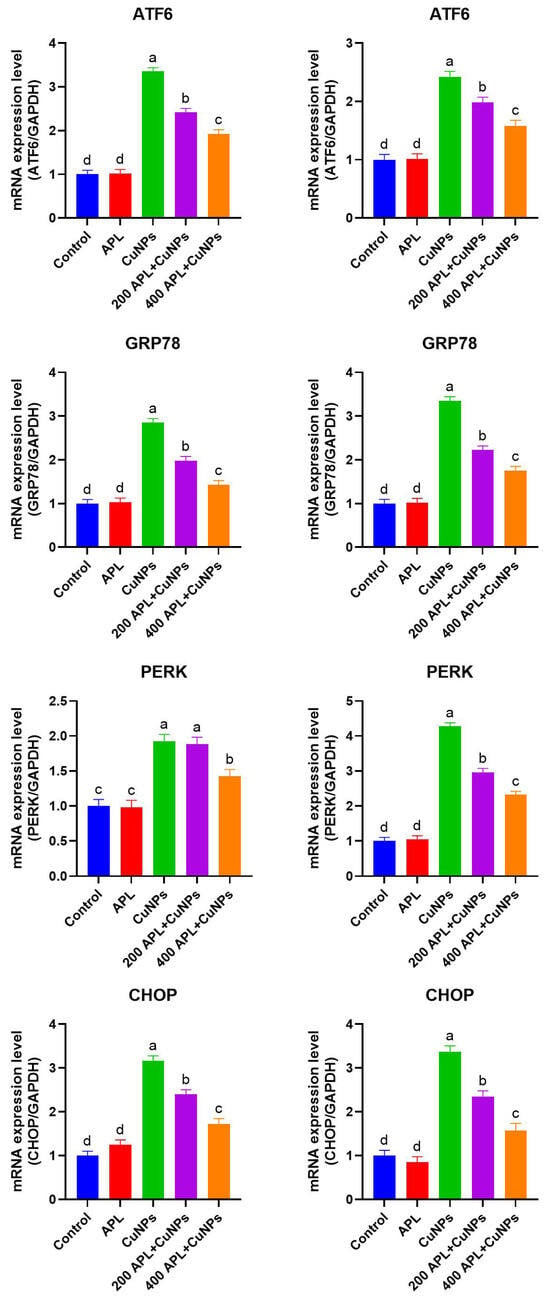
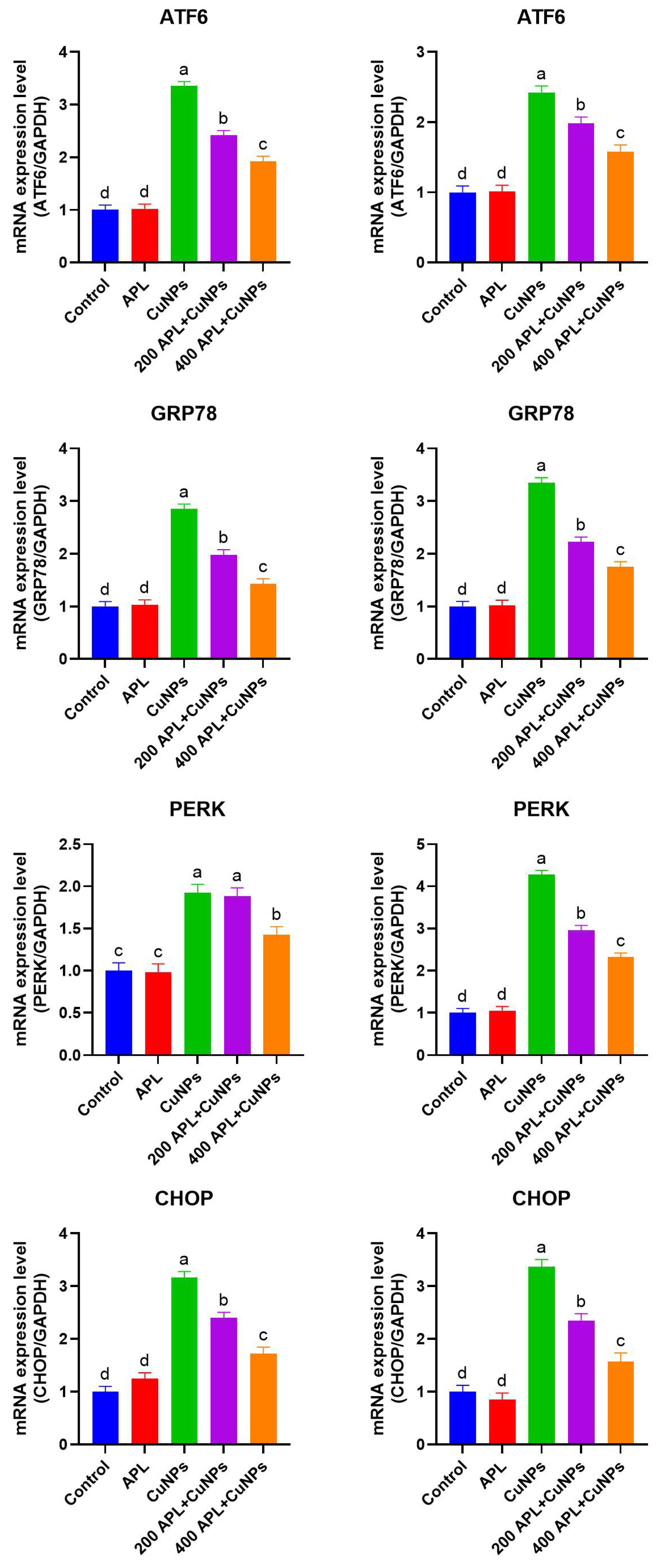
CuNPs exposure significantly increased the mRNA expression of ER stress-related markers in liver and kidney tissues. The expression levels of ATF6, GRP78, PERK and CHOP were markedly increased in the CuNPs-treated group compared with the control group (p < 0.05) (Figure 4).
Figure 4.
Relative mRNA expression levels of ER stress-related markers in the liver (left panel) and kidney (right panel) tissues of the experimental groups. A substantial discrepancy was identified among the means designated by different superscripts (a, b, c and d) (p < 0.05).
APL administration significantly reduced the mRNA expression of ER stress-related markers in both tissues. The reductions were more prominent in the 400 mg/kg APL group, which exhibited expression levels approaching those of the control group (p < 0.05).
2.5. Histopathology Results
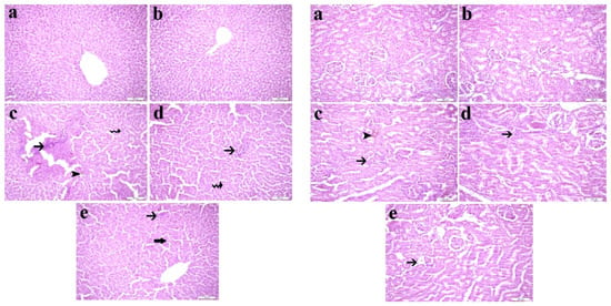
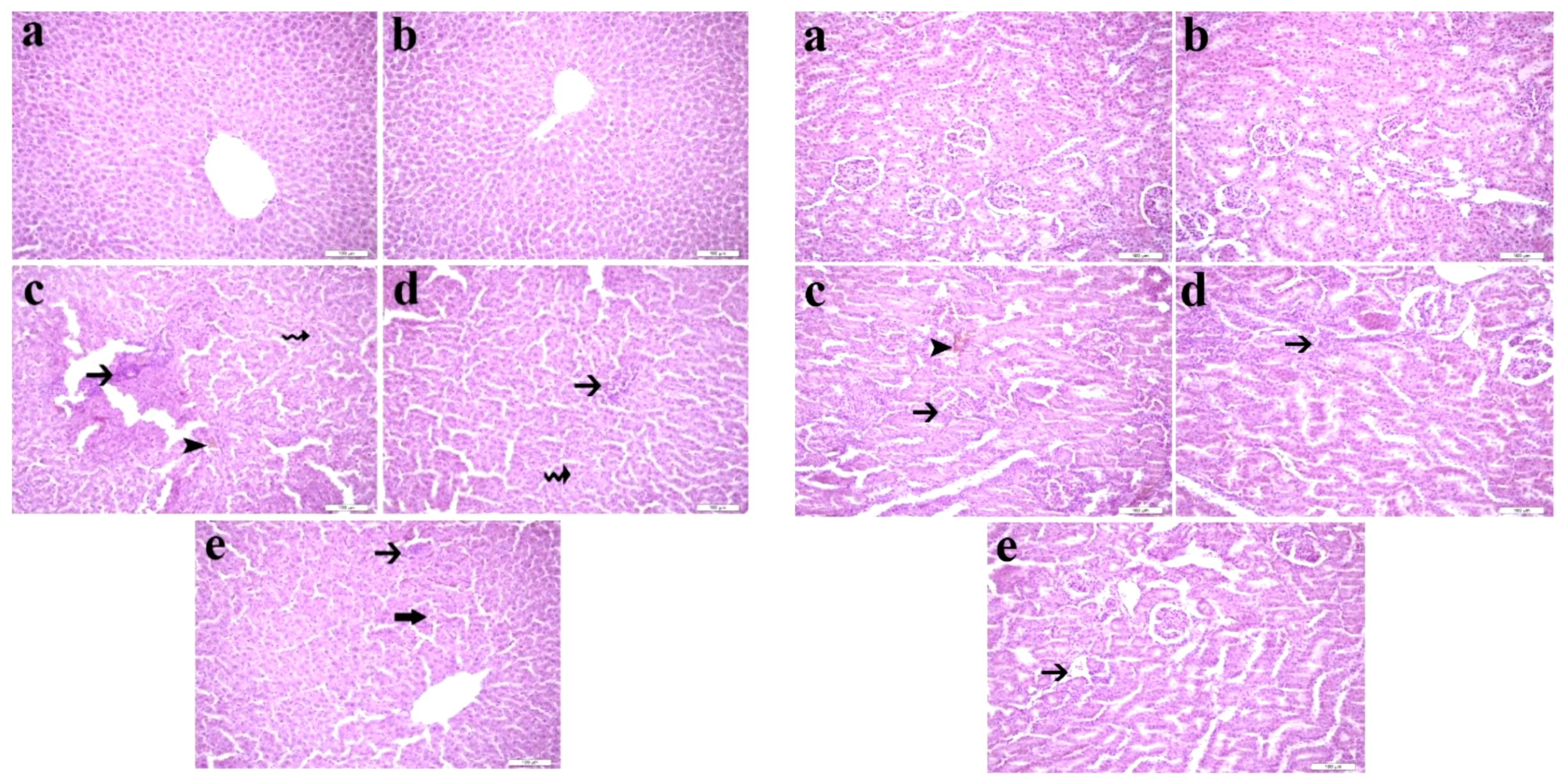
A histopathological examination of the livers of rats in the control and APL groups revealed normal hepatic parenchyma that was homogeneously distributed and showed no pathological alterations. In the CuNPs-treated group, the normal hepatic radial architecture was disrupted, accompanied by congestion, inflammatory cell infiltration, and necrotic changes. In the CuNPs + APL200-treated group, the hepatic radial architecture showed partial improvement, and inflammatory cell infiltration was reduced, although necrotic changes were still evident. In the CuNPs + APL400-treated group, the hepatic architecture closely resembled that of the control group, with markedly reduced inflammatory cell infiltration and only mild degenerative changes observed instead of necrotic lesions. Representative histopathological findings of the liver and kidney tissues are presented in Figure 5.
Figure 5.
The effect of APL on CuNPs-induced histopathology of the liver and kidneys. (a) Control; (b) APL 400 group: normal histological structure of the hepatic lobule in liver sections and of the glomerulus and tubules in kidney sections; (c) CuNPs-treated rats: cytoplasmic vacuolation of hepatocytes, inflammatory cell infiltration (thin arrow), necrosis (zigzag arrow), and congestion in hepatic sinusoids (arrowhead); intertubular hemorrhage (arrowhead) and intense inflammatory cell infiltration in kidney tissue; (d) CuNPs + APL 200-treated rats: inflammatory cell infiltration (thin arrow) and necrosis (zigzag arrow) in the liver, and intense inflammatory cell infiltration (thin arrow) in kidney tissue; (e) CuNPs + APL 400-treated rats: mild inflammatory cell infiltration (thin arrow) and hydropic degeneration (thick arrow) in the liver, and mild inflammatory cell infiltration (thin arrow) in the kidney. Histopathological staining was performed at ×200 magnification.
Renal sections from the control and APL groups exhibited normal histological architecture. In contrast, marked histopathological alterations were observed in the kidneys of the CuNPs-treated group, including renal tubular degeneration, glomerular atrophy, haemorrhage, and inflammatory cell infiltration. In the CuNPs + APL200-treated group, inflammatory cell infiltration and tubular degeneration were still present, although haemorrhage was less pronounced. In the CuNPs + APL400-treated group, inflammatory cell infiltration and tubular degeneration were markedly attenuated, and the renal histological appearance closely resembled that of the control group.
2.6. Immunohistochemistry Results
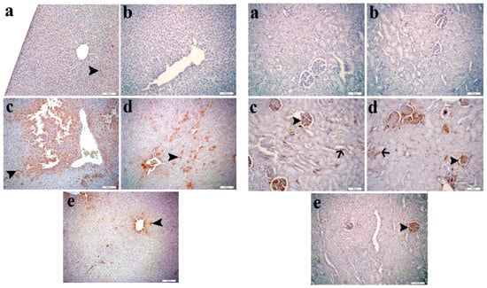
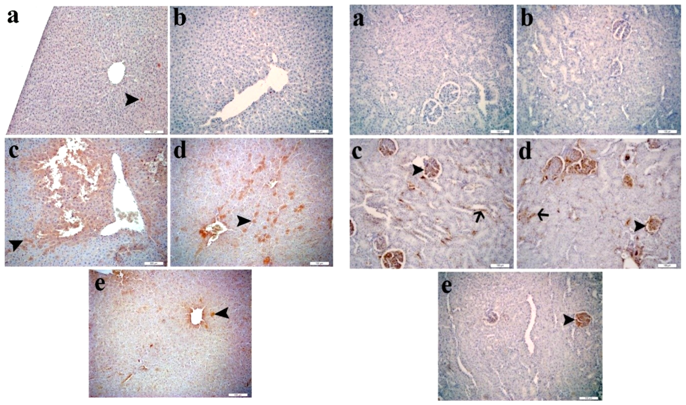
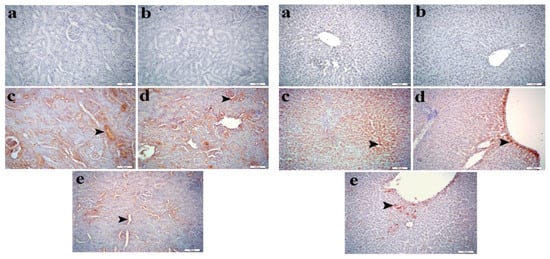
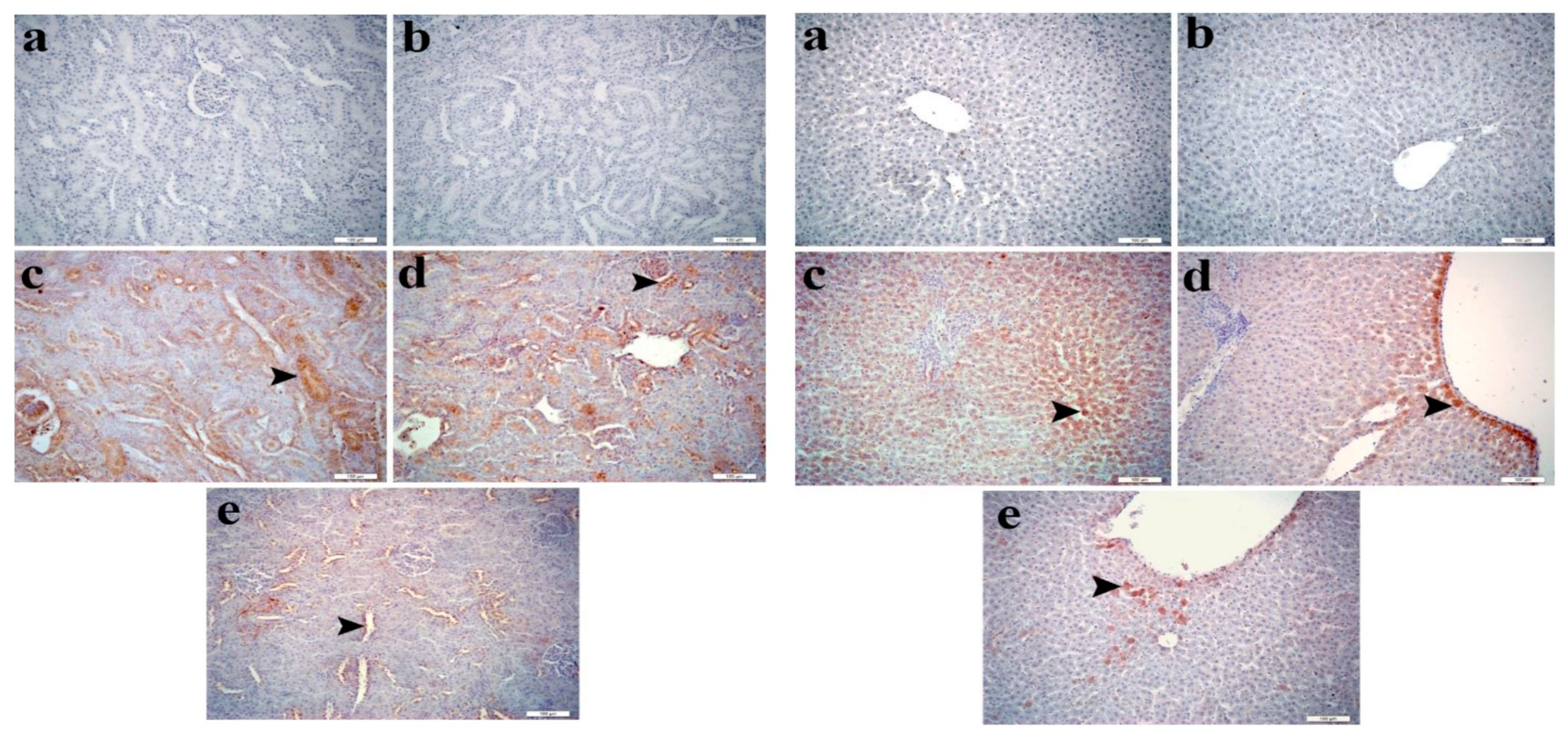
To further evaluate caspase-3 immunoreactivity in liver and kidney tissues, immunohistochemical staining was performed. In comparison with the control and APL groups, caspase-3 immunoreactivity scores were significantly higher in the CuNPs group (p < 0.05). Despite the CuNPs + APL200 treatment exhibiting a modest decrease in the elevated caspase-3 immunoreactivity scores, no statistically significant difference was observed (p > 0.05). In the group treated with CuNPs + APL400, caspase-3 immunoreactivity scores were significantly lower (p < 0.05). Representative immunohistochemical staining for caspase-3 and 8-OHdG is presented in Figure 6 and Figure 7, respectively, and the corresponding semiquantitative immunohistochemical scores are summarized in Table 1.
Figure 6.
Immunostaining for caspase-3 protein in rat liver and kidney sections; control (a) and APL (b) groups showing very mild or no caspase-3 immunopositiveness. (c) CuNPs-treated group showing a strong positive immunoreaction in liver hepatocytes (arrowhead) and kidney glomeruli (arrowhead) and tubular epithelium (arrow). (d) Severely immunoreactive liver hepatocytes (arrowhead) and kidney glomerulus (arrowhead) and tubular epithelium (arrow) in the group treated with CuNPs + APL 200. (e) Liver (arrowhead) and kidney tissue showing mildly positive immunoexpression in the group treated with CuNPs + APL 400. Immunohistochemical staining was performed at ×200 magnification. To evaluate oxidative DNA damage, 8-OHdG immunoreactivity in liver and kidney tissues was assessed by immunohistochemistry. Compared to the control and APL groups, 8-OHdG immunopositivity was significantly increased in the CuNPs group (p < 0.05). Although the CuNPs + APL200 treatment slightly reduced 8-OHdG immunopositivity, no statistically significant difference was detected (p > 0.05). In the group treated with CuNPs + APL400, 8-OHdG immunopositivity was significantly reduced (p < 0.05).
Figure 7.
Immunostaining for 8-OHdG protein in rat liver and kidney sections, control (a) and APL (b) groups showing very mild or no 8-OHdG immunoreactivity. (c) Liver hepatocytes (arrowhead) and renal tubular epithelia (arrowhead) showing strong positive immunoexpression, CuNPs-treated group. (d) Moderately immunoreactive hepatocytes (arrowhead) and positivity in tubular epithelium (arrowhead) showing strong positive immunoexpression, CuNPs + APL 200-treated group. (e) Mildly positive immunoexpression in hepatocytes (arrowhead) and tubular epithelia (arrowhead), CuNPs + APL 400-treated group. Immunohistochemical staining was performed at ×200 magnification.
Table 1.
Semiquantitative histopathological and immunohistochemical scores of caspase-3 and 8-OHdG in the liver and kidney tissues of rats following CuNPs exposure and APL administration.
3. Discussion
The present study demonstrated that CuNPs exposure induced marked hepatorenal injury, characterized by impaired liver and kidney function, oxidative imbalance, increased inflammatory protein concentrations, altered ER stress-related gene expression, oxidative DNA damage, and histopathological and immunohistochemical abnormalities. APL administration attenuated these changes, with the 400 mg/kg dose generally showing greater attenuation than the 200 mg/kg dose for several measured parameters. These findings provide an integrated biochemical, molecular, and pathological profile of CuNPs-induced hepatorenal toxicity and the effects of APL administered as a preventive co-treatment.
CuNPs can accumulate in the liver and kidneys and have been associated with oxidative stress and organ injury following oral exposure [4,5,13]. In the present study, CuNPs exposure significantly increased serum ALT, AST, ALP, urea, and creatinine levels, indicating hepatic and renal functional impairment. APL administration partially attenuated these biochemical parameters, suggesting attenuation of CuNPs-induced functional injury in this experimental model.
Oxidative stress is an important component of CuNPs-induced organ injury. In the present study, CuNPs exposure markedly increased MDA levels and decreased SOD, CAT, GPx, and GSH levels in both liver and kidney tissues, indicating disruption of the antioxidant defense system and increased lipid peroxidation. APL administration attenuated these changes, with the higher dose generally producing greater improvement in antioxidant status. These findings are consistent with previous experimental studies showing that CuNP exposure can induce oxidative imbalance in target organs and that Apilarnil can attenuate oxidative stress by reducing lipid peroxidation and enhancing antioxidant defenses [4,5,14].
Inflammation is a key contributor to the progression of CuNPs-induced organ injury and is closely associated with oxidative stress. In the present study, CuNPs exposure significantly increased the protein levels of TLR4, HMGB1, NF-κB, MCP-1 and TNF-α in both liver and kidney tissues, suggesting an association with an enhanced inflammatory response. APL administration attenuated the changes in these inflammatory protein concentrations, suggesting an association with reduced tissue concentrations of TLR4, HMGB1, NF-κB, MCP-1, and TNF-α. The effects of APL on TLR4, HMGB1, NF-κB, MCP-1 and TNF-α are consistent with previous reports highlighting the immunomodulatory and anti-inflammatory properties of apitherapy-derived products [14].
ER stress-related responses are associated with CuNP-induced toxicity. In the present study, elemental CuNPs markedly increased the mRNA expression of ATF6, GRP78, PERK, and CHOP in both liver and kidney tissues, indicating an association with ER stress-related molecular responses [15]. APL administration attenuated the expression of these ER stress-related genes [10]. These findings suggest that the protective effects of APL may be associated with changes in ER stress-related gene expression.
Histopathological examination further confirmed the hepatotoxic and nephrotoxic effects of CuNPs. Liver tissues exhibited hepatocellular degeneration, necrosis, inflammatory cell infiltration, and sinusoidal congestion, whereas kidney tissues showed tubular degeneration, glomerular alterations, and hemorrhagic changes, indicating substantial structural damage. Similar hepatic and renal histopathological alterations have been reported following oral CuNP exposure in rats [4,5,13]. The marked amelioration of these lesions following APL administration was consistent with the biochemical and molecular changes observed in the present study. Similar protective effects of Apilarnil on tissue damage have been reported in a paracetamol-induced hepatotoxicity model, in which Apilarnil attenuated histopathological alterations in liver tissue [16].
Immunohistochemical analysis provided additional evidence of CuNPs-induced cellular injury. CuNPs exposure markedly increased caspase-3 immunoreactivity in both liver and kidney tissues, whereas APL administration reduced caspase-3 immunoreactivity. These findings suggest a reduction in apoptosis-associated changes, although caspase-3 immunostaining alone is insufficient to definitively establish apoptosis. Similar effects of Apilarnil on caspase-3 expression have been reported in a paracetamol-induced hepatotoxicity model, in which Apilarnil reduced caspase-3 expression together with histopathological and oxidative stress-related alterations [16]. In addition, increased 8-OHdG immunoreactivity in CuNPs-treated tissues indicated enhanced oxidative DNA damage in the present study. The reduction in 8-OHdG immunoreactivity following APL administration suggests attenuation of oxidative DNA damage.
Nevertheless, several limitations of the present study should be acknowledged. Only a single dose of CuNPs and two doses of APL were evaluated in a 28-day repeated-dose experimental model. Importantly, because APL was administered 30 min after CuNP exposure on each day, the present design represents a preventive co-treatment model rather than a therapeutic intervention for established CuNP-induced toxicity. In addition, an APL-alone group receiving 200 mg/kg was not included; therefore, the independent effects of the lower APL dose cannot be determined. Consequently, differences observed between the 200 and 400 mg/kg APL doses should be interpreted cautiously and should not be considered definitive evidence of a dose-dependent effect. The CuNP dose used in the present study (100 mg/kg/day) represents a relatively high repeated oral exposure in an experimental animal model and should not be considered directly representative of typical environmental or occupational human exposure. Therefore, the present exposure regimen was intended to investigate toxicological responses under controlled experimental conditions rather than to reproduce typical human exposure levels. General toxicity observations, tissue copper accumulation, and CuNP toxicokinetic assessments were not investigated. In addition, nanoparticle characterization was limited to the manufacturer’s specifications, and physicochemical characterization of the actual CuNP suspension, including hydrodynamic particle size, aggregation behavior, and surface charge, could not be performed due to the technical and instrumental limitations of the available laboratory facilities. This limitation should be considered when interpreting the present findings. In future studies, physicochemical characterization of the administered CuNP suspension should be incorporated to provide a more comprehensive assessment of the exposure conditions. Future studies should also determine the dissolved copper fraction in the exposure medium to distinguish particle-associated effects from those potentially related to copper ion dissolution. Although GAPDH was used as the reference gene and melt-curve analysis was performed, additional qPCR validation procedures, including reference gene validation and primer efficiency assessment, were not evaluated. Moreover, the proposed molecular mechanisms, including ER stress-related responses, were inferred primarily from biochemical and gene expression analyses without comprehensive protein- or functional-level validation. Similarly, the inflammatory findings were based on tissue protein concentrations and did not include pathway-specific assessments such as NF-κB phosphorylation, IκB degradation, or p65 nuclear translocation. Therefore, the findings should be interpreted with these limitations in mind. Future studies incorporating diverse exposure conditions, standardized APL preparations, complementary mechanistic approaches, and advanced translational models are warranted to further elucidate the molecular targets of APL and clarify its biological effects and translational relevance.
4. Materials and Methods
4.1. Chemicals
CuNPs: Copper nanoparticles (CAS No. 7440-50-8; primary particle size: 40–60 nm) were purchased from Sigma-Aldrich (St. Louis, MO, USA). According to the manufacturer’s specifications, the particles were supplied as elemental copper nanoparticles. Fresh CuNPs were prepared daily in physiological saline immediately before administration and thoroughly vortexed to obtain a homogeneous suspension prior to oral gavage.
APL: APL used in the present study was obtained from the Faculty of Agriculture, Bingol University. The material was prepared from 3–7-day-old drone larvae, homogenized, lyophilized and stored at −20 °C until use. The APL used in this study originated from the same production batch previously characterized by Hakan İnci et al. [17] using a validated LC–MS/MS method. Therefore, the phytochemical composition reported in the previous study directly corresponds to the material used in the present investigation. In that study, 56 phytochemical compounds were identified and quantified, with quinic acid, fumaric acid, aconitic acid, kaempferol and quercetin reported as the predominant constituents. The administered doses were calculated based on the lyophilized material, which was freshly suspended in physiological saline immediately before oral gavage. Accordingly, the detailed phytochemical profile presented in Table 2 was derived from the previously published characterization of this identical production batch.
Table 2.
Phytochemical compounds identified and quantified in APL extracts (mg analyte/g dry extract).
4.2. Laboratory Animals
Thirty-five male Sprague Dawley rats weighing 220–250 g were used in the present study. The animals were obtained from the Ataturk University Medical Experimental Research Center (ATADEM). Before the experiment, all rats were acclimatized to the laboratory environment for 7 days. Throughout the study, the animals had free access to standard pellet chow and drinking water and were housed under controlled environmental conditions with a 12 h light/12 h dark cycle at 24 ± 2 °C and 45 ± 5% relative humidity.
4.3. Experimental Design
Following the acclimatization period, animals were randomly assigned to the experimental groups using a random allocation procedure. Sample size was determined a priori using G*Power software (version 3.1.9.7; Heinrich Heine University Düsseldorf, Germany) for a one-way analysis of variance (ANOVA; fixed effects, omnibus, one-way) with five experimental groups [18]. Assuming an effect size (f) of 0.65, a significance level (α) of 0.05, and a statistical power (1 − β) of 0.80, the required minimum total sample size was calculated to be 35 animals. Accordingly, seven rats were included in each experimental group.
- Group (Control): Rats received 1 mL of physiological saline by oral gavage, followed 30 min later by an additional 1 mL of physiological saline by oral gavage once daily for 28 consecutive days.
- Group (APL): Rats received 1 mL of physiological saline by oral gavage, followed 30 min later by APL (400 mg/kg) suspended in physiological saline (1 mL) by oral gavage once daily for 28 consecutive days [14].
- Group (CuNPs): Rats received CuNPs (100 mg/kg) suspended in physiological saline (1 mL) by oral gavage, followed 30 min later by 1 mL of physiological saline by oral gavage once daily for 28 consecutive days [5].
- Group (CuNPs + APL 200): Rats received CuNPs (100 mg/kg) suspended in physiological saline (1 mL) by oral gavage, followed 30 min later by APL (200 mg/kg) suspended in physiological saline (1 mL) by oral gavage once daily for 28 consecutive days.
- Group (CuNPs + APL 400): Rats received CuNPs (100 mg/kg) suspended in physiological saline (1 mL) by oral gavage, followed 30 min later by APL (400 mg/kg) suspended in physiological saline (1 mL) by oral gavage once daily for 28 consecutive days.
All experimental groups received two oral gavage administrations per day (1 mL per administration; total volume: 2 mL/rat/day) at a 30-min interval. Fresh CuNPs and APL suspensions were prepared daily in physiological saline immediately before administration. Twenty-four hours after the final treatment (day 29), the rats were deeply anesthetized with sevoflurane. Following confirmation of an adequate depth of anesthesia, the animals were euthanized by decapitation in accordance with the approved institutional animal ethics protocol. Blood, liver and kidney samples were subsequently collected for biochemical, molecular and histopathological analyses as described below. Accordingly, the present study was designed as a preventive co-treatment model, in which APL was administered concurrently with CuNP exposure, rather than as a therapeutic treatment model for established toxicity.
4.4. Collection of Serum and Tissue Samples
At the end of the experimental period, blood samples were collected from all rats and allowed to clot at room temperature. Serum was separated by centrifugation at 3500 rpm for 10 min and stored at −80 °C for subsequent biochemical analyses. Following blood collection, the liver and kidney were rapidly excised, rinsed with ice-cold physiological saline to remove residual blood and sectioned for biochemical, molecular and histopathological analyses. Samples designated for biochemical and molecular analyses were stored at −80 °C, whereas those intended for histopathological examination were fixed in 10% neutral buffered formalin until routine tissue processing.
4.5. Analysis of Liver and Kidney Oxidative Stress Markers
Oxidative stress parameters were determined in tissue homogenates using standardized spectrophotometric methodologies. Lipid peroxidation was assessed by quantifying MDA levels according to the method described by Placer et al. [19]. The activities of the antioxidant enzymes SOD and CAT were measured in accordance with the procedures reported by Sun et al. [20] and Aebi et al. [21], respectively. GSH levels were determined using the method described by Sedlak and Lindsay [22]. In addition, GPx activity was evaluated following the protocol established by Matkovics [23], total protein measured in accordance with the procedures reported by Lowry et al. [24].
4.6. Analysis of ALT, AST, Urea and Creatinine Levels in Blood Serum
Serum ALT, AST, ALP, urea, creatinine and BUN levels were determined using an automated chemistry analyzer (Randox RX Chemistry Analyzer, Randox Laboratories Ltd., Crumlin, UK).
4.7. ELISA Analyses
The tissue protein concentrations of TLR4 (No. 201-11-0081), HMGB-1 (No. 201-11-0258), NF-κB (No. 201-11-0288), MCP-1 (No. 201-11-0194) and TNF-α (No. 201-11-0765) in liver and kidney tissues were determined using the enzyme-linked immunosorbent assay (ELISA) based on the double-antibody sandwich method with commercially available kits manufactured by Sunred (Shanghai, China). Liver and kidney tissues were homogenised in phosphate-buffered saline (PBS, pH 7.4) at a 1:20 (w/v) dilution ratio. The homogenates were centrifuged at 3000 rpm for 20 min according to the manufacturer’s instructions and the resulting supernatants were collected for analysis. The protein concentrations obtained from tissue homogenates were normalized to the total protein content of each sample, which was determined using the Lowry method. All standards and tissue samples were analyzed in duplicate. Standards and tissue supernatants were added to microplate wells pre-coated with specific monoclonal antibodies, followed by the addition of biotin-labelled secondary antibodies. Subsequently, streptavidin-horseradish peroxidase (HRP) conjugate was added and the plates were incubated to facilitate immune complex formation. After incubation, the wells were washed thoroughly to remove unbound enzyme conjugates. Chromogen solutions A and B were then added and the plates were incubated to allow colour development. The enzymatic reaction generated a blue colour, which turned yellow following the addition of the stop solution. The optical density was measured at 450 nm using a microplate ELISA reader and the concentrations of the target proteins were calculated according to the corresponding standard curves.
4.8. Real-Time PCR Analyses
Total RNA was isolated from frozen liver and kidney tissues of the experimental rats using QIAzol Lysis Reagent (Cat. No. 79306, Qiagen, Hilden, Germany) according to the manufacturer’s instructions. RNA concentration and purity were determined spectrophotometrically, RNA concentration and purity were determined spectrophotometrically and only RNA samples meeting the predefined quality criteria were used for cDNA synthesis and complementary DNA (cDNA) was synthesized from 1 μg of total RNA using the RT2 First Strand Kit (Cat. No. 330404, Qiagen, Hilden, Germany) in a final reaction volume of 20 μL according to the manufacturer’s protocol. Quantitative real-time PCR was performed using QuantiNova SYBR Green PCR Master Mix (Cat. No. 208056, Qiagen, Hilden, Germany) on a Rotor-Gene Q Real-Time PCR System (Qiagen, Hilden, Germany). Each PCR reaction was carried out in a total volume of 20 μL containing 10 μL of 2× QuantiNova SYBR Green PCR Master Mix, 1 μL of forward primer (10 μM), 1 μL of reverse primer (10 μM), 2 μL of cDNA template, and 6 μL of RNase-free water. The amplification protocol consisted of an initial denaturation at 95 °C for 2 min, followed by 40 cycles of denaturation at 95 °C for 5 s and combined annealing/extension at 60 °C for 10 s. Melt-curve analysis was performed at the end of each run to verify the specificity of the amplified products. All reactions were performed in triplicate. Quantitative real-time PCR was performed and reported in accordance with the principles outlined in the Minimum Information for Publication of Quantitative Real-Time PCR Experiments (MIQE) guidelines [25]. Gene-specific primers for ATF6, GRP78, PERK, CHOP, and the reference gene GAPDH are listed in Table 3. Relative gene expression levels were calculated using the 2−ΔΔCt method after normalization to GAPDH [26].
Table 3.
Primer sequences.
4.9. Histopathological Examination
Liver and kidney samples were processed according to standard histopathological procedures [27]. Liver and kidney samples were excised and immediately fixed in 10% neutral buffered formalin. Following fixation, the tissues underwent routine histological processing, including washing under running tap water, dehydration through graded ethanol, clearing with xylene, and paraffin embedding. Sections approximately 5 μm thick were prepared from the paraffin blocks and stained with hematoxylin and eosin (H&E) for histopathological examination. Histopathological changes were evaluated by an experienced pathologist who was blinded to the experimental groups. Histopathological lesions were evaluated using a semiquantitative scoring system based on the severity of the observed lesions. For each animal, 10 randomly selected microscopic fields were evaluated from the liver and kidney sections. Histopathological changes were evaluated by an experienced pathologist who was blinded to the experimental groups.
4.10. Immunohistochemistry of Caspase-3 and 8-OHdG in Liver and Kidney Tissue
Immunohistochemical staining was performed using a standard protocol for formalin-fixed, paraffin-embedded tissues with heat-induced antigen retrieval and chromogenic detection [28]. For immunohistochemical analysis, 4-μm-thick sections were prepared from formalin-fixed, paraffin-embedded liver and kidney tissues and mounted on poly-L-lysine-coated slides. The sections were deparaffinized in xylene, rehydrated through graded ethanol solutions and rinsed in phosphate-buffered saline (PBS, 0.1 M, pH 7.4). Antigen retrieval was performed in Tris–ethylenediaminetetraacetic acid (EDTA) buffer (pH 9.0) using a microwave oven at 600 W for 10 min. Endogenous peroxidase activity was blocked by incubating the sections with 3% hydrogen peroxide for 10 min. The sections were then incubated for 1 h at room temperature with the primary antibodies anti-caspase-3 (SC-56053, Santa Cruz Biotechnology, Dallas, TX, USA; 1:100 dilution) and anti-8-OHdG (SC-66036, Santa Cruz Biotechnology, Dallas, TX, USA; 1:100 dilution). Following washing with PBS, the sections were incubated with the appropriate secondary antibody for 30 min. Immunoreactivity was visualized using 3,3′-diaminobenzidine (DAB) and the sections were counterstained with Mayer’s hematoxylin. Cells exhibiting brown cytoplasmic or nuclear staining were considered immunopositive. Nuclear and cytoplasmic staining were not evaluated separately; all positively stained cells were included in the semiquantitative analysis.
4.11. Statistical Analysis
Statistical analyses of the biochemical, ELISA and RT-qPCR data were performed using IBM SPSS Statistics version 20 (IBM Corp., Armonk, NY, USA). Data are presented as the mean ± standard error of the mean (SEM). Group differences were evaluated using one-way analysis of variance (ANOVA) followed by Tukey’s multiple comparisons test. A p value < 0.05 was considered statistically significant. Sample size was determined by an a priori power analysis using G*Power software (version 3.1.9.7) and no animals or data were excluded from the analysis. The investigators involved in histopathological, immunohistochemical and molecular assessments were blinded to the experimental groups during sample evaluation and data analysis.
4.12. Histopathological and Immunohistochemical Scoring and Statistical Analysis
Histopathological and immunohistochemical findings were independently evaluated by two blinded observers using a semiquantitative scoring system on an ordinal scale (0–4), where 0 indicated no detectable lesion or immunoreactivity, 1 mild, 2 moderate, 3 marked, and 4 severe lesion or strong immunoreactivity. In cases of disagreement, the final score was assigned by consensus following joint review of the stained sections. Histopathological and immunohistochemical scores are expressed as mean ± SD and were analyzed using the Kruskal–Wallis test followed by Dunn’s multiple comparisons test using GraphPad Prism version 10.3.1 (GraphPad Software, Boston, MA, USA). Different superscript letters indicate statistically significant differences between groups according to Dunn’s multiple comparisons test. A p value < 0.05 was considered statistically significant.
5. Conclusions
The present study demonstrates that CuNPs induce severe hepatorenal injury accompanied by alterations in oxidative stress parameters, inflammatory protein concentrations, ER stress-related gene expression, oxidative DNA damage, and caspase-3 immunoreactivity, resulting in marked biochemical, molecular, and histopathological alterations. APL attenuated these pathological changes in the CuNPs + APL200 and CuNPs + APL400 groups, with differences between the two APL doses observed across several measured outcomes. The observed protective effects of APL were associated with improved antioxidant status, reduced tissue concentrations of inflammatory proteins, decreased expression of ER stress-related genes, reduced oxidative DNA damage and caspase-3 immunoreactivity, and preservation of liver and kidney architecture. Collectively, these findings suggest that the protective effects of APL may be associated with concurrent changes in oxidative stress parameters, inflammation-associated protein concentrations, ER stress-related gene expression, oxidative DNA damage, and caspase-3 immunoreactivity rather than demonstrating direct modulation of specific cellular signaling pathways. Although further mechanistic and translational investigations are required, the present findings indicate that APL exerted protective effects against CuNPs-induced hepatorenal injury in this experimental co-exposure model. The active constituents, molecular targets, pharmacokinetic properties, and translational relevance of APL remain to be established.
Author Contributions
A.G. and F.M.K. conceived and designed the study; A.G., E.D.K. and S.O. performed the experiments and collected the data; S.K. and E.D.K. conducted the analysis and interpretation of the results; A.G. drafted the manuscript; S.C. and E.D.K. critically revised the manuscript; S.K. supervised the study. All authors have read and agreed to the published version of the manuscript.
Funding
The present study received financial support from Ataturk University Scientific Research Projects Coordination Unit, with the project code TDK-2023-11779.
Institutional Review Board Statement
The animal protocol was approved by the Ataturk University Animal Experimentation Ethics Committee (Protocol No: 2022, E-75296309, 2022-10-27). All animal experiments were carried out in accordance with the ARRIVE guidelines, the U.K. Animals (Scientific Procedures) Act 1986, and Directive 2010/63/EU of the European Parliament and of the Council on the protection of animals used for scientific purposes.
Informed Consent Statement
Not applicable.
Data Availability Statement
The data supporting the findings of this study are available from the corresponding author upon reasonable request.
Acknowledgments
This manuscript is based on the doctoral thesis of Aydin GENC (Thesis ID: 897172), submitted to Ataturk University, Turkiye. The authors used ChatGPT (OpenAI, GPT-5.5) exclusively to improve the grammar, language, and readability of the manuscript. The authors are solely responsible for the scientific content, interpretation of the results, and conclusions presented in this study.
Conflicts of Interest
The authors declare no conflicts of interest.
References
- Gumpu, M.B.; Sethuraman, S.; Krishnan, U.M.; Rayappan, J.B.B. A Review on Detection of Heavy Metal Ions in Water—An Electrochemical Approach. Sens. Actuators B Chem. 2015, 213, 515–533. [Google Scholar] [CrossRef] [Scilit]
- Singh, R.; Gautam, N.; Mishra, A.; Gupta, R. Heavy Metals and Living Systems: An Overview. Indian J. Pharmacol. 2011, 43, 246–253. [Google Scholar] [CrossRef] [Scilit] [PubMed]
- Vargas, I.T.; Pavissich, J.P.; Olivares, T.E.; Jeria, G.A.; Cienfuegos, R.A.; Pastén, P.A.; Pizarro, G.E. Increase of the Concentration of Dissolved Copper in Drinking Water Systems Due to Flow-Induced Nanoparticle Release from Surface Corrosion by-Products. Corros. Sci. 2010, 52, 3492–3503. [Google Scholar] [CrossRef] [Scilit]
- Xu, M.; Tang, H.; Zhou, X.; Chen, H.; Dong, Q.; Zhang, Y.; Ye, G.; Shi, F.; Lv, C.; Jing, B.; et al. Effects and Mechanisms of Sub-Chronic Exposure to Copper Nanoparticles on Renal Cytochrome P450 Enzymes in Rats. Environ. Toxicol. Pharmacol. 2018, 63, 135–146. [Google Scholar] [CrossRef] [Scilit] [PubMed]
- Lee, I.-C.; Ko, J.-W.; Park, S.-H.; Shin, N.-R.; Shin, I.-S.; Moon, C.; Kim, J.-H.; Kim, H.-C.; Kim, J.-C. Comparative Toxicity and Biodistribution Assessments in Rats Following Subchronic Oral Exposure to Copper Nanoparticles and Microparticles. Part Fibre Toxicol. 2016, 13, 56. [Google Scholar] [CrossRef] [Scilit] [PubMed]
- Petejova, N.; Martinek, A.; Zadrazil, J.; Teplan, V. Acute Toxic Kidney Injury. Ren. Fail. 2019, 41, 576–594. [Google Scholar] [CrossRef] [Scilit] [PubMed]
- Erisgin, Z.; Atasever, M.; Cetinkaya, K.; Akarca Dizakar, S.Ö.; Omeroglu, S.; Sahin, H. Protective Effects of Nigella Sativa Oil against Carboplatin-Induced Liver Damage in Rats. Biomed. Pharmacother. 2019, 110, 742–747. [Google Scholar] [CrossRef] [Scilit] [PubMed]
- Ileriturk, M.; Kandemir, O.; Akaras, N.; Simsek, H.; Genc, A.; Kandemir, F.M. Hesperidin Has a Protective Effect on Paclitaxel-Induced Testicular Toxicity through Regulating Oxidative Stress, Apoptosis, Inflammation and Endoplasmic Reticulum Stress. Reprod. Toxicol. 2023, 118, 108369. [Google Scholar] [CrossRef] [Scilit] [PubMed]
- Kandemir, F.M.; Kucukler, S.; Caglayan, C.; Gur, C.; Batil, A.A.; Gülçin, İ. Therapeutic Effects of Silymarin and Naringin on Methotrexate-Induced Nephrotoxicity in Rats: Biochemical Evaluation of Anti-Inflammatory, Antiapoptotic, and Antiautophagic Properties. J. Food Biochem. 2017, 41, e12398. [Google Scholar] [CrossRef] [Scilit]
- Takkaç, M.; Akarsu, S.A.; Uçak, G.; Küçükler, S.; Gür, C.; Akaras, N.; İleritürk, M.; Kandemir, F.M. Investigating the Effects of Apilarnil on Vincristine-Induced Testicular Toxicity in Rats. Basic Clin. Pharmacol. Toxicol. 2026, 138, e70179. [Google Scholar] [CrossRef] [Scilit] [PubMed]
- Kekeçoğlu, M.; BelliCi, A.E.; Yildirim, İ.; Kinalikaya, A.; Çaprazli, T.; Yüksel, B. Adverse Effects and Example Cases Due to the Use of Bee Products and Apitherapy Applications: Systematic Review. J. Tradit. Med. Complement. Ther. 2023, 6, 169–183. [Google Scholar] [CrossRef] [Scilit]
- Sawczuk, R.; Karpinska, J.; Miltyk, W. What Do We Need to Know about Drone Brood Homogenate and What Is Known. J. Ethnopharmacol. 2019, 245, 111581. [Google Scholar] [CrossRef] [Scilit] [PubMed]
- Tang, H.; Xu, M.; Luo, J.; Zhao, L.; Ye, G.; Shi, F.; Lv, C.; Chen, H.; Wang, Y.; Li, Y. Liver Toxicity Assessments in Rats Following Sub-Chronic Oral Exposure to Copper Nanoparticles. Environ. Sci. Eur. 2019, 31, 30. [Google Scholar] [CrossRef] [Scilit]
- Doğanyiğit, Z.; Okan, A.; Kaymak, E.; Pandır, D.; Silici, S. Investigation of Protective Effects of Apilarnil against Lipopolysaccharide Induced Liver Injury in Rats via TLR 4/HMGB-1/NF-κB Pathway. Biomed. Pharmacother. 2020, 125, 109967. [Google Scholar] [CrossRef] [Scilit] [PubMed]
- Wang, T.; Wei, X.; Sun, Y.; Hu, Y.; Li, J.; Zhang, X.; Yin, S.; Shi, Y.; Zhu, Y. Copper Nanoparticles Induce the Formation of Fatty Liver in Takifugu Fasciatus Triggered by the PERK-EIF2α- SREBP-1c Pathway. NanoImpact 2021, 21, 100280. [Google Scholar] [CrossRef] [Scilit] [PubMed]
- Bıçakçı, N.; Karaboğa, İ.; Bıçakçı, S.; Okuyan, H.M.; Duran, Y.; Polat, F.R.; Çelikkol, A.; Yılmaz, A. Apilarnil Alleviates Paracetamol-Induced Hepatotoxicity by Modulating Apoptosis and Oxidative Stress. Biotech. Histochem. Off. Publ. Biol. Stain Comm. 2025, 100, 119–128. [Google Scholar] [CrossRef] [Scilit] [PubMed]
- Inci, H.; Izol, E.; Yilmaz, M.A.; Ilkaya, M.; Bingöl, Z.; Gülçin, I. Comprehensive Phytochemical Content by LC/MS/MS and Anticholinergic, Antiglaucoma, Antiepilepsy, and Antioxidant Activity of Apilarnil (Drone Larvae). Chem. Biodivers. 2023, 20, e202300654. [Google Scholar] [CrossRef] [Scilit] [PubMed]
- Faul, F.; Erdfelder, E.; Lang, A.-G.; Buchner, A. G*Power 3: A Flexible Statistical Power Analysis Program for the Social, Behavioral, and Biomedical Sciences. Behav. Res. Methods 2007, 39, 175–191. [Google Scholar] [CrossRef] [Scilit] [PubMed]
- Placer, Z.A.; Cushman, L.L.; Johnson, B.C. Estimation of Product of Lipid Peroxidation (Malonyl Dialdehyde) in Biochemical Systems. Anal. Biochem. 1966, 16, 359–364. [Google Scholar] [CrossRef] [Scilit] [PubMed]
- Sun, Y.; Oberley, L.W.; Li, Y. A Simple Method for Clinical Assay of Superoxide Dismutase. Clin. Chem. 1988, 34, 497–500. [Google Scholar] [CrossRef] [Scilit]
- Aebi, H. Catalase in Vitro. Methods Enzymol. 1984, 105, 121–126. [Google Scholar] [CrossRef] [Scilit] [PubMed]
- Sedlak, J.; Lindsay, R.H. Estimation of Total, Protein-Bound, and Nonprotein Sulfhydryl Groups in Tissue with Ellman’s Reagent. Anal. Biochem. 1968, 25, 192–205. [Google Scholar] [CrossRef] [Scilit] [PubMed]
- Matkovics, B. Determination of Enzyme Activity in Lipid Peroxidation and Glutathione Pathways. Lab. Diagn. 1988, 15, 248–250. [Google Scholar]
- Lowry, O.H.; Rosebrough, N.J.; Farr, A.L.; Randall, R.J. Protein Measurement with the Folin Phenol Reagent. J. Biol. Chem. 1951, 193, 265–275. [Google Scholar] [CrossRef] [Scilit]
- Bustin, S.A.; Benes, V.; Garson, J.A.; Hellemans, J.; Huggett, J.; Kubista, M.; Mueller, R.; Nolan, T.; Pfaffl, M.W.; Shipley, G.L.; et al. The MIQE Guidelines: Minimum Information for Publication of Quantitative Real-Time PCR Experiments. Clin. Chem. 2009, 55, 611–622. [Google Scholar] [CrossRef] [Scilit] [PubMed]
- Livak, K.J.; Schmittgen, T.D. Analysis of Relative Gene Expression Data Using Real-Time Quantitative PCR and the 2−ΔΔCT Method. Methods 2001, 25, 402–408. [Google Scholar] [CrossRef] [Scilit] [PubMed]
- Slaoui, M.; Fiette, L. Histopathology Procedures: From Tissue Sampling to Histopathological Evaluation. Methods Mol. Biol. 2011, 691, 69–82. [Google Scholar] [CrossRef] [Scilit] [PubMed]
- Ramos-Vara, J.A. Principles and Methods of Immunohistochemistry. Methods Mol. Biol. 2017, 1641, 115–128. [Google Scholar] [CrossRef] [Scilit] [PubMed]

Disclaimer/Publisher’s Note: The statements, opinions and data contained in all publications are solely those of the individual author(s) and contributor(s) and not of MDPI and/or the editor(s). MDPI and/or the editor(s) disclaim responsibility for any injury to people or property resulting from any ideas, methods, instructions or products referred to in the content. |
© 2026 by the authors. Licensee MDPI, Basel, Switzerland. This article is an open access article distributed under the terms and conditions of the Creative Commons Attribution (CC BY) license.