


Abstract
Background: The effectiveness of non-surgical periodontal therapy largely depends on the ability of instruments to efficiently remove plaque and calculus from root surfaces. In recent years, innovative instruments such as LM Sharp Diamond™ curettes have been introduced, featuring a coated blade that maintains sharpness without the need for resharpening. The aim of this pilot in vitro study was to compare the cleaning efficacy of LM Sharp Diamond curettes with that of conventional stainless-steel curettes, hypothesizing that the diamond-coated instruments would demonstrate superior deposit removal efficiency. Materials and Methods: Extracted teeth were selected, fixed on a support, and the root surfaces were coated with black varnish to simulate plaque and calculus deposits. Standardized photographs were taken at baseline (T0). The coated root surfaces were then instrumented using LM Sharp Diamond™ curettes (test group) or traditional Gracey curettes (control group). After treatment (T1), the images were analyzed with Image Color Summarizer software 0.82 to calculate the percentage of cleaned surface. The results were subjected to statistical analysis performed by a professional statistician. Results: The results, obtained from the treatment of nine surfaces, demonstrated that LM Sharp Diamond™ curettes achieved greater and more consistent removal efficiency, with a mean reduction of 1.27 percentage points in residual coated area (RCA) deposits compared to traditional curettes (p = 4.45 × 10⁻⁶). Conclusions: Preliminary evidence suggests that LM Sharp Diamond™ curettes may ensure more effective residue removal than traditional steel curettes. Statistically, it has been shown that LM Sharp Diamond™ curettes provide a cleaner and more uniform surface than traditional steel curettes.
1. Introduction
Dental diseases represent a major cause of global morbidity and significantly affect both oral and general health. Among these, plaque-induced diseases are the most prevalent worldwide. Dental plaque plays a central role in the development of caries and periodontal diseases, making effective plaque control essential for prevention. Becker et al. demonstrated that periodontal surgical therapy performed without an adequate maintenance program and plaque control yields outcomes comparable to no treatment [1].
Dental plaque is a structured biofilm composed of bacteria, saliva, and food debris that forms continuously on tooth surfaces [2]. If not regularly removed, it mineralizes into calculus, promoting inflammatory conditions of hard and soft oral tissues [3]. A direct relationship between plaque accumulation and gingival inflammation was demonstrated by Löe et al. [4], although individual susceptibility varies due to genetic, immunological, and systemic factors [5].
Clinically, periodontitis is defined by clinical attachment loss (CAL), alveolar bone loss, periodontal pockets, and bleeding on probing, with attachment loss representing the key diagnostic criterion. Major risk factors include poor oral hygiene, smoking, diabetes mellitus, genetic predisposition, and stress. Although untreated periodontitis may lead to tooth loss, it is preventable and manageable in its early stages through non-surgical periodontal therapy and maintenance care. The 1999 AAP classification lacked the concept of periodontal health and a prognostic framework [6].
These limitations were addressed by the 2017 classification developed by the SIdP and endorsed by the EFP, which introduced the concept of periodontal health and a staging (Stage I–IV) and grading (Grade A–C) system based on disease severity, progression rate, and risk factors [7].
Gingivitis is a reversible inflammatory condition that commonly precedes periodontitis if untreated [8]. Prevention relies on patient motivation, effective home oral hygiene, and regular professional care, with the dental hygienist playing a key clinical and educational role. The 2017 classification defines periodontal health as the complete absence of inflammation in a healthy periodontium [9]. Professional management of gingivitis includes mechanical plaque and calculus removal through polishing, scaling, and root planing, leading to improved gingival health and reduced bleeding. Non-surgical periodontal therapy results in significant reductions in probing pocket depth (mean reduction 1.20 mm), with greater improvements in deep pockets (≥6 mm), and increased proportions of sites with PPD ≤ 3 mm [10,11,12].
Active periodontal therapy begins with non-surgical causal treatment aimed at eliminating plaque and calculus to reduce inflammation and arrest disease progression. Patient plaque control is a major predictor of long-term success [13]. According to Cobb, non-surgical therapy achieves a mean probing depth reduction of 1–2 mm in moderate pockets and approximately 1 mm of clinical attachment gain. Adjunctive antimicrobial therapy may be indicated in selected cases [14,15]. Periodontal maintenance programs ensure stable clinical attachment levels and low tooth-loss rates over time [16].
Both manual and ultrasonic instruments are used in non-surgical therapy, with studies showing no significant biological differences in root surface detoxification between the two approaches [16,17]. Checchi and Pelliccioni demonstrated that manual curettes and ultrasonic scalers equally rendered periodontally affected root surfaces non-toxic to fibroblasts [17]. Therefore, instrument choice should be guided by clinical factors such as pocket depth, root anatomy, and patient tolerance.
Manual instruments—particularly curettes—provide superior tactile sensitivity, allowing precise calculus detection and controlled pressure application [18,19]. Modified designs (After Five, Mini Five) improve access in deep pockets and complex anatomies. Current EFP guidelines recommend individualized instrument selection rather than preference for a single technique [20,21]. Specialized non-metallic instruments are required for implant maintenance to prevent surface damage [22].
Ultrasonic instruments remove deposits through vibration, cavitation, and acoustic microstreaming, enhancing biofilm disruption beyond direct tip contact [23,24]. Magnetostrictive systems produce multidirectional motion [25], while piezoelectric devices generate linear movement with greater precision [26]. Ultrasonics are particularly effective in deep pockets and complex sites, although no significant differences in overall deposit removal compared to manual instruments have been reported [27]. Optimal outcomes depend on appropriate insert selection and proper technique [28,29].
Following scaling and root planing, professional polishing removes residual stains and smooths surfaces, reducing plaque retention [30]. Conventional polishing requires careful selection of abrasive pastes to avoid surface damage [31], while air-polishing with low-abrasive powders such as glycine is effective and safe for root surfaces and implants [32,33].
Curettes are classified as universal or area-specific. Universal curettes feature two cutting edges and are adaptable to all tooth surfaces [34]. Gracey curettes have a single cutting edge and an oblique blade angulation of approximately 70–80°, allowing precise instrumentation of specific areas [35]. Modified and hybrid designs enhance access in deep pockets and furcation defects. For implant maintenance, curettes made of plastic or carbon composite are mandatory [36].
Proper sharpening is essential to maintain instrument effectiveness while preserving original design. Sharpening should follow strict protocols, be performed outside clinical procedures, and be based on detailed knowledge of instrument morphology [37]. Manual sharpening techniques include the stationary-instrument and stationary-stone methods, both of which effectively restore the cutting edge when correctly performed. Sharpening effectiveness must be verified using an acrylic test stick [38].
Technological advances have introduced mechanical sharpening devices that improve precision and standardization. LM Rondo uses a rotating ceramic stone with a fixed instrument, requiring no lubrication. Periostar automates this method by standardizing angulation, speed, and pressure, although glycerin lubrication is required. Sidekick represents a simpler and more economical alternative, using three-dimensional guides to assist correct instrument positioning [39].
LM Sharp Diamond™ curettes (LM-Instruments Oy, Parainen, Finland) represent a major innovation in manual periodontal instrumentation. A patented micrometric diamond-like coating eliminates the need for sharpening and allows the cutting edge to last up to 82% longer than conventional stainless-steel instruments [40].
The aim of this study is to compare the effectiveness of traditional periodontal instruments with periodontal instruments that do not require sharpening.
2. Materials and Methods
To address the study aim, this in vitro comparative cross-sectional study compared LM Sharp Diamond™ curettes with traditional Gracey curettes (freshly sharpened before the experimental procedures to ensure optimal cutting efficiency) in their ability to remove deposits from dental root surfaces. Four periodontally compromised extracted teeth were included in the study, providing a total of nine root surfaces for analysis. The same root surfaces were treated under both experimental conditions: LM Sharp Diamond™ curettes (test group) and traditional Gracey curettes (control group), with five repeated measurements recorded for each surface and instrument. All instrumentation procedures were performed by the same operator to ensure procedural consistency. Instrumentation was continued until the operator judged maximal macroscopic removal was achieved (defined as no visible residual coating under standardized lighting conditions). While this simulates clinical practice, it introduces operator subjectivity. Standardized stroke counts or time limits will be considered in future studies. Prior to the experimental phase, the operator underwent a calibration/training session on pilot specimens to standardize the instrumentation sequence, angulation, stroke pattern, and endpoint criteria. The endpoint of instrumentation was defined a priori as the complete macroscopic removal of the simulated deposits. This was a pilot in vitro study designed to generate preliminary comparative data. Therefore, no formal sample size calculation was performed.
Extracted teeth were fixed in a stable holder, and the root surfaces were subsequently coated with black varnish to simulate plaque and calculus deposits; this model was used as a reproducible surrogate of plaque/calculus-like coverage to allow objective evaluation of removal efficacy under controlled in vitro conditions, although it does not reproduce the exact composition, hardness, or adhesion properties of natural calculus. Standardized photographs were then taken of each root surface (mesial, buccal, distal, and lingual) for each tooth at baseline (T0) using a Nikon (Nikon Corporation, Tokyo, Japan) digital single-lens reflex camera equipped with a Sigma 105 mm macro lens and a ring flash. Images were acquired inside a light box with standardized camera settings (f/32, 1/125 s, ISO 100).
Following baseline documentation, each root surface was instrumented using LM Sharp Diamond™ curettes (test group) or traditional Gracey curettes (control group) under standardized operating conditions. Instrumentation was continued until the operator judged that no further coating could be macroscopically removed, after which, residual coated area was quantified by image analysis. Standardized photographs of each treated root surface were then obtained at T1 for both groups. The photographs obtained for each root surface at baseline (T0) and after instrumentation (T1) were analyzed using Image Color Summarizer software (version 0.82, 2006–25). For both the test and control groups, image analysis was used to quantify the percentage of residual coated area after instrumentation. Statistical analysis was performed blindly by a professional statistician using R software (version 4.3.2) with the lme4 package for linear mixed-effects modeling. The analyst was not informed of group allocations until model fitting was complete. The photographs obtained for each root surface at baseline (T0) and after instrumentation (T1) in the test group were analyzed and compared using Image Color Summarizer software (version 0.82, 2006–25). The same analytical procedure was applied to the control group, with baseline (T0) and post-instrumentation (T1) images.
The percentage of residual coated areas was calculated for both the test and control groups. Statistical analysis, performed by a professional statistician, was used to evaluate the presence of statistically significant differences in residual coated area between the test group (T1) and the control group (T1).
3. Results
3.1. Study Sample and Outcome Measure
Post-treatment results obtained with LM Sharp Diamond™ instruments and stainless-steel instruments are reported in Table 1 and Table 2, respectively. Descriptive results are presented first, followed by mixed-effects model estimates to account for repeated measurements and tooth-level clustering.
Table 1.
Percentage after curettage with Sharp DiamondTM curettes.
Table 2.
Percentage after curettage with stainless-steel curettes.
3.2. Exploratory and Descriptive Analysis
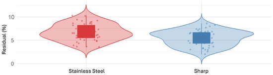
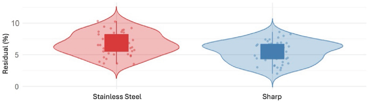
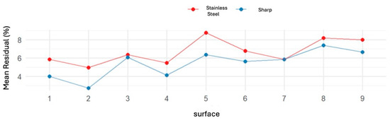
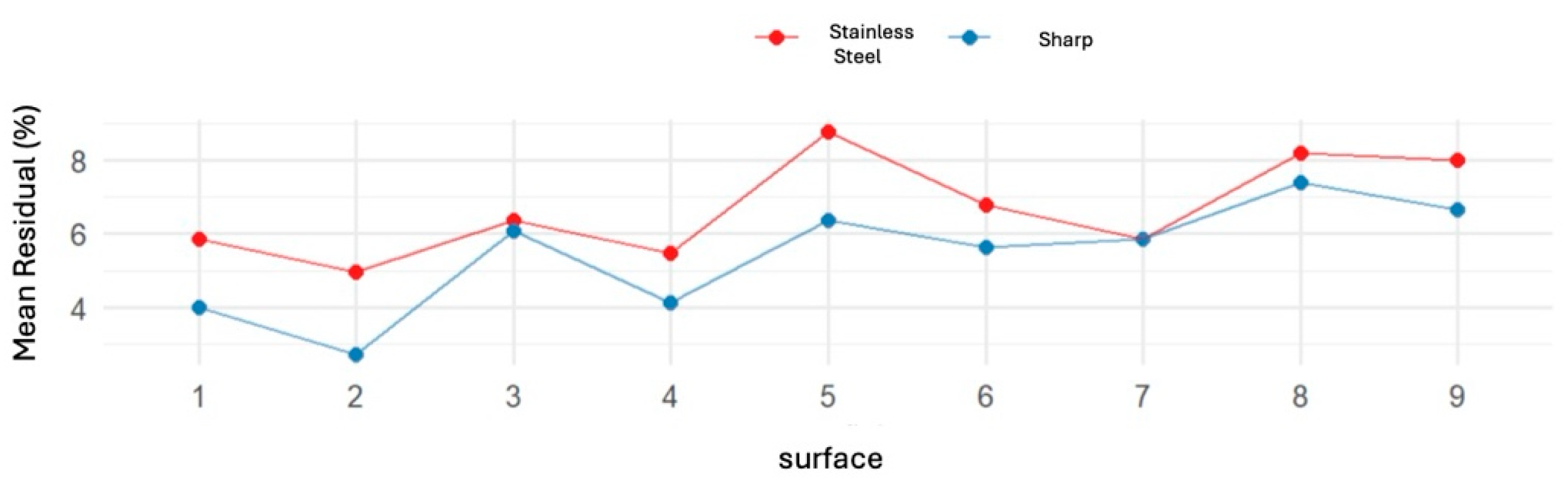
Exploratory analyses were conducted to assess the distribution of residual calculus (%) by instrument type (Figure 1) and the mean residual percentage per treated surface (Figure 2).
Figure 1.
Distribution of the RCA by instrument.
Figure 2.
Distribution of the RCA percentage of single treated surface.
In the stainless-steel group, residual calculus values were mainly distributed between 5% and 10%, with a median of approximately 6–7% and an overall range of approximately 2–11% (Table 3). In contrast, the Sharp Diamond™ group showed lower residual values, with a median of approximately 5% and a more compact distribution ranging from 3% to 8% (Figure 3).
Table 3.
Percentage of the RCA distribution.
Figure 3.
Marginal means of the RCA by instrument.
Mean residual coated area values in the stainless-steel group ranged from 5.0% to 8.5%, whereas values in the Sharp Diamond™ group ranged from 3.2% to 7.2%. The average difference between instruments corresponded to approximately 1.5–2.0% less residual calculus for Sharp Diamond™, with the greatest differences observed for teeth 1, 2, and 5.
3.3. Mixed-Effects Model Analysis
To account for the clustered data structure of the dataset, including repeated measurements within the same tooth surface and tooth-level variability, a linear mixed-effects model was fitted. This analysis was performed to estimate the effect of instrument type while accounting for the non-independence of observations. The model included residual calculus (%) as the outcome variable, instrument type as the fixed effect (stainless-steel as reference), and tooth as a random intercept.
Estimated model coefficients are reported in Table 4 (REML estimation).
Table 4.
Estimated coefficients of the linear mixed-effects model (REML method).
The intraclass correlation coefficient (ICC) was 0.535, indicating that approximately 54% of the total variance in residual calculus was attributable to between-tooth variability (Table 5).
Table 5.
Random effects and variabilities of the models.
3.4. Estimated Marginal Means and Model Diagnostics
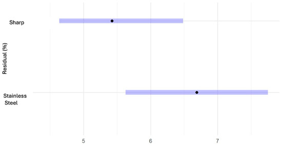
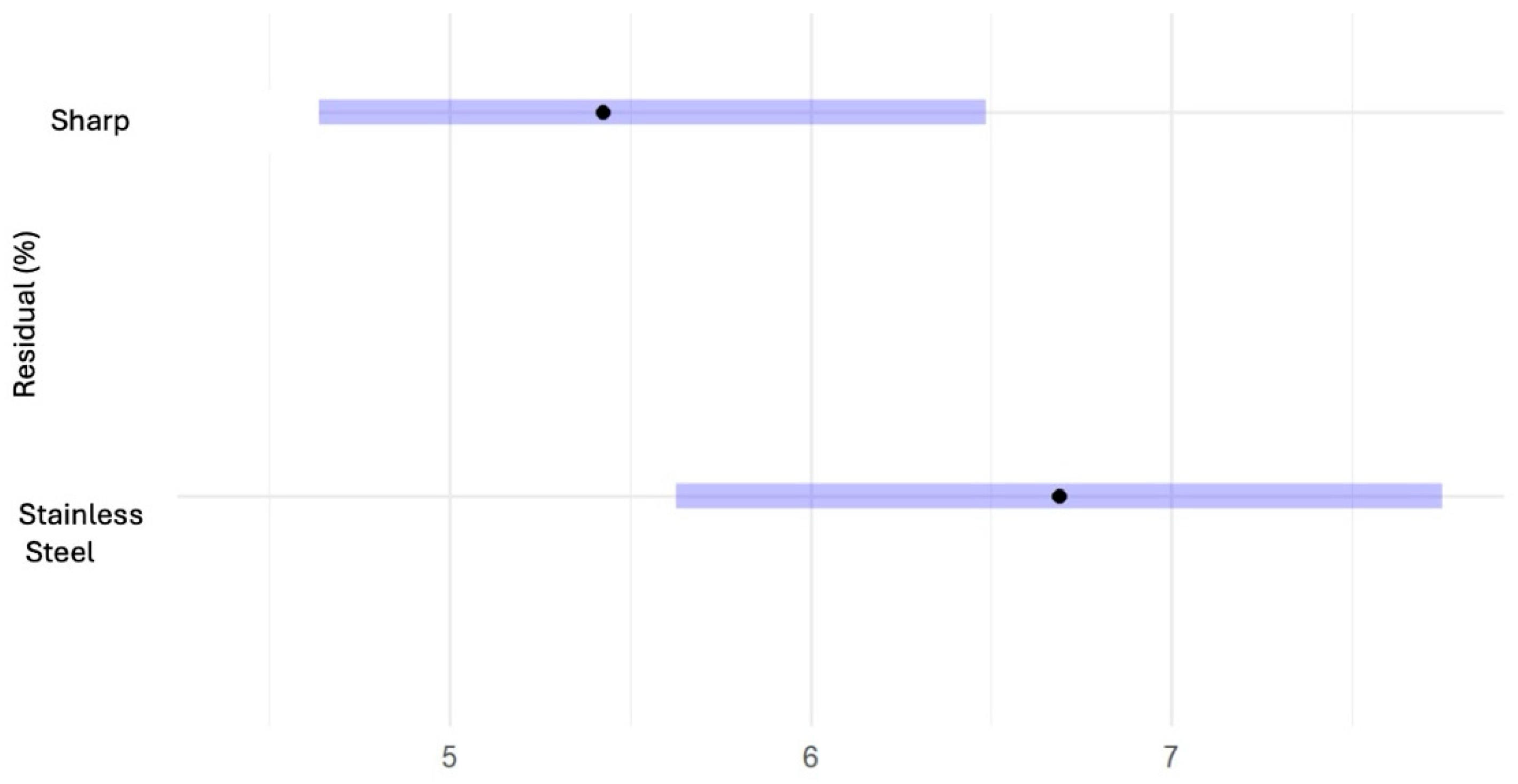
Estimated marginal means of residual calculus (%) by instrument type, derived from the mixed-effects model, are presented in Table 6. Point estimates represent adjusted mean residual coated area values, while error bars denote 95% confidence intervals (95% CI).
Table 6.
Estimated marginal means of the RCA (%) by instrument type.
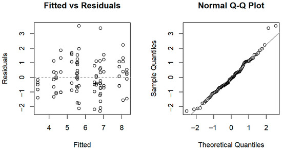
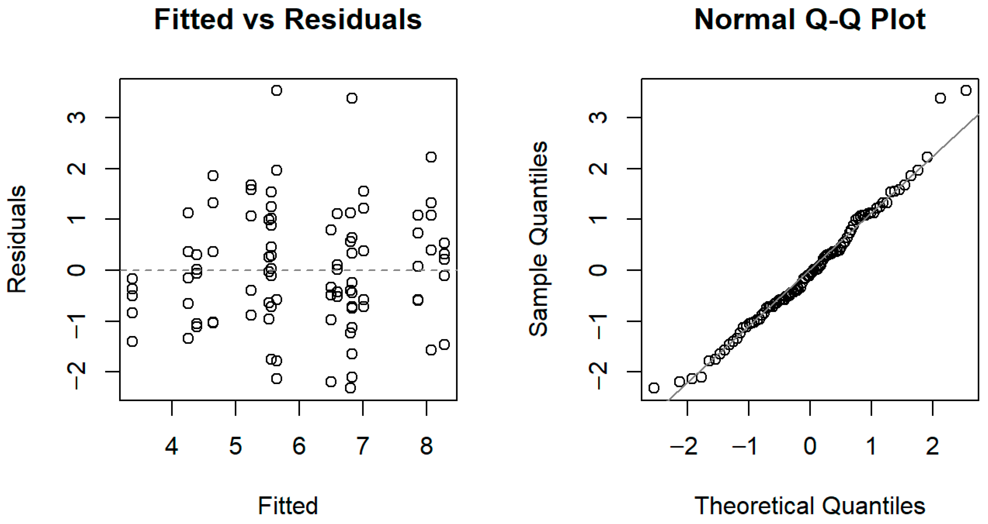
Model assumptions were evaluated using residual diagnostic plots, including fitted values versus residual coated areas and Q–Q plots (Figure 4), which indicated an adequate model fit.
Figure 4.
(Left): “Fitted vs. RCA” plot. (Right): Q–Q plot of the RCA.
4. Discussion
In this in vitro investigation, LM Sharp Diamond™ curettes demonstrated superior effectiveness in removing simulated root surface residues compared with conventional stainless-steel curettes. To the best of our knowledge, this study represents one of the first controlled experimental evaluations focused specifically on diamond-coated manual curettes under standardized conditions.
The primary objective of nonsurgical periodontal therapy is the removal of subgingival biofilm, calculus, and altered cementum to achieve a biologically acceptable root surface and halt disease progression. Traditional scaling and root planing with manual instruments such as Gracey curettes has long been established as an effective modality for achieving these objectives, with proven clinical and microbiological benefits in chronic periodontitis treatment [1,2]. However, no consensus exists regarding the superiority of one instrumentation method over another, particularly when comparing manual versus alternative or adjunctive technologies [3,4].
Clinical comparisons of different instrumentation systems have shown mixed results. For example, a randomized clinical trial comparing Gracey curettes, ultrasonic scalers, diamond burs, and piezosurgery ultrasonic devices found that curettes and ultrasonic instruments were more effective in improving clinical attachment levels and reducing probing pocket depth than diamond burs, although ultrasonic devices required less chairside time. This finding suggests that instrument design and working mechanics can significantly influence treatment outcomes but also underscores that diamond abrasives per se do not always translate into superior clinical results, especially when delivered via powered instruments rather than hand instruments [5].
Ultrasonic instrumentation with diamond-coated inserts has previously been investigated. Scott et al. reported improved efficacy in calculus removal from furcation areas using diamond-coated ultrasonic inserts compared with conventional ultrasonic and hand instruments, suggesting that diamond abrasives can enhance debridement effectiveness in challenging anatomical contexts [6]. However, surface roughness findings were variable across different studies, with some indicating increased surface roughness following diamond tip instrumentation relative to hand curettes [7], which could have implications for plaque re-accumulation and tissue healing. In this context, the present results should be interpreted cautiously: although improved deposit removal was observed, the study did not evaluate root surface roughness or other biologically relevant surface changes after instrumentation. Additional in vitro evidence also shows that diamond-coated sonic scaler tips can produce greater surface roughness than ultrasonic universal tips and hand curettes [8].
Despite the limited available evidence on diamond-coated manual curettes, the present findings suggest that LM Sharp Diamond™ instruments may remove simulated root surface deposits more effectively than conventional stainless-steel curettes under standardized in vitro conditions. The observed reduction in residual deposits was statistically significant and remained consistent after accounting for tooth-related variability. This result suggests that the improved performance was associated with instrument type rather than being explained only by differences among specimens. From a practical perspective, even a modest reduction in residual deposits may be clinically relevant, since more thorough root surface debridement is a key objective of non-surgical periodontal therapy. From a clinical perspective, the observed improvement in deposit removal suggests that the diamond coating on Sharp Diamond™ curettes may enhance instrumentation efficiency while maintaining edge performance over time. This aspect is particularly relevant in daily periodontal practice, where reduced sharpness of conventional curettes may compromise effectiveness and increase operator fatigue [9,10]. If confirmed in clinical settings, the use of diamond-coated manual curettes could represent a practical advantage in terms of debridement quality, handling consistency, and reduced need for repeated sharpening [11]. The diagnostic evaluation of the mixed model confirmed the adequacy of key statistical assumptions. The Fitted vs. Residuals plot showed residuals randomly distributed around zero, indicating homoscedasticity and the absence of unmodeled structure. The Q–Q plot revealed acceptable alignment with the normal distribution, providing support for the use of parametric inference procedures in this context.
Despite these promising results, several limitations should be acknowledged. First, the study was conducted in vitro using simulated deposits and a limited number of specimens. The small number of teeth included in the study (only 4 teeth (9 surfaces), which represents too small a sample for generalizability) should be considered a methodological limitation, and the findings should therefore be interpreted with caution. In addition, instrumentation was continued until complete macroscopic removal was achieved, rather than being standardized by time or number of strokes. This choice was intended to reproduce routine clinical handling, but it may have introduced operator-dependent variability and should be considered when interpreting the results. Moreover, the reliability of the image-based measurement method was not formally tested, and this should be considered when interpreting the quantitative results. Surface roughness and potential tissue effects were not directly measured; these factors may influence healing and long-term periodontal stability and warrant further investigation, especially given the conflicting literature on the impact of different instrumentation modalities on root surface morphology [7,8]. While this controlled setting enhances internal validity, clinical factors such as patient anatomy, access limitations, soft tissue interference, and patient discomfort cannot be replicated fully in in vitro conditions. Future clinical studies are needed to confirm whether the improved residue removal observed here translates into better periodontal outcomes in patients.
Notwithstanding these limitations, the present findings align with the broader periodontal literature indicating that instrumentation choice can influence the completeness of biofilm and deposit removal, a critical determinant of periodontal healing [3,4]. This study provides foundational evidence supporting the effectiveness of diamond-coated manual curettes and highlights the need for additional comparative research.
Overall, the present study provides preliminary evidence that diamond-coated manual curettes may improve the removal of simulated root surface deposits compared with conventional curettes. Although the experimental design does not allow direct clinical extrapolation, the findings support further investigation in larger and clinically oriented studies, including outcomes related not only to deposit removal, but also to root surface alterations, periodontal healing, and operator performance.
5. Conclusions
Under controlled in vitro conditions, LM Sharp Diamond™ curettes demonstrated preliminary superiority in simulated root debridement compared with traditional stainless-steel curettes (mean RCA reduction: −1.27 percentage points, 95% CI: −1.78 to −0.75; p = 4.45 × 10⁻⁶). These findings offer novel evidence that diamond-coated instruments address a key limitation of conventional curettes by maintaining cutting efficiency without frequent resharpening, potentially enhancing clinical consistency in non-surgical periodontal therapy. Future confirmatory research should prioritize powered RCTs (n ≥ 40 patients/side) measuring CAL gain, PPD reduction, and BOP at 6/12 months, alongside root surface profilometry/SEM analysis, standardized stroke-count protocols (e.g., 20 strokes/surface), and blinded multi-operator validation to assess reproducibility.
Author Contributions
Conceptualization, F.S.L. and G.G.; methodology, D.S.; software, V.B.; validation, E.S., E.C., S.M. and M.R.; formal analysis, F.S.L.; investigation, D.S.; resources, V.B.; data curation, F.S.L. and R.G.P.; writing—original draft preparation, F.S.L. and R.G.P.; writing—review and editing, F.S.L. and R.G.P.; visualization, F.S.L., D.S. and G.G.; supervision, E.S., S.M., E.C. and S.M.; project administration, E.S. All authors have read and agreed to the published version of the manuscript.
Funding
This research received no external funding.
Institutional Review Board Statement
The study was observational and non-interventional in nature. Participation was voluntary and completely anonymous; all participants were informed of the study’s objectives and how their data would be processed, in compliance with the European General Data Protection Regulation (GDPR 679/2016). Given the nature of the study, approval by an Ethics Committee was not required.
Informed Consent Statement
Not applicable.
Data Availability Statement
The original contributions presented in this study are included in the article. Further inquiries can be directed to the corresponding author.
Acknowledgments
The authors have reviewed and edited the output and take full responsibility for the content of this publication.
Conflicts of Interest
The authors declare no conflicts of interest.
References
- Lindhe, J.; Lang, N.P. Clinical Periodontology and Implant Dentistry, 6th ed.; Wiley-Blackwell: Chichester, UK, 2015. [Google Scholar]
- López-Valverde, N.; Quispe-López, N.; Rueda, J.A.B. Inflammation and immune response in the development of periodontal disease: A narrative review. Front. Cell. Infect. Microbiol. 2024, 14, 1493818. [Google Scholar] [CrossRef] [Scilit] [PubMed]
- Wei, Y.; Dang, G.P.; Ren, Z.Y.; Wan, M.C.; Wang, C.Y.; Li, H.B.; Zhang, T.; Tay, F.R.; Niu, L.N. Recent advances in the pathogenesis and prevention strategies of dental calculus. npj Biofilms Microbiomes 2024, 10, 56. [Google Scholar] [CrossRef] [Scilit] [PubMed] [PubMed Central]
- Löe, H.; Theilade, E.; Jensen, S.B. Experimental gingivitis in man. J. Periodontol. 1965, 36, 177–187. [Google Scholar] [CrossRef] [Scilit]
- Abdulkareem, A.A.; Al-Taweel, F.B.; Al-Sharqi, A.J.; Gul, S.S.; Sha, A.; Chapple, I.L. Current concepts in the pathogenesis of periodontitis: From symbiosis to dysbiosis. J. Oral. Microbiol. 2023, 15, 2197779. [Google Scholar] [CrossRef] [Scilit]
- American Academy of Periodontology. 1999 International Workshop for a Classification of Periodontal Diseases and Conditions. Ann. Periodontol. 1999, 4, 1–112. [Google Scholar]
- Tonetti, M.S.; Greenwell, H.; Kornman, K.S. Staging and grading of periodontitis: Framework and proposal of a new classification and case definition. J. Periodontol. 2018, 89, S159–S172. [Google Scholar] [CrossRef] [Scilit] [PubMed]
- Pjetursson, B.E.; Helbling, C.; Weber, H.P.; Matuliene, G.; Salvi, G.E.; Brägger, U.; Schmidlin, K.; Zwahlen, M.; Lang, N.P. Peri-implantitis susceptibility as it relates to periodontal therapy and supportive care. Clin. Oral. Implants Res. 2012, 23, 888–894. [Google Scholar] [CrossRef] [Scilit]
- Caton, J.G.; Armitage, G.; Berglundh, T.; Chapple, I.L.C.; Jepsen, S.; Kornman, K.S.; Mealey, B.L.; Papapanou, P.N.; Sanz, M.; Tonetti, M.S. A new classification scheme for periodontal and peri-implant diseases and conditions—Introduction and key changes from the 1999 classification. J. Periodontol. 2018, 89, S1–S8. [Google Scholar] [CrossRef] [Scilit]
- Werner, N.; Heck, K.; Walter, E.; Ern, C.; Bumm, C.V.; Folwaczny, M. Probing pocket depth reduction after non-surgical periodontal therapy: Tooth-related factors. J. Periodontol. 2024, 95, 29–39. [Google Scholar] [CrossRef] [Scilit] [PubMed]
- Citterio, F.; Gualini, G.; Chang, M.; Piccoli, G.M.; Giraudi, M.; Manavella, V.; Baima, G.; Mariani, G.M.; Romano, F.; Aimetti, M. Pocket closure and residual pockets after non-surgical periodontal therapy: A systematic review and meta-analysis. J. Clin. Periodontol. 2022, 49, 2–14. [Google Scholar] [CrossRef] [Scilit]
- Joseph, P.; Prabhakar, P.; Holtfreter, B.; Pink, C.; Suvan, J.; Kocher, T.; Pitchika, V. Systematic review and meta-analysis of randomized controlled trials evaluating the efficacy of non-surgical periodontal treatment in patients with concurrent systemic conditions. Clin. Oral. Investig. 2023, 28, 21. [Google Scholar] [CrossRef] [Scilit]
- Pihlstrom, B.L. Surgical Versus Non-Surgical Treatment of Periodontitis: The Past, the Present, the Future. J. Periodontal Res. 2025, 60, 519–523. [Google Scholar] [CrossRef] [Scilit]
- Cobb, C.M. Non-surgical pocket therapy: Mechanical. Ann. Periodontol. 1996, 1, 443–490. [Google Scholar] [CrossRef] [Scilit] [PubMed]
- Van Winkelhoff, A.J.; Rodenburg, J.P.; Goené, R.J.; Abbas, F.; Winkel, E.G.; de Graaff, J. Metronidazole plus amoxicillin in the treatment of Actinobacillus actinomycetemcomitans-associated periodontitis. J. Clin. Periodontol. 1996, 23, 960–964. [Google Scholar]
- Trombelli, L.; Farina, R.; Perinetti, G. The effect of professional mechanical plaque removal on the reduction of gingival inflammation in patients with chronic periodontitis. J. Clin. Periodontol. 2015, 42, 133–143. [Google Scholar]
- Checchi, L.; Pelliccioni, G.A. Manual and ultrasonic instrumentation in the removal of endotoxins from root surfaces in vitro. J. Periodontol. 1988, 59, 398–402. [Google Scholar] [CrossRef] [Scilit]
- Patil, S.B.; Kalburgi, V.; Goni, R.; Rao, K.A. Scanning electron microscopy of standard and modified Gracey curettes: A comparative analysis. J. Indian. Soc. Periodontol. 2022, 26, 344–349. [Google Scholar]
- Ji, Y.J.; Kim, Y.T.; Lee, J.B.; Jeong, S.N. Comparative root surface changes by various Gracey curettes: SEM analysis. J. Periodontal Implant. Sci. 2022, 52, 33–41. [Google Scholar]
- Sanz, M.; Herrera, D.; Kebschull, M.; Chapple, I.; Jepsen, S.; Berglundh, T.; Sculean, A.; Tonetti, M.S.; Consultants, T.E.W.P.A.M.; Lambert, N.L.F. Treatment of stage I–III periodontitis—The EFP S3 level clinical practice guideline. J. Clin. Periodontol. 2020, 47, 4–60. [Google Scholar] [CrossRef] [Scilit] [PubMed]
- Smiley, C.J.; Tracy, S.L.; Abt, E.; Michalowicz, B.S.; John, M.T.; Gunsolley, J.; Cobb, C.M.; Rossmann, J.; Harrel, S.K.; Forrest, J.L.; et al. Systematic review and meta-analysis on the nonsurgical treatment of chronic periodontitis by means of scaling and root planing with or without adjuncts. J. Am. Dent. Assoc. 2015, 146, 508–524.e5. [Google Scholar] [CrossRef] [Scilit] [PubMed]
- Renvert, S.; Polyzois, I. Clinical approaches to biofilm management around dental implants. Int. J. Oral. Maxillofac. Implant. 2015, 30, 125–134. [Google Scholar]
- Wilson, T.G.; Kornman, K.S. Fundamentals of Periodontal Instrumentation and Advanced Root Instrumentation, 8th ed.; Lippincott Williams & Wilkins: Waltham, MA, USA, 2020. [Google Scholar]
- Smiley, C.J.; Tracy, S.L.; Abt, E.; Michalowicz, B.S.; John, M.T.; Gunsolley, J.; Cobb, C.M.; Rossmann, J.; Harrel, S.K.; Forrest, J.L.; et al. Evidence-based clinical practice guideline on the nonsurgical treatment of chronic periodontitis. J. Am. Dent. Assoc. 2016, 147, 551–561.e8. [Google Scholar]
- Jepsen, K.; Jepsen, S. Clinical efficacy of subgingival debridement with sonic and ultrasonic devices. Periodontol. 2000 2016, 71, 91–115. [Google Scholar]
- Ritz, L.; Hefti, A.F.; Rateitschak, K.H. Mechanical debridement with hand and ultrasonic instruments: An in vitro study. J. Clin. Periodontol. 1991, 18, 698–705. [Google Scholar]
- Flemmig, T.F.; Beikler, T. Control of oral biofilms. Periodontol. 2000 2011, 55, 9–15. [Google Scholar] [CrossRef] [Scilit]
- Persson, G.R. Micropowered ultrasonic devices in periodontal therapy. Dent. Clin. N. Am. 2015, 59, 861–874. [Google Scholar]
- Louropoulou, A.; Slot, D.E.; van der Weijden, G.A. Influence of instrument material on the integrity of titanium implant surfaces: A systematic review. Clin. Oral. Implant. Res. 2012, 23, 643–650. [Google Scholar] [CrossRef] [Scilit]
- Herrera, D.; Sanz, M.; Kebschull, M.; Jepsen, S.; Sculean, A.; Berglundh, T.; Papapanou, P.N.; Chapple, I.; Tonetti, M.S.; EFP Workshop Participants and Methodological Consultant. Treatment of stage IV periodontitis: The EFP S3 level clinical practice guideline. J. Clin. Periodontol. 2022, 49, 4–71. [Google Scholar] [CrossRef] [Scilit] [PubMed]
- Axelsson, P.; Lindhe, J. The significance of maintenance care in the treatment of periodontal disease. J. Clin. Periodontol. 1981, 8, 281–294. [Google Scholar] [CrossRef] [Scilit]
- Ganss, C.; Klimek, J.; Schwarz, N. A comparative study of air polishing powders on dentin substance loss, roughness, and surface characteristics. Caries Res. 1999, 33, 292–299. [Google Scholar]
- Graumann, S.J.; Sensat, M.L.; Stoltenberg, J.L. Air polishing: A review of current literature. J. Dent. Hyg. 2013, 87, 173–180. [Google Scholar]
- Carranza, F.A.; Newman, M.G.; Takei, H.H.; Klokkevold, P.R. Carranza’s Clinical Periodontology, 13th ed.; Elsevier Saunders: Philadelphia, PA, USA, 2019. [Google Scholar]
- Pattison, G. Sharpening periodontal curets and scalers. Dent. Clin. N. Am. 1988, 32, 695–709. [Google Scholar]
- Louropoulou, A.; Slot, D.E.; Van der Weijden, F. Influence of mechanical instruments on the biocompatibility of titanium dental implants surfaces: A systematic review. Clin. Oral. Implant. Res. 2015, 26, 841–850. [Google Scholar] [CrossRef] [Scilit] [PubMed]
- Yam, N.; Levi, J.P.A.; Chen, C.J.; Barootchi, S.; Colella, N.S. Comparative Analysis of Sharpening Devices on Periodontal Hand Instruments and Their Impact on Root Surface Roughness: An In Vitro Study. Int. J. Periodontics Restor. Dent. 2025. Epub ahead of print. [Google Scholar] [CrossRef] [Scilit] [PubMed]
- Riaz, S.; Ahmed, S.; Shabbir, S.; Khan, Z.R.; Zaidi, S.J.A.; Naeem, M.M.; Farooqui, W.A. Analysing root roughness and smear layer relationship by comparing contemporary dental curettes with conventional dental curettes: A randomised controlled trial. BMC Oral. Health 2022, 22, 237. [Google Scholar] [CrossRef] [Scilit] [PubMed] [PubMed Central]
- Abbinante, A.; Cortesi Ardizzone, V.; Ottolenghi, L. Igienista Orale. Teoria e Pratica Professionale, 2a ed.; Edra: Milano, France, 2023. [Google Scholar]
- Nevala, N.; Sormunen, E.; Remes, J.; Suomalainen, K. Evaluation of Ergonomics and Efficacy of Instruments in Dentistry. Ergon. Open J. 2013, 6, 6–12. [Google Scholar] [CrossRef] [Scilit]

Disclaimer/Publisher’s Note: The statements, opinions and data contained in all publications are solely those of the individual author(s) and contributor(s) and not of MDPI and/or the editor(s). MDPI and/or the editor(s) disclaim responsibility for any injury to people or property resulting from any ideas, methods, instructions or products referred to in the content. |
© 2026 by the authors. Licensee MDPI, Basel, Switzerland. This article is an open access article distributed under the terms and conditions of the Creative Commons Attribution (CC BY) license.