


Abstract
The article examines the phylogenetic relationships between two large genera of pennate biraphid diatoms—Pinnularia and Caloneis. The monophyly of these genera is assessed using molecular-genetic and morphological analyses. Reconstruction of molecular phylogeny reveals the need for a comprehensive revision of the taxonomic composition of Pinnularia and Caloneis. This conclusion is supported by new morphological data on major species groups within both genera. We initiate the revision of the taxonomic group by investigating the circumscriptions of Pinnularia s.s. and Caloneis s.s. The diagnoses of the two genera are emended and supported by a critical analysis of their type species, Pinnularia viridis and Caloneis amphisbaena, which are analyzed here based on both wild populations and cultured specimens. In the course of the morphological analysis, we discuss the evolutionary and phylogenetic significance of different valve features, particularly alveoli, which are relevant to genera of the family Pinnulariaceae.
1. Introduction
Pinnularia Ehrenberg is one of the biggest genera of pennate diatoms, globally distributed across freshwater, marine and wetsoil ecosystems [1,2,3]. Pinnularia has a long history of taxonomic research: the name was initially introduced by Ehrenberg [4] as a subgenus within Navicula Bory. Subsequently, Pinnularia was granted the rank of an independent genus [5]. Since then, new species with the following set of morphological features have been assigned to this genus: single plate-like or H-shaped chloroplast, biraphid naviculoid valves with central raphe system, steep mantle and alveoli (alveolate striae). Each alveolus is represented by multiple rows of small pores externally and a single large transapical groove internally [6]. A similar set of morphological features of the protoplast and valves is found in another genus of naviculoid diatoms, Caloneis P.T. Cleve, described half a century later than Pinnularia [7]. Caloneis differs from Pinnularia by longitudinal lines, finer striation (with smaller inner apertures of alveoli), and greater occurrence in brackish and saline waterbodies [8].
In fact, morphological features seem quite homogeneous in the two genera; therefore, many new species and varieties of Pinnularia and Caloneis have been described to date. Hitherto, taxonomic databases report varying estimates for the two genera. AlgaeBase [9] lists over 950 taxa in Pinnularia and ca. 250 in Caloneis. DiatomBase [10] estimates more than 1000 species of Pinnularia and over 350 of Caloneis. In contrast, Krammer [3], suggested that “there are over 1500 Pinnularia species worldwide”, although his estimation is now a quarter-century old. A significant number of species of Pinnularia and Caloneis known to date were either described or investigated by Krammer himself [3,11,12,13]. He also highlighted several taxonomic and systematic issues in this complex group.
One of the existing problems is related to the type species of Pinnularia—Pinnularia viridis (Nitzsch) Ehrenberg. The type material of this taxon, previously known as Bacillaria viridis Nitzsch, originates from the collection of C.L. Nitzsch and, as Krammer [3] notes, remains lost to date. The issue was further complicated by Ehrenberg [5], who attributed a set of taxa with significant morphological diversity to a single species—Pinnularia viridis. His concept was later upheld by Cleve [14] and eventually became generally accepted. Krammer [3] took the next step towards resolving this issue by proposing a neotype of P. viridis based on his material from Frankenwald, Germany. Since then, however, the species has remained poorly studied by means of molecular phylogenetics, and its neotype has never been subjected to molecular analysis either. Given this species’ similarity to related taxa, such as Pinnularia neomajor Krammer and Pinnularia viridiformis Krammer, further critical analysis incorporating both molecular and morphological data is needed. Molecular typification of P. viridis would be a useful step. This approach has previously resolved systematic and taxonomic issues for certain species, e.g., Hantzschia amphioxys (Ehrenberg) Grunow [15], Stephanocyclus planum Skabitschevsky [16], Platessa conspicua (Ant. Mayer) Lange-Bertalot [17] and Pinnularia acidicola Van de Vijver & Le Cohu [18]. The case of Caloneis amphisbaena (Bory) Cleve, the type species of Caloneis, is similar to that of P. viridis. Its type material, analyzed by Cleve [7], originates from the collection of J.-B. Bory de Saint Vincent [19] and has never been analyzed from a molecular phylogenetic perspective. As the taxonomic challenge concerning the generic status of Caloneis remains open, the elaboration of reference molecular and morphological data for this species and, possibly, its molecular epitypification are lacking.
As it was already mentioned, a uniting feature of Pinnularia and Caloneis is the presence of multiseriate chambered striae (alveoli). However, the nature of this diagnostic character remained ambiguous when Caloneis was erected [7]. The detailed observation of alveoli and other fine valve structures became possible only after the adoption of electron microscopy in the 1950s [20,21,22]. These studies revealed that the longitudinal lines observed in Caloneis represent the internal structure of striae, homologous to that found in Pinnularia. Consequently, it became clear that the two genera cannot be reliably differentiated based solely on striae morphology. This point of view was later upheld by Round et al. [1]: according to the newly proposed system of the order Naviculales, Bessey family Pinnulariaceae, D.G. Mann included genera Diatomella Greville, Oestrupia Heiden ex Hustedt, Dimidiata Hajós and Pinnularia. Caloneis was neglected therein, as the authors were “unable to find a satisfactory basis for the traditional separation of Pinnularia from Caloneis”. Similarly, Mann [23] found “no adequate basis for the traditional Pinnularia-Caloneis distinction”. Krammer [3], however, took a different view. On the one hand, he stated that habitual differences between Pinnularia and Caloneis are obvious even in LM. On the other hand, he admitted that formal species differentiation between Pinnularia and Caloneis is complicated based on SEM, because the morphological features are quite similar, while the two genera are “almost certainly polyphyletic”. Therefore, a solution proposed by Krammer [3] encouraged further fragmentation of Pinnularia and Caloneis into minimal monophyletic genera, rather than their consolidation into a single artificial, catch-all group.
Advances in molecular phylogenetics provided evidence for Krammer’s [3] concept of the Pinnularia-Caloneis complex, demonstrating the polyphyly of both genera [24,25,26]. However, the interpretation of the obtained results is of particular interest. The authors almost unanimously concluded that the status of Caloneis as an independent genus lacks molecular-phylogenetic support, thereby justifying its merger with Pinnularia. Consequently, no attempts to revise the Pinnularia-Caloneis complex were made. Modern studies have shifted toward analyzing cryptic species and individual species groups within Pinnularia [18,27,28,29,30,31,32], while the need for a critical revision of Pinnularia and Caloneis remains unfulfilled. Our study represents a taxonomic revision using an integrative approach that combines molecular phylogenetic and morphological data. In the first phase of the revision, we aim to:
- Evaluate the recognition of Pinnularia and Caloneis as distinct genera using molecular and morphological evidence;
- Verify the polyphyletic nature of both genera;
- Differentiate Pinnularia s.s. and Caloneis s.s. We examine new strains and wild populations that, based on the evidence of our integrative analysis, represent Pinnularia viridis and Caloneis amphisbaena, the type species of the respective genera. We propose emendations to the diagnoses of Pinnularia s.s. and Caloneis s.s. following our discoveries.
2. Materials and Methods
Three samples originating from freshwater bodies in Moscow and the Kamchatka Peninsula (Russia) were utilized for the current study. Table 1 summarizes the sample information.
Table 1.
List of studied samples.
Phytoplankton sample M6 was collected with an Apstein net (mesh size: 29 µm); a 250 mL concentrate from 100 L of water was used. Samples K85 and K180 of benthos were collected from sand with biofilms and silty stones at shallow depth (less than 30 mm) by brushing with clean toothbrushes. Duplicates of each sample were fixed with 40% formaldehyde to 4% final concentration. All samples were stored in sterile 15 mL test tubes before processing in the laboratory. Hydrochemical parameters (temperature, t; pH; conductivity, σ) were measured at sampling sites using the Hanna Combo HI98129 device (Hanna Instruments, Inc., Woonsocket, RI, USA).
2.1. Sample Collection
In the laboratory, suspensions from the samples were immediately transferred to 40 mm Petri dishes and filled with WC liquid medium [33]. Monoclonal strains were isolated by micropipetting single cells under a Zeiss Axio Vert A1 inverted light microscope (ZEISS, Oberkochen, Germany). Isolation was performed in semi-sterile conditions, using sterile Pasteur pipettes and slide glasses. Isolated cells were transferred to meshes in 1 mL microtiter plates and filled with 300 µL of WC liquid medium. Cultures were maintained in an illuminated chamber (light intensity = 4000 lux) at 20 °C with a 12:12 h light–dark photoperiod. We visually inspected strain purity weekly using an inverted light microscope. The newly acquired strains were deposited in the Culture and Barcode Collection of Microalgae and Cyanobacteria “Algabank” (CBMC) in K.A. Timiryazev Institute of Plant Physiology RAS (Moscow, Russia).
2.2. Slide Preparation and Morphological Analysis
Slides for light microscopy (LM) were prepared from wild populations and cultured materials. For this purpose, suspensions were boiled with concentrated hydrogen peroxide for4–5 h. After treatment with hydrogen peroxide, the suspensions were washed and decanted repeatedly (4–5 times) with deionized water at 12 h intervals. Following the final round of decantation, the suspensions were mounted in Naphrax® (Brunel Microscopes, Chippenham, UK). For LM observations, a Zeiss AxioScope A1 (Zeiss, Oberkochen, Germany) microscope equipped with an oil immersion objective (Zeiss Plan-Apo ×100/NA1.4, Nomarski differential interference contrast, DIC) was utilized, as well as a MicMed-2 var. 2 (LOMO, Saint Petersburg, Russia) microscope, equipped with an oil immersion objective Opton Plan-Apo 63x (NA = 1.40) (Oberkochen, West Germany) and camera Levenhuk M1000 PLUS (Levenhuk, Inc., Tampa, FL, USA). To analyze the cultures by scanning electron microscopy (SEM), we placed parts of each culture suspension onto aluminum stubs. The stubs with drops of suspensions were air-dried for 24 h and then sputter-coated with 50 nm of gold in an Eiko IB 3 (Eiko Engineering, Ltd., Tokyo, Japan). Valve ultrastructure was examined by a TESCAN Vega III scanning electron microscope (TESCAN, Brno, Czech Republic), operated at 10 kV accelerating voltage and 7 mm distance.
All slides analyzed herein are housed in the Diatom Herbarium (HD) of the K.A. Timiryazev Institute of Plant Physiology RAS (Moscow, Russia).
2.3. DNA Isolation and Amplification
After two to four weeks of cultivation, strains were centrifuged in 1.5 mL Eppendorfs for 5 min at 6000 rpm. DNA was extracted using the Chelex100 Chelating Resin kit, following the manufacturer’s protocol (protocol 2.2.—Bio-Rad Laboratories, Hercules, CA, USA). PCRs were carried out using ScreenMix (Evrogen, Moscow, Russia). Amplification of V4 18S rDNA fragments was performed with D512for and D978rev primers [34]; amplification of rbcL—with rbcL404+ and rbcL1444− primers [35]. Therefore, the parameters of PCR for 18S rDNA were selected as follows: initial denaturation (95 °C, 5 min), denaturation (35 cycles, 94 °C, 30 s), annealing (52 °C, 30 s), elongation (72 °C, 50 s), final extension (72 °C, 7 min), and maintenance (12 °C). For rbcL, the procedure of PCR included initial denaturation (94 °C, 5 min), denaturation (44 cycles, 94 °C, 50 s), annealing (53 °C, 50 s), elongation (72 °C, 80 s), final extension (72 °C, 10 min), and maintenance (12 °C). Acquired products were visualized via horizontal electrophoresis in a 1.0% agarose gel with SYBR Safe stain (Life Technologies, Carlsbad, CA, USA). For purification of PCR products, we used sterile distilled water, FastAP mastermix, 10× FastAP Buffer, and Exonuclease I (Thermo Fisher Scientific, Waltham, MA, USA). We performed sequencing using the BigDye® Direct Cycle Sequencing (Applied Biosystems, Waltham, MA, USA), followed by electrophoresis on a Genetic Analyzer 3500 sequencer (Applied Biosystems, Waltham, MA, USA).
2.4. Phylogenetic Analysis
Chromatograms obtained after sequencing were trimmed and edited in Ridom TraceEdit (Ridom© GmbH, Münster, Germany). Forward and reverse reads were compared and pasted into MEGA7.1 (Pennsylvania State University, Pennsylvania, PA, USA; [36]). Matrices for 18S rDNA and rbcL were prepared separately, using the newly acquired sequences and reference sequences from the NCBI GenBank nucleotide database. Each analyzed strain was represented by both an 18S rDNA sequence and a rbcL sequence, i.e., each matrix included 192 sequences. Four strains (CBMC662m, CBMC663m, CBMC159kam, CBMC393kam) were analyzed de novo, and 188 strains were studied as references. Among them, three strains of Epithemia Kützing represented an outgroup. Other reference strains represented different genera of Pinnulariaceae and Sellaphoraceae Mereschkowsky. After assembly, the 18S rDNA matrix was aligned by means of T-Coffee via the EMBL-EBI framework [37]. rbcL matrix was aligned in MAFFT ver. 7 (RIMD, Osaka, Japan; [38]) according to the G-INS-I algorithm. Next, we trimmed the matrices in MEGA7.1; we removed unpaired regions and concatenated the 18S rDNA and rbcL datasets.
Before concatenation, the congruence of the two markers was assessed by comparing the strongly supported nodes bootstrap > 0.95) of the separate 18S rDNA and rbcL analyses. To assess marker congruence before concatenation, we performed a preliminary Maximum Likelihood (ML) analysis of the 18S rDNA and rbcL datasets separately in MEGA7.1, using the default analysis settings (GTR + G substitution model, Nearest-Neighbor-Interchange heuristic search with an initial Neighbour-Joining tree). No strongly supported topological conflicts (with bootstrap values ≥ 70 for corresponding nodes) were revealed between the two single-marker analyses. Therefore, for all phylogenetic tests described below, we used a single concatenated matrix of 1375 nucleotides—948 nucleotides for rbcL and 427 nucleotides for 18S rDNA.
Two-gene phylogenetic reconstruction was carried out using four methods: Bayesian Inference (BI), Maximum Likelihood (ML), Neighbor-joining (NJ), and Maximum Parsimony (MP). We estimated the robustness and statistical significance of the inferred tree topologies using Bayesian and bootstrap analyses with 1000 replicates. BI was performed in BEAST ver.1.10.1 software (BEAST Developers, Auckland, New Zealand; [39]). Specifications of BI analysis were assessed using jModelTest ver.2.1.10 [40] according to the Bayesian information criterion (BIC). For 18S rDNA, the specifications were calculated as follows: TrN + I + G4 partition-specific substitution model, shape parameter α = 0.2474, proportion of invariable sites (pinvar) = 0.5692. For rbcL, the specifications were TPM1uf + I + G4 model, α = 0.1510, pinvar = 0.6811 for the first codon position; JC, α = 0.1470, pinvar = 0.7359 for the second codon position; JC, α = 0.5944, pinvar = 0.2459 for the third codon position. However, the TN93 model was applied instead of TrN, HKY81—instead of TPM1uf and F81—instead of JC, as the most similar options available in BEAST ver.1.10.1. The BI was executed with the Yule process tree prior, in 10 independent runs of 10 million generations each. Convergence was assessed in Tracer ver. 1.7.1 (part of the BEAST package; [39]); the ESS values for all sampled parameters exceeded 400, indicating adequate mixing. The posterior tree samples were combined in LogCombiner ver. 1.10.1 (part of the BEAST package; [39]) after removal of the initial 10% of trees as burn-in. Rapid bootstrapping and subsequent ML search (RAxML) were carried out in RAxML GUI 2.0 [41]. For NJ and MP, the concatenated matrix was analyzed in MEGA7.1. The topology for the final phylogram was derived from the BI analysis. It was viewed in FigTree ver. 1.4.4 (University of Edinburgh, Edinburgh, UK) and edited manually via Adobe Photoshop CC ver.19.0 (Adobe, San Jose, CA, USA). GenBank accessions for the analyzed strains and the input dataset for molecular analysis are available in the Supplementary Files (Files S1 and S2, respectively).
2.5. Terminology
In the process of morphological analysis and description of new taxa, the terminology of diatom valve structures follows Krammer [3] and Gogorev et al. [42,43].
3. Results
3.1. Molecular Phylogeny
The phylogram constructed in the process of molecular analysis reveals the phylogenetic relationships between species of Pinnularia, Caloneis, and related taxa from different genera of the suborder Sellaphorineae D.G. Mann (Figure 1).
Figure 1.
Molecular phylogeny of the Pinnularia-Caloneis complex and related genera (18S rRNA and rbcL genes). Tree topology derives from BI analysis. Monophyletic clades comprising the strains of a single species or a single non-target genus are collapsed. The number of strains within each collapsed clade is indicated in brackets. Strains analyzed in this study are highlighted in bold. Branches highlighted in bold are highly supported in all four analyses (posterior probabilities, PP > 0.95, calculated by BEAST; bootstrap values, BV > 70, calculated by raxmlGUI 2.0 and MEGA7.1 using RAxML [BVML], NJ [BVNJ] and MP [BVMP]). *—var. sublinearis; **—var. nonfasciata; ***—includes the strains of S. pupula (Kützing) Mereschkowsky species complex sensu Mann et al. [44].
The phylogram shows that species and genera of the suborder Sellaphorineae form a monophyletic group, maximally supported by all four analyses. However, several distinct lineages are identified within this taxonomic group. The basal portion of the phylogram comprises genera belonging to the family Sellaphoraceae. Notably, two of these genera—Mayamaea Lange-Bertalot and Eolimna Lange-Bertalot & W. Schiller—form separate monophyletic clades, each granted with high statistical support (PP = 1/BVML = 100/BVNJ = 99/BVMP = 100 and PP = 1/BVML = 86/BVNJ = 83/BVMP = 100, respectively). The clades corresponding to Chamaepinnularia Lange-Bertalot & Krammer and Fallacia Stickle & D.G. Mann (with the exception of Fallacia sp. GU52X-3) are also monophyletic. However, the monophyly of Chamaepinnularia receives only moderate support in RAxML (BVML = 64), whereas the clade of Fallacia shows low support in NJ (BVNJ = 48).
The major part of the phylogram unites the species of Pinnularia s.l. and Caloneis s.l. This complex is highly supported by all analyses (PP = 0.96/BVML = 74/BVNJ = 71/BVMP = 91). Within this major alliance, strains of Caloneis amphisbaena (Bory) Cleve and Pinnularia borealis Ehrenberg are grouped together, indicating a close phylogenetic relationship; their common node is supported by BI and MP (PP = 1, BVMP = 100).
The clade of Caloneis amphisbaena strains (Figure 1, highlighted in blue) is highly supported in our study (PP = 1/BVML = 100/BVNJ = 99/BVMP = 100). It includes the strain CBMC393kam, which is analyzed herein. The mentioned clade corresponds to Caloneis s.s., but so far it is represented exclusively by the strains of the genus type.
Another clade, positioned in a sister-group relationship, corresponds to the P. borealis species complex. It is strongly supported across all methods as well (PP = 1/BVML = 100/BVNJ = 99/BVMP = 100). In our phylogram, it is represented by the strains of P. borealis itself, Pinnularia catenaborealis Pinseel, Hejduková, Vanormelingen, Kopalová, Vyverman a Van de Vijver, and Pinnularia paradubitabilis Kezlya, Maltsev, Krivova and Kulikovskiy.
The remaining strains of Caloneis spp., included in the analysis, are distinct from the Caloneis s.s.-clade. They represent Caloneis silicula (Ehrenberg) Cleve, Caloneis fontinalis (Grunow) A. Cleve, and Caloneis lewisii R.M. Patrick. Two more strains are labelled as Caloneis sp.; one of them, CBMC159kam, is newly isolated in our study. Altogether, the mentioned strains form a distinct monophyletic group, with exceptionally strong support across all analyses (PP = 1/BVML = 99/BVNJ = 96/BVMP = 100).
The clade of Pinnularia divergens W.Smith species complex exhibit a sister-like relationship to the C. silicula-fontinalis-lewisii-clade. This relationship is strongly supported by three out of the four tested analyses (PP = 1/BVML = 83/BVNJ = 39/BVMP = 100). In particular, the clade of P. divergens demonstrates robust statistical support (PP = 1/BVML = 99/BVNJ = 85/BVMP = 100). In addition to strain D31_023 of P. divergens, the clade comprises two strains of Pinnularia baicalodivergens Kulikovskiy, Glushchenko, Kezlya and Maltsev, two strains of Pinnularia valida Hustedt, five strains of Pinnularia spp., strain D11_014, corresponding to Pinnularia stomatophora (Grunow) Cleve and strain VP563, corresponding to Pinnularia ministomatophora Kezlya, Maltsev, Krivova and Kulikovskiy.
Our phylogram reveals close phylogenetic relationships between such species as Pinnularia isselana Krammer, Pinnularia subcommutata Krammer, Pinnularia acuminata W. Smith, Pinnularia neomajor and Pinnularia viridiformis. The taxa under consideration were primarily described by Krammer [3], who highlighted their morphological similarities with P. viridis, type species of the genus Pinnularia. The close phylogenetic relationship between these taxa and P. viridis is supported by our molecular analysis, which reveals a well-defined monophyletic “core” group within Pinnularia (PP = 1/BVML = 84/BVNJ = 71/BVMP = 100; Figure 1, highlighted in green). In this work we delimit this “core” assemblage as Pinnularia s.s. Thus, in order to characterize P. viridis, the generitype of Pinnularia s.s., we include two new strains of this species—CBMC662m and CBMC663m—in our molecular-phylogenetic study. According to the constructed phylogeny, the two strains are positioned together, and are most closely related to the subclade of P. neomajor with seven strains.
Our analysis confirms the polyphyly of Pinnularia in its current concept. Numerous species of the genus are distributed across distinct clades. In addition to the species mentioned earlier, e.g., P. divergens and P. borealis, many others are located in the top section of the phylogram, which includes two clades. The larger of is the largest in our molecular tree: it unites 63 strains, and is strongly supported by statistical analyses (PP = 1/BVML = 90/BVNJ = 72/BVMP = 100). Notably, the clade comprises several widely distributed taxa, including Pinnularia anglica Krammer, Pinnularia gibba (Ehrenberg) Ehrenberg, and Pinnularia grunowii Krammer. Another clade is much smaller, consisting of seven strains: six assigned to Pinnularia acrosphaeria W. Smith and one labelled as Pinnularia sp. This alliance receives nearly maximal statistical support across four tests (PP = 1/BVML = 100/BVNJ = 99/BVMP = 100).
3.2. Morphological Analysis
In this subsection, we propose emended diagnoses for Pinnularia s.s. and Caloneis s.s. The revised diagnosis of Pinnularia s.s. is explicitly supported by the molecular data discussed above and by new morphological data obtained via LM and SEM for specimens of Pinnularia viridis. The emendation to the diagnosis of Caloneis s.s. likewise derives from our integrative analysis, which involved the study of new specimens of Caloneis amphisbaena. However, we note that this emended diagnosis is preliminary, due to the limited molecular and morphological sampling of the species belonging to this genus. We elaborate on this issue in the Section 4.
3.2.1. Emended Diagnoses of Pinnularia and Caloneis
Pinnularia Ehrenberg emend. Mironov, Glushchenko and Kulikovskiy
Diagnosis: Cells solitary, valves naviculoid, linear-lanceolate to elliptic. Valve margins usually parallel, less often convex. Apices not protracted, gradually rounded to cuneate. Axial and central areas variable, usually moderately developed, narrow. Valve face is flat. Raphe with dilated, moderately unilaterally bent proximal ends. Internally, central portion of the raphe is continuous, i.e., proximal ends hidden under the central nodule. Distal raphe fissures unilateral, usually in the form of a question mark (recurved), internally with helictoglossae. Striae alveolate, wide, separated by very thin virgae. Externally, pores in alveoli represented by small pores with hymenate occlusions. Alveoli partially covered: ca. 1/2 of the length of each alveolus is covered by the axial plate, ca. 1/4—by the mantle plate. Thus, the internal opening is shorter than the whole alveolus.
Type species: Pinnularia viridis (Nitzsch) Ehrenberg.
Caloneis P.T. Cleve emend. Mironov, Glushchenko and Kulikovskiy
Diagnosis: Cells solitary, naviculoid. Outlines typically elliptic, apices subcapitate to capitate. Axial area narrow and linear in the apical region, distinctly widening towards the central area. The latter is rhomboid or rhombic-lanceolate. Valve face is flat. Raphe filiform to weakly lateral in the central valve portion. Central raphe ends externally significantly enlarged, straight. Internally, central ends visible, slightly offset, i.e., the central nodule is lying beside. Distal raphe fissures question mark-shaped in external view and simple, straight in internal view, with small helictoglossae. Striae alveolate, with 3 or 4 rows of small external pores with hymenate occlusions. In internal view, the alveoli are partially covered, i.e., the greater part (ca. 1/2 of the length) is covered by the axial plate, the lesser (ca. 1/4)—by the mantle plate.
Type species: Caloneis amphisbaena (Bory) P.T. Cleve.
3.2.2. Morphology of Pinnularia viridis and Caloneis amphisbaena
Pinnularia viridis (Nitzsch) Ehrenberg
Analyzed materials: wild population from sample M6 (Figure 2 and Figure 3), cultured specimens from strains CBMC662m and CBMC 663m (Figure 4, Figure 5, Figure 6 and Figure 7).
Figure 2.
(A–D). Pinnularia viridis. LM, DIC. Wild population from sample M6, slide HD10532. Size diminution series. Scale bar—10 µm.
Figure 3.
(A–C). Pinnularia viridis. LM, DIC. Wild population from sample M6, slide HD10532. Size diminution series. Scale bar—10 µm.
Figure 4.
(A–C). Pinnularia viridis. LM, DIC. Oxidized culture from strain CBMC662m, slide HD10075. Size diminution series. Scale bar—10 µm.
Figure 5.
(A–D). Pinnularia viridis. LM, DIC. Oxidized culture from strain CBMC663m, slide HD10076. Size diminution series. Scale bar—10 µm.
Figure 6.
(A–H). Pinnularia viridis. SEM. Oxidized culture from strain CBMC662m, slide HD10075. (A–C) External views. (D–H) Internal views. (A,D) Whole valve. (B,E) Central area. (C,F) Valve end. (G,H) Inner structure of alveoli. Black arrows show the circumferential alveoli (F); white arrow shows the helictoglossa (F); black ellipses show the parts of alveoli covered by the mantle plate (E,F); white ellipses show the parts of alveoli covered by the axial plate (E,F). Scale bars—20 µm (A,D), 10 µm (E), 5 µm (C,F), 2 µm (B,G), 1 µm (H).
Figure 7.
(A–H). Pinnularia viridis. SEM. Oxidized culture from strain CBMC663m, slide HD10076. (A–C) External views. (D–H) Internal views. (A,D) Whole valve. (B,E) Central area. (C,F) Valve end. (G,H) Inner structure of alveoli. Black arrows show the regular alveoli (B,C), irregular pores on the mantle (E,H), circumferential alveoli (F); white arrows show the interstriae (B,C), helictoglossa (F), pores on the valvocopula (E); black ellipses show the parts of alveoli covered by the mantle plate (E,F); white ellipses show the parts of alveoli covered by the axial plate (E,F). Scale bars—20 µm (A,D), 10 µm (B,C,E,F), 5 µm (G,H).
Description: In LM (Figure 2, Figure 3, Figure 4 and Figure 5), valves with linear, mostly parallel outlines, gradually narrowing towards the apices. In the central portion, margins slightly convex. Valve apices broad, gradually rounded. Valve dimensions: length 110–214 µm; width 20–31 µm. Length to width (l/w) ratio—5.4–6.8. Axial area narrow, linear, narrowing in the apical portion and expanding towards the central area. Central area oval-elliptic, relatively narrow (ca. 1/3 of valve width), somewhat asymmetrical. Raphe lateral. Central raphe ends rounded, unilaterally bent. Distal raphe fissures unilateral, question mark-shaped (i.e., with a recurvature = viridis-like sensu Krammer [3]). Striae thick, moderately radiate in the center, then parallel and slightly convergent around the apices. Striae density 6–8/10 µm. longitudinal lines running along the valve, corresponding to the borders of axial and mantle plates.
In SEM (Figure 6 and Figure 7), valve face is flat, smooth (Figure 6A and Figure 7A). Externally, raphe gently undulate, with unilateral, expanded (teardrop-shaped) central ends (Figure 6B and Figure 7B). Distal raphe fissures recurved, bent in the direction opposite to the central ends (Figure 6C and Figure 7C). Internally, raphe continuous, nearly straight (Figure 6D and Figure 7D). Central nodule lying above the central raphe ends (Figure 6E and Figure 7E). Distally, internal raphe fissures with small helictoglossae, deflected to one side of the valve (Figure 6F and Figure 7F, white arrows). Striae = alveoli, externally represented by multiseriate round pores (difficult to resolve due to incomplete corrosion of the valve surface, see Figure 7B,C, black arrows), continuing onto the valve mantle. Interstriae very thin (Figure 7B,C, white arrows). Internally, each alveolus with a transapically elongated opening. The opening is ca. 1/4–1/5 of the total valve width, slightly longer in the central portion and shorter towards the apices. Axial plate lying in between the internal openings and the axial area, ca. 1/2 of the valve width (Figure 6E,F and Figure 7E,F, white ellipses). Mantle plate situated at the periphery, thinner than the axial plate (Figure 6E,F and Figure 7E,F, black ellipses). External pores of alveoli are visible through the inner openings. They are small, round, in 5–6 rows for each alveolus (Figure 6G,H and Figure 7G,H). Several shortened alveoli positioned at the valve apices, arranged circumferentially (Figure 6F and Figure 7F, black arrows). Additionally, simple minute pores scattered along the valve mantle (Figure 7E,H, black arrows). Valvocopula equipped with a single row of small perpendicular perforations (Figure 7E, white arrows).
Caloneis amphisbaena (Bory) Cleve
Analyzed materials: wild population from sample K85 (Figure 8), cultured specimens from strain CBMC393kam (Figure 9, Figure 10 and Figure 11).
Figure 8.
(A–H). Caloneis amphisbaena. LM, DIC. Wild population from sample K85, slide HD07808. Size diminution series. Scale bar—10 µm.
Figure 9.
(A–D). Caloneis amphisbaena. LM, DIC. Oxidized culture from strain CBMC393kam, slide HD08225. Size diminution series. Scale bar—10 µm.
Figure 10.
(A–E). Caloneis amphisbaena. SEM, external views. Oxidized culture from strain CBMC393kam, slide HD08225. (A) Whole valve. (B,C) Central area. (D) Central portion of the valve. (E) Valve end. Black arrows show the striae (B,C), longitudinal lines (D), the notch (E); white arrows show the interstriae (B,C), terminal part of the raphe fissure (E); black ellipses show the parts of alveoli covered by the mantle plate (D); white ellipses show the parts of alveoli covered by the axial plate (D). Scale bars—10 µm (A), 5 µm (B,C), 2 µm (D,E).
Figure 11.
(A–F). Caloneis amphisbaena. SEM, internal (A–E) and girdle (F) views. Oxidized culture from strain CBMC393kam, slide HD08225. (A) Whole valve. (B) Central area. (C) Inner structure of alveoli. (D,E) Valve end. (F) Valvocopula. Black arrows show the circumferential alveoli (D,E); white arrows show the external pores in striae (C), helictoglossae (D,E); black ellipses show the parts of alveoli covered by the mantle plate (C); white ellipses show the parts of alveoli covered by the axial plate (C). Scale bars—10 µm (A,F), 5 µm (B,C), 2 µm (D,E).
Description: In LM (Figure 8 and Figure 9), valves broadly elliptical. Apices distinctly protracted, subcapitate or occasionally capitate (Figure 8E–H). Valve dimensions: length 61–68.5 µm; width 21.5–24 µm; l/w ratio—2.6–3. Axial area very narrow and linear near the apices, distinctly widening towards the rhombic-lanceolate central area. Raphe almost straight, moderately lateral. Central raphe ends large, round, slightly offset in the same direction. Distal fissures in a form of a question mark (recurved), unilateral. Striae radiate, parallel (against the central nodule), convergent (near the apices). Striae density 14.5–18/10 µm. longitudinal lines evident on each side of the valve.
In SEM (Figure 10 and Figure 11), valve face is flat, smooth (Figure 10A). In external view, valve branches nearly straight. Central raphe ends slightly offset, expanded, rounded, dented into the valve (Figure 10B,C). Distal raphe fissures unilaterally curved; the terminal part of each fissure (Figure 10E, white arrow) is bent at the valve mantle at an obtuse angle, equipped with a notch (Figure 10E, black arrow). Internally, raphe interrupted in the center by a central nodule, i.e., central raphe ends visible on the inside of the valve (Figure 11B). Distal raphe fissures terminate with small helictoglossae (Figure 11D,E, white arrows), which are moderately offset to the same valve side. Striae represented by typical alveoli. External pores of alveoli positioned in multiseriate rows (difficult to resolve due to incomplete corrosion of the valve surface, see Figure 10B,C, black arrows). Interstriae nearly as wide as striae (Figure 10B,C, white arrows). Striae continue onto the valve mantle. In internal view, alveoli represented by transapical, rectangular-elliptic openings (Figure 11C). The openings are ca. 1/4 of the total valve width. The openings are variable in size, i.e., the largest positioned mid-valve, the smallest—near the apices and against the central nodule. External pores in alveoli discernible from the interior of the valve (Figure 11C, white arrows). They are very small, round. Axial plate large, repeating the shape of the axial area throughout the valve (Figure 11C, white ellipses). Mantle plate smaller, of nearly equal width in all parts of the valve (Figure 11C, black ellipses). The presence of axial and mantle plates is also evident in external view (Figure 10D, white and black ellipses). Note the longitudinal lines visible externally, bordering the open part of alveoli (Figure 10D, black arrows). Additionally, shortened alveoli located circumferentially around the distal raphe fissures (Figure 11D,E, black arrows). Valvocopula lacks perforations (Figure 11F).
4. Discussion
In the present section, we continue the examination of the taxonomy of Caloneis s.s. and Pinnularia s.s., discussing the limits of these genera from the perspectives of molecular phylogenetics (Section 4.1) and morphology (Section 4.2). We supplement the discussion with a detailed morphological analysis of the specimens of P. viridis and C. amphisbaena, supporting their taxonomic identities (Section 4.3). This section is particularly important, since the species mentioned are the generitypes of Caloneis and Pinnularia, two genera whose diagnoses are emended in our work. We conclude the discussion with a brief outlook on the prospects for further revisions of the Pinnularia-Caloneis complex (Section 4.4).
4.1. Interpretation of Molecular Phylogeny
The results of our molecular analysis are generally consistent with the findings from prior major molecular-phylogenetic studies on the Pinnularia-Caloneis complex [25,26,32], once again demonstrating the polyphyletic nature of both Pinnularia and Caloneis. Both earlier studies and our current analysis confirm that the Pinnularia-Caloneis alliance derives from a common ancestor (Figure 1: PP = 0.96/BVML = 74/BVNJ = 71/BVMP = 91). This robust support may explain why previous works largely confined their discussion of polyphyly, without pursuing the issue further; i.e., no suggestions for more advanced revisions were proposed.
To address the problem of polyphyly, we distinguish two clades corresponding to Pinnularia s.s. and Caloneis s.s. Other major species groups examined in the molecular study are positioned outside these two sensu stricto clades. Several of these groups are particularly notable. First, the Pinnularia borealis group, represented by five strains, is distinctly separated from other Pinnularia s.l. species and forms a lineage together with Caloneis amphisbaena (i.e., Caloneis s.s.). Second, the Pinnularia divergens-group (of 12 strains) occupies a basal position relative to the Pinnularia s.s. clade. Third, the C. silicula-fontinalis-lewisii-group is sister to the P. divergens-group and remains outside the Caloneis s.s. clade. Additionally, a well-supported clade comprising six strains of Pinnularia ascrosphaeria is clearly differentiated, with strong statistical support in all analyses (Figure 1: PP = 1/BVML = 100/BVNJ = 99/BVMP = 100). However, the presence of several independent clades within the Pinnularia-Caloneis complex in the phylogram does not, by itself, constitute sufficient evidence for the taxonomic independence of these species groups from Pinnularia s.s. and Caloneis s.s. Rather, it is the suite of morphological characters that distinguishes the sensu stricto groups from the other species complexes.
We also note that the designated clades of Pinnularia s.s. and Caloneis s.s. differ in volume. The Pinnularia s.s. clade is species-rich, whereas the Caloneis s.s. clade is represented exclusively by strains of Caloneis amphisbaena. This molecular representation of the type species allows us to differentiate Caloneis s.s. from the perspective of molecular phylogeny; its differentiating morphological features of morphology are also discussed in the sections below. At the same time, the question of the taxonomic limits of Caloneis s.s. remains open and warrants further assessment, in particular through the addition of molecular data on other species of the genus.
4.2. Morphological Features in the Pinnularia-Caloneis Complex
4.2.1. Alveoli
The synapomorphy of Pinnularia and Caloneis is the presence of alveoli—chambered striae, with multiseriate rows of external pores corresponding to a single large opening on the inside of the valve. However, different species exhibit a certain degree of variability in the morphology of alveoli. This variability is linked to the size and position of the inner openings of alveoli. As we mentioned within the descriptions of the studied specimens of Pinnularia viridis and Calonies amphisbaena, the size of the opening is determined by the structure of two silica plates—the axial plate (adjacent to the raphe) and the mantle plate (adjacent to the valve mantle). In the distinguished sensu stricto groups, the inner openings of alveoli are similar—occupying ca. 1/4–1/5 of the total valve width, bordered by a wider axial plate and a narrower mantle plate (Figure 6E,F, Figure 7E,F and Figure 11C, white ellipses for axial plate, black ellipses for mantle plate). In C. amphisbaena, the arrangement of the plates can even be discerned from the external view (Figure 10D). Similarly, this arrangement results in the presence of longitudinal lines, visible in LM (Figure 2, Figure 3, Figure 4, Figure 5, Figure 8 and Figure 9). The described type of alveoli was previously illustrated for C. amphisbaena ([25] Figure 11M,N) and numerous species of Pinnularia s.s. Those are several species represented in our phylogram (Figure 1: colored in green): Pinnularia viridis ([25] Figure 10L,M), Pinnularia acuminata W. Smith ([3] Plate 148, Figure 6), Pinnularia neomajor ([3] Plate 173, Figures 1 and 3), Pinnularia viridiformis ([25] Figure 10J,K), Pinnularia substreptoraphe Krammer ([25] Figure 10N,O), Pinnularia baicalgenkalii Kulikovskiy, Glushchenko, Kezlya and Maltsev ([32] Figure 2C,D), Pinnularia baicalflexuosa Kulikovskiy, Glushchenko, Kezlya and Maltsev ([32] Figure 5C,D). In addition, partially covered (intermediate) alveoli were illustrated for Pinnularia transversiformis Krammer ([3] Plate 189, Figures 4 and 5), Pinnularia subrupesrtis Krammer ([3] Plate 117, Figures 1–6) and Pinnularia sudetica (Hilse) Hilse ([3] Plate 126, Figures 6 and 7).
In other Pinnularia and Caloneis species groups, alveoli are organized differently. On the one hand, inner openings of alveoli can be significantly reduced, i.e., nearly fully covered by the axial plate, as in Pinnularia acrosphaeria W. Smith ([3] Plate 19, Figures 5 and 6; [25] Figure 11I,J). It is worth mentioning that according to our phylogram, Pinnularia acrosphaeria (and an unidentified strain Pinnularia sp. VN400) is isolated from other species groups of the genus. Species of Caloneis clustering outside the Caloneis s.s. clade have a nearly identical type of alveoli: see the illustrations of Caloneis silicula ([26] Figure 2B) and Caloneis lewisii R.M. Patrick ([45] Figure 44). Several other species with very short alveoli are not represented in our molecular-genetic analysis: Caloneis lauta J.R. Carter ([25] Figure 11K,L), Caloneis mendosina Frenguelli ([46] Figures 53 and 54), Caloneis schumanniana (Grunow) Cleve ([45] Figures 41 and 42) and Caloneis paraegena Arsad, Bąk, Gastineau and Witkowski ([47] Figure 3F–H).
On the other hand, the inner openings of the alveoli can be elongated and bordered by narrow mantle and axial plates, leaving the chambers of alveoli fully open. Because of that, the lengths of the alveoli are identical on both the outer and inner sides of the valve, and no longitudinal lines are visible in LM. This type of alveoli is actually shared by multiple species of Pinnularia outside the sensu stricto group. Notably, it occurs in the Pinnularia gibba-group, which includes: Pinnularia subgibba Krammer ([3] Plate 72, Figures 2–6), Pinnularia grunowii Krammer ([3] Plate 82, Figures 7 and 8), Pinnularia minigibba Kezlya, Maltsev, Krivova and Kulikovskiy ([31] Figure 4), Pinnularia vietnamogibba Kezlya, Maltsev, Krivova and Kulikovskiy ([31] Figure 6), Pinnularia microgibba Kezlya, Maltsev, Krivova and Kulikovskiy ([31] Figure 8). Another is the Pinnularia divergens-group, in which open alveoli have been documented as well, for Pinnularia divergens itself ([3] Plate 28, Figures 5 and 6; Plate 35, Figure 4), Pinnularia baicalodivergens ([32] Figures 10 and 11) and Pinnularia ministomatophora ([31] Figure 12). Additional species exhibiting the same structure are: Pinnularia ovata Krammer ([3] Plate 37, Figure 4), Pinnularia subrostrata (A. Cleve) A. Cleve ([3] Plate 11, Figure 11 as P. divergentissima var. subrostrata A. Cleve), Pinnularia decrescens (Héribaud-Joseph) F.W. Mills ([3] Plate 38, Figures 6–8; Plate 40, Figure 7), Pinnularia brebissonii (Kützing) Rabenhorst ([3] Plate 49, Figures 5–7) and Pinnularia rhombarea Krammer ([3] Plate 53, Figure 10).
Pinnularia borealis group displays a similar, yet distinct morphology of alveoli. Species of this complex can be easily distinguished in LM by their notably broad striae. In external SEM view, each stria comprises multiple rows (typically 8–12) of small pores arranged chequerwise. Other species of the genus always have fewer rows of pores per stria. In internal view, alveoli appear fully open, with narrow axial and mantle plates—the latter being particularly reduced due to the shallow mantle. Illustrations for this kind of alveoli are available for Pinnularia borealis ([3] Plate 6, Figure 6), Pinnularia kulikovskiyana Gogorev ([3] Plate 6, Figures 1–4; as Pinnularia rabenhorstii var. franconica Krammer) and Pinnularia paradubitabilis ([31] Figure 14). In our phylogram, this species group forms a compact and strongly supported clade of five strains (Figure 1: PP = 1/BVML = 100/BVNJ = 71/BVMP = 100). As previously noted, this clade occupies a sister-like position in relation to Caloneis s.s.
We conclude that clades comprising Pinnularia species with open alveoli are scattered across the phylogenetic tree of Pinnularia-Caloneis. Such distribution implies that each clade represents an independent taxonomic group. This, in turn, suggests that the morphological trait of open alveoli may have evolved repeatedly in these distinct groups.
4.2.2. Central Nodule
Central nodule is a formation located on the interior of the valve in raphid diatoms, between the two branches of the raphe [42]. It is often thickened and/or elevated above the valve surface. The morphology of the central nodule is a well-established diagnostic character used to differentiate raphid diatoms at both generic and infra-generic levels. For instance, it plays a key role in the differentiation system for the families within Cymbellales [48]. In this case, the differentiating character is the interposition of the central nodule and central raphe ends. Two types of this spatial arrangement are recognized. In the first type, the central nodule overlays the raphe ends, causing the inner raphe slit to appear continuous. In the second type, the central nodule lies adjacent to the raphe, leaving the central raphe ends visible. Thus, the inner morphology of the raphe corresponds to the external configuration.
The overlaying type of central nodule (first type) is demonstrated above for Pinnularia viridis (Figure 6D,E and Figure 7D,E). Notably, Krammer [3] documented the same configuration in the following species of the sensu stricto group—Pinnularia subrupestris Krammer ([3] Plate 117, Figures 1–3 and 5), P. acuminata ([3] Plate 148, Figure 6) and P. transversiformis ([3] Plate 189, Figure 5). Kulikovskiy et al. [32] revealed the same in P. baicalgenkalii ([32] Figure 2D) and P. baicalflexuosa ([32] Figure 5D).
There are additional groups/clades featuring the overlaying central nodule. One of them is the Pinnularia gibba-group. The mentioned type of central nodule is typical for P. subgibba ([3] Plate 72, Figures 3 and 5), Pinnularia subgibba var. undulata Krammer ([3] Plate 64, Figure 10), P. minigibba ([31] Figure 4D,E), P. vietnamogibba ([31] Figure 6D,E), P. microgibba ([31] Figure 8D,E). Another group is the Pinnularia acidicola-group. Overlaying central nodule has been recently illustrated by Kollár et al. [18] for P. acidicola ([18] Figure 4G) and its related species Pinnularia acidireunionensis Kollár, T.J. Kohler, Kopalová and Van de Vijver ([18] Figure 4H) and Pinnularia vixconspicua Chattová, Metzeltin and Van de Vijver ([18] Figure 4I). Krammer [3] and then Kulikovskiy et al. [32] visualized the overlaying type of the central nodule for several species of Pinnularia grunowii-group—Pinnularia grunowii ([3] Plate 81, Figure 17; Plate 82, Figure 8), Pinnularia microfrauenbergiana Kulikovskiy, Glushchenko, Kezlya and Maltsev ([32] Figure 6W), Pinnularia pergrunowii Kulikovskiy, Glushenko, Kezlya and Maltsev ([32] Figure 7P) and Pinnularia siberiosinistra Kulikovskiy, Glushchenko, Kezlya and Maltsev ([32] Figure 8X). Notably, species from the three groups (P. gibba-, P. acidicola-, and P. grunowii-) are closely related phylogenetically, forming a top clade in our phylogram (Figure 1). The overlaying central nodule is also characteristic of P. acrosphaeria ([3] Plate 19, Figure 6). This species appears as a “maverick” in our phylogenetic analysis.
In contrast, the adjacent type of central nodule (second type) is observed in Caloneis amphisbaena (Figure 11A,B). Bruder et al. ([25] Figure 11N) similarly demonstrated it for C. amphisbaena, and other species of Caloneis—Caloneis budensis (Grunow) Krammer ([25] Figure 11P) and Caloneis lauta ([25] Figure 11L). The same configuration is known for C. paraegena ([47] Figure 3G), C. schumanniana ([45] Figures 41 and 42), C. mendosina ([46] Figure 53). Moreover, the adjacent central nodule is present in the Pinnularia divergens group. Illustrations are provided by Krammer [3], for P. divergens ([3] Plate 28, Figure 5; Plate 35, Figure 4) and P. decrescens ([3] Plate 38, Figure 8; Plate 40, Figure 7), and by Kezlya et al. [31], for P. ministomatophora ([31] Figure 12B,E). The described configuration is also found in the representatives of the Pinnularia borealis-group: P. borealis ([3] Plate 6, Figure 6), P. kulikovskiyana ([3] Plate 6, Figures 1–3) and P. paradubitabilis ([31] Figure 14D,E).
It is evident that the configuration of the central nodule, when examined at the species-group level, does not correspond to the morphology of alveoli. Notably, this trait appears consistent across both the Caloneis s.s. and the C. silicula-fontinalis-lewisii groups. We suppose that this phenomenon does not negate the phylogenetic and morphological differentiation of Caloneis species, but rather indicates that the structure of the central nodule alone is insufficient as a systematic criterion. Thus, a comprehensive analysis of a suite of morphological features should be utilized to accurately identify the species groups and genera, and reveal their phylogenetic relationships.
In the present study, we have focused on the structure of alveoli and the central nodule. However, a broader comparative analysis of other valve features in Pinnularia and Caloneis is needed for future taxonomic revisions. For example, studying the valve surface could provide additional insight into the group’s systematics. The valve in the Pinnularia-Caloneis complex is typically flat but notably ridged in P. acrosphaeria ([3] Plate 19, Figures 1–6). The presence of external grooves situated beside the central nodule in various species ([3] Plate 98, Figures 7 and 8; [45] Figures 39, 40 and 43; [47] Figure 3D) is another interesting morphological trait, which could become informative for delimitation of species groups.
4.3. Morphology and Taxonomy of the Studied Specimens
4.3.1. Morphology of Wild and Cultured Populations
For both P. viridis and C. amphisbaena, specimens from wild populations and cultures are generally uniform in valve morphology. The differences are mostly expressed in valve dimensions. In P. viridis, specimens from the wild population are notably larger (valve length 156–214 µm, valve width 27.5–31 µm) than those from cultures (valve length 110–162 µm, valve width 20–28 µm), with only a small overlap between the two ranges. The striae density, in contrast, overlaps: 6–7 in 10 µm in the wild population vs. 6–8 in 10 µm in cultures. In cultures, the l/w ratio (5.5–6.7) is within the range of this parameter for wild populations (5.4–6.8). Nevertheless, the other features are identical across the populations: the valve outline is linear with parallel margins, narrowing towards the apices; the raphe is always lateral; the central area is oval-elliptic; the axial area is narrowly linear. Additionally, longitudinal lines are notable in both wild (Figure 2 and Figure 3) and cultured (Figure 4 and Figure 5) specimens.
The analyzed specimens of C. amphisbaena are very similar to each other as well. The wild specimens are, in general, slightly larger than the cultured ones (valve length—63.5–68.5 µm in wild population vs. 61–65 µm in culture), but have the same valve width (21.5–24 µm vs. 22–24 µm) and striae density (15–18/10 µm vs. 14.5–17/10 µm). The l/w ratio range is slightly broader for the wild specimens (2.6–3), than for cultured specimens (2.6–2.8). The valves are always broadly elliptical, with usually subcapitate apices. Notably, the apices can be more protracted, strongly capitate in some wild specimens (Figure 8E–H). Other features, such as the straight raphe, large central raphe ends, question-mark-shaped distal raphe fissures, and rhombic-lanceolate central area, are identical across all specimens of C. amphisbaena illustrated herein.
4.3.2. Morphology of the Analyzed and Reference Populations
First, the specimens identified above as Pinnularia viridis should be compared with those illustrated by Krammer ([3] Plates 185–187, 194). The specimens of P. viridis sensu Krammer are similar to ours in valve length (100–182 µm in Krammer [3] vs. 110–214 µm herein), valve width (21–30 vs. 20–31 µm), l/w ratio (5–6.5 vs. 5.4–6.8) and striae density (6–7 vs. 6–8 in 10 µm). The specimens from Krammer’s neotype material ([3] Plate 186, Figures 1–4) are even closer to our population in morphometric parameters: their valves are 130–196 µm long and 21–28 µm wide, with a striae density of 6–7 in 10 µm. These values fall within the ranges observed in our specimens. The valve morphology across the populations is similar but somewhat variable. In particular, most specimens in our material have valves with slightly triundulate margins; the largest ones are moderately swollen against the central nodule (Figure 2A,C, Figure 3A–C and Figure 5C). The same set of features was reported by Krammer for his morphotypes 1, 2 and 3 of P. viridis ([3] Plate 185, Figures 1–4; Plate 186, Figures 1–4; Plate 194, Figures 3–5). In the neotype, the largest illustrated valve is also slightly triundulate, with a central inflation ([3] Plate 186, Figure 1). Morphotype 4 of Krammer ([3] Plate 187, Figures 1–4) differs the most from our specimens: it is distinguished by generally smaller valves (95–142 µm long) with more cuneiform apices. Overall, the key morphological features are shared by the different morphotypes of P. viridis, its neotype and our specimens. Those are linear outlines, a lateral raphe, a narrow axial area, an oval-elliptic and sometimes asymmetric central area, and parallel to slightly convergent striae. The observed morphological variation most likely reflects intraspecific differences among populations of this cosmopolitan species, P. viridis. Based on the combined analysis of morphological characters, we conclude that our material indeed represents P. viridis.
Our specimens can also be compared with the species of the P. viridis species group, which was studied most thoroughly by Krammer [3]. The species that merit comparison due to their morphological similarity are P. neomajor, P. viridiformis, P. neglectiformis Krammer, P. baicalgenkalii and P. baicalflexuosa. These comparisons are also relevant because the strains assigned to these species are most closely related to our strains CBMC662m and CBMC663m, according to the reconstructed molecular phylogeny (Figure 1).
P. neomajor is close to our specimens in valve dimensions (114–250 µm long, 17–30 µm wide) and striae density (6–8 in 10 µm). It also shows significant resemblance in valve outline, morphology of the axial and central areas, and striae ([3] Plate 163, Figures 1 and 2; Plate 170, Figure 3; Plate 171, Figures 1–5; Plate 172, Figures 1–4; Plate 173, Figures 1–6). The specimens of P. viridis illustrated above can be distinguished from P. neomajor by their generally smaller but wider valves and, hence, a lower l/w ratio: in our material, this ratio is 5.4–6.8, typical of P. viridis, whereas in P. neomajor it is 7.3–9 [3]. Otherwise, the morphology of the two species is very similar, and they are most closely related according to the phylogenetic analysis (Figure 1), which suggests that further investigation of this taxonomic group is needed.
P. viridiformis and P. neglectiformis are two closely similar species that are, in turn, similar to P. viridis. Valve outline, morphology of the apices and raphe, shape of the axial and central areas, and arrangement of the striae are all close in P. neglectiformis ([3] Plate 158, Figures 1–8), P. viridiformis ([3] Plate 160, Figures 1–4; Plate 161, Figures 1–4; Plate 163, Figures 3 and 4), and our specimens. As stated by Krammer [3], the species in question can be distinguished from P. viridis by their morphometric characters. Representatives of P. neglectiformis have valves 80–130 µm long, 16–20 µm wide, with 8–9 striae in 10 µm. Specimens of P. viridiformis have the following parameters: valve length 67–145 µm, valve width 14–21 µm, striae density 7–9 in 10 µm. Thus, both species are generally smaller than our specimens and have more densely arranged striae. Notably, P. neglectiformis has an l/w ratio identical to that of P. viridis sensu Krammer [3] (5–6.5) and comparable to that observed in our specimens (5.4–6.8), whereas the l/w ratio of P. viridiformis is more variable (3.8–7.5). As in the case of P. neomajor, further investigation of P. viridiformis and P. neglectiformis is needed, as previously stated by Krammer [3].
In comparison with our specimens, representatives of P. baicalgenkalii are characterized by smaller and narrower valves (92–99 µm long, 19.5–20 µm wide). The species, however, has slightly coarser striae (at most 6 in 10 µm), a distinctly undulating raphe and a less prominent central area. In fact, in P. baicalgenkalii the axial and central areas are not delimited, having practically the same width ([32] Figures 1 and 2).
Another related species, P. baicalflexuosa, exhibits smaller valve dimensions (109–116 µm long, 17.5–19 µm wide) compared with our specimens of P. viridis. These features, coupled with always parallel, somewhat rectangular valve outlines, distinguish P. baicalflexuosa from our material, whereas its other valve characters—broadly rounded apices, a narrow axial area and an elliptical, asymmetric central area—are shared with our specimens ([32] Figures 3–5).
As mentioned above, Caloneis amphisbaena is a well-known, morphologically distinct diatom species, easily recognizable by its broadly elliptical to elliptic-lanceolate valves with subcapitate to capitate apices and a large rhombic-lanceolate central area. This set of features is typical of our specimens and is also reported in the protologue of the species ([7] p. 58). In the type material, the valves are 60–80 µm long, 22.5–30 µm wide, with 16–17 striae in 10 µm; our specimens fall within these ranges in valve length and width (61–68.5 µm and 21.5–24 µm, respectively) but show a slightly wider range of striae density (14.5–18 in 10 µm). Unfortunately, the type material was not illustrated by Cleve [7], which makes direct comparison problematic.
Recent records of C. amphisbaena repeatedly show specimens with valves similar to ours, with the set of morphological features mentioned in the previous paragraph. Of the two forms most commonly reported for the species according to Krammer and Lange-Bertalot [11] and later Lange-Bertalot et al. [49]—f. amphisbaena and f. subsalina (Donkin) Cleve—the former is encountered more frequently. Our specimens correspond to f. amphisbaena most notably in having subcapitate to capitate apices ([11] Plate 168, Figure 4; [49] Plate 71, Figures 1–4), whereas in f. subsalina the apices are rostrate ([11] Plate 168, Figure 5; [49] Plate 71, Figure 5; [50] Plate 46, Figures 1–4). The cited populations also resemble our specimens in their morphometric characters: both sources [11,49] provide a broader range of valve dimensions for C. amphisbaena (length 36–80 µm, width 20–30 µm, striae density 15–18 in 10 µm). The same set of valve parameters is given in several other sources: Patrick and Reimer [51] for C. amphisbaena from North America, Witkowski et al. [52] in their marine diatom flora, Werum et al. [50] for a European population of the species, and Kulikovskiy et al. [53] in their atlas of Russian diatoms. The last of these sources provides an illustration of the species ([53] Plate 62, Figure 1) with broadly elliptic outlines, nearly capitate apices and a rhombic-lanceolate central area, closely resembling our material. Among other records worth noting are the drawings of C. amphisbaena in “An Atlas of British Diatoms” ([54] Plate 39, Figure 4): those valves are close to ours in valve dimensions (length 47–70 µm, width 21.5–28 µm, striae density 16–18 in 10 µm) and in overall morphology as well. A certain degree of variation in the shape of the apices is also evident there: they are illustrated as subcapitate to nearly rostrate. Recently, Reavie [55] illustrated specimens of C. amphisbaena from the Laurentian Great Lakes in North America ([55] Figure 440); the illustrated valve is 66 µm long and 23 µm wide, with a striae density of 16 in 10 µm, matching both our material and the type population. However, its apices are less protracted and somewhat rostrate than those of our specimens. Overall, the discussed records of C. amphisbaena are consistent with our specimens in their morphometric parameters and valve morphology, which supports the taxonomic identification of our material.
Lastly, it is important to note the species morphologically most similar to C. amphisbaena—those of the “Gruppe um Caloneis amphisbaena” designated by Krammer and Lange-Bertalot [11]: Caloneis permagna (Bailey) Cleve, Caloneis obtusa (W. Smith) Cleve, Caloneis latiuscula (Kützing) Cleve and Caloneis westii (W. Smith) Hendey. These taxa are characterized by relatively large valves with broadly elliptic to elliptic-lanceolate outlines. In contrast to C. amphisbaena, their apices are usually more acute, neither protracted nor constricted, and the axial and central areas are narrower ([11] Plate 168, Figures 1–3; Plate 169, Figures 1–7; Plate 170, Figures 1 and 2). Given their overall similarity to C. amphisbaena, it can be assumed that these species could be assigned to Caloneis s.s.; however, this remains more of a speculation in the absence of detailed SEM or TEM analyses and reference molecular data for these taxa.
4.4. Perspectives for Further Revisions
As it was noted earlier, the issue of polyphyly within the Pinnularia-Caloneis complex has been repeatedly recognized since the pioneering molecular-phylogenetic studies on pennate diatoms [24,25], yet it has remained largely unresolved in those works. Krammer [3] drew a parallel with the case of Navicula s.l., observing that both Navicula and Pinnularia have acquired the reputation of “catch-all” genera. He suggested that Pinnularia might follow the trajectory of Navicula and undergo segregation into several monophyletic genera. However, Krammer also expressed concern that such kind of reconstruction could become problematic: genera split off from Pinnularia or Caloneis might end up being either too large (i.e., again catch-all) or extremely small, even monotypic. Obviously, this imbalance would rather destabilize the taxonomic framework than stimulate the development of a natural classification system for Pinnulariaceae. Consequently, neither Pinnularia nor Caloneis has undergone the kind of subdivision seen in Navicula in the recent decades.
It is worth mentioning that genera that have been separated from Navicula are quite different in terms of their “species volume”. Some are relatively large and exhibit considerable variation in morphology, ecology, and distribution—for example, Geissleria Lange-Bertalot and Metzeltin, Neidium Pfitzer, Placoneis Mereschkowsky, and Sellaphora. In contrast, others are quite small, comprising only 3–5 species with close morphological features and restricted geographic ranges—such as Navicymbula Krammer, Ninastrelnikovia H. Lange-Bertalot and A. Fuhrmann, Sieminskia D. Metzeltin and Lange-Bertalot, and Veigaludwigia Lange-Bertalot and U. Rumrich. An analogous situation is observed in the case of Caloneis s.s. Here, we circumscribe the genus based on new data for Caloneis amphisbaena, its type species. This is the only species represented in the molecular analysis. Undoubtedly, there are grounds for including other species in this group, but the lack of molecular and detailed morphological data on them prevents us from strictly defining the limits of Caloneis s.s.
This raises the following question: should revisions of catch-all genera be suspended because species are unevenly distributed among newly erected genera? We argue that the establishment of new taxa during taxonomic revisions of well-known diatom genera, such as Pinnularia and Caloneis, can be justified if the following criteria are met:
- Robust evidence for segregation. The case for erecting a new genus should be supported by integrative data, e.g., both molecular-genetic and morphological evidence;
- Monophyly and clear delimitation. The proposed genus must be monophyletic, with clearly defined boundaries, as demonstrated by molecular-phylogenetic and morphological data;
- Characterization of the sensu stricto genus. When a new genus is separated from a sensu lato group, the sensu stricto core of the original genus should also be adequately characterized. For instance, an emended diagnosis can be proposed.
We believe these guidelines could be valuable for developing natural classification systems for complex taxonomic groups of diatoms. They may also help avoid the description of heterogeneous, unnatural genera, which would require repeated revisions. In the presented study, we applied these principles to the revision of the Pinnularia-Caloneis complex.
Supplementary Materials
The following supporting information can be downloaded at https://www.mdpi.com/article/10.3390/phycology6040129/s1, File S1. Information about analyzed strains and reference sequences; File S2. Alignment of the rbcL and 18S rRNA genes used for phylogenetic analysis.
Author Contributions
Conceptualization, A.M. and M.K.; methodology, A.G. and Y.M.; software, Y.M.; validation, A.G., Y.M. and M.K.; formal analysis, A.M. and A.G.; investigation, A.M. and A.G.; resources, A.G.; data curation, M.K.; writing—original draft preparation, A.M.; writing—review and editing, Y.M. and M.K.; visualization, A.M. and A.G.; supervision, M.K.; project administration, A.G. and M.K.; funding acquisition, A.G. and M.K. All authors have read and agreed to the published version of the manuscript.
Funding
Publication is based on research carried out with financial support by the Russian Science Foundation (25-14-00286, https://rscf.ru/project/25-14-00286/ accessed on 18 August 2026) for molecular investigation, light and scanning electron microscopy, the percentage contribution is 90%; by the framework of state assignment of the Ministry of Science and Higher Education of the Russian Federation (theme 126012015839-6) for culturing and finishing the manuscript, the percentage contribution is 10%.
Institutional Review Board Statement
Not applicable.
Informed Consent Statement
Not applicable.
Data Availability Statement
Samples collected for this study and prepared permanent slides are deposited in Diatom Herbarium (HD; https://sweetgum.nybg.org/science/ih/herbarium-details/?irn=267855, accessed on 18 August 2026) in the K.A. Timiryazev Institute of Plant Physiology RAS, Moscow, Russia. Strains analyzed herein are housed https://rscf.ru/project/25-14-00286/at Culture and Barcode Collection of Microalgae and Cyanobacteria “Algabank” (CBMC; https://algabank.ru/, accessed on 18 August 2026) in K.A. Timiryazev Institute of Plant Physiology RAS, Moscow, Russia. The sequences obtained during the current study for strains CBMC662m, CBMC663m, CBMC159kam and CBMC393kam are available in the NCBI nucleotide database under the following accession numbers: QB269392–QB269395 for 18S rRNA gene sequences and QB298075—QB298078 for rbcL gene sequences. Molecular datasets and results of performed molecular analyses can be accessed in the Supplemental Material. Additional data requests should be addressed to the corresponding author.
Acknowledgments
The authors express their gratitude to R.A. Rakitov (from the instrument analytics room of the Borissiak Paleontological Institute of the Russian Academy of Sciences, PIN RAS) for assistance in working with the scanning electron microscope.
Conflicts of Interest
The authors declare no conflicts of interest.
Abbreviations
The following abbreviations are used in this manuscript:
| BI | Bayesian Inference |
| BIC | Bayesian Information Criterion |
| DIC | Differential Interference Contrast |
| GTR | General Time-Reversible |
| LB | Likelihood Bootstrap |
| LM | Light Microscopy |
| l/w | Length to Width (ratio) |
| ML | Maximum Likelihood |
| MP | Maximum Parsimony |
| NA | Numerical Aperture |
| NJ | Neighbor-joining |
| PP | Posterior Probability |
| RAxML | Randomized Accelerated Maximum Likelihood |
| SEM | Scanning Electron Microscopy |
References
- Round, F.E.; Crawford, R.M.; Mann, D.G. The Diatoms. Biology and Morphology of the Genera; Cambridge University Press: Cambridge, UK, 1990; pp. 1–747. [Google Scholar]
- Hasle, G.R.; Syvertsen, E.E. Marine diatoms. In Identifying Marine Phytoplankton; Tomas, C.R., Ed.; Academic Press: San Diego, CA, USA, 1996; pp. 3–385. [Google Scholar]
- Krammer, K. The genus Pinnularia. In Diatoms of the European Inland Waters and Comparable Habitats; Lange-Bertalot, H., Ed.; A. R. G. Gantner Verlag K. G: Ruggell, Liechtenstein, 2000; Volume 1, pp. 1–703. [Google Scholar]
- Ehrenberg, C.G. Characteristik von 274 neuen Arten von Infusorien. Ber. Verh. K. Preuss. Akad. Wiss. Berl. 1840, 1, 197–219. [Google Scholar]
- Ehrenberg, C.G. Mittheilungen über 2 neue asiatische Lager fossiler Infusorien-Erden aus dem russischen Trans-Kaukasien (Grusien) und Sibirien. Ber. Verh. K. Preuss. Akad. Wiss. Berl. 1843, 1, 43–49. [Google Scholar]
- Cox, E.J.; Ross, R. The striae of pennate diatoms. In Proceedings of the Sixth Symposium on Fossil and Recent Diatoms, Budapest, Hungary, 1–5 September 1980. [Google Scholar]
- Cleve, P.T. Synopsis of the Naviculoid Diatoms. Part I; Kongliga Svenska Vetenskapsakademiens Handlingar Series 4: Stockholm, Sweden, 1894; Volume 26, pp. 1–194. [Google Scholar]
- Krammer, K.; Lange-Bertalot, H. Naviculaceae Neue und wenig bekannte Taxa, neue Kombinationen und Synonyme sowie Bemerkungen zu einigen Gattungen. In Bibliotheca Diatomologica; Gebrüder Borntraeger Verlagsbuchhandlung: Stuttgart, Germany, 1985; Volume 9, pp. 1–230. [Google Scholar]
- Guiry, M.D.; Guiry, G.M. AlgaeBase; World-Wide Electronic Publication, University of Galway: Galway, Ireland, 2026; Available online: https://www.algaebase.org (accessed on 18 August 2026).
- Kociolek, J.P.; Blanco, S.; Coste, M.; Ector, L.; Liu, Y.; Karthick, B.; Kulikovskiy, M.; Lundholm, N.; Ludwig, T.; Potapova, M.; et al. DiatomBase. Available online: https://www.diatombase.org (accessed on 23 August 2026).
- Krammer, K.; Lange-Bertalot, H. Süßwasserflora von Mitteleuropa. Herausgegeben von H. Ettl. Begründet von A. Pascher J Gerloff H. Heyning D. Mollenhauer Band 2/1. Bacillariophyceae: 1. Teil: Naviculaceae; Gustav Fischer Verlag: Stuttgart, Germany, 1986; pp. 1–876. [Google Scholar]
- Krammer, K. Pinnularia. Eine Monographie der europäischen Taxa. Bibl. Diatomol. 1992, 26, 1–353. [Google Scholar]
- Krammer, K. Die Bacillariophyceen Gattung Pinnularia in Bayern. Bemerkungen zu 80 hinterlassenen Tafeln von Anton Mayer sowie Beschreibungen der bisher in Bayern gefundenen Pinnularia-Taxa. Hoppea 1992, 52, 1–304. [Google Scholar]
- Cleve, P.T. Synopsis of the Naviculoid Diatoms. Part II. Kongliga Sven. Vetenskapsak Akad. Handl. 1895, 27, 1–219. [Google Scholar]
- Jahn, R.; Kusber, W.H.; Lange-Bertalot, H. Typification and taxonomy of Hantzschia amphioxys (Ehrenberg) Grunow (Bacillariophyta): Type of the genus name Hantzschia Grunow. Nova Hedwig. 2014, 143, 103–110. [Google Scholar]
- Kulikovskiy, M.; Genkal, S.; Maltsev, Y.; Glushchenko, A.; Kuznetsova, I.; Kapustin, D.; Gusev, E.; Martynenko, N.; Kociolek, J.P. Resurrection of the diatom genus Stephanocyclus (Coscinodiscophyceae: Stephanodiscaceae) on the basis of an integrated molecular and morphological approach. Fottea 2022, 22, 181–191. [Google Scholar] [CrossRef] [Scilit]
- Tseplik, N.; Glushchenko, A.; Mironov, A.; Maltsev, Y.; Nergui, S.; Kulikovskiy, M. First data on molecular phylogeny of the monoraphid genus Platessa (Achnanthidiaceae, Bacillariophyceae) on the basis of P. conspicua from Mongolia. Phytotaxa 2025, 731, 102–114. [Google Scholar] [CrossRef] [Scilit]
- Kollár, J.; Van de Vijver, B.; Kulichová, J.; Hejduková, E.; Viso, R.; Kohler, T.J.; Pinseel, E.; Poulíčková, E.; Zimmermann, J.; Neustupa, J.; et al. A polyphasic taxonomy of the pseudocryptic diatom species complex Pinnularia acidicola, including description of new species. Eur. J. Phycol. 2026, 61, 117–141. [Google Scholar] [CrossRef] [Scilit]
- Lamouroux, J.V.F.; Bory de Saint-Vincent, J.B.; Eudes-Deslongchamps, J.A. Encyclopédie méthodique ou par ordre de matières. In Histoire Naturelle des Zoophytes, ou Animaux Rayonnés, Faisant Suite à L’histoire Naturelle des vers de Bruguière; Chez Mme veuve Agasse: Paris, France, 1827; pp. 377–819. [Google Scholar]
- Helmcke, J.G. Die Feinstruktur der Kieselsäure und ihre physiologische Bedeutung in Diatomeenschalen. Naturwissenschaften 1954, 41, 254–255. [Google Scholar] [CrossRef] [Scilit]
- Kolbe, R.W.; Gölz, E. Elektronenmikroskopische Diatomeenstudien. Berichte Dtsch. Bot. Ges. 1943, 61, 91–98. [Google Scholar] [CrossRef] [Scilit]
- Okuno, H. Fossil diatoms. In Diatomeenschalen im Elektronenmikroskopischen Bild; Helmcke, J.G., Krieger, W., Eds.; J. Cramer: Weinheim, Germany, 1964; Volume 5, pp. 414–513. [Google Scholar]
- Mann, D.G. A discussion of Caloneis and related genera. Diatom 2001, 17, 29–36. [Google Scholar] [CrossRef]
- Bruder, K.; Medlin, L.K. Morphological and molecular investigations of naviculoid diatoms. II. Selected genera and families. Diatom Res. 2008, 23, 283–329. [Google Scholar] [CrossRef] [Scilit]
- Bruder, K.; Sato, S.; Medlin, L.K. Morphological and molecular investigations of naviculoid diatoms IV. Pinnularia vs. Caloneis. Diatom 2008, 24, 8–24. [Google Scholar]
- Souffreau, C.; Verbruggen, H.; Wolfe, A.P.; Vanormelingen, P.; Siver, P.A.; Cox, E.J.; Mann, D.G.; Van de Vijver, B.; Sabbe, K.; Vyverman, W. A time-calibrated multi-gene phylogeny of the diatom genus Pinnularia. Mol. Phylogenet. Evol. 2011, 61, 866–879. [Google Scholar] [CrossRef] [Scilit]
- Pinseel, E.; Hejduková, E.; Vanormelingen, P.; Kopalová, K.; Vyverman, W.; Van de Vijver, B. Pinnularia catenaborealis sp. nov. (Bacillariophyceae), a unique chain-forming diatom species from James Ross Island and Vega Island (Maritime Antarctica). Phycologia 2017, 56, 94–107. [Google Scholar] [CrossRef] [Scilit]
- Pinseel, E.; Kulichová, J.; Scharfen, V.; Urbánková, P.; Van de Vijver, B.; Vyverman, W. Extensive cryptic diversity in the terrestrial diatom Pinnularia borealis (Bacillariophyceae). Protist 2019, 170, 121–140. [Google Scholar] [CrossRef] [Scilit]
- Kollár, J.; Pinseel, E.; Vanormelingen, P.; Poulíčková, A.; Souffreau, C.; Dvořák, P.; Vyverman, W. A polyphasic approach to the delimitation of diatom species: A case study for the genus Pinnularia (Bacillariophyta). J. Phycol. 2019, 55, 365–379. [Google Scholar] [CrossRef] [Scilit]
- Kollár, J.; Pinseel, E.; Vyverman, W.; Poulickova, A. A time-calibrated multi-gene phylogeny provides insights into the evolution, taxonomy and DNA barcoding of the Pinnularia gibba group (Bacillariophyta). Fottea 2021, 21, 62–72. [Google Scholar] [CrossRef] [Scilit]
- Kezlya, E.; Maltsev, Y.; Genkal, S.; Krivova, Z.; Kulikovskiy, M. Phylogeny and Fatty Acid Profiles of New Pinnularia (Bacillariophyta) Species from Soils of Vietnam. Cells 2022, 11, 2446. [Google Scholar] [CrossRef] [Scilit]
- Kulikovskiy, M.; Glushchenko, A.; Kezlya, E.; Kuznetsova, I.; Kociolek, J.P.; Maltsev, Y. The Genus Pinnularia Ehrenberg (Bacillariophyta) from the Transbaikal Area (Russia, Siberia): Description of Seven New Species on the Basis of Morphology and Molecular Data with Discussion of the Phylogenetic Position of Caloneis. Plants 2023, 12, 3552. [Google Scholar] [CrossRef] [Scilit]
- Guillard, R.R.L.; Lorenzen, C.J. Yellow-green algae with chlorophyllide c1,2. J. Phycol. 1972, 8, 10–14. [Google Scholar] [CrossRef] [Scilit]
- Zimmermann, J.; Jahn, R.; Gemeinholzer, B. Barcoding diatoms: Evaluation of the V4 subregion on the 18S rRNA gene, including new primers and protocols. Org. Divers. Evol. 2011, 11, 173–192. [Google Scholar] [CrossRef] [Scilit]
- Ruck, E.C.; Theriot, E.C. Origin and evolution of the canal raphe system in diatoms. Protist 2011, 162, 723–737. [Google Scholar] [CrossRef] [Scilit]
- Kumar, S.; Stecher, G.; Tamura, K. MEGA7: Molecular evolutionary genetics analysis version 7.0 for bigger datasets. Mol. Biol. Evol. 2016, 33, 1870–1874. [Google Scholar] [CrossRef] [Scilit]
- Madeira, F.; Madhusoodanan, N.; Lee, J.; Eusebi, A.; Niewielska, A.; Tivey, A.R.N.; Lopez, R.; Butcher, S. The EMBL-EBI Job Dispatcher sequence analysis tools framework in 2024. Nucleic Acids Res. 2024, 52, 521–525. [Google Scholar] [CrossRef] [Scilit]
- Katoh, K.; Toh, H. Parallelization of the MAFFT multiple sequence alignment program. Bioinform. 2010, 26, 1899–1900. [Google Scholar] [CrossRef] [Scilit]
- Drummond, A.J.; Rambaut, A. BEAST: Bayesian evolutionary analysis by sampling trees. BMC Ecol. Evol. 2007, 7, 214. [Google Scholar] [CrossRef] [Scilit]
- Darriba, D.; Taboada, G.L.; Doallo, R.; Posada, D. jModelTest 2: More models, new heuristics and high-performance computing. Nat. Methods 2012, 9, 772. [Google Scholar] [CrossRef] [Scilit]
- Edler, D.; Klein, J.; Antonelli, A.; Silvestro, D. raxmlGUI 2.0: A graphical interface and toolkit for phylogenetic analyses using RAxML. Methods Ecol. Evol. 2021, 12, 373–377. [Google Scholar] [CrossRef] [Scilit]
- Gogorev, R.M.; Chudaev, D.A.; Gololobova, M.A. Addition to the glossary of diatom morphology. Nov. Sist. Nizshikh Rastenii 2024, 58, 35–58. [Google Scholar] [CrossRef] [Scilit]
- Gogorev, R.M.; Chudaev, D.A.; Stepanova, V.A.; Kulikovskiy, M.S. Russian and English terminological glossary on morphology of diatoms. Nov. Sist. Nizshikh Rastenii 2018, 52, 265–309. [Google Scholar] [CrossRef] [Scilit]
- Mann, D.G.; McDonald, S.M.; Bayer, M.M.; Droop, S.J.; Chepurnov, V.A.; Loke, R.E.; Ciobanu, A.; Du Buf, J.H. The Sellaphora pupula species complex (Bacillariophyceae): Morphometric analysis, ultrastructure and mating data provide evidence for five new species. Phycologia 2004, 43, 459–482. [Google Scholar] [CrossRef] [Scilit]
- Stancheva, R.; Manoylov, K.; Gillett, N. Morphological variation of the Caloneis schumanniana species complex (Bacillariophyceae) from different environmental conditions in North American streams. Hydrobiologia 2009, 635, 157–170. [Google Scholar] [CrossRef] [Scilit]
- Sunesen, I.; Tardivo Kubis, J.A.; Sar, E.A. A re-investigation of three Frenguelli’s Caloneis taxa (Pinnulariaceae, Bacillariophyta) from Argentina. Phytotaxa 2017, 305, 165–178. [Google Scholar] [CrossRef] [Scilit]
- Arsad, S.; Gastineau, R.; Bak, M.; Rybak, M.; Gorecka, E.; Lemieux, C.; Turmel, M.; Prasetiya, F.S.; Lobban, C.S. Not Caloneis egena: Morphological description and complete plastid and mitochondrial genomes of Caloneis paraegena sp. nov. from the Gulf of Tomini, Indonesia. Front. Mar. Sci. 2026, 13, 1749044. [Google Scholar] [CrossRef] [Scilit]
- Mironov, A.; Maltsev, Y.; Kociolek, J.P.; Kezlya, E.; Liu, Y.; Kulikovskiy, M. Description of Encyonemataceae fam. nov. and Witkowskiaceae fam. nov. (Bacillariophyceae, Cymbellales) based on molecular and morphological analyses. Sci. Rep. 2024, 14, 31045. [Google Scholar] [CrossRef] [Scilit]
- Lange-Bertalot, H.; Hofmann, G.; Werum, M.; Cantonati, M. Freshwater Benthic Diatoms of Central Europe: Over 800 Common Species Used in Ecological Assessments (English Edition with Updated Taxonomy and Added Species); Koeltz Botanical Books: Schmitten-Oberreifenberg, Germany, 2017; pp. 1–942. [Google Scholar]
- Werum, M.; Reichhardt, E.; Dressler, M.; Werner, P.; Lange-Bertalot, H. Ergänzungsband Diatomeen im Süßwasser-Benthos von Mitteleuropa: Bestimmungsflora für Kieselalgen für die Ökologische Praxis; Koeltz Botanical Books: Königstein, Germany, 2024; pp. 1–625. [Google Scholar]
- Patrick, R.M.; Reimer, C.W. The Diatoms of the United States Exclusive of Alaska and Hawaii. Volume 1: Fragilariaceae, Eunotiaceae, Achnanthaceae, Naviculaceae; Monographs of the Academy of Natural Sciences of Philadelphia No. 13; Academy of Natural Sciences of Philadelphia: Philadelphia, PA, USA, 1966; pp. 1–688. [Google Scholar]
- Witkowski, A.; Lange-Bertalot, H.; Metzeltin, D. Diatom Flora of Marine Coasts I. Iconographia Diatomologica; Koeltz Scientific Books: Königstein, Germany, 2000; Volume 7, pp. 1–925. [Google Scholar]
- Kulikovskiy, M.; Glushchenko, A.M.; Kuznetsova, I.V.; Genkal, S.I. Identification Book of Diatoms from Russia; Filigran: Yaroslavl, Russia, 2016; pp. 1–804. [Google Scholar]
- Hartley, B.; Barber, H.G.; Carter, J.R. An Atlas of British Diatoms; Biopress Ltd.: Bristol, UK, 1996; pp. 1–601. [Google Scholar]
- Reavie, E.D. Symmetric biraphid diatoms (largely non-Navicula) from the Laurentian Great Lakes. Diatom Res. 2022, 37, 351–409. [Google Scholar] [CrossRef] [Scilit]
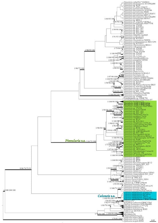
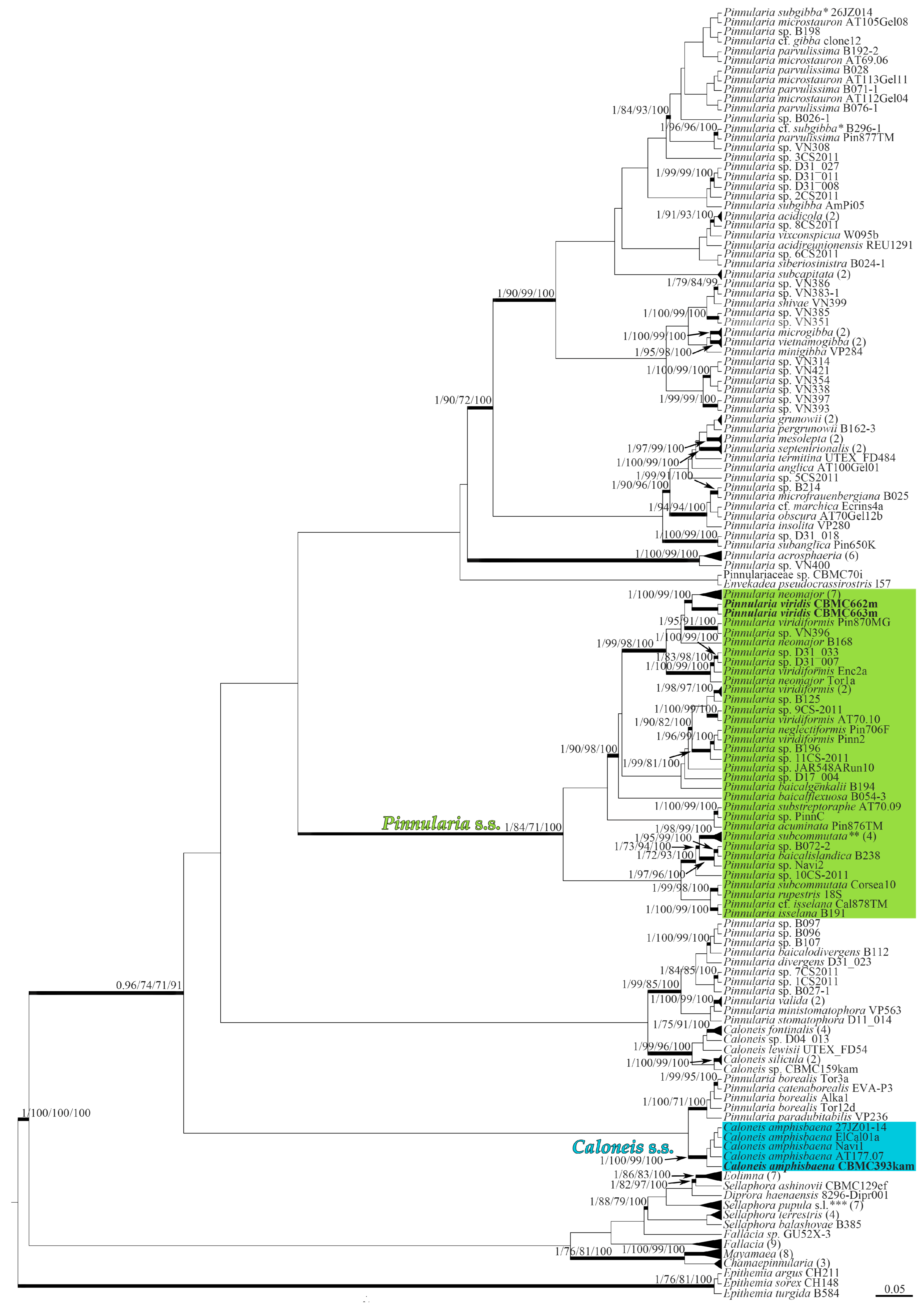
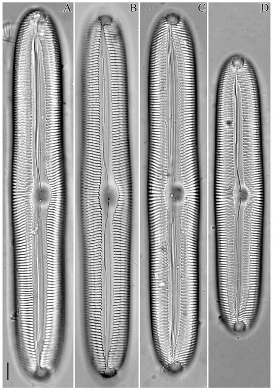
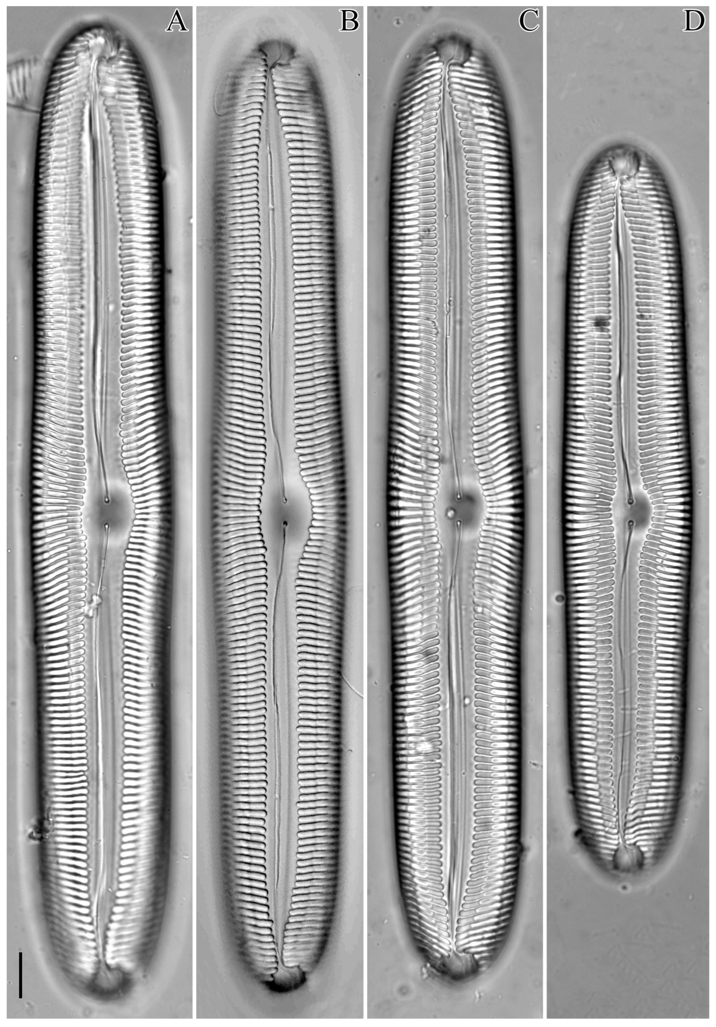
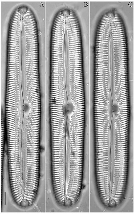
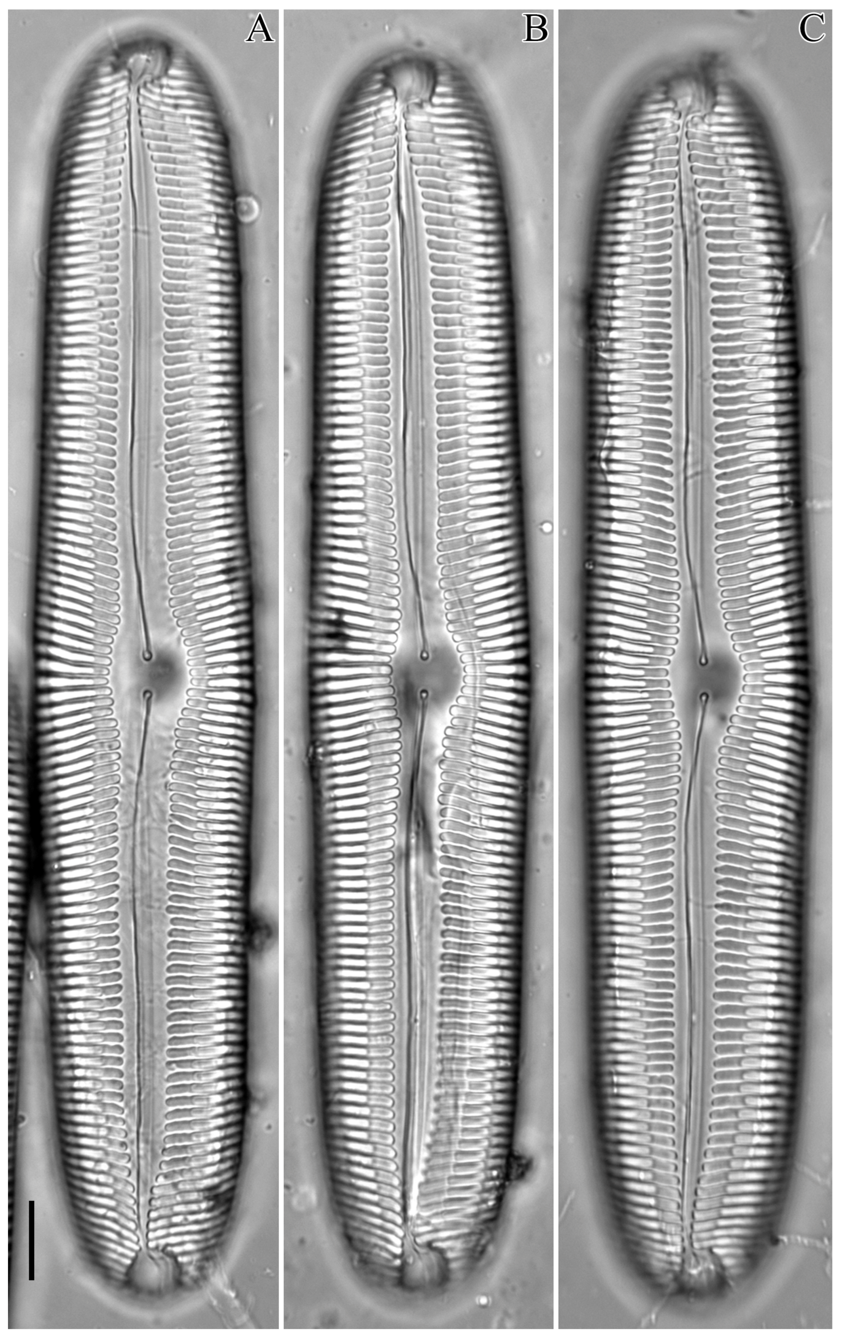
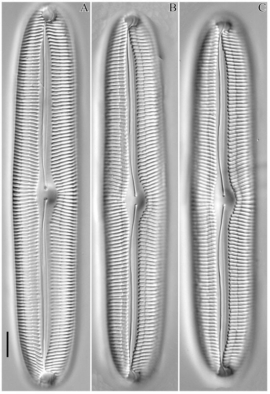
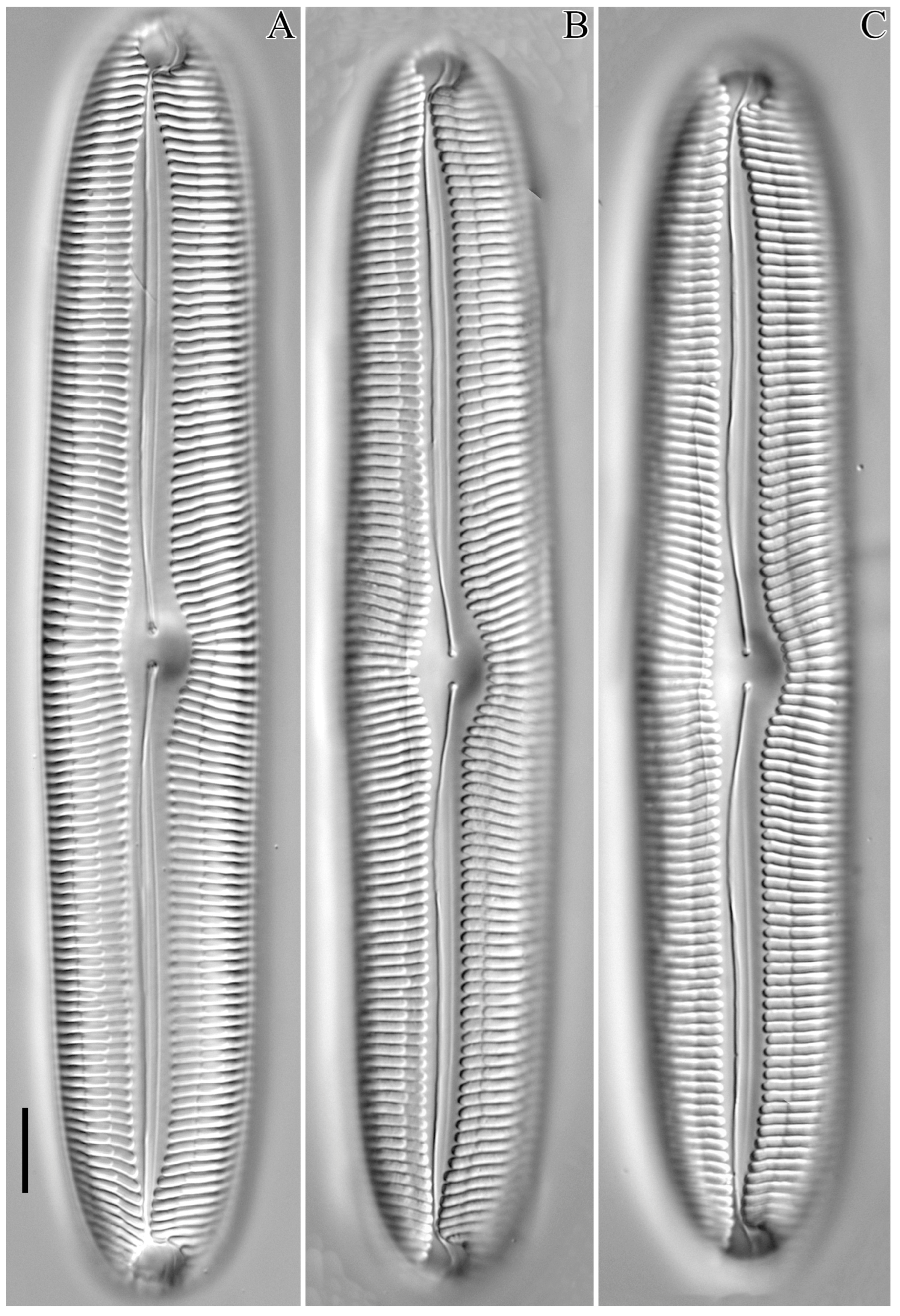
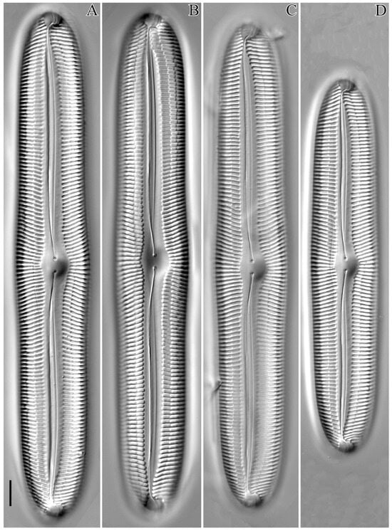
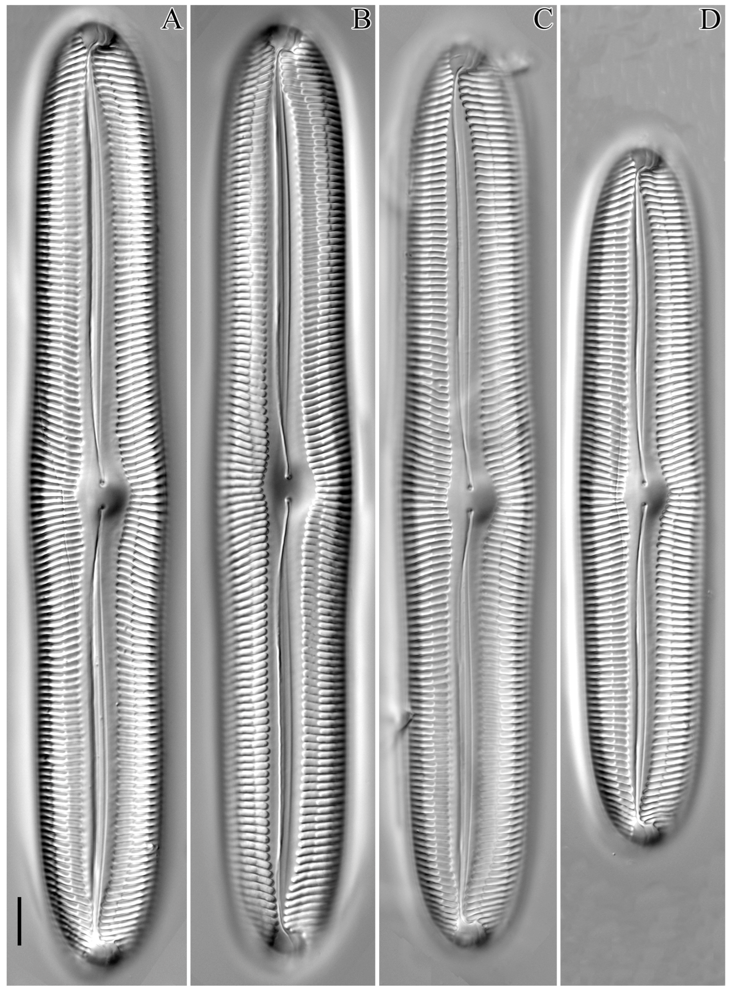
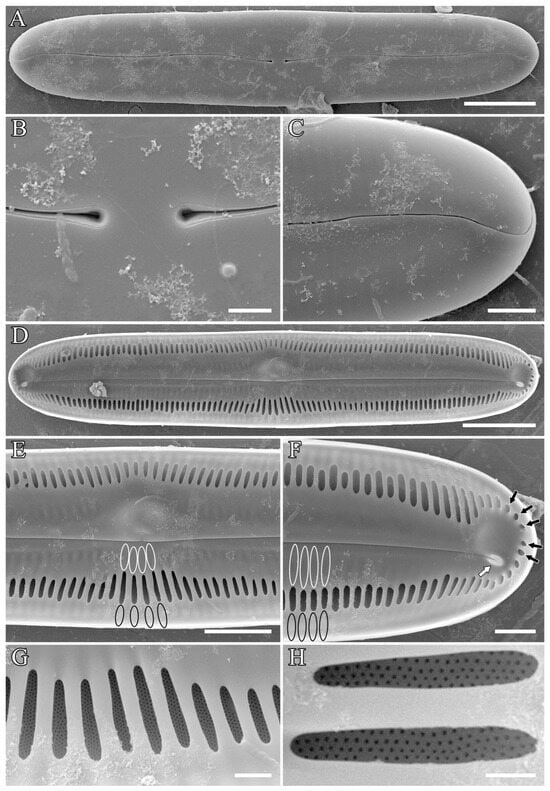
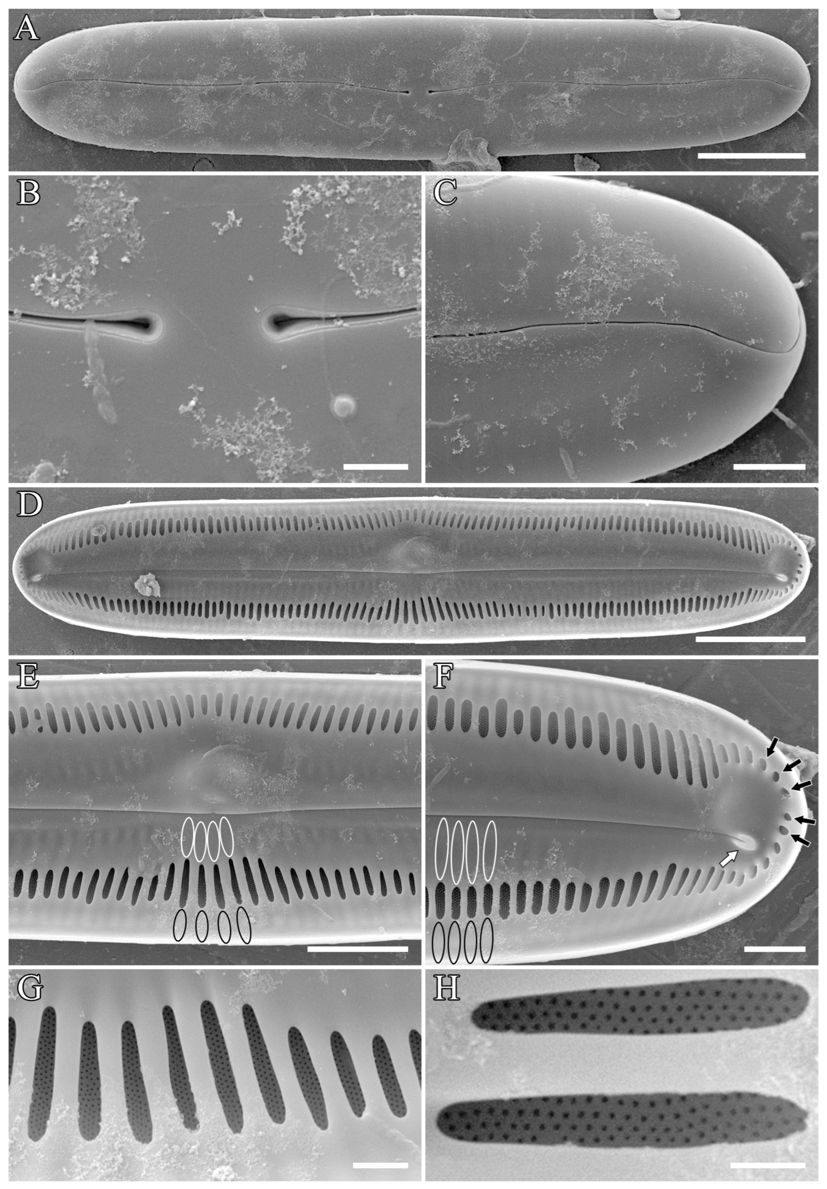
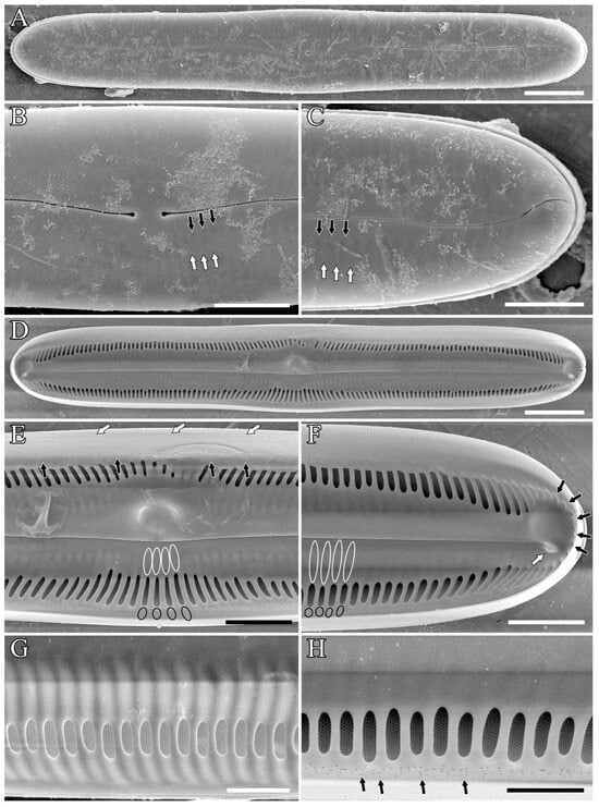
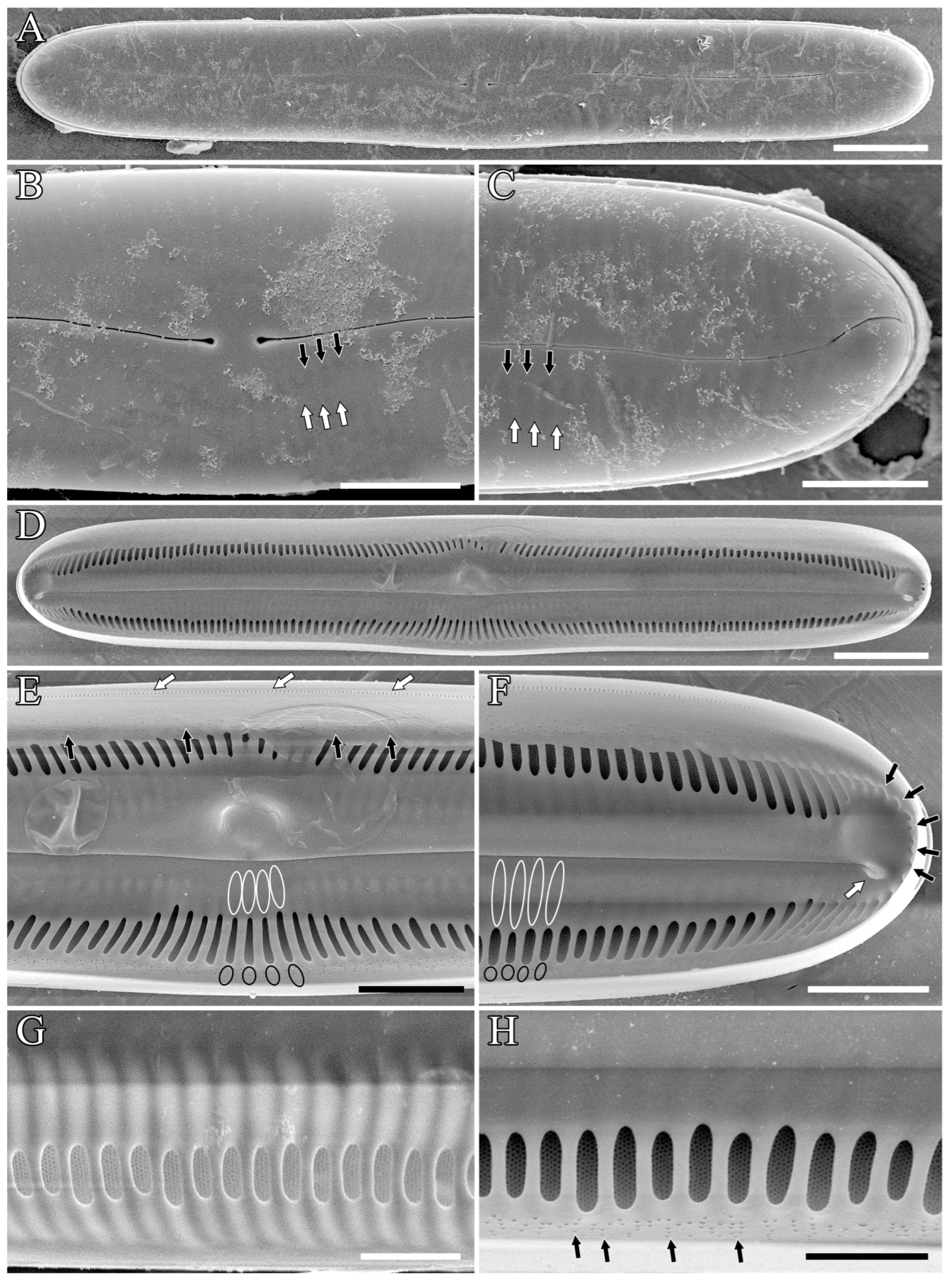
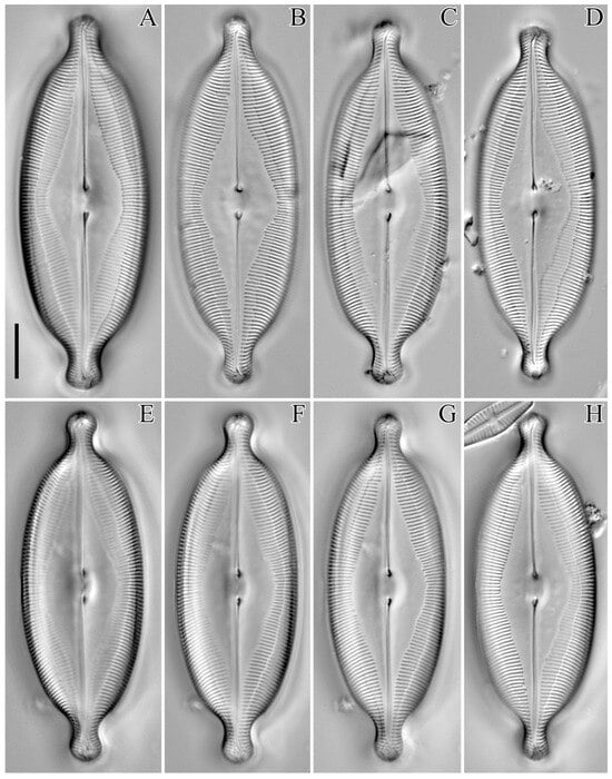
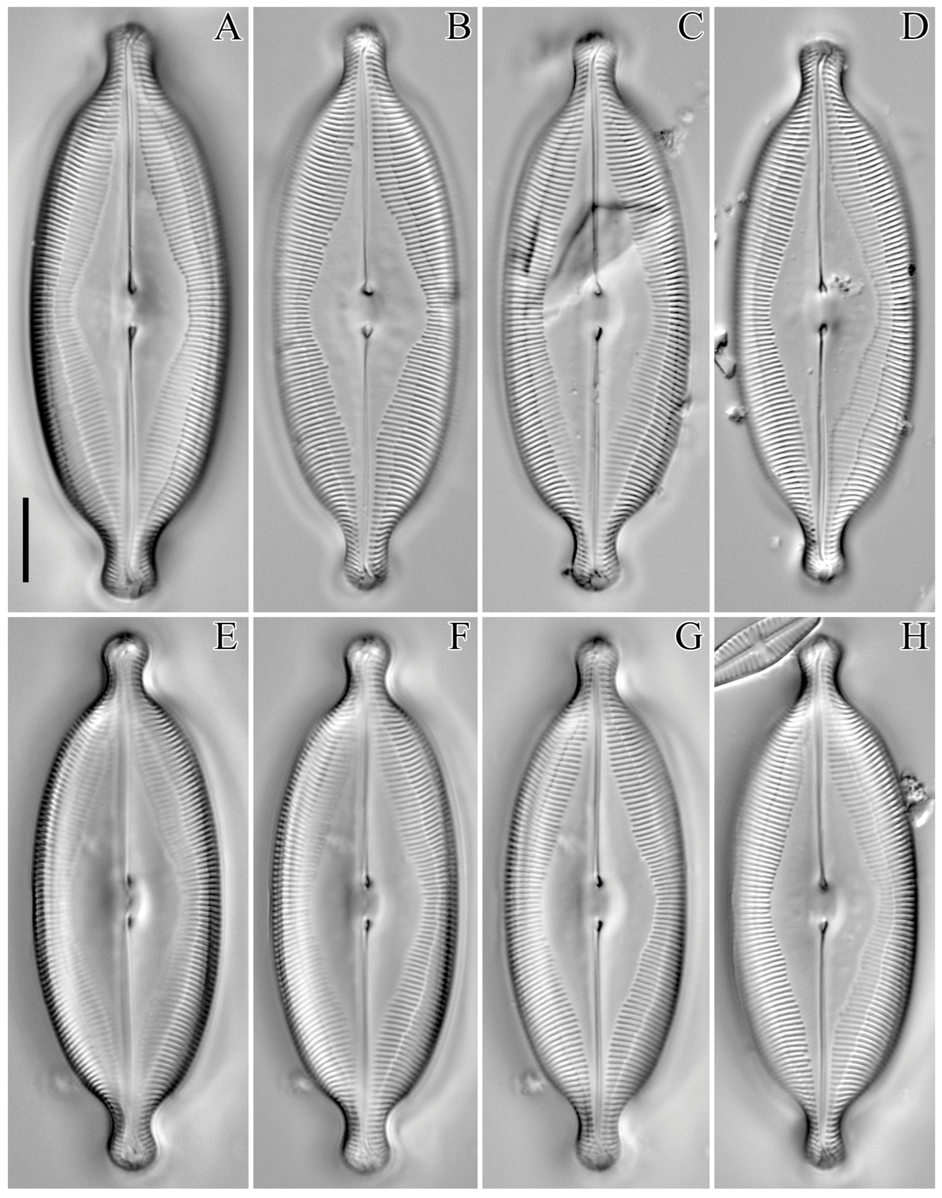
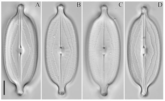
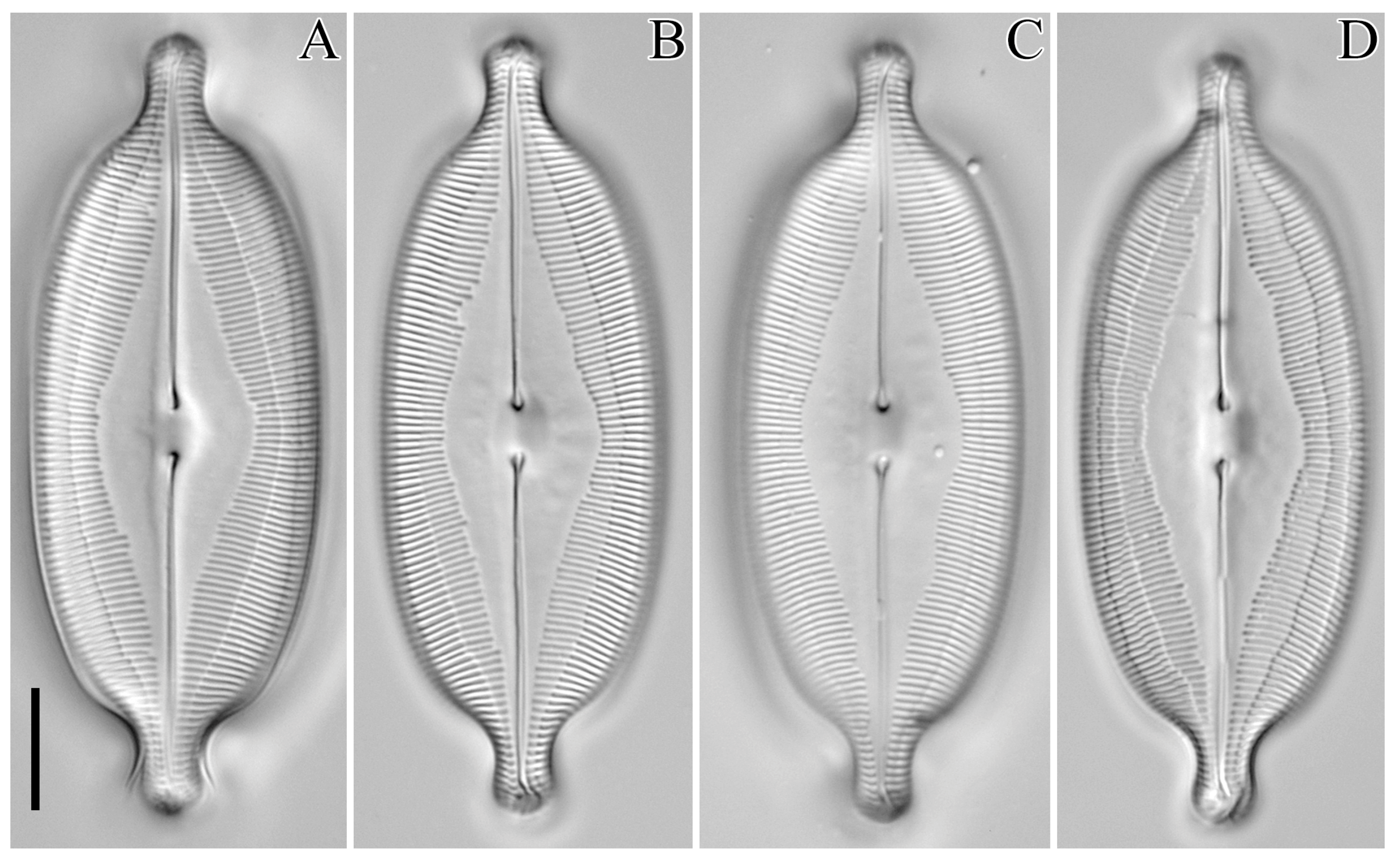
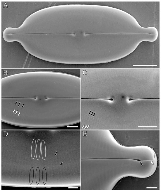
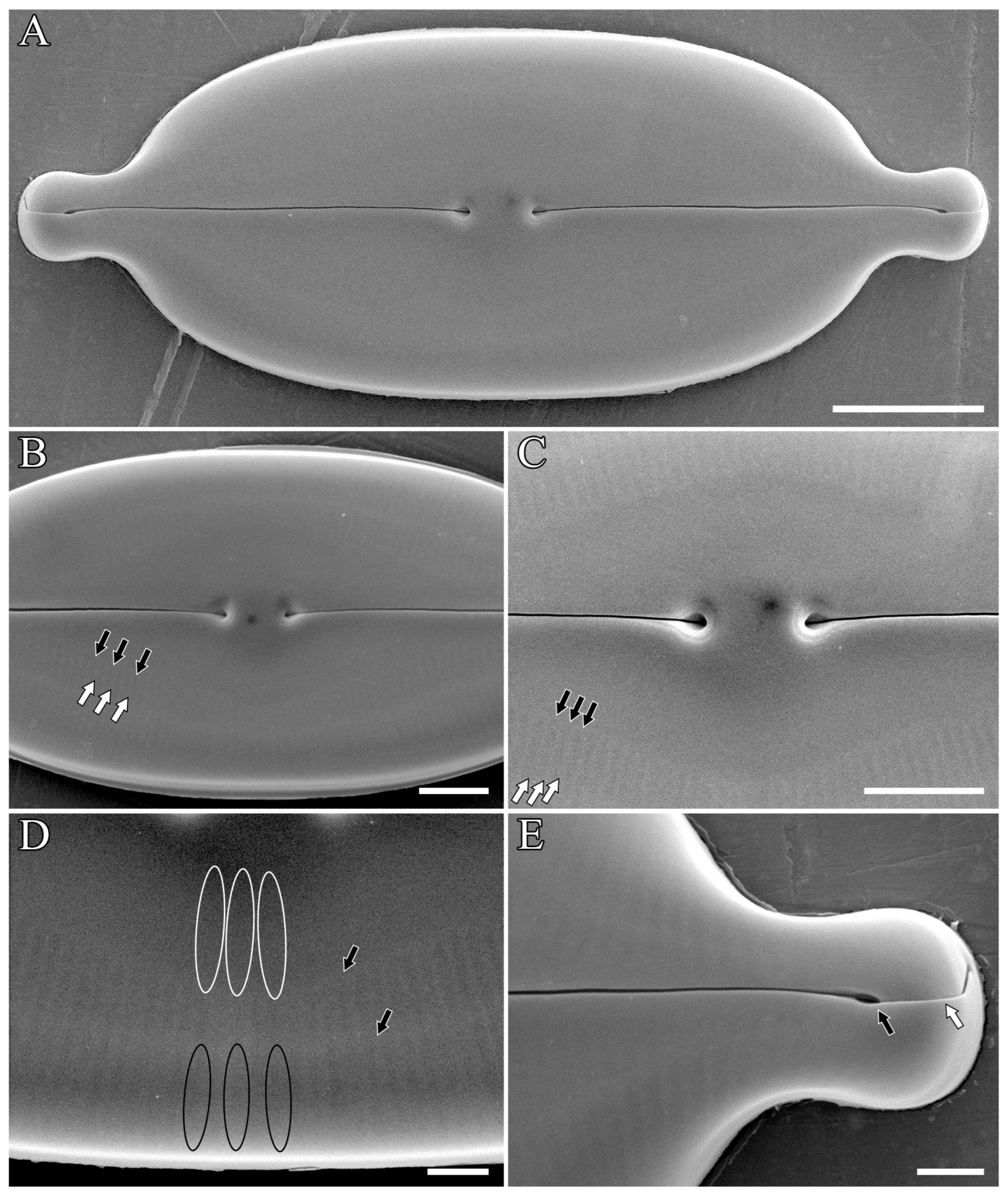
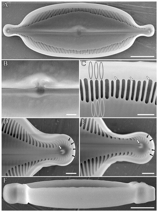
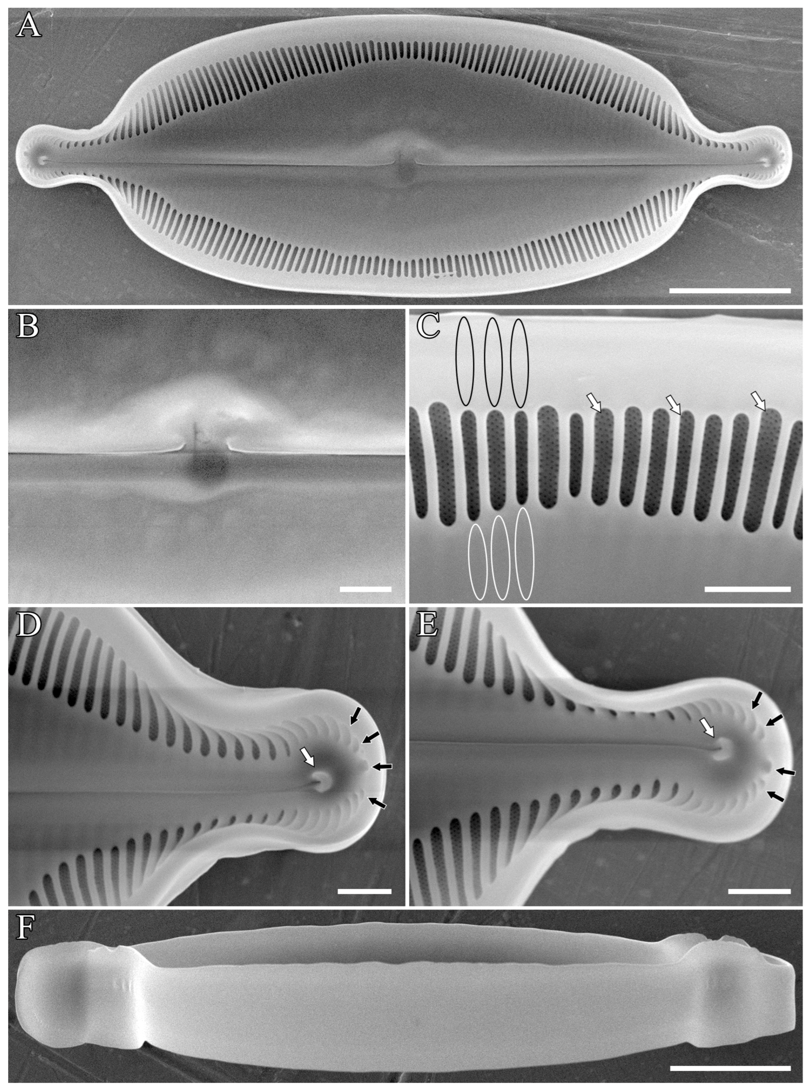

Disclaimer/Publisher’s Note: The statements, opinions and data contained in all publications are solely those of the individual author(s) and contributor(s) and not of MDPI and/or the editor(s). MDPI and/or the editor(s) disclaim responsibility for any injury to people or property resulting from any ideas, methods, instructions or products referred to in the content. |
© 2026 by the authors. Licensee MDPI, Basel, Switzerland. This article is an open access article distributed under the terms and conditions of the Creative Commons Attribution (CC BY) license.